Nanoparticle-assisted STED nanoscopy with gold nanospheres Nicolai T. Urban and Stefan W. Hell Max Planck Institute for Biophysical Chemistry, Göttingen, Germany Matthew R. Foreman Blackett Laboratory, Department of Physics, Imperial College London, London SW7 2AZ Yonatan Sivan * Unit of Electro-optics Engineering, Faculty of Engineering Sciences, Ben-Gurion University of the Negev, P.O. Box 653, Israel, 8410501 Abstract We demonstrate stimulated emission depletion (STED) microscopy with 20 nm gold nanospheres coated by fluorescently doped silica. We demonstrate significantly improved spatial resolution down to 75nm, which is the first time that hybrid NPs are used in STED imaging beyond the diffraction limit of confocal microscopy. Unlike previous demonstrations of super-resolution with metal nanoparticles with different techniques, this 3.3-fold resolution improvement was limited only by the particle size. The STED intensity required for this is almost twice lower than for conventional STED based on dye alone, and we observe no melting or displacement of the NPs at the utilized intensities. Moreover, we show that the nanoparticles can be imaged in an aqueous environment, demonstrating the relevance to bio-imaging. Finally, we also show, for the first time in this context, an up to 3-fold reduction in the rate of photobleaching compared to standard dye-based STED, thus enabling sustainably brighter images. * [email protected] 1

Welcome message from author
This document is posted to help you gain knowledge. Please leave a comment to let me know what you think about it! Share it to your friends and learn new things together.
Transcript
Nanoparticle-assisted STED nanoscopy with gold nanospheres
Nicolai T. Urban and Stefan W. Hell
Max Planck Institute for Biophysical Chemistry, Göttingen, Germany
Matthew R. Foreman
Blackett Laboratory, Department of Physics,
Imperial College London, London SW7 2AZ
Yonatan Sivan∗
Unit of Electro-optics Engineering, Faculty of Engineering Sciences,
Ben-Gurion University of the Negev, P.O. Box 653, Israel, 8410501
AbstractWe demonstrate stimulated emission depletion (STED) microscopy with 20 nm gold nanospheres
coated by fluorescently doped silica. We demonstrate significantly improved spatial resolution
down to 75nm, which is the first time that hybrid NPs are used in STED imaging beyond the
diffraction limit of confocal microscopy. Unlike previous demonstrations of super-resolution with
metal nanoparticles with different techniques, this 3.3-fold resolution improvement was limited
only by the particle size. The STED intensity required for this is almost twice lower than for
conventional STED based on dye alone, and we observe no melting or displacement of the NPs at
the utilized intensities. Moreover, we show that the nanoparticles can be imaged in an aqueous
environment, demonstrating the relevance to bio-imaging. Finally, we also show, for the first time
in this context, an up to 3-fold reduction in the rate of photobleaching compared to standard
dye-based STED, thus enabling sustainably brighter images.
1

In recent years, there has been a rapid growth in the use of nanoparticles (NPs) forvarious applications. Metal nanoparticles [1] in particular, are finding applications in lightconcentration and harvesting in solar cell applications [2], sanitation [3, 4], sensing [5],surface-enhanced Raman scattering [6], triggering chemical reactions [7], super-resolutionmicroscopy [8–10] and many others. In the context of biological applications, they are beingused for drug delivery [11] and cancer therapy [12], for bio-diagnostics [13], as nanoscalesources of heat and acoustic waves [14, 15], as contrast agents in phase interferometry [16]and more.
Frequently, a metal NP or nanostructure is combined with a standard fluorescent labelused in bio-imaging so as to create a hybrid fluorescent label, with the goal of exploit-ing the strong fields occurring at the localized plasmon resonance (LPR) [1] to boost theperformance of the label. Specifically, such labels have been used for controlling the fluores-cence lifetime and yield (also known as fluorescence engineering) and as a means to mitigatephotobleaching [17–20] and increase signal brightness [21].
Recently, Sivan et al. proposed and demonstrated how such hybrid fluorescent labels canbe used within the context of the super-resolution imaging technique called stimulated emis-sion depletion (STED) nanoscopy [22]. Specifically, it was shown theoretically [23–26] thatthe near-field enhancement occurring at the LPR can be exploited to reduce the requiredintensity of the STED beam and to reduce the bleaching rates through the shortening ofthe lifetime of the excited singlet level; this technique is referred to as nanoparticle-assistedSTED nanoscopy (NP-STED). The lowering of the STED intensity was demonstrated ex-perimentally using 150 nm gold shell NPs with a STED beam at λSTED ≈ 780 nm [27].However, the resolution level demonstrated in that work was limited to only a slight im-provement with respect to the diffraction limit. A more recent study [28] demonstrateda similar level of resolution even though the NPs used were much smaller. In addition, areduction of the bleaching rate has to date not been demonstrated in this context.
Clearly, realistic applications of NP-STED in bio-imaging may ensue only once substan-tial resolution improvement below the diffraction limit is demonstrated with a NP-STEDlabel. In particular, more than a 2-fold improvement with respect to the diffraction limitis necessary in order to out-perform alternative approaches such as Structured IlluminationMicroscopy [29]. This can be done with NP-STED labels which are small enough to be ableto benefit from the enhanced spatial resolution, to enjoy the maximum field enhancement
2
levels, and to minimize interference with the biological system. NP-STED would also benefitfrom extending the range of operating wavelengths and functions of the hybrid fluorescentlabels.
In this article, we provide a step toward fulfilling these goals. Specifically, a 3.3-foldimprovement of resolution with respect to the diffraction limit of confocal microscopy isachieved, reaching resolutions of ∼ 75 nm, while requiring ∼ 2 times lower input intensitythan needed for the standard dye. This is achieved in an aqueous environment and with nodamage to the NPs, thus demonstrating the relevance to imaging of biological samples. Wealso demonstrate, for the first time in this context, a reduced photobleaching rate inducedby the presence of the metal NPs. Finally, we compare the performance of our NPs tothose reported previously [27, 28], and discuss the route towards realizing the full potentialof NP-STED [23]. We also argue that even the existing implementation of NP-STED canbe immediately combined with a wide range of imaging and treatment procedures utilizingmetal NPs in a biological context.
I. PRINCIPLES OF NP-STED
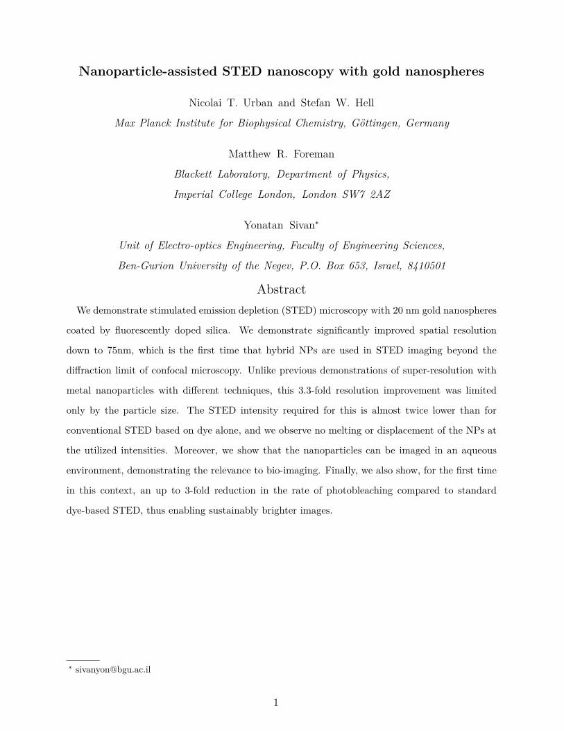
The hybrid plasmonic-fluorescent labels used in this study consisted of 18.8 ± 1.9 nmdiameter gold spheres coated with a 20.5± 1.1 nm glass shell doped with a standard greenSTED dye (Atto 488, which absorbs at ∼ 500 nm, emits at ∼ 525 nm and has a lifetime in thenanosecond range). The fluorophores were uniformly and symmetrically distributed in thesilica shell, thus avoiding issues associated with undesired shifts of the emitted signal [30].
The particles, shown in Fig. 1A, synthesized by Nanocomposix Inc., are substantiallysmaller than the gold shells used before [27], and are substantially easier to fabricate (likethe somewhat even smaller rods employed in [28]) as compared to the thin gold nanoshellsrequired for optimal NP-STED performance [23, 24]. The STED intensity reduction due tothe average near-field enhancement levels associated with this gold-core design is, however,substantially lower than those predicted for thin metal shells. The reason for this is that,unlike the rather uniform field experienced by dye inside a metal shell, the dye around ametal sphere experiences a field that decays as a function of the distance from the metalshell.
Indeed, Fig. 1B shows the near-field enhancement (defined as ΓI in [24] and taken relative
3
1.0
0.8
0.6
0.4
0.2
0.0
no
rm. e
mis
sio
n
650600550500wavelength [nm]
Atto 488 (Mowiol) hybrid NPs
0.16
0.14
0.12
0.10
0.08
600595590
1.3
1.2
1.1
1.0
0.9
ne
ar
!e
ld e
nh
an
cem
en
t
700650600550500450wavelength [nm]
50 nm
BA C
in Mowiol
hybrid NPsTEM
FIG. 1. Hybrid nanoparticles. (A) TEM image of the synthesized NPs, with 20 nm Au cores
surrounded by 20 nm thick, dye-doped glass shells. (B) Theoretical spatially and orientationally
averaged near-field intensity enhancement for the hybrid NPs immersed in water; highlighted is the
enhancement value of 1.18 at the used STED wavelength (595 nm). (C) Experimental normalized
emission spectra for both hybrid NPs and free Atto 488 in Mowiol; note the difference at (595 nm)
of 0.14 (NPs) and 0.10 (Mowiol).
to a homogeneous Mowiol environment - see below) that was calculated using the parametersindicated above and averaged over the volume of the dye-doped glass coating for NPs in anaqueous environment. Note that, in general, we will denote the enhancement ratio of aNP enhanced quantity relative to the standard dye by Γj. It can be seen that at the localplasmon resonance, λLPR ≈ 553 nm, the (spatially) averaged near-field enhancement [31] isabout ∼ 1.34. It was shown previously [24, 25] that this value gives a reasonable estimatefor the actual effective STED intensity reduction as calculated accurately with a doughnutshaped illumination (as e.g., performed in the initial studies of NP-STED [23]). In our setup,we opted for a STED wavelength of λSTED = 595 nm, which is standard for green dyes suchas Atto 488, but is slightly detuned from the plasmon resonance. At this wavelength theaverage near-field enhancement is ∼ 1.18+0.12
−0.1 , where the bounds account for size variationswithin the manufacturer tolerances.
Besides overlapping with the STED wavelength, the plasmon resonance overlaps partiallywith the emission line of the dye, giving rise to a shortening of the fluorescence lifetime [1, 32].In general, the decay rate enhancement depends strongly on position and the relative orienta-tion of the emitter with respect to the metal surface. The cumulative signal emanating from
4
a collection of such emitters typically exhibits a multi-exponential decay pattern [1, 33]. Fre-quently, however, the decay curve is approximated with low-order or even mono-exponentialdecay curves [1, 34]. In the latter case, one can define the decay rate enhancement, Γk, withrespect to the free dye decay rate [24]. As long as the STED pulse duration is substantiallyshorter than the dye lifetime, there should be little influence on the STED efficiency of thenanoscope [24, 35, 36]. If, however, the STED pulse duration approaches the dye lifetimeor even becomes longer, then the STED efficiency will notably decrease. The effects of thiscan be (partially) compensated by using time-gated detection, with the caveat that time-gating will result in somewhat lower signal brightness. Under such time-gated collection,we have shown theoretically that the averaged near-field enhancement ΓI provides a goodestimate of the STED intensity reduction that can be expected from the presence of themetal NP [24, 31]. Additionally, the fluorescence signal will further decrease due to thereduction of the apparent quantum yield (quenching) originating to energy transfer to themetal, an effect enabled by the proximity of the fluorescent dye to the metal.
The lifetime shortening due to the NP has a positive effect as well, namely an associatedreduction of bleaching. Indeed, the shortened time the molecule spends in an excited statereduces the overall number of chemical reactions that may lead to photobleaching (such asabsorption to an excited state, or intersystem crossing to a triplet state [37–39]). This effectwas described by Enderlein [40, 41], and recently was demonstrated experimentally [18,19] on single fluorescent molecules, showing a 5-fold reduction of the spatially-averagedbleaching rate. This value was found to be in good agreement with the theoretical prediction(for monochromatic and uniform excitation) of the total photon yield being equal to theratio of the emission and bleaching rates. A fuller analysis, accounting for the spatialvariation of both the lifetime shortening and near field enhancements [20], has further showntheoretically and confirmed experimentally that the resulting aggregate bleaching behaviorcan exhibit a multi-exponential behavior. An additional advantage associated with thedecay rate enhancement is that it allows higher excitation powers to be used, since thesaturation threshold increases [21]. All these effects naturally enable much brighter signals,thus, compensating for the decrease of the quantum yield.
5
A
4
56
0.1
2
3
4
56
1n
orm
. lu
min
esc
en
ce
43210time [ns]
in H2O
in Mowiolhybrid NPscalculated
B
2
4
0.01
2
4
0.1
2
4
1
no
rm. l
um
ine
sce
nc
e
43210time [ns]
low STED power488 nm excitation
hig
h S
TE
D p
ow
er
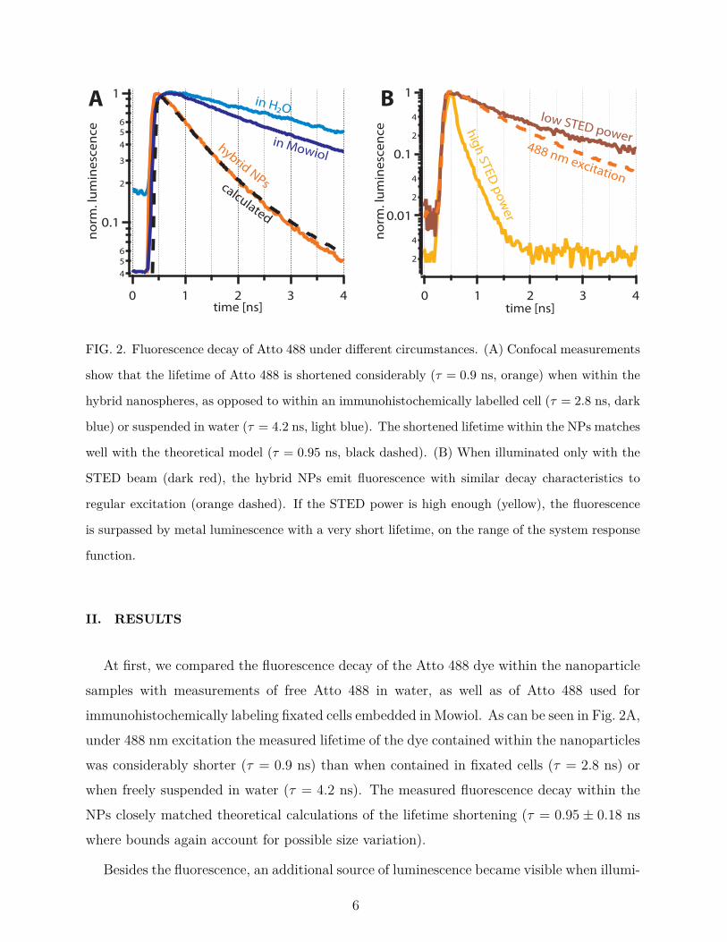
FIG. 2. Fluorescence decay of Atto 488 under different circumstances. (A) Confocal measurements
show that the lifetime of Atto 488 is shortened considerably (τ = 0.9 ns, orange) when within the
hybrid nanospheres, as opposed to within an immunohistochemically labelled cell (τ = 2.8 ns, dark
blue) or suspended in water (τ = 4.2 ns, light blue). The shortened lifetime within the NPs matches
well with the theoretical model (τ = 0.95 ns, black dashed). (B) When illuminated only with the
STED beam (dark red), the hybrid NPs emit fluorescence with similar decay characteristics to
regular excitation (orange dashed). If the STED power is high enough (yellow), the fluorescence
is surpassed by metal luminescence with a very short lifetime, on the range of the system response
function.
II. RESULTS
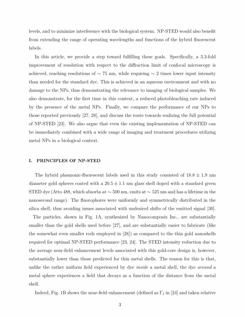
At first, we compared the fluorescence decay of the Atto 488 dye within the nanoparticlesamples with measurements of free Atto 488 in water, as well as of Atto 488 used forimmunohistochemically labeling fixated cells embedded in Mowiol. As can be seen in Fig. 2A,under 488 nm excitation the measured lifetime of the dye contained within the nanoparticleswas considerably shorter (τ = 0.9 ns) than when contained in fixated cells (τ = 2.8 ns) orwhen freely suspended in water (τ = 4.2 ns). The measured fluorescence decay within theNPs closely matched theoretical calculations of the lifetime shortening (τ = 0.95 ± 0.18 nswhere bounds again account for possible size variation).
Besides the fluorescence, an additional source of luminescence became visible when illumi-
6
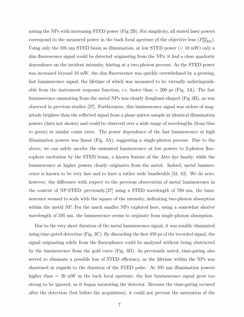
nating the NPs with increasing STED power (Fig 2B). For simplicity, all stated laser powerscorrespond to the measured power in the back focal aperture of the objective lens (P inc
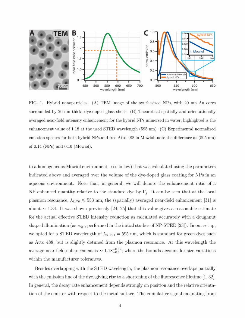
STED).Using only the 595 nm STED beam as illumination, at low STED power (< 10 mW) only adim fluorescence signal could be detected originating from the NPs; it had a clear quadraticdependence on the incident intensity, hinting at a two-photon process. As the STED powerwas increased beyond 10 mW, the dim fluorescence was quickly overwhelmed by a growing,fast luminescence signal, the lifetime of which was measured to be virtually indistinguish-able from the instrument response function, i.e. faster than ∼ 200 ps (Fig. 3A). The fastluminescence emanating from the metal NPs was clearly doughnut-shaped (Fig 3B), as wasobserved in previous studies [27]. Furthermore, this luminescence signal was orders of mag-nitude brighter than the reflected signal from a plane mirror sample at identical illuminationpowers (data not shown) and could be observed over a wide range of wavelengths (from blueto green) at similar count rates. The power dependence of the fast luminescence at highillumination powers was linear (Fig. 3A), suggesting a single-photon process. Due to theabove, we can safely ascribe the unwanted luminescence at low powers to 2-photon fluo-rophore excitation by the STED beam, a known feature of the Atto dye family, while theluminescence at higher powers clearly originates from the metal. Indeed, metal lumines-cence is known to be very fast and to have a rather wide bandwidth [42, 43]. We do note,however, the difference with respect to the previous observation of metal luminescence inthe context of NP-STED: previously,[27] using a STED wavelength of 780 nm, the lumi-nescence seemed to scale with the square of the intensity, indicating two-photon absorptionwithin the metal NP. For the much smaller NPs explored here, using a somewhat shorterwavelength of 595 nm, the luminescence seems to originate from single-photon absorption.
Due to the very short duration of the metal luminescence signal, it was readily eliminatedusing time-gated detection (Fig. 3C). By discarding the first 450 ps of the recorded signal, thesignal originating solely from the fluorophores could be analyzed without being obstructedby the luminescence from the gold cores (Fig. 3D). As previously noted, time-gating alsoserved to eliminate a possible loss of STED efficiency, as the lifetime within the NPs wasshortened in regards to the duration of the STED pulse. At 595 nm illumination powershigher than ∼ 20 mW in the back focal aperture, the fast luminescence signal grew toostrong to be ignored, as it began saturating the detector. Because the time-gating occuredafter the detection (but before the acquisition), it could not prevent the saturation of the
7
B
A
time gated
ungated
C
0
194
5
5
65
76
D
500 nm
500 nm
500 nm
0.11
0.10
0.09
0.08
0.07
8642
8
6
4
2
0
lum
ine
sce
nc
e [
10
5 c
ou
nts
]
252015105
STED power [mW]
experimental quadr. !t, <10mW linear !t, >10mW
metal luminescence
STED
STED
FIG. 3. Metal luminescence from the NPs under intense STED illumination. (A) When illumi-
nating the NPs with only 595 nm light < 10 mW, a weak fluorescence signal is visible (orange),
which scales quadratically with the applied STED power. If the illumination intensity is increased
above ∼ 10 mW, then a second source of luminescence appears (yellow), which increases linearly
in power and quickly drowns out the fluorescence. This metal luminescence displays a very short
lifetime and a doughnut-shaped intensity profile (B). When recording proper STED images, the
metal luminescence can be mostly discarded if the detection is time-gated (C), as opposed to using
the entire detected signal (D).
detector.
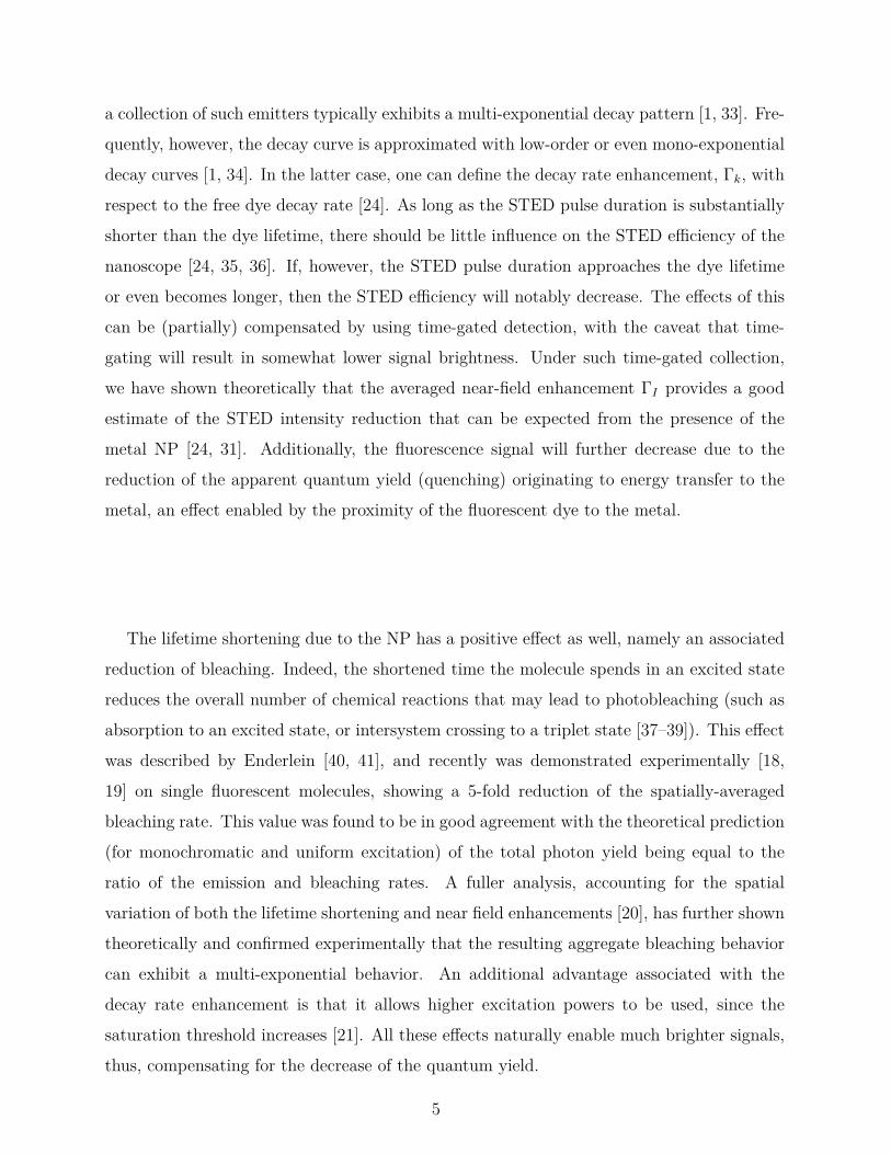
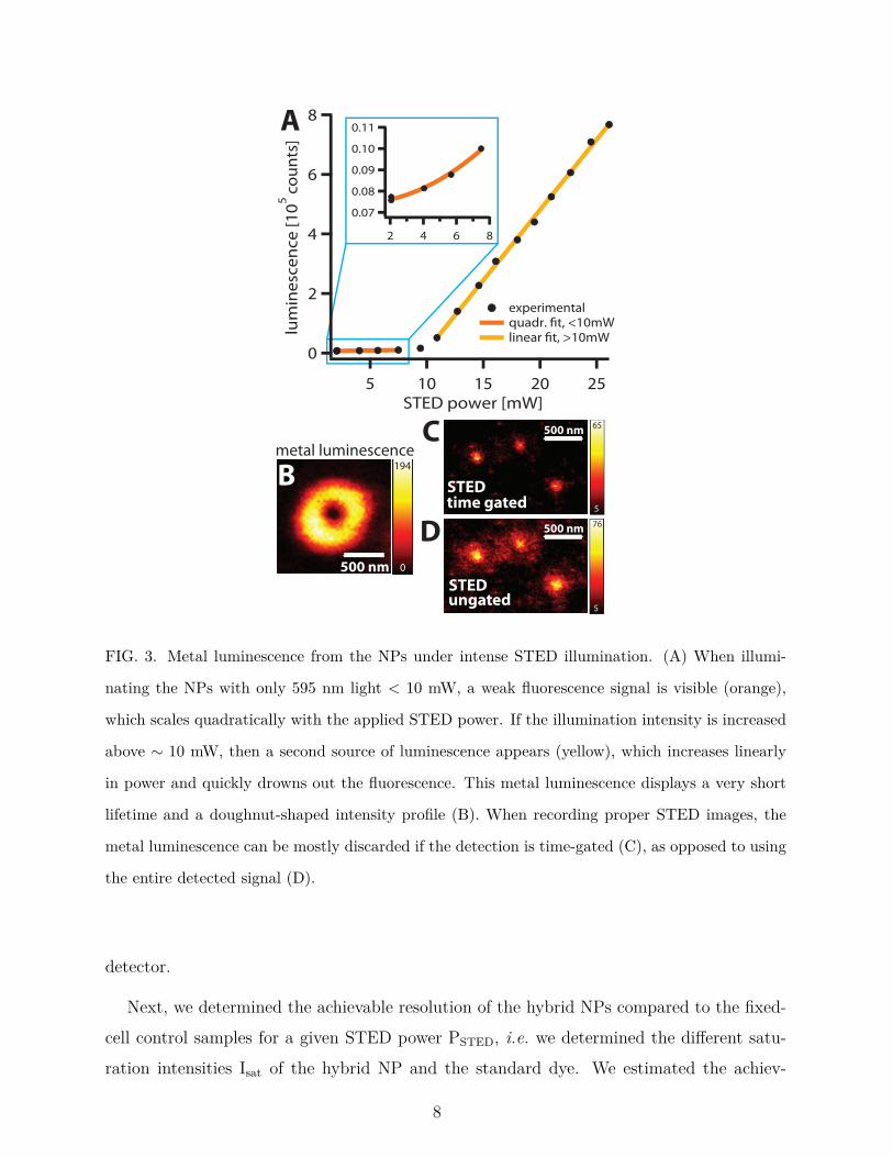
Next, we determined the achievable resolution of the hybrid NPs compared to the fixed-cell control samples for a given STED power PSTED, i.e. we determined the different satu-ration intensities Isat of the hybrid NP and the standard dye. We estimated the achiev-
8
DC
0
156
Confocal
500 nm
A
5
110
STED
500 nm
B
hybrid NPs
control
hybrid NPs
control
250
200
150
100
FW
HM
[n
m]
20151050STED power [mW]
Atto 488 immuno !t, P*sat= 5.2 mW
hybrid NPs !t, P*sat= 3.0 mW
long STED pulses 250
200
150
100
50
FW
HM
[n
m]
2520151050STED power [mW]
Atto 488 immuno !t, P*sat= 4.2 mW
hybrid NPs !t, P*sat= 2.3 mW
short STED pulses
FIG. 4. STED resolution enhancement for NP and control samples. Hybrid NPs recorded in (A)
confocal and (B) STED mode (P incSTED = 10 mW). At identical STED powers, the hybrid NPs
exhibited smaller FWHMs than the control samples. Displayed are averages ± standard deviation,
for ≥ 25 samples. Data was fitted with an inverse square-root law; the resulting saturation powers
P ∗sat are denoted in the figure. The ratio of saturation powers remained the same when measured
using longer (200 ps, C) or shorter (50 ps, D) pulses, even though shorter STED pulses allowed
higher absolute spatial resolutions of ∼ 75 nm (limited only by the NP size), in part due to a more
efficient gating of the metal luminescence background.
able resolution by measuring the average FWHM (full-width at half-maximum) of theimaged structures (beads or filaments) at different STED powers, using time-gated de-tection. For simplicity, we again use the measured incident STED power P inc
STED in theback focal aperture of the objective lens as a reference value. The resolution increaseover confocal imaging due to STED (see Fig. 4A,B) follows an inverse square-root law,dSTED ∼ dconf/
√1 + P inc
STED/P∗sat, where the effective saturation level is P ∗
sat ≡ Psat/ΓI andPsat ∼ hc/[λSTEDσem(λSTED)TSTED],[44]. Here σem, λSTED and TSTED are the emission cross
9
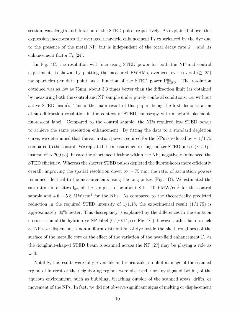
section, wavelength and duration of the STED pulse, respectively. As explained above, thisexpression incorporates the averaged near-field enhancement ΓI experienced by the dye dueto the presence of the metal NP, but is independent of the total decay rate ktot and itsenhancement factor Γk [24].
In Fig. 4C, the resolution with increasing STED power for both the NP and controlexperiments is shown, by plotting the measured FWHMs, averaged over several (≥ 25)nanoparticles per data point, as a function of the STED power Pinc
STED. The resolutionobtained was as low as 75nm, about 3.3 times better than the diffraction limit (as obtainedby measuring both the control and NP sample under purely confocal conditions, i.e. withoutactive STED beam). This is the main result of this paper, being the first demonstrationof sub-diffraction resolution in the context of STED nanoscopy with a hybrid plasmonicfluorescent label. Compared to the control sample, the NPs required less STED powerto achieve the same resolution enhancement. By fitting the data to a standard depletioncurve, we determined that the saturation power required for the NPs is reduced by ∼ 1/1.75
compared to the control. We repeated the measurements using shorter STED pulses (∼ 50 psinstead of ∼ 200 ps), in case the shortened lifetime within the NPs negatively influenced theSTED efficiency. Whereas the shorter STED pulses depleted the fluorophores more efficientlyoverall, improving the spatial resolution down to ∼ 75 nm, the ratio of saturation powersremained identical to the measurements using the long pulses (Fig. 4D). We estimated thesaturation intensities Isat of the samples to be about 8.1 − 10.0 MW/cm2 for the controlsample and 4.6 − 5.8 MW/cm2 for the NPs. As compared to the theoretically predictedreduction in the required STED intensity of 1/1.18, the experimental result (1/1.75) isapproximately 30% better. This discrepancy is explained by the differences in the emissioncross-section of the hybrid dye-NP label (0.1/0.14, see Fig. 1C), however, other factors suchas NP size dispersion, a non-uniform distribution of dye inside the shell, roughness of thesurface of the metallic core or the effect of the variation of the near-field enhancement ΓI asthe doughnut-shaped STED beam is scanned across the NP [27] may be playing a role aswell.
Notably, the results were fully reversible and repeatable; no photodamage of the scannedregion of interest or the neighboring regions were observed, nor any signs of boiling of theaqueous environment, such as bubbling, bleaching outside of the scanned areas, drifts, ormovement of the NPs. In fact, we did not observe significant signs of melting or displacement
10
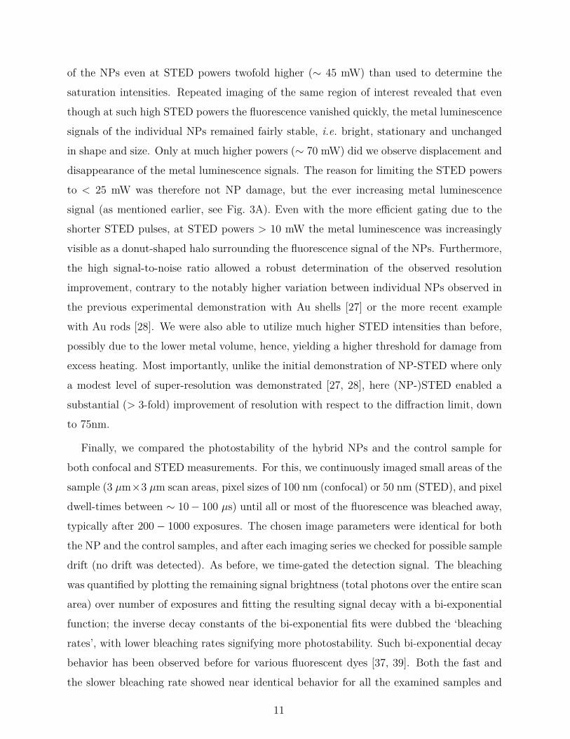
of the NPs even at STED powers twofold higher (∼ 45 mW) than used to determine thesaturation intensities. Repeated imaging of the same region of interest revealed that eventhough at such high STED powers the fluorescence vanished quickly, the metal luminescencesignals of the individual NPs remained fairly stable, i.e. bright, stationary and unchangedin shape and size. Only at much higher powers (∼ 70 mW) did we observe displacement anddisappearance of the metal luminescence signals. The reason for limiting the STED powersto < 25 mW was therefore not NP damage, but the ever increasing metal luminescencesignal (as mentioned earlier, see Fig. 3A). Even with the more efficient gating due to theshorter STED pulses, at STED powers > 10 mW the metal luminescence was increasinglyvisible as a donut-shaped halo surrounding the fluorescence signal of the NPs. Furthermore,the high signal-to-noise ratio allowed a robust determination of the observed resolutionimprovement, contrary to the notably higher variation between individual NPs observed inthe previous experimental demonstration with Au shells [27] or the more recent examplewith Au rods [28]. We were also able to utilize much higher STED intensities than before,possibly due to the lower metal volume, hence, yielding a higher threshold for damage fromexcess heating. Most importantly, unlike the initial demonstration of NP-STED where onlya modest level of super-resolution was demonstrated [27, 28], here (NP-)STED enabled asubstantial (> 3-fold) improvement of resolution with respect to the diffraction limit, downto 75nm.
Finally, we compared the photostability of the hybrid NPs and the control sample forboth confocal and STED measurements. For this, we continuously imaged small areas of thesample (3 µm×3 µm scan areas, pixel sizes of 100 nm (confocal) or 50 nm (STED), and pixeldwell-times between ∼ 10− 100 µs) until all or most of the fluorescence was bleached away,typically after 200− 1000 exposures. The chosen image parameters were identical for boththe NP and the control samples, and after each imaging series we checked for possible sampledrift (no drift was detected). As before, we time-gated the detection signal. The bleachingwas quantified by plotting the remaining signal brightness (total photons over the entire scanarea) over number of exposures and fitting the resulting signal decay with a bi-exponentialfunction; the inverse decay constants of the bi-exponential fits were dubbed the ‘bleachingrates’, with lower bleaching rates signifying more photostability. Such bi-exponential decaybehavior has been observed before for various fluorescent dyes [37, 39]. Both the fast andthe slower bleaching rate showed near identical behavior for all the examined samples and
11

A
C D
B
STED
confocal6
5
4
3
2
1
ble
ach
ing
ra
tio
543210excitation power [µW]
averaged individual theoretical
120
80
40
0b
lea
ch
ing
ra
te86420
3
2
1
ble
ach
ing
ra
tio
86420STED power [mW]
ratio NPs/immuno
inv
ers
e
1.0
0.8
0.6
0.4
0.2
0.0
no
rm. b
rig
htn
ess
100806040200Exposures
hybrid NPs, 1.6 mW hybrid NPs, 3.4 mW hybrid NPs, 6.5 mW Atto 488 immuno, 1.6 mW Atto 488 immuno, 3.4 mW Atto 488 immuno, 6.5 mW
1.0
0.8
0.6
0.4
0.2
no
rm. b
rig
htn
ess
10008006004002000Exposures
hybrid NPs, 1.25 µW hybrid NPs, 2.5 µW Atto 488 immuno, 1.25 µW Atto 488 immuno, 2.5 µW
hybrid NPs
control
hybrid NPs
control
hybrid NPs
control
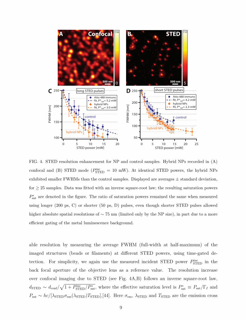
FIG. 5. Bleaching behavior of hybrid NPs. (A) When illuminated with identical excitation powers
(488 nm), the confocal signal recorded from the hybrid NPs (orange) bleaches away much more
slowly than the control sample (blue). (B) The increased photostability of the NPs is mostly
independent of the excitation power. (C) When imaging in STED mode (with both a 488 nm
excitation and a 595 nm STED beam), the NPs (orange) again bleach more slowly than the control
sample (blue). (D) The photostability is increased threefold at low STED powers, but as the STED
intensity increases, the increased photostability of the NPs is counteracted, possibly by thermal
destruction of the dye.
imaging modalities. Thus, for simplicity, we will ignore the faster decay (with the highervariance) in favor of the slower one. As a measure of the improved photostability, wecalculated the ratio of the inverse bleaching rates from the NP- and the control sample,denoted as Γb.
Fig. 5 shows the fluorescence decay using confocal and STED-enhanced imaging modali-
12

ties. When imaging confocally, i.e. without the STED beam, the control samples bleachedconsiderably faster than the dye protected within the hybrid NPs (Fig. 5A). The bleachingratio was found to be Γb ∼ 2 − 4, seemingly irrespective of the excitation power (Fig. 5B),in good agreement with the theoretically predicted value of 2.75 (red dashed). When per-forming STED imaging, we again observed slower bleaching of the hybrid NPs (Fig. 5C); atlow STED intensities, the bleaching rate of the signal from the hybrid labels was ∼ 3 timeslower than the bare dye. For higher STED power, however, the improved photostabilityof the hybrid labels decreased. Whereas the bleaching rate of the control sample remainedroughly constant for increasing STED power, the bleaching of the hybrid NPs intensified,such that Γb → 1 (Fig. 5D). We ascribe this effect to damage occurring to the dye due tointeraction between the STED beam and the gold cores (e.g., heating of the metal and itsenvironment) [20], as this intense bleaching was also observed when illuminating the samplewith only the STED beam, i.e. in the absence of the excitation beam.
III. DISCUSSION
The results detailed above provide a clear improvement with respect to previous demon-strations of NP-STED [27, 28] where only a modest super-resolution level was demonstrated(observed optical resolution was worse than 200 nm). Specifically, our demonstration ofsub-diffraction resolution of ∼ 75 nm is the first time that hybrid plasmonic fluorescentnanoparticles are used to image well beyond the diffraction barrier of visible light in thecontext of STED microscopy. This is enabled by the 20-fold reduction of the label volume— from ∼ 160 nm gold shells [27] to particles which are ∼ 60 nm diameter. Crucially, thismeans resolution levels better than those attainable by other approaches that offer limitedsub-diffraction resolution, including Structured Illumination Microscopy [29] or other metalNP-based super-resolution techniques [9, 15]; unlike the latter techniques, our approach canbe applied to particles only a few nm in size. In addition, our results show nearly a ∼ 2-foldreduction of STED intensity required for achieving the same resolution with the currentgeneration of organic labels.
Our study also confirms, for the first time in the context of STED, the prediction regardingthe reduction of bleaching ensuing from the use of the hybrid metal-fluorescent labels [23] —up to a 3-fold bleaching rate reduction was observed (in both confocal and STED modalities).
13

This value is in good agreement with the theoretical prediction based on the theory presentedpreviously [20], thus showing that both additional bleaching reduction due to plasmonic-enhanced triplet lifetime shortening at the (resonant) STED wavelength [17] as well asbleaching due to multi-photon processes is probably of lesser importance, at least for thedye used in this study.
Note that the bleaching reduction (without using STED) reported here is qualitativelysimilar to previous observations based on either single molecules [19], dye adsorbed onto athin gold layer [20], or dye contained within a polyelectrolyte multilayer film, onto whichgold beads have been added [18]. Thus, we provide what seems to be the first demonstra-tion of this concept using hybrid fluorescent labels, arguably, a more useful configuration.More importantly however, in the confocal configuration our demonstration was obtainedat substantially higher peak intensity — about 2 orders of magnitude higher due to thespatial and temporal focused nature of the illumination. Moreover, in the presence of aSTED beam, the associated intensities were an additional 3− 4 orders of magnitude higher.In the latter case, the depletion pulse, which enhances the decay from the excited level,modifies the quantitative description of the bleaching reduction [20] through the addition ofan extra decay pathway. Unfortunately, the bleaching reduction persists only for relativelylow STED powers, as for higher powers the interaction between the STED beam and themetal seems to cause dye destruction (possibly due to heating) [20]. Yet, importantly, thebleaching rate is lower than in the bare-dye case in the whole range of intensities shownhere.
Maybe the most important advance demonstrated in this study is that the imaging ofthe hybrid labels was performed in water (rather than in index matching oil, as was done sofar [27, 28]). This highlights the possibility of applying NP-STED to the study of biologicalsamples. The applicability of NP-STED to live cell imaging, however, requires cell viabilitystudies [45, 46] to be carried out, in light of the transient temperature rise associated withthe absorption in the metal. A discussion of ways to mitigate the (average) temperature riseis provided by Cortés et al. [28]; potentially the most efficient way to achieve that wouldbe to employ stochastic spatial scanning (as already demonstrated [47]), yielding minimalaverage temperature rise.
Despite the important advances reported here, the performance improvement offered bythe NP-STED hybrid labels used here is still limited. The major obstacle to achieving even
14
better performance is NP miniaturization, and specifically, decreasing the metal volume.For one, this may enable higher near-field enhancement (hence, higher intensity reductionand maximal resolution) due to suppression of residual radiation damping [1]. Also, it wouldfurther limit the temperature rise due to absorption in the metal, as the latter is propor-tional to the physical cross section of the NP [28, 48, 49]. As importantly, a smaller metalnanoparticle will give rise to lower metal luminescence, thus facilitating the achievementof even better resolution (see discussion above) by enabling the use of even higher intensi-ties and the extraction of brighter fluorescence signals. Thus, (beside the obvious reduceddamage to the biological system) NP size reduction would extend the benefit of bleachingreduction to higher intensities. In that regard, the shorter STED wavelength we employed(with respect to the more standard STED wavelength of 780nm) is advantageous: it allowsuse of gold spheres, or slightly elongated gold rods, as the LPR of these particles is suffi-ciently close to the STED wavelength used in our system. Whereas we used particles with20 nm Au cores, further miniaturization of the Au core and fluorescent shell is relativelyeasy to achieve, as was recently demonstrated [28]. We note, however, that the minimumtotal size of a good NP-STED label can practically not be much below 10− 20 nm, since forfluorophore-metal separations of less than a few nanometers, the apparent quantum yield ofthe fluorophore will be too low due to fluorescence quenching. This could be compensatedby the lower bleaching and by the possibility to increase the excitation intensity [21]. Thus,we expect that particles 2–3 times smaller than those used in the current studies are realisticand should yield superior imaging performance.
A second concern is obtaining a more significant field enhancement, and thereby a higherreduction of the STED intensity. Further intensity reduction can be achieved by optimallymatching the STED wavelength to the LPR band of the Au sphere [23, 24]. This canbe achieved by either shortening the STED wavelength, or by using a STED dye with anemission line centered at even shorter wavelengths, such as AIE luminogen fluorophores [50].Rod-shaped nanoparticles give rise to even better average intensity enhancement [28], andfurther control over the LPR wavelength. Furthermore, the ability of selective positioningof the dye with respect to the metal surface is continuously being advanced, such that NP-STED labels with improved performance, namely, higher field enhancement, lower quantumyield reduction, and moderate level of lifetime shortening, can be designed and synthesized.A step in this direction was already demonstrated [28] by replacing the thick dye-doped
15
silica shell used here with fluorophores linked at a specific distance from the metal surface;they reported an 8-fold intensity enhancement, yet without improving the optical resolutionbeyond the previous demonstration. Applying a similar procedure for the Au nanospheresused here can lead to up to 3-fold higher field enhancement. Alternatively, in the context offixed cell imaging, i.e. absent of living organisms, one could also employ Ag rods (consideredtoxic), for which the plasmonic performance far exceeds that of Au thus enabling greaterpotential performance improvement in the NP-STED context.
Much higher intensity reduction (up to 50–100 fold) could be achieved with thin shells(instead of cores), such as those demonstrated previously [51]; in that work the cores weremade of TiO2, whereas for our purposes a core material that can be doped with fluorophoresis desired, such as silica. Similarly, one could also employ quantum-dot cores, as STED hasalso recently been demonstrated with these emitters [52]. The somewhat shorter wavelengthused in the current study also poses less stringent constraints on the thickness of gold shells.Synthesis of the necessary thin shells is still challenging, but within reach.
Overall, the excellent match of experimental results with the theoretical modelling givesus high confidence in our ability to reach the optimal performance predicted originally bySivan et al. [23]. Finally, the contrast between the very low damage threshold observed inprevious demonstrations (with a gold shell [27] and a bare gold rod [28]) and its practicalabsence in the current study may imply that the silica shell serves also as a protection layer tothe gold core. If so, the ideal NP-STED label design may rely on the smallest possible metalrods coated by a thin protecting layer that includes a thin layer of fluorophores/emittingsemiconductor out of the quenching range.
Despite the limited improvement of performance demonstrated so far, there are threeavenues where the existing hybrid labels could find immediate use. One is to use theuseful characteristics of the hybrid NPs to bolster existing methods, which do not cur-rently use nanoparticles. The reduced saturation intensities could benefit parallelized STEDschemes [53, 54], where the lack of sufficient power can be a limiting factor in enabling thescan speed increase. Also, the reduced saturation intensities may also allow the applicationof STED to even thicker samples where the strong scattering reduces the incident STEDintensities as one probes deeper into the tissue.
The second avenue entails using hybrid NPs as multi-functional labels to enable the com-bined use of very different imaging methods. Specifically, on one hand, operating in STED
16
mode, one can benefit from the high spatial resolution levels at the emission wavelength;in the absence of a STED beam, and for sufficiently intense excitation, one can benefitfrom the photothermal/photoacoustic response of the gold core [14, 15, 55] and the strong,spectrally-narrow signal achievable above the nanolasing (also known as spasing) thresholdenabled by the presence of feedback from the plasmonic cavity [56]. In the same modal-ity, one can perform either bleaching-free imaging at enhanced resolution at the excitationwavelength via LPR-based Structured Illumination [10, 57] using low intensities, quantita-tive phase contrast microscopy via the photothermal effect [16] using moderate intensities,or plasmonics-based saturated excitation (SAX) microscopy [8, 9] using intense monochro-matic illumination. One can also correlate the super-resolved STED signal with 2-photonimaging [58] using moderate to high intensities. Ultimately, the hybrid labels studied herecan help move towards correlated light electron imaging [59].
The third avenue relies on enhancing the many existing bio-related applications of metalNPs (see [60]) by adding a significant level of super-resolution imaging. These applicationsmay include, for example, gene therapy treatments [61], stimulation, monitoring and sig-nalling in neurons [62], and drug delivery [11]. One promising area is biodiagnostics [13]based on spherical nucleic acids [63], which are quite similar to the NPs used in this study.The wide variety of associated applications includes protein targeting and selective colorimet-ric detection of polynucleotides [64], scanometric DNA array detection [65] and intracellulargene regulation [66], to name just a few. These procedures, as well as any other that arecurrently imaged using confocal microscopy could benefit from the substantial improvementof the imaging resolution offered by NP-STED.
IV. METHODS
Sample preparation The hybrid nanoparticles were diluted in pure water, then soni-cated to ensure that no agglomeration occurred, and finally applied to coverslips coated witha thin Poly-L Lysine layer (for increased stickiness of the surface). The coverslips were thenbriefly rinsed with pure water (to remove excess NPs and dye) and subsequently dried, be-fore re-immersing them in a small drop of water and affixing the coverslip to the microscopeslide. The particles had a density of approximately 5–10 beads per 1 µm2, and only seldomlywere there clusters of agglomerated beads. This way, we avoided tweezing effects and NP
17
diffusion, which occurred previously. The control samples consisted of MeOH-fixated Verocells, immunohistochemically stained with a primary anti-vimentin antibody (Sigma V6389,1:100 dilution) and a secondary Atto-488 sheep/anti-mouse antibody (1 : 50 dilution) andmounted in Mowiol (with a refractive index of n = 1.45).
STED nanoscope All experiments were performed on a custom-built STED nanoscope [67]optimized for use with green fluorophores. For excitation, a 488 nm pulsed diode laser (Pi-coTA, Toptica Photonics, Graefelfing, Germany) was used with pulse lengths of ≈ 110 psand typical laser powers between 1–10 µW in the back focal aperture of the objective lens.The STED beam was created using a Ti:sapphire laser (80 MHz; MaiTai, Spectra-Physics,Darmstadt, Germany), which was frequency-shifted using an optical parametric oscillator(APE, Berlin, Germany) to a wavelength of 595 nm. The initially femtosecond pulses werepre-stretched using SF6 glass rods before being stretched further by either a short (25m) ora long (110m) polarization maintaining glass fiber (OZ Optics, Ottawa, Canada), resultingin pulse widths of either approximately 50 ps or 200 ps, respectively. The typical power ofthe STED beam in the back focal aperture was between 1− 25 mW. For our system, whichemploys a 80 MHz pulse repetition rate, a rough estimate shows that these power levelscorrespond to ∼ 17− 425 MW/cm2.
The beams were overlapped using custom-made dichroic mirrors and focused using a1.3 NA glycerol immersion objective lens (63x, PL APO, CORR CS; Leica, Wetzlar, Ger-many). The fast scan axis was performed using resonant mirror scanning (15 kHz; SC-30,EOPC, Glendale, NY) and the slow axis using a piezo stage (P-733; Physik-Instrumente,Karlsruhe, Germany). The fluorescence passed through a bandpass filter (535/60) and wasrecorded using a single-photon avalanche photo diode (SPAD, PDM series, Micro Photon De-vices, Bolzano, Italy); images were recorded using the TTL-output of the SPAD, whereas thefluorescence decay measurements used the fast NIM-output with 35 ps time resolution andwere counted using a time-correlated single-photon counting module (SPC150N, Becker &Hickl, Berlin, Germany). A second detection window (450/60) could be used simultaneouslyfor observing light outside of the Atto 488 fluorescence spectrum (i.e. metal luminescence).
Measurement details All STED images were recorded using time-gated detection, inorder to distinguish fluorescence from any occurring metal luminescence. For this, the signalfrom the detector was time-gated electronically using a home-built circuit that separatedthe first 450 ps after onset of the excitation pulse from the rest of the signal, before relaying
18

both signal components to the acquisition electronics, which were based on a custom fieldprogrammable gate array (FPGA) board. Nevertheless, when imaging the hybrid nanopar-ticles, we typically limited the applied STED power to < 10 mW, to avoid saturating thedetector, risk imaging artifacts due to residual metal luminescence and/or damage to theNPs due to heating. This limited the achievable resolution to >100 nm (thus not sufficientto fully resolve the 60 nm hybrid nanoparticles), instead of the better than ∼ 50 − 70 nmresolution, which the STED nanoscope is capable of (when imaging control samples). AtSTED powers between ∼ 10− 20 mW we still observed no NP damage or displacement, themetal luminescence, however, could not be gated away entirely, appearing as a donut-shapedhalo surrounding the fluorescence signal of the NP. The lifetime of the dye under the variouscircumstances was determined by fitting the fluorescence decay using a routine implementedin Matlab (Mathworks, MA, USA), which iteratively reconvolved the instrument responsefunction (IRF) with a mono- or bi-exponential decay function and then optimized the pa-rameters. The IRF was acquired by reflecting the excitation or STED-beam, respectively,off of a plane mirror sample and recorded using the same detection path as for the experi-ments. The IRF displayed a timing resolution of ∼ 110 ps FWHM for the 488 nm excitationbeam and ∼ 200 ps FWHM for the 595 nm STED beam.
Theoretical calculations and modelling To determine the electric field distributionfor the core-shell geometry NPs, standard Mie theory [68] was used assuming an incidentcircularly polarised plane wave. Nominal values, provided by Nanocomposix Inc. of 18.8 nmand 20.5 nm were used for the inner core diameter and shell thickness, respectively. Inall calculations, the properties of gold were taken from [69] whilst the refractive index ofthe silica coating and the aqueous host were taken as 1.4584 and 1.3324. The near fieldenhancement ΓI was determined by taking the ratio of the resulting intensity distributionas compared to that of a plane wave in a homogeneous Mowiol environment (n = 1.45), soas to match experimental controls. Volume averages were taken within the NP coating only.Results were verified using COMSOL 4.3a.
Position dependent radiative Purcell factors, Fr = Pr/P0 for dipole emitters oriented bothradially and tangentially with respect to the core were calculated using a method based onaccepted theory [70]. Specifically, the integrated power flow through a spherical surface in thefar field (Pr), was compared to that of a free emitter embedded in Mowiol (P0), for differentemission wavelengths λem. The latter was found using the Larmour formula. Similarly,
19
non-radiative Purcell factors (Fnr = Pnr/P0) followed by calculating the power absorbedin the metal core, Pnr. The lifetime trace in Fig. 2 was then calculated by consideringthe number of photons emitted by a dipole of given orientation p and position (neglectingphotobleaching) which, as has been discussed previously [33], is proportional to N(r, p, t) ∼
|p ·E|Fr exp[−(Fr+Fnr+η−10 −1)t/τ0], where η0 = 0.8 (τ0) is the quantum yield (lifetime) of
a free emitter, caret notation denotes a unit vector and E is the electric field at the positionof the dipole upon illumination of the NP with a plane wave. We note that although theimportance of the orientation of the dye molecule was known [18], in many earlier theoreticaltreatments a fixed emitter orientation was considered, e.g. due to preferential adsorption [20].In contrast, since our system comprises of dye molecules doped in a silica coating, we allowedfor a random distribution of emitter orientations in turn requiring a further averaging stepto be introduced in our modelling. Specifically, we assumed that the dye molecules wereoriented randomly over the full 4π solid angle Ω, whereby the total collected signal NT
was found by integrating over the volume of the shell and averaging over the orientation.Each spectral component was further weighted according to the Atto 488 emission spectrumw(λem) such that the fluorescence intensity scales as
I(t) ∼ 1
4π
∫ ∞
0
∫Ω
∫V
w(λem)NT (r, p, t, λem)drdpdλem. (1)
Note that the Fr factor present in NT accounts for modification of the spectral profile of theisolated fluorophore [71, 72]. Finally, the resulting fluorescence trace was convolved with themeasurement IRF. We note there were no free parameters in our calculation. The effectivelifetime was extracted using a mono-exponential fit. Calculations were repeated for innercore diameters and shell thicknesses of 18.8 ± 1.9 nm and 20.5 ± 1.1 nm to estimate theeffects of size dispersion as quoted in the main text.
Since the lifetime of differently oriented and positioned emitters within the NP is modifiedto varying extents, the relative competition between radiative decay, non-radiative decayand photobleaching also varies. The effective bleaching rate of a given emitter can bedetermined as in [20], however, the resultant bleaching curves (c.f. Fig. 5 will, similarly tothe lifetime traces, be formed of the superposition of multiple exponential curves. Using anormalized time coordinate, the decay constants in the exponents are given by αΓI/[Fr +
Fnr + η−10 − 1], where α = |p · E|2/|p · E0|2 describes the relative polarization rotation from
the NP. The relative weighting of the individual bleaching curves depends on the local near
20

field intensity enhancement ΓI and the ratio Fr/(Fr + Fnr) [20]. Volume, orientation andwavelength averages were taken similarly to (1). To parallel the experimental treatment,mono-exponential fits were used to determine effective decay rates and hence the theoreticalbleaching ratio Γb.
ACKNOWLEDGEMENTS
We would like to thank T. Klar, K. Matsuzaki, S. Sahl and H. Sinclair for helpful discus-sions. MRF is funded by a Royal Society University Research Fellowship. YS acknowledgessupport from the People Programme (Marie Curie Actions) of the EU’s Seventh FrameworkProgramme (FP7/2007-2013) under REA grant agreement no. 333790, the Alumni programof the Royal Society International Newton Fellowship program and the Israeli NationalNanotechnology Initiative.
[1] V. Giannini, A. I. Fernández-Domínguez, S. C. Heck, and S. A. Maier, Chem. Rev. 111, 3888
(2011).
[2] H. A. Atwater and A. Polman, Nature Materials 9, 205 (2010).
[3] O. Neumann, A. S. Urban, J. Day, S. Lal, P. Nordlander, and N. J. Halas, ACS Nano 7, 42
(2013).
[4] Z. Fang, Y. R. Zhen, O. Neumann, A. Polman, F. J. G. de Abajo, P. Nordlander, and N. J.
Halas, Nano Lett. 1736-1742, 13 (2013).
[5] M. D. Baaske and F. Vollmer, Nat. Photon. 10, 733 (2016).
[6] E. C. Le Ru and P. G. Etchegoin, Principles of surface-enhanced Raman spectroscopy and
related plasmonic effects (Elsevier, Amsterdam, 2009).
[7] G. Baffou and R. Quidant, Chem. Soc. Rev. 43, 3898 (2014).
[8] S.-W. Chu, H.-Y. Wu, Y.-T. Huang, T.-Y. Su, H. Lee, Y. Yonemaru, M. Yamanaka, R. Oke-
tani, S. Kawata, and K. Fujita, ACS Photonics 1, 32 (2013).
[9] S.-W. Chu, T.-Y. Su, R. Oketani, Y.-T. Huang, H.-Y. Wu, Y. Yonemaru, M. Yamanaka,
H. Lee, G.-Y. Zhuo, M.-Y. Lee, S. Kawata, and K. Fujita, Phys. Rev. Lett. 112, 017402
(2014).
21
[10] J. L. Ponsetto, F. Wei, and Z. Liu, Nanoscale 6, 5807 (2014).
[11] X. Xia and Y. Xia, Front. Phys. 9, 378 (2014).
[12] L. R. Hirsch, R. J. Stafford, J. A. Bankson, S. R. Sershen, B. Rivera, R. E. Price, J. D. Hazle,
N. J. Halas, and J. L. West, Proc. Nat. Acad. Sci. U.S.A 100, 13549 (2003).
[13] N. L. Rosi and C. A. Mirkin, Chem. Rev. 1547-1562, 105 (2005).
[14] V. P. Zharov, Nature Photonics 5, 110 (2011).
[15] A. Danielli, K. Maslov, A. G. Uribe, A. Winkler, C. Li, L. Wang, Y. Chen, G. Dorn, and
L. V. Wang, J. Biomed. Opt. 19, 086006 (2014).
[16] O. Blum and N. Shaked, Light: Science and Applications 4, 322 (2015).
[17] S. Kéna-Cohen, A. Wiener, Y. Sivan, P. N. Stavrinou, D. D. C. Bradley, A. Horsefield, and
S. A. Maier, ACS Nano 5, 9958 (2011).
[18] H. Cang, Y. Liu, Y. Wang, X. Yin, and X. Zhang, Nano Lett. 13, 5949?5953 (2013).
[19] J. V. Pellegrotti, G. P. Acuna, A. Puchkova, P. Holzmeister, A. Gietl, B. Lalkens, F. D.
Stefani, and P. Tinnefeld, Nano Lett. 14, 2831 (2014).
[20] C. M. Galloway, C. Artur, J. Grand, and E. Le Ru, J. Phys. Chem. C 118, 28820 (2014).
[21] E. Wientjes, J. Renger, R. Cogdell, and N. F. van Hulst, J. Phys. Chem. Lett. 7, 1604 (2016).
[22] S. W. Hell, Rev. Mod. Phys. 87, 1169 (2015).
[23] Y. Sivan, Y. Sonnefraud, S. Kéna-Cohen, J. B. Pendry, and S. A. Maier, ACS Nano 6, 5291
(2012).
[24] Y. Sivan, Appl. Phys. Lett. 101, 021111 (2012).
[25] Y. Sivan and Y. Sonnefraud, Plasmonics and super resolution imaging (edited by Z. Liu, PAN
Stanford, to appear, 2017).
[26] F. Balzarotti and F. Stefani, ACS Nano 6, 4580 (2012).
[27] Y. Sonnefraud, H. G. Sinclair, Y. Sivan, M. R. Foreman, C. Dunsby, M. A. A. Neil, P. M.
French, and S. A. Maier, Nano Letters 14, 4449 (2014).
[28] E. Cortés, P. A. Huidobro, H. G. Sinclair, S. Guldbrand, W. J. Peveler, T. Davies, S. Parrinello,
F. Görlitz, C. Dunsby, M. A. A. Neil, Y. Sivan, I. P. Parkin, P. M. French, and S. A. Maier,
ACS Nano 10, 10454 (2016).
[29] M. Gustafsson, J. of Microscopy 198, 82 (2000).
[30] M. Raab, C. Vietz, F. D. Stefani, G. P. Acuna, and P. Tinnefeld, Nano Lett. 8, 13966 (2017).
[31] M. R. Foreman, Y. Sivan, S. A. Maier, and P. Török, Phys. Rev. B 86, 155441 (2012).
22

[32] J. Lakowicz, Principles of fluorescence spectroscopy, 3rd ed. (Springer, New York, 2006).
[33] G. M. Akselrod, C. Argyropoulos, T. B. Hoang, C. Ciraci, C. Fang, J. Huang, D. R. Smith,
and M. H. Mikkelsen, Nature Phot. 8, 835 (2014).
[34] R. M. Bakker, V. P. Drachev, Z. Liu, H. K. Yuan, R. H. Pedersen, A. Boltasseva, J. Chen,
J. Irudayaraj, A. V. Kildishev, and V. M. Shalaev, New J. Phys. 10, 125022 (2008).
[35] J. R. Moffitt, C. Osseforth, and J. Michaelis, Opt. Exp. 19, 4242 (2011).
[36] G. Vicidomini, G. Moneron, K. Y. Han, V. Westphal, H. Ta, M. Reuss, J. Engelhardt,
C. Eggeling, and S. W. Hell, Nat. Methods 8, 571 (2011).
[37] C. Eggeling, J. Widengren, R. Rigler, and C. A. M. Seidel, Anal. Chem. 70, 2651 (1998).
[38] D. A. V. Bout and L. A. Deschenes, Chem. Phys. Lett. 365, 387 (2002).
[39] C. Eggeling, A. Volkmer, and C. A. M. Seidel, ChemPhysChem 6, 791 (2005).
[40] J. Enderlein, Appl. Phys. Lett. 80, 315 (2002).
[41] J. Enderlein, Phys. Chem. Chem. Phys. 4, 2780 (2002).
[42] A. Mooradian, Phys. Rev. Lett. 22, 185 (1969).
[43] J. Park, A. Estrada, K. Sharp, K. Sang, J. A. Schwartz, D. K. Smith, C. Coleman, J. D.
Payne, B. A. Korgel, A. K. Dunn, and J. W. Tunnell, Optics Express 16, 1590 (2008).
[44] Note that this expression differs by a factor of (ktotTSTED)−1 from the expression for the
saturation intensity used, e.g., in molecular physics, laser systems etc., see discussion in [25?
].
[45] M. W. Dewhirst, B. L. Viglianti, M. Lora-Michiels, M. Hanson, and P. J. Hoopes, Int. J.
Hyperthermia 19, 267 (2003).
[46] S. Wäldchen, J. Lehmann, T. Klein, S. van de Linde, and M. Sauer, Scientific Reports 5,
15348 (2015).
[47] J. Schneider, J. Zahn, M. Maglione, S. J. Sigrist, J. Marquard, J. Chojnacki, H.-G. Kräusslich,
S. J. Sahl, J. Engelhardt, and S. W. Hell, Nat. Methods 12, 827 (2015).
[48] G. Baffou and R. Quidant, Laser Photonics Rev. 7, 171 (2013).
[49] Y. Sivan and S.-W. Chu, Nanophotonics 6, 317 (2017).
[50] J. Yu, X. Sun, F. Cai, Z. Zhu, A. Qin, J. Qian, B. Tang, and S. He, Opt. Lett. 40, 2313
(2015).
[51] X. Dang, J. Qi, M. T. Klug, P.-Y. Chen, D. S. Yun, N. X. Fang, P. T. Hammond, and A. M.
Belcher, Nano Lett. 13, 637 (2013).
23
[52] J. Hanne, H. J. Falk, F. Görlitz, P. Hoyer, J. Engelhardt, S. J. Sahl, and S. W. Hell, Nat.
Commun. 6, 7127 (2015).
[53] B. B. Yang, F. Przybilla, M. Mestre, J. B. Trebbia, and B. Lounis, Opt. Express 22, 5581
(2014).
[54] F. Bergermann, L. Alber, S. J. Sahl, J. Engelhardt, and S. W. Hell, Opt. Express 23, 211
(2015).
[55] V. P. Zharov and D. O. Lapotko, IEEE J. Sel. Top. Quantum Electron. 11, 733 (2005).
[56] E. I. Galanzha, R. Weingold, D. A. Nedosekin, M. Sarimollaoglu, A. S. Kuchyanov, R. G.
Parkhomenko, A. I. Plekhanov, M. I. Stockman, and V. P. Zharov, arXiv:1501.00342 [cond-
mat.mes-hall] (2015).
[57] A. I. Fernández-Domínguez, Z. Liu, and J. B. Pendry, ACS Photonics 2, 341 (2015).
[58] N. J. Durr, T. Larson, D. K. Smith, B. Korgel, K. Sokolov, and A. Ben-Yakar, Nano Lett. 4,
941 (2007).
[59] M. Perkovic, M. Kunz, U. Endesfelder, S. Bunse, C. Wigge, Z. Yu, V. V. Hodirnau, M. P.
Scheffer, A. Seybert, S. Malkusch, E. M. Schuman, M. Heilemann, and A. S. Frangakis, J.
Struct. Bio. 186, 205 (2014).
[60] J. A. Webb and R. Bardhan, Nanoscale, 2014 6, 941 (2014).
[61] R. Lachaine, C. Boutopoulos, P.-Y. Lajoie, E. Boulais, and M. Meunier, Nano Lett. 16,
3187?3194 (2016).
[62] F. Lavoie-Cardinal, C. Salesse, E. Bergeron, M. Meunier, and P. D. Koninck, Scientific Reports
6, 20619 (2016).
[63] C. A. Mirkin, Nature 382, 607 (1996).
[64] R. Elghanian, J. J. Storhoff, R. C. Mucic, R. L. Letsinger, and C. A. Mirkin, Science 277,
1078 (1997).
[65] T. A. Taton, C. A. Mirkin, and R. L. Letsinger, Science 289, 1757 (2000).
[66] K. L. Young, A. W. Scott, L. Hao, S. E. Mirkin, G. Liu, and C. A. Mirkin, Nano Lett. 12,
3867 (2012).
[67] N. T. Urban, K. I. Willig, S. W. Hell, and U. V. Nägerl, Biophys. J. 101, 1277 (2011).
[68] A. L. Aden and M. Kerker, J. Appl. Phys. 22, 1242 (1951).
[69] E. Palik, Handbook of Optical Constants of Solids, 2nd ed. (Academic Press, 1998).
[70] A. Moroz, Ann. Phys. 315, 352 (2005).
24

[71] E. C. Le Ru, P. G. Etchegoin, J. Grand, N. Félidj, J. Aubard, and G. Lévi, J. Phys. Chem.
C 111, 16076 (2007).
[72] M. Ringler, A. Schwemer, M. Wunderlich, A. Nichtl, K. Kürzinger, T. A. Klar, and J. Feld-
mann, Phys. Rev. Lett. 100, 203002 (2008).
25
Related Documents