Zirconia Reinvented Clinical dentistry and photography by David Hornbrook, DDS, FAACD Restorations fabricated by Utah Valley Dental Lab Every dental material ever created by a manufacturer has gone on a journey. Zirconium dioxide, or simply, zirconia, has had a longer journey than most and has steadily advanced since its inception. For many years, ceramists that worked with zirconia would tell you the same things: it’s changed how we produce restorations, it has very impressive strength; however, the esthetics are only just okay. During the past few years we’ve advanced greatly in material science, capitalizing on monolithic zirconia restorations to improve upon the layered interface approach, while maximizing the strength for which this material is known. Simultaneously, some very advanced dyeing liquids have been introduced that have elevated its esthetics considerably. However, ceramists who’ve used these materials will stain them, add glazes; but in the end, the materials can still be a little opaque. How do we take zirconia to the next level? Add-on vs. Built-in The very reason dentists like a full-contour zirconia crown is because there are no weak areas. When it’s milled and fully sintered monolithically in the lab, it’s as strong as it will ever be. Prior to this, you must think about the layers of a tooth structure. How can we get those layers out of a monolithic crown? Enter the shade gradient, where we have built-in translucency throughout, a zone with enamel shading followed by a transition zone, and then a body shaded layer worked right into the zirconia disc (Fig. 1). In the case of a recently introduced material (3M ™ Lava ™ Esthetic Fluorescent Full-Contour Zirconia), a new technology was developed where color ions found in dyeing liquids are incorporated as tiny colorant clusters dispersed within the disc in a manner that, after sintering, results in a natural gradient shading. By using color ions, the zirconia is shaded while minimizing light scattering, which helps maintain translucency for esthetics, flexural strength and fracture toughness. If the potential of a restoration to mimic the natural tooth is based on translucency and color, this process gets us much of the way there via built-in features. This equates to not needing to incorporate add-ons, like stains, which take more time and are subject to inconsistencies. The Missing Link — Fluorescence So, if our esthetics are most of the way there, what’s missing? Well, beyond the basics of understanding lighting conditions, we also know that sunlight has a UV component and that natural dentition interacts with the UV light and fluoresces. This fluorescence is what gives natural teeth their vitality, or life! While porcelains and dental restorative materials have been fluorescent for years, the ability to produce zirconia based restorations with fluorescence has been limited to the use of special dyeing liquids, which have been ineffective on darker shades, or the use of surface applied fluorescent glazes, which can wear over time, or even chip off. So, what we need in our ideal zirconia is to have inherent fluorescence throughout the entire restoration so it interacts with light like natural teeth (Fig. 2). Case Study In the following case we used the recently introduced Lava Esthetic Zirconia. We found that it fits better than other materials we have used, due to it being digitally designed and milled, and is as translucent and esthetic as materials that are significantly weaker. It incorporates the elements we discussed above – full-contour, monolithic, built-in shade gradient, inherent fluorescence – and has all the advantages of traditional zirconia, but it looks better! Our patient was a 42-year-old female who came in with a fractured onlay (IPS Empress, Ivoclar Vivadent) on tooth #4 (Fig. 3) that was placed seven years prior. She fractured the buccal aspect of the onlay while eating. Due to lack of retention of the preparation, we treatment planned for a full contour crown. CLINICAL CASE Fig. 2: Inherent fluorescence of Lava Esthetic Zirconia Fig. 3: As presented fractured Empress on tooth 4 Fig. 1: Cross-section of 3M ™ Lava ™ Esthetic Fluorescent Full-Contour Zirconia Disc 98 mm 3 heights: 14 mm 18 mm 22 mm 3 mm incisal zone 3 mm transition zone 8, 12 or 16 mm body zone

Welcome message from author
This document is posted to help you gain knowledge. Please leave a comment to let me know what you think about it! Share it to your friends and learn new things together.
Transcript
Zirconia ReinventedClinical dentistry and photography by David Hornbrook, DDS, FAACDRestorations fabricated by Utah Valley Dental Lab
Every dental material ever created by a manufacturer has gone on a journey. Zirconium dioxide, or simply, zirconia, has had a longer journey than most and has steadily advanced since its inception. For many years, ceramists that worked with zirconia would tell you the same things: it’s changed how we produce restorations, it has very impressive strength; however, the esthetics are only just okay.
During the past few years we’ve advanced greatly in material science, capitalizing on monolithic zirconia restorations to improve upon the layered interface approach, while maximizing the strength for which this material is known. Simultaneously, some very advanced dyeing liquids have been introduced that have elevated its esthetics considerably. However, ceramists who’ve used these materials will stain them, add glazes; but in the end, the materials can still be a little opaque. How do we take zirconia to the next level?
Add-on vs. Built-inThe very reason dentists like a full-contour zirconia crown is because there are no weak areas. When it’s milled and fully sintered monolithically in the lab, it’s as strong as it will ever be. Prior to this, you must think about the layers of a tooth structure. How can we get those layers out of a monolithic crown? Enter the shade gradient, where we have built-in translucency throughout, a zone with enamel shading followed by a transition zone, and then a body shaded layer worked right into the zirconia disc (Fig. 1).
In the case of a recently introduced material (3M™ Lava™ Esthetic Fluorescent Full-Contour Zirconia), a new technology was developed where color ions found in dyeing liquids are incorporated as tiny colorant clusters dispersed within the disc in a manner that, after sintering, results in a natural gradient shading. By using color ions, the zirconia is shaded while minimizing light scattering, which helps maintain translucency for esthetics, flexural strength and fracture toughness. If the potential of a restoration to mimic the natural tooth is based on translucency
and color, this process gets us much of the way there via built-in features. This equates to not needing to incorporate add-ons, like stains, which take more time and are subject to inconsistencies.
The Missing Link — FluorescenceSo, if our esthetics are most of the way there, what’s missing? Well, beyond the basics of understanding lighting conditions, we also know that sunlight has a UV component and that natural dentition interacts with the UV light and fluoresces. This fluorescence is what gives natural teeth their vitality, or life! While porcelains and dental restorative materials have been fluorescent for years, the ability to produce zirconia based restorations with fluorescence has been limited to the use of special dyeing liquids, which have been ineffective on darker shades, or the use of surface applied fluorescent glazes, which can wear over time, or even chip off. So, what we need in our ideal zirconia is to have inherent fluorescence throughout the entire restoration so it interacts with light like natural teeth (Fig. 2).
Case StudyIn the following case we used the recently introduced Lava Esthetic Zirconia. We found that it fits better than other materials we have used, due to it being digitally designed and milled, and is as translucent and esthetic as materials that are significantly weaker. It incorporates the elements we discussed above – full-contour, monolithic, built-in shade gradient, inherent fluorescence – and has all the advantages of traditional zirconia, but it looks better!
Our patient was a 42-year-old female who came in with a fractured onlay (IPS Empress, Ivoclar Vivadent) on tooth #4 (Fig. 3) that was placed seven years prior. She fractured the buccal aspect of the onlay while eating. Due to lack of retention of the preparation, we treatment planned for a full contour crown.
CLINICAL CASE
Fig. 2: Inherent fluorescence of Lava Esthetic Zirconia
Fig. 3: As presented fractured Empress on tooth 4
Fig. 1: Cross-section of 3M™ Lava™ Esthetic Fluorescent Full-Contour Zirconia Disc
98 mm
3 heights:14 mm18 mm22 mm
3 mm incisal zone3 mm transition zone
8, 12 or 16 mm body zone
3M, Lava, RelyX and Sof-Lex are trademarks of 3M. Used under license in Canada. All other trademarks are property of their respective owners. Please recycle. Printed in U.S.A. © 3M 2017. All rights reserved.
3M Oral Care2510 Conway AvenueSt. Paul, MN 55144-1000 USA
Phone 1-800-634-2249 Web 3M.com/Dental
Dr. Hornbrook has received honorarium from 3M Oral Care.
3M.com/Dental
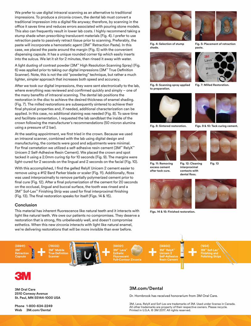
We prefer to use digital intraoral scanning as an alternative to traditional impressions. To produce a zirconia crown, the dental lab must convert a traditional impression into a digital file anyway; therefore, by scanning in the office it saves time and reduces errors associated with pouring stone models. This also can frequently result in lower lab costs. I highly recommend taking a stump shade when prescribing translucent materials (Fig. 4). I prefer to use retraction paste to passively retract tissue prior to scanning. Preferably, the paste will incorporate a hemostatic agent (3M™ Retraction Paste). In this case, we placed the paste around the margin (Fig. 5) with the convenient dispensing capsule. It has a unique rounded corner tip which easily inserts into the sulcus. We let it sit for 2 minutes, then rinsed it away with water.
A light dusting of contrast powder (3M™ High Resolution Scanning Spray) (Fig. 6) was applied prior to taking our digital impressions (3M™ True Definition Scanner). Note, this is not the old “powdering” technique, but rather a much lighter, simpler approach that increases both speed and accuracy.
After we took our digital impressions, they were sent electronically to the lab, where everything was reviewed and confirmed quickly and simply – one of the many benefits of intraoral scanning. The dental lab positions the restoration in the disc to achieve the desired thickness of enamel shading. (Fig. 7). The milled restorations are subsequently sintered to achieve their final physical properties and, if needed, additional characterization can be applied. In this case, no additional staining was needed (Fig. 8). To save time and facilitate cementation, I requested the lab sandblast the inside of the crown following the manufacturer’s recommendations (50 micron alumina using a pressure of 2 bar).
At the seating appointment, we first tried in the crown. Because we used an intraoral scanner, combined with the lab using digital design and manufacturing, the contacts were good and adjustments were minimal. For final cemetation we utilized a self-adhesive resin cement (3M™ RelyX™ Unicem 2 Self-Adhesive Resin Cement). We placed the crown and spot tacked it using a 2.0mm curing tip for 10 seconds (Fig. 9). The margins were light-cured for 2 seconds on the lingual and 2 seconds on the facial (Fig. 10).
With this accomplished, I find the gelled RelyX Unicem 2 cement easier to remove using a #12 Bard Parker blade or scaler (Fig. 11). Additionally, floss was used interproximally to remove partially polymerized cement prior to final cure (Fig. 12). After a final polymerization of the cement for 20 seconds on the occlusal, lingual and buccal surface, the tooth was rinsed and a 3M™ Sof-Lex™ Finishing Strip was used for final interproximal finishing (Fig. 13). The final restoration speaks for itself (Figs. 14 & 15).
ConclusionThis material has inherent fluorescence like natural teeth and it interacts with light like natural teeth. We owe our patients no compromises. They deserve a restoration that is strong, fits unbelievably well, and doesn’t compromise esthetics. When this new zirconia interacts with light like natural enamel, we’re delivering restorations that will be more invisible than ever before.
Fig. 11: Removing excess cement after tack cure.
Fig. 13Fig. 12: Cleaning interproximal contacts with dental floss.
Fig. 6: Scanning spray applied to preparation.
Fig. 7: Milled Restoration.
Figs. 14 & 15: Finished restoration.
Fig. 4: Selection of stump shade.
Fig. 5: Placement of retraction paste.
Fig. 8: Sintered restoration. Figs. 9 & 10: Tack curing cement.
(56941)3M™ Retraction Capsule
(78032) 3M™ Mobile True Definition Scanner
(1954) 3M™ Sof-Lex™ Finishing and Polishing Strips
(69321) 3M™ Lava™ Esthetic Fluorescent Full-Contour Zirconia
(56850) 3M™ RelyX™ Unicem 2 Self-Adhesive Resin Cement
Related Documents