Fungi Journal of Article Yeasts in Liquid Swine Diets: Identification Methods, Growth Temperatures and Gas-Formation Potential Birgit Keller 1, *, Henrike Kuder 1 , Christian Visscher 1 , Ute Siesenop 2 and Josef Kamphues 1 1 Institute for Animal Nutrition, University of Veterinary Medicine Hannover, Foundation, 30173 Hannover, Germany; [email protected] (H.K.); [email protected] (C.V.); [email protected] (J.K.) 2 Institute for Microbiology, University of Veterinary Medicine Hannover, Foundation, 30173 Hannover, Germany; [email protected] * Correspondence: [email protected] Received: 30 September 2020; Accepted: 2 December 2020; Published: 4 December 2020 Abstract: Liquid feed is susceptible to microbiological growth. Yeasts are said to cause sudden death in swine due to intestinal gas formation. As not all animals given high yeast content feed fall ill, growth and gas formation potential at body temperature were investigated as possible causally required properties. The best identification method for these environmental yeasts should be tested beforehand. Yeasts derived from liquid diets without (LD - S) and liquid diets with maize silage (LD + S) were examined biochemically (ID32C-test) and with MALDI-TOF with direct smear (DS) and an extraction method (EX). Growth temperature and gas-forming potential were measured. With MALDI-EX, most yeast isolates were identified: Candida krusei most often in LD - S, and C. lambica most often in LD + S, significantly more than in LD - S. Larger colonies, 58.75% of all yeast isolates, were formed at 25 ◦ C rather than at 37 ◦ C; 17.5% of all isolates did not grow at 37 ◦ C at all. Most C. krusei isolates formed high gas amounts within 24 h, whereas none of the C. lambica, C. holmii and most other isolates did. The gas pressure formed by yeast isolates varied more than tenfold. Only a minority of the yeasts were able to produce gas at temperatures common in the pig gut. Keywords: yeasts; liquid swine diets; MALDI-TOF; biochemical identification; growth temperature Ancom Gas Production System; Candida krusei; Candida lambica 1. Introduction Yeasts, about 600 species of which are known [1], are ubiquitous in nature and can also be found on feedstuffs[2]. They pose a risk factor regarding hygiene in liquid diets associated with off-flavor and loss of nutrients [3–5]. Depending on the species or strain, as well as on the growth conditions like temperature, substrate and its aw-value (activity of water), yeasts are able to metabolize numerous sugars, starch, protein, amino acids or even fats, and therefore lead to a loss of nutrients and energy in the feed [3–5]. In pig fattening, these energy losses in the feed are particularly undesirable [4,5]. In addition, the flavor and smell of the feed can be negatively affected [6,7]. High cell counts of yeasts in liquid swine diets due to pronounced metabolic activity are often seen in the presence of easily fermentable, low molecular weight sugars [5]. Choosing maize silage for pig feed was used with the aim of feeding the pigs to increase the feeling of satiety without making them fat [8]. The relatively high initial yeast flora of the feed has to be taken into account [8]. Therefore, feed hygiene related to yeast content was of special concern. In liquid feeds, mostly microflora develops, which is dominated by lactic acid-producing bacteria [7]. A pH-value lower than 5.0, which significantly reduces several bacteria, is often achieved J. Fungi 2020, 6, 337; doi:10.3390/jof6040337 www.mdpi.com/journal/jof

Welcome message from author
This document is posted to help you gain knowledge. Please leave a comment to let me know what you think about it! Share it to your friends and learn new things together.
Transcript

FungiJournal of
Article
Yeasts in Liquid Swine Diets: Identification Methods,Growth Temperatures and Gas-Formation Potential
Birgit Keller 1,*, Henrike Kuder 1, Christian Visscher 1 , Ute Siesenop 2 and Josef Kamphues 1
1 Institute for Animal Nutrition, University of Veterinary Medicine Hannover, Foundation,30173 Hannover, Germany; [email protected] (H.K.); [email protected] (C.V.);[email protected] (J.K.)
2 Institute for Microbiology, University of Veterinary Medicine Hannover, Foundation,30173 Hannover, Germany; [email protected]
* Correspondence: [email protected]
Received: 30 September 2020; Accepted: 2 December 2020; Published: 4 December 2020�����������������
Abstract: Liquid feed is susceptible to microbiological growth. Yeasts are said to cause sudden deathin swine due to intestinal gas formation. As not all animals given high yeast content feed fall ill, growthand gas formation potential at body temperature were investigated as possible causally requiredproperties. The best identification method for these environmental yeasts should be tested beforehand.Yeasts derived from liquid diets without (LD − S) and liquid diets with maize silage (LD + S) wereexamined biochemically (ID32C-test) and with MALDI-TOF with direct smear (DS) and an extractionmethod (EX). Growth temperature and gas-forming potential were measured. With MALDI-EX, mostyeast isolates were identified: Candida krusei most often in LD − S, and C. lambica most often in LD + S,significantly more than in LD − S. Larger colonies, 58.75% of all yeast isolates, were formed at 25 ◦Crather than at 37 ◦C; 17.5% of all isolates did not grow at 37 ◦C at all. Most C. krusei isolates formedhigh gas amounts within 24 h, whereas none of the C. lambica, C. holmii and most other isolates did.The gas pressure formed by yeast isolates varied more than tenfold. Only a minority of the yeastswere able to produce gas at temperatures common in the pig gut.
Keywords: yeasts; liquid swine diets; MALDI-TOF; biochemical identification; growth temperatureAncom Gas Production System; Candida krusei; Candida lambica
1. Introduction
Yeasts, about 600 species of which are known [1], are ubiquitous in nature and can also be foundon feedstuffs [2]. They pose a risk factor regarding hygiene in liquid diets associated with off-flavorand loss of nutrients [3–5]. Depending on the species or strain, as well as on the growth conditions liketemperature, substrate and its aw-value (activity of water), yeasts are able to metabolize numeroussugars, starch, protein, amino acids or even fats, and therefore lead to a loss of nutrients and energyin the feed [3–5]. In pig fattening, these energy losses in the feed are particularly undesirable [4,5].In addition, the flavor and smell of the feed can be negatively affected [6,7]. High cell counts of yeastsin liquid swine diets due to pronounced metabolic activity are often seen in the presence of easilyfermentable, low molecular weight sugars [5]. Choosing maize silage for pig feed was used with theaim of feeding the pigs to increase the feeling of satiety without making them fat [8]. The relativelyhigh initial yeast flora of the feed has to be taken into account [8]. Therefore, feed hygiene related toyeast content was of special concern.
In liquid feeds, mostly microflora develops, which is dominated by lactic acid-producingbacteria [7]. A pH-value lower than 5.0, which significantly reduces several bacteria, is often achieved
J. Fungi 2020, 6, 337; doi:10.3390/jof6040337 www.mdpi.com/journal/jof

J. Fungi 2020, 6, 337 2 of 20
in a shorter time with the use of starter cultures for fermented liquid feeds [3,9,10]. Yeasts are not onlyable to stay alive but also continue growing in fermented feeds [11], even if the pH-value is 4.5 [12].
Besides these complications concerning feed composition and quality, animal health may be affecteddue to the yeast content in the feed [11,13–15]. Hemorrhagic bowel syndrome (HBS), mainly caused byyeasts [13], is supposed to be causally responsible for gastric torsion and gastrointestinal tympani [16],being sometimes associated with liquid feeding [15]. HBS preferentially affects fattened pigs in thesecond half of the fattening period [16]. Those animals most affected are, as a rule, the better developedpigs in the group [14]. The fact that the affected animals are in excellent health makes this disease ofparticular economic importance [15].
In feed analyses, yeasts, irrespective of the species, are classified as spoilage indicators in animalfeed [17]. A liquid diet with more than 106 cfu yeasts/g original substance (OS) is considered assignificantly increased, while less than 105 cfu/g feed OS in liquid feed is considered as normal [18].On the other hand, selected yeasts are authorized feed additives in human nutrition and animalfeedstuffs as they synthesize vitamin B1, B2, B6, B12, folic acid, niacin, pantothenic acid and biotin,as well as containing some minerals (potassium, sodium, calcium, zinc and iron) [1]. For swine diets,viable S. cerevisiae is authorized as a feed additive as intestinal flora stabilizers, digestibility enhancersand microorganisms with a minimum concentration of 1 × 10⁹ cfu/kg complete feed (88% DM) [19].
Pathogenicity factors of yeasts have been analyzed to identify high-risk yeasts and their effects onhumans and animals. In their study about potential virulence of food-borne yeasts, Rajkowska et al. [20]stated that the ability to grow at 37 ◦C was crucial; hence, they referred to this as preliminarycriterion for pathogenicity. Adaptation to pH-value was also suggested to be a key to pathogenicity,especially important for yeasts entering the digestive tract where the pH-value changes from pH 2to pH 8 [21,22]. The ability to form biofilms also on abiotic surfaces [21] or even to colonize them isa prerequisite for colonizing the liquid feeding system, which allows the yeasts to stay alive even ifthe hygiene of the liquid feed was improved [23]. Stalljohann et al. [3] distinguished yeasts accordingto their ability to produce high or low amounts of CO2 with regard to their pathogenicity for swine,but did not mention which species produced the high gas amounts. Such detailed information on thesepossible indicators of pathogenicity is provided in the present paper.
The hypothesis of this study was that different yeast species could be found in different feedstuffs.For this reason, a comparison of biochemical differentiation and identification with MALDI-TOF wasperformed to determine the method with the most reliable identification. Presumably, only distinctspecies would be able to grow and to produce high amounts of gas at 37 ◦C.
A comparison between gas amounts produced from yeasts measured with the Ancom GasProduction System under defined conditions in a standardized Sabouraud glucose bouillon, had, to thebest of the authors’ knowledge, never been carried out previously. This permits a comparison of yeastisolates not only to see whether but also how much gas can be produced by yeasts within a certaintime period regardless of feedstuffs. Further studies must clarify whether and to what extent theseproperties have an influence on the development of diseases such as HBS. These new aspects couldthen allow to make better predictions concerning the ability of high yeast cell counts in liquid diets tocause clinical problems.
2. Materials and Methods
2.1. Sample Origin
For our project, samples from farms with liquid feeding common to all samples were collected.We obtained these samples either by contacting farms in the area, or from our own studies, which werealso carried out on similar farms. In total, 42 liquid feed samples were analyzed. Of these samples,33 were submitted for diagnostic purposes to the Institute for Animal Nutrition, University of VeterinaryMedicine Hannover, Foundation, Germany. These included common liquid feeds to which no silagehad been added, referred to as liquid diets without silage (LD − S). The remaining nine samples

J. Fungi 2020, 6, 337 3 of 20
obtained from field trials carried out by the Institute for Animal Nutrition, additionally containedwhole plant maize-silage (up to 66% DM; liquid diets with silage, LD + S). For collecting the liquidfeed samples, a standard laboratory protocol was used for both the submissions and the samplesfrom the studies. The protocol required that the samples were taken fresh, packed directly into asterile, unbreakable vessel, filled to 2/3 at most, immediately cooled and not sent before the weekend.All samples were processed directly, or in case they arrived late in the afternoon, refrigerated andprocessed the following morning.
2.2. Detection Techniques
Yeasts were isolated and morphologically characterized on Sabouraud glucose agar (SAB-Agar,PO 5096A, Thermo Fisher Scientific GmbH, Bremen, Germany) and then incubated at 30 ◦C. Only yeaststhat grew at the highest decimal dilution levels of the agar plates were considered.
2.2.1. Biochemical Differentiation
Biochemical differentiation of the yeast isolates was performed by ID 32 C strip (bioMérieux SA,Marcy-l‘Ètoile, France). This was performed in accordance with the manufacturer’s instructions.The strip consisted of 32 cavities, each containing a dehydrated carbohydrate substrate, testing theassimilation by the yeast. Pure culture yeast material of 44–48 h-grown subcultures was suspendedin 3 mL aqua destillatum. Turbidity was set in accordance with a McFarland standard of 2.0 usinga Densitometer DEN-1B (BioSan, Riga, Lettland). From this solution, 250 µL were added to the APIC medium included in the test kit. After careful vortexing, 135 µL were transferred from this liquidmedium to each well of the test strip. The strip was incubated at 30 ◦C for 44–48 h. Yeast growth resultedin turbidity of the liquid medium in the cupules, which was visually evaluated. The obtained resultswere noted on a result sheet. The values corresponding to the positive reactions were then added upwithin groups. Three results each were added up for a group. Group values were coded into a numericalprofile. This was analyzed by means of identification software (APIWEBTM, bioMérieux). The resultsof two of these carbohydrates, N-acetylglucosamine (NAG) and lactic acid (LAT), are examined inmore detail below. Only results that received good, very good or excellent (classified as “Very goodidentification”) ratings were evaluated. Rice agar (Thermo Fisher Scientific GmbH), incubated at25 ◦C for 44–48 h, was selected for some isolates if the identification software required this deficiencymedium, with a cover glass placed over the inoculum for an oxygen-reduced atmosphere.
2.2.2. MALDI-TOF
In the MALDI-TOF analysis, the sample (e.g., bacteria or yeasts) was ionized by a laser beam.These ions were then accelerated differently depending on their mass and charge. The time required topass through the length of the flight tube was determined [24]. In this way, a characteristic spectrum canbe generated for bacteria or fungi, which usually allows a species diagnosis [24,25]. As an advantage,less time is required for this method compared to biochemical methods [24].
MALDI-TOF analyses were performed on a Microflex LT/SH MALDI-MS Biotyper (Bruker DaltonikGmbH, Bremen, Germany) with the direct smear method (MALDI-DS) and with a formic acid–ethanolextraction (MALDI-EX). The latter is used for hardly soluble bacteria or yeasts.
For MALDI-DS, direct on-plate smearing was performed with yeasts incubated 44–48 h on anSAB-plate at 30 ◦C. Small amounts of colony material of every isolate were evenly applied with atoothpick to two circles of the target plate (8280800 MSP 96 Target polished steel BC, Bruker Daltonik).After air drying the sample material at room temperature for five to ten minutes, 1 µL of anα-cyano-4-hydroxycinnamic acid (HCCA, 19182, Sigma Aldrich Inc., St. Louis, MO, USA) matrixsolution was applied to each circle of the target plate and dried again at room temperature.
For MALDI-EX, a 1 µL loop of 20–24 h-grown yeast material on an SAB-plate was vortexedin 300 µL deionized water at 30 ◦C; 900 µL of ethanol absolute, HPLC-grade, was added and itwas vortexed again. The samples were pelleted by centrifuging for two minutes (13,000 U/min;

J. Fungi 2020, 6, 337 4 of 20
approximately 3000× g); the supernatant was discarded, centrifuged and then the supernatant wasdiscarded again. The pellet in the Eppendorf tube was air dried for three to five minutes andresuspended in 40 µL 70% formic acid. After adding 40 µL acetonitrile (ACN, Acetonitrile HPLCGradient Grade, 20060.320 VWR International Inc., Radnor, PA, USA), this was followed by the samecentrifugation step as above. Eppendorf tubes were taken carefully out of the centrifuge and 1 µLof the supernatant was applied to a circle of the target and air dried for 5 min at room temperature.Immediately afterwards, 1 µL of the same matrix solution that had been previously used for theMALDI-DS method was applied to a circle of the target and air dried for 5 min at room temperature.
For the control of both methods, 1 µL BTS (Bacterial Test-Standard, 8290190 Bruker IVD BacterialTest-Standard) was placed on every target plate and, additionally, a control strain, E. coli DH5 α,was tested on every target plate in two circles.
Each sample was analyzed by a Microflex LT/SH MALDI-TOF MS in the linear mode across amass-to-charge ratio range between 2000 and 20,000. The obtained data were analyzed automaticallyby using the MBT Compass Library BDAL and MBT Flex Control software, BTyp2.0-Sec.Library,1.0. Every strain was tested in two circles, with a decreased concentration in the second circle. The resultfrom the two circles that achieved the higher score value was used for analysis.
The identification cut-off scores were interpreted as per Bruker’s recommendation scores as follows:obtaining scoring thresholds between 2.30 and 3.0 suggested highly probable species identification;2.00–2.29, probable species identification; 1.70–1.99, identification at the genus level was postulated;whereas cut-off scores <1.70 indicated no reliable identification.
2.2.3. Method Comparison
The results “excellent identification” and “very good identification” adopted in the softwareAPIWEBTM were equated to “highly probable species identification” from the MALDI-TOF analysis.“Good identification” was equated to “probable species identification”; “probable genus identification”was equated to “good identification at genus level”; and “doubtful profile” and “no identification”were equated to “unacceptable profile”.
2.3. Temperature Comparison
After cultivating the yeasts from liquid feeds on SAB-agar and subcultivating a single colony, asubculture on two SAB-agar plates was produced for the temperature comparison. One agar plateeach was cultivated in an incubator at either 25 ◦C or 37 ◦C for 48 h. The colony growth (diameter) wascompared visually.
2.4. PH-Value
Using a calibrated glass electrode (HI 2211 pH/ORP Meter, Hanna Instruments Inc., Woonsocket,RI, USA), pH-values were measured. Results of 25/33 LD − S and 16/17 LD + S were obtainedimmediately after dividing the samples for further microbiological testing.
2.5. Gas Pressure Measurement
To measure yeast activity in gas production, 40 selected yeast isolates were examined for 24 hat 37 ◦C with the ANKOM RF Gas Production System (ANCOM Technology, Macedon, NY, USA),which remotes pressure under controlled pressure measurements and records these on a standardExcel spreadsheet. Gas production curves were generated. In 100 mL glass bottles containing 100 mLSabouraud glucose broth (SAB-B; CM 0147 B, Thermo Fisher Scientific GmbH), a 10 µL yeast suspension,McFarland standard 0.3 (Densitometer DEN-1B, Biosan, Riga, Lettland), in a physiologic salt solution,was added. SAB-B conforms to the parameters from the harmonized EP/USP/JP Microbial Limit Testingfor the microbial enumeration tests and tests for specified microorganisms. The bottles were placedon magnetic stirring panels (MIXdrive magnetic e motion with Mixcontrol 20, 2mag AG, Munich,Germany) for permanent mixing at 210 rpm. Gas pressure was measured over a 24-h period, taking into

J. Fungi 2020, 6, 337 5 of 20
account that feed normally does not normally stay longer in animals’ gastrointestinal tract. Line chartsof the cumulated gas production were generated with the Ancom Gas Pressure Monitor. Each isolatewas tested at least twice.
The results were divided into two groups. A very small pressure increase (<100 mbar) at thebeginning was also observed if no further gas was produced thereafter. If less than 100 mbar ofcumulated pressure within 24 h was observed, the result was determined as negative. If a yeastwas able to produce more than 800 mbar within 24 h, the result was determined as gas production.This value was defined based on the results found, because no yeast produced gas amounts between100 and 800 mbar.
2.6. Statistics
Data were statistically analyzed using the SAS® Enterprise Guide® (version 7.1, Fa. SAS InstituteInc. Cary, NC, USA). Pearson’s chi-square homogeneity test and Fisher’s exact test, used to analyzequalitative analytical characteristics, were applied to check if a yeast was found significantly more withone feed; if one of the identification tests found significantly more reliable results; whether yeasts grewbetter at a certain temperature; or if yeasts built up a distinct pressure at 37 ◦C within 24 h. Fisher’sexact test was used especially for low absolute frequencies.
3. Results
In total, 95 morphologically different yeast colonies (color, size and surface structure) were isolatedfrom a total of 42 feed samples. In each feed sample, one to four different yeast species were found.
3.1. Identification
In spite of a different morphology, yeast identification led to the same result for 15 yeast isolates.Yeasts diagnosed twice in the same sample were not considered in the evaluation of the number ofyeast species or species of yeast-like organisms that were found in the respective feed sample.
The isolates originated from six genera (Candida (76.25%; n = 61), Geotrichum (7.5%, n = 6),Trichosporon (3.75%, n = 3), Saprochaete (2.5%, n = 2), Rhodotorula (1.25%, n = 1), Pichia (1.25%, n = 1) andnon-identified yeasts (7.5%, n = 6)). A total of 19 different yeast species were identified.
In Table 1, the most often isolated species are listed. Less frequently isolated (one to three times)were C. pelliculosa (n = 3, 2× LD + S, 1× LD − S), C. valida (n = 3; 2× LD − S, 1× LD + S), Sap. suaveolens(n = 2, both from LD − S), C. rugosa (n = 2, both from LD − S), C. kefyr (n = 1, LD − S), C. variabilis (n = 1,LD − S), C. spherica (n = 1, LD − S), T. asahii (n = 1, LD + S), T. coremiiforme (n = 1, LD − S), T. laibachii(n = 1, LD − S), P. manshurica (n = 1, LD − S), Rhodotorula mucilaginosa (n = 1, LD − S) and Candida spp.(n = 1, LD − S). Six isolates (5× in LD − S; 1× in LD + S) were not reliably identified.
Table 1. Often isolated yeast species.
Liquid Diet Often Isolated Yeast Species in %
LD – S(n = 63)
C. krusei (23.8), C. holmii (12.7), C. humilis (7.9), isolates not identified (7.9), C. lambica (6.3),S. cerevisiae (6.3), G. silvicola (6.3)
LD + S(n = 17)
C. lambica (29.4), C. krusei (23.5), C. holmii (17.6), C. pelliculosa (11.8), S. cerevisiae (5.9),C. valida (5.9), isolates not identified (5.9)
Total(n = 80)
C. krusei (23.75), C. holmii (13.75), C. lambica (11.25), isolates not identified (7.5),C. humilis (6.25), S. cerevisiae (6.25), G. silvicola (6.25)
C. = Candida; S. = Saccharomyces; G. = Geotrichum.
Only C. lambica was determined to have a significantly higher incidence in LD + S (p < 0.0126).The occurrence in LD − S or LD + S did not significantly change (Figure 1).

J. Fungi 2020, 6, 337 6 of 20
J. Fungi 2020, 6, x FOR PEER REVIEW 6 of 20
Only C. lambica was determined to have a significantly higher incidence in LD + S (p < 0.0126). The occurrence in LD − S or LD + S did not significantly change (Figure 1).
Figure 1. Incidence of yeasts according to their feed origin. An asterisk (*) indicates a significant difference (* p < 0.05). C. = Candida; S. = Saccharomyces.
If, despite different colony morphology, two identical yeast species were diagnosed in one feed sample, only one yeast species was further examined. In 33 LD − S, 7/70 yeast colonies with different morphologies were selected, but identified as the same yeast species within the same diet. In LD + S, 8/25 morphologically different yeasts were identified as the same yeast within the same diet. Therefore, 63 yeasts from LD − S and 17 yeast from LD + S were further examined. As a result of this, significantly more different yeast morphologies were observed from the LD + S samples (p < 0.0211).
On the other hand, no differences between the two feeding groups were found regarding the actual (not morphologically) different yeasts. In both diet groups, an average of 1.9 different yeasts per feed sample were diagnosed: 63 different yeasts of 33 LD − S diets, and 17 different yeasts of nine LD + S diets.
3.1.1. Method Comparison
The comparison of methods revealed the most reliable results with MALDI-EX (78.75% reliable species identification), closely followed by ID32C with 75.0% reliable results. These test results did not differ statistically significantly (p < 0.6762). Among the reliable results, ID32C provided differentiation of ten isolates (8%), seven isolates with species identification and three isolates were at least identified up to genus level, which MALDI-EX was not able to differentiate. Hence, taking the results of both methods together, 71 (88.75%) of all isolates were identified up to species level. A probable identification or rather identification only at genus level was possible in three (3.75%) isolates. No identification with any of the three diagnostic methods was made for 6/80 (7.5%) yeast isolates. Only few results (37.5%) were provided by MALDI-DS (Table 2). When evaluating the reliable results, both MALDI-EX and ID32C differed significantly (both p < 0.001) from the MALDI-DS. No identification with any of the three diagnostic methods was made for 6/80 yeast isolates (7.5%). For better comparison, the results of all three methods were presented together in one table. However, the yeast names (according to their teleomorphic or anamorphic growth) given in the results varied sometimes according to the evaluation software.
Table 2. Comparison of performance of methods with 80 yeast isolates in %.
Evaluation Score of MALDI-TOF MALDI-EX MALDI-DS ID-32C
Highly probable species identification (>2.3) 38.75 (n = 31)
78.75 *,a
(n = 63)
5.0 (n = 4) 37.5 *,b
(n = 30)
47.5 (n = 38) 75.0 **,a (n = 60) Probable species
identification (2.0–2.29) 40.0 (n = 32) 32.5 (n = 26) 27.5 (n = 22)
Figure 1. Incidence of yeasts according to their feed origin. An asterisk (*) indicates a significantdifference (* p < 0.05). C. = Candida; S. = Saccharomyces.
If, despite different colony morphology, two identical yeast species were diagnosed in onefeed sample, only one yeast species was further examined. In 33 LD − S, 7/70 yeast colonies withdifferent morphologies were selected, but identified as the same yeast species within the same diet.In LD + S, 8/25 morphologically different yeasts were identified as the same yeast within the same diet.Therefore, 63 yeasts from LD − S and 17 yeast from LD + S were further examined. As a result of this,significantly more different yeast morphologies were observed from the LD + S samples (p < 0.0211).
On the other hand, no differences between the two feeding groups were found regarding theactual (not morphologically) different yeasts. In both diet groups, an average of 1.9 different yeastsper feed sample were diagnosed: 63 different yeasts of 33 LD − S diets, and 17 different yeasts of nineLD + S diets.
3.1.1. Method Comparison
The comparison of methods revealed the most reliable results with MALDI-EX (78.75% reliablespecies identification), closely followed by ID32C with 75.0% reliable results. These test results did notdiffer statistically significantly (p < 0.6762). Among the reliable results, ID32C provided differentiationof ten isolates (8%), seven isolates with species identification and three isolates were at least identifiedup to genus level, which MALDI-EX was not able to differentiate. Hence, taking the results of bothmethods together, 71 (88.75%) of all isolates were identified up to species level. A probable identificationor rather identification only at genus level was possible in three (3.75%) isolates. No identification withany of the three diagnostic methods was made for 6/80 (7.5%) yeast isolates. Only few results (37.5%)were provided by MALDI-DS (Table 2). When evaluating the reliable results, both MALDI-EX andID32C differed significantly (both p < 0.001) from the MALDI-DS. No identification with any of thethree diagnostic methods was made for 6/80 yeast isolates (7.5%). For better comparison, the resultsof all three methods were presented together in one table. However, the yeast names (according totheir teleomorphic or anamorphic growth) given in the results varied sometimes according to theevaluation software.
Some yeasts were diagnosed more accurately with one or the other method. Five isolatesdifferentiated with MALDI-EX as C. humilis were diagnosed as C. holmii in ID 32 C. C. humilis wasnot included in the database used to evaluate the ID 32 C. Both of these species had very similarbiochemical reactions. Therefore, identification with MALDI-EX was chosen to be more accurate.
The two yeasts identified as C. pararugosa in MALDI-EX were identified as C. rugosa in ID 32C. Results in MALDI-EX were only 1.88 and 1.79, respectively. Therefore, they had to be namedaccording to ID 32 C, where the results for both isolates revealed very good identification scores,namely, 99.8% and 99.5% for C. rugosa. On the other hand, C. pararugosa was not included in thedatabase used to evaluate the ID 32 C.

J. Fungi 2020, 6, 337 7 of 20
Table 2. Comparison of performance of methods with 80 yeast isolates in %.
Evaluation Score ofMALDI-TOF MALDI-EX MALDI-DS ID-32C
Highly probable speciesidentification (>2.3) 38.75 (n = 31)
78.75 *,a
(n = 63)
5.0 (n = 4)37.5 *,b
(n = 30)
47.5 (n = 38)75.0 **,a
(n = 60)Probable speciesidentification (2.0–2.29) 40.0 (n = 32) 32.5 (n = 26) 27.5 (n = 22)
Probable genusidentification (1.7–1.99) 7.5 (n = 6) 21.25 ***
(n = 17)
33.75 (n = 27) 62.5 ***(n = 70)
3.75 (n = 3) 25.0 ***(n = 20)
No identification (<1.7) 13.75 (n = 11) 28.75 (n = 23) 21.25 (n = 17)a,b Different superscripts differ significantly in a row. * Reliable identification: Highly probable species identificationand probable species identification; ** Reliable identification: Good and very good identification together;***: No species identification.
Even with the formic acid extraction method, the slimy and red growing Rhodotorula (R.)mucilaginosa could not be detected with MALDI-TOF; there was no reliable identification, although itwas registered in the database.
In the ID 32 C, two isolates were diagnosed as Cryptococcus (C. curvatus and C. laurentii),which showed no mucus capsule in the Indian ink preparation, but showed hyphal growth, arthro-and blastosporogenesis on rice agar under the cover glass (Figure 2), and were identified in MALDI-EXas T. coremiiforme and T. laibachii (Table 3). On the other hand, two T. asahii isolates could be recognizedwell or very well by both methods.
J. Fungi 2020, 6, x FOR PEER REVIEW 7 of 20
Probable genus identification (1.7–1.99)
7.5 (n = 6) 21.25 *** (n = 17)
33.75 (n = 27) 62.5 *** (n = 70)
3.75 (n = 3) 25.0 *** (n = 20)
No identification (<1.7) 13.75 (n = 11) 28.75 (n = 23) 21.25 (n = 17) a,b Different superscripts differ significantly in a row. * Reliable identification: Highly probable species identification and probable species identification; ** Reliable identification: Good and very good identification together; ***: No species identification.
Some yeasts were diagnosed more accurately with one or the other method. Five isolates differentiated with MALDI-EX as C. humilis were diagnosed as C. holmii in ID 32 C. C. humilis was not included in the database used to evaluate the ID 32 C. Both of these species had very similar biochemical reactions. Therefore, identification with MALDI-EX was chosen to be more accurate.
The two yeasts identified as C. pararugosa in MALDI-EX were identified as C. rugosa in ID 32 C. Results in MALDI-EX were only 1.88 and 1.79, respectively. Therefore, they had to be named according to ID 32 C, where the results for both isolates revealed very good identification scores, namely, 99.8% and 99.5% for C. rugosa. On the other hand, C. pararugosa was not included in the database used to evaluate the ID 32 C.
Even with the formic acid extraction method, the slimy and red growing Rhodotorula (R.) mucilaginosa could not be detected with MALDI-TOF; there was no reliable identification, although it was registered in the database.
In the ID 32 C, two isolates were diagnosed as Cryptococcus (C. curvatus and C. laurentii), which showed no mucus capsule in the Indian ink preparation, but showed hyphal growth, arthro- and blastosporogenesis on rice agar under the cover glass (Figure 2), and were identified in MALDI-EX as T. coremiiforme and T. laibachii (Table 3). On the other hand, two T. asahii isolates could be recognized well or very well by both methods.
Figure 2. Trichophyton coremiiforme on rice agar: arthrospores (a) and blastospores (b). 400× magnification.
Table 3. Isolates differentially diagnosed with MALDI-EX and ID 32C.
Number of Isolates MALDI-EX ID 32C 5 Candida humilis Candida holmii 2 Candida pararugosa Candida rugosa 1 No identification Rhodotorula mucilaginosa 1 Trichosporon coremiiforme Cryptococcus curvatus 1 Trichosporon laibachii Cryptococcus laurentii
Yeasts highlighted in bold represent the selected diagnoses.
Six isolates had a score between 1.79 and 1.98 in the MALDI-EX. Of these isolates, S. cerevisiae (score 1.98) and C. holmii (score 1.97) had the same result in ID32C (see Table 4), with very good
a
b
Figure 2. Trichophyton coremiiforme on rice agar: arthrospores (a) and blastospores (b).400×magnification.
Table 3. Isolates differentially diagnosed with MALDI-EX and ID 32C.
Number of Isolates MALDI-EX ID 32C
5 Candida humilis Candida holmii2 Candida pararugosa Candida rugosa1 No identification Rhodotorula mucilaginosa1 Trichosporon coremiiforme Cryptococcus curvatus1 Trichosporon laibachii Cryptococcus laurentii
Yeasts highlighted in bold represent the selected diagnoses.
Six isolates had a score between 1.79 and 1.98 in the MALDI-EX. Of these isolates, S. cerevisiae(score 1.98) and C. holmii (score 1.97) had the same result in ID32C (see Table 4), with very goodidentification. Furthermore, two isolates were identified as C. pararugosa in the MALDI-EX (with scores

J. Fungi 2020, 6, 337 8 of 20
1.88 and 1.63). C. pararugosa was not included in the identification software (APIWEBTM, bioMérieux).The remaining two isolates consisted of Saprochaete (Sap.) suaveolens (score 1.88), which was diagnosedas a Geotrichum spp. in ID32C, and Pichia occidentalis (score 1.83), diagnosed with 99.7% as C. kruseiin ID32C. Bearing in mind that Sap. suaveolens was formerly diagnosed as Geotrichum fragrans,the diagnosis made by MALDI-EX was most likely the one with the currently correct name. The nameof the yeast in ID32C was probably out of date; the yeast was still correctly identified. Only one isolatewas differently identified by the two methods as P. occidentalis (MALDI-TOF) and C. krusei (ID32C;Table 4).
Table 4. Isolates in MALDI-EX rated as probable genus identification (1.70 and 1.99).
MALDI-EX ID32C
Diagnosis Score Diagnosis Identification %C. pararugosa 1.88 C. rugosa very good 99.8
Sap. suaveolens 1.88 Geotrichum spp. very good 99.7P. occidentalis 1.83 C. krusei very good 99.7
C. holmii 1.97 C. holmii very good 99.2C. pararugosa 1.79 C. rugosa very good 99.5S. cerevisiae 1.98 S. cerevisiae very good 99.7
Yeasts in bold represent the selected diagnoses C. = Candida; Sap. = Saprochaete; P. = Pichia; S. = Saccharomyces.
3.1.2. Biochemical Reactions
In total, all the investigated yeasts were able to assimilate glucose and no yeast grew in the cupulewhere no substrate was present (cupule F). These reactions were considered as the positive growthcontrol or negative control (no contamination). From the large number of biochemical reactions,two of them will be examined in more detail in the following section, since the ability of the yeasts tometabolize them could be an advantage, especially in maize silage.
Metabolization of N-Acetylglucosamine (NAG)
In the ID 32 C-Test, 27 yeasts from a total of 63 yeasts in the LD − S samples were able tometabolize N-acetylglucosamine and 36 yeasts were not. In LD + S, 11 yeasts were able to buildN-acetylglucosamine and six yeasts were not. Despite the fact that this is insignificant (p = 0.0788),the ability to build NAG was more often seen in yeasts from LD + S.
Metabolization of Lactic Acid (LAT)
In the ID 32 C-Test, 36 yeasts of a total of 63 yeasts in the LD − S samples were able to metabolizelactic acid and 27 yeasts were not. In LD + S, 12 yeasts were able to metabolize lactic acid and fiveyeasts were not. These results were insignificant (p = 0.4075). Nevertheless, the ability to metabolizelactic acid could be found more often with yeasts that had to stay alive or even grow in maize silagethan for yeasts in LD − S.
3.2. Temperature Comparison
Most yeast isolates (n = 47; 58.75%) formed larger colonies at 25 ◦C than at 37 ◦C, among them,14 isolates (17.5%) did not grow at 37 ◦C at all. These included ten isolates from LD − S (3× C. holmii,1× each for C. humilis, C. lambica, T. laibachii, C. pelliculosa, Geotrichum spp., S. cerevisiae and an isolatenot identified) and four from LD + S (3× C. holmii and 1× isolate not identified). Among all yeasts,which grew better or only at 25 ◦C, many isolates of C. holmii, C. humilis and C. lambica were found.Only 23.75% of isolates grew better at 3 ◦C compared to 25 ◦C; this was often the case for C. krusei(12/19 isolates) and S. cerevisiae (4/5 isolates). Additionally, C. kefyr (1/1), C. holmii (1/11 isolates) andone isolate that could not be identified (1/6) showed better growth at 37 ◦C. All S. cerevisiae isolateswere harvested from LD − S samples. Larger colonies at 37 ◦C were formed from nine C. krusei isolates

J. Fungi 2020, 6, 337 9 of 20
gained from LD− S and three from LD + S, while three isolates from LD− S and one from LS + S formedlarger colonies at 25 ◦C; one isolate showed equal colony growth at 25 ◦C or 37 ◦C. Hence, for C. krusei,no difference was observed, regardless of which feed it was isolated from. When comparing bothfeeds, it was noticeable that especially yeasts isolated from LD + S grew poorly at 37 ◦C (see Table 5).Better growth at 37 ◦C than at 25 ◦C for yeasts harvested from LD + S was only seen for three isolates,all of which were C. krusei. However, there was no statistically significant difference (p < 0.3862)between the two feed sources concerning growth performance (colony size) of the yeasts at either ofthe temperatures.
Table 5. Growth performance of the yeasts at different temperatures depending on feed.
Yeast Isolates No Growth at 37 ◦C Better Growth at 25 ◦C 25 ◦C = 37 ◦C Better Growth at 37 ◦C
LD − S (n = 63) 10 (15.9%) 24 (38.1%) 13 (20.6%) 16 (25.4%)LD + S (n = 17) 4 (23.5%) 9 (52.9%) 1 (5.9%) 3 (17.6%)Total (n = 80) 14 (17.5%) 33 (41.25%) 14 (17.5%) 19 (23.75%)
3.3. pH-Value in Liquid Swine Diets
The pH-values of LD − S (n 25/33) ranged from 3.87–5.78, while the pH-values of LD + S (n = 8/9)achieved higher pH-values ranging from pH 4.79 to pH 5.61. Related to the feed origin, 44 yeastsisolated from LD − S were harvested from liquid feed, with an average pH-value of 4.59. Yeasts gainedfrom LD + S were harvested from liquid feed, with an average pH-value of 5.51. C. krusei was isolatedfrom liquid swine diets with the lowest (pH 3.87) and highest pH-values (pH 5.78) as well. C. humilisand C. holmii were found in diets with lower pH-values (pH 3.9 to pH 5.11), whereas C. lambica wasisolated once from a diet with a pH-value of 4.45. However, other isolates were harvested from dietswith higher pH-values (ranging from pH 4.97 to pH 5.61).
3.4. Gas Production
The results of the duplicate testing of each isolate showed small deviations, possibly caused bysmall differences in cell counts at the beginning as well as differences in replication time and counts ofspores formed by each yeast cell during the 24-h incubation period.
Only two groups were formed: yeasts that produced virtually no gas within 24 h at 37 ◦C andyeasts that produced more than 800 mbar. A further subdivision of the yeasts into groups producinglittle or a lot of gas was omitted, because too little information was available from the literature as towhich quantities could be classified as a lot or little.
No yeast produced gas amounts between 100 mbar and 800 mbar. More yeasts harvested fromLD − S produced gas than yeasts that were found in LD + S, but the quantity was not significant(p < 0.2216).
Gas production with more than 800 mbar was observed for a total of 13 (40.6%) isolates (Table 6):10/11 C. krusei-isolates, 2/3 S. cerevisiae-isolates, 1/1 C. kefyr and 1/1 C. humilis. C. kefyr formed thehighest gas pressure, with 10,419 mbar, followed by the C. krusei isolates (7134.5, 7073, 6659, 6487, 6383,6164.5, 4839, 4147.5, 3954.5 and 3940.5 mbar), both isolates of S. cerevisiae (1160.5 and 1466.5 mbar)and C. humilis (888 mbar). Eleven of these isolates grew better at 37 ◦C than at 25 ◦C within 48 h(see Section 3.2). Nonetheless, two isolates, which also grew better at 37 ◦C, were not able to producemore than 100 mbar gas in 24 h. These two yeasts were one C. holmii and one C. krusei isolate harvestedfrom LD + S. The latter one produced these high gas quantities only after a 40 h incubation time.On the other hand, one isolate of C. krusei, harvested from LD − S, which grew better at 25 ◦C than at37 ◦C, nevertheless produced 3954.5 mbar gas within 24 h. Only one yeast isolate from LD + S couldproduce significant quantities of gas under the abovenamed circumstances within 24 h (Table 4).
J. Fungi 2020, 6, 337 10 of 20
Table 6. Gas production (mbar) at 37 ◦C within 24 h.
Sample Number <100 mbar >800 mbar
LD − S n = 32 19 (59.4%) 13 (40.6%)LD + S n = 8 7 (87.5%) 1 (12.5%)
total n = 40 26 (65.0%) 14 (35.0%)
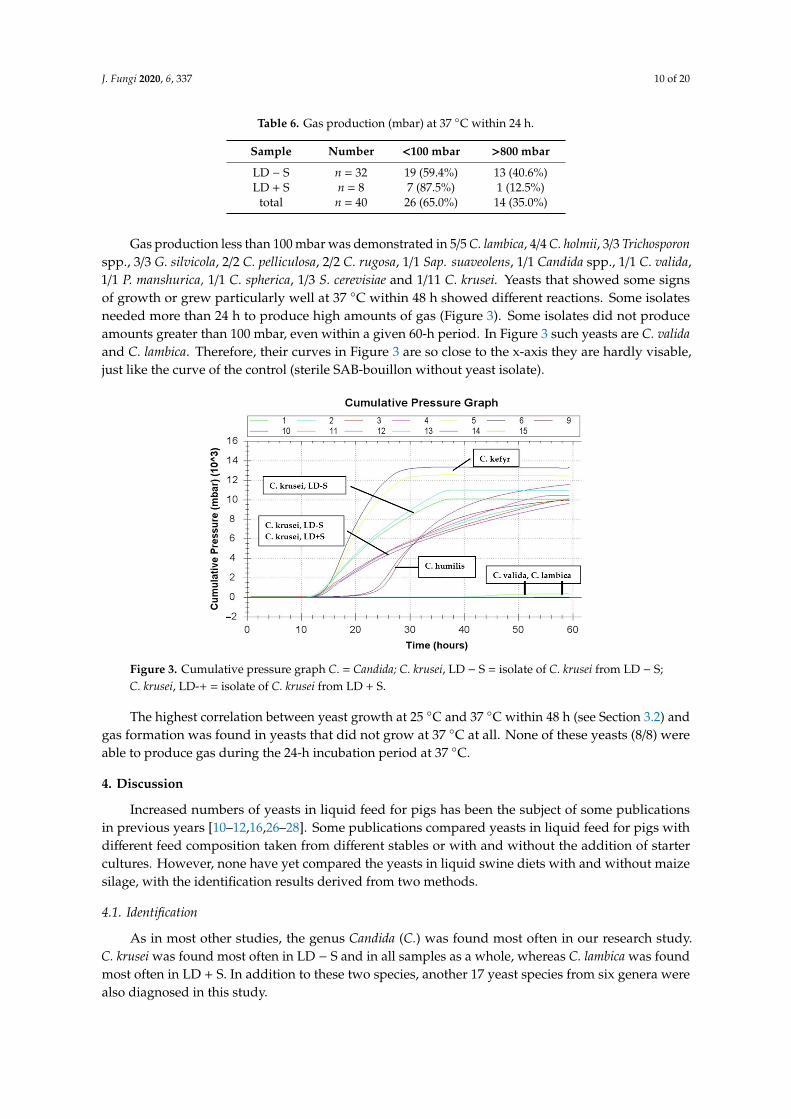
Gas production less than 100 mbar was demonstrated in 5/5 C. lambica, 4/4 C. holmii, 3/3 Trichosporonspp., 3/3 G. silvicola, 2/2 C. pelliculosa, 2/2 C. rugosa, 1/1 Sap. suaveolens, 1/1 Candida spp., 1/1 C. valida,1/1 P. manshurica, 1/1 C. spherica, 1/3 S. cerevisiae and 1/11 C. krusei. Yeasts that showed some signsof growth or grew particularly well at 37 ◦C within 48 h showed different reactions. Some isolatesneeded more than 24 h to produce high amounts of gas (Figure 3). Some isolates did not produceamounts greater than 100 mbar, even within a given 60-h period. In Figure 3 such yeasts are C. validaand C. lambica. Therefore, their curves in Figure 3 are so close to the x-axis they are hardly visable,just like the curve of the control (sterile SAB-bouillon without yeast isolate).
J. Fungi 2020, 6, x FOR PEER REVIEW 10 of 20
total n = 40 26 (65.0%) 14 (35.0%)
Gas production less than 100 mbar was demonstrated in 5/5 C. lambica, 4/4 C. holmii, 3/3 Trichosporon spp., 3/3 G. silvicola, 2/2 C. pelliculosa, 2/2 C. rugosa, 1/1 Sap. suaveolens, 1/1 Candida spp., 1/1 C. valida, 1/1 P. manshurica, 1/1 C. spherica, 1/3 S. cerevisiae and 1/11 C. krusei. Yeasts that showed some signs of growth or grew particularly well at 37 °C within 48 h showed different reactions. Some isolates needed more than 24 h to produce high amounts of gas (Figure 3). Some isolates did not produce amounts greater than 100 mbar, even within a given 60-h period. In Figure 3 such yeasts are C. valida and C. lambica. Therefore, their curves in Figure 3 are so close to the x-axis they are hardly visable, just like the curve of the control (sterile SAB-bouillon without yeast isolate).
Figure 3. Cumulative pressure graph C. = Candida; C. krusei, LD − S = isolate of C. krusei from LD − S; C. krusei, LD-+ = isolate of C. krusei from LD + S.
The highest correlation between yeast growth at 25 °C and 37 °C within 48 h (see Section 3.2) and gas formation was found in yeasts that did not grow at 37 °C at all. None of these yeasts (8/8) were able to produce gas during the 24-h incubation period at 37 °C.
4. Discussion
Increased numbers of yeasts in liquid feed for pigs has been the subject of some publications in previous years [10–12,16,26–28]. Some publications compared yeasts in liquid feed for pigs with different feed composition taken from different stables or with and without the addition of starter cultures. However, none have yet compared the yeasts in liquid swine diets with and without maize silage, with the identification results derived from two methods.
4.1. Identification
As in most other studies, the genus Candida (C.) was found most often in our research study. C. krusei was found most often in LD − S and in all samples as a whole, whereas C. lambica was found most often in LD + S. In addition to these two species, another 17 yeast species from six genera were also diagnosed in this study.
4.1.1. C. krusei
Overall, the most often found yeast in this study, C. krusei, was isolated from feed samples with the lowest and highest pH-values. From the literature, it is known to grow at low pH-values [29], ferment up to a pH-value of 3.6 [30] and can also form biofilms [31]. These properties are likely to be beneficial for yeast persistence in the liquid feed and the feeding system. In part, C. krusei is capable
Figure 3. Cumulative pressure graph C. = Candida; C. krusei, LD − S = isolate of C. krusei from LD − S;C. krusei, LD-+ = isolate of C. krusei from LD + S.
The highest correlation between yeast growth at 25 ◦C and 37 ◦C within 48 h (see Section 3.2) andgas formation was found in yeasts that did not grow at 37 ◦C at all. None of these yeasts (8/8) wereable to produce gas during the 24-h incubation period at 37 ◦C.
4. Discussion
Increased numbers of yeasts in liquid feed for pigs has been the subject of some publicationsin previous years [10–12,16,26–28]. Some publications compared yeasts in liquid feed for pigs withdifferent feed composition taken from different stables or with and without the addition of startercultures. However, none have yet compared the yeasts in liquid swine diets with and without maizesilage, with the identification results derived from two methods.
4.1. Identification
As in most other studies, the genus Candida (C.) was found most often in our research study.C. krusei was found most often in LD − S and in all samples as a whole, whereas C. lambica was foundmost often in LD + S. In addition to these two species, another 17 yeast species from six genera werealso diagnosed in this study.

J. Fungi 2020, 6, 337 11 of 20
4.1.1. C. krusei
Overall, the most often found yeast in this study, C. krusei, was isolated from feed samples withthe lowest and highest pH-values. From the literature, it is known to grow at low pH-values [29],ferment up to a pH-value of 3.6 [30] and can also form biofilms [31]. These properties are likely to bebeneficial for yeast persistence in the liquid feed and the feeding system. In part, C. krusei is capable ofpseudohyphae formation and mostly of growing at 37 ◦C, both characteristics that could contributeto HBS.
C. krusei is responsible for about 2% of yeast infections caused by Candida species in humans [32].Pichia kudriavzevii, Issatchenkia orientalis and Candida glycerinogenes are proven to be the same yeastwith collinear genomes 99.6% identical in DNA sequence. Under these names, the yeast is usedfor industrial-scale production of glycerol and succinate, and is also used to make some fermentedfoods [32]. The latter use in fermented foods also explains the frequent occurrence in liquid feed forpigs, which also has a low pH value (see Section 3.3).
4.1.2. C. lambica
The significantly higher presence of C. lambica in LD + S is possibly due to maize silage in the feedbut could also be due to the lower storage temperature of the maize silage outdoors during winter [33].As LD + S samples were gained from the institute’s own research projects, it is known that animalsdid not develop HBS or any other disease and that they ate a lot more with the ad libitum feeding ofLD + S in comparison to the previous feed intake with commercial feed (Jörling, 2017) [8]. This wasobserved, although the yeast content of the feed was temporarily more than 1 × 10⁸ cfu/g feed (Jörling,personal observations, results of which have not yet been published). Olstorpe et al. [26] discoveredthat Pichia fermentans (C. lambica) was dominant in all their experiments. They assumed that C. lambicawas able to improve palatability as it has been described to improve the flavor composition duringwine- and cheese-making [26]. Presumably, the yeast species within the diet might be more importantthan the orientation values, which are the same for all yeasts when the hygiene status of the liquiddiets is under debate.
4.1.3. Yeasts from Liquid Diets for Pigs
In the present study, mostly C. krusei, C. holmii, C. lambica, S. cerevisiae, C. humilis and Geotrichumspp. were identified (see Section 3.1, Table 1). Together, they accounted for 66.5% of all yeast isolates.Other species could not be identified (7.5%, Table 1) or were only detected in lower proportions (26%;see Section 3.1).
Middelhoven et al. [33] observed, in whole-crop maize ensiled for two weeks, similar yeastspecies compared to those found in LD + S. They predominately found C. holmii, C. lambica, C. milleri(current name: C. humilis), Hansenula anomala (current name: Wickerhamomyces anomalis, anamorph:C. pelliculosa) and Saccharomyces dairensis [33], whereby only the latter yeast did not occur in our study.The comparability of the yeast species in both studies could indicate that it is not so much the storageover winter but rather the substrate that influences the yeast occurrence.
The biochemical profiles of both yeasts are very similar and therefore sometimes misidentified [34].Both yeasts are able to assimilate mostly glucose, lactose, glycerol, inositol and N-acetylglucosamine,while xylose is only metabolized from C. lambica. On the other hand, the next frequently identifiedyeasts, C. holmii, C. humilis and S. cerevisiae, cannot perform this metabolic function, with the exceptionof glucose. Instead, they metabolize galactose and raffinose, and, in part, trehalose (C. holmii andC. humilis), sucrose (C. holmii and partly also S. cerevisiae) as well as maltose (S. cerevisiae). This couldsuggest that the assimilative capacities of the yeasts are not essential for their presence or absence indifferent liquid feeds for pigs.
Likewise, many different yeasts were identified in studies on yeast determination from liquidfeed samples for pigs, and different species dominated in the different feed samples [6,26–28].

J. Fungi 2020, 6, 337 12 of 20
Similarities to the isolated yeasts in the present study were observed in the studies by Olstorpeet al. [26], who examined liquid feeds based on a cereal grain mix and wet wheat distiller’s grainwith and without starter cultures. Without starter cultures, they observed P. fermentans (C. lambica),C. pararugosa, C. rugosa, P. galeiformis (current name: P. mandshurica), T. asahii, Issatchenkia orientalis(C. krusei, P. kurdriavzevii), C. ethanolica and C. vini. These yeasts were found in the present study aswell, except the last three mentioned ones.
Olstorpe et al. [26] isolated C. kefyr from wheat-based liquid feed as the dominant yeast, which wasisolated only once in the present study. This previous publication also found C. krusei, C. pelliculosa andPichia membranaefaciens (C. valida). Plumed-Ferrer and Wright [35] most frequently observed K. exigua(C. humilis), Debaromyces hansenii and Pichia derserticola in fresh batches of liquid feed, of which onlyK. exigua was often observed in the present study. On the other hand, in this previous study, other yeastswere also isolated, such as Pichia kurdriavzevii (C. krusei), S. exiguous (C. holmii), Pichia membranaefaciens(C. valida) and Wickerhamomyces anomalus (C. pelliculosa), which were identified in the present study, too.
Significantly more morphologically different yeasts were observed in LD + S (see Section 3.1),which had lower pH-values than conventional feed and were stored outdoors during winter, which,as a consequence, were exposed to changing temperatures. These different colonial morphologiescould be a result of changing environmental conditions, as explained in previous publications [36,37].
The genera Geotrichum and Trichosporon are classified as yeast or yeast-like organisms,but Geotrichum was formerly classified as a mold [38–40]. The colony morphology is very similar toother yeasts and therefore was described in many previous studies concerning yeasts in liquid swinediets [25,27,28], so that comparability with other studies is possible. Saprochaete suaveolens, formerlyclassified as Geotrichum fragrans, is also classified as a yeast or yeast-like organism (mycobank.org [41]).Hereafter, for the sake of simplicity, all genera are referred to as yeasts, even if the term yeast oryeast-like organism would be more accurate.
4.1.4. Method Comparison
The present study compared different methods for identifying yeasts to find the best methodfor the chosen substrate and the yeasts contained in it. From previous studies [6,10,26,28,42], it wasknown that many tests for identifying yeasts from the environment produce fewer results than thosefrom clinical material [43–45]. Additionally, different databases on which different test procedures arebased also influence the obtained results [45,46].
For clinical samples consisting mainly of Candida spp., the method of MALDI-TOF outperformedthe diagnosis capacities of the phenotypic tests by reducing the delay in results and improving thereliable identification rate at species level [43]. On the other hand, this method requires significantlyhigher acquisition costs for the equipment. Therefore, this method was compared with the ID 32 C test,which has virtually no purchase costs.
ID32C
In our study, 5.2% less reliable results were observed with the ID 32 C test in comparison toMALDI-EX. Nevertheless, in individual cases, correct identification could only be made with this simplebiochemical method (see Table 4). C. rugosa was twice identified with more than 99.5% accuracy as “verygood identification”, while MALDI-EX identified these two yeast isolates as C. pararugosa. The latterwas not included in the ID32C-database. Considering the fact that several authors [44,45,47] proposeda lower identification score for the yeast identification with MALDI (see below), perhaps the MALDIresults are the correct ones. In the case of Geotrichum spp. and Saprochaete suaveolens, the situation issimilar. Saprochaete suaveolens was not included in the database of the ID32C test. Comparable to thefinding in the present study, namely that Rhodotorula was better identified with ID32C, Olstorpe et al. [6]reported that, with the applied PCR fingerprinting, two Rhodotorula glutinis isolates were incorrectlyclassified as Cryptococcus satoi or Pichia membranaefaciens, but correctly identified with ID32C.
J. Fungi 2020, 6, 337 13 of 20
The ID32C test can be easily performed in any laboratory and does not require an expensive device.In addition to species identification, the biochemical test has the advantage of showing which enzymescan be produced by the respective yeast isolate. This, in turn, could allow or exclude opportunities foridentifying which feed components could be metabolized by the yeast.
Selected Biochemical Reactions of the ID32C-Test
Metabolization of N-Acetylglucosamine (NAG): More yeasts from LD + S, even if not significant,were able to metabolize the amino sugar NAG. This is the monomeric constituent of chitin, which isone of the most abundant renewable resources found in nature [48]. The uptake of NAG into the yeastcell, its metabolites in the cell and conversion to cell wall formation have already been described forvarious yeasts [48]. The cell wall reinforced by NAG (chitin) offers protection against low pH-values inthe environment [22]. Although the pH-values in the LD − S were not significantly lower than those ofLD + S, the prolonged period of survival in silage (see below) may have led to the ability of yeasts tometabolize NAG.
Metabolization of Lactic Acid (LAT): More yeasts from LD + S were able to metabolize lacticacid. As this finding is not statistically significant, the ability to metabolize lactic acid obviously isnot a prerequisite for yeasts in LD + S. Lactic acid bacteria are the predominant group of bacteriafound in maize silages, and are able to multiply in liquid feed, lactic acid being a main product of theirmetabolism [49]. Maize silage used for LD + S in the present study was kept outdoors during winterand early spring until it was fed to the animals in late spring and early summer. Being able to use asubstrate present in the environment is presumed to be an advantage for yeasts [50,51], which have tosurvive in these conditions for a long time.
MALDI-DS: The less time-consuming and less expensive MALDI-DS reduced the identificationrate significantly (p < 0.001) by more than half compared to MALDI-EX (37.5% vs. 78.75% reliableidentification). Thus, the use of this method is clearly limited, at least if different yeasts are to beidentified from environmental samples. In contrast to bacteria, yeasts possess a thick and chitinous cellwall, which might lead to the difficulties encountered with the MALDI-DS method [52].
MALDI-EX: In our study, 78.75% of the 80 different yeasts could be identified by MALDI-EX and75.0% by the ID 32 C. While 11 isolates were not identified at all, six isolates achieved only probableresults at the genus level (see Table 4). An incorrect diagnosis was observed only once, mistakenlyidentifying P. occidentalis instead of C. krusei (see Section 3.1.1).
A comparison of identification of 96 foodborne yeasts with MALDI-TOF and two conventionaltests, of which one was ID 32 C, was made by Pavlovic et al. 2014 [53]. In their study, more yeastisolates could be identified with MALDI-TOF than with the ID32C test, too.
The identification rate of the different methods in the present study was comparable to those ofothers in which yeasts were isolated from the environment rather than from clinical material [6,54–56].As already shown by these and other authors [1,57], none of the methods were capable of reliablydetecting all yeast isolates from the liquid feed. Many authors attribute these differentiation failuresto the background of the ID32C, MALDI-TOF and other commercially available tests, as these weredeveloped and established for clinically relevant yeasts in human beings and not for yeasts in animalfeed [1,45,57]. None of the available methods can be considered as the golden standard for thedifferentiation of yeasts from liquid feeds. With respect to the low examination costs, low workload,fast availability of results and available databases, which means the highest rate of correct identification,the different methods exhibit advantages and disadvantages.
Although MALDI-EX was the best method for gaining the most reliable identification results inthis study, it has to be considered that results from this method are only as good as the underlyingdatabase [46]. Vlek et al. [46] identified 61.5% of their yeasts from human patients using the BrukerDaltonic database (BDAL), but improved their identification rate up to 86.8% by adding their databasewith the in-house database from the Centraalbureau voor Schimmelcultures (Central Bureau for FungalCultures) (BDAL + CBS in-house). This allows the assumption to be made that even more yeasts will
J. Fungi 2020, 6, 337 14 of 20
be identified with this method in the future, if correspondingly relevant data continue to be added,especially for the non-clinical yeasts found in the surroundings. An improvement in the identificationresults of 845 environmental yeasts by one third was also described by Augustini et al. [45] afterdeveloping a supplementary database.
Besides the databases as reason for missing reliable yeast identification, Augustini et al. [45]stated that identification scores <2.00 are not able to unequivocally affirm that the identification atspecies level is unreliable. They cited studies that showed identification results under 2.00, but withcorrect identifications. This observation was underlined in the studies by Tan et al. [44]. RepeatingMALDI-TOF attempts in 10.2% of the yeast isolates, which had indicated spectral scores as beingunacceptable on the first attempt (scores < 2.00), resulted in acceptable scores (>2.00). Most of theseachieved a correct identification on the first attempt [44]. The authors concluded that lowering theidentification score from <2.00 to <1.70 could reduce the repetition rate [44]. With a cut-off of <1.70,Lee et al. [50] also improved the identification rate of their 284 pathogenic yeasts from clinical samplescompared to the required cut-off value of >2.00 [49]. When comparing the results of two differentMALDI-TOF systems (Biotyper from Bruker and ASTA MALDI-TOF MS), Lee at al. [50] found thatonly 39.5% of the isolates with confirmed identification with molecular sequencing met the cut-off
score in both systems. The majority of the isolates (58.6%) ranged between 1.70 and 2.00 when usingthe Bruker Biotyper and scores > 140 using ASTA MALDI-TOF.
Lee et al. [52] performed a formic acid extraction with a shorter protocol. Most of the yeastsobtained from samples of clinically infected humans were identified correctly, but the method failedto identify the slimy Cryptococcus spp. Considering the fact that in our study no Cryptococcus spp.were found, possibly this shorter, easier and inexpensive method could have provided as good resultsas MALDI-EX. On the other hand, different Cryptococcus spp. were isolated from liquid swine diets instudies by Olstorpe et al. [6]. Therefore, MALDI-EX seemed to be the best method to reliably identifyas many yeasts species as possible.
Extending databases, lowering the identification score for yeasts as well as shorter protocols couldimprove the ratio of reliable results of environmental yeasts with MALDI-EX in the future, so that theresults of this method could be highlighted even more.
Various molecular biological methods described in the literature were not included in this study,although previous authors achieved good results [27]. Gori et al. [27] had difficulties in separating thetwo most commonly occurring yeasts in their study with 26S rRNA sequencing: C. humilis (formerlynamed C. milleri; 58.4%) and C. holmii (Kasachstania exigua; 17.5%), together accounting for 75.9% of allresults (n = 766 yeasts). They distinguished the two yeasts biochemically according to their sucroseand raffinose metabolism [27]. In the present study, C. humilis and C. holmii accounted together for20% of all results (n = 16). In retrospect, it can be assumed that the 26S rRNA method would not havebeen advantageous in these cases.
4.2. Temperature
In the present study, clearly more than half of the yeasts (52.9%) grew better at 25 ◦C than at 37 ◦Cor did not grow at 37 ◦C at all (23.5%). Those yeasts that did not grow at 37 ◦C at all will presumablynot grow in the intestines of pigs, where the internal body temperature normally still exceeds 37 ◦C.
Considering only yeasts isolated from LD + S, there are even more isolates that prefer coolertemperatures (see Table 5). An explanation for these yeasts preferring lower temperatures than yeastsfrom LD − S could be the chosen time of sampling of LD + S in late spring and early summer in the twoprojects, when the liquid diets were composed. After harvesting the maize plants and making silage inthe fall in the respective projects, this was stored outdoors during winter, where yeasts had to copewith low temperatures. Thus, yeasts may have adapted to these temperatures or died. Storing feedmaterials or liquid diets at cool temperatures possibly reduces the yeast species, which prefer 37 ◦C,and as a result have little or no impact on gut health.

J. Fungi 2020, 6, 337 15 of 20
The present results could also indicate an adaptation of the yeasts to their feed origin andstorage temperature. These results were obtained directly after the cultivation of the yeasts fromthe respective feed (see Section 2.3). Therefore, yeasts had little or no opportunity to adapt to thenew temperatures. This is in the broadest sense comparable with the climatic conditions during thelong period between the fall and spring. On the other hand, the possibility to adapt would existat warmer outside temperatures and in case of the pre-fermentation of the liquid feed (24 h, 38 ◦C),as is sometimes practiced, especially with controlled fermentation [58]. Suutari et al. [59] reportedmorphological changes in some yeasts that had to adapt to cooler or very warm temperatures in abouillon. The investigations in this previous study on growth performance at different temperatureswas made on agar plates. The possible easier adaptation to new temperature conditions in a bouilloncould also be an explanation for the observations that some yeasts only produce gas at 37 ◦C after alonger period of time (see Section 3.4, C. humilis in Figure 3).
Margesin et al. [60] isolated yeasts and bacteria from cold-adapted habitats and classified 60%of the yeasts but only 8% of bacteria to be true psychrophils, which showed no growth above 20 ◦C,indicating that the remaining microorganisms are able to adapt to warmer temperatures. Yeasts thatdo not grow or grow very poorly at 37 ◦C are thought to have little or no effect on gut health [20].As a result, the lack of or partly low clinical symptoms on farms with a high yeast load in the feed areexplicable. Correspondingly, yeasts that did not grow at all or worse at 37 ◦C than at 30 ◦C were alsofound on yeasts obtained from swab samples from milking machines [55]. A large majority of themcould not be recovered from the milk collected with these milking machines.
In both groups (LD − S and LD + S), 1.9 different yeasts were identified. On the other hand,significantly more different colonial morphologies of the yeasts were found in LD + S, possibly indicatingthat temperature could have an influence on morphology, as was also observed by Nadeem et al. [37].
4.3. pH-Value
In the present study, the LD + S had on average slightly higher pH-values than LD − S andthey contained significantly more C. lambica. Whether this connection is accidental or related to thehigher pH-value can only be suspected due to the small number of farms of origin. Lack of growthat 37 ◦C [34], a good smell/taste [26] but no described ability of biofilm formation, as found by theOlostorpe et al. [26], could mean that this yeast is expected to be less harmful as a feed contaminantand for gut health than other yeasts. On the other hand, some yeasts are known to adapt to pH-values,to temperature and to different media [37], so that the safety of C. lambica in liquid swine diets stillneeds to be tested.
For fungi as well as bacteria, one of the most important environmental conditions is ambient pH.Changes in external pH result in phenotypic, metabolic and physical changes of the microorganisms [22].The low pH-values in liquid feeds, especially fermented ones or those containing silage compared tonormal feed for pigs, in general favor yeasts. This is due to the fact that at pH-values < 5.0, many bacteriaare not able to stay alive or to grow as fast as they do at higher pH-values [61]. Molds depend onoxygen, but yeasts are able to grow at low pH-values with and without oxygen [3]. Some yeasts areknown to be able to adapt to low pH-values in their surroundings by forming a thicker cell wall withchitin (N-acetyl glucuronidase) [22]: the high buffering capacity in the cytosol, high H+-ATP-ase and/orhigh endogenous energy reserves of C. krusei [29]. Therefore, fermented liquid feeds, especially aftercontrolled fermentation, always poses a certain risk of increased yeast content.
4.4. Gas Production
Quantitatively comparing gas production of different yeasts under standardized conditions withAncom RF Gas Production System was, to the best of our knowledge, performed for the first time.Investigations in a bouillon, produced in accordance with European and US Pharmacopoeia guidelines,allows for a comparison of gas-producing yeasts irrespective of feed or water. The SAB-bouillonprovides ideal conditions for yeasts and contains high amounts of glucose (20 g/L). However, the total

J. Fungi 2020, 6, 337 16 of 20
gas quantities measured do not describe quantities that would also be produced in the feed or in theanimal, since the competing flora is always different, and feed is not composed like a bouillon or anagar for yeasts.
Different generation times, sizes and numbers of buds make it difficult to precisely calculate theyeast quantity with density determination or even with quantitative cell counting. Hence, the amountsof gas production were not precisely determined but categorized to two major groups, as describedabove. Additionally, not the exact yeast numbers per milliliter were determined but only the densityby means of the McFarland standard. Exemplarily, for some samples with the density of McFarland0.3, the yeasts were counted, resulting in 1–4 × 10⁵ cfu yeasts per mL. Thus, these yeast counts are justabout acceptable regarding the requirements in liquid feed according to Kamphues et al. [17].
In the present study, slightly increasing gas pressures were also measured for yeasts that didnot produce gas at the beginning of the experiment. This could be explained by the rising roomtemperature during processing to the 37 ◦C in the incubator.
The gas formation capacity of the yeasts differed very clearly between 888 mbar and 10,419 mbar.C. kefyr formed over ten times more gas than one of the S. cerevisiae isolates. Only one yeast isolatefrom LD + S was able to produce higher amounts of gas at 37 ◦C within 24 h. This was partly causedby its preference for cooler temperatures, as described above. The reason for the differing amounts ofgas production of yeasts may to some extent be seen in the lack of oxygen produced in the Ancom GasProduction System, which is also found in the pig’s colon. Some yeasts like C. sphaerica, C. variabilis,C. kefyr, C. lambica, C. krusei, S. cerevisiae and C. pelliculosa are known to metabolize glucose underanaerobic conditions; variable metabolization is expected from other yeasts like C. valida, G. candidumand G. capitatum, while C. rugosa and Rhodotorula spp. are mostly not capable of fermentation [61].The latter cannot be expected to produce gas amounts under the conditions available in the presentstudy as well as those found in the gastrointestinal tract of pigs. As such, they cannot be expected tocause a disease such as HBS. Comparing the growth of a yeast from liquid feed for pigs at 37 ◦C and25 ◦C can give a good indication of whether a yeast is likely to cause HBS. However, it is not possible tomake an accurate prediction because yeasts are partially capable of adapting to temperatures and someyeasts hardly ferment under anaerobic conditions despite growth at 37 ◦C. On the other hand, no highgas production within 24 h was observed in the present study when a yeast isolate did not grow at37 ◦C. Presumably, those yeasts are not supposed to cause HBS. A test of growth at 37 ◦C would beeasy to perform in every laboratory and could give a hint at whether a yeast would be able to grow ina pig’s alimentary tract. Further studies will be needed to clarify which amount of gas production cangenerally be called high or low. Apart from this, it has to be considered that a yeast, even if it is notable to form a biofilm itself, may colonize the biofilm of the lines of the feeding system. Such yeastscould potentially be capable of adapting to warmer temperatures, especially in the summer months.
4.5. Summary
In several studies of liquid feed, samples for pigs’ yeasts were identified, which were also foundin the present study. The most commonly detected yeast in our study was C. krusei. This is thefirst study of liquid feed with and without maize silage. In liquid feed with maize silage (LD + S),significantly more C. lambica was found.
MALDI-EX provided the most reliable results (78.75%), but the ID 32 C-test, easy to performin every laboratory, was sufficient for confirming 75.0% of the identified yeasts. Both tests togetheridentified 88.75% of the yeasts because some yeasts were only reliably identified with one or the othertest. The quicker MALDI-DS-method provided only 37.5% reliable results, this being significantly lessthan the other two methods. Thus, a formic acid/acetonitrile extraction (MALDI-EX) before analysisshould be preferred.
Clearly more than half of all yeast isolates grew better at 25 ◦C than at 37 ◦C. Fourteen isolatesshowed no growth at all at 37 ◦C. Gas amounts produced by the different yeast isolates differed morethan tenfold within a 24-h incubation period at 37 ◦C in SAB-bouillon measured with the Ancom

J. Fungi 2020, 6, 337 17 of 20
Gas Production System. Most of the tested C. krusei and S. cerevisiae but none of the tested C. holmii,Trichosporon spp., G. silvicola and C. pelliculosa were able to produce gas. While only one yeast fromLD + S was able to produce gas within 24 h, more yeasts (40.6%) from LD − S were able to do so.None of the yeasts that did not grow on the SAB-agar at 37 ◦C were able to produce high amounts ofgas within a 24-h incubation period at 37 ◦C in the bouillon, presuming that those yeasts could onlyslightly affect the animals’ health.
Due to the fact that the majority of C. krusei isolates were able to grow at 37 ◦C, produce highamounts of gas, grow in low pH conditions and form biofilms, as is known from the literature, this yeastspecies seems to be predestinated to grow in liquid diets and to remain in a biofilm in the pipelinesserving the liquid diet. Therefore, special interest should be given to this yeast species. The evaluationof yeast levels in liquid feed for pigs has so far only been determined on the basis of the number ofyeasts per gram feed. Laboratory values alone could possibly incorrectly estimate the influence ofyeasts on the health of the animals as either being too low or too high. Additional investigations areneeded to further characterize the effect of each yeast species on pig health. Moreover, investigatingthe effect of having the storage temperature of the feed significantly below body temperature couldbe interesting.
Author Contributions: Conceptualization, B.K.; methodology, B.K.; software, B.K. and U.S.; validation, B.K., H.K.,U.S. and C.V.; formal analysis, B.K. and C.V.; investigation, H.K. and B.K.; resources, J.K. and C.V.; writing—originaldraft preparation, B.K.; writing—review and editing, B.K., C.V. and U.S.; visualization, B.K. and C.V.; supervision,C.V.; project administration, B.K.; funding acquisition, J.K. All authors have read and agreed to the publishedversion of the manuscript.
Funding: This publication was supported by the German Research Foundation (Deutsche Forschungsgemeinschaft)and the University of Veterinary Medicine Hannover, Foundation, Hannover, Germany within the funding programOpen Access Publishing.
Acknowledgments: We would like to thank Frances Sherwood-Brock for proof-reading the manuscript to ensurecorrect English.
Conflicts of Interest: The funders had no role in the design of the study; in the collection, analyses, or interpretationof data; in the writing of the manuscript, or in the decision to publish the results.
References
1. Fiedler, B. Hefen: Lebensmittelassoziierte Mikroorganismen Fermentationsleistung und Verderbspotential, 2nd ed.;Behr’s Verlag GmbH: Hamburg, Germany, 2017; pp. 7, 12, 56.
2. Büchl, N.R. Identifizierung von Hefen durch Fourier-transform Infrarotspektroskopie und künstlich neuronaleNetzen. Ph.D. Thesis, Faculty Sciene Center Weihenstephan, Weihenstephan, Germany, 19 June 2009.Available online: http://mediatum.ub.tum.de/?id=684162 (accessed on 2 December 2020).
3. Stalljohann, G. Tiergesundheitsmanagement auf betrieblicher Ebene. In Tiergesundheit Schwein; Brede, W.,Blaha, T., Hoy, S., Eds.; DLG-Verlags-GmbH: Frankfurt am Main, Germany, 2010; pp. 236–241.
4. Santos, M.C.; Golt, C.; Joerger, R.D.; Mechor, G.D.; Murao, G.B.; Kung, L., Jr. Identification of the majoryeasts isolated from high moisture corn and corn silages in the United States using genetic and biochemicalmethods. J. Dairy Sci. 2017, 100, 1151–1160. [CrossRef] [PubMed]
5. Nagel, M. Hygiene in Flüssigfütterungsanlagen. In Themen zu Tierernährung, Fachtagung DeutscheVilomix Tierernährung GmbH 2004/2005. 2005. Available online: http://docplayer.org/32990535-Hygiene-in-fluessigfuetterungsanlagen.html (accessed on 2 December 2020).
6. Olstorpe, M.; Lyberg, K.; Lindberg, J.E.; Schnürer, J.; Passoth, V. Population diversity of yeasts and lactic acidbacteria in pig feed fermented with whey, wet wheat distillers’ grains or water at different temperatures.Appl. Environ. Microbiol. 2008, 74, 1696–1703. [CrossRef] [PubMed]
7. Brooks, P.H. Fermented liquid feed for pigs. CAB Rev. Perspect. Agric. Veter- Sci. Nutr. Nat. Resour. 2008, 3.[CrossRef]
8. Jörling, U. Untersuchung zum Ansatzverhalten und zur Mikroflora im Kot von Mastschweinenunter den Bedingungen einer Konditionsbasierten Energie- und Nährstoffversorgung undAd-libitum-fütterungsbedingungen. Master Thesis, University of Veterinary Medicine Hannover, Hanover,Germany, 2017.

J. Fungi 2020, 6, 337 18 of 20
9. Jensen, B.B. The impact of feed additives on the microbial ecology of the gut in young pigs. J. Anim. Feed Sci.1998, 7, 45–64. [CrossRef]
10. Canibe, B.; Jensen, B.B. Fermented liquid feed—Microbial and nutritional aspects and impact on entericdiseases in pigs. Anim. Feed Sci. Technol. 2012, 173, 17–40. [CrossRef]
11. Missotten, J.A.; Michiels, J.; Ovyn, A.; De Smet, S.; Dierick, N.A. Fermented liquid feed for pigs.Arch. Anim. Nutr. 2010, 64, 437–466. [CrossRef]
12. Canibe, N.; Jensen, B.B. Fermented and nonfermented liquid feed to growing pigs: Effect on aspects ofgastrointestinal ecology and growth performance. J. Anim. Sci. 2003, 81, 2019–2031. [CrossRef]
13. Johannsen, U.; Strijkstra, G.; Klarmann, D.; Janthur, I. Untersuchungen zum Enterohämorrhagischen Syndrom(EHS) der Schweine. Prakt. Tierarzt. 2000, 12, 440–451.
14. Reiner, G. Enterohaemorrhagisches Syndrom. In Krankes Schwein—Kranker Bestand; Eugen Ulmer Verlag:Stuttgart, Germany, 2015; pp. 132–134.
15. Straw, B.; Dewey, C.; Kober, J.S.; Henry, C. Factors associated with death due to hemorrhagic bowel syndromein two large commercial swine farms. J. Swine Health Prod. 2002, 10, 75–79.
16. Missotten, J.; Michiels, A.; Degroote, J.; De Smet, S. Fermented liquid feed for pigs: An ancient technique forthe future. J. Anim. Sci. Biotechnol. 2015, 6, 4. [CrossRef]
17. Kamphues, J.; Wolf, P.; Coenen, M.; Eder, K.; Iben, C.; Kienzle, E.; Liesegang, A.; Männer, K.; Zebeli, Q.;Zentek, J. Beurteilung der Mikrobiologischen Qualität von FM. In Supplemente zur Tierernährung für Studiumund Praxis, 12th ed.; Schaper Verlag: Hannover, Germany, 2014; p. 206.
18. Nagel, M. Mikrobiologische Vorgänge in Flüssigfutter für Schweine. In Handbuch der Tierischen Veredlung;Kamlage-Verlag: Osnabrück, Germany, 1998; Volume 23, pp. 189–200.
19. Grüne Broschüre 2020 Das geltende Futtermittelrecht. (Band FZ: Futtermittelzusatzstoffe), 2nd ed.; Allround MediaService e. K: Rheinbach, Germany, 2019; pp. 310–315, 322–323, 326–327, 338–339, 352–353.
20. Rajkowska, K.; Kunicka-Styczynska, A. Typing and virulence factors of food-borne Candida spp. isolates. Int.J. Food Microbiol. 2018, 279, 57–63. [CrossRef]
21. Mayer, F.L.; Wilson, D.; Hube, B. Candida albicans pathogenicity mechanisms. Virulence 2013, 4, 119–128.[CrossRef] [PubMed]
22. Sherrington, S.L.; Sorsby, E.; Mahtey, N.; Kumwenda, P.M.; Lenardon, D.; Brown, I.E.; Ballou, R.D.;MacCallum, M.; Hall, R.A. Adaptation of Candida albicans to environmental pH induces cell wall remodellingand enhances innate immune recognition. PLoS Pathog. 2017, 13. [CrossRef] [PubMed]
23. Puligundla, P.; Mok, C. Potential applications of nonthermal plasmas againstbiofilm-associatedmicro-organisms in vitro. J. Appl. Microbiol. 2017, 122, 1134–1148. [CrossRef] [PubMed]
24. Singhal, N.; Kumar, M.; Kanaujia, P.K.; Jugsharan, S.; Virdi, J.S. MALDI-TOF mass spectrometry: An emergingtechnology for microbial identification and diagnosis. Front. Microbiol. 2015, 6, 791. [CrossRef] [PubMed]
25. Lee, H.; Park, J.H.; Oh, J.; Cho, S.; Koo, J.; Park, I.C.; Kim, J.; Park, S.; Choi, J.S.; Shin, S.Y.; et al. Evaluation of anew matrix-assisted laser desorption/ionization time-of-flight mass spectrometry system for the identificationof yeast isolation. J. Clin. Lab. Anal. 2018, 33, e22685. [CrossRef]
26. Olstorpe, M.; Axelsson, L.; Schnürer, J.; Passoth, V. Effect of starter culture inoculation on feed hygiene andmicrobial population development in fermented pig feed composed of a cereal grain mix with wet wheatdistillers’ grain. J. Appl. Microbiol. 2010, 108, 129–138. [CrossRef]
27. Gori, K.; Kryger Bjørklund, M.; Canibe, N.; Petersen, A.O.; Jespersen, L. Occurrence and Identification ofYeast Species in Fermented Liquid Feed for Piglets. Microbial. Ecol. 2011, 61, 146–153. [CrossRef]
28. Urubschurow, V.; Janczyk, P.; Pieper, R.; Souffrant, W.B. Biological diversity of yeasts in the gastrointestinaltract of weaned piglets under different farm conditions. FEMS Yeast Res. 2009, 8, 1349–1356. [CrossRef]
29. Halm, M.; Hornbaek, T.; Arneborg, N.; Sefa-Dedeh, S.; Jespersen, L. Lactic acid tolerance determined bymeasurement of intracellular pH of single cells of Candida krusei and Saccharomyces cerevisiae isolated fromfermented maize dough. Int. J. Food Microbiol. 2004, 94, 97–103. [CrossRef]
30. Spicher, G.; Schröder, R. Die Mikroflora des Sauerteiges. Z. Lebensm. Unters. Forsch. 1980, 170, 119–123.[CrossRef]
31. Silva, S.; Rodrigues, C.F.; Araùjo, D.; Rodrigues, M.E.; Henriques, M. Candida species biofilms’ antifungalresistance. J. Fungi 2017, 3, 8. [CrossRef]
J. Fungi 2020, 6, 337 19 of 20
32. Douglass, A.P.; Offei, B.; Braun-Galleani, S.; Coughlan, A.Y.; Martos, A.A.R. Population genomics shows nodistinction between pathogenic Candida krusei and environmental Pichia kudriavzevii: One species, four names.PLoS Pathog. 2018, 14, e1007138. [CrossRef] [PubMed]
33. Middelhoven, W.J.; de Jong, I.M.; de Winter, M. Yeasts and fungi occurring in ensiled whole-crop maize a7dother ensiled vegetable crops. Antonie Van Leeuwenhoek 1990, 57, 153–158. [CrossRef]
34. Vervaeke, S.; Vandamme, K.; Boone, E.; De Laere, E.; Swinne, D.; Surmont, I. A case of Candida lambicafungemia misidentified as Candida krusei in an intravenous drug abuser. Med. Mycol. 2008, 46, 853–856.[CrossRef] [PubMed]
35. Plumed-Ferrer, C.; von Wright, A. Antimicrobial activity of weak acids in liquid feed fermentations, and itseffects on yeasts and lactic acid bacteria. J. Sci. Food Agric. 2011, 91, 1032–1040. [CrossRef]
36. Kuthan, M.; Devaux, F.; Janderová, B.; Jacq, C.I.; Palkov, Z. Domestication of wild Saccharomyces cerevisiaeis accompanied by changes in gene expression and colony morphology. Mol. Microbiol. 2003, 47, 745–754.[CrossRef]
37. Nadeem, S.G.; Shafik, A.; Hakim, S.T.; Anjum, Y.; Kazm, S.U. Effect of growth media, pH and temperatureon yeast to hyphal transition in Candida albicans. Open J. Med. Microbiol. 2013, 3, 185–192. [CrossRef]
38. Bauer, J.; Schwaiger, K. Spezielle Mykologie und Prototheken. In Tiermedizinische Mikrobiologie, Infektions- undSeuchenlehre; Selbitz, H.-J., Truyen, U., Valentin-Weigand, P., Eds.; Enke Verlag: Stuttgart, Germany,2015; p. 365.
39. Pohlmann, L.M.; Chengappa, M.M. Yeasts—Cryptococcus, Malassezia and Candida. In Veterinary Microbiology,3rd ed.; McVey, S.D., Kennedy, M., Chengappa, M.M., Eds.; Willey-Blackwell: Ames, IA, USA, 2013;pp. 319–320.
40. mycobank.org. Available online: www.mycobank.org/BioloMICS.aspx?TableKey=14682616000000067&Rec=
39099 (accessed on 14 August 2020).41. mycobank.org. Available online: www.mycobank.org/name/Geotrichumfragrans (accessed on 14 August
2020).42. Canibe, N.; Pedersen, A.O.; Jensen, B.B.; Jespersen, L. Microbiological and biochemical characterization of
fermented liquid feed samples from 40 Danish farms. Livestock Sci. 2010, 134, 158–161. [CrossRef]43. Stefaniuk, E.; Baraniak, A.; Fortuna, M.; Hryniewicz, W. Usefulness of CHROMagar Candida medium,
biochemical methods—API ID32C and VITEK 2 Compact and two MALDI-TOF MS Systems for Candida spp.Identification. Polish J. Microbiol. 2016, 65, 111–114. [CrossRef]
44. Tan, B.; Ellis, C.; Lee, R.; Stamper, P.D.; Zhang, S.X.; Carroll, K.C. Prospective Evaluation of a Matrix-AssistedLaser Desorption Ionization–Time of Flight Mass Spectrometry System in a Hospital Clinical MicrobiologyLaboratory for Identification of Bacteria and Yeasts: A Bench-by-Bench Study for Assessing the Impact onTime to Identification and Cost-Effectiveness. J. Clin. Microbiol. 2012, 50, 3301–3308. [CrossRef] [PubMed]
45. Augustini, B.; Silva, L.P.; Bloch, C. Evaluation of MALDI-TOF mass spectrometry for identification ofenvironmental yeasts and development of supplementary database. Appl. Microbiol. Biotechnol. 2014, 98,5645–5654. [CrossRef] [PubMed]
46. Vlek, A.; Koecka, A.; Khayhan, K.; Theelen, B.; Groenewald, M.; Boel, E.; Multicenter Study Group;Boekhout, T. Interlaboratory comparison of sample preparation methods, database expansions, and cutoff
values for identification of yeasts by matrix-assisted laser desorption ionization-time of flight massspectrometry using a yeast test panel. J. Clin. Microbiol. 2014, 52, 3023–3029. [CrossRef] [PubMed]
47. Koht, P.D.; Farrance, C.E. Evaluation of MALDI-TOF Mass Spectrometry for Identification of YeastsCommonly Found During Environmental Monitoring. Am. Pharm. Rev. 2016. Available online:https://www.americanpharmaceuticalreview.com/Featured-Articles/331621-Evaluation-of-MALDI-TOF-Mass-Spectrometry-for-Identification-of-Yeasts-Commonly-Found-During-Environmental-Monitoring/
(accessed on 3 December 2020).48. Inokuma, K.; Matsuda, M.; Sasaki, D.; Hasunuma, T.; Kondo, A. Widespread effect of N-acetyl-d-glucosamine
assimilation on the metabolisms of amino acids, purines, and pyrimidines in Scheffersomyces stipitis. Microb.Cell Factoriest 2018, 17, 153. [CrossRef] [PubMed]
49. Dellaglio, F.; Torriani, S. DNA-DNA homology, physiological characteristics and distribution of lactic acidbacteria isolated from maize silage. J. Appl. Bacteriol. 1986, 60, 83–92. [CrossRef]
50. Drihuis, F.; Oude Elferink, S.J.W.H. The impact of the quality of silage on animal health and food safety:A review. Vet. Q. 2000, 22, 212–217. [CrossRef] [PubMed]

J. Fungi 2020, 6, 337 20 of 20
51. Ponomarova, O.; Gabrielle, N.; Séverin, D.C.; Mülleder, M.; Zimgibl, K.; Bulyha, K.; Andrejev, S.; Kafkia, E.;Typas, A.; Sauer, U.; et al. Yeast creates a niche for symbiotic lactic acid, bacteria through nitrogen overflow.Cell Syst. 2017, 5, 345–357. [CrossRef]
52. Lee, H.S.; Shin, J.H.; Choi, M.J.; Won, E.J.; Kee, S.J.; Kim, S.H.; Shin, M.G.; Suh, S.P. Comparison of the BrukerBiotyper and VITEK MS Matrix-Assisted Laser Desorption/Ionization Time-of-Flight Mass SpectrometrySystems Using a Formic Acid Extraction Method to Identify Common and Uncommon Yeast Isolates.Ann. Lab. Med. 2017, 37, 223–230. [CrossRef]
53. Pavlovica, M.; Mewesa, A.; Maggipintoa, M.; Schmidta, W.; Messelhäußera, U.; Balsliemkeb, J.;Hörmansdorfer, S.; Buscha, U.; Huber, I. MALDI-TOF MS based identification of food-borne yeast isolates.J. Microbiol. Methods 2014, 106, 123–128. [CrossRef]
54. Latouche, G.N.; Daniel, H.-M.; Lee, O.K.C.; Mitchell, T.G.; Sorrell, T.C.; Meyer, W. Comparison of use ofphenotypic and genotypic characteristics for identification of species of the anamorph genus Candida andrelated teleomorph yeast species. J. Clin. Microbiol. 1997, 35, 3171–3180. [CrossRef]
55. Keller, B.; Scheibl, P.; Bleckmann, E.; Hoedemaker, M. Differenzierung von Hefen in Mastitismilch. Mycoses2000, 43 (Suppl. 1), 17–19. [PubMed]
56. Hoedemaker, M.; Schmidt, A.; Keller, B.; Bleckmann, E.; Böhm, K.H. Isolation von Hefen aus Milch vonKühen mit Mastitis und aus Tupferproben von der Melkanlage. Prakt. Tierarzt. 2006, 87, 890–898.
57. Buchan, B.W.; Ledeboer, N.A. Advances in identification of clinical yeast isolates by use of Matrix-AssistedLaser Desorption Ionization-Time of Flight mass spectrometry. J. Clin. Microbiol. 2013, 51, 1359–1366.[CrossRef] [PubMed]
58. Bunte, S. The Fermentation of Liquid Diets on Feeding Concept in Fattening Pigs—Potentials, but alsoRisks from the View of Animal Nutrition and Veterinary Medicine. Master Thesis, University of VeterinaryMedicine Hannover, Hanover, Germany, 2018.
59. Suutari, M.; Liukkonen, K.; Laakso, S. Temperature adaptation in yeasts: The role of fatty acids.J. Gen. Microbiol. 1990, 136, 1469–1474. [CrossRef] [PubMed]
60. Margesin, R.; Gander, S.; Zacke, G.; Gounot, A.M.; Schinner, F. Hydrocarbon degradation and enzymeactivities of cold-adapted bacteria and yeasts. Extremophiles 2003, 7, 451–458. [CrossRef]
61. Rüschendorf, A. Medizinische Mykologie. Bestimmung und Differenzierung von Sproßpilzen, Schimmelopilzen,Dermatophyten und dimorphen Pilzen, 3rd ed.; Lehmanns Media: Berlin, Germany, 2014; pp. 30, 33, 37, 60,ISBN 978-3-86541-629-2.
Publisher’s Note: MDPI stays neutral with regard to jurisdictional claims in published maps and institutionalaffiliations.
© 2020 by the authors. Licensee MDPI, Basel, Switzerland. This article is an open accessarticle distributed under the terms and conditions of the Creative Commons Attribution(CC BY) license (http://creativecommons.org/licenses/by/4.0/).
Related Documents