Yeast Ull1/Siz1 Is a Novel SUMO1/Smt3-ligase for Septin Components and Functions as an Adaptor between Conjugating Enzyme and Substrates Yoshimitsu Takahashi* , Tomoaki Kahyo , Akio Toh-e*, Hideyo Yasuda , and Yoshiko Kikuchi* ‡ ‡ From *Department of Biological Sciences, Graduate School of Science, The University of Tokyo, 7-3-1, Hongo, Bunkyo-ku, Tokyo, 113-0033, Japan and School of Life Science, Tokyo University of Pharmacy and Life Science, 1432-1 Horinouchi, Hachioji, Tokyo, 192-0392, Japan Running title; Identification of a Novel Yeast SUMO1/Smt3-ligase ƒ These authors contribute equally. ‡To whom correspondence should be addressed: Department of Biological Sciences, Graduate School of Science, The University of Tokyo, 7-3-1, Hongo, Bunkyo-ku, Tokyo, 113-0033, Japan. Tel. and Fax: 81-3-5684-9420; E-mail: [email protected] The abbreviations used are: DTT, dithiothreitol; GFP, green fluorescent protein; GST, glutathione S-transferase; HA, hemagglutinin; NEM, N-ethylmaleimide; PAGE, polyacrylamide gel electrophoresis; PCR, polymerase chain reaction; PIAS, a protein inhibitor of activated STAT; SMT3, suppressor of mif2 ; SUMO, small ubiquitin-like modifier; ULL1 , ubiquitin-like protein ligase 1. by guest on December 28, 2018 http://www.jbc.org/ Downloaded from

Welcome message from author
This document is posted to help you gain knowledge. Please leave a comment to let me know what you think about it! Share it to your friends and learn new things together.
Transcript

Yeast Ull1/Siz1 Is a Novel SUMO1/Smt3-ligase for Septin
Components and Functions as an Adaptor between
Conjugating Enzyme and Substrates
Yoshimitsu Takahashi* , Tomoaki Kahyo , Akio Toh-e*,
Hideyo Yasuda , and Yoshiko Kikuchi*‡‡
From *Department of Biological Sciences, Graduate School of Science,
The University of Tokyo, 7-3-1, Hongo, Bunkyo-ku, Tokyo, 113-0033,
Japan and School of Life Science, Tokyo University of Pharmacy and
Life Science, 1432-1 Horinouchi, Hachioji, Tokyo, 192-0392, Japan
Running title; Identification of a Novel Yeast SUMO1/Smt3-ligase
ƒ These authors contribute equally.
‡To whom correspondence should be addressed: Department of
Biological Sciences, Graduate School of Science, The University of
Tokyo, 7-3-1, Hongo, Bunkyo-ku, Tokyo, 113-0033, Japan. Tel. and
Fax: 81-3-5684-9420; E-mail: [email protected]
The abbreviations used are: DTT, dithiothreitol; GFP, green
fluorescent protein; GST, glutathione S-transferase; HA, hemagglutinin;
NEM, N-ethylmaleimide; PAGE, polyacrylamide gel electrophoresis;
PCR, polymerase chain reaction; PIAS, a protein inhibitor of activated
STAT; SMT3, suppressor of mif2; SUMO, small ubiquitin-like
modifier; ULL1, ubiquitin-like protein ligase 1.
by guest on Decem
ber 28, 2018http://w
ww
.jbc.org/D
ownloaded from

SUMMARY
SUMO1/Smt3, a ubiquitin-like protein modifier, is
known to conjugate to other proteins and modulate their
functions in various important processes. Similar to the
ubiquitin-conjugation system, SUMO/Smt3 is transferred to
substrate lysine residues through the thioester cascade of E1
(activating enzyme) and E2 (conjugating enzyme). In our
previous report (Takahashi, Y., Toh-e, A., & Kikuchi, Y.
(2001) Gene in press), we showed that Siz1/Ull1 (YDR409w)
of budding yeast, a member of human PIAS family containing
a Ring-like domain, is a strong candidate of SUMO1/Smt3
ligase, because the SUMO1/Smt3-modification of septin
components was abolished in the ull1 mutant and Ull1
associated with E2 (Ubc9) and the substrates (septin
components) in immunoprecipitation experiments. Here we
have developed an in vitro Smt3-conjugation system for a
septin component (Cdc3), using purified recombinant
proteins. In this system, Ull1 is additionally required, as well
as E1 (Sua1/Uba2-complex), E2 (Ubc9) and ATP. A cysteine
residue of the Ring-like domain was essential for the
conjugation both in vivo and in vitro. Furthermore, a region
containing the Ring-like domain directly interacted with
Ubc9 and Cdc3. Thus, this SUMO/Smt3-ligase functions as an
adaptor between E2 and the target proteins.
INTRODUCTION
SUMO (small ubiquitin-like modifier)/Smt3 is a member of
growing family of ubiquitin-related proteins and is known to be
by guest on Decem
ber 28, 2018http://w
ww
.jbc.org/D
ownloaded from

conjugated to RanGAP1, PML, IκBα, p53, septins, etc (1-4). Not only
are the amino acid sequences and the three-dimensional structures
similar between SUMO/Smt3 and ubiquitin, but their conjugation
systems and the enzymes involved are highly related (5-9). In the
ubiquitin pathway a third enzyme, ubiquitin ligase (E3), is often
required for the final transfer of this modifier and plays a crucial role
by recognizing target proteins and by promoting their conjugation (10).
It remains unknown, however, whether any SUMO1/Smt3 ligases (E3s)
are involved in this conjugation pathway.
In the ubiquitin pathway, some E3 components such as Apc11 of
the anaphase promoting complex and Rbx1 of the SCF-ubiquitin ligase
complex contain a zinc-binding Ring-domain with an octet of ordered
cysteine and histidine residues forming a cross-brace around two zinc
atoms (11, 12). This type of ubiquitin ligases (E3s) has to interact with
E2 and the substrate at the same time, because apparently they do not
form the thioester bond with ubiquitin. In the case of c-Cbl proto-
oncoprotein, its Ring-domain interacts with UbcH7 (E2), and TKB
domain, a region close to the Ring-domain, recognizes its substrate.
Thus, c-Cbl proto-oncoprotein functions as a bridging-molecule
between E2 and its substrate (13).
In budding yeast, Smt3 is the only member of the SUMO family,
and the Smt3-conjugation system is essential for mitotic growth. The
lethality of the smt3 deletion mutant can be suppressed by expressing
human SUMO1, suggesting that SUMO1 is a functional homologue of
yeast Smt3 (14). As the substrate proteins in yeast, three components of
septins (Cdc3, Cdc11 and Shs1) have been identified so far (14, 15).
Septins are a highly conserved group of GTP-binding proteins from
by guest on Decem
ber 28, 2018http://w
ww
.jbc.org/D
ownloaded from
yeast to human and required for the completion of cytokinesis, polar
growth and morphogenesis checkpoint control (16, 17).
In our previous work, we showed that Siz1 (YDR409w), a
member of a new family including human PIAS3 (18), containing a
Ring-like domain, is required for the Smt3-conjugation to septins in
vivo and associates with Ubc9 and septins, assayed by
immunoprecipitation experiments (19). Thereby, Siz1 could be a novel
Smt3/SUMO1 ligase. In this report, we have developed an in vitro
Smt3-conjugation system and demonstrate that Siz1 is a bona fide
SUMO1/Smt3-ligase. Thus we propose the gene name of YDR409w, as
ULL1 (Ubiquitin-like protein ligase). Furthermore, we show that a
region containing the Ring-like domain directly interacts with Ubc9 and
Cdc3.
EXPERIMENTAL PROCEDURES
Strains and Genetic Manipulations - Escherichia coli strains,
DH5α and BL21 (DE3), were used for plasmid propagation and protein
purification, respectively. Strains of Saccharomyces cerevisiae, T-13
(ull1::cgHIS3) and T-20 (ull1::cgHIS3 CDC3HA-TRP1), isogenic to
W303-1A (MATa ade2 ura3 trp1 leu2 his3 can1 ssd-d2), were described
previously (19). PJ69-4A (MATa ura3 trp1 leu2 his3 gal4 gal80
LYS2::GAL1-HIS3 GAL2-ADE2 met2::GAL7-lacZ) was used for the
two-hybrid system (20). Media and genetic techniques for yeast were
described by Kaiser et al (21).
Plasmids – pT-17 (pTS901CL-ULL1HA), pT-18 (pTS901CL-
ull1C377SHA), pT-20 (pTS904CU-ULL1myc), pT-21 (pTS904CU-
ull1C377Smyc) and pT-22 (pGBDU-Ring-like domain) were described
previously (19). Each plasmid pT-23 (pTS910CU-ULL1GFP), pT-24
(pTS910CU-ull1C361SGFP), pT-25 (pTS910CU-ull1C400SGFP), pT-26
by guest on Decem
ber 28, 2018http://w
ww
.jbc.org/D
ownloaded from

(pTS910CU-ull1 C377S, C400SGFP) or pT-27 (pTS910CU-ull1S460CGFP) has
the same fragment of pT-17 except carrying each mutation, on a GFP-
tagging vector pTS910CU (22). pT-29 (pTS901CL-ull1C361SHA), pT-30
(pTS901CL-ull1C361S, C400SHA), pT-31 (pTS901CL-ull1C377S, C400SHA) and
pT-32 (pTS901CL-ull1S460CHA) have the same fragment of pT-17 except
carrying each mutation, on an HA-tagging vector pTS901CL (22).
Mutations were introduced by PCR (polymerase chain reaction)-based
site-directed mutagenesis. For the mutations of C361S, C400S and
S460C, the following primer pairs were used, respectively; C361S-SS
(AGTCTGCAATCTCCAATTTCG) and C361S-AS
(CGAAATTGGAGATTGCAGACT), C400S-SS
(CGTGGCAATCCCCAGTATG) and C400S-AS
(CATACTGGGGATTGCCACG), S460C-SS
(TGGTAGTAGATGCCCAGAAAAA) and S460C-AS
(TTTTTCTGGGCATCTACTACCA) (mutant nucleotides are
underlined). Plasmids pT-33 (pTS904EU-ULL1myc) and pT-34
(pTS904EU-ull1C377Smyc) have the same fragments of pT-20 and pT-21,
respectively, on a multi-copy plasmid pTS904EU. To construct pT-35
(pGEX-KG-SMT3gg), a DNA fragment carrying the SMT3 open
reading frame (ORF) lacking the two C-terminal amino acids was
amplified by PCR using the following primers: SMT3-N1
(CGCGGATCCATGTCGGACTCAGAAGTCAAT) and SMT3-C-GG
(GCGCGTCGACTAACCACCAATCTGTTCTCTGT) and genomic
DNA as template, cut with BamHI and SalI, and inserted into pGEX-KG
vector. pT-36 (pET21b-CDC3); the DNA fragment containing the ORF
of CDC3 was cloned from pGAD-CDC3 (19) into pET21b (Novagen)
for expression of T7-tagged N-terminal and His-tagged C-terminal Cdc3
in E. coli. pT-37 (pGBDU-C1-ull1C377S) contains the same fragment of
by guest on Decem
ber 28, 2018http://w
ww
.jbc.org/D
ownloaded from

pT-22 except carrying the mutation C377S. pT-38 (pGAD-C1-UBC9)
and pT-42 (pET21a-UBC9) contain a PCR fragment of UBC9,
amplified using a pACT-cDNA bank (a gift of S. J. Elledge) as template
and cloned into pGAD-C1 (20) and pET21a, respectively. pT-39
(pGAD-C1-SMT3) contains the same fragment of pMK02 carrying ORF
and its 3’-region of SMT3, described previously (14). pT-40 (pGEX-
KG-Ring-like domain) contains the same fragment of pT-22 on pGEX-
KG. To construct pGSTFastBacHT-ULL1 and pGSTFastBacHT-ull1-
C377S, DNA fragments of the wild-type ULL1 and mutant ull1C377S
genes were inserted into the SalI site of pGSTFastBacHT vector
(GIBCOBRL) for expression of GST-tagged Ull1 proteins by the
bacurovirus protein expression system. pGAD-hSUMO1 (pGAD424-
hSUMO1) was derived from pAS2-1-SUMO1, described previously
(23). pGAD-mUbc9 (pACT-mUbc9) contains murine UBC9 cDNA on
pACT-I vector.
Preparation of Yeast Cell Lysates and Immunoblot Analysis -
Cells were grown in minimal medium at 25℃. To arrest cell growth at
G2/M phase, nocodazole (15 µg/ml; Sigma) was added to cultures for
3.5 h. The cells were collected, resuspended in 50 µl of lysis buffer 1
[100 mM Tris-HCl (pH 8.0), 200 mM NaCl, 1 mM EDTA, 5 %
glycerol, 0.5 mM dithiothreitol (DTT), 5 mM N-ethylmaleimide
(NEM), 0.1 % TritonX-100] containing proteinase inhibitors, and
disrupted by shaking with glass beads. Cell lysates were prepared and
immunoblotting was performed, as described previously (19), using 2
µg/ml α-HA (16B12; BAbCO) or α-myc (Ab-1; Oncogene) monoclonal
antibody, and 0.2 µg/ml α-PSTAIRE (sc-53; Santa Cruz Biotech.)
rabbit polyclonal antibody for 2 h, and 1.5 µl of goat anti-mouse IgG
conjugated to horseradish peroxidase (Promega) for 1 h at room
by guest on Decem
ber 28, 2018http://w
ww
.jbc.org/D
ownloaded from

temperature. Bands were detected with the detection reagent (New Life
Science Products), as instructed by the manufacturer.
Purification of the Proteins for in vitro Conjugation Assay - For
preparation of T7- and His-tagged Cdc3 (T7-Cdc3-His) protein, pT-36
was transformed into E. coli BL21 (DE3). The transformants were
grown to mid-log phase and isopropyl β-D-thiogalactopyranoside
(IPTG) was added to the culture (final concentration: 1 mM) to induce
the recombinant protein for 2 h. Cells from 200 ml cultures were
collected, resuspended in lysate buffer [10 mM Tris-HCl (pH 7.4), 3
mM MgCl2, 0.1 mM PMSF] plus 1 mM PMSF, 0.1% Nonidet P-40 and
200 mM NaCl, and disrupted by sonication. The lysates were
centrifuged at 15,000 x g for 15 min. TALON Metal Affinity Resin
(CLONTECH) was added to the supernatant for incubation at 4℃ for
30 min, washed three times with lysate buffer, loaded into a column and
washed stepwise with 0, 20, 50 and 100 mM imidazole in elution buffer
(50 mM sodium phosphate, 300 mM NaCl). The T7-Cdc3-His protein
was eluted with 150 mM imidazole in the elution buffer and dialyzed
against 50 mM Tris-HCl (pH 7.4) containing 0.1 mM PMSF. GST or
GST-Smt3gg was expressed from plasmid pGEX-KG or pT-35 in E.
coli BL21 (DE3). Each cell lysate was mixed and incubated with
Glutathione Sepharose 4B (Amersham Pharmacia) at 4℃ for 30 min.
After the beads were washed three times with lysate buffer, each protein
was eluted with 20 mM glutathione in 50 mM Tris-HCl (pH 8.0) and
dialyzed against 50 mM Tris-HCl (pH 7.4) containing 0.1 mM PMSF.
To prepare untagged Smt3gg, GST-Smt3gg bound to Glutathione
Sepharose 4B beads was treated with thrombin, and 2 mM PMSF was
added to the fraction of free Smt3gg. Purification of E1-complex (GST-
tagged human Sua1 and His-tagged Uba2) from the lysate of Sf-9 cells
by guest on Decem
ber 28, 2018http://w
ww
.jbc.org/D
ownloaded from
and T7-tagged hUbc9 from E. coli lysate was described previously (24).
To prepare GST-tagged Ull1 and GST-tagged ull1-C377S mutant
proteins, the recombinant proteins were expressed in Sf-9 cells by use
of the bacurovirus protein expression system (GIBCOBRL). About 3 x
107 cells were collected and resuspended in lysate buffer containing 1
mM PMSF, 0.1% Nonidet P-40 and 400 mM NaCl, and disrupted by
sonication. The low speed supernatant fractions were mixed and
incubated with Glutathione Sepharose 4B beads at 4℃ for 30 min. The
beads were washed three times with lysate buffer containing 0.01% Brij
58. The proteins were eluted by incubation with 50 mM Tris-HCl (pH
8.0) buffer containing 5 mM glutathione at 4℃ for 1 h.
In vitro Smt3-conjugation Assay - The complete in vitro system
using recombinant proteins contains: 0.5 µg E1-complex, 0.02 µg E2,
0.04 µg GST-Ull1, 0.8 µg His-T7-tagged Cdc3 and 1.0 µg GST-tagged
mature form of Smt3 in a 30-µl reaction mixture containing 50 mM
Tris-HCl (pH 7.4), 3.3 mM ATP, 5 mM MgCl2 and 2 mM DTT. Or 0.5
µg mature form of Smt3 was added in place of GST-Smt3gg. In a
negative control, GST (1.0 µg) was added in place of GST-Smt3gg. The
GST-tagged ull1-C377S mutant protein (0.04 µg) was added in place of
GST-Ull1. The mixtures were incubated at 25℃ for 40 min. Cdc3 was
probed with α-T7 (Novagen) in immunoblotting analysis.
In vitro Binding Assay – Transformants of E. coli BL21 (DE3)
with pT-40 (pGEX-KG-Ring-like domain), pGEX-KG, pT-42 (pET21a-
UBC9), pT-36 (pET21b-CDC3) or pET21a were grown to mid-log
phase and each recombinant protein was induced by adding 0.1 mM
IPTG at 37℃ for 3 h. Cells were collected, resuspended in lysis buffer
2 [10 mM Tris-HCl (pH 7.5), 1 mM EDTA, 0.1% Nonidet P-40 and 1
mM PMSF] and disrupted by sonication. Cell lysates were centrifuged at
by guest on Decem
ber 28, 2018http://w
ww
.jbc.org/D
ownloaded from

15,000 x g for 15 min and supernatant fraction containing GST or GST-
Ring-like domain was incubated with Glutathione Sepharose 4B beads,
as instructed by the manufacture (Amersham Pharmacia). To prepare
cell lysate containing T7-tagged Ubc9, T7-Cdc3 or untagged control,
100 µl of the supernatant fraction (about 100 µg proteins) was diluted
twice with lysis buffer 2, and 5 µl bead suspension (about 0.5 µg GST-
proteins were bound) was added. The mixtures were incubated at room
temperature for 1 h. The beads were washed three times with 75% lysis
buffer 2/25% RIPA buffer [50 mM Tris-HCl (pH 7.5), 200 mM NaCl,
1% TritonX-100, 0.5% sodium deoxycholate, 0.1% SDS, 1% BSA].
Bound proteins were subjected to immunoblotting analysis, using α-T7
and α-GST.
RESULTS
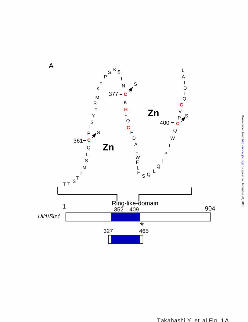
Cysteine Residues in the Ring-like Domain of Ull1 Are Necessary
for the Smt3-conjugation – Ull1 contains a Ring-like domain in the
central region (Fig. 1A ). When the cysteine-377 in this domain was
changed to serine, the Smt3-conjugation to septins was abolished in vivo
(19). In order to see whether other cysteine residues in the Ring-like
domain are also essential for septin-sumoylation, we constructed
plasmids carrying mutant genes (ull1C361S, ull1C400S, ull1C377S/C400S; see Fig.
1A ) and those plasmids were introduced into the ull1 disruptant that
expressed HA-tagged CDC3, replacing the endogenous wild-type CDC3
gene. Cultures of the transformants were treated with a microtubule-
depolymerizing drug, nocodazole, for 3.5 h to arrest cells at the G2/M
boundary. Cell extracts were prepared and subjected to immunoblotting.
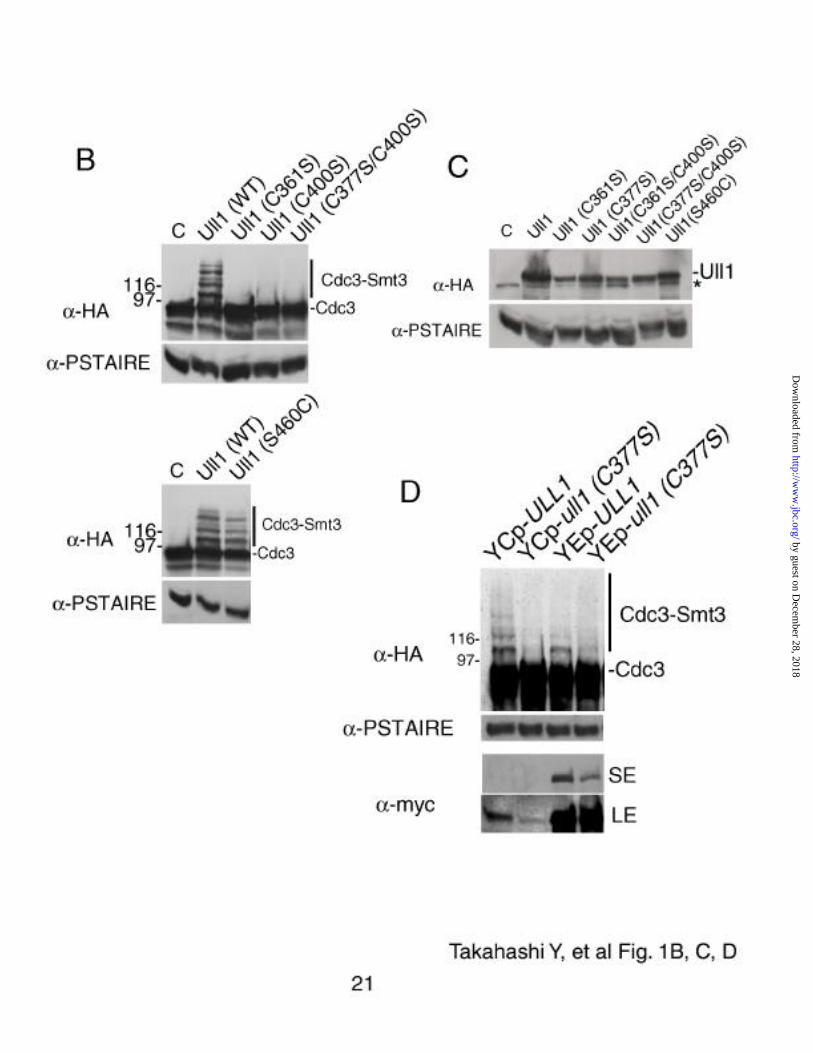
Cdc3 was probed with anti-HA antibody. As shown in 1B, several
higher molecular weight bands of Smt3-Cdc3 conjugates were detected
by guest on Decem
ber 28, 2018http://w
ww
.jbc.org/D
ownloaded from

in extracts of ULL1 cells, in accordance with previous reports (15, 19).
In contrast, all the mutants failed to conjugate Smt3 to Cdc3 (Fig. 1B).
When wild-type or each mutant protein was tagged with the HA-
epitope and prepared as described above, the protein level of the mutant
proteins decreased by a few-fold, compared with the wild-type Ull1
protein (Fig. 1C). Even when a myc-tagged mutant protein (C377S) was
expressed from a multi-copy plasmid and the protein level was higher
than the wild-type level, it did not fully recover the Smt3-conjugation to
HA-tagged Cdc3, although the mutant protein may still retain some
activity (Fig. 1D). Thus we conclude that those cysteine residues in the
Ring-like domain of Ull1 are important for the Smt3-conjugation.
Ull1 is phosphorylated especially in the M-phase (19). There is
one potential CDK-target site (SPXK) near the Ring-like domain. We
changed this serine-460 residue to cysteine, but the septin-sumoylation
was not impaired (Fig. 1B).
Ull1 Promotes Septin-sumoylation In vitro – In order to
demonstrate that Ull1 is a bona fide Smt3/SUMO-ligase (E3), we have
developed an in vitro system for septin-sumoylation. As a substrate, T7-
His-tagged Cdc3 was purified from E. coli lysate. The mature form of
GST-tagged Smt3 was purified from E. coli lysate. The mature form of
untagged Smt3 was prepared from GST-Smt3 by the treatment with
thrombin. As E1 and E2 enzymes, we used GST-tagged human
Sua1/His-tagged Uba2-complex purified from Sf-9 cells and T7-tagged
human Ubc9 purified from E. coli lysate, respectively. These E1 and E2
enzymes successfully promoted the in vitro SUMO1-conjugation to
RanGAP1 (24). GST-tagged Ull1 or ull1C377S mutant protein was
expressed and purified by the bacurovirus protein expression system.
The purified recombinant proteins were subjected to SDS-PAGE and
by guest on Decem
ber 28, 2018http://w
ww
.jbc.org/D
ownloaded from

the gels were stained with Coomassie Brilliant Blue, as shown in Fig.
2A . The Smt3 fraction contained a break-down product of Smt3, since
the band of the smaller size was also stained with α-Smt3 (data not
shown). In the lane of the E1-complex fraction, the band of Uba2 was
very weak, since we purified the E1 fraction of GST-Sua1/Uba2
heterodimer through Glutathione Sepharose 4B resin. Also in this
fraction, GST from Sf-9 cells was co-purified, which is marked by
asterisk.
For an in vitro conjugation assay, the various reaction mixtures
shown in Fig. 2B, were incubated with 3.3 mM ATP and 2 mM DTT at
25℃ for 40 min and subjected to immunoblotting. Cdc3 was probed
with anti-T7 antibody. A new band corresponding to the Cdc3 modified
with GST-Smt3 was detected in the complete reaction mixture in lane 6.
Appearance of this new band depended on the presence of E1 (lane 1),
E2 (lane 2), GST-Ull1 (lane 3) or GST-Smt3 (lane 5). Higher molecular
weight bands detected by the antibody, were derived from the T7-Cdc3-
His fraction, since these bands were missing in lane 4, and were present
even without other components (lane 9 and lane 12). Whether those are
Cdc3-polymers or unrelated proteins that are reactive to this antibody,
remains unknown.
When untagged Smt3 was used in place of GST-Smt3, a new band
of a smaller size was detected in lane 7. The size difference between
these bands corresponds to the size of GST. Furthermore, production of
the new band was abolished in the presence of 5 mM NEM (Fig. 2C),
which is consistent with a notion that thioester bond formation should be
involved in these reactions. This is the first demonstration that
sumoylation requires an additional factor, besides E1 and E2. Taken
by guest on Decem
ber 28, 2018http://w
ww
.jbc.org/D
ownloaded from

together with our previous study (19), we conclude that Ull1 is a
SUMO1/Smt3-ligase for septin components.
When the ull1C377S mutant protein was added to this in vitro
system, the production of the Smt3-conjugates decreased at least several
fold (Fig. 2B, lane 8). Thus the cysteine-377 in the Ring-like domain is
essential for this conjugation in vitro, as well as in vivo.
The Ring-like domain interacts with Ubc9 and Cdc3 – We previously
showed that the region (from 327th to 465th) containing the Ring-like
domain of Ull1 interacted with Cdc3 in the two-hybrid system (19).
This region also interacted with human SUMO1 and mouse Ubc9 (Fig.
3A ), as well as yeast Smt3 and Ubc9 in the two-hybrid system (Fig. 3B).
The C377S mutation within the Ring-like domain impaired the
interaction both with Ubc9 and Smt3 (Fig. 3B).
In order to examine whether the Ring-like domain interacts with
E2 and the substrate directly, an in vitro binding assay was performed.
GST-tagged Ring-like domain (from 327th to 465th) of Ull1 or GST was
expressed in E. coli and bound to Glutathione Sepharose 4B beads. T7-
tagged Ubc9 and T7-Cdc3 were separately expressed in E. coli, and
each of those cell lysates was mixed with the beads bound with GST-
Ring-like domain or GST. After washing, bound proteins were
subjected to immunoblotting. As shown in Fig. 3C, both Ubc9 and Cdc3
were bound to the GST-Ring-like domain. In contrast, neither Ubc9 nor
Cdc3 interacted with GST. Thus both Ubc9 (E2) and Cdc3 (substrate)
directly and specifically interact with the region containing the Ring-
like domain of Ull1.
DISCUSSION
In our previous study, we showed that the SUMO1/Smt3-
conjugation to Cdc3 depends on Ull1 in vivo, and that Ull1 interacts
with E2 and the substrates in immunoprecipitation analysis (19). In the
by guest on Decem
ber 28, 2018http://w
ww
.jbc.org/D
ownloaded from

present study, we have developed an in vitro system, where Ull1 is
required for the Smt3-modification of Cdc3, in addition to E1 and E2
enzymes (Fig. 2). These results exclude a possibility that the absence of
septin-sumoylation in the ull1 mutant is due to a defect in expression of
hypothetical factors in the Smt3-conjugation pathway in the mutant.
Although it has been published that E3 is not required in the
SUMO1/Smt3 conjugation pathway (24), we suspect that it may be an in
vitro artifact, because only E1 and E2 enzymes promoted the
modification in our system, when a large amount of E2 was added to the
reaction (our unpublished results).
Human SUMO1 rescues the lethality of the yeast smt3 deletion
mutant (14), and both SUMO1 and mouse Ubc9 interacted with yeast
Ull1 in the two-hybrid system (Fig. 3A ). Furthermore, human E1 and
E2 enzymes successfully promoted the in vitro yeast Smt3-conjugation
to septin component (Fig. 2). These results indicate that enzymes in the
SUMO1/Smt3 conjugation pathway are well conserved from yeast to
human.
Certain ubiquitin ligases are known to carry a zinc-binding Ring-
finger domain that often interacts with ubiquitin conjugating enzymes.
In accordance with these facts, Ull1 as a SUMO1/Smt3-ligase contains a
Ring-like domain and the conserved cysteine residues in the domain are
important for the Smt3-protein conjugation in vivo (19, Fig. 1) and in
vitro (Fig. 2). It is not known, however, whether the structure of this
Ring-like domain is similar to the authentic Ring-finger domain of a
bracelet structure containing two zinc atoms. This issue remains to be
elucidated.
The region containing the Ring-like domain (from 327th to 465th)
of Ull1 interacted with Ubc9 in the two-hybrid system (Fig. 3B) and in
by guest on Decem
ber 28, 2018http://w
ww
.jbc.org/D
ownloaded from

vitro binding assay (Fig. 3C). On the other hand, the ull1-C377S mutant
protein did not interact with Ubc9 in the two-hybrid system (Fig. 3B).
Also, this domain directly interacted with Cdc3 (Fig. 3C). Among
ubiquitin ligases, only the hect-proteins are known to bind a ubiquitin
molecule through thioester bond formation. The other ubiquitin ligases
have to interact with E2 and the substrate at the same time, because
those ubiquitin ligases apparently do not form the thioester bond with
ubiquitin, and function as bridging molecules between E2 and the
substrates. In the case of c-Cbl proto-oncoprotein, the Ring-domain
interacts with UbcH7 (E2), and the region (TKB domain) close to the
Ring-domain is known to be a recognition site for its substrate (13). Just
like this c-Cbl proto-oncoprotein, the region containing the Ring-like
domain of Ull1 binds to Ubc9 (E2) and Cdc3 (substrate) (Fig. 3).
Taken together, Ull1 should be qualified as an E3 (SUMO1/Smt3
ligase) in the SUMO1/Smt3 conjugation pathway, and enzymes both in
the SUMO1/Smt3 and ubiquitin conjugation pathways are conserved.
Acknowledgments – We would like to thank S. J. Elledge for
cDNA bank, E. Craig and T. Sasaki for plasmids. Y. T. is a recipient of
the Japan Society for the Promotion of Science Fellowship for Young
Scientists. This work was partly supported by Grant-in-Aid from the
Ministry of Education, Science, Sports and Culture of Japan to Y. K.
and from Uehara Memorial Foundation to H. Y.
REFERENCES
1. Müller, S., Hoege, C., Pyrowolakis, G., and Jentsch, S. (2001)
Nature Rev. Mol. Cell. Biol. 2, 202-210
2. Melchior, F. (2000) Annu. Rev. Cell Dev. Biol. 16, 591-626
by guest on Decem
ber 28, 2018http://w
ww
.jbc.org/D
ownloaded from

3. Hochstrasser, M. (2000) Nature Cell Boil. 2, E153-E157
4. Weissman, A. M. (2001) Nature Rev. Mol. Cell Biol. 2, 169-178
5. Meluh, P. B., and Koshland, D. (1995) Mol. Biol. Cell 6, 793-
807
6. Dohmen, R. J., Stappen, R., McGrath, J. P., Forrová, H.,
Kolarov, J., Goffeau, A., and Varshavsky, A. (1995) J. Biol.
Chem. 270, 18099-18109
7. Johnson, E. S., Schwienhorst, I., Dohmen, R. J., and Blobel, G.
(1997) EMBO J. 16, 5509-5519
8. Johnson, E. S., and Blobel, G. (1997) J. Biol. Chem. 272, 26799-
26802
9. Schwarz, S. E., Matuschewski, K., Liakopoulos, D., Scheffner,
M., and Jentsch, S. (1998) Proc. Natl. Acad. Sci. USA 95, 560-
564
10. Hershko, A., and Ciechanover, A. (1998) Annu. Rev. Biochem.
67, 425-479
11. Saurin, A. J., Borden, K. L. B., Boddy, M. N., and Freemont, P.
S. (1996) Trends in Biochem. Sci. 21, 208-214
12. Jackson, P. K., Eldridge, A. G., Freed, E., Furstenthal, L., Hsu,
J. Y., Kaiser, B. K., and Reimann, J. D. R. (2000) Trends in Cell
Biol. 10, 429-439
13. Zheng, N., Wang, P., Jeffrey, P. D., and Pavletich, N. P. (2000)
Cell 102, 533-539
14. Takahashi, Y., Iwase, M., Konishi, M., Tanaka, M., Toh-e, A.,
and Kikuchi, Y. (1999) Biochem. Biophys. Res. Commun. 259,
582-587
15. Johnson, E. S., and Blobel, G. (1999) J. Cell Biol. 147, 981-994
by guest on Decem
ber 28, 2018http://w
ww
.jbc.org/D
ownloaded from

16. Longtine, M. S., DeMarini, D. J., Valencik, M. L., Al-Awar, O.
S., Fares, H., De Virgilio, C., and Pringle, J. R. (1996) Curr.
Opin. Cell Biol. 8, 106-119
17. Field, C. M., and Kellogg, D. (1999) Trends Cell Biol. 9, 387-
394
18. Chung, C. D., Liao, J., Liu, B., Rao, X., Jay, P., Berta, P., Shuai,
K. (1997) Science 278, 1803-1805
19. Takahashi, Y., Toh-e, A., and Kikuchi, Y. (2001) Gene in press
20. James, P., Halladay, J., and Craig, E. A. (1996) Genetics 144,
1425-1436
21. Kaiser, C., Michaelis, S., and Mitchell, A. (1994) in Methods in
Yeast Genetics (Cold Spring Harbor Laboratory Press, Cold
Spring Harbor, NY.
22. Sasaki, T., Toh-e, A., and Kikuchi, Y. (2000) Mol. Cell. Biol.
20, 7971-7979
23. Nishida, T., Tanaka, H., and Yasuda, H. (2000) Eur. J. Biochem.,
267, 6423-6427
24. Okuma, T., Honda, R., Ichikawa, G., Tsumagari, N., and Yasuda,
H. (1999) Biochem. Biophy. Res. Comm. 254, 693-698
FIGURE LEGENDS
FIG. 1. The Ring-like domain of Ull1 is essential for
septin-sumoylation. A , Schematic structure of Ull1 and amino acid
sequence of its Ring-like domain. The Ring-like domain (from 352th to
409th amino acid) and a potential CDK target site (marked by asterisk)
of Ull1 are shown. The cysteine residue-361, -377, or -400 within the
Ring-like domain was changed to serine. The region (from 327th to 465th
amino acid) containing the Ring-like domain, used for the two-hybrid
by guest on Decem
ber 28, 2018http://w
ww
.jbc.org/D
ownloaded from

system and binding assay, is shown as a box. B, Mutational analysis of
the Ring-like domain of Ull1. Cells of T-20 (ull1 CDC3HA) were
transformed with low-copy plasmid pT-23 (ULL1), pT-24 (ull1C361S),
pT-25 (ull1C400S), pT-26 (ull1C377S, C400S), pT-27 (ull1S460C) or vector (C;
control), and the transformants were arrested at G2/M with 15 µg/ml
nocodazole. Cell lysates were prepared and subjected to
immunoblotting. Cdc3 was probed with α-HA. The α-PSTAIRE
staining (lower panels) serves as internal control for loading. C, Protein
level of ull1 mutants. The ull1 disruptant (T-13) was transformed with a
low-copy plasmid pT-17 (HA-tagged ULL1), pT-29 (HA-tagged
ull1C361S), pT-18 (HA-tagged ull1C377S), pT-30 (HA-tagged ull1C361S, C400S),
pT-31 (HA-tagged ull1C377S, C400S), pT-32 (HA-tagged ull1S460C), or
pTS901CL vector (C; control). Cell lysates of the transformants were
prepared and immunoblotting was performed, using α-HA. A non-
specific band is marked by asterisk. Equal loading of each sample was
confirmed by α-PSTAIRE staining (lower panel). D, Excess amount of
the mutant protein did not fully recover the septin-sumoylation. Cells of
T-20 (ull1 CDC3HA) were transformed with low-copy plasmid pT-20
(myc-tagged ULL1) or pT-21 (myc-tagged ull1C377S), and multi-copy
plasmid pT-33 (myc-tagged ULL1) or pT-34 (myc-tagged ull1C377S).
Cell lysates were prepared and subjected to immunoblotting. Cdc3 and
Ull1 were probed with α-HA and α-myc, respectively. Equal loading of
each sample was confirmed by α-PSTAIRE staining. SE; short
exposure, LE; long exposure.
FIG. 2. Ull1 is required for in vitro Smt3-conjugation to
Cdc3. A, Preparation of recombinant proteins for in vitro assay. The
proteins prepared for the in vitro system, were subjected to SDS-PAGE
and the gels were stained with Coomassie Brilliant Blue. Loaded
by guest on Decem
ber 28, 2018http://w
ww
.jbc.org/D
ownloaded from
samples are; 6.3 µg GST, 3.9 µg GST-Smt3, 0.4 µg Smt3 (* indicates a
break-down product of Smt3), 1.2 µg E1-complex (His-Uba2, GST-
Sua1. * indicates GST co-purified from Sf-9 cells), 1.0 µg E2 (T7-
hUbc9), 0.8 µg T7-Cdc3-His, 0.08 µg GST-Ull1 and 0.08 µg GST-
ull1C377S. Each component is marked by an arrowhead. B, The Smt3-
conjugation to Cdc3 requires Ull1 in vitro and the cysteine-377 of Ull1
is essential for the modification. The complete reaction mixture contains
0.5 µg E1-complex (GST-Sua1/His-hUba2), 0.02 µg E2 (T7-hUbc9),
0.04 µg GST-Ull1, 0.8 µg T7-Cdc3-His and 1.0 µg GST-Smt3. +,
presence of the factor; -, absence of the factor; G, 1.0 µg GST was
added instead of GST-Smt3; S, 0.5 µg Smt3 was added instead of GST-
Smt3; M, 0.04 µg GST-ull1C377S mutant protein was added instead of
GST-Ull1. Various reaction mixtures as indicated, were incubated with
3.3 mM ATP and 2 mM DTT at 25℃ for 40 min, and were subjected to
immunoblotting. Cdc3 was probed with α-T7. The right-side panel
shows various control experiments with (+), or without (-) incubation in
the buffer containing ATP. Molecular weights of marker proteins are
shown in the leftside of the figure. Non-specific bands are marked by
asterisks. C, The conjugation is NEM sensitive. The reaction mixtures
contained all the components with (+) or without (-) 2 mM DTT or 5
mM NEM, as indicated.
FIG. 3. The Ring-like domain of Ull1 interacts with Ubc9
and Cdc3. A & B. Two-hybrid interaction. Yeast strain PJ69-4A was
co-transformed with pT-22 (pGBDU-Ring-like domain), pT-37
(pGBDU-Ring-like domainC377S) or pGBDU-vector, together with
various GAD-plasmids; pGAD-hSUMO1 (pGAD424-hSUMO1), pGAD-
mUbc9 (pACT-mUbc9), pT-38 (pGAD-Ubc9), pT-39 (pGAD-Smt3) or
pGAD-vector. The transformants were streaked on a minimal plate
by guest on Decem
ber 28, 2018http://w
ww
.jbc.org/D
ownloaded from

lacking histidine, uracil and leucine, and incubated at 30℃ for 3 days.
C. In vitro binding assay. GST and GST-tagged Ring-like domain of
Ull1 were bound to Glutathione Sepharose 4B beads. E. coli lysate
containing T7-Ubc9 or T7-Cdc3, as well as untagged control lysate, was
incubated with those beads. After washing, bound proteins were
subjected to immunoblotting. Ubc9 and Cdc3 were probed with α-T7
(lanes 1-9). GST and GST-Ring-like domain were probed with α-GST
(lanes 10-17). Lane 1, total lysate of untagged control; 2, total lysate of
T7-Ubc9; 3, total lysate of T7-Cdc3; 4, 11, GST-beads plus untagged
lysate; 5, 12, GST-beads plus T7-Ubc9 lysate; 6, 13, GST-beads plus
T7-Cdc3 lysate, 7, 15, GST-Ring-beads plus untagged lysate; 8, 16,
GST-Ring-beads plus T7-Ubc9 lysate; 9, 17, GST-Ring-beads plus T7-
Cdc3 lysate; 10, total lysate of GST; 14, total lysate of GST-Ring.
by guest on Decem
ber 28, 2018http://w
ww
.jbc.org/D
ownloaded from
L
T T ST
IM
SL
Q
CPISYTRM
KY
PS
K S
I
N
C
K
HLQ
CFDALWFL
S QL
QI
P
T
W
Q
CPVC
QIDI
A
Zn
Zn
S
377
361
400
S
S
1 904Ull1/Siz1
Ring-like-domain352 409
465327*
H
A
Takahashi Y, et al Fig. 1A
by guest on Decem
ber 28, 2018http://w
ww
.jbc.org/D
ownloaded from

- + + +- - + -+ + + -
Ull1mM NEMmM DTT
Smt3-Cdc3
Cdc3
-T7 Takahashi Y, et al Fig. 2A, B, C22
C
A
B
lane 9 10 11 12 13 14 15-T7
E1+E2GST-Ull1T7-Cdc3-HisGST-Smt3Buf.+ATP+Inc.
- - - - - - +- + - - + - ++ - - + - - +- - + - - + +- - - + + + +
**
- + + + + + + ++ - + + + + + ++ + - + + + + M+ + + - + + + ++ + + + G + S +
66k
97k116k
200k
Cdc3Smt3-Cdc3GST-Smt3-Cdc3
lane 1 2 3 4 5 6 7 8-T7
45k
66k
97k116k
200k
GS
T-u
ll1(C
377S
)
GS
T-U
ll1 (
WT
)
T7-
Cdc
3-H
is
12ST-Ull17-Cdc3-HisST-Smt3
8k
14k
21k
31k
45k
97k66k
E1-
com
plex
E2
Sm
t3
GS
T-S
mt3
GS
T
*
*
**
GST-Smt3-Cdc3
Cdc3
by guest on Decem
ber 28, 2018http://w
ww
.jbc.org/D
ownloaded from


Yoshimitsu Takahashi, Tomoaki Kahyo, Akio Toh-e, Hideyo Yasuda and Yoshiko Kikuchian adaptor between conjugating enzyme and substrates
Yeast Ull1/Siz1 is a novel SUMO1/Smt3-ligase for septin components and functions as
published online September 27, 2001J. Biol. Chem.
10.1074/jbc.M109295200Access the most updated version of this article at doi:
Alerts:
When a correction for this article is posted•
When this article is cited•
to choose from all of JBC's e-mail alertsClick here
by guest on Decem
ber 28, 2018http://w
ww
.jbc.org/D
ownloaded from
Related Documents