DOI: 10.1542/peds.2005-2969 2007;119;e232-e240 Pediatrics Catherine Stayer, Alan C. Evans and Carole A. Samango-Sprouse Elizabeth Molloy Wells, Jonathan D. Blumenthal, Jean E. Nelson, Julia W. Tossell, Jay N. Giedd, Liv S. Clasen, Gregory L. Wallace, Rhoshel K. Lenroot, Jason P. Lerch, Resonance Imaging Case-Control Study XXY (Klinefelter Syndrome): A Pediatric Quantitative Brain Magnetic http://www.pediatrics.org/cgi/content/full/119/1/e232 located on the World Wide Web at: The online version of this article, along with updated information and services, is rights reserved. Print ISSN: 0031-4005. Online ISSN: 1098-4275. Grove Village, Illinois, 60007. Copyright © 2007 by the American Academy of Pediatrics. All and trademarked by the American Academy of Pediatrics, 141 Northwest Point Boulevard, Elk publication, it has been published continuously since 1948. PEDIATRICS is owned, published, PEDIATRICS is the official journal of the American Academy of Pediatrics. A monthly at GERSTEIN SCIENCE INFO CTR on April 21, 2008 www.pediatrics.org Downloaded from

Welcome message from author
This document is posted to help you gain knowledge. Please leave a comment to let me know what you think about it! Share it to your friends and learn new things together.
Transcript

DOI: 10.1542/peds.2005-2969 2007;119;e232-e240 Pediatrics
Catherine Stayer, Alan C. Evans and Carole A. Samango-Sprouse Elizabeth Molloy Wells, Jonathan D. Blumenthal, Jean E. Nelson, Julia W. Tossell,
Jay N. Giedd, Liv S. Clasen, Gregory L. Wallace, Rhoshel K. Lenroot, Jason P. Lerch, Resonance Imaging Case-Control Study
XXY (Klinefelter Syndrome): A Pediatric Quantitative Brain Magnetic
http://www.pediatrics.org/cgi/content/full/119/1/e232located on the World Wide Web at:
The online version of this article, along with updated information and services, is
rights reserved. Print ISSN: 0031-4005. Online ISSN: 1098-4275. Grove Village, Illinois, 60007. Copyright © 2007 by the American Academy of Pediatrics. All and trademarked by the American Academy of Pediatrics, 141 Northwest Point Boulevard, Elkpublication, it has been published continuously since 1948. PEDIATRICS is owned, published, PEDIATRICS is the official journal of the American Academy of Pediatrics. A monthly
at GERSTEIN SCIENCE INFO CTR on April 21, 2008 www.pediatrics.orgDownloaded from

ARTICLE
XXY (Klinefelter Syndrome): A PediatricQuantitative Brain Magnetic Resonance ImagingCase-Control StudyJay N. Giedd, MDa, Liv S. Clasen, PhDa, Gregory L. Wallace, MAa, Rhoshel K. Lenroot, MDa, Jason P. Lerch, PhDb, Elizabeth Molloy Wells, MDa,
Jonathan D. Blumenthal, MAa, Jean E. Nelson, MHSa, Julia W. Tossell, MDa, Catherine Stayer, MD, PhDa, Alan C. Evans, PhDb,
Carole A. Samango-Sprouse, EdDc,d
aChild Psychiatry Branch, National Institute of Mental Health, National Institutes of Health, Bethesda, Maryland; bMcConnell Brain Imaging Centre, Montreal NeurologicalInstitute, McGill University, Montreal, Quebec, Canada; cDepartment of Pediatrics, George Washington University, Washington, DC; dNeurodevelopmental DiagnosticCenter for Young Children, Davidson, Maryland
The authors have indicated they have no financial relationships relevant to this article to disclose.
ABSTRACT
OBJECTIVE.An extra X chromosome in males (XXY), known as Klinefelter syndrome,is associated with characteristic physical, cognitive, and behavioral features ofvariable severity. The objective of this study was to examine possible neuroana-tomical substrates of these cognitive and behavioral features during childhood andadolescence.
METHODS.MRI brain scans were acquired for 42 XXY and 87 healthy XY age-matched control males. We compared these 2 groups on regional brain volumesand cortical thickness.
RESULTS. Total cerebral volume and all lobar volumes except parietal white matterwere significantly smaller in the XXY group, whereas lateral-ventricle volume waslarger. Consistent with the cognitive profile, the cortex was significantly thinner inthe XXY group in left inferior frontal, temporal, and superior motor regions.
CONCLUSION. The brain-imaging findings of preferentially affected frontal, temporal,and motor regions and relative sparing of parietal regions are consistent withobserved cognitive and behavioral strengths and weaknesses in XXY subjects.
www.pediatrics.org/cgi/doi/10.1542/peds.2005-2969
doi:10.1542/peds.2005-2969
KeyWordssex chromosome aneuploidy, Klinefeltersyndrome, magnetic resonance imaging,children, adolescents, brain, XXY
AbbreviationsKS—Klinefelter syndromeTCV—total cerebral volumeTRT—testosterone-replacement therapyNIH—National Institutes of HealthCSF—cerebrospinal fluidANOVA—analysis of varianceANCOVA—analysis of covarianceSES—socioeconomic status
Accepted for publication Jul 26, 2006
Address correspondence to Jay N. Giedd, MD,Child Psychiatry Branch/NIMH, Building 10,Room 4C110, 10 Center Dr, MSC 1367,Bethesda, MD 20892. E-mail: [email protected]
PEDIATRICS (ISSN Numbers: Print, 0031-4005;Online, 1098-4275). Copyright © 2007 by theAmerican Academy of Pediatrics
e232 GIEDD et al at GERSTEIN SCIENCE INFO CTR on April 21, 2008 www.pediatrics.orgDownloaded from

KLINEFELTER SYNDROME (KS) (also known as47,XXY), the occurrence of an additional X chro-
mosome in males, is the most common sex-chromosomeaneuploidy and is found in between 1 of 6001,2 and 1 of10003 live male births. Supernumerary X chromosomestypically arise from nondisjunction during either mater-nal or paternal meiotic cell division4 and can affect de-velopment of the cardiac, endocrine, reproductive, skel-etal, and central nervous systems.
The original phenotype, described by Hans Klinefelterin 1942,5 consisted of hypogonadism, gynecomastia,sparse body hair, eunuchoid body habitus, above-aver-age height, and infertility. Other physical characteristicssubsequently identified included long legs and arm span,decreased bone mineral density, taurodontism,6 and lowtestosterone levels. Except for hypogonadism, which ispresent in nearly all individuals with XXY, the physicalphenotype may be quite variable. Because there is somedebate regarding whether those with XXY without theKS phenotype should be assigned the KS diagnosis, forthis report we will use the designation XXY.
Similar to those with the physical phenotype, XXYmales have characteristic but highly variable cognitiveand behavioral features. The most frequently docu-mented cognitive impairments, occurring in �80% ofthose with XXY, are language-based learning disor-ders.7–12 Graham et al13 found that �50% of boys withXXY were reading 1 or more grade levels below averagefor their age. Also common are disorders of executivefunction,14,15 particularly difficulties with planning12 andinhibitory control.16 Motor delay with truncal hypotoniaand motor planning deficits have been observed in in-fancy and throughout childhood.17,18 Other deficits re-ported in XXY have been in short-term auditory mem-ory13,17 and auditory processing.19,20 In contrast, visual-spatial processing (eg, facility with puzzles, computers,and machinery) seems to be relatively preserved, and insome cases enhanced, in XXY males.19,21,22
Few studies have been conducted regarding possibleneurobiological substrates of the observed cognitive andbehavioral phenotypes. Decreased head circumference
has been found in children with KS from birth to 9 yearsof age.23 MRI findings in adults have implicated reducedleft temporal lobe gray matter,2 smaller total cerebralvolume (TCV), a decrease in amygdala volume in thepresence of preserved hippocampal volume,24 and bilat-erally enlarged lateral ventricles.25
In this study we used MRI to investigate the effects ofa supernumerary X chromosome in a relatively largesample of children and adolescents with XXY. Pediatricsubjects were studied earlier in their developmental tra-jectories than adults and have had less time in which tobe exposed to environmental influences. Therefore, theymay be better suited than an adult population for eluci-dating early neurobiological effects of the additional Xchromosome.
On the basis of these previously identified cognitivefeatures, we hypothesized that we would find:
1. smaller TCV in subjects with XXY compared withcontrol subjects, consistent with smaller head size;
2. decreased cortical thickness, specifically in the lefttemporal, prefrontal, and motor cortices, which arebrain regions thought to underlie cognitive impair-ments commonly observed in subjects with XXY; and
3. preserved parietal cortex in the XXY group commen-surate with their relatively good visual-spatial skills.
METHODS
ParticipantsThe XXY group consisted of 42 nonmosaic XXY malesranging in age from 5.3 to 26.0 years (Table 1). Theethnic composition of the XXY group was 40 white, 1black, and 1 Hispanic. Thirty-three subjects were right-handed, 4 were left-handed, and 5 were mixed-handed.One of the XXY boys was born premature at 35 weeks’gestation, and the remaining 41 had gestational ages of�37 weeks.
Consistent with current clinical practice guidelines,none of the 24 XXY males aged �12 were undergoingtestosterone-replacement therapy (TRT), and all 14 of
TABLE 1 XXY and XY Demographics
XXY (N � 42) XY (N � 87) t P
Mean (SD) n Mean (SD) n
Age, y 12.8 (5.0) 42 12.7 (5.0) 87 0.1 .902Height, in 61.6 (9.9) 42 60.1 (9.1) 84 0.9 .386Height age �13 y 54.4 (6.3) 24 54.0 (6.0) 49 0.3 .787Height age �13 y 71.2 (3.6) 18 68.6 (4.7) 35 2.1 .041a
Weight, lb 118.2 (62.8) 42 110.5 (47.4) 86 0.7 .485Tanner stage 2.8 (1.5) 41 2.8 (1.6) 86 0.0 .979SES 51.0 (22.7) 42 38.6 (20.7) 83 3.1 .003a
IQ 95.2 (17.1) 39 120.3 (11.3) 82 8.4 �.0001a
Vocabulary 8.1 (3.4) 39 13.2 (2.5) 82 8.3 �.0001a
Block design 10.0 (3.4) 39 13.6 (2.7) 82 6.2 �.0001a
a Statistically significant.
PEDIATRICS Volume 119, Number 1, January 2007 e233 at GERSTEIN SCIENCE INFO CTR on April 21, 2008 www.pediatrics.orgDownloaded from

the XXY males aged �14 were undergoing TRT. Of the 4remaining XXY boys (all aged 13 years), 2 were under-going TRT and 2 were not. Because the ages of the TRTand non-TRT XXY subgroups were so disparate, it wasnot possible to examine the effects of TRT on brainmorphometry in our sample.
We recruited XXY males nationwide with the help of2 parent advocacy groups: the American Association forKlinefelter Syndrome Information and Support (AAK-SIS) and Klinefelter Syndrome and Associates (KS&A).Parents of XXY males were interviewed by telephoneand asked to report their child’s health, developmental,and educational history. Children with severe head in-juries or other conditions that might have affected grossbrain development were not accepted into the study.
During their visit to the National Institutes of Health(NIH), subjects underwent physical and neurologic as-sessment and IQ testing (using the Wechsler Scales ofIntelligence26–28) whenever possible (IQ was not obtainedfor 3 XXY participants: 1 because of refusal to undergotesting and 2 because of scheduling difficulties). Thepresence of XXY was confirmed with karyotype testingon all subjects. High resolution G-band karyotyping wasperformed on phytohemagglutinin-stimulated patientperipheral blood cultures. A minimum of 50 metaphaseswere analyzed, and 3 karyotypes per patient were pro-duced. (All karyotyping was performed by the Cytoge-netics Laboratory, Department of Obstetrics and Gyne-cology, Georgetown University Hospital.) Subjects wereincluded on the basis of an XXY karyotype and not onthe presence of the characteristic Klinefelter phenotypeor other specific clinical features. However, we did notethat, generally consistent with the extant literature,7–9,11
30 (71%) of the 42 XXY males in our sample had beendiagnosed with speech and/or language delays.
Eighty-seven healthy XY males ranging in age from5.2 to 25.5 years were selected as controls. These con-trols were age-matched to the 42 XXY boys in the sam-ple. They were a subset of healthy volunteers recruitedfrom the community through the NIH Normal VolunteerOffice, newspaper advertisements, and outreach toWashington, DC, area schools. The ethnic compositionof this group was 80 white, 2 black, 1 Asian, 1 Hispanic,and 3 biracial (black/white). Eighty controls were right-handed, 4 were left-handed, and 3 were mixed-handed.One of the XY boys was born premature at 34 weeks’gestation, and the remaining 86 were born term at ges-tational ages of �37 weeks.
Healthy controls were screened via an initial tele-phone interview, parent and teacher rating versions ofthe Child Behavior Checklist,29 and physical and neuro-logic assessment. Exclusion criteria included psychiatricdiagnosis in the subject or a first-degree relative andhead injury or other conditions that might have affectedgross brain development.
IQ scores were obtained by using the Wechsler scales
in the form of the full test, the 4-subtest abbreviatedversion (the Wechsler Abbreviated Scale of Intelligence),or the 2-subtest (vocabulary and block design) shortform scored by using Sattler’s technique.30 Five controlparticipants did not receive IQ assessment, all because ofscheduling difficulties. Because healthy control partici-pants were taking part in an ongoing longitudinal studyof brain development, several different versions of theWechsler IQ tests were used depending on the time ofassessment.
For all study participants, handedness was assessed byusing the Physical and Neurological Examination forSoft Signs (PANESS).31 In this examination, individualsare asked to write their name and then demonstrate howthey perform 11 other activities (eg, throwing a ball,using a saw, etc). If a child writes with his or her righthand and performs 10 or all of the remaining 11 itemswith the right hand (or right hand first, indicating pref-erence), the child is categorized as right-handed. Con-versely, if the child writes with his or her left hand andperforms 10 or all of the remaining 11 items with the lefthand (or left hand first), the child is categorized asleft-handed. All other outcomes result in categorizationas mixed-handed.
We obtained verbal or written assent from the childand written consent from the parents for their partici-pation in the study. The National Institute of MentalHealth Institutional Review Board approved the proto-col.
MRI AcquisitionAll images were acquired on the same General Electric1.5-T Signa scanner (Waukesha, WI), which was locatedat the NIH clinical center in Bethesda, Maryland. A3-dimensional spoiled-gradient recalled-echo sequencein the steady-state sequence, designed to optimize dis-crimination between gray matter, white matter, and ce-rebrospinal fluid (CSF), was used to acquire 124 contig-uous 1.5-mm-thick slices in the axial plane (echo time: 5milliseconds; repetition time: 24 milliseconds; flip angle:45°; matrix: 256 � 192; number of excitations: 1; field ofview: 24 cm; acquisition time: 9 minutes, 52 seconds). Afast-spin-echo/proton-density weighted imaging se-quence was also acquired for clinical evaluation.
Quantification of Cortical Thickness and Regions of InterestThe native MRI scans were registered into standardizedstereotaxic space by using a linear transformation32 andcorrected for nonuniformity artifacts.33 The registeredand corrected volumes were segmented into white mat-ter, gray matter, and CSF by using a neural net clas-sifier.34 Region-of-interest analysis was performed bycombining tissue-classification information with a prob-abilistic atlas. The regions that had been validated bycomparison with other methods were the midsagittalarea of the corpus callosum; volumes of the cerebellum,
e234 GIEDD et al at GERSTEIN SCIENCE INFO CTR on April 21, 2008 www.pediatrics.orgDownloaded from
caudate nucleus, and lateral ventricles; and gray andwhite matter volumes of the total cerebrum, frontallobes, parietal lobes, and temporal lobes.35
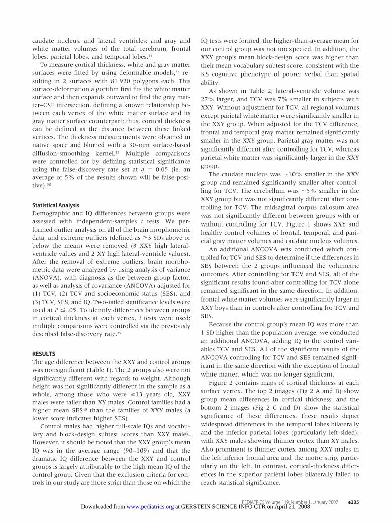
To measure cortical thickness, white and gray mattersurfaces were fitted by using deformable models,36 re-sulting in 2 surfaces with 81 920 polygons each. Thissurface-deformation algorithm first fits the white mattersurface and then expands outward to find the gray mat-ter–CSF intersection, defining a known relationship be-tween each vertex of the white matter surface and itsgray matter surface counterpart; thus, cortical thicknesscan be defined as the distance between these linkedvertices. The thickness measurements were obtained innative space and blurred with a 30-mm surface-baseddiffusion-smoothing kernel.37 Multiple comparisonswere controlled for by defining statistical significanceusing the false-discovery rate set at q � 0.05 (ie, anaverage of 5% of the results shown will be false-posi-tive).38
Statistical AnalysisDemographic and IQ differences between groups wereassessed with independent-samples t tests. We per-formed outlier analysis on all of the brain morphometricdata, and extreme outliers (defined as �3 SDs above orbelow the mean) were removed (3 XXY high lateral-ventricle values and 2 XY high lateral-ventricle values).After the removal of extreme outliers, brain morpho-metric data were analyzed by using analysis of variance(ANOVA), with diagnosis as the between-group factor,as well as analysis of covariance (ANCOVA) adjusted for(1) TCV, (2) TCV and socioeconomic status (SES), and(3) TCV, SES, and IQ. Two-tailed significance levels wereused at P � .05. To identify differences between groupsin cortical thickness at each vertex, t tests were used;multiple comparisons were controlled via the previouslydescribed false-discovery rate.39
RESULTSThe age difference between the XXY and control groupswas nonsignificant (Table 1). The 2 groups also were notsignificantly different with regards to weight. Althoughheight was not significantly different in the sample as awhole, among those who were �13 years old, XXYmales were taller than XY males. Control families had ahigher mean SES40 than the families of XXY males (alower score indicates higher SES).
Control males had higher full-scale IQs and vocabu-lary and block-design subtest scores than XXY males.However, it should be noted that the XXY group’s meanIQ was in the average range (90–109) and that thedramatic IQ difference between the XXY and controlgroups is largely attributable to the high mean IQ of thecontrol group. Given that the exclusion criteria for con-trols in our study are more strict than those on which the
IQ tests were formed, the higher-than-average mean forour control group was not unexpected. In addition, theXXY group’s mean block-design score was higher thantheir mean vocabulary subtest score, consistent with theKS cognitive phenotype of poorer verbal than spatialability.
As shown in Table 2, lateral-ventricle volume was27% larger, and TCV was 7% smaller in subjects withXXY. Without adjustment for TCV, all regional volumesexcept parietal white matter were significantly smaller inthe XXY group. When adjusted for the TCV difference,frontal and temporal gray matter remained significantlysmaller in the XXY group. Parietal gray matter was notsignificantly different after controlling for TCV, whereasparietal white matter was significantly larger in the XXYgroup.
The caudate nucleus was �10% smaller in the XXYgroup and remained significantly smaller after control-ling for TCV. The cerebellum was �5% smaller in theXXY group but was not significantly different after con-trolling for TCV. The midsagittal corpus callosum areawas not significantly different between groups with orwithout controlling for TCV. Figure 1 shows XXY andhealthy control volumes of frontal, temporal, and pari-etal gray matter volumes and caudate nucleus volumes.
An additional ANCOVA was conducted which con-trolled for TCV and SES to determine if the differences inSES between the 2 groups influenced the volumetricoutcomes. After controlling for TCV and SES, all of thesignificant results found after controlling for TCV aloneremained significant in the same direction. In addition,frontal white matter volumes were significantly larger inXXY boys than in controls after controlling for TCV andSES.
Because the control group’s mean IQ was more than1 SD higher than the population average, we conductedan additional ANCOVA, adding IQ to the control vari-ables TCV and SES. All of the significant results of theANCOVA controlling for TCV and SES remained signif-icant in the same direction with the exception of frontalwhite matter, which was no longer significant.
Figure 2 contains maps of cortical thickness at eachsurface vertex. The top 2 images (Fig 2 A and B) showgroup mean differences in cortical thickness, and thebottom 2 images (Fig 2 C and D) show the statisticalsignificance of these differences. These results depictwidespread differences in the temporal lobes bilaterallyand the inferior parietal lobes (particularly left-sided),with XXY males showing thinner cortex than XY males.Also prominent is thinner cortex among XXY males inthe left inferior frontal area and the motor strip, partic-ularly on the left. In contrast, cortical-thickness differ-ences in the superior parietal lobes bilaterally failed toreach statistical significance.
PEDIATRICS Volume 119, Number 1, January 2007 e235 at GERSTEIN SCIENCE INFO CTR on April 21, 2008 www.pediatrics.orgDownloaded from

DISCUSSIONIn this study we found both general and regionally spe-cific effects of a supernumerary X chromosome on braindevelopment. TCV was found to be smaller in XXY in-dividuals. Specific effects include thinner cortex in tem-poral and frontal regions, smaller frontal, temporal, andcaudate volumes, and preserved parietal regions, all con-sistent with the cognitive/behavioral phenotypes re-ported for XXY such as impaired language function.
The frontal and caudate findings seen here are similarto those seen in MRI studies of attention-deficit/hyper-activity disorder, a similarity which suggests that anom-
alies of frontal-striatal circuitry41 may also underlie as-pects of executive dysfunction in XXY.14,16,22 Thesestructures, along with the preferential thinning of motorcortex, may also contribute to impairment in the plan-ning and integration of motor movements.8,18 The areasof the motor strip in which cortical thinning is mostprominent are the superior region, associated with con-trol of the upper trunk and shoulders, a noted area ofmuscular weakness in XXY, and the left inferior region,associated with control of the anatomy involved inspeech production such as the lips, jaw, and pharynx.42
Dyspraxia in these areas has been hypothesized to con-
TABLE 2 Regional XXY and XY Brain Volumes (mL), Unadjusted and Adjusted for (1) TCV, (2) TCV and SES, and (3) TCV, SES, and IQ
Brain Structure Covariates XXY (N � 42), Mean (SD)/EMMean (SE), mL
XY (N � 87), Mean (SD)/EMMean (SE) , mL
ANOVA/ANCOVA, F
P
TCV None 1066.6 (119.3) 1145.8 (103.4) 15.0 �.001a,b
Gray matter None 681.7 (69.0) 742.5 (71.1) 21.1 �.0001a,b
Frontal gray matter None 205.9 (20.7) 227.8 (23.5) 26.5 �.0001a,b
TCV 215.2 (2.0) 223.3 (1.4) 10.7 .0014a,b
TCV, SES 214.7 (2.0) 223.7 (1.4) 12.5 �.001a,b
TCV, SES, IQ 215.4 (2.4) 224.9 (1.5) 8.9 .003a
Temporal gray matter None 170.5 (15.1) 187.6 (16.5) 32.1 �.0001a,b
TCV 177.3 (1.3) 184.3 (0.9) 18.8 �.0001a,b
TCV, SES 177.0 (1.3) 184.7 (0.9) 20.9 �.0001a,b
TCV, SES, IQ 177.9 (1.6) 185.1 (1.0) 11.6 .001a,b
Parietal gray matter None 113.2 (14.5) 122.2 (13.6) 11.6 �.001a,b
TCV 118.5 (1.4) 119.6 (1.0) 0.4 .517TCV, SES 118.1 (1.5) 119.8 (1.0) 0.9 .353TCV, SES, IQ 119.5 (1.7) 119.9 (1.1) 0.0 .862
White matter None 384.9 (67.3) 403.3 (51.4) 3.0 .087Frontal white matter None 142.1 (25.8) 150.5 (18.8) 4.5 .037a
TCV 150.6 (2.0) 146.4 (1.4) 2.8 .098TCV, SES 151.6 (2.0) 146.6 (1.4) 4.0 .048a
TCV, SES, IQ 149.9 (2.4) 147.9 (1.5) 0.4 .520Temporal white matter None 78.7 (13.4) 84.1 (11.2) 5.7 .018a
TCV 83.0 (1.3) 82.0 (0.9) 0.4 .550TCV, SES 83.4 (1.3) 82.3 (0.9) 0.4 .529TCV, SES, IQ 82.9 (1.6) 82.3 (1.0) 0.1 .748
Parietal white matter None 76.3 (13.0) 79.6 (10.8) 2.4 .126TCV 80.9 (1.1) 77.4 (0.7) 6.7 .011a
TCV, SES 81.3 (1.1) 77.5 (0.7) 8.0 .006a
TCV, SES, IQ 81.3 (1.3) 77.4 (0.8) 5.3 .023a
Caudate None 9.8 (1.2) 10.9 (1.1) 28.3 �.0001a,b
TCV 10.1 (0.2) 10.8 (0.1) 12.6 .005a
TCV, SES 10.1 (0.2) 10.7 (0.1) 9.0 .003a,b
TCV, SES, IQ 10.1 (0.2) 10.8 (0.1) 8.3 .005a
Cerebellum None 128.3 (12.3) 135.6 (11.3) 11.2 .001a,b
TCV 132.5 (1.3) 133.6 (0.9) 0.4 .515TCV, SES 133.0 (1.3) 133.6 (0.9) 0.1 .741TCV, SES, IQ 131.7 (1.6) 134.4 (1.0) 1.7 .201
Corpus callosum area, mm2 None 535.9 (91.8) 541.9 (77.4) 0.3 .558TCV 550.2 (12.0) 533.6 (8.2) 1.3 .265TCV, SES 553.3 (12.2) 533.1 (8.5) 1.7 .193TCV, SES, IQ 540.8 (14.7) 536.8 (9.4) 0.0 .838
Lateral ventriclesc None 13.6 (8.3) 10.7 (4.7) 9.3 .003a,b
TCV 14.0 (0.8) 10.5 (0.5) 12.9 �.001a,b
TCV, SES 13.8 (0.8) 10.8 (0.6) 8.8 .004a,b
TCV, SES, IQ 13.6 (1.0) 10.9 (0.6) 4.3 .040a
EM mean indicates estimated marginal mean.a Statistically significant.b ANOVA/ANCOVA survived Bonferroni adjustment at P � .0048 (.05/13 comparisons �number of structures�).c For lateral ventricles, XXY n � 39 and XY n � 85 (3 XXY and 2 XY extreme high outliers were dropped).
e236 GIEDD et al at GERSTEIN SCIENCE INFO CTR on April 21, 2008 www.pediatrics.orgDownloaded from

tribute to the language-learning disability of these XXYchildren.11
In addition to more generalized potential effects onmotor function, abnormalities of the caudate nucleushave been associated with developmental speech andlanguage dyspraxias.43,44 The reduced caudate volumesfound here are consistent with the oral motor dysfunc-tion and deficits in imitation of nonverbal mouth move-ments previously described in males with XXY.11 Addi-tional investigation of this region and its influence onspeech and language acquisition is warranted.
The relative sparing of gray matter and larger white
matter volume in the parietal region is intriguing in lightof reports indicating average or higher-level perfor-mance of XXY subjects on nonmotor perceptual tasks,which are thought to rely on intact parietal function-ing.9,15 Average range ability in nonverbal visual mem-ory, also found to rely on intact parietal functioning,45
has been found in XXY.8 In addition, XXY males seem tohave a strong preference for visual stimulation, oftenevident within the first months of life, while showingsigns of deficits in auditory localization.11 Among theWechsler subtests, block design (particularly its spatial-perception component), the subtest on which the XXY
FIGURE 1XY and XXY frontal (A), temporal (B), and parietal (C) graymatter volumes and caudate nucleus volumes (D).
PEDIATRICS Volume 119, Number 1, January 2007 e237 at GERSTEIN SCIENCE INFO CTR on April 21, 2008 www.pediatrics.orgDownloaded from

FIGURE 2Cortical-thickness maps at each surface vertex. A and B show group mean differences in cortical thickness; C and D show the statistical significance of these differences. NV indicatesnormal volunteer.
e238 GIEDD et al at GERSTEIN SCIENCE INFO CTR on April 21, 2008 www.pediatrics.orgDownloaded from

participants in our study performed best, is most stronglyassociated with superior parietal lobe function.46
A recent article describing the use of an automatedwhole-brain voxel-based morphometric technique on asubset of the data described here reported results largelyconsistent with these, including smaller whole-brainvolumes of gray and white matter and larger ventriclesin XXY subjects.47 Although direct comparison of indi-vidual structures between the 2 methods is precluded bydifferences in definition of anatomic areas, the auto-mated method also found several areas with decreasedgray matter in the temporal lobes, in addition to limbicand occipital regions. The 1 reported discrepancy was inparietal white matter, which was reported as smaller onthe right side using the voxel-based method, in contrastto our finding that total parietal white matter wasslightly larger in XXY boys. It is not clear whether thisdiscrepancy is attributable to a true hemispheric differ-ence or is related to differences in methods; we arecurrently investigating this question.
The impact of TRT on brain development in XXYmales is not known. An earlier study2 of 10 adult XXYsubjects and 10 controls reported that the 5 XXY subjectswho had TRT showed preservation of temporal graymatter compared with those who did not receive TRT.Although this study was limited by small sample size, theknown beneficial effect of TRT on physical and behav-ioral domains in XXY adolescents makes it plausible thatTRT may have a normalizing effect on brain structuralmeasures. We are continuing to collect data on TRT inour subjects to obtain sufficient power to determine itseffects on the trajectory of brain development.
CONCLUSIONSBrain measures in children and adolescents with XXYshow regional discrepancies from control subjects con-sistent with their behavioral and cognitive differences. Astriking characteristic of the XXY population is the widevariability of phenotypes. Many XXY individuals haveno cognitive or behavioral deficits, obtain advanced de-grees, and function without impairment vocationally orsocially. However, study of the group differences in braindevelopment may help clarify the mechanisms by whicha supernumerary X chromosome affects brain develop-ment and lead to more targeted interventions.
ACKNOWLEDGMENTSThis research was supported in its entirety by the Intra-mural Program of the NIH, National Institute of MentalHealth.
We thank the families who participated in this re-search and the American Association for Klinefelter Syn-drome Information and Support and Klinefelter Syn-drome and Associates for assisting us in the recruitmentof participants.
REFERENCES1. Linden MG, Bender BG, Robinson A. Genetic counseling for
sex chromosome abnormalities. Am J Med Genet. 2002;110:3–10
2. Patwardhan AJ, Eliez S, Bender B, Linden MG, Reiss AL. Brainmorphology in Klinefelter syndrome: extra X chromosome andtestosterone supplementation. Neurology. 2000;54:2218–2223
3. Nussbaum RL, McInnes RR, Willard HF, Thompson MW.Thompson & Thompson Genetics in Medicine. 7th ed. Philadelphia,PA: Saunders; 2004
4. Thomas NS, Hassold TJ. Aberrant recombination and the originof Klinefelter syndrome. Hum Reprod Update. 2003;9:309–317
5. Klinefelter HF, Reifenstein EC, Albright F. Syndrome charac-terized by gynecomastia aspermatogenesis without aleydigismand increased excretion of follicle stimulating hormone. J ClinEndocrinol Metab. 1942;2:615–627
6. Varrela J, Alvesalo L. Taurodontism in 47,XXY males: an effectof the extra X chromosome on root development. J Dent Res.1988;67:501–502
7. Bender BG, Linden MG, Robinson A. Neuropsychological im-pairment in 42 adolescents with sex chromosome abnormali-ties. Am J Med Genet. 1993;48:169–173
8. Geschwind DH, Gregg J, Boone K, et al. Klinefelter’s syndromeas a model of anomalous cerebral laterality: testing gene dosagein the X chromosome pseudoautosomal region using a DNAmicroarray. Dev Genet. 1998;23:215–229
9. Geschwind DH, Boone KB, Miller BL, Swerdloff RS. Neurobe-havioral phenotype of Klinefelter syndrome. Ment Retard DevDisabil Res Rev. 2000;6:107–116
10. Mandoki MW, Sumner GS, Hoffman RP, Riconda DL. A reviewof Klinefelter’s syndrome in children and adolescents [pub-lished correction appears in J Am Acad Child Adolesc Psychiatry.1991;30:516]. J Am Acad Child Adolesc Psychiatry. 1991;30:167–172
11. Samango-Sprouse CA, Rogol A. XXY: the hidden disability anda prototype for an infantile presentation of developmentaldyspraxia (IDD). Infants Young Child. 2002;15:11–18
12. Visootsak J, Aylstock M, Graham JM Jr. Klinefelter syndromeand its variants: an update and review for the primary pedia-trician. Clin Pediatr (Phila). 2001;40:639–651
13. Graham JM Jr, Bashir AS, Stark RE, Silbert A, Walzer S. Oraland written language abilities of XXY boys: implications foranticipatory guidance. Pediatrics. 1988;81:795–806
14. Boone KB, Swerdloff RS, Miller BL, et al. Neuropsychologicalprofiles of adults with Klinefelter syndrome. J Int NeuropsycholSoc. 2001;7:446–456
15. Samango-Sprouse CA, Law P. The neurocognitive profile ofthe young child with XXY. Eur J Hum Genet. 2001;9(suppl1):193
16. Temple CM, Sanfilippo PM. Executive skills in Klinefelter’ssyndrome. Neuropsychologia. 2003;41:1547–1559
17. Bender BG, Puck MH, Salbenblatt JA, Robinson A. Dyslexia in47,XXY boys identified at birth. Behav Genet. 1986;16:343–354
18. Salbenblatt JA, Meyers DC, Bender BG, Linden MG, RobinsonA. Gross and fine motor development in 47,XXY and 47,XYYmales. Pediatrics. 1987;80:240–244
19. Walzer S, Graham JM Jr, Bashir AS, Silbert AR. Preliminaryobservations on language and learning in XXY boys. BirthDefects Orig Artic Ser. 1982;18(4):185–192
20. Mandoki MW, Sumner GS. Klinefelter syndrome: the need forearly identification and treatment. Clin Pediatr (Phila). 1991;30:161–164
21. Rovet J, Netley C, Keenan M, Bailey J, Stewart D. The psycho-educational profile of boys with Klinefelter syndrome. J LearnDisabil. 1996;29:180–196
22. Fales CL, Knowlton BJ, Holyoak KJ, Geschwind DH, Swerdloff
PEDIATRICS Volume 119, Number 1, January 2007 e239 at GERSTEIN SCIENCE INFO CTR on April 21, 2008 www.pediatrics.orgDownloaded from

RS, Gonzalo IG. Working memory and relational reasoning inKlinefelter syndrome. J Int Neuropsychol Soc. 2003;9:839–846
23. Ratcliffe SG, Masera N, Pan H, McKie M. Head circumferenceand IQ of children with sex chromosome abnormalities. DevMed Child Neurol. 1994;36:533–544
24. Patwardhan AJ, Brown WE, Bender BG, Linden MG, Eliez S,Reiss AL. Reduced size of the amygdala in individuals with47,XXY and 47,XXX karyotypes. Am J Med Genet. 2002;114:93–98
25. Warwick MM, Doody GA, Lawrie SM, Kestelman JN, Best JJ,Johnstone EC. Volumetric magnetic resonance imaging studyof the brain in subjects with sex chromosome aneuploidies.J Neurol Neurosurg Psychiatry. 1999;66:628–632
26. Wechsler D. Wechsler Abbreviated Scale of Intelligence (WASI)Manual. San Antonio, TX: Psychological Corporation; 1999
27. Wechsler D. WISC-III: Wechsler Intelligence Scale for Children:Manual. 3rd ed. San Antonio, TX: Psychological Corporation/Harcourt Brace Jovanovich; 1991
28. Wechsler D, Corporation P. WPPSI-R, Manual: Wechsler Preschooland Primary Scale of Intelligence. Revised ed. San Antonio, TX:Psychological Corporation/Harcourt Brace Jovanovich; 1989
29. Achenbach TM, Ruffle TM. The Child Behavior Checklist andrelated forms for assessing behavioral/emotional problems andcompetencies. Pediatr Rev. 2000;21:265–271
30. Sattler JM. Assessment of Children. Revised/updated, 3rd ed. SanDiego, CA: J.M. Sattler; 1992
31. Denckla MB. Revised physical and neurological examinationfor subtle signs. Psychopharmacol Bull. 1985;21:773–800
32. Collins DL, Neelin P, Peters TM, Evans AC. Automatic 3Dintersubject registration of MR volumetric data in standardizedTalairach space. J Comput Assist Tomogr. 1994;18:192–205
33. Sled JG, Zijdenbos AP, Evans AC. A nonparametric method forautomatic correction of intensity nonuniformity in MRI data.IEEE Trans Med Imaging. 1998;17:87–97
34. Zijdenbos AP, Forghani R, Evans AC. Automatic “pipeline”analysis of 3-D MRI data for clinical trials: application to mul-tiple sclerosis. IEEE Trans Med Imaging. 2002;21:1280–1291
35. Collins DL, Holmes CJ, Peters TM, Evans AC. Automatic 3-Dmodel-based neuroanatomical segmentation. Hum Brain Mapp.1995;3:190–208
36. MacDonald D, Kabani N, Avis D, Evans AC. Automated 3-Dextraction of inner and outer surfaces of cerebral cortex fromMRI. Neuroimage. 2000;12:340–356
37. Chung MK, Worsley KJ, Robbins S, et al. Deformation-basedsurface morphometry applied to gray matter deformation. Neu-roimage. 2003;18:198–213
38. Genovese CR, Lazar NA, Nichols T. Thresholding of statisticalmaps in functional neuroimaging using the false discovery rate.Neuroimage. 2002;15:870–878
39. Lerch JP, Evans AC. Cortical thickness analysis examinedthrough power analysis and a population simulation. Neuroim-age. 2005;24:163–173
40. Hollingshead AdB, Redlich FC. Social Class and Mental Illness: ACommunity Study. New York, NY: Wiley; 1958
41. Giedd JN, Blumenthal J, Molloy E, Castellanos FX. Brain im-aging of attention deficit/hyperactivity disorder. Ann N Y AcadSci. 2001;931:33–49
42. Parent A. Cerebral cortex. In: Carpenter’s Human Neuroanatomy.9th ed. Baltimore, MD: Williams & Wilkins; 1996:890
43. Jernigan TL, Hesselink JR, Sowell E, Tallal PA. Cerebral struc-ture on magnetic resonance imaging in language- and learn-ing-impaired children. Arch Neurol. 1991;48:539–545
44. Watkins KE, Gadian DG, Vargha-Khadem F. Functional andstructural brain abnormalities associated with a genetic disor-der of speech and language. Am J Hum Genet. 1999;65:1215–1221
45. Todd JJ, Marois R. Capacity limit of visual short-term memoryin human posterior parietal cortex. Nature. 2004;428:751–754
46. Meyer-Lindenberg A, Kohn P, Mervis CB, et al. Neural basis ofgenetically determined visuospatial construction deficit in Wil-liams syndrome. Neuron. 2004;43:623–631
47. Shen D, Liu D, Liu H, Clasen L, Giedd J, Davatzikos C. Auto-mated morphometric study of brain variation in XXY males.Neuroimage. 2004;23:648–653
e240 GIEDD et al at GERSTEIN SCIENCE INFO CTR on April 21, 2008 www.pediatrics.orgDownloaded from

DOI: 10.1542/peds.2005-2969 2007;119;e232-e240 Pediatrics
Catherine Stayer, Alan C. Evans and Carole A. Samango-Sprouse Elizabeth Molloy Wells, Jonathan D. Blumenthal, Jean E. Nelson, Julia W. Tossell,
Jay N. Giedd, Liv S. Clasen, Gregory L. Wallace, Rhoshel K. Lenroot, Jason P. Lerch, Resonance Imaging Case-Control Study
XXY (Klinefelter Syndrome): A Pediatric Quantitative Brain Magnetic
& ServicesUpdated Information
http://www.pediatrics.org/cgi/content/full/119/1/e232including high-resolution figures, can be found at:
References
http://www.pediatrics.org/cgi/content/full/119/1/e232#BIBLat: This article cites 40 articles, 10 of which you can access for free
Subspecialty Collections
ologyhttp://www.pediatrics.org/cgi/collection/genetics_and_dysmorph
Genetics & Dysmorphologyfollowing collection(s): This article, along with others on similar topics, appears in the
Permissions & Licensing
http://www.pediatrics.org/misc/Permissions.shtmltables) or in its entirety can be found online at: Information about reproducing this article in parts (figures,
Reprints http://www.pediatrics.org/misc/reprints.shtml
Information about ordering reprints can be found online:
at GERSTEIN SCIENCE INFO CTR on April 21, 2008 www.pediatrics.orgDownloaded from
Related Documents