viruses Review Who’s Driving? Human Cytomegalovirus, Interferon, and NFκB Signaling Christopher M. Goodwin, Jessica H. Ciesla and Joshua Munger * Department of Biochemistry and Biophysics, University of Rochester Medical Center, Rochester, NY 14642, USA; [email protected] (C.M.G.); [email protected] (J.H.C.) * Correspondence: [email protected]; Tel.: +1-585-273-4800 Received: 3 August 2018; Accepted: 18 August 2018; Published: 21 August 2018 Abstract: As essential components of the host’s innate immune response, NFκB and interferon signaling are critical determinants of the outcome of infection. Over the past 25 years, numerous Human Cytomegalovirus (HCMV) genes have been identified that antagonize or modulate the signaling of these pathways. Here we review the biology of the HCMV factors that alter NFκB and interferon signaling, including what is currently known about how these viral genes contribute to infection and persistence, as well as the major outstanding questions that remain. Keywords: human cytomegalovirus; HCMV; NFκB; interferon; IκB kinase (IKK); ISG 1. Introduction Human cytomegalovirus (HCMV) is a widespread opportunistic pathogen that causes disease in a variety of immunosuppressed populations, including the elderly, cancer patients, and AIDS patients [1,2]. HCMV infection also causes significant morbidity in transplant recipients, and it is a major cause of kidney, liver, heart and bone marrow transplant rejection [3]. HCMV infection is also a significant cause of congenital disability, as roughly 5 out of every 1000 infants born each year are infected with HCMV, and approximately 10% of that population will experience neurological symptoms [2,4,5]. The relationship between HCMV and host immunity, including recognition, priming, and the subsequent host response, is a major determinant of HCMV pathogenesis. The earliest events of this response typically involve innate immune sensing of infection in non-immune cells, induction of an anti-viral state, and secretion of anti-viral paracrine factors that both help neighboring cells resist infection as well as recruit and activate professional immune cells. Interferon and NFκB signaling are two signal transduction cascades integral to these processes that are frequently targeted by viral infection to ensure persistence. The past 25 years have seen great progress in identifying numerous HCMV viral factors that modulate these innate immune pathways. However, many questions remain about both the specific biochemical mechanisms involved as well as the contexts through which they contribute to viral pathogenesis, replication, and persistence. Given their importance to infectious outcomes, further elucidating the diverse roles of these viral innate immune modulators remains a high priority to further our understanding of viral biology, while also potentially providing fertile ground for therapeutic development. 2. HCMV and Interferon Signaling Interferons (IFN) are a broad class of cytokines initially recognized for their ability to protect cells from viral infection. Anti-viral IFN signaling in response to infection is capable of inducing a variety of host cell defenses targeting various aspects of virus biology, including global inhibition of translation via activation of protein kinase R (PKR), induction of RNAse L-mediated degradation Viruses 2018, 10, 447; doi:10.3390/v10090447 www.mdpi.com/journal/viruses

Welcome message from author
This document is posted to help you gain knowledge. Please leave a comment to let me know what you think about it! Share it to your friends and learn new things together.
Transcript
viruses
Review
Who’s Driving? Human Cytomegalovirus, Interferon,and NFκB Signaling
Christopher M. Goodwin, Jessica H. Ciesla and Joshua Munger *
Department of Biochemistry and Biophysics, University of Rochester Medical Center, Rochester, NY 14642, USA;[email protected] (C.M.G.); [email protected] (J.H.C.)* Correspondence: [email protected]; Tel.: +1-585-273-4800
Received: 3 August 2018; Accepted: 18 August 2018; Published: 21 August 2018�����������������
Abstract: As essential components of the host’s innate immune response, NFκB and interferonsignaling are critical determinants of the outcome of infection. Over the past 25 years, numerousHuman Cytomegalovirus (HCMV) genes have been identified that antagonize or modulate thesignaling of these pathways. Here we review the biology of the HCMV factors that alter NFκB andinterferon signaling, including what is currently known about how these viral genes contribute toinfection and persistence, as well as the major outstanding questions that remain.
Keywords: human cytomegalovirus; HCMV; NFκB; interferon; IκB kinase (IKK); ISG
1. Introduction
Human cytomegalovirus (HCMV) is a widespread opportunistic pathogen that causes diseasein a variety of immunosuppressed populations, including the elderly, cancer patients, and AIDSpatients [1,2]. HCMV infection also causes significant morbidity in transplant recipients, and it isa major cause of kidney, liver, heart and bone marrow transplant rejection [3]. HCMV infection isalso a significant cause of congenital disability, as roughly 5 out of every 1000 infants born each yearare infected with HCMV, and approximately 10% of that population will experience neurologicalsymptoms [2,4,5].
The relationship between HCMV and host immunity, including recognition, priming, and thesubsequent host response, is a major determinant of HCMV pathogenesis. The earliest events of thisresponse typically involve innate immune sensing of infection in non-immune cells, induction of ananti-viral state, and secretion of anti-viral paracrine factors that both help neighboring cells resistinfection as well as recruit and activate professional immune cells. Interferon and NFκB signalingare two signal transduction cascades integral to these processes that are frequently targeted by viralinfection to ensure persistence. The past 25 years have seen great progress in identifying numerousHCMV viral factors that modulate these innate immune pathways. However, many questions remainabout both the specific biochemical mechanisms involved as well as the contexts through which theycontribute to viral pathogenesis, replication, and persistence. Given their importance to infectiousoutcomes, further elucidating the diverse roles of these viral innate immune modulators remains ahigh priority to further our understanding of viral biology, while also potentially providing fertileground for therapeutic development.
2. HCMV and Interferon Signaling
Interferons (IFN) are a broad class of cytokines initially recognized for their ability to protectcells from viral infection. Anti-viral IFN signaling in response to infection is capable of inducing avariety of host cell defenses targeting various aspects of virus biology, including global inhibitionof translation via activation of protein kinase R (PKR), induction of RNAse L-mediated degradation
Viruses 2018, 10, 447; doi:10.3390/v10090447 www.mdpi.com/journal/viruses
Viruses 2018, 10, 447 2 of 18
of viral RNA, production of anti-viral nitric oxide through inducible nitric oxide synthase (iNOS),depletion of tryptophan via the indoleamine 2,3 dioxygenase (IDO) pathway, and upregulation ofantigen presentation via major histocompatibility (MHC) complex constituents [6–10]. IFN signalingis sub-classified into Type I, Type II, and Type III groups based on their specific signaling architecturesand outcomes. The majority of interferon-induced anti-viral activity is dependent on the diverseactivities of Type I IFNs, which in humans include IFNκ, IFNε, IFNω, IFNβ, and a host of IFNαisoforms derived from 13 separate genes [11]. The activities of Type I IFNs induce the expression of alarge set of IFN-stimulated genes (ISGs) that are critical for rendering the host cell environment lesspermissive for viral infection. In addition to the direct effects of Type I cytokines on ISG induction,these IFNs have been implicated in coordinated innate immune effector processes including NaturalKiller (NK) cell activation, dendritic cell (DC) maturation, and T-cell differentiation [12]. In contrast tothe diverse signaling of Type I IFNs, Type II IFN signaling is comprised entirely of the activities of asingle cytokine: IFNγ. While IFNγ is also capable of activating ISG expression, its primary functionappears to center around enabling the activation, recruitment, and survival of a diverse array ofimmune cells [13]. The final interferon subtype, Type III, was discovered more recently. Like Type IIFN signaling, Type III IFN signaling plays an important role in host cell defense against viral infection,but it relies on a different subset of IFNs (IFN-λ1, IFN-λ2, and IFN-λ3) and a distinct, heterodimericIL28Rα/IL10Rβ receptor to induce the expression of anti-viral ISGs. As expression of the Type III IFNreceptor is limited to epithelial cell populations, Type III IFN signaling is restricted to a more nichebiological context than Type I signaling [14]. HCMV gene products that interact directly with Type IIIsignaling have yet to be described. Canonical signaling of all three IFN subtypes occurs through theactivation of the JAK-STAT pathway, but each subtype coordinates with unique cellular receptors andrecruits specific STAT proteins that tailor downstream responses to respective Type I, II, and III IFNsignaling inputs (reviewed in [15]).
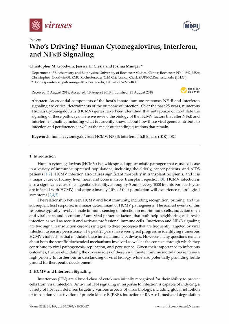
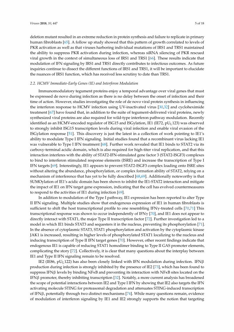
The IFN response to HCMV infection is complex, with multiple distinct mechanisms of IFNactivation and temporal peaks of IFN activity occurring over the viral life cycle. The initial interferonresponse to HCMV infection is triggered when the cell detects viral attachment and entry, resultingin an early induction of IFN synthesis and secretion [16]. The list of cellular sensors that detect andare activated by HCMV binding and entry continues to expand, and includes the TLR2 (toll-likereceptor) and CD14 receptors that interact with viral gB and gH [17,18] as well as the intracellulardsDNA sensors Z-DNA binding protein 1 (ZBP1) [19], TLR9 [20], and cGAS [20], all of which detectthe presence of the viral genome in the host cell. Seemingly in response to these cellular defenses,HCMV has evolutionarily developed a suite of IFN countermeasures that occupy a significant portionof viral coding potential (summarized in Figure 1 and reviewed in [12,21,22]).
Viruses 2018, 10, x FOR PEER REVIEW 2 of 18
variety of host cell defenses targeting various aspects of virus biology, including global inhibition of translation via activation of protein kinase R (PKR), induction of RNAse L-mediated degradation of viral RNA, production of anti-viral nitric oxide through inducible nitric oxide synthase (iNOS), depletion of tryptophan via the indoleamine 2,3 dioxygenase (IDO) pathway, and upregulation of antigen presentation via major histocompatibility (MHC) complex constituents [6–10]. IFN signaling is sub-classified into Type I, Type II, and Type III groups based on their specific signaling architectures and outcomes. The majority of interferon-induced anti-viral activity is dependent on the diverse activities of Type I IFNs, which in humans include IFNκ, IFNε, IFNω, IFNβ, and a host of IFNα isoforms derived from 13 separate genes [11]. The activities of Type I IFNs induce the expression of a large set of IFN-stimulated genes (ISGs) that are critical for rendering the host cell environment less permissive for viral infection. In addition to the direct effects of Type I cytokines on ISG induction, these IFNs have been implicated in coordinated innate immune effector processes including Natural Killer (NK) cell activation, dendritic cell (DC) maturation, and T-cell differentiation [12]. In contrast to the diverse signaling of Type I IFNs, Type II IFN signaling is comprised entirely of the activities of a single cytokine: IFNγ. While IFNγ is also capable of activating ISG expression, its primary function appears to center around enabling the activation, recruitment, and survival of a diverse array of immune cells [13]. The final interferon subtype, Type III, was discovered more recently. Like Type I IFN signaling, Type III IFN signaling plays an important role in host cell defense against viral infection, but it relies on a different subset of IFNs (IFN-λ1, IFN-λ2, and IFN-λ3) and a distinct, heterodimeric IL28Rα/IL10Rβ receptor to induce the expression of anti-viral ISGs. As expression of the Type III IFN receptor is limited to epithelial cell populations, Type III IFN signaling is restricted to a more niche biological context than Type I signaling [14]. HCMV gene products that interact directly with Type III signaling have yet to be described. Canonical signaling of all three IFN subtypes occurs through the activation of the JAK-STAT pathway, but each subtype coordinates with unique cellular receptors and recruits specific STAT proteins that tailor downstream responses to respective Type I, II, and III IFN signaling inputs (reviewed in [15]).
The IFN response to HCMV infection is complex, with multiple distinct mechanisms of IFN activation and temporal peaks of IFN activity occurring over the viral life cycle. The initial interferon response to HCMV infection is triggered when the cell detects viral attachment and entry, resulting in an early induction of IFN synthesis and secretion [16]. The list of cellular sensors that detect and are activated by HCMV binding and entry continues to expand, and includes the TLR2 (toll-like receptor) and CD14 receptors that interact with viral gB and gH [17,18] as well as the intracellular dsDNA sensors Z-DNA binding protein 1 (ZBP1) [19], TLR9 [20], and cGAS [20], all of which detect the presence of the viral genome in the host cell. Seemingly in response to these cellular defenses, HCMV has evolutionarily developed a suite of IFN countermeasures that occupy a significant portion of viral coding potential (summarized in Figure 1 and reviewed in [12,21,22]).
Figure 1. HCMV-mediated modulation of Interferon signaling. Figure 1. HCMV-mediated modulation of Interferon signaling.

Viruses 2018, 10, 447 3 of 18
2.1. The HCMV Tegument Proteins and Interferon Modulation
Given that the IFN response to infection is a swift anti-viral response set in motion by the firstinteractions between virion glycoproteins and cellular receptors, it is logical that HCMV encodesviral effectors that immediately antagonize these early anti-viral events, and downregulation of IFNsignaling during HCMV infection is well-documented [23–26]. Many of these early viral anti-interferonfactors are tegument proteins that are delivered to the cellular cytoplasm upon initial envelope fusion.One of the first of these HCMV-encoded IFN modulators described was the UL83-encoded tegumentprotein pp65, which is packaged into the virion in such abundance that it is the major constituentof the infectious particle [27]. The pp65 protein plays a major role in inhibiting both innate andadaptive immune responses to HCMV infection by interfering with antigen presentation [28] andNK cell activation [29]. HCMV pp65 was initially characterized as a Type I IFN inhibitor throughearly reports utilizing a UL83-deletion virus that induced IFN-associated transcriptional patternsduring infection. Varying hypotheses of pp65’s mechanistic function emerged as some groups reportedthat pp65 was sufficient to block activation and nuclear localization of IRF3, a primary mediator ofType I IFN signaling [30], while others reported no change in IRF3 activity but observed that pp65loss significantly increased IRF1 and NFκB nuclear localization [31]. Subsequent studies suggestedthat the pp65 deletion mutant virus exhibited impaired expression of other important HCMV genes,including the immediate-early protein 2 (IE2), and found that a less-disruptive mutation of the pp65ORF maintained viral inhibition of Type I IFN signaling during infection [32], indicating that pp65may be more dispensable for inhibition of IFN signaling than was initially presumed.
The question of how pp65 contributes to HCMV-mediated inhibition of IFN activity is largelystill unresolved. While some IFN phenotypes originally attributed to pp65 appear to be due to theactions of other viral gene products such as IE2, new roles for pp65 are continually being revealed,such as the interaction between pp65 and activators of the stimulator of interferon genes (STING),an IFN inducer. Signaling via STING and the dsDNA sensor cGAS has emerged as an importantcellular defense against viral infection [33]. A novel binding interaction between pp65 and cGASwas recently identified as a mechanism through which pp65 inhibits the release of cGAMP, therebypreventing STING recruitment to cGAS and impeding the expression of IFNβ [34]. STING signaling isalso reported to be inhibited by pp65 via interaction with the upstream STING activator interferongamma-inducible protein 16 (IFI16). Evidence suggests that pp65 binds to IFI16 and occludes the pyrindomains required for IFI16 oligomerization in the nucleus, preventing STING activation and anti-viralcytokine expression [35]. Contributing to this picture, pp65 has also been shown to form a complexwith UL97 [36], a conserved herpesviral modulator of IFN signaling that downregulates IFN secretionvia inhibition of IRF3 [37], and may have further direct or indirect effects on IFI16 activation mediatedthrough this interaction. These recent findings underscore the extent to which pp65’s interactions withIFN signaling are continuing to be elucidated.
Evidence has emerged implicating several other tegument proteins in innate immune modulatoryactivities. These include the UL23, UL26, and pp71 proteins. The pp71 tegument protein, encoded by theUL82 gene, shares significant homology with its neighboring HCMV gene product pp65 [38,39] and hasbeen identified as a key activator of the HCMV major immediate-early promoter (MIEP) [40]. Knownto be required for high-titer viral replication [41], pp71 has been discovered to provide an integraldefense against silencing of the viral MIEP by the host innate immune effector Daxx [42]. Throughdirect binding, pp71 functions to disrupt complex formation between Daxx and other transcriptionalrepressors such as ATRX [43], as well as target Daxx itself for proteasomal degradation [44].Underscoring the relevance of this interaction is the observation that ablation of pp71 results inattenuated expression of HCMV immediate-early genes and impaired lytic replication, which canbe rescued via inhibition of Daxx [41,45]. pp71 has also recently been found to bind STING andprevent the formation of complexes required for its translocation, sequestering it away from thenucleus [46]. Further, RNAi-mediated knockdown or gene knockout of pp71 increases host anti-viral
Viruses 2018, 10, 447 4 of 18
gene expression, further emphasizing the importance of pp71 for attenuating innate immunity duringinfection [46].
Tegument proteins have also been shown to modulate the Type II IFN response to infection.Type II signaling, induced by IFNγ, is heavily dependent on STAT1 transcription factor homodimersthat bind and activate transcription at promoters containing gamma interferon activation sequences(GAS) [47]. Recent work using UL23-deficient HCMV mutants shows that Type II IFN gene targets areupregulated during infection in the absence of UL23. These mutants are also more sensitive to challengewith IFNγ [48]. A putative binding interaction between UL23 and the STAT effector molecule N-mycinteractor (NMI), identified via yeast two-hybrid screening, is hypothesized to prevent the properactivation and translocation of the STAT1 homodimers required for Type II IFN signaling [48]. Theseearly results provide insight into HCMV’s modulation of Type II IFN, which is less well-characterizedthan its interplay with Type I signaling.
Complex relationships between tegument proteins and individual interferon stimulated genesare also currently being elucidated. The ubiquitin-like modifier ISG15 is a Type I IFN target that iscovalently bound to target molecules, thereby altering their function (reviewed in [49]). Multiplereports have implicated ISGylation as an anti-viral defense mechanism that is triggered early duringHCMV infection via cGAS-STING viral DNA sensing and restricts HCMV replication [50,51]. UL26was recently found to interact with ISG15 as well as multiple enzymes involved in the activationand ligation of ISG15 to target proteins [51]. Interestingly, some of these ISG15 enzymes have otherpotentially relevant roles in the immune response outside of their ISG15-proximal functions, suchas UBP43, which ligates ISG15 to target proteins but also binds IFNAR2 and occludes its interactionwith JAK1 to downregulate IFN signaling [52]. During infection in the absence of immediate-earlyprotein 1 (IE1), UL26 appears to play a role in suppressing the accumulation of ISGylated proteins [51].Notably, UL26 itself is subject to ISG15 modification [51]; however, many questions remain about howthe interactions between UL26 and the ISG15-machinery contribute to viral infection.
In addition to proteins involved in cytoplasmic DNA sensing, e.g., IFI16, ZBP1, and cGAS,the host cell also encodes proteins that sense cytoplasmic dsRNA and activate similar responses,with one of the most prominent being PKR (reviewed in [53]). PKR is an ISG with an N-terminaldsRNA-binding domain that can be activated by the presence of dsRNA as well as by a host of otherstimuli including oxidative stress, cytokines, and other cellular kinases [54]. Active PKR functions byhomodimerizing and autophosphorylating itself to enable a binding interaction between its C-terminaldomain and the eukaryotic transcription initiation factor eIF2α, globally repressing transcription ofboth viral and cellular genes by interfering with the formation of the eIF2α-tRNAMet-GTP transcriptioninitiation complex [55,56]. PKR signaling can result in a diverse array of immune outcomes includingupregulated Type I IFN signaling [57] as well as increased NFκB pathway activity [58]. HCMV encodestwo immediate-early gene products that specifically target PKR as a means of downregulating the IFNresponse and maintaining high levels of viral gene transcription during infection: IRS1 and TRS1.
Co-infection of HCMV was found to be sufficient to rescue protein translation of a Vaccinia virusmutant (VV∆E3L) that is sensitive to PKR activity [59], which ultimately led to the identificationof IRS1 and TRS1 as key PKR modulatory factors [60]. Further investigation into TRS1 found thattwo distinct regions of this protein modulate PKR in separable fashions, with the C-terminal regioncapable of directly binding dsRNA and presumably preventing it from binding PKR and other dsRNAsensors, and the N-terminal region binding PKR directly and sequestering it in the nucleus to preventits activation and downstream signaling [61–63]. Interestingly, both regions of TSR1 are required tofully rescue VV∆E3L-PKR related phenotypes, which include viral replication and host cell range [61],though more recent results suggest that the N-terminal region is responsible for the majority of thePKR inhibition phenotype [64].
To assess the contribution of these proteins to overall HCMV replication, mutants containingdeletions of IRS1 and TRS1 both individually and in tandem were used to show that loss of eithergene product in isolation did not strongly affect viral growth, but infection with an IRS1/TRS1 double
Viruses 2018, 10, 447 5 of 18
deletion mutant resulted in an extreme reduction in protein synthesis and failure to replicate in primaryhuman fibroblasts [65]. A follow up study showed that this pattern of growth correlated to levels ofPKR activation as well as that viruses harboring individual mutations of IRS1 and TRS1 maintainedthe ability to suppress PKR activation during infection, whereas siRNA silencing of PKR rescuedviral growth in the context of simultaneous loss of IRS1 and TRS1 [66]. These results indicate thatmodulation of IFN signaling by IRS1 and TRS1 directly contributes to infectious outcomes. As futureinquiries continue to dissect the different functions of IRS1 and TRS1, it will be important to elucidatethe nuances of IRS1 function, which has received less scrutiny to date than TRS1.
2.2. HCMV Immediate-Early Genes (IE) and Interferon Modulation
Immunomodulatory tegument proteins enjoy a temporal advantage over viral genes that mustbe expressed de novo during infection as there is no delay between the onset of infection and theirtime of action. However, studies investigating the role of de novo viral protein synthesis in influencingthe interferon response to HCMV infection using UV-inactivated virus [30,32] and cycloheximidetreatment [67] have found that, in addition to the suite of tegument-delivered viral proteins, newlysynthesized viral proteins are also required for wild-type interferon pathway modulation. Recentlyidentified as an HCMV-encoded regulator of ISG15 and ISGylation, IE1 (IE72, pUL123) was observedto strongly inhibit ISG15 transcription levels during viral infection and enable viral evasion of theISGylation response [51]. This discovery is just the latest in a collection of work pointing to IE1’sability to modulate Type I IFN signaling. Initial studies found that a recombinant virus lacking IE1was vulnerable to Type I IFN treatment [68]. Further work revealed that IE1 binds to STAT2 via itscarboxy-terminal acidic domain, which is also required for high-titer viral replication, and that thisinteraction interferes with the ability of STAT2-IFN-stimulated gene factor 3 (STAT2-ISGF3) complexesto bind to interferon stimulated response elements (ISRE) and increase the transcription of Type IIFN targets [69]. Interestingly, IE1 appears to prevent STAT2-ISGF3 complex loading onto ISRE siteswithout altering the abundance, phosphorylation, or complex formation ability of STAT2, relying on amechanism of interference that has yet to be fully described [68,69]. Additionally noteworthy is thatSUMOylation of IE1’s acidic domain has been shown to inhibit the IE1-STAT2 interaction and mitigatethe impact of IE1 on IFN target gene expression, indicating that the cell has evolved countermeasuresto respond to the activities of IE1 during infection [69].
In addition to modulation of the Type I pathway, IE1 expression has been reported to alter TypeII IFN signaling. Multiple studies show that endogenous expression of IE1 in human fibroblasts issufficient to shift the host transcriptional profile to one resembling IFNγ-treated cells [70,71] Thistranscriptional response was shown to occur independently of IFNγ [70], and IE1 does not appear todirectly interact with STAT1, the major Type II transcription factor [72]. Further investigation led to amodel in which IE1 binds STAT3 and sequesters it in the nucleus, preventing its phosphorylation [71].In the absence of cytoplasmic STAT3, STAT1 phosphorylation and activation by the cytoplasmic kinaseJAK1 is increased, resulting in higher levels of phosphorylated STAT1 localizing to the nucleus andinducing transcription of Type II IFN target genes [70]. However, other recent findings indicate thatendogenous IE1 is capable of reducing STAT1 homodimer binding to Type II GAS promoter elements,complicating the story [72]. Collectively, it is clear that many questions about the interplay betweenIE1 and Type II IFN signaling remain to be resolved.
IE2 (IE86, pUL122) has also been closely linked with IFN modulation during infection. IFNβproduction during infection is strongly inhibited by the presence of IE2 [73], which has been found tosuppress IFNβ levels by binding NFκB and preventing its interaction with NFκB sites located on theIFNβ promoter, thereby inhibiting transcription [32]. Notably, a more current analysis has broadenedthe scope of potential interactions between IE2 and Type I IFN by showing that IE2 also targets the IFNactivating molecule STING for proteasomal degradation and attenuates STING-induced transcriptionof IFNβ, potentially through two distinct mechanisms [74]. While many questions remain, evidenceof modulation of interferon signaling by IE1 and IE2 strongly supports the notion that targeting
Viruses 2018, 10, 447 6 of 18
interferon signaling at immediate-early times of infection is critical for successful HCMV infectionand persistence.
2.3. HCMV-Mediated Modulation of Interferon at Later Times of Infection
The unique short region of the HCMV genome contains a series of adjacent genes with limitedsequence similarity, US2–US11, that encode a set of glycoproteins implicated in a variety of activitiesincluding immune modulation [75,76]. US9 is expressed with early kinetics during viral infection andlocalizes to the mitochondria and endoplasmic reticulum [77], where it appears to modulate TypeI IFN signaling and IFNβ production through two distinct interactions with the MAVS and STINGadaptor proteins [78]. MAVS and STING both function similarly as adaptors that recruit the TBK1kinase with its substrate, IRF3, which is then activated and localized to the nucleus to upregulate ISGtranscription [78]. Expression of US9 in cells results in a strong downregulation of IRF3 activation andnuclear accumulation [78]. US9 appears to achieve this effect by damaging the cell’s mitochondriaand preventing its retention of MAVS, as well as by directly binding STING and preventing itsdimerization and downstream activation of IRF3 [78]. As a non-tegument HCMV protein expressedwith early kinetics, US9 is of particular interest because most HCMV-encoded IFN modulatory proteinsdiscovered to date are either delivered with the tegument or expressed immediately upon infection.In contrast, US9 is scarcely detectable within the cell prior to 6 hpi and reaches its highest level ofaccumulation around 48 hpi [23], appearing to be exclusively dedicated to protecting and enabling thelater phases of the viral life cycle.
As discussed above, the identification of pp71 as a cGAS-STING interactor was achieved throughthe use of an HCMV gene expression library containing 131 constructs each encoding a unique HCMVprotein [46]. Another HCMV gene product that emerged from this screen was UL31, a true-late proteinrequired for wild-type viral growth [79,80] that was found to be capable of binding both viral DNA andhost cell cGAS via its N- and C-terminal regions, respectively. UL31’s high binding affinity for cGAS,but not for DNA, suggests a non-competitive mechanism of action wherein UL31 preferentially bindscGAS in a manner that dissociates DNA from the molecule. UL31 fails to inhibit cGAMP induction ofType I ISGs but is capable of inhibiting the interferon-associated gene transcription stimulated by bothHCMV infection and dsDNA, signifying that this key interaction with cGAS is critical for modulatingdownstream immune signaling [81]. Further, knockdown of cGAS is capable of rescuing the growthdefect of UL31-deficient viral infection, indicating that the UL31/cGAS interaction has implications foroverall viral growth [81]. Notably, UL31-mediated inhibition of cGAS appears to have a broad immunefootprint, affecting multiple immune signaling archetypes, as infection with a UL31-deficient HCMVmutant strongly induced both ISG’s and NFκB target genes, and plasmid overexpression of UL31 infibroblasts inhibited the activation of both ISRE-containing and NFκB reporter elements [81].
Another upstream inducer of STING, IFI16, is targeted for inhibition by the HCMV early-lategene UL97. Conserved among herpesviruses, UL97 is a viral kinase that has been shown to bind andphosphorylate IFI16, inducing its relocalization out of the nucleus of infected cells into the cytoplasm,where it is prevented from inducing an IFN response [82]. Nuclear retention of IFI16 can be rescued bytreatment with an inhibitor that reduces UL97 kinase function or by removing the UL97 reading framefrom the HCMV genome, further establishing this link between UL97 and IFI16 export [82].
2.4. Modulation of Interferon during Latency
Work using an IRS1 and TRS1 double mutant (∆IRS1/∆TRS1) to investigate PKR modulationduring infection was instrumental in uncovering another HCMV gene product that alters the hostcell’s ability to sense viral dsRNA: ORF94 (UL126a). Oligoadenylate synthetase (OAS) proteins aredsRNA-binding enzymes that activate cellular RNases in response to dsRNA detection, degradinghost and viral RNA and down regulating overall rates of protein synthesis (reviewed in [83]). HCMVinfection institutes a block to OAS signaling, and it was observed that while HCMV-encoded TRS1and IRS1 are capable of downregulating this pathway in certain contexts [60], a mutant HCMV lacking
Viruses 2018, 10, 447 7 of 18
both of these open reading frames still inhibited OAS activation [65]. HCMV ORF94 was identified asa potential candidate that may be mediating this OAS block by inhibiting the expression of OAS1 inmultiple contexts, including during productive infection and in the face of interferon stimulation [84].Notably, HCMV ORF94 is a latency-associated ORF encoded by transcripts expressed during latentinfection [85]. While extremely intriguing, it still remains to be seen how ORF94 and its modulation ofthe IFN response contributes to latency establishment, maintenance, or reactivation.
3. HCMV Modulation of NFκB Signaling
The NFκB signaling network regulates a wide variety of pro-inflammatory processes thatultimately shape innate and adaptive immune responses via transcriptional regulation of numerousNFκB responsive genes. NFκB signaling can be activated by a myriad of inducers including infectiousagents, paracrine signaling factors, or environmental stress [86]. Highlighting its importance asa defense against infection, a wide variety of evolutionarily diverse viruses including HumanImmunodeficiency Virus-1 (HIV-1), HCMV, Herpes Simplex Virus-1 (HSV-1), and Epstein Barr Virus(EBV) have evolved mechanisms to modulate NFκB signaling [87]. Numerous interactions betweenHCMV and NFκB signaling have been described over the years (Figure 2, and reviewed in [87,88]).HCMV gene products have been shown to both inhibit NFκB signaling and activate facets of the NFκBpathway to support lytic replication or induce reactivation from latency [89,90], suggesting a nuancedrelationship. This highlights the reality that NFκB signaling is not simply binary, but rather that NFκBendpoint signaling is capable of multiple distinct transcriptional landscapes depending on specificupstream stimuli. The relationship between HCMV and NFκB is shaped by multiple HCMV geneproducts, including both proteins and miRNAs, which serve to modulate various aspects of NFκBsignaling. Less clear are how the various facets of HCMV-mediated modulation of NFκB contribute tothe variety of biological contexts of HCMV infection, including viral persistence, shedding, latency,reactivation, tropism, and pathogenesis.
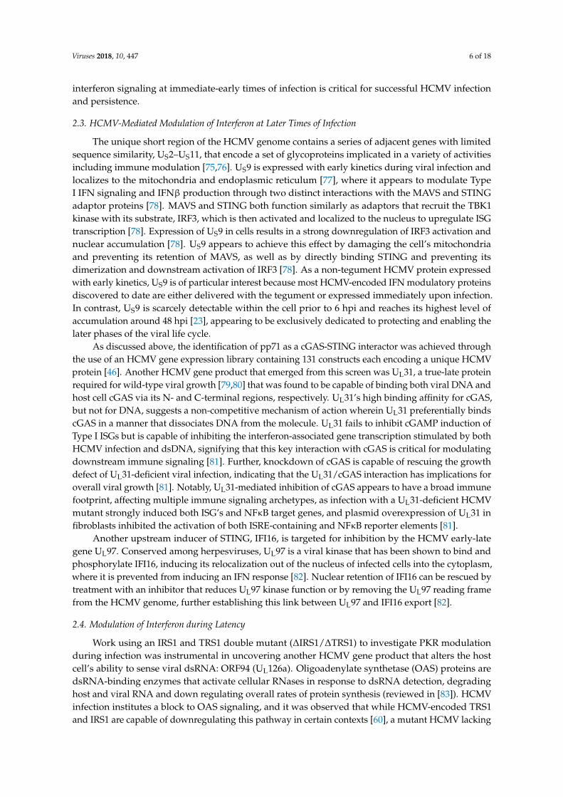
Most effectors of the NFκB response converge upon the activation of a serine-specific IκB kinase(IKK) complex comprised of different combinations of three distinct subunits: IKKα, IKKβ andIKKγ/NEMO (Figure 2). Canonical NFκB signaling relies on a tripartite complex consisting of one ofeach IKK subunit, while non-canonical NFκB functions through an IKKα dimer [86]. The mechanismsthrough which the cytoplasmic NFκB subunit complexes are activated represent a major differencebetween these two sub-pathways (Figure 2). In canonical NFκB signaling, an IKK complex containingNEMO phosphorylates the repressor protein IκB, marking it for ubiquitination and degradation. IκBrepresses the canonical NFκB transcription factors p50 and p65 (RelA) in the cytoplasm, which arefreed upon IκB phosphorylation/degradation, resulting in their nuclear translocation and subsequenttranscriptional activation of NFκB targets [80]. During non-canonical NFκB signaling an IKKαhomodimer acts as the kinase that phosphorylates the C-terminus of p100, which resides in aninhibitory complex with RelB in the cytoplasm. This phosphorylation of p100 results in its processingto p52, which in complex with RelB, can enter the nucleus to modulate NFκB target transcription [91](Figure 2).
During HCMV infection of fibroblasts NFκB activation appears to follow a specific sequence inwhich the pathway is active early in infection, but is then repressed from middle to late time points ofthe viral life cycle. At the earliest time of infection, i.e. envelope fusion, the HCMV glycoproteins B andH (gB/gH), encoded by UL55 and UL75, respectively, bind to Toll-like receptor 2 (TLR-2) on the surfaceof the cell and induce a canonical NFκB signaling cascade resulting in the excretion of pro-inflammatoryand anti-viral cytokines [92]. This immediate enhancement of NFκB activation appears to be pro-viral,as HCMV’s MIEP possesses NFκB binding motifs [93] that facilitate the expression of IE genes, aneffect that can be enhanced by TNFα stimulation [94]. Further evidence supporting a pro-viral aspectof early NFκB activation for infection include the findings that dominant-negative constructs targetingkey NFκB constituents such as IKKα, IKKβ, and IκBα reduce MIEP activation [95]. These findingssuggest that NFκB activation at early times is important for optimal transactivation of the MIEP.

Viruses 2018, 10, 447 8 of 18
However, the utilization of HCMV mutants lacking the NFκB motifs in the MIEP did not result insignificant attenuation of infection or IE gene product accumulation [96], suggesting that additionalhost transcription factor binding motifs present in the MIEP, including CREB (cAMP response elementbinding) and ATF (activating transcription factor) sites [97], may be sufficient to activate IE genetranscription even in the absence of p65/p50 binding. This remains an unresolved issue, and furtherinquiries have suggested that this nuanced interaction between viral transcription and NFκB signalingcan be influenced by numerous factors including the host cell’s progression through the cell cycle [89],the adaptation of HCMV strains to laboratory passage conditions [98], and host cell lineage [99].
Viruses 2018, 10, x FOR PEER REVIEW 8 of 18
unresolved issue, and further inquiries have suggested that this nuanced interaction between viral transcription and NFκB signaling can be influenced by numerous factors including the host cell’s progression through the cell cycle [89], the adaptation of HCMV strains to laboratory passage conditions [98], and host cell lineage [99].
Figure 2. HCMV-mediated modulation of NFκB signaling
3.1. HCMV Tegument Proteins and NFκB Modulation
Concurrent with envelope fusion, the virion releases viral tegument proteins into the cytoplasm which disseminate and begin to modulate a number of cellular pathways. The pp65 protein, as discussed above, likely plays a role in blocking the host IFN response during early infection that is not yet fully understood. In addition to this IFN modulatory role, infection with a pp65-deficient mutant HCMV increases the accumulation of NFκB target genes and induces the nuclear binding activity of NFκB transcription factors [31], suggesting an important contribution to NFκB regulation. Mechanistically, however, it is unclear how pp65 modulates NFκB activity. Further, the extent to which pp65’s modulation of IFN and NFκB signaling might be functionally related is uncertain.
The UL26 protein is also delivered with the tegument and has been shown to antagonize NFκB activity. A UL26 deletion mutant virus is severely attenuated, and UL26 has been shown to be necessary to inhibit IKK complex phosphorylation and RelB translocation during infection [100,101]. Further, expression of UL26 by itself is sufficient to block TNFα-mediated NFκB activation [100,101]. However, despite its presence in the tegument, UL26 does not block the activation of NFκB at the earliest times of infection. UL26 is thought to have a stronger role in the attenuation of NFκB activity that occurs later during infection, when UL26 localizes to the cytoplasm, as opposed to early times when it localizes in the nucleus [102]. However, the possibility of an interaction between UL26 and NFκB at early times during infection cannot be ruled out, as UL26-deficient virus is more sensitive to challenge with TNFα [100]. Notably, UL26 is capable of inhibiting IKK complex activation in the face of diverse upstream stimuli including TNFα signaling and Sendai virus infection, suggesting that it is acting at the level of the IKK complex, where these signaling cascades converge [100], but the exact mechanism of UL26’s inhibition of NFκB signaling remains to be elucidated
Figure 2. HCMV-mediated modulation of NFκB signaling
3.1. HCMV Tegument Proteins and NFκB Modulation
Concurrent with envelope fusion, the virion releases viral tegument proteins into the cytoplasmwhich disseminate and begin to modulate a number of cellular pathways. The pp65 protein, asdiscussed above, likely plays a role in blocking the host IFN response during early infection that isnot yet fully understood. In addition to this IFN modulatory role, infection with a pp65-deficientmutant HCMV increases the accumulation of NFκB target genes and induces the nuclear bindingactivity of NFκB transcription factors [31], suggesting an important contribution to NFκB regulation.Mechanistically, however, it is unclear how pp65 modulates NFκB activity. Further, the extent to whichpp65’s modulation of IFN and NFκB signaling might be functionally related is uncertain.
The UL26 protein is also delivered with the tegument and has been shown to antagonize NFκBactivity. A UL26 deletion mutant virus is severely attenuated, and UL26 has been shown to be necessaryto inhibit IKK complex phosphorylation and RelB translocation during infection [100,101]. Further,expression of UL26 by itself is sufficient to block TNFα-mediated NFκB activation [100,101]. However,despite its presence in the tegument, UL26 does not block the activation of NFκB at the earliest times ofinfection. UL26 is thought to have a stronger role in the attenuation of NFκB activity that occurs laterduring infection, when UL26 localizes to the cytoplasm, as opposed to early times when it localizesin the nucleus [102]. However, the possibility of an interaction between UL26 and NFκB at early

Viruses 2018, 10, 447 9 of 18
times during infection cannot be ruled out, as UL26-deficient virus is more sensitive to challengewith TNFα [100]. Notably, UL26 is capable of inhibiting IKK complex activation in the face of diverseupstream stimuli including TNFα signaling and Sendai virus infection, suggesting that it is acting atthe level of the IKK complex, where these signaling cascades converge [100], but the exact mechanismof UL26’s inhibition of NFκB signaling remains to be elucidated
HCMV tegument proteins are also capable of inducing pro-viral NFκB signaling. UL76,a tegument-associated endonuclease, is reported to activate the canonical NFκB pathway throughthe DNA damage response and induce IL-8 production [103]. This UL76-mediated increase in IL-8production was shown to be dependent on the cellular kinases Ataxia-telangiectasia mutated (ATM)and IKKβ; however, ablation of ATM expression in cells or of the key endonuclease motif aminoacids present in UL76 failed to completely restore IL-8 production back to wild-type levels duringinfection [103], implying that additional aspects of NFκB signaling might be contributing to thisphenotype. A UL76 deletion mutant has a significant growth defect [104], but the contributions ofincreased IL-8 production to this attenuation are not clear.
HCMV virions have been found to incorporate cellular mRNAs and proteins [27,105]. Thisraises the possibility that, in addition to viral factors, virion-associated cellular factors could also bemodulating NFκB signaling. One such example is the virion packaging of casein kinase II (CKII),which has been found in the virion tegument and is reported to activate NFκB signaling throughphosphorylation of the IκB repressor, thereby releasing the associated NFκB subunits to localize tothe nucleus and induce NFκB-dependent transcription [106]. The extent to which cellularly-derived,virion-associated NFκB modulators contribute to the various facets of HCMV infection is still largelynot known.
3.2. HCMV Immediate-Early Genes and NFκB Modulation
The IE proteins expressed upon MIEP stimulation interact with the NFκB pathway in diverse ways.IE1, a promiscuous transactivator of NFκB pathway constituents and their downstream targets duringinfection, has been implicated in the upregulation of p65, IL-6, TNF-α and IL-8 as well as the inductionof p52/RelB heterodimer binding activity in the nucleus [88]. At immediate-early times, the virusalso produces UL144, a TNF-receptor-like transmembrane receptor with immediate-early expressionkinetics [107], which activates expression of the immune cytokine CCL22 [108]. Mechanistically,UL144 complexes with TRAF6 in perinuclear regions of the cell to enable NFκB transcription factortranslocation and binding, and siRNA targeting UL144, TRAF6, or NFκB all ablate the downstreamCCL22 expression induced by infection [108]. The CCL22 cytokine has been noted to play achemoattractant role in recruiting Th2 and regulatory T-cells (Tregs) to mediate adaptive immuneresponses [108]. The UL144 open reading frame is lost in extensively laboratory passaged strains ofHCMV, potentially due to its activation of NFκB signaling, which could be a detriment to viral fitnessduring in vitro fibroblast infection [109,110].
In addition to immediate-early expression of the NFκB agonists IE1 and UL144, IE2 inhibits hostNFκB signaling at all points during HCMV infection through a still-controversial model: either byblocking NFκB subunit dimer interactions or preventing subunit interactions with specific NFκBtarget promoters, e.g., IL-6 [32,111]. Interestingly, the antagonistic effects of IE2 do not prevent UL144from inducing NFκB [112], which highlights the specificity with which HCMV is able to tailor NFκBsignaling. Collectively the data suggest that at early points of infection the virus seems to be operatingwithin an optimal pro-inflammatory signaling window, with just enough NFκB transcription factorbinding to transactivate the viral MIEP, but still staying below a threshold that might trigger a broaderanti-viral immune response.
3.3. HCMV-Mediated Modulation of NFκB at Later Times of Infection
At some point during the transition from the immediate-early stages of infection, where activeNFκB signaling is observed, to the later stages of infection, the viral actions towards NFκB become
Viruses 2018, 10, 447 10 of 18
much more inhibitory. This transition coincides with the increased expression of multiple HCMV genesthat antagonize NFκB activity. One such gene is UL111a, also known as cmvIL-10 for its functionalsimilarity to the cellular cytokine IL-10. cmvIL-10 shares only approximately 27% homology withhost IL-10, but binds as a homodimer to the human IL-10 receptor and blocks NFκB signaling bypreventing IκBα degradation in a similar manner to IL-10 [113–115]. In addition, cmvIL-10 also hasa significant immunosuppressive effect on interferon signaling. In peripheral blood mononuclearcells (PBMCs), TLR-agonist treated media from wild-type AD169 infection is sufficient to inhibit theproduction of IFNα when compared to similarly treated media from a cmvIL-10 knockout virus [116].Further, cmvIL-10 alone is sufficient to do the same in plasmacytoid DCs [117]. The mechanism behindthese inhibitions appears to involve activation of STAT3 [118], but is still poorly understood. It remainsunknown if the suppressive effects of cmvIL-10 on IFN and NFκB signaling are separable. Studies ofthe cmvIL-10/interferon signaling axis suggest that the molecule may be acting in a paracrine mannerto activate defenses in uninfected cells, which could represent a promising avenue of further inquiry.
3.4. HCMV miRNAs and NFκB Modulation
Viral miRNAs represent additional tools that HCMV employs to undermine host cell defensesat later points of infection. HCMV encodes 26 miRNAs, each approximately 22nt in length, thathave been implicated in modulating a wide variety of cellular pathways and processes includingvesicle transport, cytokine secretion, immune signaling, and progression through the cell cycle,(reviewed in [119] and [120]). Throughout the course of infection, beginning with immediate-earlygene expression, HCMV miRNAs begin to accumulate, becoming abundant by late infection [119,121].HCMV miR-US5-1 and miRUL112-3p have been shown to prevent NFκB cytokine signaling byspecifically downregulating the expression of the key kinases IKKα and IKKβ [122]. In additionto blocking the NFκB signal relay set off by cytokine detection, miR-US5-2 is able to block the infectedcells’ secretion of cytokines [123], thereby ceasing the positive feedback loop of NFκB activation andultimately returning the pathway to its initial inactive state. During latent infection, miR-UL148Dis one of the most highly expressed miRNAs, and has been demonstrated to block NFκB upstreamadapters and repress IL-6 production [124], thus allowing the infected cell to evade host immunity.
3.5. Latency-Associated NFκB Modulators
A hallmark of all herpesviruses is the ability to enter latency, persisting with limited lytic viralreplication in the face of a primed immune response and capable of reactivation during times ofstress or immunosuppression, resulting in viral dissemination and potential pathologies. The signalsthat reactivate HCMV from latency remain incompletely understood, but immunosuppression andinflammation are thought to play major roles (reviewed in [125]). Consistent with this view, reportsindicate that viral genes activate NFκB during reactivation [126], and, further, NFκB activation hasbeen linked to HCMV reactivation via NFκB subunit enhancement of MIEP expression [127,128]. Theviral chemokine receptor US28, expressed with early kinetics during lytic infection, is one of the fewviral proteins expressed during latency as demonstrated in latently infected THP-1 monocytes [128].US28 has been implicated in activating the MIEP through NFκB signaling [126]; it is possible thatduring latent expression, US28 activates the MIEP and aids in reactivation from latency. US28 inducesconstitutive NFκB activation through its interaction with the Gq/11 G protein, which mediates therelease of Gβγ subunits that induce downstream NFκB activity [129]. Although in general US28 hasappeared to stimulate NFκB activity, recent work suggests that US28 attenuates multiple cell signalingpathways including NFκB, which is required to maintain latency as mutants lacking US28 return totheir lytic phase and infected cells are subsequently targeted by T-cells [130]. It is clear that US28 playsa complicated role in the HCMV life cycle; and, like other viral factors, possesses more than one rolethat may seem counterintuitive, but that are likely important in different infectious contexts.
Another viral protein expressed during latency, UL138, acts by activating and stabilizing thecell surface expression of TNFR1 [131–133]. Interestingly, while UL138 appears to promote the
Viruses 2018, 10, 447 11 of 18
sensitivity of latently infected cells to TNFα, reporter assays show that UL138 strongly repressesMIEP transactivation [134] and ChIP (chromatin-immunoprecipitation) assays suggest it preventscellular demethylases from interacting with the MIEP [135], leading to the conclusion that this proteinis also playing a central role in maintaining HCMV’s latent state. These results represent some ofthe first forays into exploring the immunomodulatory potential of HCMV latency-associated genes,and the data suggest that there is a complex interplay between pro- and anti-viral manipulations ofimmune signaling as the virus maintains latent infection despite silencing much of its transcription.
4. Conclusions and Future Perspectives
Reports over the past 25 years have made it abundantly clear that HCMV devotes substantialgenetic resources towards manipulation of interferon and NFκB signaling. The list of involved genesis large and still growing, with a variety of HCMV gene products playing modulatory roles in variouscontexts. While a multitude of questions remain, certain themes and patterns have emerged. Forexample, with respect to IFN signaling, while some ISGs such as viperin and Cox2 (reviewed in [12])appear to be co-opted by the virus for pro-viral activities, almost all HCMV gene products that havebeen identified to play a role in IFN signaling serve to attenuate this host cell response to support robustinfection, suggesting that HCMV goes to great lengths to inhibit IFN signaling to enable infection.
In contrast, the literature regarding HCMV’s modulation of NFκB suggests a more complex picture.NFκB activation during lytic infection of fibroblasts appears to be biphasic with a pro-inflammatory,pro-viral NFκB signaling environment instituted during the earliest times of infection, whichsubsequently shifts to broadly inhibitory of NFκB at later time points of infection. As discussedabove, current data suggest a model in which early NFκB activation supports infection throughincreased MIEP transcription, whereas later NFκB inhibition prevents the secretion of NFκB-regulatedanti-viral factors, e.g., cytokines. However, many questions remain. For one, the sheer number ofHCMV gene products that modulate NFκB inflammatory signaling in diverse ways suggests a morenuanced story. It seems likely that these NFκB modulatory gene products work in different infectiouscontexts to support varied aspects of viral infection, including robust lytic infection, the establishmentof latency, and reactivation from the latent state. In this regard, the activities of NFκB-modulatory viralproteins will likely be sensitive to a variety of cellular states including cell type, differentiation status,and the inflammatory environment.
A typical in vivo infection progresses from the mucosal epithelial cells to responding immunecells, with subsequent seeding of lymph nodes and infection of progenitor cells that will serve aslatency reservoirs, followed by some level of either subclinical or pathogenic reactivation back in themucosal epithelia. We propose that cell-type and context specific HCMV-mediated modulation ofNFκB will be critical at each step of this process. Furthering our understanding of how the relationshipbetween HCMV and NFκB shapes viral biology and pathogenesis will require elucidating how specificmechanisms of HCMV-mediated modulation of NFκB contribute to key events during the in vivo virallife cycle in physiologically relevant cell types. While this will be challenging given the limitations ofour current in vivo and in vitro models, recent developments in humanized mice, as well as explantand organoid culture techniques will yield exciting, novel opportunities to address these issues. Thefield has made substantial progress, likely identifying the majority of the viral players involved in NFκBmodulation. It is time to turn our attention to identifying how these viral NFκB modulators contributeto various infectious contexts, and to developing the tools and experimental systems required to doso. Given the importance of these interactions to infectious outcomes, further elucidating how theycontribute to infection will likely provide novel avenues to limit HCMV-associated pathogenesis.
Author Contributions: Conceptualization and Writing, C.M.G., J.H.C., and J.M.
Funding: C.M.G. was supported by an NIH training grant from the Training Program in Oral Science(T90-DE021985-07). The work was also supported by an NIH grant AI127370 to J.M. and by a Research ScholarGrant from the American Cancer Society (RSG-15-049-01-MPC).
Conflicts of Interest: The authors declare no conflicts of interest.

Viruses 2018, 10, 447 12 of 18
References
1. Gerna, G.; Baldanti, F.; Revello, M.G. Pathogenesis of human cytomegalovirus infection and cellular targets.Hum. Immunol. 2004, 65, 381–386. [CrossRef] [PubMed]
2. Pass, R.F. Cytomegalovirus. In Fields’ Virology, 4th ed.; Knipe, D.M., Howley, P.M., Eds.; Lippincott-Williamsand Wilkins: New York, NY, USA, 2001; pp. 2675–2705.
3. Neiman, P.; Wasserman, P.B.; Wentworth, B.B.; Kao, G.F.; Lerner, K.G.; Storb, R.; Buckner, C.D.; Clift, R.A.;Fefer, A.; Fass, L.; et al. Interstitial pneumonia and cytomegalovirus infection as complications of humanmarrow transplantation. Transplantation 1973, 15, 478–485. [CrossRef] [PubMed]
4. Pinninti, S.G.; Ross, S.A.; Shimamura, M.; Novak, Z.; Palmer, A.L.; Ahmed, A.; Tolan, R.W., Jr.; Bernstein, D.I.;Michaels, M.G.; Sánchez, P.J. Comparison of saliva PCR assay versus rapid culture for detection of congenitalcytomegalovirus infection. Pediatr. Infect. Dis. J. 2015, 34, 536–537. [CrossRef] [PubMed]
5. Boppana, S.B.; Ross, S.A.; Shimamura, M.; Palmer, A.L.; Ahmed, A.; Michaels, M.G.; Sánchez, P.J.;Bernstein, D.I.; Tolan, R.W., Jr.; Novak, Z. Saliva polymerase-chain-reaction assay for cytomegalovirusscreening in newborns. N. Engl. J. Med. 2011, 364, 2111–2118. [CrossRef] [PubMed]
6. Dauber, B.; Wolff, T. Activation of the antiviral kinase PKR and viral countermeasures. Viruses 2009, 1,523–544. [CrossRef] [PubMed]
7. Samuel, C.E. Adenosine deaminases acting on RNA (ADARS) are both antiviral and proviral. Virology 2011,411, 180–193. [CrossRef] [PubMed]
8. Grandvaux, N.; Servant, M.J.; Hiscott, J. The interferon antiviral response: From viral invasion to evasion.Curr. Opin. Infect. Dis. 2002, 15, 259–267. [CrossRef] [PubMed]
9. Mehta, D.R.; Ashkar, A.A.; Mossman, K.L. The nitric oxide pathway provides innate antiviral protectionin conjunction with the type I interferon pathway in fibroblasts. PLoS ONE 2012, 7, e31688. [CrossRef][PubMed]
10. Zimmermann, A.; Hauka, S.; Maywald, M.; Le, V.T.K.; Schmidt, S.K.; Däubener, W.; Hengel, H. Checks andbalances between human cytomegalovirus replication and indoleamine-2,3-dioxygenase. J. Gen. Virol. 2014,95, 659–670. [CrossRef] [PubMed]
11. Bekisz, J.; Schmeisser, H.; Hernandez, J.; Goldman, N.D.; Zoon, K.C. Mini review Human interferons alpha,beta and omega. Growth Factors 2004, 22, 243–251. [CrossRef] [PubMed]
12. Amsler, L.; Verweij, M.C.; DeFilippis, V.R. The tiers and dimensions of evasion of the type I interferonresponse by human cytomegalovirus. J. Mol. Biol. 2013, 425, 4857–4871. [CrossRef] [PubMed]
13. Saha, B.; Prasanna, S.J.; Chandrasekar, B.; Nandi, D. Gene modulation and immunoregulatory roles ofinterferonγ. Cytokine 2010, 50, 1–14. [CrossRef] [PubMed]
14. Sommereyns, C.; Paul, S.; Staeheli, P.; Michiels, T. IFN-lambda (IFN-λ) is expressed in a tissue-dependentfashion and primarily acts on epithelial cells in vivo. PLoS Pathog. 2008, 4, e1000017. [CrossRef] [PubMed]
15. Weerd, N.A.; Nguyen, T. The interferons and their receptors—Distribution and regulation. Immunol. Cell Biol.2012, 90, 483–491. [CrossRef] [PubMed]
16. Zhu, H.; Cong, J.-P.; Shenk, T. Use of differential display analysis to assess the effect of humancytomegalovirus infection on the accumulation of cellular RNAS: Induction of interferon-responsive RNAS.Proc. Natl. Acad. Sci. USA 1997, 94, 13985–13990. [CrossRef] [PubMed]
17. Compton, T.; Kurt-Jones, E.A.; Boehme, K.W.; Belko, J.; Latz, E.; Golenbock, D.T.; Finberg, R.W. Humancytomegalovirus activates inflammatory cytokine responses via CD14 and toll-like receptor 2. J. Virol. 2003,77, 4588–4596. [CrossRef] [PubMed]
18. Boehme, K.W.; Guerrero, M.; Compton, T. Human cytomegalovirus envelope glycoproteins B and H arenecessary for TLR2 activation in permissive cells. J. Immunol. 2006, 177, 7094–7102. [CrossRef] [PubMed]
19. DeFilippis, V.R.; Alvarado, D.; Sali, T.; Rothenburg, S.; Früh, K. Human cytomegalovirus induces theinterferon response via the DNA sensor zbp1. J. Virol. 2010, 84, 585–598. [CrossRef] [PubMed]
20. Paijo, J.; Döring, M.; Spanier, J.; Grabski, E.; Nooruzzaman, M.; Schmidt, T.; Witte, G.; Messerle, M.;Hornung, V.; Kaever, V. Cgas senses human cytomegalovirus and induces type I interferon responses inhuman monocyte-derived cells. PLoS Pathog. 2016, 12, e1005546. [CrossRef] [PubMed]
21. Le-Trilling, V.; Trilling, M. Attack, parry and riposte: Molecular fencing between the innate immune systemand human herpesviruses. Tissue Antigens 2015, 86, 1–13. [CrossRef] [PubMed]
Viruses 2018, 10, 447 13 of 18
22. Trilling, M.; Le, V.T.K.; Hengel, H. Interplay between cmvs and interferon signaling: Implications forpathogenesis and therapeutic intervention. Future Microbiol. 2012, 7, 1269–1282. [CrossRef] [PubMed]
23. Weekes, M.P.; Tomasec, P.; Huttlin, E.L.; Fielding, C.A.; Nusinow, D.; Stanton, R.J.; Wang, E.C.; Aicheler, R.;Murrell, I.; Wilkinson, G.W. Quantitative temporal viromics: An approach to investigate host-pathogeninteraction. Cell 2014, 157, 1460–1472. [CrossRef] [PubMed]
24. Le, V.T.K.; Trilling, M.; Wilborn, M.; Hengel, H.; Zimmermann, A. Human cytomegalovirus interferes withsignal transducer and activator of transcription (STAT) 2 protein stability and tyrosine phosphorylation.J. Gen. Virol. 2008, 89, 2416–2426. [CrossRef] [PubMed]
25. Miller, D.M.; Zhang, Y.; Rahill, B.M.; Waldman, W.J.; Sedmak, D.D. Human cytomegalovirus inhibitsIFN-α-stimulated antiviral and immunoregulatory responses by blocking multiple levels of IFN-α signaltransduction. J. Immunol. 1999, 162, 6107–6113. [PubMed]
26. Miller, D.M.; Rahill, B.M.; Boss, J.M.; Lairmore, M.D.; Durbin, J.E.; Waldman, J.W.; Sedmak, D.D. Humancytomegalovirus inhibits major histocompatibility complex class II expression by disruption of the JAK/STATpathway. J. Exp. Med. 1998, 187, 675–683. [CrossRef] [PubMed]
27. Varnum, S.M.; Streblow, D.N.; Monroe, M.E.; Smith, P.; Auberry, K.J.; Paša-Tolic, L.; Wang, D.; Camp, D.G.;Rodland, K.; Wiley, S. Identification of proteins in human cytomegalovirus (HCMV) particles: The HCMVproteome. J. Virol. 2004, 78, 10960–10966. [CrossRef] [PubMed]
28. Gilbert, M.J.; Riddell, S.R.; Plachter, B.; Greenberg, P.D. Cytomegalovirus selectively blocks antigenprocessing and presentation of its immediate—Early gene product. Nature 1996, 383, 720–722. [CrossRef][PubMed]
29. Arnon, T.I.; Achdout, H.; Levi, O.; Markel, G.; Saleh, N.; Katz, G.; Gazit, R.; Gonen-Gross, T.; Hanna, J.;Nahari, E. Inhibition of the NKp30 activating receptor by pp65 of human cytomegalovirus. Nat. Immunol.2005, 6, 515–523. [CrossRef] [PubMed]
30. Abate, D.A.; Watanabe, S.; Mocarski, E.S. Major human cytomegalovirus structural protein pp65 (ppUL83)prevents interferon response factor 3 activation in the interferon response. J. Virol. 2004, 78, 10995–11006.[CrossRef] [PubMed]
31. Browne, E.P.; Shenk, T. Human cytomegalovirus ul83-coded pp65 virion protein inhibits antiviral geneexpression in infected cells. Proc. Natl. Acad. Sci. USA 2003, 100, 11439–11444. [CrossRef] [PubMed]
32. Taylor, R.T.; Bresnahan, W.A. Human cytomegalovirus immediate—Early 2 protein IE86 blocks virus-inducedchemokine expression. J. Virol. 2006, 80, 920–928. [CrossRef] [PubMed]
33. Ma, Z.; Damania, B. The CGAS-sting defense pathway and its counteraction by viruses. Cell Host Microbe2016, 19, 150–158. [CrossRef] [PubMed]
34. Biolatti, M.; Dell’Oste, V.; Pautasso, S.; Gugliesi, F.; von Einem, J.; Krapp, C.; Jakobsen, M.R.; Borgogna, C.;Gariglio, M.; de Andrea, M. Human cytomegalovirus tegument protein pp65 (pUL83) dampens type Iinterferon production by inactivating the DNA sensor cGAS without affecting STING. J. Virol. 2018, 92,e01774-01717. [CrossRef] [PubMed]
35. Li, T.; Chen, J.; Cristea, I.M. Human cytomegalovirus tegument protein pUL83 inhibits IFI16-mediated DNAsensing for immune evasion. Cell Host Microbe 2013, 14, 591–599. [CrossRef] [PubMed]
36. Kamil, J.P.; Coen, D.M. Human cytomegalovirus protein kinase UL97 forms a complex with the tegumentphosphoprotein pp65. J. Virol. 2007, 81, 10659–10668. [CrossRef] [PubMed]
37. Hwang, S.; Kim, K.S.; Flano, E.; Wu, T.-T.; Tong, L.M.; Park, A.N.; Song, M.J.; Sanchez, D.J.; O’Connell, R.M.;Cheng, G. Conserved herpesviral kinase promotes viral persistence by inhibiting the IRF-3-mediated type Iinterferon response. Cell Host Microbe 2009, 5, 166–178. [CrossRef] [PubMed]
38. Nowak, B.; Gmeiner, A.; Sarnow, P.; Levine, A.J.; Fleckenstein, B. Physical mapping of humancytomegalovirus genes: Identification of DNA sequences coding for a virion phosphoprotein of 71 kDa anda viral 65-kDa polypeptide. Virology 1984, 134, 91–102. [CrossRef]
39. Rüger, B.; Klages, S.; Walla, B.; Albrecht, J.; Fleckenstein, B.; Tomlinson, P.; Barrell, B. Primary structureand transcription of the genes coding for the two virion phosphoproteins pp65 and pp71 of humancytomegalovirus. J. Virol. 1987, 61, 446–453. [PubMed]
40. Liu, B.; Stinski, M.F. Human cytomegalovirus contains a tegument protein that enhances transcription frompromoters with upstream ATF and AP-1 cis-acting elements. J. Virol. 1992, 66, 4434–4444. [PubMed]

Viruses 2018, 10, 447 14 of 18
41. Bresnahan, W.A.; Shenk, T.E. Ul82 virion protein activates expression of immediate early viral genes inhuman cytomegalovirus-infected cells. Proc. Natl. Acad. Sci. USA 2000, 97, 14506–14511. [CrossRef][PubMed]
42. Cantrell, S.R.; Bresnahan, W.A. Human cytomegalovirus (HCMV) UL82 gene product (pp71) relieveshDaxx-mediated repression of HCMV replication. J. Virol. 2006, 80, 6188–6191. [CrossRef] [PubMed]
43. Lukashchuk, V.; McFarlane, S.; Everett, R.D.; Preston, C.M. Human cytomegalovirus protein pp71 displacesthe chromatin-associated factor ATRX from nuclear domain 10 at early stages of infection. J. Virol. 2008, 82,12543–12554. [CrossRef] [PubMed]
44. Hwang, J.; Kalejta, R.F. Proteasome-dependent, ubiquitin-independent degradation of Daxx by the viralpp71 protein in human cytomegalovirus-infected cells. Virology 2007, 367, 334–338. [CrossRef] [PubMed]
45. Tavalai, N.; Papior, P.; Rechter, S.; Stamminger, T. Nuclear domain 10 components promyelocytic leukemiaprotein and hDaxx independently contribute to an intrinsic antiviral defense against human cytomegalovirusinfection. J. Virol. 2008, 82, 126–137. [CrossRef] [PubMed]
46. Fu, Y.-Z.; Su, S.; Gao, Y.-Q.; Wang, P.-P.; Huang, Z.-F.; Hu, M.-M.; Luo, W.-W.; Li, S.; Luo, M.-H.; Wang, Y.-Y.Human cytomegalovirus tegument protein UL82 inhibits sting-mediated signaling to evade antiviralimmunity. Cell Host Microbe 2017, 21, 231–243. [CrossRef] [PubMed]
47. Platanias, L.C. Mechanisms of type-I-and type-II-interferon-mediated signalling. Nat. Rev. Immunol. 2005, 5,375–386. [CrossRef] [PubMed]
48. Feng, L.; Sheng, J.; Vu, G.-P.; Liu, Y.; Foo, C.; Wu, S.; Trang, P.; Paliza-Carre, M.; Ran, Y.; Yang, X.Human cytomegalovirus UL23 inhibits transcription of interferon-γ stimulated genes and blocks antiviralinterferon-γ responses by interacting with human n-myc interactor protein. PLoS Pathog. 2018, 14, e1006867.[CrossRef] [PubMed]
49. Perng, Y.-C.; Lenschow, D.J. ISG15 in antiviral immunity and beyond. Nat. Rev. Microbiol. 2018, 16, 423–439.[CrossRef] [PubMed]
50. Bianco, C.; Mohr, I. Restriction of human cytomegalovirus replication by ISG15, a host effector regulated bycGAS-STING double-stranded-DNA sensing. J. Virol. 2017, 91. [CrossRef] [PubMed]
51. Kim, Y.J.; Kim, E.T.; Kim, Y.E.; Lee, M.K.; Kwon, K.M.; Kim, K.I.; Stamminger, T.; Ahn, J.H. Consecutiveinhibition of ISG15 expression and isgylation by cytomegalovirus regulators. PLoS Pathog. 2016, 12, e1005850.[CrossRef] [PubMed]
52. Malakhova, O.A.; Kim, K.I.; Luo, J.K.; Zou, W.; Kumar, K.S.; Fuchs, S.Y.; Shuai, K.; Zhang, D.E. UBP43 isa novel regulator of interferon signaling independent of its ISG15 isopeptidase activity. EMBO J. 2006, 25,2358–2367. [CrossRef] [PubMed]
53. Munir, M.; Berg, M. The multiple faces of proteinkinase r in antiviral defense. Virulence 2013, 4, 85–89.[CrossRef] [PubMed]
54. Feng, G.; Chong, K.; Kumar, A.; Williams, B. Identification of double-stranded RNA-binding domains inthe interferon-induced double-stranded RNA-activated p68 kinase. Proc. Natl. Acad. Sci. USA 1992, 89,5447–5451. [CrossRef] [PubMed]
55. Garcia, M.; Gil, J.; Ventoso, I.; Guerra, S.; Domingo, E.; Rivas, C.; Esteban, M. Impact of protein kinase PKRin cell biology: From antiviral to antiproliferative action. Microbiol. Mol. Biol. Rev. 2006, 70, 1032–1060.[CrossRef] [PubMed]
56. Taylor, D.; Frank, J.; Kinzy, T.; Mathews, M.; Sonenberg, N.; Hershey, J. Translational control in biologyand medicine. In Structure and Function of the Eukaryotic Ribosome and Elongation Factors; Mathews, M.B.,Sonenberg, N., Hershey, J.W.B., Eds.; Cold Spring Harbor Laboratory Press: Cold Spring Harbor, NY, USA,2007; pp. 71–72.
57. Schulz, O.; Pichlmair, A.; Rehwinkel, J.; Rogers, N.C.; Scheuner, D.; Kato, H.; Takeuchi, O.; Akira, S.;Kaufman, R.J.; Sousa, C.R. Protein kinase r contributes to IFN-α/β production during viral infection byregulating IFN mRNA integrity. Cell Host Microbe 2010, 7, 354. [CrossRef] [PubMed]
58. Zamanian-Daryoush, M.; Mogensen, T.H.; DiDonato, J.A.; Williams, B.R. NF-κB activation bydouble-stranded-RNA-activated protein kinase (PKR) is mediated through NF-κB -inducing kinase and IκBkinase. Mol. Cell. Biol. 2000, 20, 1278–1290. [CrossRef] [PubMed]
59. Child, S.J.; Jarrahian, S.; Harper, V.M.; Geballe, A.P. Complementation of vaccinia virus lacking thedouble-stranded RNA-binding protein gene E3L by human cytomegalovirus. J. Virol. 2002, 76, 4912–4918.[CrossRef] [PubMed]

Viruses 2018, 10, 447 15 of 18
60. Child, S.J.; Hakki, M.; de Niro, K.L.; Geballe, A.P. Evasion of cellular antiviral responses by humancytomegalovirus TRS1 and IRS1. J. Virol. 2004, 78, 197–205. [CrossRef] [PubMed]
61. Hakki, M.; Geballe, A.P. Double-stranded RNA binding by human cytomegalovirus PTRS1. J. Virol. 2005, 79,7311–7318. [CrossRef] [PubMed]
62. Hakki, M.; Marshall, E.E.; De Niro, K.L.; Geballe, A.P. Binding and nuclear relocalization of protein kinase Rby human cytomegalovirus TRS1. J. Virol. 2006, 80, 11817–11826. [CrossRef] [PubMed]
63. Bierle, C.J.; Semmens, K.M.; Geballe, A.P. Double-stranded RNA binding by the human cytomegalovirusPKR antagonist TRS1. Virology 2013, 442, 28–37. [CrossRef] [PubMed]
64. Vincent, H.A.; Ziehr, B.; Moorman, N.J. Mechanism of protein kinase R inhibition by human cytomegaloviruspTRS1. J. Virol. 2017, 91, e01574-16. [CrossRef] [PubMed]
65. Marshall, E.E.; Bierle, C.J.; Brune, W.; Geballe, A.P. Essential role for either TRS1 or IRS1 in humancytomegalovirus replication. J. Virol. 2009, 83, 4112–4120. [CrossRef] [PubMed]
66. Ziehr, B.; Vincent, H.A.; Moorman, N.J. Human cytomegalovirus pTRS1 and pIRS1 antagonize protein kinaser to facilitate virus replication. J. Virol. 2016, 90, 3839–3848. [CrossRef] [PubMed]
67. Browne, E.P.; Wing, B.; Coleman, D.; Shenk, T. Altered cellular mRNA levels in humancytomegalovirus-infected fibroblasts: Viral block to the accumulation of antiviral mRNAs. J. Virol. 2001, 75,12319–12330. [CrossRef] [PubMed]
68. Paulus, C.; Krauss, S.; Nevels, M. A human cytomegalovirus antagonist of type I IFN-dependent signaltransducer and activator of transcription signaling. Proc. Natl. Acad. Sci. USA 2006, 103, 3840–3845.[CrossRef] [PubMed]
69. Huh, Y.H.; Kim, Y.E.; Kim, E.T.; Park, J.J.; Song, M.J.; Zhu, H.; Hayward, G.S.; Ahn, J.-H. Binding STAT2by the acidic domain of human cytomegalovirus IE1 promotes viral growth and is negatively regulated bySUMO. J. Virol. 2008, 82, 10444–10454. [CrossRef] [PubMed]
70. Knoblach, T.; Grandel, B.; Seiler, J.; Nevels, M.; Paulus, C. Human cytomegalovirus IE1 protein elicits a typeII interferon-like host cell response that depends on activated stat1 but not interferon-γ. PLoS Pathog. 2011, 7,e1002016. [CrossRef] [PubMed]
71. Harwardt, T.; Lukas, S.; Zenger, M.; Reitberger, T.; Danzer, D.; Übner, T.; Munday, D.C.; Nevels, M.; Paulus, C.Human cytomegalovirus immediate-early 1 protein rewires upstream STAT3 to downstream stat1 signalingswitching an il6-type to an IFNΓ-like response. PLoS Pathog. 2016, 12, e1005748. [CrossRef] [PubMed]
72. Raghavan, B.; Cook, C.H.; Trgovcich, J. The carboxy terminal region of the human cytomegalovirusimmediate early 1 (IE1) protein disrupts type ii inteferon signaling. Viruses 2014, 6, 1502–1524. [CrossRef][PubMed]
73. Taylor, R.T.; Bresnahan, W.A. Human cytomegalovirus immediate-early 2 gene expression blocksvirus-induced β interferon production. J. Virol. 2005, 79, 3873–3877. [CrossRef] [PubMed]
74. Kim, J.-E.; Kim, Y.-E.; Stinski, M.F.; Ahn, J.-H.; Song, Y.-J. Human cytomegalovirus IE2 86 kDa protein inducessting degradation and inhibits cGAMP-mediated IFN-β induction. Front. Microbiol. 2017, 8, 1854. [CrossRef][PubMed]
75. Jones, T.R.; Muzithras, V.P. A cluster of dispensable genes within the human cytomegalovirus genome shortcomponent: IRS1, US1 through US5, and the US6 family. J. Virol. 1992, 66, 2541–2546. [PubMed]
76. Huber, M.T.; Tomazin, R.; Wisner, T.; Boname, J.; Johnson, D.C. Human cytomegalovirus US7, US8, US9, andUS10 are cytoplasmic glycoproteins, not found at cell surfaces, and US9 does not mediate cell-to-cell spread.J. Virol. 2002, 76, 5748–5758. [CrossRef] [PubMed]
77. Mandic, L.; Miller, M.S.; Coulter, C.; Munshaw, B.; Hertel, L. Human cytomegalovirus US9 protein containsan N-terminal signal sequence and a C-terminal mitochondrial localization domain, and does not altercellular sensitivity to apoptosis. J. Gen. Virol. 2009, 90, 1172–1182. [CrossRef] [PubMed]
78. Jian Choi, H.; Park, A.; Kang, S.; Lee, E.; Lee, T.A.; Ra, E.A.; Lee, J.; Lee, S.; Park, B. Humancytomegalovirus-encoded US9 targets MAVS and STING signaling to evade type I interferon immuneresponses. Nat. Commun. 2018, 9, 125. [CrossRef] [PubMed]
79. Westdorp, K.N.; Sand, A.; Moorman, N.J.; Terhune, S.S. Cytomegalovirus late protein pUL31 alters Pre-rRNAexpression and nuclear organization during infection. J. Virol. 2017. [CrossRef] [PubMed]
80. Dunn, W.; Chou, C.; Li, H.; Hai, R.; Patterson, D.; Stolc, V.; Zhu, H.; Liu, F. Functional profiling of a humancytomegalovirus genome. Proc. Natl. Acad. Sci. USA 2003, 100, 14223–14228. [CrossRef] [PubMed]
Viruses 2018, 10, 447 16 of 18
81. Huang, Z.-F.; Zou, H.-M.; Liao, B.-W.; Zhang, H.-Y.; Yang, Y.; Fu, Y.-Z.; Wang, S.-Y.; Luo, M.-H.; Wang, Y.-Y.Human cytomegalovirus protein UL31 inhibits DNA sensing of CGAS to mediate immune evasion.Cell Host Microbe 2018, 24, 69–80. [CrossRef] [PubMed]
82. Dell’Oste, V.; Gatti, D.; Gugliesi, F.; de Andrea, M.; Bawadekar, M.; Cigno, I.L.; Biolatti, M.; Vallino, M.;Marschall, M.; Gariglio, M. Innate nuclear sensor ifi16 translocates into the cytoplasm during early stage ofin vitro HCMV infection and is entrapped in the egressing virions during late stage. J. Virol. 2014. [CrossRef][PubMed]
83. Choi, U.Y.; Kang, J.-S.; Hwang, Y.S.; Kim, Y.-J. Oligoadenylate synthase-like (OASL) proteins: Dual functionsand associations with diseases. Exp. Mol. Med. 2015, 47, e144. [CrossRef] [PubMed]
84. Tan, J.C.; Avdic, S.; Cao, J.Z.; Mocarski, E.S.; White, K.L.; Abendroth, A.; Slobedman, B. Inhibition of 2′,5′-oligoadenylate synthetase expression and function by the human cytomegalovirus ORF94 gene product.J. Virol. 2011, 85, 5696–5700. [CrossRef] [PubMed]
85. Kondo, K.; Xu, J.; Mocarski, E.S. Human cytomegalovirus latent gene expression in granulocyte-macrophageprogenitors in culture and in seropositive individuals. Proc. Natl. Acad. Sci. USA 1996, 93, 11137–11142.[CrossRef] [PubMed]
86. Gilmore, T.G. Nf-κb transcription factors. Boston University.87. Hiscott, J.; Kwon, H.; Génin, P. Hostile takeovers: Viral appropriation of the NF-κB pathway. J. Clin. Investig.
2001, 107, 143–151. [CrossRef] [PubMed]88. Hancock, M.H.; Nelson, J.A. Modulation of the NF-κB signalling pathway by human cytomegalovirus.
Virology (Hyderabad) 2017, 1, 104. [PubMed]89. Caposio, P.; Luganini, A.; Hahn, G.; Landolfo, S.; Gribaudo, G. Activation of the virus-induced IKK/ NF-κB
signalling axis is critical for the replication of human cytomegalovirus in quiescent cells. Cell. Microbiol. 2007,9, 2040–2054. [CrossRef] [PubMed]
90. Goodrum, F.; Reeves, M.; Sinclair, J.; High, K.; Shenk, T. Human cytomegalovirus sequences expressedin latently infected individuals promote a latent infection in vitro. Blood 2007, 110, 937–945. [CrossRef][PubMed]
91. Moynagh, P.N. The NF-κB pathway. J. Cell Sci. 2005, 118, 4589–4592. [CrossRef] [PubMed]92. Yurochko, A.D.; Hwang, E.S.; Rasmussen, L.; Keay, S.; Pereira, L.; Huang, E.S. The human cytomegalovirus
UL55 (gB) and UL 75 (gH) glycoprotein ligands initiate the rapid activation of Sp1 and NF-κB duringinfection. J. Virol. 1997, 71, 5051–5059. [PubMed]
93. Cherrington, J.M.; Mocarski, E.S. Human cytomegalovirus ie1 transactivates the α promoter-enhancer via an18-base-pair repeat element. J. Virol. 1989, 63, 1435–1440. [PubMed]
94. Prösch, S.; Staak, K.; Stein, J.; Liebenthal, C.; Stamminger, T.; Volk, H.-D.; Krüger, D.H. Stimulation of thehuman cytomegalovirus ie enhancer/promoter in hl-60 cells by tnfα is mediated via induction of NF-κB.Virology 1995, 208, 197–206. [CrossRef] [PubMed]
95. DeMeritt, I.B.; Milford, L.E.; Yurochko, A.D. Activation of the NF-κB pathway in humancytomegalovirus-infected cells is necessary for efficient transactivation of the major immediate-earlypromoter. J. Virol. 2004, 78, 4498–4507. [CrossRef] [PubMed]
96. Gustems, M.; Borst, E.; Benedict, C.A.; Pérez, C.; Messerle, M.; Ghazal, P.; Angulo, A. Regulation of thetranscription and replication cycle of human cytomegalovirus is insensitive to genetic elimination of thecognate NF-κB binding sites in the enhancer. J. Virol. 2006, 80, 9899–9904. [CrossRef] [PubMed]
97. Mocarski, E.S.; Shenk, T.; Pass, R.F. Cytomegalovirus. In Fields Virology, 5th ed.; Wolters Kluwer Health:Philadelphia, PA, USA, 2006; Volume 1.
98. Ho, C.M.; I’ah, Z.; Tan, L.; Zhang, T.; Gray, N.S.; Strang, B.L. Inhibition of IKK α by bay61-3606 reveals IKKα-dependent histone h3 phosphorylation in human cytomegalovirus infected cells. PLoS ONE 2016, 11,e0150339. [CrossRef] [PubMed]
99. Khan, K.A.; Coaquette, A.; Davrinche, C.; Herbein, G. Bcl-3-regulated transcription from majorimmediate-early promoter of human cytomegalovirus in monocyte-derived macrophages. J. Immunol.2009, 182, 7784–7794. [CrossRef] [PubMed]
100. Mathers, C.; Schafer, X.; Martinez-Sobrido, L.; Munger, J. The human cytomegalovirus UL26 proteinantagonizes NF-κB activation. J. Virol. 2014, 88, 14289–14300. [CrossRef] [PubMed]

Viruses 2018, 10, 447 17 of 18
101. Lorz, K.; Hofmann, H.; Berndt, A.; Tavalai, N.; Mueller, R.; Schlotzer-Schrehardt, U.; Stamminger, T. Deletionof open reading frame UL26 from the human cytomegalovirus genome results in reduced viral growth,which involves impaired stability of viral particles. J. Virol. 2006, 80, 5423–5434. [CrossRef] [PubMed]
102. Munger, J.; Yu, D.; Shenk, T. UL26-deficient human cytomegalovirus produces virions withhypophosphorylated pp28 tegument protein that is unstable within newly infected cells. J. Virol. 2006, 80,3541–3548. [CrossRef] [PubMed]
103. Costa, H.; Nascimento, R.; Sinclair, J.; Parkhouse, R.M.E. Human cytomegalovirus gene UL76 induces IL-8expression through activation of the DNA damage response. PLoS Pathog. 2013, 9, e1003609. [CrossRef][PubMed]
104. Yu, D.; Silva, M.C.; Shenk, T. Functional map of human cytomegalovirus AD169 defined by global mutationalanalysis. Proc. Natl. Acad. Sci. USA 2003, 100, 12396–12401. [CrossRef] [PubMed]
105. Terhune, S.S.; Schroer, J.; Shenk, T. RNAs are packaged into human cytomegalovirus virions in proportion totheir intracellular concentration. J. Virol. 2004, 78, 10390–10398. [CrossRef] [PubMed]
106. Nogalski, M.T.; Podduturi, J.P.; DeMeritt, I.B.; Milford, L.E.; Yurochko, A.D. The human cytomegalovirusvirion possesses an activated casein kinase II that allows for the rapid phosphorylation of the inhibitor ofNF-κB, IκBα. J. Virol. 2007, 81, 5305–5314. [CrossRef] [PubMed]
107. Benedict, C.A.; Butrovich, K.D.; Lurain, N.S.; Corbeil, J.; Rooney, I.; Schneider, P.; Tschopp, J.; Ware, C.F.Cutting edge: A novel viral TNF receptor superfamily member in virulent strains of human cytomegalovirus.J. Immunol. 1999, 162, 6967–6970. [PubMed]
108. Poole, E.; King, C.A.; Sinclair, J.H.; Alcami, A. The UL144 gene product of human cytomegalovirus activatesNF-κB via a TRAF6-dependent mechanism. EMBO J. 2006, 25, 4390–4399. [CrossRef] [PubMed]
109. Yoshie, O.; Imai, T.; Nomiyama, H. Chemokines in immunity. In Advances in Immunology; Elsevier: New York,NY, USA, 2001; Volume 78, pp. 57–110.
110. Cha, T.-A.; Tom, E.; Kemble, G.W.; Duke, G.M.; Mocarski, E.S.; Spaete, R.R. Human cytomegalovirus clinicalisolates carry at least 19 genes not found in laboratory strains. J. Virol. 1996, 70, 78–83. [PubMed]
111. Gealy, C.; Humphreys, C.; Dickinson, V.; Stinski, M.; Caswell, R. An activation-defective mutant of thehuman cytomegalovirus IE2P86 protein inhibits NF-κB-mediated stimulation of the human interleukin-6promoter. J. Gen. Virol. 2007, 88, 2435–2440. [CrossRef] [PubMed]
112. Poole, E.; Atkins, E.; Nakayama, T.; Yoshie, O.; Groves, I.; Alcami, A.; Sinclair, J. NF-κB-mediated activationof the chemokine CCL22 by the product of the human cytomegalovirus gene UL144 escapes regulation byviral IE86. J. Virol. 2008, 82, 4250–4256. [CrossRef] [PubMed]
113. Nachtwey, J.; Spencer, J.V. Hcmv il-10 suppresses cytokine expression in monocytes through inhibition ofNF-κB. Viral Immunol. 2008, 21, 477–482. [CrossRef] [PubMed]
114. Kotenko, S.V.; Saccani, S.; Izotova, L.S.; Mirochnitchenko, O.V.; Pestka, S. Human cytomegalovirus harborsits own unique IL-10 homolog (cmvil-10). Proc. Natl. Acad. Sci. USA 2000, 97, 1695–1700. [CrossRef][PubMed]
115. Jones, B.C.; Logsdon, N.J.; Josephson, K.; Cook, J.; Barry, P.A.; Walter, M.R. Crystal structure of humancytomegalovirus IL-10 bound to soluble human IL-10R1. Proc. Natl. Acad. Sci. USA 2002, 99, 9404–9409.[CrossRef] [PubMed]
116. Sester, M.; Sester, U.; Gärtner, B.; Kubuschok, B.; Girndt, M.; Meyerhans, A.; Köhler, H. Sustained highfrequencies of specific CD4 T cells restricted to a single persistent virus. J. Virol. 2002, 76, 3748–3755.[CrossRef] [PubMed]
117. Chang, W.W.; Barry, P.A.; Szubin, R.; Wang, D.; Baumgarth, N. Human cytomegalovirus suppresses type Iinterferon secretion by plasmacytoid dendritic cells through its interleukin 10 homolog. Virology 2009, 390,330–337. [CrossRef] [PubMed]
118. Spencer, J.V.; Cadaoas, J.; Castillo, P.R.; Saini, V.; Slobedman, B. Stimulation of b lymphocytes by cmvil-10but not lacmvil-10. Virology 2008, 374, 164–169. [CrossRef] [PubMed]
119. Grey, F.; Antoniewicz, A.; Allen, E.; Saugstad, J.; McShea, A.; Carrington, J.C.; Nelson, J. Identification andcharacterization of human cytomegalovirus-encoded microRNAs. J. Virol. 2005, 79, 12095–12099. [CrossRef][PubMed]
120. Hook, L.; Hancock, M.; Landais, I.; Grabski, R.; Britt, W.; Nelson, J.A. Cytomegalovirus microRNAs.Curr. Opin. Virol. 2014, 7, 40–46. [CrossRef] [PubMed]

Viruses 2018, 10, 447 18 of 18
121. Stark, T.J.; Arnold, J.D.; Spector, D.H.; Yeo, G.W. High-resolution profiling and analysis of viral and hostsmall RNAs during human cytomegalovirus infection. J. Virol. 2012, 86, 226–235. [CrossRef] [PubMed]
122. Hancock, M.H.; Hook, L.M.; Mitchell, J.; Nelson, J.A. Human cytomegalovirus microRNAs MIR-US5-1 andMIR-UL112-3p block proinflammatory cytokine production in response to NF-κB-activating factors throughdirect downregulation of IKKA and IKKB. mBio 2017, 8, e00109-00117. [CrossRef] [PubMed]
123. Hook, L.M.; Grey, F.; Grabski, R.; Tirabassi, R.; Doyle, T.; Hancock, M.; Landais, I.; Jeng, S.; McWeeney, S.;Britt, W.; et al. Cytomegalovirus miRNAs target secretory pathway genes to facilitate formation of thevirion assembly compartment and reduce cytokine secretion. Cell Host Microbe 2014, 15, 363–373. [CrossRef][PubMed]
124. Lau, B.; Poole, E.; Krishna, B.; Montanuy, I.; Wills, M.R.; Murphy, E.; Sinclair, J. The expression of humancytomegalovirus microRNA mir-ul148d during latent infection in primary myeloid cells inhibits activina-triggered secretion of IL-6. Sci. Rep. 2016, 6, 31205. [CrossRef] [PubMed]
125. Sinclair, J. Latency and reactivation of human cytomegalovirus. J. Gen. Virol. 2006, 87, 1763–1779. [CrossRef][PubMed]
126. Boomker, J.M.; The, T.H.; de Leij, L.F.M.H.; Harmsen, M.C. The human cytomegalovirus-encoded receptorUS28 increases the activity of the major immediate-early promoter/enhancer. Virus Res. 2006, 118, 196–200.[CrossRef] [PubMed]
127. Söderberg-Nauclér, C.; Fish, K.N.; Nelson, J.A. Reactivation of latent human cytomegalovirus by allogeneicstimulation of blood cells from healthy donors. Cell 1997, 91, 119–126. [CrossRef]
128. Beisser, P.S.; Laurent, L.; Virelizier, J.-L.; Michelson, S. Human cytomegalovirus chemokine receptor geneUS28 is transcribed in latently infected THP-1 monocytes. J. Virol. 2001, 75, 5949–5957. [CrossRef] [PubMed]
129. Casarosa, P.; Bakker, R.A.; Verzijl, D.; Navis, M.; Timmerman, H.; Leurs, R.; Smit, M.J. Constitutive signalingof the human cytomegalovirus-encoded chemokine receptor US28. J. Biol. Chem. 2001, 276, 1133–1137.[CrossRef] [PubMed]
130. Krishna, B.A.; Poole, E.L.; Jackson, S.E.; Smit, M.J.; Wills, M.R.; Sinclair, J.H. Latency-associated expression ofhuman cytomegalovirus US28 attenuates cell signaling pathways to maintain latent infection. mBio 2017, 8,e01754-17. [CrossRef] [PubMed]
131. Weekes, M.P.; Tan, S.Y.; Poole, E.; Talbot, S.; Antrobus, R.; Smith, D.L.; Montag, C.; Gygi, S.P.;Sinclair, J.H.; Lehner, P.J. Latency-associated degradation of the mrp1 drug transporter during latent humancytomegalovirus infection. Science 2013, 340, 199–202. [CrossRef] [PubMed]
132. Montag, C.; Wagner, J.A.; Gruska, I.; Vetter, B.; Wiebusch, L.; Hagemeier, C. The latency-associated UL138gene product of human cytomegalovirus sensitizes cells to TNFα signaling by upregulating TNFα receptor 1cell surface expression. J. Virol. 2011. [CrossRef] [PubMed]
133. Le, V.T.K.; Trilling, M.; Hengel, H. The cytomegaloviral protein pUL138 acts as potentiator of tumor necrosisfactor (TNF) receptor 1 surface density to enhance ULb’-encoded modulation of TNF-signaling. J. Virol. 2011,85, 13260–13270. [CrossRef] [PubMed]
134. Lee, S.H.; Caviness, K.; Albright, E.R.; Lee, J.-H.; Gelbmann, C.B.; Rak, M.; Goodrum, F.; Kalejta, R.F. Longand short isoforms of the human cytomegalovirus UL138 protein silence ie1 transcription and promotelatency. J. Virol. 2016. [CrossRef] [PubMed]
135. Lee, S.H.; Albright, E.R.; Lee, J.-H.; Jacobs, D.; Kalejta, R.F. Cellular defense against latent colonization foiledby human cytomegalovirus UL138 protein. Sci. Adv. 2015, 1, e1501164. [CrossRef] [PubMed]
© 2018 by the authors. Licensee MDPI, Basel, Switzerland. This article is an open accessarticle distributed under the terms and conditions of the Creative Commons Attribution(CC BY) license (http://creativecommons.org/licenses/by/4.0/).
Related Documents