RESEARCH ARTICLE Open Access White cell count and platelet count associate with histological alcoholic hepatitis in jaundiced harmful drinkers Timothy Hardy 1,2 , Christopher Wells 1 , Stuart Kendrick 1 , Mark Hudson 1 , Christopher P Day 2 , Alastair D Burt 2 , Steven Masson 1 and Stephen F Stewart 1,3* Abstract Background: Patients with suspected alcoholic hepatitis and a Discriminant Function ≥32 underwent liver biopsy to confirm the diagnosis. Of these (n = 58), 43 had histological features of alcoholic hepatitis and 15 (25%) did not. We aimed to determine the laboratory features that differentiated those patients with a histological diagnosis of alcoholic hepatitis from those without, and assess potential clinical utility. Methods: Laboratory investigations at presentation for each of the histologically confirmed cases of alcoholic hepatitis (n = 43) were compared to those without (n = 15) to determine whether there were differences between the two groups. Univariate analysis was by Mann Whitney U Test and Multivariate analysis was by a stepwise approach. Results: White cell count (16.2 ± 10.5 v 6.9 ± 3.5 (× 10 9 /L); p = 0.0001) and platelet count (178 ± 81 v 98.4 ± 43 (× 10 9 /L); p = 0.0005) were higher in the patients with histological features of alcoholic hepatitis than in those without. The area under the ROC curve for AH diagnosis was estimated to be 0.83 (0.73, 0.94) and 0.81 (0.69, 0.93) for white cell count and platelet count respectively. Conclusions: Clinicians cannot accurately differentiate patients with or without alcoholic hepatitis without liver biopsy. This is critically important when deciding on specific therapies such as corticosteroids or when interpreting data from future trials in which biopsy is not mandated. In situations where liver biopsy is unsuitable or unavailable the white cell and platelet counts can be used to determine the likelihood of histological alcoholic hepatitis and guide treatment. Keywords: Alcoholic hepatitis, Diagnosis, Accuracy, Biopsy, Biochemical markers Background Alcoholic hepatitis (AH) is defined by a constellation of histological abnormalities. These consist of liver cell dam- age (in the form of ballooning degeneration), a neutrophil inflammatory cell infiltrate, Mallory bodies and pericellular fibrosis [1]. This fibrosis characteristically commences in the perivenular zone [2] and has a “chicken wire appea- rance”. These histological features are associated with a clinical syndrome of alcoholic hepatitis, but the severity of the clinical syndrome does not closely reflect the severity of the histology. The clinical syndrome can be mild with non-specific symptoms and mild liver transaminase abnor- malities, or severe with jaundice, ascites and encephalo- pathy [3,4]. Prognosis in alcoholic hepatitis correlates with the se- verity of the underlying histological lesion [5,6]. In se- vere cases, however, this prognostic information is hard to obtain as transabdominal liver biopsy is precluded on the grounds of coagulopathy associated with the liver failure. Transjugular liver biopsy is an alternative but is not always available. In addition, it has a small but defin- ite mortality in this population [7-9]. For this reason, prognostic scores based on clinical parameters such as * Correspondence: [email protected] 1 The Liver Unit, Freeman Hospital, Newcastle upon Tyne NE7 7DN, UK 3 The Centre for Liver Disease, Mater Misericordiae University Hospital, 55 Eccles Street, Dublin 7, Irish Republic Full list of author information is available at the end of the article © 2013 Hardy et al.; licensee BioMed Central Ltd. This is an Open Access article distributed under the terms of the Creative Commons Attribution License (http://creativecommons.org/licenses/by/2.0), which permits unrestricted use, distribution, and reproduction in any medium, provided the original work is properly cited. Hardy et al. BMC Gastroenterology 2013, 13:55 http://www.biomedcentral.com/1471-230X/13/55

Welcome message from author
This document is posted to help you gain knowledge. Please leave a comment to let me know what you think about it! Share it to your friends and learn new things together.
Transcript
Hardy et al. BMC Gastroenterology 2013, 13:55http://www.biomedcentral.com/1471-230X/13/55
RESEARCH ARTICLE Open Access
White cell count and platelet count associate withhistological alcoholic hepatitis in jaundicedharmful drinkersTimothy Hardy1,2, Christopher Wells1, Stuart Kendrick1, Mark Hudson1, Christopher P Day2, Alastair D Burt2,Steven Masson1 and Stephen F Stewart1,3*
Abstract
Background: Patients with suspected alcoholic hepatitis and a Discriminant Function ≥32 underwent liver biopsyto confirm the diagnosis. Of these (n = 58), 43 had histological features of alcoholic hepatitis and 15 (25%) did not.We aimed to determine the laboratory features that differentiated those patients with a histological diagnosis ofalcoholic hepatitis from those without, and assess potential clinical utility.
Methods: Laboratory investigations at presentation for each of the histologically confirmed cases of alcoholichepatitis (n = 43) were compared to those without (n = 15) to determine whether there were differences betweenthe two groups. Univariate analysis was by Mann Whitney U Test and Multivariate analysis was by a stepwiseapproach.
Results: White cell count (16.2 ± 10.5 v 6.9 ± 3.5 (× 109/L); p = 0.0001) and platelet count (178 ± 81 v 98.4 ± 43(× 109/L); p = 0.0005) were higher in the patients with histological features of alcoholic hepatitis than in thosewithout. The area under the ROC curve for AH diagnosis was estimated to be 0.83 (0.73, 0.94) and 0.81 (0.69, 0.93)for white cell count and platelet count respectively.
Conclusions: Clinicians cannot accurately differentiate patients with or without alcoholic hepatitis without liverbiopsy. This is critically important when deciding on specific therapies such as corticosteroids or when interpretingdata from future trials in which biopsy is not mandated. In situations where liver biopsy is unsuitable or unavailablethe white cell and platelet counts can be used to determine the likelihood of histological alcoholic hepatitis andguide treatment.
Keywords: Alcoholic hepatitis, Diagnosis, Accuracy, Biopsy, Biochemical markers
BackgroundAlcoholic hepatitis (AH) is defined by a constellation ofhistological abnormalities. These consist of liver cell dam-age (in the form of ballooning degeneration), a neutrophilinflammatory cell infiltrate, Mallory bodies and pericellularfibrosis [1]. This fibrosis characteristically commences inthe perivenular zone [2] and has a “chicken wire appea-rance”. These histological features are associated with aclinical syndrome of alcoholic hepatitis, but the severity of
* Correspondence: [email protected] Liver Unit, Freeman Hospital, Newcastle upon Tyne NE7 7DN, UK3The Centre for Liver Disease, Mater Misericordiae University Hospital, 55Eccles Street, Dublin 7, Irish RepublicFull list of author information is available at the end of the article
© 2013 Hardy et al.; licensee BioMed Central LCommons Attribution License (http://creativecreproduction in any medium, provided the or
the clinical syndrome does not closely reflect the severityof the histology. The clinical syndrome can be mild withnon-specific symptoms and mild liver transaminase abnor-malities, or severe with jaundice, ascites and encephalo-pathy [3,4].Prognosis in alcoholic hepatitis correlates with the se-
verity of the underlying histological lesion [5,6]. In se-vere cases, however, this prognostic information is hardto obtain as transabdominal liver biopsy is precluded onthe grounds of coagulopathy associated with the liverfailure. Transjugular liver biopsy is an alternative but isnot always available. In addition, it has a small but defin-ite mortality in this population [7-9]. For this reason,prognostic scores based on clinical parameters such as
td. This is an Open Access article distributed under the terms of the Creativeommons.org/licenses/by/2.0), which permits unrestricted use, distribution, andiginal work is properly cited.

Hardy et al. BMC Gastroenterology 2013, 13:55 Page 2 of 6http://www.biomedcentral.com/1471-230X/13/55
the prothrombin time, bilirubin, white cell count andrenal function have been developed [10,11]. These arenow used in many units to make treatment decisions, par-ticularly to decide whether to give the patient corticoste-roids or not. The clinical utility of these scores means thatmany physicians do not confirm the diagnosis of alcoholichepatitis with biopsy but treat according to Maddrey’sDiscriminant Function (DF) [10] or the Glasgow AlcoholicHepatitis Score [11] This has a pragmatic attraction, par-ticularly in centres where transjugular liver biopsy is notavailable. There is a concern however that some cases thatare treated are not alcoholic hepatitis at all but decompen-sated cirrhosis. The outcome of these patients whentreated with steroids is not known.In this study, our aims were to report on the frequency
with which the clinical diagnosis of alcoholic hepatitis isinaccurate and to determine if there were any clinical dif-ferences between the group with or without alcoholichepatitis on biopsy. We also sought to assess the clinicalutility of any observed differences between the two groups.
MethodsPatients with suspected alcoholic hepatitis and a DF ≥ 32underwent liver biopsy to “confirm” alcoholic hepatitis.Most of these patients underwent biopsy prior to entryinto a randomized controlled trial of antioxidants versusplacebo in alcoholic hepatitis, which was carried out inthe Freeman Hospital Liver Unit in Newcastle uponTyne, UK [12]. The inclusion criteria were a recent his-tory of alcohol excess, jaundice and a discriminant func-tion ≥32 [13]. Exclusion criteria were active infection,spontaneous bacterial peritonitis, upper GI bleed andprevious cardiac or respiratory disease [12]. All patientsthat fulfilled the criteria were biopsied to confirm or re-fute the clinical suspicion of alcoholic hepatitis [12].Some patients were subsequently excluded because theyhad no histological features of alcoholic hepatitis. To in-crease patient numbers we have included patients subse-quently admitted to our unit who were biopsied toinvestigate presumed alcoholic hepatitis and were foundnot to have the relevant histological features, using thesame criteria.Laboratory data (full blood count, serum bilirubin, liver
enzymes, prothrombin time (PT), creatinine and albumin)from the time of admission were collected. From thesedata, the Child-Pugh Score [13] and Discriminant Function[10] were derived. Liver biopsies were reviewed, classifiedand staged by an expert pathologist (ADB) according tocriteria published previously [1].Descriptive statistics are provided as the mean (±SD),
median and range or percentage for quantitative andqualitative variables, respectively. Comparisons betweengroups were performed with the Mann–Whitney U test.For multivariate analysis a stepwise approach was used.
Fisher’s exact test was used to test the association ofhistological stage with presence of histological AH. A pvalue of < 0.05 was considered statistically significant.Receiver operating characteristic (ROC) curve analysiswas used to assess the utility of White Cell Count andPlatelet Count in the diagnosis of AH. IBM SPSS 17 wasused to perform all analyses.Patients gave written informed consent for entry into
the trial. Ethics for the original antioxidant trial wasgranted by Newcastle and North Tyneside Local EthicsCommittee [12]; research was conducted in compliancewith the Helsinki Declaration.
ResultsThe main characteristics of the 58 patients are given inTable 1. Of these, 49 patients were included from theoriginal antioxidant study, with 9 additional patients iden-tified subsequently. From these 58, 43 (74%) had histo-logical features of alcoholic hepatitis and 15 were found tobe cirrhotic with no histological features of alcoholichepatitis. Of the 43 patients with histological features ofalcoholic hepatitis, 35 (81%) were found to be cirrhotic.Histological features of the 58 patients are given in Table 2.Lab indices according to fibrosis stage are given in Table 3.Diagnoses of 12 patients without histological features ofalcoholic hepatitis were spontaneous bacterial peritonitis,pneumonia, sepsis of unknown source or GI bleed. In theremaining three patients no clear diagnosis could be madeother than end stage alcoholic cirrhosis.White cell count (16.2 ± 10.5 v 6.9 ± 3.5 (×109/L); p =
0.0001) and platelet count (178 ± 81 v 98.4 ± 43 (× 109/L);p = 0.0005) were higher in the patients with histologicalfeatures of alcoholic hepatitis than in those with none.Both clinical parameters remained significant on multi-variate analysis.Using the area under the receiver operating character-
istic (ROC) curve approach, we next calculated potentialcut-off values to separate patients with AH from thosewithout AH based on White Cell Count and PlateletCount. The area under the ROC curve (AUC) was esti-mated to be 0.83 (0.73, 0.94) (Figure 1) for white cellcount and 0.81 (0.69, 0.93) for platelet count (Figure 2).We chose a cut off of 10.75 × 10⁹/L for white cell count
and 147.5 × 10⁹/L for platelet count for further analysis. Awhite cell count of > 10.75 × 10⁹/L had a sensitivity of 65%and a specificity of 93% for detecting AH (PPV 97% andNPV 48%). A platelet count of >147.5 × 10⁹/L had a sensi-tivity of 56% and a specificity of 93% (PPV 96% and NPV56%). A white cell count > 10.75 × 10⁹/L and a plateletcount of >147.5 × 10⁹/L was seen in 19 patients and had asensitivity of 44% and a specificity of 100% for detectingAH (PPV 100% and NPV 38%).We then chose a cut off of 5.95 × 10⁹/L for white cell
count and 86 × 109/L for platelet count to determine a
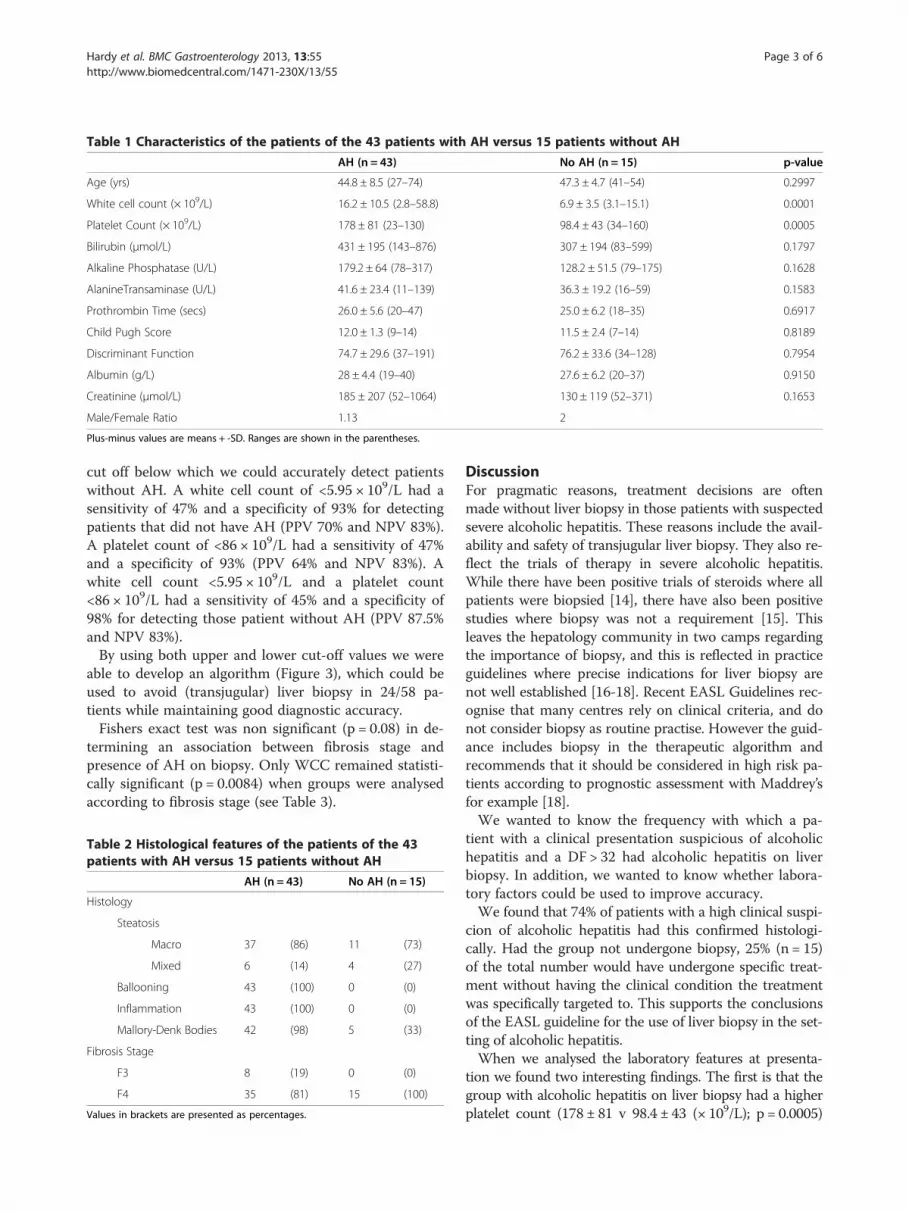
Table 1 Characteristics of the patients of the 43 patients with AH versus 15 patients without AH
AH (n = 43) No AH (n = 15) p-value
Age (yrs) 44.8 ± 8.5 (27–74) 47.3 ± 4.7 (41–54) 0.2997
White cell count (× 109/L) 16.2 ± 10.5 (2.8–58.8) 6.9 ± 3.5 (3.1–15.1) 0.0001
Platelet Count (× 109/L) 178 ± 81 (23–130) 98.4 ± 43 (34–160) 0.0005
Bilirubin (μmol/L) 431 ± 195 (143–876) 307 ± 194 (83–599) 0.1797
Alkaline Phosphatase (U/L) 179.2 ± 64 (78–317) 128.2 ± 51.5 (79–175) 0.1628
AlanineTransaminase (U/L) 41.6 ± 23.4 (11–139) 36.3 ± 19.2 (16–59) 0.1583
Prothrombin Time (secs) 26.0 ± 5.6 (20–47) 25.0 ± 6.2 (18–35) 0.6917
Child Pugh Score 12.0 ± 1.3 (9–14) 11.5 ± 2.4 (7–14) 0.8189
Discriminant Function 74.7 ± 29.6 (37–191) 76.2 ± 33.6 (34–128) 0.7954
Albumin (g/L) 28 ± 4.4 (19–40) 27.6 ± 6.2 (20–37) 0.9150
Creatinine (μmol/L) 185 ± 207 (52–1064) 130 ± 119 (52–371) 0.1653
Male/Female Ratio 1.13 2
Plus-minus values are means + -SD. Ranges are shown in the parentheses.
Hardy et al. BMC Gastroenterology 2013, 13:55 Page 3 of 6http://www.biomedcentral.com/1471-230X/13/55
cut off below which we could accurately detect patientswithout AH. A white cell count of <5.95 × 10⁹/L had asensitivity of 47% and a specificity of 93% for detectingpatients that did not have AH (PPV 70% and NPV 83%).A platelet count of <86 × 109/L had a sensitivity of 47%and a specificity of 93% (PPV 64% and NPV 83%). Awhite cell count <5.95 × 10⁹/L and a platelet count<86 × 109/L had a sensitivity of 45% and a specificity of98% for detecting those patient without AH (PPV 87.5%and NPV 83%).By using both upper and lower cut-off values we were
able to develop an algorithm (Figure 3), which could beused to avoid (transjugular) liver biopsy in 24/58 pa-tients while maintaining good diagnostic accuracy.Fishers exact test was non significant (p = 0.08) in de-
termining an association between fibrosis stage andpresence of AH on biopsy. Only WCC remained statisti-cally significant (p = 0.0084) when groups were analysedaccording to fibrosis stage (see Table 3).
Table 2 Histological features of the patients of the 43patients with AH versus 15 patients without AH
AH (n = 43) No AH (n = 15)
Histology
Steatosis
Macro 37 (86) 11 (73)
Mixed 6 (14) 4 (27)
Ballooning 43 (100) 0 (0)
Inflammation 43 (100) 0 (0)
Mallory-Denk Bodies 42 (98) 5 (33)
Fibrosis Stage
F3 8 (19) 0 (0)
F4 35 (81) 15 (100)
Values in brackets are presented as percentages.
DiscussionFor pragmatic reasons, treatment decisions are oftenmade without liver biopsy in those patients with suspectedsevere alcoholic hepatitis. These reasons include the avail-ability and safety of transjugular liver biopsy. They also re-flect the trials of therapy in severe alcoholic hepatitis.While there have been positive trials of steroids where allpatients were biopsied [14], there have also been positivestudies where biopsy was not a requirement [15]. Thisleaves the hepatology community in two camps regardingthe importance of biopsy, and this is reflected in practiceguidelines where precise indications for liver biopsy arenot well established [16-18]. Recent EASL Guidelines rec-ognise that many centres rely on clinical criteria, and donot consider biopsy as routine practise. However the guid-ance includes biopsy in the therapeutic algorithm andrecommends that it should be considered in high risk pa-tients according to prognostic assessment with Maddrey’sfor example [18].We wanted to know the frequency with which a pa-
tient with a clinical presentation suspicious of alcoholichepatitis and a DF > 32 had alcoholic hepatitis on liverbiopsy. In addition, we wanted to know whether labora-tory factors could be used to improve accuracy.We found that 74% of patients with a high clinical suspi-
cion of alcoholic hepatitis had this confirmed histologi-cally. Had the group not undergone biopsy, 25% (n = 15)of the total number would have undergone specific treat-ment without having the clinical condition the treatmentwas specifically targeted to. This supports the conclusionsof the EASL guideline for the use of liver biopsy in the set-ting of alcoholic hepatitis.When we analysed the laboratory features at presenta-
tion we found two interesting findings. The first is that thegroup with alcoholic hepatitis on liver biopsy had a higherplatelet count (178 ± 81 v 98.4 ± 43 (× 109/L); p = 0.0005)

Table 3 Lab indices of the 8 patients with F3 fibrosis versus 50 patients with F4 fibrosis
F3 (n = 8) F4 (n = 50) p-value
Age (yrs) 45.3 ± 6.3 (34–53) 45.1 ± 8.5 (27–74) 0.9552
White cell count (× 109/L) 24.5 ± 17 (6.9–58.8) 13.6 ± 8.2 (2.8–44.7) 0.0084
Platelet Count (× 109/L) 186 ± 101 (88–354) 165 ± 80 (34–379) 0.5519
Bilirubin (μmol/L) 403 ± 198 (143–663) 419 ± 200 (83–876) 0.8519
Alkaline Phosphatase (U/L) 179.7 ± 70 (105–317) 170 ± 64 (69–297) 0.8186
Alanine Transaminase (U/L) 43.4 ± 17.5 (23–71) 40.6 ± 23.4 (13–139) 0.5396
Prothrombin Time (secs) 29 ± 9.1 (22–47) 25.4 ± 4.8 (18–41) 0.1202
Child Pugh Score 12.5 ± 1 (11–14) 11.9 ± 1.5 (7–14) 0.2477
Discriminant Function 89.9 ± 48.6 (51–191) 72.4 ± 25.4 (34–151) 0.1523
Albumin (g/L) 28 ± 4.8 (24–38) 28 ± 4.7 (19–40) 0.9507
Creatinine (μmol/L) 209 ± 157 (52–546) 173 ± 206 (52–1064) 0.6627
Plus-minus values are means + -SD. Ranges are shown in the parentheses.
Hardy et al. BMC Gastroenterology 2013, 13:55 Page 4 of 6http://www.biomedcentral.com/1471-230X/13/55
than those without alcoholic hepatitis. This is likely to bedue to the fact that patients with no AH have more ad-vanced fibrotic liver disease with portal hypertension lead-ing to decompensation. The second was that the groupwith alcoholic hepatitis on liver biopsy had a higher meanwhite cell count (16.2 ± 10.5 v 6.9 ± 3.5 (× 109/L); p =0.0001). Using a cut off value of >10.75 × 10⁹/L for whitecell count and >147.5 × 10⁹/L for platelet count, accuratelydiagnoses AH with a combined specificity 100%. A lowerboundary of <5.95 × 10⁹/L and <86 × 109/L for white cellcount and platelet count respectively, accurately rules outAH as a diagnosis with a combined sensitivity of 98%.There are two important clinical ramifications of this
Figure 1 White cell count for diagnosis of AH. The area under the ROCdiscriminating AH from no AH diagnosis.
finding. The first is that there should be suspicion regard-ing labelling patients with a normal white cell count as al-coholic hepatitis. The second is that alcoholic hepatitis isan inflammatory condition and a high white cell count isentirely in keeping with this. A raised white cell countshould not therefore be used as a reason not to give treat-ment because of concerns about infection when there areno other features of overt sepsis.Together these laboratory tests may help the clinician
to make a decision about whom to biopsy; particularly ifthis means sending them to another hospital. We wouldadvise caution in making a diagnosis of acute alcoholichepatitis, regardless of the clinical presentation in any
curve is shown for the performance of the White Cell Count for

Figure 2 Platelet count for diagnosis of AH. The area under the ROC curve is shown for the performance of the Platelet Count fordiscriminating AH from no AH diagnosis.
Hardy et al. BMC Gastroenterology 2013, 13:55 Page 5 of 6http://www.biomedcentral.com/1471-230X/13/55
patient with a normal white cell count, particularly ifthis is associated with a very low platelet count.The majority of the patients in our study were cir-
rhotic 50/58 (86%). As expected, all 15 of the patientsthat were found to not have alcoholic hepatitis on liverbiopsy were cirrhotic. These are the patients thatprogressed to decompensation due to very advancedliver disease rather than superimposed hepatitis. 8/43patients with AH were non-cirrhotic; all of these wereF3. When we compared laboratory indices between thecirrhotic patients and the non-cirrhotic patients wefound the only statistically significant finding to be thedifference in average WCC. This is largely due to the
White cell count/platelet count N=58
False Positive: 0Correct: 19
Grey ZoneLiver Biopsy
Correct: 5False Negative: 1
White cell count
Platelet count 10.75
147.5N=19
White cell count 5.95- <10.75Platelet count86 - <147.5N=33
White cell count<5.95Platelet count <86N=6
NPV-83% PPV-100%
Figure 3 Prediction of alcoholic hepatitis by white cell countand platelet count.
fact that the F4 cohort contained all the patients that didnot have histological alcoholic hepatitis.Our results suggest that in future clinical trials where
biopsy is not mandated, 25% of patients maybe falselyincluded. This clearly has implications for the validityand reliability of data testing specific therapies in pa-tients whom may not suffer from the disease they aretargeted to; this is reflected in the EASL Clinical Prac-tical Guideline in Alcohol whereby performing liver bi-opsy prior to trial commencement is recommended.What is needed now are data from large prospective
studies where biopsy is included in the diagnostic algo-rithm but where patients are included in the therapeuticstudy regardless of the result. This will help to elucidatethe response of the patient presenting with the alcoholichepatitis syndrome but a non-confirmatory biopsy tostandard of care treatment. Should these studies showthat treatment is detrimental to this group, biopsyshould in future be mandated prior to commencingtreatment. In situations where transjugular biopsy is im-practical or simply unavailable the surrogate markers ofwhite cell count and platelet count may prove to be ofgreat use in determining which patients receive treatment.
ConclusionsIn conclusion, we have shown that the diagnosis of alco-holic hepatitis is difficult on clinical grounds; we havealso found two clinical parameters that can assist theclinician with the diagnosis. This may in time prove tobe very useful when determining treatment strategies.

Hardy et al. BMC Gastroenterology 2013, 13:55 Page 6 of 6http://www.biomedcentral.com/1471-230X/13/55
Competing interestsThe authors declare that they have no competing interests.
Authors’ contributionsTH: acquisition of data, analysis and interpretation of data, drafting themanuscript. CW, SK, MH, CP: acquisition of data. AB: acquisition of data,analysis and interpretation of data (liver biopsies). SS: Conception anddesign, analysis and interpretation of data, drafting and revising forimportant intellectual content. All authors read and approved the finalmanuscript.
Author details1The Liver Unit, Freeman Hospital, Newcastle upon Tyne NE7 7DN, UK.2Institute of Cellular Medicine, Newcastle University, Newcastle upon TyneNE2 4HH, UK. 3The Centre for Liver Disease, Mater Misericordiae UniversityHospital, 55 Eccles Street, Dublin 7, Irish Republic.
Received: 15 August 2012 Accepted: 15 March 2013Published: 26 March 2013
References1. MacSween RN, Burt AD: Histologic spectrum of alcoholic liver disease.
Semin Liver Dis 1986, 6:221–232.2. Lucey MR, Mathurin P, Morgan TR: Alcoholic hepatitis. New Engl J Med
2009, 360:2758–2769.3. MacSween RN: Alcoholic liver disease: morphological manifestations.
Review by an international group. Lancet 1981, 1:707–711.4. Ishak KG, Zimmerman HJ, Ray MB: Alcoholic liver disease: pathologic,
pathogenetic and clinical aspects. Alcohol Clin Exp Res 1991, 15:45–66.5. Chedid A, Mendenhall CL, Gartside P, et al: Prognostic factors in alcoholic liver
disease. VA cooperative study group. Am J Gastroenterology 1991, 86:210–216.6. Mookerjee RP, Lackner C, Stauber R, Stadlbauer V, Deheragoda A, Jalan R:
The role of liver biopsy in the diagnosis and prognosis of patients withacute deterioration of alcoholic cirrhosis. J Hepatol 2011, 55:1103–1111.
7. Gamble P, Colapinto RF, Stronell RD: Transjugular liver biopsy: a review of461 biopsies. Radiology 1985, 157:589–593.
8. Velt PM, Choy OG, Shimkin PM, et al: Transjugular liver biopsy in high riskpatients with hepatic disease. Radiology 1984, 153:91–93.
9. Vlavianos P, Bird G, Portmann B, et al: Transjugular liver biopsy: use in aselected high risk population. Eur J Gastroenterol Hepatol 1991, 3:469–472.
10. Maddrey WC, Boitnott JK, Bedine MS, et al: Corticosteroid therapy ofalcoholic hepatitis. Gastroenterology 1978, 75:193–199.
11. Forrest EH, Evans CD, Stewart S, et al: Analysis of factors predictive ofmortality in alcoholic hepatitis and derivation and validation of theGlasgow alcoholic hepatitis score. Gut 2005, 54:1174–1179.
12. Stewart S, Prince M, Bassendine M, et al: A randomized trial of antioxidanttherapy alone or with corticosteroids in acute alcoholic hepatitis. J Hepatol2007, 47(2):277–283.
13. Pugh RN, Murray-Lyon IM, Dawson JL, et al: Transection of the oesophagusfor bleeding oesophageal varices. Br J Surg 1973, 60(8):646–649.
14. Ramond M-J, Poynard T, Rueff B, et al: A Randomised trial of prednisolone inpatients with severe alcoholic hepatitis. New Engl J Med 1992, 326:507–512.
15. Carithers RL, Herlong F, Diehl AM, et al: Methylprednisolone therapy inpatients with severe alcoholic hepatitis. Ann Intern Med 1989, 110:685–690.
16. O’Shea RS, Dasarathy S, McCullough AJ, Practice Guideline Committee ofthe American Association for the Study of Liver Diseases; PracticeParameters Committee of the American College of Gastroenterology:Alcoholic liver disease. Hepatology 2010, 51:307–328.
17. Alcohol use disorders: diagnosis and clinical management of alcohol-related physical complications. Clinical guideline 100. (Accessed Jan 2011)http://www.nice.org.uk/nicemedia/live/12995/48989/48989.pdf.
18. Clinical Practical Guidelines EASL: Management of alcoholic liver disease.J Hepatol 2012, 57:399–420.
doi:10.1186/1471-230X-13-55Cite this article as: Hardy et al.: White cell count and platelet countassociate with histological alcoholic hepatitis in jaundiced harmfuldrinkers. BMC Gastroenterology 2013 13:55.
Submit your next manuscript to BioMed Centraland take full advantage of:
• Convenient online submission
• Thorough peer review
• No space constraints or color figure charges
• Immediate publication on acceptance
• Inclusion in PubMed, CAS, Scopus and Google Scholar
• Research which is freely available for redistribution
Submit your manuscript at www.biomedcentral.com/submit
Related Documents