West Nile virus infection of the placenta Justin G. Julander a , Quinton A. Winger b , Lee F. Rickords b , Pei-Yong Shi c , Mark Tilgner c , Iwona Binduga-Gajewska c , Robert W. Sidwell a , John D. Morrey a, * a The Institute for Antiviral Research, Utah State University, Logan, UT 84322, USA b Department of Animal, Dairy, and Veterinary Sciences, Utah State University, Logan, UT 84322, USA c Wadsworth Center, New York State Department of Health, Albany, NY 12208, USA Received 23 September 2005; returned to author for revision 17 October 2005; accepted 19 November 2005 Available online 9 January 2006 Abstract Intrauterine infection of fetuses with West Nile virus (WNV) has been implicated in cases of women infected during pregnancy. Infection of timed-pregnant mice on 5.5, 7.5, and 9.5 days post-coitus (dpc) resulted in fetal infection. Infection of dams on 11.5 and 14.5 dpc resulted in little and no fetal infection, respectively. Pre-implantation embryos in culture were also infected with WNV after the blastocyst stage and the formation of trophectoderm. Green fluorescent protein (GFP) expression was observed in a trophoblast stem (TS) cell line after infection with a GFP- expressing WNV construct. However, no fluorescence was observed in differentiated trophoblast giant cell (TGC) cultures. GFP fluorescence was present in TGC cultures if infected TS cells were induced to differentiate. These results suggest that embryos are susceptible to WNV infection after the formation of the trophectoderm around 3.5 dpc through the formation of the functional placenta around 10.5 dpc. D 2005 Elsevier Inc. All rights reserved. Keywords: West Nile virus; Placenta; Trophoblast; Intrauterine; Vertical transmission; Pregnancy Introduction West Nile virus (WNV) causes disease in man, including encephalitis, paralysis, and death (Anderson et al., 2004). WNV may also infect horses, dogs, cats, and alpacas, as well as other species such as alligators (Abutarbush et al., 2004; Austgen et al., 2004; Kutzler et al., 2004; Miller et al., 2003; Yaeger et al., 2004). The primary vector for human transmis- sion is the mosquito, however, other modes of infection have been observed (Sardelis et al., 2001; Turell et al., 2001). Virus has been transferred in human patients by blood and organ transplantation, as well as by accidental laboratory infection (Laboratory-acquired West Nile Virus, 2002; Macedo de Oliveira et al., 2004; Wadei et al., 2004). Some animal species have become infected after ingestion of infected materials or contact, such as feather picking or grooming, with infected individuals (Banet-Noach et al., 2003; Miller et al., 2003; Odelola and Oduye, 1977). Intrauterine infection of fetuses with WNV has been implicated (Intrauterine West Nile virus, 2002), but other reports of maternal infection with WNV during pregnancy have shown no evidence for morbidity of the fetus (Bruno et al., 2004). Many other WNV cases of maternal infection during pregnancy are under investigation (Interim Guide- lines, 2004). A woman infected with WNV during pregnan- cy gave birth to a WNV-seropositive baby with chorioretinal scarring and some brain abnormalities that may have been due to maternal infection with WNV during the second trimester of gestation (Alpert et al., 2003). Fetal viral infections are generally transmitted from maternal viremia across the placenta to fetal circulation, so an understanding of viral interactions with the placenta is important (Kaplan, 1993). Infection of mouse fetuses was recently demonstrated in our laboratory (Julander et al., 2005). In that study, dams infected with WNV 7.5 days post-coitus (dpc) had a high rate of passage of maternal virus to fetuses as compared to low frequency of fetal infection when dams were infected 11.5 dpc. 0042-6822/$ - see front matter D 2005 Elsevier Inc. All rights reserved. doi:10.1016/j.virol.2005.11.040 * Corresponding author. Mailing address: Biotechnology Center 305, Utah State University, 4700 Old Main Hill, Logan, UT 84322-4700, USA. Fax: +1 435 797-2766. E-mail address: [email protected] (J.D. Morrey). Virology 347 (2006) 175 – 182 www.elsevier.com/locate/yviro

Welcome message from author
This document is posted to help you gain knowledge. Please leave a comment to let me know what you think about it! Share it to your friends and learn new things together.
Transcript

lsevier.com/locate/yviro
Virology 347 (200
West Nile virus infection of the placenta
Justin G. Julander a, Quinton A. Winger b, Lee F. Rickords b, Pei-Yong Shi c, Mark Tilgner c,
Iwona Binduga-Gajewska c, Robert W. Sidwell a, John D. Morrey a,*
a The Institute for Antiviral Research, Utah State University, Logan, UT 84322, USAb Department of Animal, Dairy, and Veterinary Sciences, Utah State University, Logan, UT 84322, USA
c Wadsworth Center, New York State Department of Health, Albany, NY 12208, USA
Received 23 September 2005; returned to author for revision 17 October 2005; accepted 19 November 2005
Available online 9 January 2006
Abstract
Intrauterine infection of fetuses with West Nile virus (WNV) has been implicated in cases of women infected during pregnancy. Infection of
timed-pregnant mice on 5.5, 7.5, and 9.5 days post-coitus (dpc) resulted in fetal infection. Infection of dams on 11.5 and 14.5 dpc resulted in little
and no fetal infection, respectively. Pre-implantation embryos in culture were also infected with WNVafter the blastocyst stage and the formation
of trophectoderm. Green fluorescent protein (GFP) expression was observed in a trophoblast stem (TS) cell line after infection with a GFP-
expressing WNV construct. However, no fluorescence was observed in differentiated trophoblast giant cell (TGC) cultures. GFP fluorescence was
present in TGC cultures if infected TS cells were induced to differentiate. These results suggest that embryos are susceptible to WNV infection
after the formation of the trophectoderm around 3.5 dpc through the formation of the functional placenta around 10.5 dpc.
D 2005 Elsevier Inc. All rights reserved.
Keywords: West Nile virus; Placenta; Trophoblast; Intrauterine; Vertical transmission; Pregnancy
Introduction
West Nile virus (WNV) causes disease in man, including
encephalitis, paralysis, and death (Anderson et al., 2004).
WNV may also infect horses, dogs, cats, and alpacas, as well as
other species such as alligators (Abutarbush et al., 2004;
Austgen et al., 2004; Kutzler et al., 2004; Miller et al., 2003;
Yaeger et al., 2004). The primary vector for human transmis-
sion is the mosquito, however, other modes of infection have
been observed (Sardelis et al., 2001; Turell et al., 2001). Virus
has been transferred in human patients by blood and organ
transplantation, as well as by accidental laboratory infection
(Laboratory-acquired West Nile Virus, 2002; Macedo de
Oliveira et al., 2004; Wadei et al., 2004). Some animal species
have become infected after ingestion of infected materials or
contact, such as feather picking or grooming, with infected
0042-6822/$ - see front matter D 2005 Elsevier Inc. All rights reserved.
doi:10.1016/j.virol.2005.11.040
* Corresponding author. Mailing address: Biotechnology Center 305, Utah
State University, 4700 Old Main Hill, Logan, UT 84322-4700, USA. Fax: +1
435 797-2766.
E-mail address: [email protected] (J.D. Morrey).
individuals (Banet-Noach et al., 2003; Miller et al., 2003;
Odelola and Oduye, 1977).
Intrauterine infection of fetuses with WNV has been
implicated (Intrauterine West Nile virus, 2002), but other
reports of maternal infection with WNV during pregnancy
have shown no evidence for morbidity of the fetus (Bruno
et al., 2004). Many other WNV cases of maternal infection
during pregnancy are under investigation (Interim Guide-
lines, 2004). A woman infected with WNV during pregnan-
cy gave birth to a WNV-seropositive baby with chorioretinal
scarring and some brain abnormalities that may have been
due to maternal infection with WNV during the second
trimester of gestation (Alpert et al., 2003). Fetal viral
infections are generally transmitted from maternal viremia
across the placenta to fetal circulation, so an understanding
of viral interactions with the placenta is important (Kaplan,
1993).
Infection of mouse fetuses was recently demonstrated in our
laboratory (Julander et al., 2005). In that study, dams infected
with WNV 7.5 days post-coitus (dpc) had a high rate of
passage of maternal virus to fetuses as compared to low
frequency of fetal infection when dams were infected 11.5 dpc.
6) 175 – 182
www.e
J.G. Julander et al. / Virology 347 (2006) 175–182176
The placenta had elevated viral titer compared to other
maternal organs regardless of the gestational time point of
infection. Dams had high mortality and generally died prior to,
or during, delivery unless treated with WNV-specific immu-
noglobulin. Immunoglobulin treatment allowed dams to
conceive and raise pups.
Placental development is a dynamic process involving the
interaction between invasive fetal-derived trophoblast cells
and maternal decidual cells of the uterus (Fazleabas et al.,
2004). At the blastocyst stage (3.5 dpc), just prior to
implantation, surface cells of the embryo will differentiate
into trophectodermal cells that will eventually give rise to
the placenta and other extraembryonic structures (Cross et
al., 1994). Trophoblast cells invade the maternal decidua
during development and establish an interface between
maternal and fetal blood for the transfer of nutrients to the
developing fetus. The placental barrier between maternal and
fetal blood is established in mice around 10.5 dpc and
consists of one layer of mononuclear trophoblast cells
(cytotrophoblast) and two layers of differentiated syncytial
trophoblast (syncytiotrophoblast) (Georgiades et al., 2002).
The placental barrier functions to allow selective transfer of
nutrients and to inhibit the transfer of harmful materials, but
this barrier may be breached by different chemicals or
microorganisms (Koi et al., 2001a, 2001b).
A trophoblast stem (TS) cell line has been established by
culturing blastocysts or early post-implantation trophoblasts in
media containing fetal growth factor-4 (FGF-4), haprin, and
fibroblast conditioned media (Tanaka et al., 1998). Upon
removal of these components, the TS cells differentiate into
other trophoblast cell types including trophoblast giant cells.
The TS cell line serves as a model for the replicative and
differentiated trophoblast cells of the placenta.
An understanding of the mechanism of WNV intrauterine
infection may be important for preventing clinical cases as well
as for the development of therapies to reduce fetal disease and
associated symptoms. The objectives of this study were to
delineate the timing of viral passage from infected dam to fetus
and to identify the placental cell types susceptible to viral
infection in vitro.
Table 1
West Nile virus (WNV) titers recovered from tissues and fetuses from mice infecte
Infectedd Necropsyf Meana virus titer T SDb in maternal tissue samples (
(dpce) (dpc) Brain Kidney Spleen
5.5 11.5 7.0 T 1.8 (3/6) 5.5 T 0.9 (2/6) 5.6 T 0.3 (6
7.5 13.5 7.7 T 2.0 (2/4) <3.6 T 0 (0/4) 6.2 T 1.4 (3
9.5 15.5 6.7 T 2.8 (2/5) 5.5 T 0.4 (2/5) 6.6 T 0.3 (5
11.5 16.5 6.8 T 2.8 (2/7) 5.6 T 0.5 (3/5) N/T
14.5 19.5 <3.6 T 0 (0/6) 5.7 T 0.1 (3/6) N/T
a Mean virus titer is the average TCID50/g tissue titer from positive samples thatb Standard deviation.c Tissue samples with detectable WNV titers per total samples tested.d Day of gestation on which dam was challenged with WNV.e Days post-coitus.f Day on which tissue samples were harvested from infected dams.g Not tested.
Results
Timing of fetal infection
To determine the gestational timing of fetal infection
with WNV, timed-pregnant dams were challenged with
WNV on 5.5, 7.5, 9.5, 11.5, and 14.5 dpc. Whole fetus,
placenta, and maternal brain, kidney, and spleen were titered
for WNV by infectious cell culture assay (Table 1). Virus
was present in fetuses 6 days post-maternal challenge when
dams were challenged on 5.5, 7.5, and 9.5 dpc. Fetuses
from dams challenged 9.5 dpc had higher WNV titers than
fetuses from dams challenged 7.5 dpc (Table 1). Little or
no virus was present in fetuses from dams challenged 11.5
or 14.5 dpc. High WNV titers were present in the placenta
regardless of gestational state at the time of infection.
Maternal tissues had some detectable virus, but titers in
maternal organs were much lower than titers in fetuses and
placentas.
Infection of pre-implantation embryos
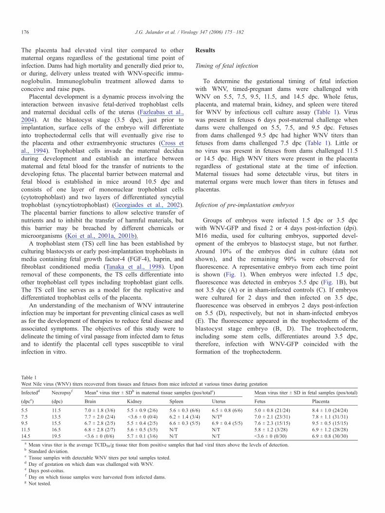
Groups of embryos were infected 1.5 dpc or 3.5 dpc
with WNV-GFP and fixed 2 or 4 days post-infection (dpi).
M16 media, used for culturing embryos, supported devel-
opment of the embryos to blastocyst stage, but not further.
Around 10% of the embryos died in culture (data not
shown), and the remaining 90% were observed for
fluorescence. A representative embryo from each time point
is shown (Fig. 1). When embryos were infected 1.5 dpc,
fluorescence was detected in embryos 5.5 dpc (Fig. 1B), but
not 3.5 dpc (A) or in sham-infected controls (C). If embryos
were cultured for 2 days and then infected on 3.5 dpc,
fluorescence was observed in embryos 2 days post-infection
on 5.5 (D), respectively, but not in sham-infected embryos
(E). The fluorescence appeared in the trophectoderm of the
blastocyst stage embryo (B, D). The trophectoderm,
including some stem cells, differentiates around 3.5 dpc,
therefore, infection with WNV-GFP coincided with the
formation of the trophectoderm.
d at various times during gestation
pos/totalc) Mean virus titer T SD in fetal samples (pos/total)
Uterus Fetus Placenta
/6) 6.5 T 0.8 (6/6) 5.0 T 0.8 (21/24) 8.4 T 1.0 (24/24)
/4) N/Tg 7.0 T 2.1 (23/31) 7.8 T 1.1 (31/31)
/5) 6.9 T 0.4 (5/5) 7.6 T 2.3 (15/15) 9.5 T 0.5 (15/15)
N/T 5.8 T 1.2 (3/28) 6.9 T 1.2 (28/28)
N/T <3.6 T 0 (0/30) 6.9 T 0.8 (30/30)
had viral titers above the levels of detection.
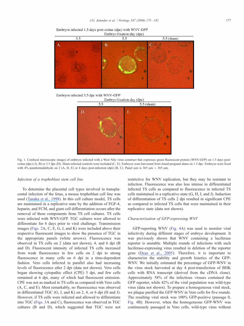
Fig. 1. Confocal microscopic images of embryos infected with a West Nile virus construct that expresses green fluorescent protein (WNV-GFP) on 1.5 days post-
coitus (dpc) (A, B) or 3.5 dpc (D). Sham-infected controls were included (C, E). Embryos were harvested from timed-pregnant dams on 1.5 dpc. Embryos were fixed
with 4% paraformaldehyde on 2 (A, D, E) or 4 days post-infection (dpi) (B, C). Panel size is 365 Am � 365 Am.
J.G. Julander et al. / Virology 347 (2006) 175–182 177
Infection of a trophoblast stem cell line
To determine the placental cell types involved in transpla-
cental infection of the fetus, a mouse trophoblast cell line was
used (Tanaka et al., 1998). In this cell culture model, TS cells
are maintained in a replicative state by the addition of FGF-4,
heparin, and FCM, and giant cell differentiation occurs after the
removal of these components from TS cell cultures. TS cells
were infected with WNV-GFP. TGC cultures were allowed to
differentiate for 6 days prior to viral challenge. Transmission
images (Figs. 2A, C, E, G, I, and K) were included above their
respective fluorescent images to show the presence of TGC in
the appropriate panels (white arrows). Fluorescence was
observed in TS cells on 2 (data not shown), 4, and 6 dpi (B
and D). Fluorescent intensity of infected TS cells increased
from weak fluorescence in few cells on 2 dpi to strong
fluorescence in many cells on 6 dpi in a time-dependent
fashion. Vero cells infected in parallel also had increasing
levels of fluorescence after 2 dpi (data not shown). Vero cells
began showing cytopathic effect (CPE) 5 dpi, and few cells
remained at 6 dpi, many of which had fluorescent emission.
CPE was not as marked in TS cells as compared with Vero cells
(A, C, and E). Most remarkably, no fluorescence was observed
in differentiated TGC (G, I, and K) on 2, 4, or 6 dpi (H and J).
However, if TS cells were infected and allowed to differentiate
into TGC (Figs. 3A and C), fluorescence was observed in TGC
cultures (B and D), which suggested that TGC were not
restrictive for WNV replication, but they may be resistant to
infection. Fluorescence was also less intense in differentiated
infected TS cells as compared to fluorescence in infected TS
cells maintained in a replicative state (G, H, I, and J). Induction
of differentiation of TS cells 2 dpi resulted in significant CPE
as compared to infected TS cells that were maintained in their
replicative state (data not shown).
Characterization of GFP-expressing WNV
GFP-reporting WNV (Fig. 4A) was used to monitor viral
infectivity during different stages of embryo development. It
was previously shown that WNV containing a luciferase
reporter is unstable. Multiple rounds of infections with such
luciferase-expressing virus resulted in deletion of the reporter
gene (Deas et al., 2005). Therefore, it is important to
characterize the stability and growth kinetics of the GFP-
WNV. We initially estimated the percentage of GFP-WNV in
the virus stock harvested at day 4 post-transfection of BHK
cells with RNA transcript (derived from the cDNA clone).
Approximately 58% of the infectious viruses contained the
GFP reporter, while 42% of the viral population was wild-type
virus (data not shown). To prepare a homogeneous viral stock,
we plaque purified the GFP-WNV in Vero cells for five rounds.
The resulting viral stock was 100% GFP-positive (passage 0,
Fig. 4B). However, when the homogeneous GFP-WNV was
continuously passaged in Vero cells, wild-type virus without

Fig. 2. Infection of trophoblast stem (TS) cells (A–F) and trophoblast giant cells (TGC) (G–L) with a West Nile virus construct that expresses green fluorescent
protein (WNV-GFP). Fluorescence was observed in TS cells starting 2 days post-infection (dpi) and increasing in intensity 4 (B) and 6 (D) dpi. No fluorescence was
observed in sham-infected TS cells (F). No fluorescence was detected in WNV-GFP-infected TGC (H and J) or in sham-infected TGC (L). Giant cells are indicated
by white arrows (G, I, and K).
J.G. Julander et al. / Virology 347 (2006) 175–182178
GFP gradually dominated the population. After the first,
second, and third rounds of passage, 72%, 35%, and 4% of
the infectious viruses were GFP-positive, respectively (Fig.
4B). Next, we compared the growth kinetics of the GFP-WNV
(passage 0) with that of wild-type virus in Vero cells. The GFP-
WNV replicated slower with a lower peak titer than those of the
wild-type virus (Fig. 4C). Overall, the results suggest that the
plaque-purified GFP-WNV is not stable in maintaining the
reporter gene. However, unpassaged plaque-purified GFP-
WNV is homogeneous and could be used for detection of
initial WNV infection.
Discussion
The findings of this study suggested that differentiated
syncytiotrophoblasts of the maturing placenta are a barrier to
infection of mouse fetuses by WNV. The percentage of fetuses
becoming infected with WNV was greater if dams were
infected before, but not after 10.5 dpc. At this gestational time,
the placental barrier between maternal and fetal blood is
established in mice and consists of one layer of cellular
trophoblast and two layers of differentiated syncytiotrophoblast
possessing tight cellular structure (Georgiades et al., 2002). In
this study, differentiated TGC in culture were resistant to
infection with WNV, where no virus was detected up to 6 dpi
when TGC were infected with WNV. Pre-implantation
embryos were susceptible to viral infection after the formation
of the trophectoderm at the blastocyst stage, which suggests
that viral infection of embryos may be dependent on the
presence of susceptible trophoblast cells. This indicates a
possible mechanism for infection of the fetus with WNV
through replicative trophoblast cells that are resistant after
differentiation and formation of the placental barrier. Many
more studies are available that demonstrate the dependence of
viral infection on cellular differentiation. Human choriocarci-
noma cells are susceptible to transduction with replication-
incompetent adenovirus and herpes simplex virus, unless the
cells are chemically differentiated (Parry et al., 1997). This loss
of recombinant adenovirus and herpesvirus transduction is
likely due to the downregulation of the Coxsackie adenovirus
receptor during differentiation (Koi et al., 2001a, 2001b) and
reduction in viral uptake, respectively. Conversely, adeno-
associated virus, a parvovirus, has a higher transduction rate in
differentiated cells as compared with undifferentiated cells (Koi
et al., 2001a, 2001b).
Trophoblast cell differentiation in the placenta is an ongoing
process. Undifferentiated placental cells are present in the
placenta throughout gestation, so, during the formation of the

Fig. 3. Fluorescent and light images of trophoblast stem (TS) cells infected with a West Nile construct expressing green fluorescent protein (WNV-GFP). One group
of cells (A–F) was differentiated to trophoblast giant cells (TGC), and another group (G–L) was maintained in a replicative state. Faint fluorescence was observed in
TGC cultures (B) starting 4 days post infection (dpi), which increased in intensity on 6 dpi (D). Fluorescent cells were also observed in replicative TS cells on 4 (H)
and 6 dpi (J). No GFP was observed in sham control cells (F and L). TGC were present in cultures of differentiated TS cells (D and F white arrows).
J.G. Julander et al. / Virology 347 (2006) 175–182 179
placental barrier, there is a possibility for TS cells or other
progenitor trophoblast cell types to become infected and then
differentiate into syncytiotrophoblast in vivo. We observed that
if cultured TS cells were infected and then differentiation was
induced 2 dpi, the resulting differentiated TGC had high virus-
induced cell damage as compared to replicative TS cells.
Differentiation of infected trophoblast stem cells into syncytio-
trophoblast cells in vivo and subsequent cytopathogenesis of
these cells could account for placental dysfunction and
spontaneous abortion observed in potential viral infections of
the placenta. Similarly, infection of extravillous cytotropho-
blast cells with adenovirus resulted in apoptosis of the cells in
vitro, which is a possible explanation for placental dysfunction
observed in in vivo adenovirus infection (Koi et al., 2001a,
2001b). Another example of viral-induced pathogenesis of
differentiated placental cells is HCMV infection of villious
syncytiotrophoblast, which results in an upregulation of
intercellular adhesion molecule ICAM-1 that causes blood
monocytes to bind to these cells and induce apoptosis (Chan et
al., 2004). Increased apoptosis within villous trophoblast cells,
resulting in placental failure and fetal death, was correlated
with parvovirus B-19 infection and presents further evidence of
differentiated trophoblast destruction as a result virus infection
as a cause for placental failure (Jordan and Butchko, 2002). We
also observed fetal death if dams were infected before 10.5 dpc
and not treated with WNV-reactive antibody (Julander et al.,
2005).
Although the placenta did reduce the transfer of WNV to
fetuses after 10.5 dpc, the placenta had high viral titers relative
to all other tissues assayed, regardless of the gestational state at
the time of infection, indicating the presence of susceptible
cells within the mature placenta. This was significant because
placental infection, even without fetal infection, can result in
fetal loss or other complications of normal pregnancy and may
contribute to certain congenital abnormalities (Kaplan, 1993).
It will be important in future studies to double-stain for WNV
antigens and cellular markers in placental tissue to identify
which cells are infected in the placenta at different stages of
gestation.
We hypothesize that the reduction of susceptibility of TGC
may be due to a reduction of specific cellular receptors that the
virus uses for entry into the trophoblast cell. The expression of
cell surface receptors for herpes simplex virus-1 is reduced in
third term villous syncytiotrophoblast, which prevents infection
of these cells during late gestation and constitutes a barrier to
herpes simplex virus-1 infection of the fetus (Koi et al., 2002).
The coxsackie and adenovirus receptor is present on extra-
villous cytotrophoblast cells, which are susceptible to infection,
but once the cytotrophoblast cells differentiate into syncytio-
trophoblast cells, the receptor is not expressed and the

Fig. 4. A full-length WNV containing a GFP reporter (GFP-WNV). (A) GFP
gene driven by an EMCV IRES was engineered at nucleotide position 10,436
within the 3V-UTR of WNV genome. (B) A GFP-WNV stock (designated as
passage 0, prepared by five rounds of plaque purification) was continuously
passaged in Vero cells (MOI 0.1) for three rounds. At 48 h post-infection for
each round, an immuno-plaque was performed to quantify GFP-positive
viruses, using mouse monoclonal antibody against GFP and anti-mouse
antibody conjugated with horseradish peroxidase as primary and secondary
antibodies, respectively. Standard plaques assays were used to quantify total
infectious WNV. The percentage of GFP-WNV and wild-type WNV in viral
population was presented for each passage. (C) Growth kinetics was compared
between GFP-WNV and wild-type WNV. Vero cells were infected with
indicated viruses at MOI of 2. Viral production was quantified at various time
points post-infection by standard plaque assays.
J.G. Julander et al. / Virology 347 (2006) 175–182180
syncytiotrophoblast cells are not susceptible to infection (Koi
et al., 2001a, 2001b). Cytomegalovirus can infect both
cytotrophoblast-like or syncytiotrophoblast-like cells in vitro,
indicating that differentiated cells of the functional placenta
cannot always serve as a protective barrier to viral infection
(Chan et al., 2004). It was anticipated that infection of
trophoblast cells may be due to the presence of integrin
avh3 on the cell surface because of the several lines of
evidence that WNV uses integrin avh3 as a receptor for
attachment and entry on Vero or other cell types (Chu and Ng,
2004). To address this hypothesis, we stained for integrin avh3on the surface of replicative and differentiated trophoblast cell
types. Similar levels of integrin avh3 were present on both
replicative and differentiated trophoblast cells, and blocking
with antibodies to this integrin did not reduce viral attachment
and entry as seen in Vero cells (data not shown).
It is possible that the TGC will not support WNV replication
because they are not actively dividing, but this is likely not the
case. When TS cells were infected prior to differentiation into
TGC, this resulted in differentiated placental cell types that
supported the replication of the virus, and GFP expression was
observed in these cells. Furthermore, cells that do not actively
divide, such as neuronal cells, are susceptible to WNV
infection (Ceccaldi et al., 2004; Hunsperger and Roehrig,
2005).
In summary, the involvement of trophoblast cell differenti-
ation and placental development as a barrier to WNV infection
extends our basic understanding of WNV biology and may
provide information to help manage WNV infection of
pregnant human patients.
Materials and methods
Animals
FVB/N mice were used (Charles River Laboratories,
Wilmington, MA) because they are commonly employed to
obtain embryos for micromanipulation procedures. Moreover,
the FVB/N mice were susceptible to WNV disease and
mortality. WNV-infected pregnant females had a 100%
mortality rate and generally died prior to parturition (Julander
et al., 2005). Animals were housed in the Biosafety Level 3
(BL-3) area of the AAALAC-accredited Laboratory Animal
Research Center (LARC) at Utah State University (USU). Nine
to 12-week-old animals were also bred in-house, and potential
pregnancy was identified according to the presence of vaginal
plugs, which indicated 0.5 dpc. Animal use and care were in
compliance with the USU Institutional Animal Care and Use
Committee.
Virus
A New York WNV crow brain stock (NY, CDC 996625,
V1 D3 11/10/1999) was grown on MA-104 cells and had a
titer of 106.75 50% cell culture infectious doses/0.1 ml
(TCID50). Virus was diluted in Minimal Essential Media
(MEM) with no fetal bovine serum (FBS) for injection into
timed-pregnant mice. Virus was administered subcutaneously
(sc) at 105.3 TCID50/mouse.
A WNV construct that expresses green fluorescent protein
(WNV-GFP) was used in in vitro infection studies (Fig. 4). An
infectious cDNA clone of an epidemic WNV (Shi et al., 2002a,
2002b) was used to construct the WNV-GFP. The GFP reporter
driven by an encephalomyocarditis virus internal ribosomal
entry site (EMCV IRES) was inserted into the WNV 3V-UTR at
nucleotide position 10,436 (GenBank accession number
AF404756). The WNV-GFP cDNA plasmid was prepared by
swapping the SpeI–SacII fragment (nt 8022 to 10,822)
between the wild-type infectious clone (Shi et al., 2002a,
2002b) and a replicon clone containing the IRES-GFP insertion
(Shi et al., 2002a, 2002b). The WNV-GFP RNA was in vitro
transcribed and transfected into BHK cells to generate the
GFP-reporting virus stock (Shi et al., 2002a, 2002b). A similar
WNV containing a luciferase reporter was recently reported
(Deas et al., 2005). Since such reporting WNV was shown to
be unstable (Deas et al., 2005), the GFP-expressing WNV used
in this study was plaque-purified to maximize its homogeneity
(see details in Results). Upon infection of susceptible cells with

J.G. Julander et al. / Virology 347 (2006) 175–182 181
the WNV-GFP, GFP protein was expressed only during viral
replication allowing the distinction between virus attached to
cells and replicating virus.
Infectious cell culture assay
The virus titers in tissues or plasma were assayed using an
infectious cell culture assay (Morrey et al., 2002) where a
specific volume of tissue homogenate was added to the first
tube of a series of dilution tubes. Serial dilutions were made
and added to Vero cells (ATCC #CCL-81). Six to seven days
later, cytopathic effect (CPE) was used to identify the end-point
of infection (Sidwell and Huffman, 1971). Four replicates were
used to calculate the infectious doses per gram of tissue (Reed
and Muench, 1938). Tissue samples were washed thoroughly
to remove contaminating virus as previously described
(Julander et al., 2005).
Infection of pre-implantation embryos
Superovulation was induced in weanling female FVB/N
mice by intraperitoneal injection with 5 international units
(IU) pregnant mare’s serum gonadotropin (PMSG) (Sioux
Biochemical, Inc.) followed 42–48 h by an injection with 6
IU human chorionic gonadotropin (hCG) (Calbiochem). After
injection with hCG, mice were set with males and checked
for vaginal plugs the following morning. Embryos were
harvested between 2- and 4-cell stage on 1.5 dpc. The oviduct
was flushed with M2 medium (Sigma-Aldrich, Inc.) contain-
ing 3 mg/ml bovine serum albumin. The zona pelucida was
removed from half of the embryos using acidic Tyrode’s
solution. Three replicate dishes containing 5 zona-intact and 5
zona-free embryos were placed together in a well of a 96-well
plate and cultured in M16 media (Sigma-Aldrich, Inc.).
Embryos were infected with 106 TCID50/well WNV-GFP on
1.5 and 3.5 dpc. Infected embryos were fixed 20–30 min
with 4% paraformaldehyde (Ted Pella, Inc.) 2 or 4 days after
infection. After fixation, cells were observed using confocal
microscopy.
Cultivation and infection of trophoblast stem cells and giant
cells
Trophoblast stem (TS) cells were maintained as previously
reported (Tanaka et al., 1998). Fibroblast conditioned media
(FCM) containing 25 ng/ml fibroblast growth factor-4 (FGF-4)
(Sigma-Aldrich, Inc.) and 1 Ag/ml heparin (Sigma-Aldrich,
Inc.) was used to maintain the TS cells in their replicative state.
Once FGF-4, heparin, and FCM were removed from the media,
cells will terminally differentiate into trophoblast giant cells
(TGC) and other trophoblast cell types. Differentiated TGC
were easily distinguished from other trophoblast cell types by
the presence of polyploid nuclei and their large size. Cells were
plated in 6- or 12-well plates with and without glass inserts.
Two milliliters of media were added to each well, and cells
were incubated at 37 -C for up to 6 days. Media was changed
every 2 days.
WNV-GFP was added to each well containing 500 Al ofmedia and incubated at 37 -C on a rocker for 30 min. To
observe fluorescence, cells on glass slides were washed twice
with PBS and then fixed with 4% paraformaldehyde for 20–30
min. Cells were washed 3 times with PBS and then mounted to
a coverslip with Fluoromount G mounting media (Electron
Microscopy Sciences, Hatfield, PA) or observed directly in the
bottom of the wells of the culture plate.
Acknowledgments
Wewould like to thank Grant Roper, J.R. Haynie, Dan Olsen,
Josh Durrant, Kyle Greer, Landon Preece, and Heather Gavin for
their assistance in this project.Wewould also thank Dr.Mah-Lee
Mary Ng for help with receptor blocking studies. Dr. Douglas A.
Gregg helped with immunohistochemistry, which was also
appreciated. This work was partially supported by Contract
NO1-AI-15435 from the Virology Branch, NIAID, NIH and
1U01AI061193-01 from the National Institutes of Health.
References
Abutarbush, S.M., O’Connor, B.P., Clark, C., Sampieri, F., Naylor, J.M., 2004.
Clinical West Nile virus infection in 2 horses in western Canada. Can. Vet.
J. 45 (4), 315–317.
Alpert, S.G., Fergerson, J., Noel, L.P., 2003. Intrauterine West Nile virus:
ocular and systemic findings. Am. J. Ophthalmol. 136 (4), 733–735.
Anderson, R.C., Horn, K.B., Hoang, M.P., Gottlieb, E., Bennin, B., 2004.
Punctate exanthem of West Nile Virus infection: report of 3 cases. J. Am.
Acad. Dermatol. 51 (5), 820–823.
Austgen, L.E., Bowen, R.A., Bunning, M.L., Davis, B.S., Mitchell, C.J.,
Chang, G.J., 2004. Experimental infection of cats and dogs with West Nile
virus. Emerging Infect. Dis. 10 (1), 82–86.
Banet-Noach, C., Simanov, L., Malkinson, M., 2003. Direct (non-vector)
transmission of West Nile virus in geese. Avian Pathol. 32 (5), 489–494.
Bruno, J., Rabito Jr., F.J., Dildy III, G.A., 2004. West Nile virus meningoen-
cephalitis during pregnancy. J. La. State Med. Soc. 156 (4), 204–205.
Ceccaldi, P.E., Lucas, M., Despres, P., 2004. New insights on the neuropathol-
ogy of West Nile virus. FEMS Microbiol. Lett. 233 (1), 1–6.
Chan, G., Stinski, M.F., Guilbert, L.J., 2004. Human cytomegalovirus-induced
upregulation of intercellular cell adhesion molecule-1 on villous syncytio-
trophoblasts. Biol. Reprod. 71 (3), 797–803.
Chu, J.J., Ng, M.L., 2004. Interaction of West Nile virus with alpha v beta
3 integrin mediates virus entry into cells. J. Biol. Chem. 279 (52),
54533–54541.
Cross, J.C., Werb, Z., Fisher, S.J., 1994. Implantation and the placenta: key
pieces of the development puzzle. Science 266 (5190), 1508–1518.
Deas, T.S., Binduga-Gajewska, I., Tilgner, M., Ren, P., Stein, D.A., Moulton,
H.M., Iversen, P.L., Kauffman, E.B., Kramer, L.D., Shi, P.Y., 2005.
Inhibition of flavivirus infections by antisense oligomers specifically
suppressing viral translation and RNA replication. J. Virol. 79 (8), 1.
Fazleabas, A.T., Kim, J.J., Strakova, Z., 2004. Implantation: embryonic signals
and the modulation of the uterine environment—A review. Placenta 25
(Suppl. A), S26–S31.
Georgiades, P., Ferguson-Smith, A.C., Burton, G.J., 2002. Comparative
developmental anatomy of the murine and human definitive placentae.
Placenta 23 (1), 3–19.
Hunsperger, E., Roehrig, J.T., 2005. Characterization of West Nile viral
replication and maturation in peripheral neurons in culture. J. NeuroVirol.
11 (1), 11–22.
Interim guidelines for the evaluation of infants born to mothers infected
with West Nile virus during pregnancy, 2004. Morb. Mortal Wkly. Rep.
53 (7), 154–157.

J.G. Julander et al. / Virology 347 (2006) 175–182182
Intrauterine West Nile virus infection—New York, 2002. Morb. Mort. Wkly.
Rep. 51 (50), 1135–1136.
Jordan, J.A., Butchko, A.R., 2002. Apoptotic activity in villous trophoblast
cells during B19 infection correlates with clinical outcome: assessment by
the caspase-related M30 Cytodeath antibody. Placenta 23 (7), 547–553.
Julander, J.G., Winger, Q.A., Olsen, A.L., Day, C.W., Sidwell, R.W., Morrey,
J.D., 2005. Treatment of West Nile virus-infected mice with reactive
immunoglobulin reduces fetal titers and increases dam survival. Antiviral.
Res. 65 (2), 79–85.
Kaplan, C., 1993. The placenta and viral infections. Semin. Diagn. Pathol. 10
(3), 232–250.
Koi, H., Zhang, J., Makrigiannakis, A., Getsios, S., MacCalman, C.D., Kopf,
G.S., Strauss III, J.F., Parry, S., 2001. Differential expression of the
coxsackievirus and adenovirus receptor regulates adenovirus infection of
the placenta. Biol. Reprod. 64 (3), 1001–1009.
Koi, H., Zhang, J., Parry, S., 2001. The mechanisms of placental viral infection.
Ann. N. Y. Acad. Sci. 943, 148–156.
Koi, H., Zhang, J., Makrigiannakis, A., Getsios, S., MacCalman, C.D.,
Strauss III, J.F., Parry, S., 2002. Syncytiotrophoblast is a barrier to
maternal– fetal transmission of herpes simplex virus. Biol. Reprod. 67
(5), 1572–1579.
Kutzler, M.A., Bildfell, R.J., Gardner-Graff, K.K., Baker, R.J., Delay, J.P.,
Mattson, D.E., 2004. West Nile virus infection in two alpacas. J. Am. Vet.
Med. Assoc. 225 (6), 921–4, 880.
Laboratory-acquired West Nile virus infections—United States, 2002. Morb.
Mort. Wkly. Rep. 51 (50), 1133–1135.
Macedo de Oliveira, A., Beecham, B.D., Montgomery, S.P., Lanciotti, R.S.,
Linnen, J.M., Giachetti, C., Pietrelli, L.A., Stramer, S.L., Safranek, T.J.,
2004. West Nile virus blood transfusion-related infection despite nucleic
acid testing. Transfusion 44 (12), 1695–1699.
Miller, D.L., Mauel, M.J., Baldwin, C., Burtle, G., Ingram, D., Hines II, M.E.,
Frazier, K.S., 2003. West Nile virus in farmed alligators. Emerging Infect.
Dis. 9 (7), 794–799.
Morrey, J.D., Smee, D.F., Sidwell, R.W., Tseng, C., 2002. Identification of
active antiviral compounds against a New York isolate of West Nile virus.
Antiviral. Res. 55 (1), 107–116.
Odelola, H.A., Oduye, O.O., 1977. West Nile virus infection of adult mice by
oral route. Arch. Virol. 54 (3), 251–253.
Parry, S., Holder, J., Strauss III, J.F., 1997. Mechanisms of trophoblast–virus
interaction. J. Reprod. Immunol. 37 (1), 25–34.
Reed, L.J., Muench, C.H., 1938. A simple method of estimating fifty percent
endpoint. Am. J. Hyg. 27, 493–497.
Sardelis, M.R., Turell, M.J., Dohm, D.J., O’Guinn, M.L., 2001. Vector
competence of selected North American Culex and Coquillettidia mosqui-
toes for West Nile virus. Emerging Infect. Dis. 7 (6), 1018–1022.
Shi, P.Y., Tilgner, M., Lo, M.K., 2002. Construction and characterization of
subgenomic replicons of New York strain of West Nile virus. Virology 296
(2), 219–233.
Shi, P.Y., Tilgner, M., Lo, M.K., Kent, K.A., Bernard, K.A., 2002. Infectious
cDNA clone of the epidemic West Nile virus from New York City. J. Virol.
76 (12), 5847–5856.
Sidwell, R.W., Huffman, J.H., 1971. Use of disposable micro tissue culture
plates for antiviral and interferon induction studies. Appl. Microbiol. 22 (5),
797–801.
Tanaka, S., Kunath, T., Hadjantonakis, A.K., Nagy, A., Rossant, J., 1998.
Promotion of trophoblast stem cell proliferation by FGF4. Science 282
(5396), 2072–2075.
Turell, M.J., Sardelis, M.R., Dohm, D.J., O’Guinn, M.L., 2001. Potential North
American vectors of West Nile virus. Ann. N. Y. Acad. Sci. 951, 317–324.
Wadei, H., Alangaden, G.J., Sillix, D.H., El-Amm, J.M., Gruber, S.A., West,
M.S., Granger, D.K., Garnick, J., Chandrasekar, P., Migdal, S.D., Haririan,
A., 2004. West Nile virus encephalitis: an emerging disease in renal
transplant recipients. Clin. Transplant 18 (6), 753–758.
Yaeger, M., Yoon, K.J., Schwartz, K., Berkland, L., 2004. West Nile virus
meningoencephalitis in a Suri alpaca and Suffolk ewe. J. Vet. Diagn. Invest.
16 (1), 64–66.
Related Documents