Increased Frequency of Tim-3 Expressing T Cells Is Associated with Symptomatic West Nile Virus Infection Marion C. Lanteri 1 *, Michael S. Diamond 2 , Jacqueline P. Law 1 , Glen M. Chew 3 , Shiquan Wu 1 , Heather C. Inglis 1 , Derek Wong 1 , Michael P. Busch 1,4 , Philip J. Norris 1,4,5. , Lishomwa C. Ndhlovu 3 * . 1 Blood Systems Research Institute, University of California at San Francisco, San Francisco, California, United States of America, 2 Departments of Medicine, Molecular Microbiology, and Pathology & Immunology, Washington University School of Medicine, St. Louis, Missouri, United States of America, 3 Department of Tropical Medicine, John A. Burns School of Medicine, University of Hawaii, Honolulu, Hawaii, United States of America, 4 Department of Laboratory Medicine, University of California San Francisco, San Francisco, California, United States of America, 5 Department of Medicine, University of California San Francisco, San Francisco, California, United States of America Abstract More than a decade after West Nile virus (WNV) entered North America, and despite a significant increase in reported cases during the 2012 and 2013 seasons, no treatment or vaccine for humans is available. Although antiviral T cells contribute to the control of WNV, little is known about their regulation during acute infection. We analyzed the expression of Tim-3 and PD-1, two recently identified T cell negative immune checkpoint receptors, over the course of WNV infection. Symptomatic WNV+ donors exhibited higher frequencies of Tim-3 + cells than asymptomatic subjects within naı ¨ve/early differentiated CD28 +/– CD57 – CD4 + and differentiated CD28 – CD57 – CD8 + T cells. Our study links Tim-3-expression on T cells during acute WNV infection with the development of symptomatic disease, suggesting Tim-3 and its ligands could be targeted therapeutically to alter anti-WNV immunity and improve disease outcome. Citation: Lanteri MC, Diamond MS, Law JP, Chew GM, Wu S, et al. (2014) Increased Frequency of Tim-3 Expressing T Cells Is Associated with Symptomatic West Nile Virus Infection. PLoS ONE 9(3): e92134. doi:10.1371/journal.pone.0092134 Editor: Robyn Klein, Washington University, United States of America Received December 18, 2013; Accepted February 18, 2014; Published March 18, 2014 Copyright: ß 2014 Lanteri et al. This is an open-access article distributed under the terms of the Creative Commons Attribution License, which permits unrestricted use, distribution, and reproduction in any medium, provided the original author and source are credited. Funding: This work was supported by grants from the Centers for Disease Control and Prevention R01 C1000214 and National Heart Lung and Blood Institute RC2HL101. The funders had no role in study design, data collection and analysis, decision to publish, or preparation of the manuscript. Competing Interests: The authors have declared that no competing interests exist. * E-mail: [email protected] (MCL); [email protected] (LCN) . These authors contributed equally to this work. Introduction Over the past 13 years West Nile virus (WNV) has been responsible in the United States (US) alone for more than 38,000 documented infections, of which 16,453 developed neuroinvasive disease, and 1,579 died [1]. Based on serological analysis, it is projected that over 3 million people were infected with WNV in the US from 1999 to 2010 [2]. With no specific treatment or vaccine licensed against WNV for use in humans, an improved understanding of host-virus interface and novel approaches to therapy are needed. Increased age [3] and host genetic background [4,5,6] have been associated with symptomatic disease or failure to control WNV infection. Studies in rodents and humans have established the importance of innate and adaptive immunity in the control and clearance of WNV infection and prevention of its complica- tions [7]. Immunocompromised humans and animals develop more severe symptoms and disease after WNV infection [8]. The humoral immune response is important in the control of WNV viremia and prevention of spread to the central nervous system, and antiviral T cells function to clear WNV from infected tissues and limit the extent of WNV disease in the central nervous system (CNS) [7]. Although WNV-specific CD8 + T cell responses are required to clear WNV from the CNS, these responses can contribute to immunopathology, which is characterized by excessive neuronal injury and inflammation [9,10]. Although research has begun to clarify the relationship between immune protection and disease, the precise correlates of protective T cell immunity in humans remain uncharacterized. Determining the nature of protective rather than pathogenic T cell responses and identifying strategies to modulate such activities could reduce the risks of neurological complications or death in recently infected persons, as well as inform vaccine strategies that optimize cell-mediated immune responses. Several counter-regulatory mechanisms have been suggested to control the functional fate of T cells. While negative regulatory mechanisms limit host tissue damage by dampening inflammation, this could have adverse effects by suppressing requisite antiviral effector T cell responses [11]. In the course of chronic antigen persistence during viral infection, T cells advance through sequential stages of ‘exhaustion’ that are characterized by expression of multiple inhibitory receptors, including the pro- grammed death-receptor 1 (PD-1) [12], which is associated with T cells retaining proliferative but losing cytokine production capacities, and the T cell immunoglobulin and mucin domain 3 receptor (Tim-3), which is linked to poor proliferative and polyfunctional cytokine production capacities in T cells [12]. Higher expression of Tim-3 on PD-1 expressing T cells correlates with clinical progression in multiple chronic viral infections [13]. However, the role of inhibitory T cell receptor checkpoints in PLOS ONE | www.plosone.org 1 March 2014 | Volume 9 | Issue 3 | e92134

Welcome message from author
This document is posted to help you gain knowledge. Please leave a comment to let me know what you think about it! Share it to your friends and learn new things together.
Transcript

Increased Frequency of Tim-3 Expressing T Cells IsAssociated with Symptomatic West Nile Virus InfectionMarion C. Lanteri1*, Michael S. Diamond2, Jacqueline P. Law1, Glen M. Chew3, Shiquan Wu1,
Heather C. Inglis1, Derek Wong1, Michael P. Busch1,4, Philip J. Norris1,4,5., Lishomwa C. Ndhlovu3*.
1 Blood Systems Research Institute, University of California at San Francisco, San Francisco, California, United States of America, 2 Departments of Medicine, Molecular
Microbiology, and Pathology & Immunology, Washington University School of Medicine, St. Louis, Missouri, United States of America, 3 Department of Tropical Medicine,
John A. Burns School of Medicine, University of Hawaii, Honolulu, Hawaii, United States of America, 4 Department of Laboratory Medicine, University of California San
Francisco, San Francisco, California, United States of America, 5 Department of Medicine, University of California San Francisco, San Francisco, California, United States of
America
Abstract
More than a decade after West Nile virus (WNV) entered North America, and despite a significant increase in reported casesduring the 2012 and 2013 seasons, no treatment or vaccine for humans is available. Although antiviral T cells contribute tothe control of WNV, little is known about their regulation during acute infection. We analyzed the expression of Tim-3 andPD-1, two recently identified T cell negative immune checkpoint receptors, over the course of WNV infection. SymptomaticWNV+ donors exhibited higher frequencies of Tim-3+ cells than asymptomatic subjects within naı̈ve/early differentiatedCD28+/–CD57–CD4+ and differentiated CD28–CD57–CD8+ T cells. Our study links Tim-3-expression on T cells during acuteWNV infection with the development of symptomatic disease, suggesting Tim-3 and its ligands could be targetedtherapeutically to alter anti-WNV immunity and improve disease outcome.
Citation: Lanteri MC, Diamond MS, Law JP, Chew GM, Wu S, et al. (2014) Increased Frequency of Tim-3 Expressing T Cells Is Associated with Symptomatic WestNile Virus Infection. PLoS ONE 9(3): e92134. doi:10.1371/journal.pone.0092134
Editor: Robyn Klein, Washington University, United States of America
Received December 18, 2013; Accepted February 18, 2014; Published March 18, 2014
Copyright: � 2014 Lanteri et al. This is an open-access article distributed under the terms of the Creative Commons Attribution License, which permitsunrestricted use, distribution, and reproduction in any medium, provided the original author and source are credited.
Funding: This work was supported by grants from the Centers for Disease Control and Prevention R01 C1000214 and National Heart Lung and Blood InstituteRC2HL101. The funders had no role in study design, data collection and analysis, decision to publish, or preparation of the manuscript.
Competing Interests: The authors have declared that no competing interests exist.
* E-mail: [email protected] (MCL); [email protected] (LCN)
. These authors contributed equally to this work.
Introduction
Over the past 13 years West Nile virus (WNV) has been
responsible in the United States (US) alone for more than 38,000
documented infections, of which 16,453 developed neuroinvasive
disease, and 1,579 died [1]. Based on serological analysis, it is
projected that over 3 million people were infected with WNV in
the US from 1999 to 2010 [2]. With no specific treatment or
vaccine licensed against WNV for use in humans, an improved
understanding of host-virus interface and novel approaches to
therapy are needed.
Increased age [3] and host genetic background [4,5,6] have
been associated with symptomatic disease or failure to control
WNV infection. Studies in rodents and humans have established
the importance of innate and adaptive immunity in the control
and clearance of WNV infection and prevention of its complica-
tions [7]. Immunocompromised humans and animals develop
more severe symptoms and disease after WNV infection [8]. The
humoral immune response is important in the control of WNV
viremia and prevention of spread to the central nervous system,
and antiviral T cells function to clear WNV from infected tissues
and limit the extent of WNV disease in the central nervous system
(CNS) [7].
Although WNV-specific CD8+ T cell responses are required to
clear WNV from the CNS, these responses can contribute to
immunopathology, which is characterized by excessive neuronal
injury and inflammation [9,10]. Although research has begun to
clarify the relationship between immune protection and disease,
the precise correlates of protective T cell immunity in humans
remain uncharacterized. Determining the nature of protective
rather than pathogenic T cell responses and identifying strategies
to modulate such activities could reduce the risks of neurological
complications or death in recently infected persons, as well as
inform vaccine strategies that optimize cell-mediated immune
responses.
Several counter-regulatory mechanisms have been suggested to
control the functional fate of T cells. While negative regulatory
mechanisms limit host tissue damage by dampening inflammation,
this could have adverse effects by suppressing requisite antiviral
effector T cell responses [11]. In the course of chronic antigen
persistence during viral infection, T cells advance through
sequential stages of ‘exhaustion’ that are characterized by
expression of multiple inhibitory receptors, including the pro-
grammed death-receptor 1 (PD-1) [12], which is associated with T
cells retaining proliferative but losing cytokine production
capacities, and the T cell immunoglobulin and mucin domain 3
receptor (Tim-3), which is linked to poor proliferative and
polyfunctional cytokine production capacities in T cells [12].
Higher expression of Tim-3 on PD-1 expressing T cells correlates
with clinical progression in multiple chronic viral infections [13].
However, the role of inhibitory T cell receptor checkpoints in
PLOS ONE | www.plosone.org 1 March 2014 | Volume 9 | Issue 3 | e92134

acute viral infections such as WNV remains less well character-
ized.
The present study investigated the dynamics of Tim-3 and PD-1
inhibitory receptor expression through the evolution of acute
WNV infection. Our results reveal an association between
increased frequencies of Tim-3+ T cells and the development of
symptomatic WNV disease in humans, suggesting that Tim-3 and
its ligands could be targeted therapeutically to limit the
development of symptomatic WNV disease.
Materials and Methods
Human subjectsThirty-two WNV-infected subjects were enrolled in 2007 by the
Blood Systems Research Institute (BSRI, San Francisco, CA) from
donors in United Blood Services blood centers throughout the US.
Blood donors whose donation tested positive for WNV RNA
(WNV+) with the WNV Procleix transcription mediated amplifi-
cation assay (Novartis Diagnostics) were enrolled in follow-up
Table 1. WNV+ subject characteristics.
Subject Symptom number Genderb Age IgMc IgGc Donor Statusd
Beforedonation
Indexdonation
Afterdonation
Peaksymptomnumbera
267 0 0 0 0 F 52 - - AS
247 1 0 N/A 1 F 29 - - AS
257 0 0 1 1 M 58 - - AS
437 1 0 0 1 F 57 - - AS
227 1 0 1 1 F 69 - - AS
17 2 2 0 2 M 52 - - AS
207 1 1 2 2 F 57 - - AS
357 0 0 2 2 F 46 - - AS
387 2 0 0 2 F 18 - - AS
137 2 0 N/A 2 M 63 - - AS
407 0 1 2 2 F 65 - - AS
127 3 0 0 3 M 26 - - AS
327 3 0 0 3 M 58 - - AS
77 0 0 0 0 M 77 + - AS
497 0 0 0 0 F 69 + - AS
517 0 0 N/A 0 F 65 + - AS
487 2 0 0 2 F 65 + - AS
177 0 0 0 0 M 60 + + AS
367 0 0 0 0 M 36 + + AS
187 1 0 1 1 M 37 + + AS
297 2 0 0 2 F 61 + + AS
307 0 0 0 0 M 38 + E AS
627 2 1 0 2 F 60 + E AS
57 0 1 0 1 M 69 E + AS
87 1 0 4 4 F 59 - - S
477 0 0 4 4 M 65 - - S
47 0 0 5 5 F 25 - - S
277 6 5 0 6 F 32 - - S
37 0 0 8 8 M 49 - - S
117 8 5 2 8 F 46 - - S
317 0 0 9 9 F 38 - - S
397 9 2 N/A 9 M 26 - - S
N/A, not available.aHighest number of symptoms reported on either questionnaire.bFemale (F) and male (M).cAntibody interpretation at index: positive (+), negative (2), or equivocal (E).dAS is for asymptomatic when peak symptom number #3 and S is for symptomatic when peak symptom number $4.doi:10.1371/journal.pone.0092134.t001
Tim-3+ T Cell Dynamics and WNV Disease Outcome
PLOS ONE | www.plosone.org 2 March 2014 | Volume 9 | Issue 3 | e92134
Tim-3+ T Cell Dynamics and WNV Disease Outcome
PLOS ONE | www.plosone.org 3 March 2014 | Volume 9 | Issue 3 | e92134

studies after obtaining written informed consent. Questionnaires
were administered to determine symptoms during the three week
period around initial blood donation (index). Samples were
collected at regional blood centers during follow-up visits and
shipped to BSRI. Based on symptom data, WNV+ blood donors
were classified as asymptomatic (n = 24) or symptomatic (n = 8) as
previously described [14]. The control subjects in this study
included 16 apheresis blood donors who tested negative for WNV
RNA (WNV2) (no demographic data available) and 10 healthy
controls who had provided written consent and were negative for
WNV IgM. The research involving human participants reported
in this study was approved by the UCSF Committee on Human
Research (IRB # 10-01255 and # 11-06262).
Peripheral blood mononuclear cell isolationThe peripheral blood mononuclear cells (PBMCs) and plasma
used in this study were derived from blood drawn from 32 WNV+blood donors at regional blood centers. Samples were processed
within 24 hours, with PBMCs and plasma cryopreserved for future
use. The samples used in this study were collected a mean of 14
days, 1 month, 3 months, and 12 months post-index donation.
The PBMCs from the 16 WNV- uninfected donors were derived
from discarded leukoreduction chambers collected on apheresis
machines after platelet donations and were cryopreserved in liquid
nitrogen [15].
Human flow cytometry analysisCryopreserved human PBMCs were thawed and stained with
the LIVE/DEADH aqua amine-reactive dye (Invitrogen). The
cells were washed and surface-stained with a cocktail of antibodies
consisting of Energy Coupled Dye (ECD)-conjugated anti-CD3
(clone UCHT1, Beckman Coulter), Alexa FluorH 700 (A700)-
conjugated anti-CD4 (clone RPA-T4, BD Pharmingen), APC-
Cyano dye 7 (APC-Cy7)-conjugated anti-CD8 (clone SK1, BD
Pharmingen), Phycoerythrin-Cyano dye 7 (PE-Cy7)-conjugated
anti-CD28 (clone CD28.2, eBioscience), Fluorescein isothiocya-
nate (FITC)-conjugated anti-CD57 (clone HCD57, BioLegend),
Phycoerythrin (PE)-conjugated anti-Tim-3 (clone 344823, R&D
Systems, Inc.), and Allophycocyanin (APC)-conjugated anti-PD-1
(clone EH12.2H7, BioLegend) antibodies. Cells were then washed
and fixed with BD Stabilizing Fixative buffer (BD Bioscience). In
separate experiments cells were stained with the LIVE/DEADHaqua amine-reactive dye and surface-stained with PE-conjugated
HLA-A02-restricted SVG9 (E peptide) tetramers [16] (prepared
by the NIH Tetramer Facility, Atlanta, GA) for 45 minutes at
37uC before being washed and surface-stained with a cocktail of
antibodies consisting of ECD-conjugated anti-CD3 (clone
UCHT1, Beckman Coulter), APC-Cy7-conjugated anti-CD8
(clone SK1, BD Pharmingen), and A700-conjugated anti-Tim-3
(Clone 344823, R&D Systems, Inc.) monoclonal antibodies (mAbs)
before final wash and fixation in BD Stabilizing Fixative buffer
(BD Bioscience). In separate experiments cells were incubated for
2 hours before the addition of Brefeldin A (5 mg/mL) for overnight
incubation in unstimulated and stimulated conditions with anti-
CD3 plus anti-CD28 mAbs (BD Pharmingen), with a pool of 8
WNV peptides corresponding to immunodominant CD8+ T cell
epitopes in membrane, envelope, and nonstructural 3 and 4B
proteins identified in WNV+ subjects [17] (using 10 mg/mL of
individual peptides) along with the HLA-A02-restricted SVG9
peptide (at a 1/200 dilution from a 1.2 mg/mL stock solution in a
final volume of 100 mL of RPMI-1640 with 10% FBS). Cells were
stained with the LIVE/DEADH aqua amine-reactive dye, washed
and surface stained with FITC-conjugated anti-CD4 (clone
OKT4, BioLegend), APC-Cy7-conjugated anti-CD8 (clone SK1,
BD Bioscience), and BV421-conjugated anti-Tim-3 (Clone F38-
2E2, BioLegend) mAbs. After washing, cells were fixed and
permeabilized with Caltag mediums A and B (Invitrogen) before
addition of ECD-conjugated anti-CD3 (clone UCHT1, Beckman
Coulter) and APC-conjugated IFN-c (clone B27, BioLegend)
mAbs. Then cells were washed and fixed in 1% paraformalde-
hyde. The cells used for FMO conditions were stained and
acquired in parallel. The acquisition was performed using an
LSRII instrument (BD Biosciences). At least 100,000 events were
collected and analyzed with the FlowJo software (TreeStar).
Statistical analysisGraphPad Prism and R statistical software was used for data
analysis. A two-tailed t test was used where appropriate. For data
with non-Gaussian distributions (i.e., cell marker frequency
measurements), the non-parametric Mann-Whitney rank sum test
for two independent populations was performed. The false
discovery rates (FDR) were derived from the Benjamini and
Hochberg procedure. While accounting for data dependency, the
generalized estimating equations (GEE) method was used to
compare the panels of multiple longitudinal measurements.
Statistical significance was considered for p-values ,0.05 and
FDRs#0.1.
Results
Symptomatic WNV+ subjects have higher frequencies ofTim-3-expressing CD4+ and CD8+ T cells thanasymptomatic WNV+ subjects
We characterized the cell surface expression of Tim-3 and PD-1
on T cells over the course of acute WNV infection in a cohort of
WNV-positive (WNV+) human blood donors followed from the
earliest stage of viremia (Table 1) throughout convalescence [18].
The proportion of Tim-3 and PD-1 expressing CD4+ and CD8+ T
cells was measured by flow cytometry (Figure 1) from PBMCs
collected at days 14, 30, 90, and 365 after blood donation (post-
index) from 32 WNV+ blood donors, and was compared to cells
collected at a single time-point from 26 WNV-negative normal
controls (WNV2).
WNV+ subjects could be segregated based on their clinical
presentation during the acute viremic phase into asymptomatic
and symptomatic WNV+ subjects (Table 1) [14]. We observed
higher frequencies of total Tim-3+ CD4+ T cells in symptomatic
WNV+ subjects compared to asymptomatic WNV+ subjects as
early as day 14 post-index (p = 0.0009 FDR = 0.003) (Figure 2A)
with symptomatic WNV+ subjects maintaining significantly higher
frequencies of Tim-3+ CD4+ T cells throughout the course of
WNV infection (overall GEE, p = 0.004) (Figure 2A). In contrast,
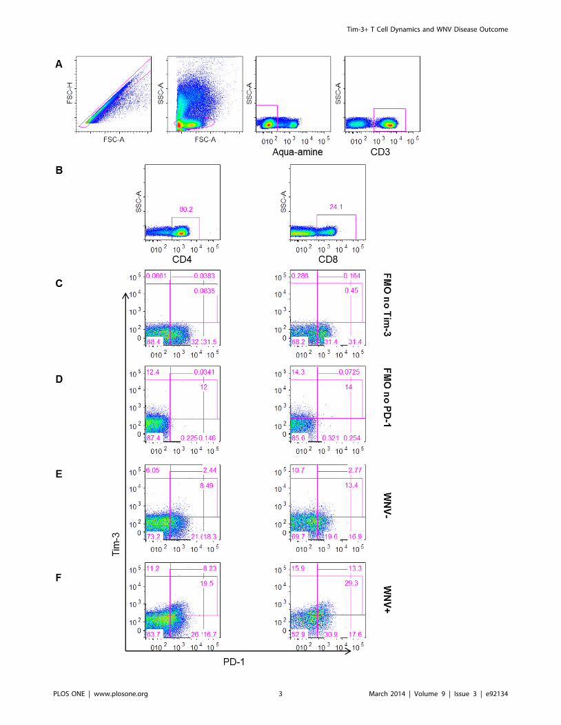
Figure 1. Gating strategy for measuring Tim-3 and PD-1 inhibitory receptors on T cells. The plots show (A) the gating strategy for liveCD3+ lymphocytes, (B) for CD4+ (left) and CD8+ (right) T cells. Gates were set on FMO no Tim-3 (C) and FMO no PD-1 (D). The gating of cells expressingTim-3 and PD-1 is shown for representative (E) WNV2 and (F) WNV+ subjects day 14 post-index donation.doi:10.1371/journal.pone.0092134.g001
Tim-3+ T Cell Dynamics and WNV Disease Outcome
PLOS ONE | www.plosone.org 4 March 2014 | Volume 9 | Issue 3 | e92134

Tim-3+ T Cell Dynamics and WNV Disease Outcome
PLOS ONE | www.plosone.org 5 March 2014 | Volume 9 | Issue 3 | e92134

asymptomatic and symptomatic WNV+ subjects expressed similar
frequencies of PD-1+ T cells (Figure 2B). Symptomatic WNV+subjects exhibited significantly higher frequencies of Tim-3+PD-12
CD4+ T cells (p,0.05 FDR,0.05) (Figure 2C) and Tim-3+ PD-
1+ CD4+ T cells (p = 0.015 FDR = 0.06) (Figure 2D) as well as
Tim-3+ PD-1+ CD8+ T cells (p = 0.023 FDR = 0.09) than
asymptomatic WNV+ subjects at day 14 post index.
Symptomatic WNV+ subjects exhibit higher frequenciesof early differentiated (CD28+/2CD57) Tim-3+ T cellpopulations
We measured the frequencies of differentiated T cells based on
CD28 and CD57 expression over the course of WNV infection by
flow cytometry (Figure 3) to assess the frequencies of Tim-3+ T
cells within naı̈ve/early differentiating (CD28+/2CD572), differ-
entiated (CD282CD572), and senescent (CD282CD57+) T cell
subsets in asymptomatic and symptomatic WNV+ subjects
(Figure 4). Symptomatic WNV+ subjects had higher frequencies
of Tim-3+ naı̈ve/early differentiating and CD4+ T cells compared
to asymptomatic WNV+ subjects from the earliest time-point at
day 14 post-index (p AS vs. S,0.0001, FDR = 0.002) and
throughout WNV infection (overall GEE, p,0.01 for both subsets)
(Figure 4A). Symptomatic WNV+ subjects also exhibited higher
frequencies of Tim-3+ differentiated CD8+ T cells than the
asymptomatic WNV+ subjects at 14 days post-index (p AS vs.
S = 0.02, FDR = 0.107) and throughout WNV infection (overall
GEE, p = 0.043) (Figure 4B). Notably, asymptomatic and
symptomatic WNV+ subjects expressed similar frequencies of
senescent T cells throughout and after resolution of WNV
infection.
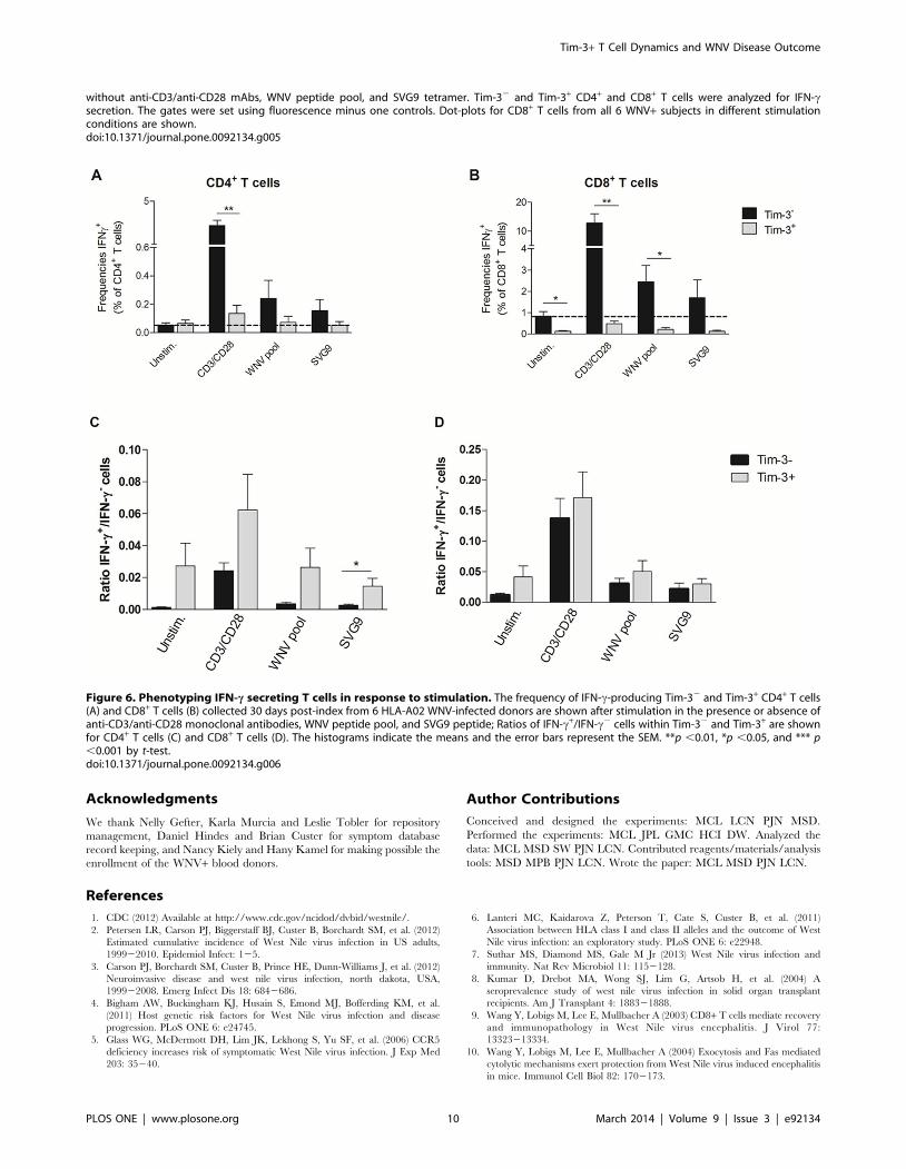
While Tim-3+ T cells are responsible for most of the IFN-csecretion, both Tim-3+ and Tim-32 T cells are competentfor IFN-c secretion
To characterize further the functional state of the Tim-3+ T
cells, we examined cytokine responses from PBMCs obtained from
6 HLA-A02 WNV+ donors incubated ex vivo with or without anti-
CD3 and anti-CD28 monoclonal antibodies, a WNV peptide pool
corresponding to immunodominant T cell epitopes in membrane,
envelope, and nonstructural 3 and 4B proteins [17], or an SVG9
peptide (HLA02 restricted SVGGVFTSV peptide in E) [16] by
flow cytometry (Figure 5). In all conditions, the majority of IFN-
c-secreting T cells were Tim-32 (Figure 6A and B). However,
the ratios of IFN-c+ cells within the Tim-32 and Tim-3+ CD4+
(Figure 6C) and CD8+ T cells (Figure 6D) were similar if not
higher in the Tim-3+ T cells.
Discussion
In this longitudinal study, we assessed the dynamics of
expression of immune inhibitory receptors during the evolution
of acute WNV infection. Our experiments revealed that symp-
tomatic WNV+ donors exhibited higher frequencies of Tim-3+
and Tim-3+PD-1+ T cells compared to asymptomatic WNV+donors throughout the course of infection, and Tim-3+ populations
phenotypically were CD28+ CD572 naı̈ve/differentiating and
CD282 CD572 differentiated CD4+ and CD8+ T cells. This is the
first report documenting the induction of Tim-3+PD-1+ T cells
after WNV infection and the association between the frequency of
Tim-3+ T cells and WNV disease outcome.
The increased expression of negative inhibitory receptors on T
cells has been observed in chronic viral infections and cancers.
Under these conditions, a phenomenon of T cell exhaustion
develops that has been associated with higher antigen load or
longer antigen exposure. Antibody blockade of exhaustion markers
leads to an improvement in antiviral and anti-tumor T cell
immunity, reduced viremia or tumor burden, and reduced severity
of disease outcome [19,20,21,22,23]. Tim-3+PD-1+ T cell induc-
tion in the acute phase of HCV infection correlated with defective
T cell responses and may contribute to the lack of immune control
and development of persistence [19]. Unexpectedly, a recent study
examining acute Mycobacterium tuberculosis infection in humans
found that Tim-3+ T cells were more active and effective in
suppressing mycobacterial infection in macrophages, suggesting a
possible differential role for Tim-3 in an acute bacterial infection
[24].
WNV infection is an acute viral infection, which in the majority
of individuals resolves within a few weeks of exposure, although
the virus may persist in some humans [25,26] and mice [27]. The
expression of T cell exhaustion markers, such as Tim-3 and PD-1,
has not been investigated extensively in acute viral infections. One
recent study, reported a protective role of Tim-3 in acute influenza
infection in mice [28]. Our findings, in contrast, suggest that
individuals with higher frequencies of Tim-3+ T cells after WNV
infection experience symptomatic disease. In mice, severe disease
following influenza infection may be due more to the immune
response and less to the virus directly [29], whereas WNV directly
promotes injury and death of some neuronal subtypes once it
reaches the CNS [7]. Thus, the difference in pathogenesis and
mechanism of injury may explain the discrepant results between
acute influenza and WNV infection.
The reasons for increased frequencies of Tim-3+ T cells in
symptomatic WNV+ subjects are not clear. Whether symptomatic
WNV+ subjects developed higher frequencies of Tim-3+ T cells
after WNV infection or had higher levels of these cells prior to
infection remains to be established. With no pre-WNV infection
samples available, our human studies are limited to an association
between elevated Tim-3+ T cell frequencies and symptomatic
disease. The functional characterization of T cells ex vivo after
restimulation revealed that while Tim-3+ T cells secrete IFN-c,
most of the IFN-c is secreted by Tim-32 T cells. In the context of
WNV infection, Tim-3+ T cells might be less functional, as
reported in other infectious models [12]. Taken together, these
results demonstrated that Tim-3+ T cells triggered after WNV
Figure 2. Frequencies of Tim-3 and PD-1 expressing T cells compared over the course of WNV infection in groups with differentdisease outcome. The graphs below demonstrate the change of frequencies of (A) Tim-3+, (B) PD-1+, (C) Tim-3+PD-12, and (D) Tim-3+PD-1+ CD4+
(left panel) and CD8+ (right panel) T cell subsets in asymptomatic (AS, circles/solid lines, n = 24) and symptomatic (S, squares/dash lines, n = 8) WNV-infected subjects 14, 30, 90, and 365 days post-index donation. The frequencies of the same cells in uninfected controls are displayed (WNV2,triangles, n = 26). The symbols indicate the means and the error bars represent the SEM. The p-values of the pairwise comparisons betweenasymptomatic and symptomatic groups are indicated by *p ,0.05, ** p ,0.01, and *** p ,0.001 above time-points when groups were compared at agiven time-point by Mann-Whitney test. The p-values of the comparison between asymptomatic and symptomatic groups are indicated by **adjacent to the cell subset title for p ,0.01 over the time of post-index by generalized estimated equation (GEE).doi:10.1371/journal.pone.0092134.g002
Tim-3+ T Cell Dynamics and WNV Disease Outcome
PLOS ONE | www.plosone.org 6 March 2014 | Volume 9 | Issue 3 | e92134

Tim-3+ T Cell Dynamics and WNV Disease Outcome
PLOS ONE | www.plosone.org 7 March 2014 | Volume 9 | Issue 3 | e92134

infection a priori are not less functional in terms of IFN-c secretion
than the Tim-32 T cells even though most of the IFN-c response
comes from Tim-32 cells.
We observed an association between symptomatic disease
outcome and Tim-3+ T cells with a CD28+/2CD572 naı̈ve/early
differentiated cellular profile. Recent studies reported that Tim-3
expression can be induced in naı̈ve [30,31], effector, and memory
T cells in an antigen-independent manner by cytokines, such as
IL-2, IL-7, IL-15 and IL-21, and also can be expressed on the
surface of naı̈ve, effector, and memory T cells [30]. In the context
of an acute viral infection this may impact the generation of an
effective antiviral T cell response. We envision that during acute
WNV infection, premature immune exhaustion among early
differentiating T cells could blunt the T cell responses that are
required to control and clear the virus, especially in the later
phases of pathogenesis. Whether symptomatic WNV+ subjects
develop higher, more persistent viremia and/or inflammation
remains to be investigated. Placing our data into the context of
prior studies showing that PBMCs from symptomatic WNV+
subjects’ PBMCs preferentially secrete IFN-c in response to WNV
peptide restimulation [17], and that lower levels of Tregs circulate
in symptomatic WNV+ subjects [14], we propose that, in
symptomatic WNV+ subjects, higher antigen load and inflamma-
tory responses could lead to increased cellular activation, with
Tim-3 marking a population of hyperactivated cells. Alternatively,
sustained or high levels of viral infection and inflammation could
induce expression of inhibitory receptors, with Tim-3+ on T cells
acting to limit collateral immune-mediated damage to host tissues.
Nonetheless, this counter-regulatory activity could be detrimental
if blunted T cell responses allowed WNV to persist longer in the
CNS, where the virus is cytopathic for neurons [7]. In this
scenario, targeting of Tim-3+ T cells (antibody blockade or
depletion) might boost the T cell immune response to control viral
replication and reduce disease severity, as reported for chronic
viral infections (HIV, HCV, and HBV) [13,19,32]. Clearly, further
investigation is required to determine whether higher frequencies
of Tim-3+ T cells are the cause or the consequence of symptomatic
WNV disease.
Figure 3. Gating strategy for measuring CD28 differentiation and CD57 senescence makers on T cells. The plots show (A) the gatingstrategy for live CD3+ lymphocytes, (B) for CD4+ (left) and CD8+ (right) T cells. Gates were set on FMO no CD28 (C) and FMO no CD57 (D). Plots areshown for representative (E) West Nile virus (WNV) uninfected controls (WNV2) and (F) WNV infected subjects (WNV+) day 14 post-index donation.doi:10.1371/journal.pone.0092134.g003
Figure 4. Differentiation status and functional capacity of Tim-3+ T cells in acute West Nile virus infection. The graphs show, throughthe course of WNV infection, the frequencies of Tim-3+ CD28+CD572, CD282CD572 and CD282CD57+ (A) CD4+ and (B) CD8+ T cell subsets inasymptomatic (AS, circles/solid lines, n = 24) and symptomatic (S, squares/dash lines, n = 8) WNV+ subjects 14, 30, 90, and 365 days post-indexdonation. The frequencies of the same cells in uninfected controls are displayed (WNV-, triangles, n = 26). The symbols indicate the means and theerror bars represent the SEM. **p ,0.01, *p ,0.05, and *** p ,0.001 by Mann-Whitney. The p-values of the comparison between asymptomatic andsymptomatic groups are indicated by ** adjacent to the cell subset title for p ,0.01 over the time post-index by GEE.doi:10.1371/journal.pone.0092134.g004
Tim-3+ T Cell Dynamics and WNV Disease Outcome
PLOS ONE | www.plosone.org 8 March 2014 | Volume 9 | Issue 3 | e92134

Figure 5. Gating strategy for phenotyping IFN-c secreting T cells in response to stimulation. The frequencies of Tim-3+ and Tim-32 IFN-csecreting CD4+ and CD8+ T cells were measured in PBMC collected at day 30 post-index from 6 HLA-A02 WNV-infected donors and incubated with or
Tim-3+ T Cell Dynamics and WNV Disease Outcome
PLOS ONE | www.plosone.org 9 March 2014 | Volume 9 | Issue 3 | e92134
Acknowledgments
We thank Nelly Gefter, Karla Murcia and Leslie Tobler for repository
management, Daniel Hindes and Brian Custer for symptom database
record keeping, and Nancy Kiely and Hany Kamel for making possible the
enrollment of the WNV+ blood donors.
Author Contributions
Conceived and designed the experiments: MCL LCN PJN MSD.
Performed the experiments: MCL JPL GMC HCI DW. Analyzed the
data: MCL MSD SW PJN LCN. Contributed reagents/materials/analysis
tools: MSD MPB PJN LCN. Wrote the paper: MCL MSD PJN LCN.
References
1. CDC (2012) Available at http://www.cdc.gov/ncidod/dvbid/westnile/.
2. Petersen LR, Carson PJ, Biggerstaff BJ, Custer B, Borchardt SM, et al. (2012)
Estimated cumulative incidence of West Nile virus infection in US adults,
199922010. Epidemiol Infect: 125.
3. Carson PJ, Borchardt SM, Custer B, Prince HE, Dunn-Williams J, et al. (2012)
Neuroinvasive disease and west nile virus infection, north dakota, USA,
199922008. Emerg Infect Dis 18: 6842686.
4. Bigham AW, Buckingham KJ, Husain S, Emond MJ, Bofferding KM, et al.
(2011) Host genetic risk factors for West Nile virus infection and disease
progression. PLoS ONE 6: e24745.
5. Glass WG, McDermott DH, Lim JK, Lekhong S, Yu SF, et al. (2006) CCR5
deficiency increases risk of symptomatic West Nile virus infection. J Exp Med
203: 35240.
6. Lanteri MC, Kaidarova Z, Peterson T, Cate S, Custer B, et al. (2011)
Association between HLA class I and class II alleles and the outcome of West
Nile virus infection: an exploratory study. PLoS ONE 6: e22948.
7. Suthar MS, Diamond MS, Gale M Jr (2013) West Nile virus infection and
immunity. Nat Rev Microbiol 11: 1152128.
8. Kumar D, Drebot MA, Wong SJ, Lim G, Artsob H, et al. (2004) A
seroprevalence study of west nile virus infection in solid organ transplant
recipients. Am J Transplant 4: 188321888.
9. Wang Y, Lobigs M, Lee E, Mullbacher A (2003) CD8+ T cells mediate recovery
and immunopathology in West Nile virus encephalitis. J Virol 77:
13323213334.
10. Wang Y, Lobigs M, Lee E, Mullbacher A (2004) Exocytosis and Fas mediated
cytolytic mechanisms exert protection from West Nile virus induced encephalitis
in mice. Immunol Cell Biol 82: 1702173.
without anti-CD3/anti-CD28 mAbs, WNV peptide pool, and SVG9 tetramer. Tim-32 and Tim-3+ CD4+ and CD8+ T cells were analyzed for IFN-csecretion. The gates were set using fluorescence minus one controls. Dot-plots for CD8+ T cells from all 6 WNV+ subjects in different stimulationconditions are shown.doi:10.1371/journal.pone.0092134.g005
Figure 6. Phenotyping IFN-c secreting T cells in response to stimulation. The frequency of IFN-c-producing Tim-32 and Tim-3+ CD4+ T cells(A) and CD8+ T cells (B) collected 30 days post-index from 6 HLA-A02 WNV-infected donors are shown after stimulation in the presence or absence ofanti-CD3/anti-CD28 monoclonal antibodies, WNV peptide pool, and SVG9 peptide; Ratios of IFN-c+/IFN-c2 cells within Tim-32 and Tim-3+ are shownfor CD4+ T cells (C) and CD8+ T cells (D). The histograms indicate the means and the error bars represent the SEM. **p ,0.01, *p ,0.05, and *** p,0.001 by t-test.doi:10.1371/journal.pone.0092134.g006
Tim-3+ T Cell Dynamics and WNV Disease Outcome
PLOS ONE | www.plosone.org 10 March 2014 | Volume 9 | Issue 3 | e92134

11. Suvas S, Azkur AK, Kim BS, Kumaraguru U, Rouse BT (2004) CD4+CD25+regulatory T cells control the severity of viral immunoinflammatory lesions. JImmunol 172: 412324132.
12. Wherry EJ (2011) T cell exhaustion. Nat Immunol 12: 4922499.
13. Wherry EJ, Ha SJ, Kaech SM, Haining WN, Sarkar S, et al. (2007) Molecularsignature of CD8+ T cell exhaustion during chronic viral infection. Immunity
27: 6702684.14. Lanteri MC, O’Brien KM, Purtha WE, Cameron MJ, Lund JM, et al. (2009)
Tregs control the development of symptomatic West Nile virus infection in
humans and mice. J Clin Invest 119: 326623277.15. Pfeiffer IA, Zinser E, Strasser E, Stein MF, Dorrie J, et al. (2013) Leukoreduction
system chambers are an efficient, valid, and economic source of functionalmonocyte-derived dendritic cells and lymphocytes. Immunobiology 218:
139221401.16. Kim S, Li L, McMurtrey CP, Hildebrand WH, Weidanz JA, et al. (2010) Single-
chain HLA-A2 MHC trimers that incorporate an immundominant peptide elicit
protective T cell immunity against lethal West Nile virus infection. J Immunol184: 442324430.
17. Lanteri MC, Heitman JW, Owen RE, Busch T, Gefter N, et al. (2008)Comprehensive analysis of west nile virus-specific T cell responses in humans. J
Infect Dis 197: 129621306.
18. Tobler LH, Cameron MJ, Lanteri MC, Prince HE, Danesh A, et al. (2008)Interferon and interferon-induced chemokine expression is associated with
control of acute viremia in West Nile virus-infected blood donors. J Infect Dis198: 9792983.
19. McMahan RH, Golden-Mason L, Nishimura MI, McMahon BJ, Kemper M, etal. (2010) Tim-3 expression on PD-1+ HCV-specific human CTLs is associated
with viral persistence, and its blockade restores hepatocyte-directed in vitro
cytotoxicity. J Clin Invest 120: 454624557.20. Nakamoto N, Cho H, Shaked A, Olthoff K, Valiga ME, et al. (2009) Synergistic
reversal of intrahepatic HCV-specific CD8 T cell exhaustion by combined PD-1/CTLA-4 blockade. PLoS Pathog 5: e1000313.
21. Nakamoto N, Kaplan DE, Coleclough J, Li Y, Valiga ME, et al. (2008)
Functional restoration of HCV-specific CD8 T cells by PD-1 blockade is definedby PD-1 expression and compartmentalization. Gastroenterology 134:
192721937, 1937 e192121922.
22. Ngiow SF, von Scheidt B, Akiba H, Yagita H, Teng MW, et al. (2011) Anti-
TIM3 antibody promotes T cell IFN-gamma-mediated antitumor immunity and
suppresses established tumors. Cancer Res 71: 354023551.
23. Sakuishi K, Apetoh L, Sullivan JM, Blazar BR, Kuchroo VK, et al. (2010)
Targeting Tim-3 and PD-1 pathways to reverse T cell exhaustion and restore
anti-tumor immunity. J Exp Med 207: 218722194.
24. Qiu Y, Chen J, Liao H, Zhang Y, Wang H, et al. (2012) Tim-3-expressing CD4+and CD8+ T cells in human tuberculosis (TB) exhibit polarized effector memory
phenotypes and stronger anti-TB effector functions. PLoS Pathog 8: e1002984.
25. Lai L, Lee TH, Tobler L, Wen L, Shi P, et al. (2011) Relative distribution of
West Nile virus RNA in blood compartments: implications for blood donor
nucleic acid amplification technology screening. Transfusion.
26. Nett RJ, Kuehnert MJ, Ison MG, Orlowski JP, Fischer M, et al. (2012) Current
practices and evaluation of screening solid organ donors for West Nile virus.
Transpl Infect Dis 14: 2682277.
27. Appler KK, Brown AN, Stewart BS, Behr MJ, Demarest VL, et al. (2010)
Persistence of West Nile virus in the central nervous system and periphery of
mice. PLoS ONE 5: e10649.
28. Cho JL, Roche MI, Sandall B, Brass AL, Seed B, et al. (2012) Enhanced Tim3
activity improves survival after influenza infection. J Immunol 189: 287922889.
29. Braciale TJ, Sun J, Kim TS (2012) Regulating the adaptive immune response to
respiratory virus infection. Nat Rev Immunol 12: 2952305.
30. Mujib S, Jones RB, Lo C, Aidarus N, Clayton K, et al. (2012) Antigen-
Independent Induction of Tim-3 Expression on Human T Cells by the Common
gamma-Chain Cytokines IL-2, IL-7, IL-15, and IL-21 Is Associated with
Proliferation and Is Dependent on the Phosphoinositide 3-Kinase Pathway. J
Immunol.
31. Ndhlovu LC, Lopez-Verges S, Barbour JD, Jones RB, Jha AR, et al. (2012) Tim-
3 marks human natural killer cell maturation and suppresses cell-mediated
cytotoxicity. Blood 119: 373423743.
32. Golden-Mason L, Palmer BE, Kassam N, Townshend-Bulson L, Livingston S, et
al. (2009) Negative immune regulator Tim-3 is overexpressed on T cells in
hepatitis C virus infection and its blockade rescues dysfunctional CD4+ and
CD8+ T cells. J Virol 83: 912229130.
Tim-3+ T Cell Dynamics and WNV Disease Outcome
PLOS ONE | www.plosone.org 11 March 2014 | Volume 9 | Issue 3 | e92134
Related Documents