Waves of awareness for occipital and parietal phosphenes perception Chiara Bagattini a,1 , Chiara Mazzi b,c,1 , Silvia Savazzi b,c,n a Cognitive Neuroscience Section, IRCCS Centro San Giovanni di Dio Fatebenefratelli, Brescia, Italy b University of Verona and National Institute of Neuroscience, Italy c Perception and Awareness (PandA) Laboratory, Department of Neurological and Movement Sciences, University of Verona, Italy article info Article history: Received 22 October 2014 Received in revised form 10 February 2015 Accepted 16 February 2015 Available online 17 February 2015 Keywords: Dorsal stream Ventral stream Primary visual cortex TMS-evoked potentials EEG abstract Transcranial magnetic stimulation (TMS) of the occipital cortex is known to induce visual sensations, i.e. phosphenes, which appear as flashes of light in the absence of an external stimulus. Recent studies have shown that TMS can produce phosphenes also when the intraparietal sulcus (IPS) is stimulated. The main question addressed in this paper is whether parietal phosphenes are generated directly by local me- chanisms or emerge through indirect activation of other visual areas. Electroencephalographic (EEG) signals were recorded while stimulating left occipital or parietal cortices inducing phosphene perception in healthy participants and in a hemianopic patient who suffered from complete destruction of the early visual cortex of the left hemisphere. Results in healthy participants showed that the onset of phosphene perception induced by occipital TMS correlated with differential cortical activity in temporal sites while the onset of phosphene perception induced by parietal TMS correlated with differential cortical activity in the stimulated parietal site. Moreover, IPS-TMS of the lesioned hemisphere of the hemianopic patient with a complete lesion to V1 showed again that the onset of phosphene perception correlated with differential cortical activity in the stimulated parietal site. The present data seem thus to suggest that temporal and parietal cortices can serve as different local early gatekeepers of perceptual awareness and that activity in the occipital cortex, although being relevant for perception in general, is not part of the neural bases of the perceptual awareness of phosphenes. & 2015 Elsevier Ltd. All rights reserved. 1. Introduction A central question in consciousness studies is to reveal which brain regions, and in what order of activation, critically determine specific conscious percepts. Several models have been proposed in order to find the brain areas (“where”) correlating with visual awareness and to determine the time-course of neural activation in interconnected areas (“when/how”) needed for awareness to emerge. With respect to the “where” question, one of the most influential models in visual processing, the so-called two-streams hypothesis (Goodale and Milner, 1992; Milner and Goodale, 2008), states that visual awareness is restricted to the ventral stream (Milner, 2012). The dorsal stream, instead, is thought not to be “in the business of providing any kind of a visual representation of the world” (Goodale and Milner, 2004, p. 114). With respect to the “when/how” question, another very influential model, Lamme's model (Lamme et al., 1998), states that recurrent processing feeding back to occipital cortex is necessary for awareness to emerge. On the basis of these two models, it can be predicted that the neural correlates of visual awareness can only be found along the ventral stream (comprising the occipital and temporal cor- tices) and that the integrity of the primary visual cortex (V1) is needed. Recent findings have challenged both of these statements. It has indeed been found that, at least under certain circum- stances, the dorsal stream can generate visual awareness (Hes- selmann and Malach, 2011; Koivisto et al., 2010; Mazzi et al., 2014) and that recurrent processing feeding back to V1 is not necessary for visual awareness (Zeki and ffytche, 1998; ffytche and Zeki, 2011; Mazzi et al., 2014). In the present paper we further tested the contribution of the dorsal stream and V1 in the emergence of awareness by combining transcranial magnetic stimulation (TMS) and electro- encephalography (EEG), two modern methodologies which can provide information both with respect to the “where” and the “when/how” questions. TMS, by being a non-invasive direct stimulation method, is one Contents lists available at ScienceDirect journal homepage: www.elsevier.com/locate/neuropsychologia Neuropsychologia http://dx.doi.org/10.1016/j.neuropsychologia.2015.02.021 0028-3932/& 2015 Elsevier Ltd. All rights reserved. n Correspondence to: Perception and Awareness (PandA) Laboratory, Section of Physiology and Psychology, Department of Neurological and Movement Sciences, Uni- versity of Verona, Strada le Grazie, 8 37134 Verona, Italy. Fax: þ39 045 8027279. E-mail address: [email protected] (S. Savazzi). 1 These authors contributed equally. Neuropsychologia 70 (2015) 114–125

Welcome message from author
This document is posted to help you gain knowledge. Please leave a comment to let me know what you think about it! Share it to your friends and learn new things together.
Transcript
Neuropsychologia 70 (2015) 114–125
Contents lists available at ScienceDirect
Neuropsychologia
http://d0028-39
n Corrversity
E-m1 Thes
journal homepage: www.elsevier.com/locate/neuropsychologia
Waves of awareness for occipital and parietal phosphenes perception
Chiara Bagattini a,1, Chiara Mazzi b,c,1, Silvia Savazzi b,c,n
a Cognitive Neuroscience Section, IRCCS Centro San Giovanni di Dio Fatebenefratelli, Brescia, Italyb University of Verona and National Institute of Neuroscience, Italyc Perception and Awareness (PandA) Laboratory, Department of Neurological and Movement Sciences, University of Verona, Italy
a r t i c l e i n f o
Article history:Received 22 October 2014Received in revised form10 February 2015Accepted 16 February 2015Available online 17 February 2015
Keywords:Dorsal streamVentral streamPrimary visual cortexTMS-evoked potentialsEEG
x.doi.org/10.1016/j.neuropsychologia.2015.02.032/& 2015 Elsevier Ltd. All rights reserved.
espondence to: Perception and Awareness (Pof Verona, Strada le Grazie, 8 37134 Verona, Iail address: [email protected] (S. Savazzi).e authors contributed equally.
a b s t r a c t
Transcranial magnetic stimulation (TMS) of the occipital cortex is known to induce visual sensations, i.e.phosphenes, which appear as flashes of light in the absence of an external stimulus. Recent studies haveshown that TMS can produce phosphenes also when the intraparietal sulcus (IPS) is stimulated. The mainquestion addressed in this paper is whether parietal phosphenes are generated directly by local me-chanisms or emerge through indirect activation of other visual areas. Electroencephalographic (EEG)signals were recorded while stimulating left occipital or parietal cortices inducing phosphene perceptionin healthy participants and in a hemianopic patient who suffered from complete destruction of the earlyvisual cortex of the left hemisphere. Results in healthy participants showed that the onset of phospheneperception induced by occipital TMS correlated with differential cortical activity in temporal sites whilethe onset of phosphene perception induced by parietal TMS correlated with differential cortical activityin the stimulated parietal site. Moreover, IPS-TMS of the lesioned hemisphere of the hemianopic patientwith a complete lesion to V1 showed again that the onset of phosphene perception correlated withdifferential cortical activity in the stimulated parietal site. The present data seem thus to suggest thattemporal and parietal cortices can serve as different local early gatekeepers of perceptual awareness andthat activity in the occipital cortex, although being relevant for perception in general, is not part of theneural bases of the perceptual awareness of phosphenes.
& 2015 Elsevier Ltd. All rights reserved.
1. Introduction
A central question in consciousness studies is to reveal whichbrain regions, and in what order of activation, critically determinespecific conscious percepts. Several models have been proposed inorder to find the brain areas (“where”) correlating with visualawareness and to determine the time-course of neural activationin interconnected areas (“when/how”) needed for awareness toemerge. With respect to the “where” question, one of the mostinfluential models in visual processing, the so-called two-streamshypothesis (Goodale and Milner, 1992; Milner and Goodale, 2008),states that visual awareness is restricted to the ventral stream(Milner, 2012). The dorsal stream, instead, is thought not to be “inthe business of providing any kind of a visual representation of theworld” (Goodale and Milner, 2004, p. 114). With respect to the“when/how” question, another very influential model, Lamme'smodel (Lamme et al., 1998), states that recurrent processingfeeding back to occipital cortex is necessary for awareness to
21
andA) Laboratory, Section of Phystaly. Fax: þ39 045 8027279.
emerge. On the basis of these two models, it can be predicted thatthe neural correlates of visual awareness can only be found alongthe ventral stream (comprising the occipital and temporal cor-tices) and that the integrity of the primary visual cortex (V1) isneeded. Recent findings have challenged both of these statements.It has indeed been found that, at least under certain circum-stances, the dorsal stream can generate visual awareness (Hes-selmann and Malach, 2011; Koivisto et al., 2010; Mazzi et al., 2014)and that recurrent processing feeding back to V1 is not necessaryfor visual awareness (Zeki and ffytche, 1998; ffytche and Zeki,2011; Mazzi et al., 2014).
In the present paper we further tested the contribution of thedorsal stream and V1 in the emergence of awareness by combiningtranscranial magnetic stimulation (TMS) and electro-encephalography (EEG), two modern methodologies which canprovide information both with respect to the “where” and the“when/how” questions.
TMS, by being a non-invasive direct stimulation method, is one
iology and Psychology, Department of Neurological and Movement Sciences, Uni-

C. Bagattini et al. / Neuropsychologia 70 (2015) 114–125 115
of the state of the art methodologies used to study whether aspecific neural area plays a crucial role in cognitive processes. Thelogic is the following: if TMS of a specific cortical area has a spe-cific effect on performance, this area and the network associated toit are crucial for the studied function. In this respect, for example,if TMS is applied to the portion of the primary motor cortex (M1)representing the contralateral hand, a motor twitch of the handcontralateral to the stimulation is elicited, thus making it possibleto conclude that activity within the stimulated area (M1) has acausal role in eliciting the motor twitch. Moreover, TMS can beuseful not only for the localization of eloquent cortical areas but itcan also determine the intensity needed to evoke a specific effect.For instance, by stimulating M1, one can determine the minimalTMS pulse intensity (the so-called motor threshold) needed toevoke a motor potential; this intensity is considered to directlyreflect the level of excitability of the stimulated cortex.
Similarly, TMS of visual areas induces conscious visual percepts,or phosphenes, i.e. the experience of flashes of light in the absenceof an external stimulus. Additionally, akin to the motor threshold,cortical excitability can be measured with the phosphene thresh-old. Typically, phosphene perception has been studied by stimu-lating areas within the occipital cortex. However, recent findings(Marzi et al., 2009; Mazzi et al., 2014; Fried et al., 2011) haveshown that phosphenes can be elicited also along the dorsalstream (Goodale and Milner, 1992; Milner and Goodale, 2008),specifically in the intra-parietal sulcus (IPS). Interestingly for thepurposes of the present paper, Mazzi and collaborators (2014)tested both healthy participants and hemianopic patients with acomplete lesion of V1 and asked them to report the presence/absence of phosphenes while their occipital or parietal cortex wasstimulated (only the parietal cortex was stimulated in hemianopicpatients). The authors reported that, in healthy participants, IPS-phosphenes have a higher phosphene-threshold and differentphenomenical characteristics than those elicited by occipital TMS.Importantly, they showed that parietal phosphenes can be ob-tained also in patients with a complete lesion to the ipsilateral V1and that their conscious visual percepts were undistinguishablefrom those obtained with healthy participants. The authors con-cluded that (1) neural activity in V1 is not necessary for visualawareness and (2) that IPS is an independent generator of theawareness of phosphenes. As a note of caution, one should con-sider the possibility that the awareness of phosphenes generatedby TMS of IPS could result from a spread of activity towards othervisual areas (i.e. extrastriate areas and the temporal cortex). Al-though the involvement of V1 seems unlikely given the resultswith hemianopic patients, the awareness of IPS phosphenes couldbe induced by a “third visual area”, or a network subtendingconnections with IPS, that could be in charge of providing accessto visual awareness (Fried et al., 2011; Mazzi et al., 2014). A likelycandidate for this hypothesis, as suggested by the previously de-scribed models (Goodale and Milner, 1992; Lamme et al., 1998)could be found along the ventral stream, e.g., in the temporalcortex which is known to play an important role in visualawareness (Goodale and Milner, 1992).
In order to test the contribution of such a “third area” in thegeneration of phosphenes, TMS-evoked potentials (TEPs) can beacquired by co-registering EEG signals while the cortex is stimu-lated. TEPs represent a clear and direct measure of cortical excit-ability and can be used to assess the state of cortical reactivity andconnectivity also in the so-called silent-areas that do not producea peripheral marker (Ilmoniemi et al., 1997). Here, we adopted aninteractive approach (Miniussi and Thut, 2010) by using EEG–TMSco-registration while the participant performed a task. This ap-proach consists in the stimulation of a circumscribed cortical areawith TMS and to monitor, with EEG, the induced electrical changesin the whole cortex. EEG–TMS co-registration conceives the
tracing of the time course of functionally relevant activity in dis-tant but functionally connected areas relevant for the task at hand(i.e. effective connectivity). The relevance of this approach istwofold: (1) it provides an empirical measure of the network ofareas implicated in a specific task as the activation induced by TMSof the targeted area propagates to functionally connected areasand (2) it provides information on the causal relationship in theconnections across the network of activated areas. Given the hightemporal resolution of EEG and the properties of the spreading ofactivity induced by TMS, if an area X results to be active prior toarea Y it can be assumed that the activity in area X causes a changein the activity of area Y through effective connections between thetwo areas.
Thanks to these characteristics, TEPs are ideal to gather in-formation on the time-course and spatio-temporal dynamics ofthe emergence of phosphene perception. Despite the relativecrudeness of spatial topography of EEG, its temporal resolution isvery high (in the range of milliseconds) and this is the most crucialcharacteristic for the purposes of the present paper. The logic isthe following: if TEPs detect an electrical difference at the stimu-lated area (e.g. IPS) between phosphene-present and phosphene-absent trials in a specific time window after TMS, to be consideredcrucial for this effect, a “third area” (e.g. temporal cortex) shoulddisplay a comparable effect (phosphene-present different thanphosphene-absent) in the same or earlier time windows. If such adifferential effect cannot be found, no causal role of the supposed“third area” (e.g. temporal cortex) can be advocated for the per-ception of phosphenes elicited by TMS of the stimulated area (e.g.IPS).
The main purpose of the present paper is to uncover the spatio-temporal dynamics of the onset of the phosphene perception asinduced by occipital and parietal TMS. To do this, healthy partici-pants and one hemianopic patient was asked to report the pre-sence/absence of a phosphene induced by stimulation of a specificoccipital and parietal site by means of TMS while monitoring andrecording TEPs. On the basis of the results of this EEG–TMS in-teractive co-registration approach, we could draw some conclu-sions both on the role of V1 in perceptual awareness and onwhether or not other functionally interconnected areas are playingsome role in the emergence of awareness after stimulation of theoccipital and parietal cortex.
2. Experiment 1
2.1. Materials and methods
2.1.1. Healthy participantsSixteen healthy volunteers (9 females) were recruited to par-
ticipate in the study. Their ages ranged between 22 and 28 years(mean 25 years, sd 1.90) and they were all right handed, as as-sessed with the Edinburgh Handedness Inventory (Oldfield, 1971).They all had normal or corrected-to-normal visual acuity and nohistory of neurological or psychiatric disorders. All participantsgave their written informed consent prior to participation. Theexperiment was carried out according to the principles laid downin the 1964 Declaration of Helsinki and approved by the localEthics Committee. As assessed by a safety screening questionnaire(adapted from Rossi et al., 2011), participants were negative for allrisk factors associated with TMS: none reported neurological dis-orders, cardiac pacemaker, any history of epilepsy or migraine,current treatment with any psychoactive medication and preg-nancy. One participant could not perceive reliable phosphenesafter either occipital or parietal sites and four participants droppedout and did not perform the second session. These participantswere thus excluded from the sample.

C. Bagattini et al. / Neuropsychologia 70 (2015) 114–125116
2.1.2. Experimental procedureParticipants were individually tested in a dimly illuminated
room. During the experiment, they sat in front of a 17 in. LCDmonitor (LG L1753HM) at a viewing distance of 57 cm with theirhead secured in a chin rest with forehead support. To enhance theexcitability of their visual cortex, participants’ eyes were coveredwith eye-patches prior to the threshold measurements and theyperformed the entire experiment blindfolded and were asked tomaintain their gaze steady in front of them. Commercial earplugswere used with the aim of protecting the participants from thenoise associated with TMS (Rossi et al., 2009) and preventing re-sponses from being affected by the intensity of the coil click.
At the beginning of each session, after applying the cap withelectrodes for EEG recording, phosphene threshold (PT) was as-sessed for occipital and parietal sites, by means of an automatic,non-adaptive, psychophysical method (“Method of constant sti-muli”) implemented with Matlab (Abrahamyan et al., 2011). Thir-teen randomly intermixed different intensities were employed(ranging from 57% to 93% of maximum stimulator output (MSO),with changes in steps of 3%) and eight pulses were given for eachstimulator output intensity (total number of pulses¼104). Thedata obtained were then fitted with a cumulative Weibull psy-chometric function via a maximum likelihood criterion using thePalamedes toolbox (http://www.palamedestoolbox.org) with Ma-tlab. The stimulation intensity at which the participant couldperceive a phosphene on 50% of trials was taken as the thresholdvalue and used in the subsequent experimental session.
The experimental session comprised 4 blocks of 80 trials each,for a total number of 320 stimulations. After each TMS pulse,participants were requested to report the presence or absence of aphosphene with a “yes/no” response by pressing respectively the“z” button (left index finger) or the “m” button (right index finger)on the keyboard without losing time. After the response was gi-ven, there was a random interval (ranging from 3000 to 3300 ms)and then the subsequent pulse was automatically delivered. Theinter-pulse interval was never shorter than 4 s, well above thecriterion assessed by safety instructions (Anand and Hotson, 2002;Wassermann, 1998).
Each participant performed two experimental sessions (con-ducted on separate days with at least 48 h between sessions), onefor each stimulation site (left occipital cortex and left parietalcortex). The order of the two sessions was counterbalanced amongparticipants. In total, each experimental session lasted about twoand a half hours, including the setup of the EEG cap and neuro-navigation system. Participants were debriefed at the end of thesecond experimental session.
2.1.3. TMS protocolFor each experimental session, single-pulse magnetic stimula-
tion (inter-pulse interval 44 s) was delivered through a 70 mmfigure-of-eight coil connected to a biphasic Magstim Rapid2 sys-tem (maximum output 3.5 T) (Magstim Company Limited, Whit-land, UK). The TMS pulse trigger and response acquisition werecontrolled with Matlab (The MathWorks, Natick, MA) for thephosphene threshold assessment, and with E-Prime 1.1 (SP3)software (Psychology Software Tools, Pittsburgh, PA) for the ex-perimental session.
Neuronavigation software (SofTaxic, E.M.S., Bologna, Italy)combined with a 3D optical digitizer (Polaris Vicra, NDI, Waterloo,Canada) was used throughout the experiment to maintain the coilposition over the participant's head within a 2 mm accuracythreshold. The TMS coil was placed tangentially on the surface ofthe scalp, parallel to the participant's sagittal midline, with thehandle pointing upwards in order to avoid unspecific activation ofneck and shoulder muscles.
The best location found for eliciting circumscribed and right-
lateralized phosphenes, the “hot spot”, was then acquired by theneuro-navigation system and the coil was fixed in the targetedposition by means of a mechanical arm (Manfrotto magic arm,Italy, www.manfrotto.com). To confirm that participants perceivedgenuine phosphenes, some criteria (Kammer et al., 2005) such asthe dependence on the stimulated hemisphere, i.e. phosphenes inthe contralateral visual field (Meyer et al., 1991), gaze direction(Meyer et al., 1991)and visibility with the eyes both open andclosed (Kammer and Beck, 2002), had to be satisfied duringtraining trials. In order to avoid any potential effects of partici-pants’ trying to comply with the experimenter's expectations,additional tests were performed (see Mazzi et al., 2014 for details).
Since previous studies reported that left hemisphere stimula-tion evokes more reliable phosphenes than the right hemisphere(Beckers and Hömberg, 1992; Stewart et al., 1999; Antal et al.,2001; Silvanto et al., 2008), we applied TMS over the left hemi-sphere. Individual hot spots were located using the functionalmethod of inducing phosphenes by stimulating with supra-threshold intensities in a region within an area of 2 cm in diametercentered on two different scalp positions (see Fig. 1A): (1) theoccipital lobe in correspondence to the O1 electrode position ofthe 10–20 International EEG system and (2) the parietal lobe incorrespondence to the P3 electrode position of the 10–20 Inter-national EEG system. These sites are most likely to correspond,respectively, to visual cortical areas V1/V2 (Thielscher et al., 2010,Salminen-Vaparanta et al., 2012) and intraparietal sulcus in allparticipants (Mazzi et al., 2014).
2.1.4. EEG recording and TMS-evoked potentials (TEPs) analysisTMS-compatible EEG equipment (BrainAmp, Brain Products
GmbH, Munich, Germany) was used to record EEG signals(BrainVision Recorder). The EEG activity was continuously re-corded from a Fast'n Easy cap with 27 TMS-compatible Ag/AgClpellet pin electrodes (EasyCap GmbH, Herrsching, Germany)placed according to the 10–20 International System (O1, O2, P7, P3,Pz, P4, P8, CP5, CP1, CP2, CP6, T7, C3, Cz, C4, T8, FC5, FC1, FC2, FC6,F7, F3, Fz, F4, F8, Fp1, Fp2). Additional electrodes were used asreference and ground and for the electro-oculogram. The groundelectrode was placed in AFz, i.e. at the maximal distance from thestimulating TMS coil. All scalp channels were online referenced tothe right mastoid (RM) and then re-referenced offline to the leftmastoid (LM). Horizontal and vertical eye movements were de-tected respectively with electrodes placed at the left and rightcanthi and up and below the right eye. The impedance of all theelectrodes was kept below 5 kΩ. The EEG was recorded at 5000 Hzsampling rate with a time constant of 10 s as low cut-off and ahigh cut-off of 1000 Hz. The EEG signal was processed off-lineusing BrainVision Analyzer 1.05.
In order to reduce TMS artifacts and to make possible to recordthe EEG signals from the electrodes placed right beneath the TMScoil, we devised a custom-made polystyrene C-shaped annulus.The annulus was positioned over the stimulated electrodes (O1,P3), making it possible to place the coil over the target electrodewithout the need to physically remove that electrode.
Continuous data were filtered offline with a 20 Hz (12 dB/oc-tave) (Oruç et al., 2011) high cut-off filter and then divided intoepochs starting from 200 ms before and ending 600 ms after theTMS pulse. Epochs were then baseline corrected (from �200 msto 0 ms) and visually inspected in order to remove all trials con-taminated by eye movements and blinking artifacts, involuntarymotor acts or excessive noisy EEG. After pre-processing, theaverage number of trials were 102.22 for “yes” responses and 74.11for “no” responses when stimulating the occipital cortex and 90.67“yes” responses and 112.33 “no” responses when stimulating theparietal cortex. TEPs were obtained by averaging epochs for eachparticipant and for each stimulation site separately for trials where

Fig. 1. Experiment 1. Results and analyses with healthy participants. (A) Individual hot-spots for occipital (blue) and parietal (red) stimulation sites eliciting reliablephosphenes. (B) Psychometric functions for the two sites of stimulation. The blue line indicates the threshold function obtained after TMS of the occipital cortex while thered line represents the threshold function obtained after TMS of the parietal cortex. Each dot indicates the mean performance across subjects at each TMS intensity for thetwo stimulation sites. (C) TEPs elicited by TMS pulses over O1 as a function of the phosphene-present (red) and phosphene-absent (black) conditions. Gray dotted boxesshow the time windows in which the two waveforms are statistically significant. The black box indicates the time window in which the TMS artifact was present.(D) Intensity plots showing the cluster-corrected significant time windows resulting from the two-tailed point-wise paired t-tests comparing phosphene-present andphosphene-absent condition following O1-TMS. The x-, y- and z-axis represent, respectively, time (from 0 ms to 600 ms after TMS pulse), electrodes and t-test values(depicted by different colors) at each data point. The figure inset depicts the SCD topographic map of the phosphene present/absent effect. The SCD focus is compatible withtemporal generators. (E) TEPs elicited by TMS pulses over P3 as a function of the phosphene-present (red) and phosphene-absent (black) conditions. Gray dotted boxes showthe time windows in which the two waveforms are statistically significant. The black box indicates the time window in which the TMS artifact was present. (F) Intensity plotsshowing the cluster-corrected significant time windows resulting from the two-tailed point-wise paired t-tests comparing phosphene-present and phosphene-absentcondition following P3-TMS. The x-, y- and z-axis represent, respectively, time (from 0 ms to 600 ms after TMS pulse), electrodes and t-test values (depicted by differentcolors) at each data point. The figure inset depicts the SCD topographic map of the phosphene present/absent effect. The SCD focus is compatible with parietal generators.(For interpretation of the references to color in this figure legend, the reader is referred to the web version of this article.)
C. Bagattini et al. / Neuropsychologia 70 (2015) 114–125 117

C. Bagattini et al. / Neuropsychologia 70 (2015) 114–125118
participants reported perceiving a phosphene (hereafter called“phosphene-present” trials/conditions) and those where TMS didnot elicit any visual percepts (hereafter called “phosphene-absent”trials/conditions).
Given that TMS artifacts influence the recording of meaningfulEEG data in a period of a few milliseconds after delivery of themagnetic pulse (Veniero et al., 2009), data recorded up to 50 msafter the TMS pulse was not analyzed. Due to the presence of noisyEEG signals in two participants, TEP analysis was performed on thedata from 9 participants (7 females; mean age 24.78 years, sd 1.79years). Moreover, despite the presence of the custom-made an-nulus over the stimulated electrode, because of the arrangementof the electrode's lead wire (Sekiguhi et al., 2011) strong TMS ar-tifacts were recorded from the P3 electrode in some participantswhen the parietal cortex was stimulated. We therefore analyzedTEPs recorded from Pz in all participants, which was the electrodeclosest to the stimulation site with EEG signals free of TMS arti-facts. Instead, TMS of the occipital cortex did not induce suchstrong artifacts and the data obtained from the O1 electrode couldbe analyzed.
2.1.5. Statistical analysisBehavioral and electrophysiological analysis were performed
on nine participants. For the statistical analysis of the phosphenethreshold functions obtained for the two stimulated sites, a one-tailed Kolmogorov–Smirnov test was applied. Response times forthe phosphene-present and phosphene-absent trials were ana-lyzed using a 2-way repeated measures ANOVA. TEPs amplitudewas analyzed separately for each stimulation site using two-tailedpoint-wise paired t-tests (Guthrie and Buchwald, 1991) to comparetrials where a phosphene was perceived (phosphene-present) andtrials where TMS pulse did not evoke any visual percepts (phos-phene-absent). The 0.05 α criterion was set as the significancelevel. The minimum number of consecutive significant data points(cluster) needed to control for the family-wise error rate was set at101 (420 ms at a 5000 Hz digitizing rate). The results of thisanalysis are represented as intensity plots (Murray et al., 2002) inwhich only cluster-corrected significant results are shown (seeFig. 1D and F) to depict the topographic distribution of differentialactivation associated with phosphene-present trials versus phos-phene-absent trials and to identify the onset of these differencesacross time.
Scalp current density (SCD) maps were used to determine thegenerators contributing to the phosphene present/absent effect.SCD maps are based on the Laplacian second derivative of the fieldpotential and have the advantage to be directly proportional to thecurrent density, to be reference-independent and to mathemati-cally eliminate the voltage gradients due to tangential currentflows. Thus SCD maps emphasize the local contributions to thesurface maps and provide a better localization of approximatelocations of intracranial generators. In the present experiment SCDtopographic maps were computed from the spherical spline in-terpolation of the surface voltage recording (Perrin et al., 1989),obtained for the phosphene present/absent effect, as implementedin BrainVision Analyzer 2.0. A fourth-order spherical spline wasused with a spline-smoothing coefficient (λ) of 1�10-6. In order toobtain the highest signal-to-noise ratio and to account for inter-individual differences, SCD maps were created on the group-averaged TEP data. Given that the previous point-wise paired t-tests showed a broadly distributed effect in late time windows, welimited our SCD analysis to the early time window, i.e. the onesignalling the onset of the phosphene present/absent effect.Moreover, in order not to bias the SCD topographic maps, wecreated a single map for the entire early time-window where thephosphene present/absent effect resulted in being significant in atleast one electrode (i.e. from 67 to 111 ms for occipital TMS and
from 66 to 139 ms for parietal TMS). To determine the display gainof the maps we visually inspected the SCD maps in the baselineperiod (from �200 ms to 0 ms) in order to appreciate the con-tribution of noise to the SCD topographic maps.
2.2. Results and discussion
2.2.1. Phosphene threshold functionEach participant was able to perceive reliable phosphenes in-
duced by stimulation to the left occipital and left parietal cortices(see Fig. 1A for individual stimulation sites). Induced phosphenesfrom stimulation of both target sites consisted of brief static fla-shes of light, mostly greyish or white, appearing in the hemifieldcontralateral to TMS. Individual occipital phosphene thresholdsranged between 60% and 76% of the MSO (mean¼66.89, sd¼4.54),while parietal thresholds ranged between 60% and 81% of the MSO(mean¼72.56, sd¼7.5). One-tailed Kolmogorov–Smirnov testshowed that the mean threshold functions obtained for the twostimulated sites were different starting from 63% MSO (Χ2¼0.084,po0.05) (see Fig. 1B). This result, in line with that obtained byFried et al. (2011) and Mazzi et al. (2014), indicates that the level ofstimulation necessary to generate conscious visual percepts afterTMS of the occipital and parietal cortex starts to be different at alow intensity and remains different for all the upper parts of thethreshold function.
2.2.2. Behavioral resultsWith respect to the occipital cortex, phosphenes were evoked
on average in 59.81% of the trials (sd¼9.50), while the remaining40.19% of trials resulted in no phosphene perception. TMS of theparietal cortex induced phosphene perception in 42.29% of thetrials (sd¼12.29), while the remaining 57.71% of trials resulted inno phosphene perception, indicating that the percentage ofphosphene detections was roughly 50% in both stimulated sites.Moreover, for both the occipital and parietal TMS, the TMS pulseintensity remained the same during the entire experimental ses-sion, allowing to assume that the trials were only different for thesubjective report: i.e. the presence or absence of a phosphene.
A 2-way repeated measures ANOVA on response times withStimulation Site (O1/ P3) and Phosphene Perception (present/ab-sent) as within-subject factors was carried out. Results showed asignificant main effect of Phosphene Perception [F(1,8)¼8.81,po0.05, ηp2¼0.52] indicating that phosphene-present trials(958.12 ms) were reacted to faster than phosphene-absent trials(1128.98 ms), thus providing evidence for the reliability of parti-cipants’ self-reports (i.e. the presence/absence of a visual percept).Furthermore, the results showed a tendency toward significancefor Stimulation Site [F(1,8)¼4.69, p¼0.06, ηp2¼0.37], indicatingfaster responses when TMS was applied over the parietal cortex(986.43 ms) compared to occipital stimulation (1100.70 ms). Theseresults are in line with those obtained by Marzi et al. (2009) whoreported shorter reaction times for phosphenes obtained by par-ietal stimulation compared to occipital phosphenes. Finally, theinteraction was found not to be significant [F(1,8)¼3.31, p¼0.11, ηp2¼0.29], indicating that the speeding up of response times in-duced by phosphene perception was not significantly differentbetween the two stimulated sites.
2.2.3. TEPs resultsTo investigate how TEPs amplitude was modulated by the
perception of a phosphene and to better characterize the spatio-temporal dynamics of the brain's responses to the presence/ab-sence of the phosphene induced by TMS, we entered TEPs am-plitude recorded at 27 of the 32 electrode locations (i.e. excludingthe left mastoid and the electrodes used to record vertical andhorizontal eye movements) into point-wise paired t-tests for TMS

C. Bagattini et al. / Neuropsychologia 70 (2015) 114–125 119
of the occipital (Fig. 1C and D) and parietal cortices (Fig. 1E and F).Cluster plots (Fig. 1D and F) represent the topographic distributionover time for the phosphene present/absent effect (i.e. significantdifferences between phosphene-present and phosphene-absentconditions are shown). Moreover, SCD topographic maps (insets ofFigs. 1D and F) display the generators of the effect for the earlytime window. Analyses were performed separately for the twosites of stimulation. Given that a difference in SCD topographicmaps implies different configurations of generators in the brain(Vaughan, 1982; Michel et al., 2004), if the SCD maps of two sti-mulation sites are different, we can conclude that the two sti-mulation sites are different with respect to the topography of theireffects.
2.2.3.1. Occipital cortex. When single-pulse TMS was applied overthe left occipital cortex, phosphene-present and phosphene-ab-sent TEPs amplitude recorded at the O1 electrode (Fig. 1C) differedsignificantly in three late time windows: between 319.2 ms and340.3 ms (lasting 21.1 ms), between 413.4 ms and 470.4 ms (last-ing 57 ms) and between 501.6 ms and 600 ms (lasting 98.4 ms)after the TMS pulse. In all the time windows, phosphene-presenttrials were characterized by a higher amplitude than the phos-phene-absent trials.
The earliest onset latency of the phosphene present/absenteffect (i.e. a significant difference in TEPs amplitude for phos-phene-present versus phosphene-absent trials) in the stimulatedhemisphere is found at 67.2 ms after the TMS pulse at scalp site T7(Fig. 1D). In the same early time window (onset latencies up to90 ms) other central scalp sites showed a significant effect in bothhemispheres (C3, CP5, FC5, Fz, C4, FC2, FC6). Importantly, the SCDtopographic map at the early time window showed that thephosphene present/absent effect occurs over temporal areas. Sig-nificant effect at posterior sites are only evident at later onset la-tencies, starting at �260 ms after the TMS pulse at parietal sites(Pz, P4), then extending to centro-parietal (CP5) and occipital (O1,O2) sites at �320 ms after TMS pulse with massive significanteffects at almost all posterior sites starting at �500 ms after TMSpulse.
2.2.3.2. Parietal cortex. When single-pulse TMS was applied overthe left parietal cortex, phosphene-present and phosphene-absentTEPs amplitude recorded at Pz electrode (Fig. 1E) were reliablydifferent in the same three late time windows found for occipitalTMS: between 283.6 ms and 352.4 ms (lasting 68.8 ms), between402 ms and 455 ms (lasting 53 ms) and between 504.6 ms and550.8 ms (lasting 46.2 ms) after the TMS pulse. In contrast to oc-cipital TMS, parietal TEPs were additionally different in one earlytime window between 84.2 ms and 119.4 ms after TMS (lasting35.2 ms). Phosphene-present trials for parietal stimulation werecharacterized by a higher amplitude than phosphene-absent trialsin all the time windows, similar to results found in occipitalstimulation
The earliest onset latency of the phosphene-present/absenteffect in the stimulated hemisphere was found at 66.2 ms after theTMS pulse at scalp site Cz (Fig. 1F). In the same early time window(onset latencies up to 100 ms) other central scalp sites showed asignificant effect in both hemispheres (C3, C4, Pz, P4, CP2, CP6, T8,FC1, Fz, F4, FC2). The SCD topographic map at the early timewindow showed that the phosphene present/absent effect occursover centro-parietal areas (see Fig. 1F inset). A second phase of thephosphene present/absent effect started at �210 ms after TMS inseveral left hemisphere sites (CP1, CP5, T7, FC1, FC5, F7) while amassive significant effect at posterior sites (O1, O2, P7, Pz, P4, P8,CP1, CP2) was only evident at later onset latencies starting at�280 ms after the TMS pulse and persisting up to the end of theanalyzed window with a �10 Hz onset frequency.
Taken together, the data of the present experiment showed thatthe time-course of phosphene perception following occipital andparietal TMS is different, with an earlier onset for parietal thanoccipital phosphenes as recorded at the stimulation sites. Analysisof the scalp distribution (Fig. 1D and F) of the phosphene present/absent effect also revealed that the generation of a phospheneafter TMS of the two stimulated sites (occipital vs. parietal) cor-related with activity at occipital sites in the late phase, whereas adifferent localization can be seen for the early phase: occipitalphosphenes correlated with the activity of centro-temporal leftsites (T7, C3, CP5, FC5) while parietal phosphenes correlated withthe activity of central, parietal and frontal sites (C3, Cz, Pz, FC1, Fz).Importantly, SCD topographic maps at the early phase are con-sistent with temporal generators for occipital phosphenes andparietal generators for parietal phosphenes (Fig. 1D and F insets).
3. Experiment 2
In the present experiment we further tested the independenceof the parietal cortex in phosphene perception in a patient with acomplete lesion of the left primary visual cortex. If neural activityin V1 is necessary for the perception of parietal phosphenes, weshould find no evidence of the presence of phosphenes afterparietal TMS. Alternatively, if the patient would show the samebehavioral performance of the healthy participants in Experiment1 and, more importantly, the same time-course of the TEPs, wecould conclude that feedback to V1 is not necessary for awarenessto emerge. Moreover, a spatio-temporal analysis of the time-course of the phosphene present/absent effect will also serve toestablish the possible contribution of other areas in the generationof a phosphene after TMS to the parietal cortex. In this respect, ifno other areas (i.e. temporal cortex) respond differently to theonset of the phosphene present/absent effect, we could concludethat the parietal cortex is an early independent generator ofawareness if directly stimulated with TMS.
3.1. Materials and methods
3.1.1 Patient SLThe hemianopic patient S.L. (right handed female, 44 years old)
suffered a right homonymous hemianopia (Fig. 2B) resulting froman ischemic stroke with hemorrhagic evolution. MRI evidenced acomplete destruction of the left striate cortex (V1) (Fig. 2A). Theabsence of islands of residual functionality in the left V1 of patientSL was tested in a previous paper (Mazzi et al., 2014): briefly, TMSwas applied at a supra-threshold intensity in different portions ofthe lesioned occipital cortex. A total of 25 sites (separated by 1 cm)were stimulated, with each site being stimulated five times, for atotal of 125 TMS pulses. This procedure did not elicit any consciousvisual percepts, thus demonstrating no residual functionally activevisual areas within the lesion.
Visual field defect was assessed by means of a computerizedperimetry (Humphrey system). The patient was tested in 2013,about 50 months after her neurological event. The patient pro-vided her written informed consent prior to participating in thestudy and she was free to withdraw at any time. The experimentwas carried out in accordance with the 1964 Declaration of Hel-sinki and approved by the local Ethics Committee.
3.1.2. TMS protocol, EEG recording and experimental procedureThe TMS protocol, EEG recording and experimental procedure
were identical to that described in Experiment 1 with healthyparticipants, with the exception that TMS was applied only to theparietal cortex of the left damaged hemisphere (corresponding tothe P3 electrode). The same criteria used for assessing the
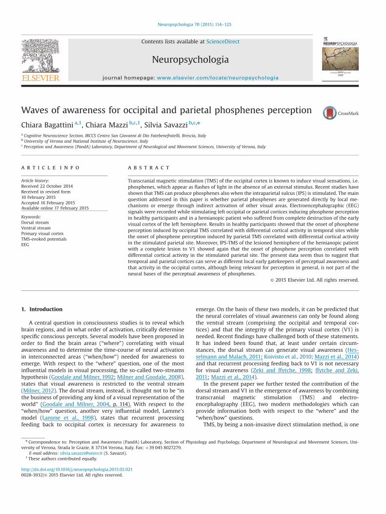
Fig. 2. Experiment 2. Results and analyses with hemianopic patient SL. (A) Brain lesion reconstruction. (B) Visual field defect. (C) Site of stimulation. The intersection of thedashed lines, superimposed on the 3D reconstruction of the patients’ brain, represents the hot-spot for parietal stimulation. (D) Psychometric threshold function obtainedwith patient SL. The red line represents the threshold function obtained after TMS of the parietal cortex. Each dot indicates the patient's performance at each TMS intensity.(E) TEPs elicited by TMS pulses over P3 as a function of the phosphene-present (red) and phosphene-absent (black) conditions. Dotted boxes mark the time windows inwhich statistical analysis were performed. Dark gray boxes indicate statistically significant results while light gray boxes the time windows resulted to be not significant. Theblack box indicates the time window in which the TMS artifact was present. The figure inset depicts the left hemisphere electrodes (red circles) significant at the early timewindow. (F) Illustration of the results of the bootstrap analysis performed at each of the four waveform peaks. Solid red bars indicate the mean amplitude of the differencebetween phosphene-present and phosphene-absent trials. Solid black lines correspond to zero. Dotted black lines mark the 5th percentile value which, if above zero, indicatea statistically significant difference (Peaks 1 and 2). The exact p-values correspond to the proportion of resamples that were smaller than zero. (For interpretation of thereferences to color in this figure legend, the reader is referred to the web version of this article.)
C. Bagattini et al. / Neuropsychologia 70 (2015) 114–125120

C. Bagattini et al. / Neuropsychologia 70 (2015) 114–125 121
genuineness of phosphenes in the healthy group was used forpatient SL.
3.1.3. Behavioral data statistical analysisTo compare the performance of patient SL with that of the
healthy participants we used two tests developed by Crawford andcolleagues which are considered the most suitable analyses whenthe normative sample is small. Specifically, we used (1) the Sin-glims_ES.exe program (Crawford and Garthwaite, 2002; Crawfordand Howell, 1998) which tested the hypothesis that the score of asingle individual lies within or outside the normal range of scoresdefined by the mean and standard deviation of control group'sdata and (2) the RSDT_ES.exe program (Crawford and Garthwaite,2005; Crawford et al., 2010) to compare differences between sin-gle-case scores on two conditions and the differences observed ina control sample. See Mazzi et al. (2014) for a more detailed de-scription of the tests.
3.1.4. TEPs analysisFor homogeneity with the analysis performed with healthy
participants we analyzed TEPs recorded at the Pz electrode (in thepresent case EEG recorded at P3 electrode was not affected by TMSartifacts, data from the P3 electrode is thus reported in the sub-sequent spatio-temporal analysis). By visually inspecting theaveraged TEPs as recorded at the Pz electrode, four time windowswith amplitude peaks could be identified: around �80 ms(phosphene present: 78 ms; phosphene absent: 77 ms), �170 ms(phosphene present: 166 ms; phosphene absent: 171 ms),�250 ms (phosphene present: 250 ms; phosphene absent:253 ms) and �395 ms (phosphene present: 393 ms; phospheneabsent: 402 ms) after the TMS pulse (Fig. 2E). Peak latencies weredetected separately for the two conditions and the different timewindows by means of the peak latency detection module im-plemented in BrainVision Analyzer 1.05. Given that using a singlepeak value when analyzing single trials data can be very noisy(Oruç et al., 2011), we used the peak export solution implementedin BrainVision Analyzer 1.05 to export, for each condition and timewindow, the mean amplitude values in a time window centeredon the previously identified peaks at an average level (a 40-mstime window for the first two peaks and 80-ms for the two laterpeaks). After pre-processing the EEG data, the phosphene-presentand phosphene-absent conditions resulted in 164 and 177 trials,respectively. Given that we cannot predict which trial in the se-quence would result in a phosphene-present or absent outcomeand, more importantly, that the two conditions did not result inthe same number of trials, before running the analysis, the order oftrials was randomized within each of the two conditions (phos-phene present/absent). For single-subject analysis we used thefirst 164 trials per condition and we calculated the difference be-tween the mean amplitude for phosphene-present and phos-phene-absent trials per each trial pair. The difference in amplitudefor the two conditions was then analyzed by means of a non-parametric Monte Carlo percentile bootstrap simulation (Efronand Tibshirani, 1993; Oruç et al., 2011). This procedure creates asimulated data distribution by re-sampling the raw data with re-placement. We created 50,000 re-samples of 164 trials each for thephosphene-present minus phosphene-absent amplitude values.The lower 5th percentile of the re-sampled data distribution servedas the critical values for the one-tailed 0.05 significance level. Ifthe 5th percentile results to be above the zero level (phosphene-present4phosphene-absent), it means that the phosphene-pre-sent condition yields a significantly larger amplitude than thephosphene-absent condition. This analysis was performed sepa-rately for each of the four time windows. For the spatio-temporalanalysis involving the other electrodes we adopted the sameprocedure described above. In a visual inspection of the entire set
of electrodes, three peaks can be detected (the last windowpeaking at �395 ms after TMS was visible only for Pz). The firstpeak detectable by visually inspecting the TEPs had a latency of�80 ms for electrodes O1, O2, P7, P3, Pz, P4, CP5, CP1, T7, C3, Cz,FC5 and F7, of �95 ms for electrodes CP2, C4, FC1, FC2, F3 and F4,and of �110 ms for electrodes P8, T8, CP6, FC6, F4 and Fz. Thesecond peak, with a latency of �165/170 ms after TMS was visiblefor all electrodes (O1, O2, P7, P3, P4, P8, CP5, CP1, CP2, CP6, T7, C3,Cz, C4, T8, FC5, FC1, FC2, FC6, F7, F3, Fz, F4, F8). The third peak wasdetectable at �250 ms after TMS for electrodes P3 and P4 and at�280 ms after TMS for electrode P8. No later peaks could beidentified for any electrodes with the exception of Pz (see above)and no analysis was performed for this time window. ElectrodesFp1 and Fp2 did not show any clear peaks at any time windowsand were not further analyzed.
3.2. Results and discussion
3.2.1. Phosphene threshold functionSL, as previously reported (Mazzi et al., 2014), could experience
phosphenes when TMS was applied over the parietal cortex of herlesioned hemisphere (Fig. 2C) with eyes both open and blind-folded. In the previous paper and in the present one, inducedphosphenes were localized in the upper right hemifield, con-tralateral to the stimulation site, and were described as very brief,static and mostly greyish or white.
The maximum stimulator output (MSO) intensity capable ofeliciting a phosphene in 50% of trials was 71%, a value not sig-nificantly different [t(8)¼�0.197; p¼0.85, Z-CC¼�0.208] fromthe mean threshold values obtained stimulating the left parietalcortex of healthy participants in Experiment 1. These results are inline with those obtained by Mazzi et al. (2014) reporting that apsychophysical threshold function (Fig. 2D) for parietal phospheneperception could be created also for a patient lacking V1 and thather threshold function did not differ from the function obtainedwith healthy participants.
3.2.2. Behavioral resultsWhen stimulating the ipsilesional parietal cortex (P3), SL reported
perceiving a phosphene in 47.22% of the trials (N¼170), while single-pulse TMS did not elicit a phosphene in the remaining 52.78% of trials(N¼190). The percentage of perceived parietal phosphenes did notdiffer from the mean percentage value obtained in Experiment 1 withhealthy participants [t(8)¼0.381; p¼0.71, Z-CC¼0.401]. Moreover, thepatient's response times showed a speeding up in phosphene-present (mean¼1459.07 ms) versus phosphene-absent(mean¼1702.79 ms) trials that was comparable [t(8)¼0.303; p¼0.77,Z-DCC¼0.356] to that observed during parietal stimulation of healthyparticipants in Experiment 1, thus providing evidence of the reliabilityof patient's self-reports (i.e. the presence/absence of a visual percept).
3.2.3. TEPs resultsFig. 2E and F shows TEPs recorded at electrode Pz for left par-
ietal cortex stimulation and the histograms of the phosphene-present/phosphene-absent contrast values obtained from thebootstrap analysis separated for the four time windows. As it canbe seen from the figures, the first two peaks were significant (Peak1, Exact p¼0.010; Peak 2, Exact p¼0.017), both having more po-sitive power values for the phosphene-present condition. The lasttwo peaks (at �250 and �395 ms after TMS pulse) were insteadnot significant (Peak 3, Exact p¼0.059; Peak 4, Exact p¼0.091).Importantly, the first peak (at �80 ms after TMS pulse) indexesthe early onset of the phosphene present/absent effect as detectedat the Pz electrode.
The spatio-temporal analysis revealed that, in addition to Pz, asignificant early difference between phosphene-present and
C. Bagattini et al. / Neuropsychologia 70 (2015) 114–125122
phosphene-absent responses was found at �79 ms after the TMSpulse at scalp site P3 (phosphene-present peak: 80 ms; phos-phene-absent peak: 78 ms, Exact p¼0.027) and at �104 ms afterthe TMS pulse at scalp site Fz (phosphene-present peak: 105 ms;phosphene-absent peak: 103 ms, Exact p¼0.013). At the secondpeak, in addition to Pz, a significant difference was found at scalpsites P3 (phosphene-present peak: 163 ms; phosphene-absentpeak: 169 ms, Exact p¼0.0003), P4 (phosphene-present peak:168 ms; phosphene-absent peak: 170 ms, Exact p¼0.0069), P8(phosphene-present peak: 170 ms; phosphene-absent peak:172 ms, Exact p¼0.0092), CP1 (phosphene-present peak: 162 ms;phosphene-absent peak: 164 ms, Exact p¼0.0088), CP2 (phos-phene-present peak: 164 ms; phosphene-absent peak: 168 ms,Exact p¼0.015), CP6 (phosphene-present peak: 168 ms; phos-phene-absent peak: 172 ms, Exact p¼0.047). At the third peaknone of the analyzed electrodes (P3, P4 and P8) showed a sig-nificant difference between phosphene-present and phosphene-absent responses (Exact p40.05).
Taken together, these results, in line with those obtained withhealthy participants, show that the onset of the phosphene pre-sent/absent effect can be found at parietal sites (P3, Pz) and thatthe effect spreads to circumscribed functionally connected centro-parietal sites. Given that temporal sites do not show any effect, itcan be concluded that these sites are not part of the functionalnetwork generating the effect, thus reinforcing the idea that theparietal cortex is an early and independent generator of phos-phenes. Moreover, the presence of this effect in a patient with acomplete lesion to the ipsilateral occipital cortex is in line with theidea that feedback to V1 is not necessary for the awareness ofparietal phosphenes.
4. General discussion
The present paper aimed to establish the neural dynamics ofthe awareness of occipital and parietal phosphenes by in-vestigating TEPs in healthy participants and one hemianopic pa-tient with a lesion to V1. Phosphenes were induced by stimulatingV1/V2 and IPS while EEG signals were recorded. In line withprevious data (Mazzi et al., 2014), the present results show thatboth healthy participants and a patient with a complete lesion ofthe ipsilateral V1 experienced phosphenes induced by TMS of IPSand that IPS-phosphenes have a higher phosphene-threshold andwere signalled faster than those elicited by occipital TMS inhealthy participants (see also Fried et al., 2011).
The novelty of the present paper relies on the use of a EEG–TMS interactive co-registration approach (Miniussi and Thut,2010), which gave us the opportunity to directly measure thespatio-temporal dynamics of the cortical reactivity and con-nectivity (Ilmoniemi et al., 1997; Miniussi and Thut, 2010) due tothe presence or absence of a phosphene and to draw some con-clusions regarding the role of the occipital and parietal corticesduring the emergence of awareness. Given the poor spatial re-solution of the EEG technique, especially when performed with alow number of electrodes, rendering source localization analysis(e.g. LORETA) unreliable, the investigation of the exact source ofneural activity in specific brain regions (e.g. a gyrus, sulcus or sub-cortical structure) goes beyond the scope of the present work.Instead, we adopted a surface-source imaging approach (the SCDanalysis), which provided us with an approximation of the localcurrent density flowing perpendicularly to the scalp, which is areliable analysis evenwith the present number of electrodes (Luck,2014, p. 165). By focusing on the different time windows of activitycorrelating with the presence/absence of a phosphene, we couldidentify both early gatekeepers of the awareness of phosphenesand late consequences of the ignition induced by previous neural
activity.In healthy participants, the awareness of phosphenes induced
by occipital TMS correlated with activity in centro-temporal sitesat an early phase (�70 ms after TMS) and in occipital sites at a latephase (starting at �320 ms after TMS). Conversely, the awarenessof phosphenes induced by parietal TMS correlated with activity incentro-parietal sites at an early phase (�70 ms after TMS) and inoccipital sites at a late phase (starting at �280 ms after TMS).Moreover, in the second experiment, IPS-TMS of the lesionedhemisphere in a patient with a complete lesion to the ipsilateralV1, showed that the awareness of phosphenes correlated withactivity in parietal sites at an early phase (�80 ms after TMS),spreading to centro-parietal sites at �165/170 ms after TMS. Theactivity induced by TMS does not extend to any later phase in-dicating that the late phase of activity in occipital sites found withhealthy participants is not necessary for the awareness of theparietal phosphenes to emerge. Therefore, these data show dif-ferent topographic maps and different time-courses of the phos-phene present/absent effect depending on the cortical area beingstimulated.
The different spatio-temporal dynamics of phosphene percep-tion for the two stimulated sites in healthy participants and thedifferences found between healthy participants and the hemi-anopic patient for parietal phosphenes give us the opportunity toadvance possible answers to the questions stated in the in-troduction about the “where” and “when/how” of the emergenceof perceptual awareness.
Previous data (Mazzi et al., 2014) and the present paper testedthe hypothesis that neural activity along the dorsal stream couldhave access to perceptual awareness. Here, we found that directstimulation of IPS induces the perception of phosphenes and thatthis effect has its onset in the parietal cortex at �70–80 ms afterTMS. This result could point to a role of the parietal cortex as alocal early gatekeeper of awareness. It could, however, be surmisedthat parietal phosphenes are generated in other visual areas (suchas V1 or areas along the ventral stream) strongly connected to IPS.For this hypothesis to be tenable, one should find early neuralactivity correlating with the awareness of IPS-phosphenes in oc-cipital or temporal areas. With respect to the involvement of V1,we found that neural activity correlating with phosphene per-ception can be detected in the occipital cortex of healthy partici-pants only at a late time window after TMS of IPS. Moreover, inline with a previous report (Mazzi et al., 2014), we showed that apatient with a complete lesion to the ipsilateral V1 can still per-ceive phosphenes induced by IPS-TMS and that no activity at oc-cipital sites (ipsi- and contra-lateral to TMS) can be detected at anytime windows of the entire epoch, thus ruling out any contribu-tions of feedback to V1 as a necessary mechanism for awareness toemerge (Zeki and ffytche, 1998; ffytche and Zeki, 2011). Similarly,with respect to the involvement of other areas along the ventralstream, we found no early activity in temporal sites after IPS-TMSneither in healthy participants (for whom SCD topographic mapsare compatible with parietal generators) or the hemianopic pa-tient (where no activity was found at temporal sites along theentire epoch), thus ruling out the possibility that the ventralstream, via its strong connections with the dorsal stream, couldhave contributed to the emergence of awareness of the IPS-phosphenes. Taken together, the results obtained with TEPs eli-cited by IPS-TMS show that IPS is an early and independent gen-erator of phosphenes.
The role of the occipital and ventral cortices need to be furtherdiscussed in relation to the generation of phosphenes induced byTMS of V1/V2. In the present paper we found that occipitalphosphenes correlated in healthy participants with an early onsetof the phosphene present/absent effect in temporal sites. In linewith a previous report (Taylor et al., 2010), occipital sites' activity

C. Bagattini et al. / Neuropsychologia 70 (2015) 114–125 123
correlating with phosphene perception can only be found in a latetime window. Unfortunately, the cited paper did not investigatethe phosphene present/absent effect in sites other than the occi-pital cortex, thus rendering it difficult to determine a directcomparison between these and the previous results in terms ofpossible early activity in temporal sites correlating with occipitalphosphenes. Interestingly, activity along the ventral stream hasbeen found recently by combining TMS with fMRI (Halko et al.,2013). In this paper, phosphenes were induced by TMS of V1 andhemodynamic responses were measured. In line with the presentresults, the authors found neural activity correlating with the TMSpulse at both the lower left V1 (the targeted area) and lateral oc-cipito-temporal cortex. Unfortunately, temporal resolution of fMRIis very poor, thus rendering it difficult to gather the temporal or-der of activations in occipital and temporal sites.
The kind of spatio-temporal dynamics found in the presentpaper could thus signal that temporal cortex is the early gate-keeper for the awareness of occipital phosphenes, in line with thenotion that activity in higher-order visual areas along the ventralstream is best correlated with visual awareness than V1 per se(Rees et al., 2002). Activity in the occipital cortex would then re-present a consequence of the feedback spreading from the tem-poral cortex, despite not having a causal role in the emergence ofthe phosphene perception. In line with the idea of a functionalcross-talk between early and late visual cortices not having acausal role in awareness, are the results of parietal phosphenes inthe hemianopic patient: no late (i.e. after 170 ms to TMS pulse)phosphene present/absent effect is found for any cortical sites.This evidence could be explained as the lack of activity in V1 dueto the brain lesion i.e., neural activity in the parietal cortex, al-though causing feedback to V1 due to its anatomical connections,cannot receive the second sweep of activity from V1 and thus nolate effects can be found in the parietal cortex.
Taken together, the results obtained with TMS of occipital andparietal sites, given the lack of early activity in occipital sitescorrelating with the awareness of phosphenes, can dismiss thecausal role of V1 as an early gatekeeper of awareness. Indeed, earlyactivity can be found only at parietal and temporal sites after TMSof parietal and occipital sites, respectively. However, a questionstill remains open as to the existence of a common site for theawareness of phosphenes, contributing to explain the summedactivity found at temporal and parietal cortical sites. Given the lackof power of the present paper in being conclusive in finding thesource of the EEG signal, only some speculations can be provided.Previous findings have shown that subcortical structures, such asthe lateral geniculate nucleus (Kastner et al., 2006) can serve as anearly gatekeeper in the control of visual attention and awareness.Future studies should therefore investigate the possibility that thismight be the case not only for visual stimuli presented to the eyesbut also for visual percepts generated by direct cortical stimulationby means of TMS. In the present paper, however, we have shownthat the SCD topographical maps are different for the two stimu-lation sites, thus suggesting different subcortical generators(Vaughan, 1982; Michel et al., 2004) for occipital and parietalphosphenes. A finer arrangement of electrodes should be used infuture studies to strengthen our conclusions that occipital andparietal phosphenes are generated by different areas. Alter-natively, an optimal, although more technically demanding, can-didate for this investigation and for overcoming the limitations ofthe present results and obtaining a finer localization of the brainsources of the awareness of occipital and parietal phosphenes,would be event-related optical imaging (Gratton and Fabiani,2010; Wolf et al., 2008). Future studies could thus consider thepossibility to study phosphene perception using this techniquewhich, thanks to its high temporal (o10 ms) and spatial (�1 cm3)resolution, would add to the present results, providing a better
understanding not only on the timing but also on the exact neuralstructures involved in the perception of occipital and parietalphosphenes.
The present data seem to be in favor of the proposal (Mou-toussis and Zeki, 2002; Beauchamp et al., 2012) that local pro-cesses in high-order visual areas, specifically, the temporal andparietal cortex, could serve as early gatekeepers in which activitygenerates the emergence of conscious visual percepts (i.e. re-flecting the correlates of “phenomenal awareness”; Block, 1996). Inthis respect, late activity found with healthy participants in theoccipital or frontal cortex could be considered as the consequenceof the “ignition” (Fisch et al., 2009) engendered in the temporal orparietal cortex (i.e. reflecting the correlates of “access awareness”;Block, 1996). However, in the literature there is a strong debate onthe early vs. late correlates of visual awareness. Several event-re-lated potential (ERP) studies have shown that the neural processesdirectly correlating with consciousness occur in the relatively earlytime window (e.g. Bachmann, 2009; Koivisto and Revonsuo, 2010;Railo et al., 2011) whereas other researchers have concluded thatthe neural processes directly correlating with visual awarenessoccur later (e.g. Dehaene and Changeux, 2011; Salti et al., 2012).The inconsistent results present in the literature render it unclearwhich time windows should be considered to correlate with theneural processing for generating consciousness (NCC), which cor-relate with the preceding processes (NCC-pr) and which with theconsequences (NCC-co) of conscious perception (Aru et al., 2012).In this respect, it is important to note that the timing at which aneural process occurs cannot per se be informative but specificexperimental manipulations need to be adopted (Aru et al., 2012).As an example in a recent paper (Pitts et al., 2014), the authorsorthogonally manipulated visual awareness and task relevanceand they found that late effects (i.e. the P300 component of ERPs)had to be considered as reflecting post-perceptual processes (NCC-co), not visual awareness per se (NCC). In the present paper, wehad the opportunity to study a patient with a complete lesion tothe ipsilateral V1 and we found that the neural processes corre-lating with the awareness of phosphenes remained confined in anearly phase (with two peaks at �70–80 ms and �165–170 msafter TMS). Importantly, no effect was present in a late phase,despite the patient being aware of the presence/absence of thephosphenes. This piece of evidence, given the lack of late effects inpatient SL but the presence of awareness, should imply that thelate activity found with healthy participants in occipital andfrontal areas as reflecting post-perceptual processes (NCC-co) andnot visual awareness per se. Indeed, if the late activity is a properneural correlate of awareness, no awareness should be possiblewithout it. Conversely, it remains unclear whether the early ac-tivity found in both healthy participants and patient SL reflectsonly the NCC or a combination of NCC and NCC-pr. Unfortunately,the present experiment is neutral in this respect, both because nomanipulation of the NCC-pr was adopted (Aru et al., 2012) andbecause a direct comparison between TEPs and ERPs latencies wasnot possible, given that in the former the entire subcorticalpathway processing real stimuli was lacking, thus leaving uswithout any clues about the nature of neural processes precedingvisual awareness when the percept was directly generated in thecortex. It would therefore be of great interest for future studies toadopt specific experimental manipulations to disentangle NCC-prfrom NCC in phosphene perception.
An interesting aspect of the present results relates to the pre-sence of visual qualia induced by TMS of the IPS that can be ob-tained also in the absence of a functioning V1. As discussed else-where (Mazzi et al., 2014; Silvanto, 2015), the presence of visualqualia in hemianopic patients has already been documented afterTMS (Silvanto et al., 2007, 2008). Differently for previous reports,however, the present data and those obtained by Mazzi et al.
C. Bagattini et al. / Neuropsychologia 70 (2015) 114–125124
(2014) show that the stimulation of the sole damaged hemispherein SL (and of patients SL and AG in Mazzi et al., 2014) was capableof inducing phosphenes in the blind field (see Silvanto, 2015 for athoughtful discussion on the dissimilarities among the differentstudies). In a similar respect, a final point that deserves someconsideration relates to the role of the primary visual cortex invision. Indeed, a lesion to the primary visual cortex abolishes theability to consciously perceive external visual stimuli. If, as thepresent data seem to suggest, V1 is not necessary for the per-ception of parietal phosphenes one should try to explain why V1seems to be essential for normal vision. An interesting discussioncan be found in a very recent review by Silvanto (2015, but see alsoSilvanto, 2008). In his view, of which we agree, one should con-sider the role played by a V1 lesion as affecting not only thefunctioning of V1 itself but that of all the visual areas in thehierarchy of visual processing. In this respect, a lesion to V1 wouldmake the entire visual cortex, not just V1, still capable of low-levelvisual functions but not of maintaining perceptual awareness.
In conclusion, the present data show that temporal and parietalcortices, at least under the present circumstances, can serve asdifferent local early gatekeepers of perceptual awareness in thehuman brain and that activity in the occipital cortex, althoughbeing relevant for perception in general (Koivisto et al., 2010;Silvanto, 2008, 2015), is not part of the neural bases of perceptualawareness (Crick and Koch, 1998).
Acknowledgments
We thank Javier Sánchez-López for the help with the SCD to-pographic maps and Ramisha S. Knight for English proofreading.
References
Abrahamyan, A., Clifford, C.W.G., Ruzzoli, M., Phillips, D., Arabzadeh, E., Harris, J.A.,2011. Accurate and rapid estimation of phosphene thresholds (REPT). PLoS One6, e22342.
Anand, S., Hotson, J., 2002. Transcranial magnetic stimulation: neurophysiologicalapplications and safety. Brain Cogn. 50, 366–386.
Antal, A., Nitsche, M.A., Paulus, W., 2001. External modulation of visual perceptionin humans. NeuroReport 12, 3553–3555.
Aru, J., Bachmann, T., Singer, W., Melloni, L., 2012. Distilling the neural correlates ofconsciousness. Neurosci. Biobehav. Rev. 36, 737–746.
Bachmann, T., 2009. Finding ERP-signatures of target awareness: puzzle persistsbecause of experimental co-variation of the objective and subjective variables.Conscious. Cogn. 18, 804–808.
Beauchamp, M.S., Sun, P., Baum, S.H., Tolias, A.S., Yoshor, D., 2012. Electro-corticography links human temporoparietal junction to visual perception. Nat.Neurosci. 15 (7), 957–959.
Beckers, G., Hömberg, V., 1992. Cerebral visual motion blindness: transitory aki-netopsia induced by transcranial magnetic stimulation of human area V5. Proc.Biol. Sci. 249, 173–178.
Block, N., 1996. How can we find the neural correlate of consciousness? TrendsNeurosci. 19 (11), 456–459.
Crawford, J.R., Garthwaite, P.H., 2002. Investigation of the single case in neu-ropsychology: confidence limits on the abnormality of test scores and test scoredifferences. Neuropsychologia 40, 1196–1208.
Crawford, J.R., Garthwaite, P.H., Porter, S., 2010. Point and interval estimates of ef-fect sizes for the case-controls design in neuropsychology: rationale, methods,implementations, and proposed reporting standards. Cogn. Neuropsychol. 27,245–260.
Crawford, J.R., Garthwaite, P.H., 2005. Testing for suspected impairments and dis-sociations in single-case studies in neuropsychology: evaluation of alternativesusing Monte Carlo simulations and revised tests for dissociations. Neu-ropsychology 19, 318–331.
Crawford, J.R., Howell, D.C., 1998. Comparing an individual's test score againstnorms derived from small samples. Clin. Neuropsychol. 12, 482–486.
Crick, F., Koch, C., 1998. Consciousness and neuroscience. Cereb. Cortex 8, 97–107.Dehaene, S., Changeux, J.P., 2011. Experimental and theoretical approaches of
conscious processing. Neuron 70, 200–227.Efron, B., Tibshirani, R.J., 1993. An Introduction to the Bootstrap. Chapman Hall,
New York, NY.Fisch, L., Privman, E., Ramot, M., Harel, M., Nir, Y., Kipervasser, S., Andelman, F.,
Neufeld, M.Y., Kramer, U., Fried, I., et al., 2009. Neural “ignition”: enhanced
activation linked to perceptual awareness in human ventral stream visualcortex. Neuron 64, 562–574.
ffytche, D.H., Zeki, S., 2011. The primary visual cortex, and feedback to it, are notnecessary for conscious vision. Brain 134, 247–257.
Fried, P.J., Elkin-Frankston, S., Rushmore, R.J., Hilgetag, C.C., Valero-Cabre, A., 2011.Characterization of visual percepts evoked by noninvasive stimulation of thehuman posterior parietal cortex. PLoS One 6, e27204.
Goodale, M.A., Milner, A.D., 1992. Separate visual pathways for perception andaction. Trends Neurosci. 15, 20–25.
Goodale, M.A., Milner, A.D., 2004. Sight Unseen. An Exploration of Conscious andUnconscious Vision. Oxford University Press, Oxford.
Gratton, G., Fabiani, M., 2010. Fast optical imaging of human brain function. Front.Hum. Neurosci. 4, 52.
Guthrie, D., Buchwald, J.S., 1991. Significance testing of difference potentials. Psy-chophysiology 28, 240–244.
Halko, M.A., Eldaief, M.C., Pascual-Leone, A., 2013. Noninvasive brain stimulation inthe study of the human visual system. J. Glaucoma 22, S39–S41.
Hesselmann, G., Malach, R., 2011. The link between fMRI-BOLD activation andperceptual awareness is “stream-invariant” in the human visual system. Cereb.Cortex 21 (12), 2829–2837.
Ilmoniemi, R.J., Virtanen, J., Ruohonen, J., Karhu, J., Aronen, H.J., Näätänen, R., Katila,T., 1997. Neuronal responses to magnetic stimulation reveal cortical reactivityand connectivity. Neuroreport 8 (16), 3537–3540.
Kammer, T., Beck, S., 2002. Phosphene thresholds evoked by transcranial magneticstimulation are insensitive to short-lasting variations in ambient light. Exp.Brain Res. 145, 407–410.
Kammer, T., Puls, K., Erb, M., Grodd, W., 2005. Transcranial magnetic stimulation inthe visual system. II. Characterization of induced phosphenes and scotomas.Exp. Brain Res. 160, 129–140.
Kastner, S., Schneider, K.A., Wunderlich, K., 2006. Beyond a relay nucleus: neuroi-maging views on the human LGN. Prog. Brain Res. 155, 125–143.
Koivisto, M., Mäntylä, T., Silvanto, J., 2010. Neuroimage 51, 828–834.Koivisto, M., Revonsuo, A., 2010. Event-related potential correlates of visual
awareness. Neurosci. Biobehav. Rev. 34, 922–934.Lamme, V.A.F., Super, H., Spekreijse, H., 1998. Feedforward, horizontal and feedback
processing in the visual cortex. Curr. Opin. Neurobiol. 8, 529–535.Luck, S.J., 2014. An Introduction to the Event-related Potential Technique, second
edition. MIT Press, Cambridge, MA.Marzi, C.A., Mancini, F., Savazzi, S., 2009. Interhemispheric transfer of phosphenes
generated by occipital versus parietal transcranial magnetic stimulation. Exp.Brain Res. 192, 431–441.
Mazzi, M., Mancini, F., Savazzi, S., 2014. Can IPS reach visual awareness without V1?Evidence from TMS in healthy subjects and hemianopic patients. Neu-ropsychologia 64, 134–144.
Meyer, B.U., Diehl, R.R., Steinmetz, H., Britton, T.C., Benecke, R., 1991. Magneticstimuli applied over motor cortex and visual cortex: influence of coil positionand field polarity on motor responses, phosphenes, and eye movements.Electroencephalogr Clin Neurophysiol Suppl. 43, 121–134.
Michel, C.M., Murray, M.M., Lantz, G., Gonzales, S., Spinelli, L., Grave de Peralta, R.,2004. EEG source imaging. Clin. Neurophysiol. 115 (10), 2195–2222.
Milner, A.D., 2012. Is visual processing in the dorsal stream accessible to con-sciousness? Proc. Biol. Sci. 279, 2289–2298.
Milner, A.D., Goodale, M.A., 2008. Two visual systems re-viewed. Neuropsychologia46, 786–802.
Miniussi, C., Thut, G., 2010. Combining TMS and EEG offers new prospects in cog-nitive neuroscience. Brain Topogr. 22, 249–256.
Moutoussis, K., Zeki, S., 2002. The relationship between cortical activation andperception investigated with invisible stimuli. Proc. Natl. Acad. Sci. USA 99,9527–9532.
Murray, M.M., Wylie, G.R., Higgins, B.A., Javitt, D.C., Schroeder, C.E., Foxe, J.J., 2002.The spatiotemporal dynamics of illusory contour processing: combined high-density electrical mapping, source analysis, and functional magnetic resonanceimaging. J. Neurosci. 22, 5055–5073.
Oldfield, R.C., 1971. The assessment and analysis of handedness: the Edinburghinventory. Neuropsychologia 9 (1), 97–113.
Oruç, I., Krigolson, O., Dalrymple, K., Nagamatsu, L.S., Handy, T.C., Barton, J.J.S., 2011.Bootstrap analysis of the single-subject with event related potentials. Cogn.Neuropsychol. 28 (5), 322–337.
Pitts, M.A., Padwal, J., Fennelly, D., Martínez, A., Hillyard, S.A., 2014. Gamma bandactivity and the P3 reflect post-perceptual processes, not visual awareness.Neuroimage 101, 337–350.
Perrin, F., Pernier, J., Bertrand, O., Echallier, J.F., 1989. Spherical splines for scalp andcurrent density mapping. Electroencephalogr. Clin. Neurophysiol. 72 (2),184–187.
Railo, H., Koivisto, M., Revonsuo, A., 2011. Tracking the processes behind consciousperception: a review of event-related potential correlates of visual conscious-ness. Conscious. Cogn. 20, 972–983.
Rees, G., Kreiman, G., Koch, C., 2002. Neural correlates of consciousness in humans.Nat. Neurosci. Rev. 2, 261–270.
Rossi, S., Hallett, M., Rossini, P.M., Pascual-Leone, A., 2009. Safety of TMS ConsensusGroup. Safety, ethical considerations, and application guidelines for the use oftranscranial magnetic stimulation in clinical practice and research. Clin. Neu-rophysiol. 120 (12), 2008–2039.
Rossi, S., Hallett, M., Rossini, P.M., Pascual-Leone, A., 2011. Screening questionnairebefore TMS: an update. Clin. Neurophysiol. 122 (8), 1686.
Salminen-Vaparanta, N., Noreika, V., Revonsuo, A., Koivisto, M., Vanni, S., 2012. Is

C. Bagattini et al. / Neuropsychologia 70 (2015) 114–125 125
selective primary visual cortex stimulation achievable with TMS? Hum. BrainMapp. 33 (3), 652–665.
Salti, M., Bar-Haim, Y., Lamy, D., 2012. The P3 component of the ERP reflects con-scious perception, not confidence. Conscious. Cogn. 21, 961–968.
Sekiguhi, H., Takeuchi, S., Katoda, H., Kohno, Y., Nakajima, Y., 2011. TMS-inducedartifacts on EEG can be reduced by rearrangement of the electrode's lead wirebefore recording. Clin. Neurophysiol. 122 (5), 984–990.
Silvanto, J., 2008. A re-evaluation of blindsight and the role of striate cortex (V1) invisual awareness. Neuropsychologia 46, 2869–2871.
Silvanto, J., 2015. Why is “blindsight” blind? A new perspective on primary visualcortex, recurrent activity and visual awareness. Conscious. Cogn. 32, 15–32.
Silvanto, J., Cowey, A., Lavie, N., Walsh, V., 2007. Making the blindsighted see.Neuropsychologia 45, 3346–3350.
Silvanto, J., Cowey, A., Walsh, V., 2008. Inducing conscious perception of colour inblindsight. Curr. Biol. 18, R950–R951.
Stewart, L., Battelli, L., Walsh, V., Cowey, A., 1999. Motion perception and perceptuallearning studied by magnetic stimulation. Electroencephalogr. Clin. Neuro-physiol. Suppl. 51, 334–350.
Taylor, P.C.J., Walsh, V., Eimer, M., 2010. The neural signature of phosphene
perception. Hum. Brain Mapp. 31, 1408–1417.Thielscher, A., Reichenbach, A., Uğurbil, K., Uludağ, K., 2010. The cortical site of
visual suppression by transcranial magnetic stimulation. Cerebr. Cortex 20,328–338.
Vaughan, H.G., 1982. The neural origins of human event-related potentials. Ann. N.Y. Acad. Sci. 388, 125–138.
Veniero, D., Bortoletto, M., Miniussi, C., 2009. TMS–EEG co-registration: on TMS-induced artifact. Clin. Neurophysiol. 120 (7), 1392–1399.
Wassermann, E.M., 1998. Risk and safety of repetitive transcranial magnetic sti-mulation: report and suggested guidelines from the International Workshop onthe Safety of Repetitive Transcranial Magnetic Stimulation. Electroencephalogr.Clin. Neurophysiol. 108, 1–16.
Wolf, M., Morren, G., Haensse, D., Karen, T., Wolf, U., Fauchere, J.C., et al., 2008. Nearinfrared spectroscopy to study the brain: an overview. Opto-Electron. Rev. 16,413–419.
Zeki, S., ffytche, D.H., 1998. The Riddoch syndrome: insights into the neurobiologyof conscious vision. Brain 121, 25–45.
Related Documents