RESEARCH Open Access Visfatin induces MUC8 and MUC5B expression via p38 MAPK/ROS/NF-κB in human airway epithelial cells Si-Youn Song 1† , Eun Chae Jung 1† , Chang Hoon Bae 1 , Yoon Seok Choi 1 and Yong-Dae Kim 1,2* Abstract Background: Among a variety of inflammatory mediators, visfatin is a proinflammatory adipocytokine associated with inflammatory reactions in obesity, metabolic syndrome, chronic inflammatory disease, and autoimmune disease. However, the biological role of visfatin in secretion of major mucins in human airway epithelial cells has not been reported. Therefore, this study was conducted in order to investigate the effect and the brief signaling pathway of visfatin on MUC8 and MUC5B expression in human airway epithelial cells. Results: Visfatin significantly induced MUC8 and MUC5B expression. Visfatin significantly activated phosphorylation of p38 MAPK. Treatment with SB203580 (p38 MAPK inhibitor) and knockdown of p38 MAPK by siRNA significantly blocked visfatin-induced MUC8 and MUC5B expression.Visfatin significantly increased ROS formation. Treatment with SB203580 significantly attenuated visfatin-induced ROS formation. Treatment with NAC (ROS scavenger) and DPI (NADPH oxidase inhibitor) significantly attenuated visfatin-induced MUC8 and MUC5B expression. However, treatment with NAC and DPI did not attenuate visfatin-activated phosphorylation of p38 MAPK. Visfatin significantly activated the phosphorylation of NF-κB. Treatment with PDTC (NF-κB inhibitor) significantly attenuated visfatin-induced MUC8 and MUC5B expression. Conclusions: These results suggest that visfatin induces MUC8 and MUC5B expression through p38 MAPK/ROS/NF-κB signaling pathway in human airway epithelial cells. Keywords: Visfatin, p38 MAPK, ROS, NF-κB, MUC8, MUC5B, Epithelial cell Background Mucins, highly glycosylated proteins, are the major com- ponents of mucus. Among the mucins, MUC8, MUC5B, and MUC5AC are representative secretory mucin genes in the human airway [1,2]. MUC8, MUC5B, and MUC5AC expression is increased by a variety of inflammatory medi- ators in airway inflammatory diseases such as chronic bronchitis [3]. Among a variety of inflammatory mediators, visfatin is a proinflammatory adipocytokine that is preferentially produced by visceral adipose tissue: visfatin is highly enriched in visceral fat and its expression level in plasma increases during development of obesity [4,5]. Although the biological role of visfatin is not entirely clear, visfatin is known to have insulin-mimetic action because it re- duces blood glucose level [6]. Several recent studies have reported an association of visfatin with inflammation and oxidative stress response: expression of visfatin is upregulated during activation of immune cells such as monocytes, macrophages, dendritic cells, T cells, and B cells, as well as in amniotic epithelial cells upon stimula- tion with lipopolysaccharide, TNF-α, IL-1β, or IL-6 [7-10]. And visfatin increases expression of inflammatory adhesion molecules in vascular endothelial cells in ROS- dependent manner through NF-κB signaling pathway [5]. Therefore, it could be hypothesized that visfatin may play a role in expression of mucin gene in human airway epithelial cells. However, the biological role of visfatin in secretion of major mucins in human airway epithelial cells has not been reported. For the this reason, this * Correspondence: [email protected] † Equal contributors 1 Department of Otorhinolaryngology-Head and Neck Surgery, College of Medicine, Yeungnam University, Daegu, Republic of Korea 2 Regional Center for Respiratory Diseases, Yeungnam University Medical Center, Daegu, Republic of Korea © 2014 Song et al.; licensee BioMed Central Ltd. This is an Open Access article distributed under the terms of the Creative Commons Attribution License (http://creativecommons.org/licenses/by/4.0), which permits unrestricted use, distribution, and reproduction in any medium, provided the original work is properly credited. The Creative Commons Public Domain Dedication waiver (http://creativecommons.org/publicdomain/zero/1.0/) applies to the data made available in this article, unless otherwise stated. Song et al. Journal of Biomedical Science 2014, 21:49 http://www.jbiomedsci.com/content/21/1/49

Welcome message from author
This document is posted to help you gain knowledge. Please leave a comment to let me know what you think about it! Share it to your friends and learn new things together.
Transcript

Song et al. Journal of Biomedical Science 2014, 21:49http://www.jbiomedsci.com/content/21/1/49
RESEARCH Open Access
Visfatin induces MUC8 and MUC5B expression viap38 MAPK/ROS/NF-κB in human airway epithelialcellsSi-Youn Song1†, Eun Chae Jung1†, Chang Hoon Bae1, Yoon Seok Choi1 and Yong-Dae Kim1,2*
Abstract
Background: Among a variety of inflammatory mediators, visfatin is a proinflammatory adipocytokine associatedwith inflammatory reactions in obesity, metabolic syndrome, chronic inflammatory disease, and autoimmunedisease. However, the biological role of visfatin in secretion of major mucins in human airway epithelial cells hasnot been reported. Therefore, this study was conducted in order to investigate the effect and the brief signalingpathway of visfatin on MUC8 and MUC5B expression in human airway epithelial cells.
Results: Visfatin significantly induced MUC8 and MUC5B expression. Visfatin significantly activated phosphorylationof p38 MAPK. Treatment with SB203580 (p38 MAPK inhibitor) and knockdown of p38 MAPK by siRNA significantlyblocked visfatin-induced MUC8 and MUC5B expression.Visfatin significantly increased ROS formation. Treatmentwith SB203580 significantly attenuated visfatin-induced ROS formation. Treatment with NAC (ROS scavenger) andDPI (NADPH oxidase inhibitor) significantly attenuated visfatin-induced MUC8 and MUC5B expression. However,treatment with NAC and DPI did not attenuate visfatin-activated phosphorylation of p38 MAPK. Visfatin significantlyactivated the phosphorylation of NF-κB. Treatment with PDTC (NF-κB inhibitor) significantly attenuated visfatin-inducedMUC8 and MUC5B expression.
Conclusions: These results suggest that visfatin induces MUC8 and MUC5B expression through p38 MAPK/ROS/NF-κBsignaling pathway in human airway epithelial cells.
Keywords: Visfatin, p38 MAPK, ROS, NF-κB, MUC8, MUC5B, Epithelial cell
BackgroundMucins, highly glycosylated proteins, are the major com-ponents of mucus. Among the mucins, MUC8, MUC5B,and MUC5AC are representative secretory mucin genes inthe human airway [1,2]. MUC8, MUC5B, and MUC5ACexpression is increased by a variety of inflammatory medi-ators in airway inflammatory diseases such as chronicbronchitis [3].Among a variety of inflammatory mediators, visfatin is
a proinflammatory adipocytokine that is preferentiallyproduced by visceral adipose tissue: visfatin is highlyenriched in visceral fat and its expression level in plasma
* Correspondence: [email protected]†Equal contributors1Department of Otorhinolaryngology-Head and Neck Surgery, College ofMedicine, Yeungnam University, Daegu, Republic of Korea2Regional Center for Respiratory Diseases, Yeungnam University MedicalCenter, Daegu, Republic of Korea
© 2014 Song et al.; licensee BioMed Central LCommons Attribution License (http://creativecreproduction in any medium, provided the orDedication waiver (http://creativecommons.orunless otherwise stated.
increases during development of obesity [4,5]. Althoughthe biological role of visfatin is not entirely clear, visfatinis known to have insulin-mimetic action because it re-duces blood glucose level [6]. Several recent studies havereported an association of visfatin with inflammationand oxidative stress response: expression of visfatin isupregulated during activation of immune cells such asmonocytes, macrophages, dendritic cells, T cells, and Bcells, as well as in amniotic epithelial cells upon stimula-tion with lipopolysaccharide, TNF-α, IL-1β, or IL-6[7-10]. And visfatin increases expression of inflammatoryadhesion molecules in vascular endothelial cells in ROS-dependent manner through NF-κB signaling pathway[5]. Therefore, it could be hypothesized that visfatin mayplay a role in expression of mucin gene in human airwayepithelial cells. However, the biological role of visfatin insecretion of major mucins in human airway epithelialcells has not been reported. For the this reason, this
td. This is an Open Access article distributed under the terms of the Creativeommons.org/licenses/by/4.0), which permits unrestricted use, distribution, andiginal work is properly credited. The Creative Commons Public Domaing/publicdomain/zero/1.0/) applies to the data made available in this article,

Figure 1 The effects of visfatin on MUC16, MUC8, MUC5B,MUC5AC, and MUC4 expression in NCI-H292 cells. RT-PCRshowed that visfatin induced remarkable MUC8 and MUC5B mRNAexpression. However, visfatin did not definitely induce MUC16,MUC5AC, and MUC4 mRNA expression. Images are representativeof three separate experiments performed in triplicate. Bars indicatethe mean ± S.D. of three independent experiments performed intriplicate. *P < 0.05 compared with zero value.
Song et al. Journal of Biomedical Science 2014, 21:49 Page 2 of 9http://www.jbiomedsci.com/content/21/1/49
study was conducted in order to investigate the effectand the brief signaling pathway of visfatin on MUC8 andMUC5B expression in human airway epithelial cells.
MethodsMaterialsVisfatin was purchased from Biovision Inc. (MountainView, CA, USA). RT-PCR kits were obtained from AppliedBiosystems (Foster City, CA, USA). Real-time PCR kitswere obtained from Roche Applied Science (Mannheim,Germany). Enhanced chemiluminescence kit was obtainedfrom Perkin Elmer Life Sciences (Boston, MA, USA).ERK1/2, phospho-ERK1/2, p38, phospho-p38, NF-κB, andphosphor-NF-κB were purchased from Cell SignalingTechnology (Danvers, MA, USA). The specific inhibitor,U0126 was purchased from Calbiochem (San Diego, CA,USA), SB203580 was purchased from BIOMOL (PlymouthMeeting, PA, USA), and NAC, DPI, and PDTC were pur-chased from Sigma-Aldrich (St. Louis, MO, USA). Prede-signed siRNA targeting p38 MAPK, negative controlsiRNA for p38 MAPK, OPTI-MEM I Reduced SerumMedium, and Lipofectamine 2000 were purchased fromInvitrogen Corporation (Carlsbad, CA, USA).For the primary culture, nasal mucosa was obtained
from normal inferior turbinate samples from 10 patientsundergoing augmentation rhinoplasty who had no per-sonal or family history of allergy, and who had negativeresults on skin-prick tests to 20 common airborne aller-gens and on multiple simultaneous allergen tests. Thisstudy was approved by the institutional review board forhuman studies at the Yeungnam University MedicalCenter and written informed consent was obtained fromeach patient.
Cell culture and treatmentHuman NCI-H292 airway epithelial cells (AmericanType Culture Collection, Manassas, VA, USA) were cul-tured in RPMI 1640 medium (Invitrogen Corporation)supplemented with 2 mM L-glutamine, 100 U/mL peni-cillin, 100 μg/mL streptomycin, and 10% fetal bovineserum (Hyclone Laboratories, Logan, UT, USA). Thecells were grown at 37˚C in 5% CO2 fully humidified airand subcultured twice weekly. When the cultures hadreached confluence, the cells were incubated in RPMI1640 medium containing 0.5% fetal bovine serum for24 h. The cells were then rinsed with serum-free RPMI1640 medium and exposed to the indicated concentra-tions of visfatin.For the primary culture of human nasal epithelial cells,
the nasal mucosal tissue was washed with PBS andimmersed in dispase (Boehringer Mannheim Biochem-ica, Mannheim, Germany) for 90 min. The tissue wasscraped off the surface of the nasal mucosa using a scal-pel; it was then added to 1% PBS and filtered through a
mesh. The cells were seeded in a 24-well plate at 2.5 ×10⁵ cells/well, followed by incubation with EpiLifemedium (Cascade Biologics, Portland, OR, USA) andhuman keratinocyte growth supplement (5 mL/500 mLof medium, Cascade Biologics). When the cultures hadreached confluence, the cells were exposed to the indi-cated concentrations of visfatin. To investigate the briefsignaling pathway of mucin gene expression, humanNCI-H292 airway epithelial cells and human nasal epi-thelial cells were pretreated with U0126, SB203580,PDTC, NAC, or DPI for 1 h before exposure to visfatin.For the controls, human NCI-H292 airway epithelialcells and human nasal epithelial cells were incubatedwith the medium alone for the same amount of time.
RT-PCR and real time PCR analysis of MUC16, MUC8,MUC5B, MUC5AC, and MUC4 mRNA expressionTotal RNA was isolated from the cultured cells accord-ing to the manufacturer’s instructions (Applied Biosys-tems). Each sample was reverse transcribed into cDNAusing the GeneAmp RNA PCR Core Kit (Applied Biosys-tems). The primer sequences and conditions used were ac-cording to previously published protocols for MUC16,MUC8, MUC5B, MUC5AC, and MUC4 [11,12]. The PCR
Song et al. Journal of Biomedical Science 2014, 21:49 Page 3 of 9http://www.jbiomedsci.com/content/21/1/49
products were electrophoresed on a 2% agarose gel,stained with ethidium bromide, and visualized by UVfluorescence. Semiquantitative analysis of the RT-PCRproduct was performed on the scanned gel images, andthe intensity of the PCR product was measured usingcommercially available imaging software (Scion, Frederick,MD, USA). The relative intensity of the individual bandson the gel image was determined as the ratio of the inten-sities of each MUC16, MUC8, MUC5B, MUC5AC, andMUC4 to the intensity of GAPDH.Real-time PCR was performed using the LC Fast Start
DNA Master SYBR Green kit (Roche Applied Science)using 0.5 μL of cDNA, corresponding to 25 ng of totalRNA in a 10 μL final volume, 2.5 mM MgCl2 and0.5 μM of each primer (final concentration). Quantita-tive PCR was performed using a LightCycler (Roche Ap-plied Science) for 45 cycles at 95˚C for 10 s, specificannealing temperature for 5 s and 72˚C for 10 s. Datawere normalized to GAPDH. Amplification specificitywas evaluated using a melting curve, following the man-ufacturer’s instructions (Roche Applied Science).
ELISA analysis of MUC8 and MUC5B proteinsMUC8 and MUC5B protein levels were determined byELISA. Samples of supernatant or cell lysates from hu-man NCI-H292 airway epithelial cells were prepared inPBS at several dilutions, and each sample was incubated
Figure 2 The effects of visfatin on MUC8 MUC5B expression in NCI-Hsignificantly increased MUC8 and MUC5B expression: visfatin-induced MUCexpression, and MUC8 and MUC5B mRNA expression peaked at 8 h after trseparate experiments performed in triplicate. Bars indicate the mean ± S.D.compared with zero value.
at 40˚C in a 96-well plate until dry. The plates were thenwashed three times with PBS, blocked with 2% bovineserum albumin for 1 h at room temperature, washedagain three times with PBS, and incubated with primaryantibody for MUC8 or MUC5B (Santa Cruz Biotechnol-ogy, Santa Cruz, CA, USA), followed by dilution at 1:200with PBS containing 0.05% Tween 20 for 1 h. The wellswere then washed three times with PBS, horseradishperoxidase-conjugated secondary antibody for MUC8 orMUC5B (Santa Cruz Biotechnology) was dispensed intoeach well. After 4 h, color was developed using 3,3’, 5,5’-tetramethylbenzidine peroxidase solution and stoppedwith 2 N-H2SO4. Optical density was measured at450 nm using an ELISA reader (EL800; BIO-TEK Instru-ments, Winooski, VT, USA).
Western blot analysis of p38 MAPK, ERK1/2 MAPK,and NF-κBSamples of cells from human NCI-H292 airway epithe-lial cells were seeded in wells of a 6-well plate andtreated with visfatin. The cells were exposed to trypsinand formed into pellets at 700 g at 4˚C pellets were re-suspended in lysis buffer. The preparation was then clari-fied by centrifugation, and the supernatant was saved as awhole-cell lysate. Proteins (20 μg) were separated using10% reducing sodium dodecyl sulfate-polyacrylamide gelelectrophoresis and electroblotted onto a nitrocellulose
292 cells. (A-D) Real time RT-PCR and ELISA showed that visfatin8 expression was significantly stronger than visfatin-induced MUC5Beatment with visfatin (300 ng/mL). Images are representative of threeof three independent experiments performed in triplicate. *P < 0.05

Figure 3 The phosphorylation of p38 MAPK in visfatin-inducedMUC8 and MUC5B expression in NCI-H292 cells. (A) Western blotanalysis showed that visfatin activated phosphorylation of p38 MAPK;however, visfatin did not activate phosphorylation of ERK1/2 MAPK.(B) RT-PCR showed that SB203580 inhibited visfatin-induced MUC8 andMUC5B expression; however, U0126 did not inhibit visfatin-inducedMUC8 and MUC5B expression. (C) RT-PCR showed the knockdown ofp38 MAPK by siRNA significantly blocked visfatin-induced MUC8 andMUC5B mRNA expression. Images are representative of three separateexperiments performed in triplicate. Bars indicate the mean ± S.D. ofthree independent experiments performed in triplicate. *P< 0.05compared with zero value. **P < 0.05 compared with visfatin alone,†P< 0.05 compared with negative control.
Song et al. Journal of Biomedical Science 2014, 21:49 Page 4 of 9http://www.jbiomedsci.com/content/21/1/49
membrane. The membrane was blocked with 5% nonfatdry milk, followed by incubation with the indicated pri-mary antibody for p38, ERK1/2, or NF-κB (Cell SignalingTechnology) for 4 h. Subsequently, the membrane was in-cubated for 1 h with secondary antibody for p38, ERK1/2,or NF-κB (Cell Signaling Technology) conjugated tohorseradish peroxidase, and developed using an enhancedchemiluminescence kit. Bands were detected after expos-ure to x-ray film for 10 s.
Cell transfection with siRNA for p38 MAPKSequences of each siRNA were as follows: p38 MAPK;forward: AUG AAU GAU GGA CUG AAA UGG UCUG and reverse: CAG ACC AUU UCA GUC CAU CAUUCA U. The transfection rate of p38 MAPK siRNA wasverified to be over 90% in human NCI-H292 airway epi-thelial cells. Transfection was performed according tothe manufacturer’s protocol (Invitrogen Corporation).Briefly, the cells were seeded in wells of a 6-well plate at1 × 105 cells/well and incubated in RPMI 1640 medium.When the cells had reached confluence, OPTI-MEM IReduced Serum Medium was added. Then, p38 MAPKsiRNA and Lipofectamine 2000 were incubated togetherin OPTI-MEM I Reduced Serum Medium to form a p38MAPK siRNA-Lipofectamine complex. The p38 MAPKsiRNA-Lipofectamine complex-containing medium wasadded to each well containing the cells to a final p38MAPK siRNA concentration of 20 nM. After 24 h oftransfection with p38 MAPK siRNA, the cells were ex-posed to the indicated concentrations of visfatin andthen harvested for RT-PCR analysis of MUC8 andMUC5B mRNA expression. The same procedure wasperformed with negative control siRNA: the cells weretransfected with Lipofectamine 2000 only in negativecontrol siRNA.
Flow cytometric measurement of ROSHuman NCI-H292 airway epithelial cells were loadedwith 50 μM 2,7-DCF-DA (Sigma-Aldrich) for 40 min,and were then washed three times with PBS before theywere harvested. The washed cells were resuspended in
Song et al. Journal of Biomedical Science 2014, 21:49 Page 5 of 9http://www.jbiomedsci.com/content/21/1/49
1 mL of PBS and kept on ice until flow cytometricanalysis was started. Measurement of ROS formationwas performed immediately by flow cytometry usingFACSort (BD bioscience, Rutherford, NJ, USA) with a488-nm excitation beam. The signals were obtained
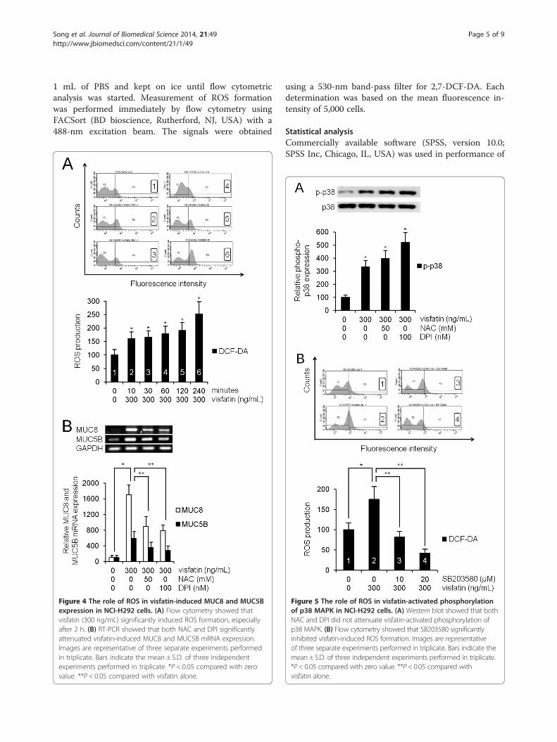
Figure 4 The role of ROS in visfatin-induced MUC8 and MUC5Bexpression in NCI-H292 cells. (A) Flow cytometry showed thatvisfatin (300 ng/mL) significantly induced ROS formation, especiallyafter 2 h. (B) RT-PCR showed that both NAC and DPI significantlyattenuated visfatin-induced MUC8 and MUC5B mRNA expression.Images are representative of three separate experiments performedin triplicate. Bars indicate the mean ± S.D. of three independentexperiments performed in triplicate. *P < 0.05 compared with zerovalue. **P < 0.05 compared with visfatin alone.
using a 530-nm band-pass filter for 2,7-DCF-DA. Eachdetermination was based on the mean fluorescence in-tensity of 5,000 cells.
Statistical analysisCommercially available software (SPSS, version 10.0;SPSS Inc, Chicago, IL, USA) was used in performance of
Figure 5 The role of ROS in visfatin-activated phosphorylationof p38 MAPK in NCI-H292 cells. (A) Western blot showed that bothNAC and DPI did not attenuate visfatin-activated phosphorylation ofp38 MAPK. (B) Flow cytometry showed that SB203580 significantlyinhibited visfatin-induced ROS formation. Images are representativeof three separate experiments performed in triplicate. Bars indicate themean ± S.D. of three independent experiments performed in triplicate.*P < 0.05 compared with zero value. **P < 0.05 compared withvisfatin alone.
Song et al. Journal of Biomedical Science 2014, 21:49 Page 6 of 9http://www.jbiomedsci.com/content/21/1/49
statistical analysis. The mean for each of the obtainedquantitative values was calculated. Comparisons weremade using the Student’s t-test. For all tests, a p value ofless than 0.05 was considered statistically significant.
ResultsVisfatin induced MUC8 and MUC5B expression inNCI-H292 cellsTo investigate the effect of visfatin on MUC16, MUC8,MUC5B, MUC5AC, and MUC4 expression in humanNCI-H292 airway epithelial cells, the cells were incu-bated with different doses of visfatin for 8 h. Result ofRT-PCR showed remarkable visfatin-induced MUC8and MUC5B mRNA expression: visfatin-induced MUC8mRNA expression was significantly stronger than visfatin-induced MUC5B mRNA expression. However, visfatin didnot definitely induce MUC16, MUC5AC, and MUC4mRNA expression (Figure 1, P < 0.05). Real time RT-PCRand ELISA analysis were performed to investigate theeffect of visfatin on MUC8 and MUC5B expression ina dose- and time-dependent manner. The cells weretreated with different doses of visfatin for 8 h or visfatin(300 ng/mL) for variable times. The results also showed asignificant increase in MUC8 and MUC5B mRNA expres-sion and protein production by treatment with all dosagesof visfatin: visfatin-induced MUC8 expression was signifi-cantly stronger than visfatin-induced MUC5B expression,and MUC8 and MUC5B mRNA expression peaked at 8 hafter treatment with visfatin (300 ng/mL) (Figure 2A-D,P < 0.05).
p38 MAPK was involved in visfatin-induced MUC8 andMUC5B expression in NCI-H292 cellsTo evaluate the brief intracellular mechanisms of visfatin-induced MUC8 and MUC5B expression, ERK1/2, or p38
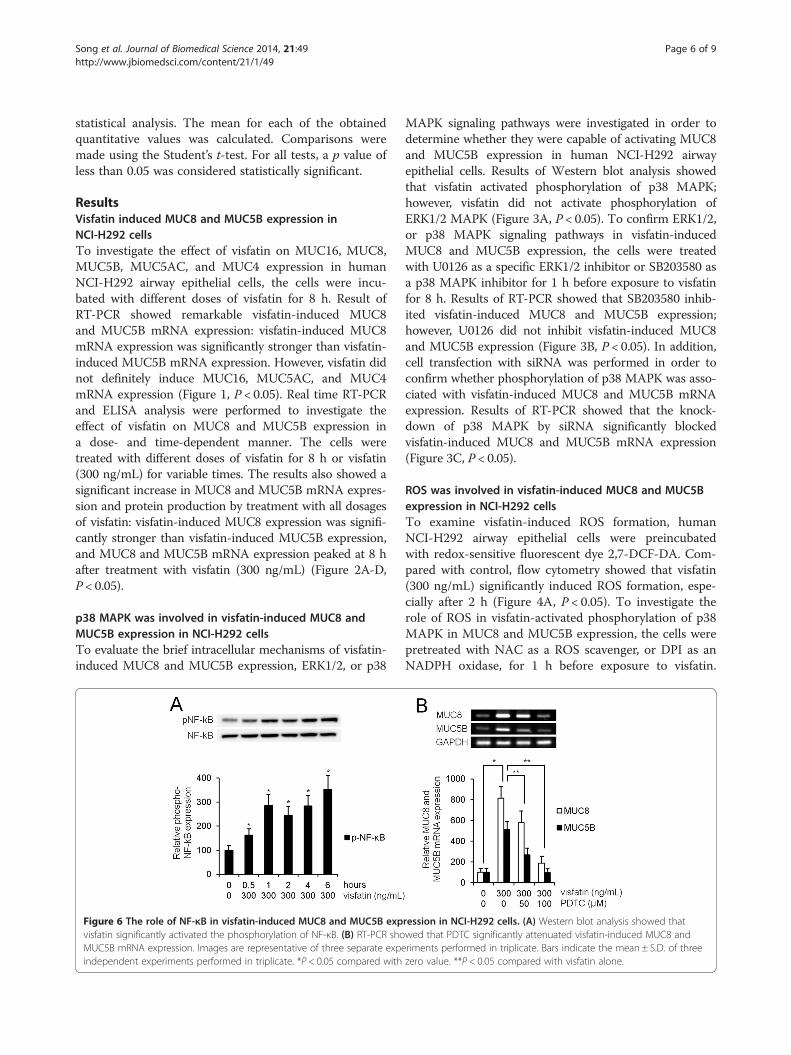
Figure 6 The role of NF-κB in visfatin-induced MUC8 and MUC5B expvisfatin significantly activated the phosphorylation of NF-κB. (B) RT-PCR shoMUC5B mRNA expression. Images are representative of three separate expeindependent experiments performed in triplicate. *P < 0.05 compared with
MAPK signaling pathways were investigated in order todetermine whether they were capable of activating MUC8and MUC5B expression in human NCI-H292 airwayepithelial cells. Results of Western blot analysis showedthat visfatin activated phosphorylation of p38 MAPK;however, visfatin did not activate phosphorylation ofERK1/2 MAPK (Figure 3A, P < 0.05). To confirm ERK1/2,or p38 MAPK signaling pathways in visfatin-inducedMUC8 and MUC5B expression, the cells were treatedwith U0126 as a specific ERK1/2 inhibitor or SB203580 asa p38 MAPK inhibitor for 1 h before exposure to visfatinfor 8 h. Results of RT-PCR showed that SB203580 inhib-ited visfatin-induced MUC8 and MUC5B expression;however, U0126 did not inhibit visfatin-induced MUC8and MUC5B expression (Figure 3B, P < 0.05). In addition,cell transfection with siRNA was performed in order toconfirm whether phosphorylation of p38 MAPK was asso-ciated with visfatin-induced MUC8 and MUC5B mRNAexpression. Results of RT-PCR showed that the knock-down of p38 MAPK by siRNA significantly blockedvisfatin-induced MUC8 and MUC5B mRNA expression(Figure 3C, P < 0.05).
ROS was involved in visfatin-induced MUC8 and MUC5Bexpression in NCI-H292 cellsTo examine visfatin-induced ROS formation, humanNCI-H292 airway epithelial cells were preincubatedwith redox-sensitive fluorescent dye 2,7-DCF-DA. Com-pared with control, flow cytometry showed that visfatin(300 ng/mL) significantly induced ROS formation, espe-cially after 2 h (Figure 4A, P < 0.05). To investigate therole of ROS in visfatin-activated phosphorylation of p38MAPK in MUC8 and MUC5B expression, the cells werepretreated with NAC as a ROS scavenger, or DPI as anNADPH oxidase, for 1 h before exposure to visfatin.
ression in NCI-H292 cells. (A) Western blot analysis showed thatwed that PDTC significantly attenuated visfatin-induced MUC8 andriments performed in triplicate. Bars indicate the mean ± S.D. of threezero value. **P < 0.05 compared with visfatin alone.
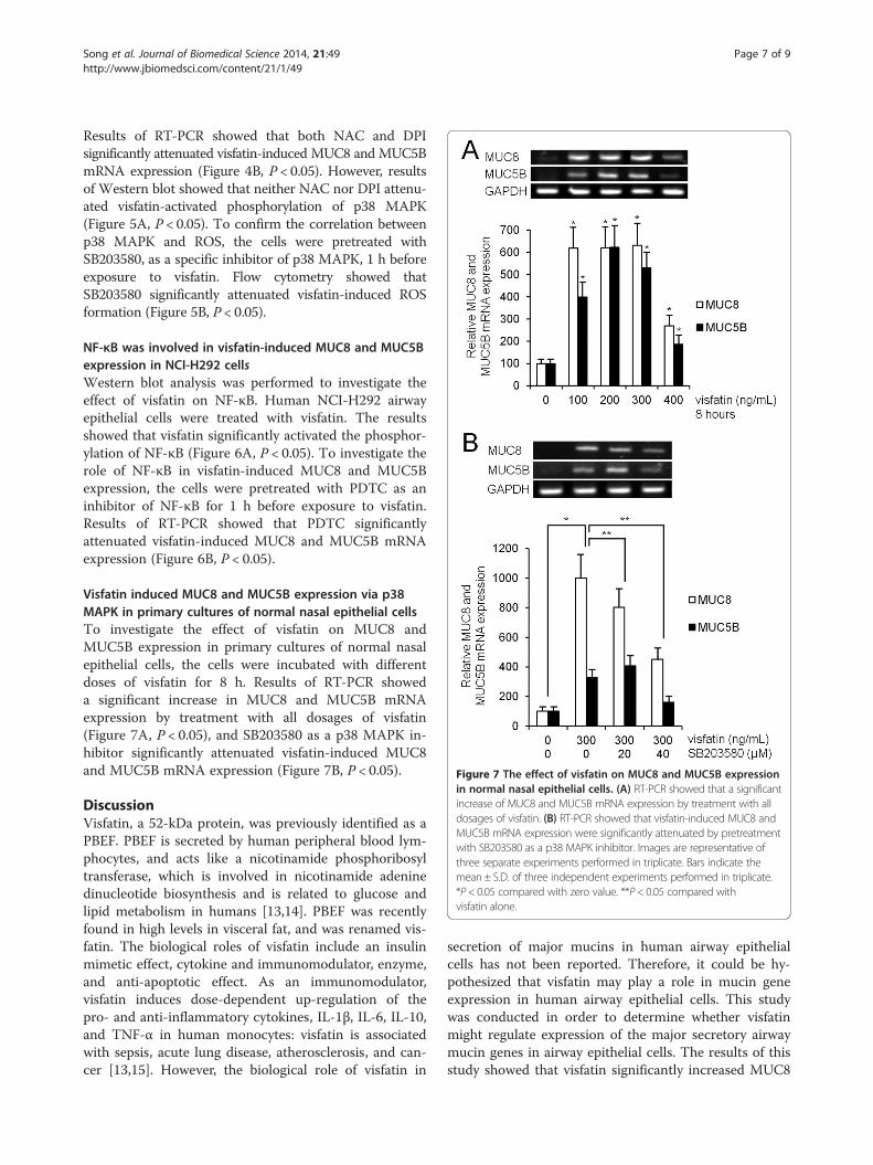
Figure 7 The effect of visfatin on MUC8 and MUC5B expressionin normal nasal epithelial cells. (A) RT-PCR showed that a significantincrease of MUC8 and MUC5B mRNA expression by treatment with alldosages of visfatin. (B) RT-PCR showed that visfatin-induced MUC8 andMUC5B mRNA expression were significantly attenuated by pretreatmentwith SB203580 as a p38 MAPK inhibitor. Images are representative ofthree separate experiments performed in triplicate. Bars indicate themean ± S.D. of three independent experiments performed in triplicate.*P < 0.05 compared with zero value. **P< 0.05 compared withvisfatin alone.
Song et al. Journal of Biomedical Science 2014, 21:49 Page 7 of 9http://www.jbiomedsci.com/content/21/1/49
Results of RT-PCR showed that both NAC and DPIsignificantly attenuated visfatin-induced MUC8 and MUC5BmRNA expression (Figure 4B, P < 0.05). However, resultsof Western blot showed that neither NAC nor DPI attenu-ated visfatin-activated phosphorylation of p38 MAPK(Figure 5A, P < 0.05). To confirm the correlation betweenp38 MAPK and ROS, the cells were pretreated withSB203580, as a specific inhibitor of p38 MAPK, 1 h beforeexposure to visfatin. Flow cytometry showed thatSB203580 significantly attenuated visfatin-induced ROSformation (Figure 5B, P < 0.05).
NF-κB was involved in visfatin-induced MUC8 and MUC5Bexpression in NCI-H292 cellsWestern blot analysis was performed to investigate theeffect of visfatin on NF-κB. Human NCI-H292 airwayepithelial cells were treated with visfatin. The resultsshowed that visfatin significantly activated the phosphor-ylation of NF-κB (Figure 6A, P < 0.05). To investigate therole of NF-κB in visfatin-induced MUC8 and MUC5Bexpression, the cells were pretreated with PDTC as aninhibitor of NF-κB for 1 h before exposure to visfatin.Results of RT-PCR showed that PDTC significantlyattenuated visfatin-induced MUC8 and MUC5B mRNAexpression (Figure 6B, P < 0.05).
Visfatin induced MUC8 and MUC5B expression via p38MAPK in primary cultures of normal nasal epithelial cellsTo investigate the effect of visfatin on MUC8 andMUC5B expression in primary cultures of normal nasalepithelial cells, the cells were incubated with differentdoses of visfatin for 8 h. Results of RT-PCR showeda significant increase in MUC8 and MUC5B mRNAexpression by treatment with all dosages of visfatin(Figure 7A, P < 0.05), and SB203580 as a p38 MAPK in-hibitor significantly attenuated visfatin-induced MUC8and MUC5B mRNA expression (Figure 7B, P < 0.05).
DiscussionVisfatin, a 52-kDa protein, was previously identified as aPBEF. PBEF is secreted by human peripheral blood lym-phocytes, and acts like a nicotinamide phosphoribosyltransferase, which is involved in nicotinamide adeninedinucleotide biosynthesis and is related to glucose andlipid metabolism in humans [13,14]. PBEF was recentlyfound in high levels in visceral fat, and was renamed vis-fatin. The biological roles of visfatin include an insulinmimetic effect, cytokine and immunomodulator, enzyme,and anti-apoptotic effect. As an immunomodulator,visfatin induces dose-dependent up-regulation of thepro- and anti-inflammatory cytokines, IL-1β, IL-6, IL-10,and TNF-α in human monocytes: visfatin is associatedwith sepsis, acute lung disease, atherosclerosis, and can-cer [13,15]. However, the biological role of visfatin in
secretion of major mucins in human airway epithelialcells has not been reported. Therefore, it could be hy-pothesized that visfatin may play a role in mucin geneexpression in human airway epithelial cells. This studywas conducted in order to determine whether visfatinmight regulate expression of the major secretory airwaymucin genes in airway epithelial cells. The results of thisstudy showed that visfatin significantly increased MUC8

Song et al. Journal of Biomedical Science 2014, 21:49 Page 8 of 9http://www.jbiomedsci.com/content/21/1/49
and MUC5B expression. However, visfatin did not definitelyinduce MUC16, MUC5AC, and MUC4 mRNA expression.These results suggest that visfatin has up-regulation ofMUC8 and MUC5B expression, like an inflammatory medi-ator: lipopolysaccharide, TNF α, and IL-1β.Within the signaling pathway, visfatin induces vascular
endothelial growth factor, and production of matrix metal-loproteinases via MAPK [8,16]. In addition, MUC8 orMUC5B expression is induced in response to a wide varietyof stimuli, including nerve activation and inflammatory cy-tokines, such as IL-1β, IL-6, TNF-α, and prostaglandin E2through a process involving p38 or ERK1/2 MAPK activa-tion [11,17,18]. And insulin-like growth factor-1, which hasan insulin mimetic effect like visfatin, induces MUC8 andMUC5B expression via ERK1 and p38 MAPK signalingpathway in human airway epithelial cells [19]. Therefore,this study focused on visfatin-induced MUC8 and MUC5Bexpression via the p38 or ERK1/2 MAPK signaling path-way. The results of this study showed that visfatin acti-vated phosphorylation of p38 MAPK. SB203580 inhibitedvisfatin-induced MUC8 and MUC5B expression. Inaddition, the knockdown of p38 MAPK by siRNA signifi-cantly blocked visfatin-induced MUC8 and MUC5BmRNA expression. These results suggest that visfatin in-duces MUC8 and MUC5B expression through the p38MAPK signaling pathway in human airway epithelial cells.ROS produced by cytokines, growth factors, and va-
soactive agents contribute to the intracellular signalingcascades associated with inflammatory responses. ROS in-duce NF-κB activation by modifying the activity of one ormore of the kinase enzymes in the NF-κB activation cas-cades [5]. Recent studies have reported that visfatin in-creases expression of inflammatory adhesion moleculesthrough an ROS-dependent NF-κB signaling pathway invascular endothelial cells [5,7,9,16,20]. Therefore, thisstudy focused on correlation between visfatin-inducedMUC8 and MUC5B expression and ROS formation viathe NF-κB signaling pathway in human airway epithelialcells. The results of this study showed that visfatin signifi-cantly induced ROS formation. Treatment with SB203580significantly attenuated visfatin-induced ROS formation.Treatment with NAC and DPI significantly attenuatedvisfatin-induced MUC8 and MUC5B expression. However,neither NAC nor DPI attenuated visfatin-activated phos-phorylation of p38 MAPK. Visfatin significantly activatedthe phosphorylation of NF-κB. PDTC significantly attenu-ated visfatin-induced MUC8 and MUC5B expression.These results suggest that visfatin induces MUC8 andMUC5B expression through the p38 MAPK/ROS/NF-κBsignaling pathway in human airway epithelial cells.
ConclusionsIn summary, the results of this study demonstrate forthe first time that visfatin induces MUC8 and MUC5B
expression in human airway epithelial cells. In addition,visfatin-induced MUC8 and MUC5B expression may beregulated through the p38 MAPK/ROS/NF-κB signalingpathway in human airway epithelial cells. These resultsprovide important information demonstrating that modu-lation of visfatin may be an appropriate pharmacologicaltarget for control of mucus-hypersecretion in treatment ofairway inflammatory diseases in patients with obesity.
AbbreviationsDCF-DA: Dichlorodihydrofluorescein diacetate; DPI: Diphenyleneiodonium;ELISA: Enzyme-linked immunosorbent assay GAPDH, Glyceraldehyde-3-phosphate dehydrogenase; IL: Interleukin; MAPK: Mitogen-activated proteinkinase; NAC: N-acetyl-cystein; NADPH: Nicotinamide adenine dinucleotidephosphate; NF-κB: Nuclear factor kappa-light-chain-enhancer of activated Bcells; PBEF: Pre-B cell colony-enhancing factor; PBS: Phosphate-bufferedsaline; PCR: Polymerase chain reaction; PDTC: Pyrrolidine dithiocarbamate;ROS: Reactive oxygen species; RT: Reverse transcriptase; SiRNA: smallinterfering RNA; TNF: Tumor necrosis factor.
Competing interestsThe authors declare that they have no competing interests.
Authors’ contributionsS-YS conceived and designed this study; ECJ drafted the manuscript andacquired data; Y-DK supervised this study and provided the critical revisionof the manuscript for important intellectual content; YSC and BCH analyzedand interpreted data; S-YS and ECJ provided the administrative, technical,or material support. All authors read and approved the final manuscript.
AcknowledgementsThis work was supported by the 2013 Yeungnam University Research Grant.
Received: 12 March 2014 Accepted: 15 May 2014Published: 20 May 2014
References1. Williams OW, Sharafkhaneh A, Kim V, Dickey BF, Evans CM: Airway mucus:
from production to secretion. Am J Respir Cell Mol Biol 2006, 34:527–536.2. Ali MS, Pearson JP: Upper airway mucin gene expression: a review.
Laryngoscope 2007, 117:932–938.3. Rose MC, Voynow JA: Respiratory tract mucin genes and mucin
glycoproteins in health and disease. Physiol Rev 2006, 86:245–278.4. Stofkova A: Resistin and visfatin: regulators of insulin sensitivity,
inflammation and immunity. Endocr Regul 2010, 44:25–36.5. Kim SR, Bae YH, Bae SK, Choi KS, Yoon KH, Koo TH, Jang HO, Yun I, Kim KW,
Kwon YG, Yoo MA, Bae MK: Visfatin enhances ICAM-1 and VCAM-1 expres-sion through ROS-dependent NF-kappaB activation in endothelial cells.Biochim Biophys Acta 2008, 1783:886–895.
6. Adeghate E: Visfatin: structure, function and relation to diabetes mellitusand other dysfunctions. Curr Med Chem 2008, 15:1851–1862.
7. Oita RC, Ferdinando D, Wilson S, Bunce C, Mazzatti DJ: Visfatin inducesoxidative stress in differentiated C2C12 myotubes in an Akt- andMAPK-independent, NFkB-dependent manner. Pflug Arch Eur J Phy 2010,459:619–630.
8. Adya R, Tan BK, Punn A, Chen J, Randeva HS: Visfatin induces humanendothelial VEGF and MMP-2/9 production via MAPK and PI3K/Aktsignalling pathways: novel insights into visfatin-induced angiogenesis.Cardiovasc Res 2008, 78:356–365.
9. Zhang LQ, Adyshev DM, Singleton P, Li H, Cepeda J, Huang SY, Zou X, VerinAD, Tu J, Garcia JG, Ye SQ: Interactions between PBEF and oxidative stressproteins-a potential new mechanism underlying PBEF in the pathogen-esis of acute lung injury. FEBS Lett 2008, 582:1802–1808.
10. Busso N, Karababa M, Nobile M, Rolaz A, Van Gool F, Galli M, Leo O, So A,De Smedt T: Pharmacological inhibition of nicotinamidephosphoribosyltransferase/visfatin enzymatic activity identifies a newinflammatory pathway linked to NAD. PLoS One 2008, 3:e2267.
11. Woo HJ, Yoo WJ, Bae CH, Song SY, Kim YW, Park SY, Kim YD: Leptinup-regulates MUC5B expression in human airway epithelial cells via
Song et al. Journal of Biomedical Science 2014, 21:49 Page 9 of 9http://www.jbiomedsci.com/content/21/1/49
mitogen-activated protein kinase pathway. Exp Lung Res 2010,36:262–269.
12. Woo HJ, Bae CH, Song SY, Lee HM, Kim YD: Expression of membrane-bound mucins in human nasal mucosa: different patterns for MUC4and MUC16. Arch Otolaryngol Head Neck Surg 2010, 136:603–609.
13. Sonoli SS, Shivprasad S, Prasad CV, Patil AB, Desai PB, Somannavar MS:Visfatin-a review. Eur Rev Med Pharmacol Sci 2011, 15:9–14.
14. Skoczylas A: The role of visfatin in the pathophysiology of human.Wiad Lek 2009, 62:190–196.
15. Pilz S, Mangge H, Obermayer-Pietsch B, März W: Visfatin/pre-B-cellcolony-enhancing factor: a protein with various suggested functions.J Endocrinol Invest 2007, 30:138–144.
16. Fan Y, Meng S, Wang Y, Cao J, Wang C: Visfatin/PBEF/Nampt inducesEMMPRIN and MMP-9 production in macrophages via the NAMPT-MAPK(p38, ERK1/2)-NF-κB signaling pathway. Int J Mol Med 2011, 27:607–615.
17. Casalino-Matsuda SM, Monzon ME, Day AJ, Forteza RM: Hyaluronanfragments/CD44 mediate oxidative stress-induced MUC5B up-regulationin airway epithelium. Am J Respir Cell Mol Biol 2009, 40:277–285.
18. Kim DE, Min KJ, Kim JS, Kwon TK: High-mobility group box-1 proteininduces mucin 8 expression through the activation of the JNK andPI3K/Akt signal pathways in human airway epithelial cells.Biochem Biophys Res Commun 2012, 421:436–441.
19. Bae CH, Kim JS, Song SY, Kim YW, Park SY, Kim YD: Insulin-like growthfactor-1 induces MUC8 and MUC5B expression via ERK1 and p38 MAPKin human airway epithelial cells. Biochem Biophys Res Commun 2013,430:683–688.
20. Adya R, Tan BK, Chen J, Randeva HS: Nuclear factor-kappaB inductionby visfatin in human vascular endothelial cells: its role in MMP-2/9production and activation. Diabetes Care 2008, 31:758–760.
doi:10.1186/1423-0127-21-49Cite this article as: Song et al.: Visfatin induces MUC8 and MUC5Bexpression via p38 MAPK/ROS/NF-κB in human airway epithelial cells.Journal of Biomedical Science 2014 21:49.
Submit your next manuscript to BioMed Centraland take full advantage of:
• Convenient online submission
• Thorough peer review
• No space constraints or color figure charges
• Immediate publication on acceptance
• Inclusion in PubMed, CAS, Scopus and Google Scholar
• Research which is freely available for redistribution
Submit your manuscript at www.biomedcentral.com/submit
Related Documents