viruses Review Dance with the Devil: Stress Granules and Signaling in Antiviral Responses Nina Eiermann 1, † , Katharina Haneke 1, † , Zhaozhi Sun 2 , Georg Stoecklin 1 and Alessia Ruggieri 2, * 1 Division of Biochemistry, Mannheim Institute for Innate Immunoscience (MI3), Medical Faculty Mannheim, Heidelberg University, 68167 Mannheim, Germany; [email protected] (N.E.); [email protected] (K.H.); [email protected] (G.S.) 2 Department of Infectious Diseases, Molecular Virology, Center for Integrative Infectious Disease Research (CIID), University of Heidelberg, 69120 Heidelberg, Germany; [email protected] * Correspondence: [email protected] † These authors contributed equally to this work. Received: 20 July 2020; Accepted: 31 August 2020; Published: 4 September 2020 Abstract: Cells have evolved highly specialized sentinels that detect viral infection and elicit an antiviral response. Among these, the stress-sensing protein kinase R, which is activated by double-stranded RNA, mediates suppression of the host translation machinery as a strategy to limit viral replication. Non-translating mRNAs rapidly condensate by phase separation into cytosolic stress granules, together with numerous RNA-binding proteins and components of signal transduction pathways. Growing evidence suggests that the integrated stress response, and stress granules in particular, contribute to antiviral defense. This review summarizes the current understanding of how stress and innate immune signaling act in concert to mount an effective response against virus infection, with a particular focus on the potential role of stress granules in the coordination of antiviral signaling cascades. Keywords: virus; stress granules; stress response; innate immune response; PKR; G3BP1; antiviral signaling 1. Introduction Viruses depend on the host translation apparatus to express viral proteins. By hijacking and redirecting ribosomes, translation factors, and RNA binding proteins (RBPs), viruses modulate the cellular translatome in favor of their needs and virus progeny production. In rare cases, viruses such as hantaviruses, myoviruses, haloarchaeal viruses, and giant viruses encode viral proteins that substitute for translation initiation, elongation, and termination factors, transfer RNAs (tRNAs), aminoacyl-tRNA synthetases [1–6], and—as lately identified via metagenome analysis—also ribosomal proteins [7]. Although the use of the host-encoded translation machinery saves coding capacity, the ensuing dependency on host factors to accomplish this critical step of the viral life cycle makes viruses vulnerable. To counteract virus reproduction, cells have evolved highly specialized stress sensors that detect viral products and actively suppress both host and viral translation. Hence, limiting the access, availability, or activity of the translation apparatus can be considered an effective defense mechanism and integral component of the antiviral response. Immune sensors detect viral nucleic acids as non-self through specific features, termed pathogen-associated molecular patterns (PAMPs), among these incoming viral DNA and RNA genomes and viral replication intermediates. The activation of immune sensors elicits an antiviral Viruses 2020, 12, 984; doi:10.3390/v12090984 www.mdpi.com/journal/viruses

Welcome message from author
This document is posted to help you gain knowledge. Please leave a comment to let me know what you think about it! Share it to your friends and learn new things together.
Transcript
viruses
Review
Dance with the Devil: Stress Granules and Signalingin Antiviral Responses
Nina Eiermann 1,†, Katharina Haneke 1,† , Zhaozhi Sun 2, Georg Stoecklin 1
and Alessia Ruggieri 2,*1 Division of Biochemistry, Mannheim Institute for Innate Immunoscience (MI3), Medical Faculty Mannheim,
Heidelberg University, 68167 Mannheim, Germany; [email protected] (N.E.);[email protected] (K.H.); [email protected] (G.S.)
2 Department of Infectious Diseases, Molecular Virology,Center for Integrative Infectious Disease Research (CIID), University of Heidelberg,69120 Heidelberg, Germany; [email protected]
* Correspondence: [email protected]† These authors contributed equally to this work.
Received: 20 July 2020; Accepted: 31 August 2020; Published: 4 September 2020�����������������
Abstract: Cells have evolved highly specialized sentinels that detect viral infection and elicitan antiviral response. Among these, the stress-sensing protein kinase R, which is activated bydouble-stranded RNA, mediates suppression of the host translation machinery as a strategy to limitviral replication. Non-translating mRNAs rapidly condensate by phase separation into cytosolic stressgranules, together with numerous RNA-binding proteins and components of signal transductionpathways. Growing evidence suggests that the integrated stress response, and stress granules inparticular, contribute to antiviral defense. This review summarizes the current understanding ofhow stress and innate immune signaling act in concert to mount an effective response against virusinfection, with a particular focus on the potential role of stress granules in the coordination of antiviralsignaling cascades.
Keywords: virus; stress granules; stress response; innate immune response; PKR; G3BP1; antiviralsignaling
1. Introduction
Viruses depend on the host translation apparatus to express viral proteins. By hijackingand redirecting ribosomes, translation factors, and RNA binding proteins (RBPs), viruses modulatethe cellular translatome in favor of their needs and virus progeny production. In rare cases, virusessuch as hantaviruses, myoviruses, haloarchaeal viruses, and giant viruses encode viral proteins thatsubstitute for translation initiation, elongation, and termination factors, transfer RNAs (tRNAs),aminoacyl-tRNA synthetases [1–6], and—as lately identified via metagenome analysis—also ribosomalproteins [7]. Although the use of the host-encoded translation machinery saves coding capacity,the ensuing dependency on host factors to accomplish this critical step of the viral life cycle makesviruses vulnerable. To counteract virus reproduction, cells have evolved highly specialized stresssensors that detect viral products and actively suppress both host and viral translation. Hence, limitingthe access, availability, or activity of the translation apparatus can be considered an effective defensemechanism and integral component of the antiviral response.
Immune sensors detect viral nucleic acids as non-self through specific features, termedpathogen-associated molecular patterns (PAMPs), among these incoming viral DNA and RNAgenomes and viral replication intermediates. The activation of immune sensors elicits an antiviral
Viruses 2020, 12, 984; doi:10.3390/v12090984 www.mdpi.com/journal/viruses
Viruses 2020, 12, 984 2 of 47
state through the production of interferons (IFNs) and pro-inflammatory cytokines [8–10]. In addition,the accumulation of viral double-stranded (ds) RNA in the cytosol and of viral proteins inthe endoplasmic reticulum (ER) triggers stress sensors such as protein kinase R (PKR) and PKR-likeendoplasmic reticulum kinase (PERK), respectively [11]. Once activated, these kinases initiatethe integrated stress response (ISR) by phosphorylating the alpha subunit of the eukaryotic translationinitiation factor-2 (eIF2α), which delivers the Methionine initiator tRNA (tRNAiMet) to the small 40Sribosomal subunit. As a consequence, translation initiation is stalled and polysomes disassemble [12].
The assembly of stress granules (SGs) is an integral part of host stress responses, frequentlyobserved upon infection with DNA or RNA viruses. SGs form by a cytosolic phase separation event,through which stalled mRNAs are separated from the remaining cytosol together with translationfactors. SGs were primarily suggested to serve as storage and triage areas for mRNAs that can easily bereengaged with polysomes in order to resume translation once the stress is resolved [13,14]. However,the abundance of RBPs and signal transducing factors in SGs has broadened our perspective onthe role of SGs in recent years. SGs are now considered to be signaling platforms that contributeto the coordination of cellular processes during stress, including apoptosis, cell growth, metaboliccontrol, and antiviral defense [15–18]. It is therefore not surprising that viruses not only escapetranslational repression through various mechanisms, but also interfere with the assembly of SGsand repurpose SG proteins for their own replication [19–22]. The diversity of these viral strategieshighlights the importance of the stress response, and SGs in particular, in antiviral defense.
Whether SGs as a whole or single proteins within SGs contribute to the antiviral response isstill an open question whose answer might depend on the virus type, the cell type, and the resultinghost–virus interactions. Recent advances in the purification of SG components have opened the routeto investigate which proteins and mRNAs are selectively recruited to SGs during viral infections. Here,we aim to summarize the current understanding of the translational stress response in the context ofvirus infection and cover the various connections between the stress response and innate immuneresponse, which are intertwined events that act in concert to establish an antiviral state. Emphasis willbe given to the potential role of SGs in the initiation and coordination of antiviral signaling events.
2. General Mechanisms of Translation Control under Homeostasis and Virus Infection
Regulation of gene expression at the level of translation is a central control mechanism that enablesrapid and reversible changes in protein levels, both spatially and temporally. In addition to essentialroles in several biological processes, including cell growth, differentiation, and apoptosis [23–25],translational control plays a major role in the host stress response to virus infection [26,27].
2.1. Translation Initiation, a Key Step in the Regulation of Protein Synthesis
Mammalian protein synthesis is primarily regulated at the level of translation initiation, whichrequires recruitment of the small ribosomal subunit to the mRNA, scanning along the mRNA 5′
untranslated region (UTR), recognition of the start codon, and subsequent joining of the largeribosomal subunit [28]. These steps are regulated by upstream open reading frames (uORFs), RNAmodifications, RNA structures and different RBPs, which together dictate mRNA translation initiationefficiency [29,30].
Most cellular mRNAs contain a cap structure at their 5′ end, composed of an inverted7-methylguanosine connected to the mRNA via a 5′-5′-triphosphate bridge. Most 5′ ends areadditionally methylated at the ribose 2′O position of the +1 ribonucleotide (cap1), and in many cases,also of the +2 ribonucleotide (cap2). This feature is not only important for mRNA translation and stability,but also for the distinction between self from non-self mRNAs [31]. Indeed, several single-stranded(ss) RNA viruses that replicate in the cytosol, including flaviviruses, picornaviruses, coronaviruses,and poxviruses, encode their own 2′O-methytransferases to escape recognition by host immunesensors [32]. Initially, the 5′ cap is bound by the eIF4F complex, which consists of the cap-binding proteineIF4E, the RNA helicase eIF4A, and the scaffold protein eIF4G. Subsequently, the 40S small ribosomal
Viruses 2020, 12, 984 3 of 47
subunit together with the initiation factors eIF1, eIF1A, eIF5, eIF3, and the eIF2–GTP–tRNAiMet ternarycomplex is loaded onto the mRNA 5′ end, forming a scanning-competent 48S preinitiation complex(PIC). The PIC then scans the 5′ UTR until it recognizes a start codon within a favorable sequencecontext, leading to hydrolysis of the eIF2-bound GTP. This crucial step results in the release of eIF2-GDP,leaving the initiator tRNAiMet base-paired with the AUG start codon [33].
2.2. Repression of Translation Initiation upon Environmental Stress
Cells respond to unfavorable conditions such as amino acid starvation, ER stress, oxidativestress, hypoxia, UV irradiation, and virus infection by rapid attenuation of translation initiation rates,mainly through controlling (i) the eIF2–GTP–tRNAiMet ternary complex and (ii) the cap-bindingcomplex. Thereby, the translation of mRNAs not critical for cell survival, e.g., housekeeping mRNAs,is strongly repressed during stress in favor of mRNAs encoding survival factors, repair enzymes,and stress-regulatory proteins [23,24].
The first mechanism involves four eIF2α-kinases, which are activated by a wide range of extrinsicand intrinsic stressors, and phosphorylate the α subunit of eIF2 at Ser51 [12]. This prevents release ofeIF2 from the GTP-GDP exchange factor eIF2B, thereby impairing regeneration of the ternary complexand slowing down protein synthesis [34]. The heme-regulated inhibitor (HRI) senses oxidative stress,osmotic stress, heat shock, and heme-depletion. PKR is activated by cellular and viral dsRNAs. PERK,a transducer of the unfolded protein response (UPR), is activated by the accumulation of misfoldedproteins in the ER. Finally, the general control nonderepressible 2 (GCN2) is mainly activated bylimited amino acid availability and UV stress. As a common feature, eIF2α-kinases were foundto be activated by stress-induced oligo- or dimerization and autophosphorylation [11]. The poolof the GTP-bound ternary complex is regenerated by dephosphorylation of eIF2α through proteinphosphatase 1 (PP1), whose catalytic subunit is recruited to eIF2α via a specific regulatory subunit.In unstressed cells, the constitutive repressor of eIF2α phosphorylation (CReP), a constitutivelyexpressed regulatory subunit of PP1, maintains low levels of eIF2α phosphorylation [35], especiallyat the ER [36]. Under stress conditions, the PP1 regulatory subunit growth arrest and DNA damageinducible protein 34 (GADD34) is both transcriptionally and translationally upregulated, antagonizingeIF2α phosphorylation and translational arrest imposed by eIF2α-kinase activation [37–40].
The second mechanism is based on regulating the activity of the eIF4F cap-binding complex, whichcontrols cap-dependent translation. Certain types of stress, such as nutrient deprivation and hypoxia,lead to inhibition of the mammalian target of rapamycin (mTOR) complex 1 (mTORC1), the key kinasethat regulates the phosphorylation state of 4E binding proteins (4E-BPs) [24]. In their unphosphorylatedform, 4E-BPs have an increased affinity for the cap-binding protein eIF4E and outcompete eIF4G,preventing efficient recruitment of eIF3 and the 40S small ribosomal subunit to the 5′ end ofthe mRNA [41]. Thereby, inhibition of mTORC1 leads to a reduction in cap-dependent translation.
A subset of cellular mRNAs contains linear motifs and secondary structures that enable proteinsynthesis under stress conditions through alternative modes of translation initiation, which areindependent of eIF2α and/or eIF4F. These include (i) transcripts containing short uORFs [42], mostlyencoding for survival-related and stress-effector proteins, e.g., the transcription factors activatingtranscription factor 4 (ATF4) and the CCAAT/enhancer-binding protein homology protein (CHOP),as well as GADD34. (ii) Approximately 10% of all cellular mRNAs are suggested to containinternal ribosome entry sites (IRES) and hence, do not require eIF4F binding, but instead, relyon IRES trans-acting factors (ITAFs) for efficient translation initiation [43–45]. IRES elements wereoriginally discovered in viruses belonging to the Picornaviridae family [46,47]. Even though theiractivity and efficiency is still under debate, numerous cellular IRES elements have been reportedand characterized, particularly in genes involved in apoptosis such as the X-linked inhibitor of apoptosisprotein (XIAP) [48] and B-cell lymphoma-2 (Bcl-2) [49], and more recently, in genes involved in cellgrowth and proliferation such as mTOR [50] and c-Src [51]. The existence and involvement of a growingnumber of cap-independent initiation mechanisms in eukaryotes, including (iii) N6-methyladenosine
Viruses 2020, 12, 984 4 of 47
(m6A)-dependent translation [52–54], and (iv) the use of alternative cap-binding complexes such aseIF4FH under hypoxic conditions or eIF4FM in response to stress or proliferative cues, respectively [55],are indisputable.
The impact of translational repression, especially on RNA viruses, is highlighted by the fact thatmany evolved alternative initiation strategies to translate their genome independently of eIF2α,including the use of IRES elements [56] and the possibility to switch from cap-dependent tocap-independent translation [57–60].
2.3. From Translational Suppression to SG Formation
When translation initiation is blocked under stress conditions, stalled translation preinitiationcomplexes accumulate. At the same time, translation elongation continues and polysomes disassembleas a consequence of ribosome run-off. Upon acute and strong inhibition of translation initiation,non-translating mRNAs condensate together with RBPs into microscopically visible, non-membranouscytosolic SGs through a liquid–liquid phase separation event [61]. Phase separation is likelyinitiated by the appearance of long stretches of mRNA not covered by ribosomes, which engage inRNA–RNA interactions [62] and serve as scaffolds for numerous RBPs that promote phase separationthrough their intrinsically disordered regions. RBPs with an essential role in nucleating SGs includeRasGAP-associated endoribonuclease 1 (G3BP1), T cell internal antigen 1 (TIA1), TIA1-related protein(TIAR), Caprin1, and fragile X mental retardation protein (FMRP) [63]. Recently, ubiquitin-associatedprotein 2-like (UBAP2L) [64–67], cold shock domain containing E1 (CSDE1), and proline-rich coiled-coil2C (PRRC2C) were added to the growing list of SG-nucleating proteins [64]. Overexpression of singleproteins such as TIA1 or G3BP1 can drive SG formation, even in the absence of stress [68,69], indicatingthat a shift in the equilibrium between solubilizing and aggregation-prone proteins is sufficient toinduce phase separation of the cytosol.
Interestingly, SGs were found to consist of a more stable inner core, stabilized by directprotein–protein and RNA–protein interactions, and a dynamic shell-like outer layer that is characterizedby multiple, multivalent low-affinity interactions between proteins and RNAs [70]. SGs are verydynamic structures, which rapidly assembly under stress conditions and disassemble within minuteswhen cells recover from stress [71,72]. Depending on the type of stress, SGs vary in size and number,and differ with respect to some of their mRNA and protein constituents. During oxidative stress, forexample, SGs move in a microtubule-dependent manner and grow in size by fusion [71,73].
Given their dynamic nature, it is not surprising that proteins and mRNAs shuttle in and out ofSGs in the seconds to minute range [71,74,75], showing that SG components are in constant exchangewith the cytosol. SGs also exchange proteins and mRNAs with processing bodies (PBs), a different typeof cytosolic RNA granule that contains RNA degrading enzymes and functions in mRNA silencing [76].SGs and PBs often exist in close proximity and may represent different stages of an “mRNP cycle” [13].
A peculiar type of SG dynamic was discovered by our laboratory upon chronic hepatitis Cvirus (HCV) infection, which leads to recurring cycles of SG assembly and disassembly, followingan oscillation of translational on- and off-states. The stochastic nature of SG assembly and disassemblyis controlled by the antagonistic action of PKR and GADD34, which repeatedly phosphorylateand dephosphorylate eIF2α. This oscillating stress response is widely observed upon infection withRNA viruses including Newcastle disease virus (NDV) and Sendai virus (SeV) [77]. Our currentunderstanding is that oscillating SGs represent a long-term strategy by which infected cells suppressviral protein production and replication intermittently, while enabling host protein production inbetween SG phases. Moreover, rapid SG oscillations seem to correlate with enhanced cell survival,suggesting a role in balancing the burden of antiviral defense with cellular homeostasis.
3. Stress Kinases—Mediators of Viral Translational Inhibition
Translation inhibition is an important pillar of the antiviral response. Viruses evolved multiplestrategies to target all steps of translation initiation in order to evade host-induced translational shutoff
Viruses 2020, 12, 984 5 of 47
and promote the synthesis of their own proteins. Since these strategies have been covered in severalexcellent reviews [26,27,56], we will focus here on how stress kinases contribute to the induction ofthe ISR upon virus infection and how viruses directly target these kinases. While global translationsuppression upon infection with RNA viruses is primarily mediated by PKR, activation of other stresskinases, alone or in combination with PKR, has been reported. They can be considered as additionalantiviral barriers, especially when viruses have evolved strategies to counteract PKR.
3.1. Protein Kinase R (EIF2AK2)
Among the four eIF2α-kinases, PKR, formerly called DAI, contributes to the sensing of viralinfection and is the object of extensive research [78–80]. PKR is an IFN-induced effector protein [81] thatis activated upon binding of dsRNA molecules [82]. The binding of dsRNA to the two dsRNA-bindingmotifs (dsRBD) within the PKR N-terminal domain promotes a structural reorientation, whichallows for PKR dimerization and subsequent activation by autophosphorylation of PKR C-terminalkinase domain. These structural rearrangements are required for binding and phosphorylation ofeIF2α [82–85]. One of the earliest molecules found to activate PKR was a hairpin loop of the hepatitisdelta virus genome, within the self-cleaving ribozyme region [86–88]. A similar hairpin structure wasdiscovered in the human immunodeficiency virus (HIV) genome within the transactivation-responseregion [89]. In general terms, viral activators of PKR represent dsRNA regions longer than 30 bp [85,86],including the genome of dsRNA viruses like rotaviruses [90], dsRNA replication intermediates ofpositive and negative ssRNA viruses [91,92] as well as dsRNA products from antiparallel transcriptionof DNA viruses such as vaccinia virus (VACV) [93]. The activation of PKR by short-stem loopshas been suggested to be 5′ triphosphate-dependent [94,95], though this observation is discussedcontroversially [96,97].
In recent years, PKR was shown to have functions, beyond the detection of viral dsRNA, in cellularprocesses such as mitosis and apoptosis [98,99], in metabolic as well as autoimmune diseases [100–103],and in long-term memory [104–106]. Cellular activators of PKR include mitochondrial dsRNAs [99],dsRNAs derived from inverted Alu repeats [98,107], non-coding small nucleolar RNAs [100],ribotoxin-induced or damaged rRNAs as well as unmodified rRNAs or tRNAs [108–110]. RNAs thatinhibit PKR have also been identified, e.g., circular RNAs [103] and the human non-coding RNA886 [111,112].
Viruses have evolved a multitude of strategies to counteract PKR. For instance, many RNAviruses encode proteins that bind and shield dsRNA from PKR detection, exemplified by VACVE3L [113], influenza A virus (IAV) accessory protein NS1 [114], Middle East respiratory syndromecoronavirus accessory protein 4a [115,116], reovirus sigma 3 protein [117], and Ebola virus proteinVP35 [118,119]. Other viruses such as human parainfluenza virus type 3 (HPIV3) sequester viral RNAin inclusion bodies to avoid detection by PKR [120]. Herpes simplex virus (HSV) 1 and 2 encodean endoribonuclease, the virion host shutoff protein that degrades RNA to avoid PKR activation earlyduring infection [121–123]. Negative ssRNA viruses such as measles virus (MV), influenza virus,and SeV avoid detection by PKR by making sure that only a small number of dsRNA replicationintermediates accumulate in the cytosol [91]. This is achieved by the virally encoded accessory protein C,which attenuates the copy-back mechanism of the viral RNA polymerase during replication [124–126].
Other viruses suppress PKR by encoding proteins that directly inhibit the kinase function, e.g., HCVnon-structural protein NS5A [127], Japanese encephalitis virus (JEV) non-structural protein NS2A [128],human cytomegalovirus protein TRS1 [129], and Kaposi’s sarcoma-associated herpesvirus lytic proteinORF57 [130]. An interesting variation of this theme is the expression of small regulatory RNAs by someDNA viruses, which antagonize PKR activation by competing with dsRNA binding. Examples includethe adenovirus VAI RNAs and the Epstein–Barr virus transcripts EBER-1 and EBER-2 [131–133], all ofwhich bind to but do not activate PKR.
An alternative strategy adopted by some viruses is mimicry. Baculovirus protein PK2, forexample, is an inactive kinase with homology to PKR, which leads to the formation of inactive PKR
Viruses 2020, 12, 984 6 of 47
heterodimers [134,135]. The VACV K3L protein is an eIF2α homologue, which competes with eIF2αfor PKR binding and thereby reduces eIF2α phosphorylation [113]. Finally, PKR can be targeted toproteasomal degradation upon infection with Rift Valley fever virus by the NSs protein [136–138].
3.2. PKR-Like Endoplasmic Reticulum Kinase (EIF2AK3)
During their life cycle, many RNA viruses perturb or hijack ER functions by (i) remodeling ERmembranes to form viral replication and assembly sites [139], (ii) utilizing and competing for the hostprotein glycosylation machinery [140,141], and (iii) encoding viral proteins with viroporin functionthat alter ER calcium homeostasis [142]. Virus-imposed interference with ER functions causes ER stressand induces the three signaling branches of the UPR through activation of IRE1-XBP1, ATF6, and PERK,whereby infected cells aim to reestablish ER homeostasis. PERK, as one player of the UPR, preventsprotein production, hence alleviating the ER burden [143]. For certain viruses, PERK activation wasreported to be beneficial, notably for NDV [144] and Seneca Valley virus [145]. Here, PERK-inducedautophagy is essential for viral replication. However, in multiple cases, PERK has adverse effects forviral replication. Transmissible gastroenteritis virus (TGEV) but also West Nile virus (WNV) and Langatvirus replication, for instance, are inhibited via PERK signaling [146–148].
Hence, it is not surprising that viruses counteract PERK activity. NDV, e.g., mediates PERKcleavage [149], whereas dengue virus (DENV) infection was found to inhibit PERK activation by a yetunknown mechanism [150]. The VACV K3L protein was reported to reduce PERK activity in vitro [151],and similar results were obtained for the HCV-encoded glycoprotein E2. This protein induces ER stresson the one hand [152], but acts as a pseudo-substrate and thereby inhibits PERK activation on the otherhand [153]. These examples illustrate that viruses strongly interfere with ER functions but at the sametime, often try to prevent the consequent activation of PERK. Interestingly, PERK can also get activatedby viral proteins such as JEV-encoded protein NS4B, which induces PERK dimerization and is knownto be important for the pathogenesis of encephalitis [154]. This is consistent with the fact that PERK’simplication in virus infection might differ depending on the virus type or infection stage.
3.3. General Control Nonderepressible 2 (EIF2AK4)
GCN2 is usually activated under conditions of amino acid starvation through its histidyl-tRNAsynthetase-related domain that binds uncharged tRNAs [155]. This domain also binds to viral RNAgenomes in vitro, notably those of sindbis virus (SINV), poliovirus, and HIV-1, and in vivo studiesconfirmed that infection with SINV and HIV-1 leads to activation of GCN2, eIF2α phosphorylation,and translational inhibition of viral RNA [156,157]. Furthermore, HIV-1 induces protease-mediatedcleavage of GCN2, indicating that GCN2 has an antiviral function that the virus tries to suppress [157].This notion is supported by the observation that GCN2-deficient or GCN2-mutant mice are moresusceptible to infection with SINV [156], murine cytomegalovirus, and human adenovirus [158].
In the future, it will be important to further characterize the mechanism behind GCN2 activationin virus-infected cells since this might also be an indirect consequence of virus- or IFN-γ-inducedamino acid deprivation.
3.4. Heme-Regulated Inhibitor (EIF2AK1)
A direct implication of HRI in virus infection was reported only recently for its fish homologues.In flounder cells (Paralichthys olivaceus), an upregulation of HRI was observed upon infection withScophthalmus maximus rhabdovirus and poly(I:C) treatment, at both the mRNA and protein levels [159].Furthermore, overexpression of HRI in orange-spotted grouper (Epinephelus coioides) resulted inan increase in eIF2α phosphorylation and inhibition of red-spotted-grouper nervous necrosis virus(RGNNV) replication [160]. Both studies suggested that HRI might have a similar function to PKRin fish.
Normally, HRI is activated by high intracellular levels of reactive oxygen species (ROS) and otherimbalances of redox homeostasis, e.g., those induced by arsenite [161]. Several viruses interfere with
Viruses 2020, 12, 984 7 of 47
mitochondrial, peroxisomal, and ER functions and can thereby lead to ROS production, as observedduring infection with influenza virus [162], flaviviruses such as DENV, WNV, and JEV [163], as well aschronic viruses such as HCV and hepatitis B virus [164–167], HIV [168], and poxviruses [169]. In manycases, ROS production represents a necessary event for viral replication or other processes such ascapping [170,171]. At the same time, viruses manipulate the antioxidative defense system to maintainROS levels in a range that is optimal for their purpose, without inducing cell death [172]. However, incontrast to fish, there are no reports, to our knowledge, showing an activation and implication of HRIupon virus infections in mammalian cells.
4. Stress Kinases—Mediators of Innate Immune Signaling
Beside their function in translational control, stress kinases were reported to be involved ina multitude of immune modulatory processes. In the following section, we summarize the currentlyknown connections between the immune and stress sensing pathways.
4.1. Innate Immune Signaling Pathways
Innate immune sensors represent the first line of defense against viral infection. They span a widerange of receptor families: RIG-I-like receptors (RLRs) including retinoic acid-inducible gene I (RIG-I)and melanoma differentiation-associated protein 5 (MDA5), Toll-like receptors (TLRs), C-type-lectinreceptors, NOD-like receptors, AIM2-like receptors, and cytosolic DNA sensors (CDSs) such as ZBP-1,GMP-AMP synthase (cGAS), DDX41, and IFI16. Due to differences in their cellular localization(plasma membrane, endosomes, cytosol) and binding preference (proteins, dsDNA, ssRNA, dsRNA),the diversity of these sensors enables the detection of a large panel of PAMPs. Upon ligand binding,receptors initiate specific intracellular signaling cascades via different signaling adapters. RLRs signalvia the mitochondrial-associated adaptor protein MAVS, CDSs via the ER adaptor molecule stimulator ofinterferon response CGAMP interactor (STING), and endosomal TLRs via TRIF and MyD88 [173–177].
Notably, all these signaling pathways converge on the activation of the transcription factorsIFN-regulatory factor (IRF) 3 and 7 by the TANK-binding kinase 1 (TBK1) and nuclear factor-κ-B(NF-κB) by the IκB kinase (IKK) complex [9,10,178,179]. Nuclear translocation of IRF3/7 phosphorylatedforms leads to the induction of type I IFN genes. Upon secretion, IFNs act in an autocrine and paracrinemanner, engage with IFN receptors and induce Janus kinase/signal transducers and activators oftranscription (JAK/STAT) signaling [180], leading to the transcriptional activation of hundreds ofIFN-stimulated genes (ISGs) with antiviral activity [181], including PKR. Thereby, type I IFNs establishan antiviral state that is critical for containment of viral infections. In parallel, phosphorylation ofIκB by the IKK complex results in its dissociation from NF-κB and its degradation. In turn, NF-κBtranslocates into the nucleus where it initiates transcription of numerous pro-inflammatory cytokinesand chemokines (e.g., interleukin (IL)-1, IL-6, IL-8, tumor necrosis factor α (TNF-α)), as well asanti-apoptotic proteins (e.g., Bcl-2, cFLIP, Fas) that promote cell survival [182,183].
The expression of cytokines and IFNs is further controlled at the posttranscriptional level viamitogen-activated kinases Erk1/2 and p38 MAPK signaling, through the downstream kinases Mnk1/2and MK2. The phosphorylation of eIF4E by Mnk1/2, for instance, upregulates translation of IκBand IRF-1 [184,185]. Moreover, MK2-dependent phosphorylation of Tristetraprolin (TTP), an RBP thatbinds to AU-rich elements (AREs) located in the 3′ UTR of many cytokine and IFN mRNAs, preventsrapid degradation of these mRNAs and further promotes their translation [186–188].
4.2. Impact of Stress Kinases on Innate Immune Signaling Pathways
IFN and cytokine production is not only regulated by the abovementioned immune sensingpathways. Several lines of evidence indicate that eIF2α-kinases modulate immune signaling pathwaysat several levels (Figure 1).
A first mechanism is a direct consequence of translational repression by the eIF2α-kinases, whichlowers the levels of key regulatory proteins, especially labile proteins such as IκB, A20, and SHIP-1,
Viruses 2020, 12, 984 8 of 47
leading to elevated activity of NF-κB and IRF3 [189–192]. A second mechanism involves CHOP,a central uORF-regulated transcription factor induced during the ISR. CHOP causes transcriptionalinhibition of peroxisome proliferator-activated receptor γ, which, in turn, is a negative regulatorof NF-κB transcriptional activity [193]. Thereby, CHOP indirectly augments NF-κB activity understress conditions. Furthermore, PKR-mediated translation inhibition was found to be required for fullactivation of the stress-activated JNK upon poly(I:C) transfection [194].
Independently of its function in translational control, PKR orchestrates a variety of immuneand survival pathways, thereby influencing cell fate decisions. For instance, PKR has been implicatedin NF-κB activation by directly phosphorylating the NF-κB inhibitor IκB [195]. However, later reportsindicated that PKR activates NF-κB signaling indirectly, either in a translation-dependent manneras described above, or by providing a signaling platform via its dsRBD, which recruits varioussignaling molecules and allows PKR to function as a scaffold independently of its kinase activity.Accordingly, PKR was shown to interact with the β subunit of the IKK complex [196–198] as wellas with TNF receptor-associated factor (TRAF) 2, TRAF5, and TRAF6 [199]. Additionally, PKR wasfound to directly interact with components of the RIG-I/MDA5 signaling pathway. For instance,during the very early response to HCV infection, PKR interacts with MAVS and TRAF3, therebyinducing ISG15. Subsequent ISGylation of RIG-I interferes with RIG-I activation and thus, limitsIFN induction [200]. In contrast, DHX36-mediated activation of PKR in response to IAV and NDVinfection promotes RIG-I activation [201]. PKR was also reported to associate with MDA5 and tostimulate IFN-β production via the MAVS-IRF3/7 signaling cascade in response to VACV infection [202].In addition, the interaction of PKR with MAVS is suggested to promote PKR activation in responseto dsRNA [202,203]. cGAS together with G3BP1 was shown to form a complex with PKR, which isimportant for cGAS activation and IFN production upon dsDNA exposure. Vice versa, PKR was alsoactivated within the complex [204]. Interactions of PKR with immune-related transcription factors,including STAT1 [205] and STAT3 [206], have also been reported. Finally, PKR coordinates IKK, JNK,and p38 MAPK activity in response to pro-inflammatory stimuli such as TNF-α and IL-1 [207,208].
Another mode by which PKR affects immune signaling is through binding to cis-acting regulatoryelements in cytokine mRNAs. Binding of PKR to an element in the TNF-α 3′ UTR was shown topromote TNF-α pre-mRNA splicing [209] and binding to the IFN-γ 5′ UTR appears to suppress IFN-γtranslation beyond the effect of PKR on global protein synthesis [210]. PKR has also been implicatedin sustaining IFN-α/β mRNA integrity in response to a subset of RNA viruses that activate MDA5specifically, but the exact mechanism still needs to be clarified [211].
Unlike those identified for PKR, links between the other stress kinases, namely GCN2, PERK,and HRI, and innate immune signaling pathways, are less well understood and are often not studied inthe context of virus infections. In vitro, HRI mediates NF-κB activation by phosphorylating IκB [212].Further to this, HRI was shown to be important in fish for the NF-κB-mediated immune responseto RGNNV [160]. Reports about GCN2 focus on its ability to negatively regulate inflammatoryresponses or positively influence antigen-presentation [213–215]. Direct interactions with immunesignaling components remain to be investigated. PERK acts as an activator of the JAK1/STAT3signaling pathway in the context of neuroinflammation [216] and was shown to be important forpoly(I:C)-induced TLR inflammatory signaling [217]. On the other hand, PERK activation was reportedto induce the degradation of the type I IFN receptor IFNAR1, and thereby inhibits JAK/STAT-mediatedproduction of many ISGs. Viruses such as HCV or vesicular stomatitis virus (VSV) take advantage ofthat and actively trigger IFNAR1 degradation via PERK activation [218,219]. Further investigationsabout a possible direct implication of HRI, GCN2, and PERK in innate immune signaling are needed.
Downstream of the eIF2α-kinases, signal transducers also link the ISR to the IFN signaling. ATF4,which is translationally activated upon eIF2α phosphorylation, directly interacts with IRF7, and affectsIFN-α/β induction in response to viral infection. This interaction is bidirectional, since IRF7 upregulatesATF4 expression and activity, while ATF4 inhibits IRF7 activation [220]. Moreover, GADD34 expressionwas proposed to be induced by the MAVS-IRF3/7 pathway in response to poly (I:C) or infection with
Viruses 2020, 12, 984 9 of 47
VSV [192]. Additionally, GADD34 is involved in negative feedback upon TNF-α activation: it recruitsCUE domain-containing 2 (CUEDC2) to PP1 and thereby leads to the dephosphorylation of IKKαand β and a decrease in NF-κB activity [221].Viruses 2020, 12, x 10 of 47
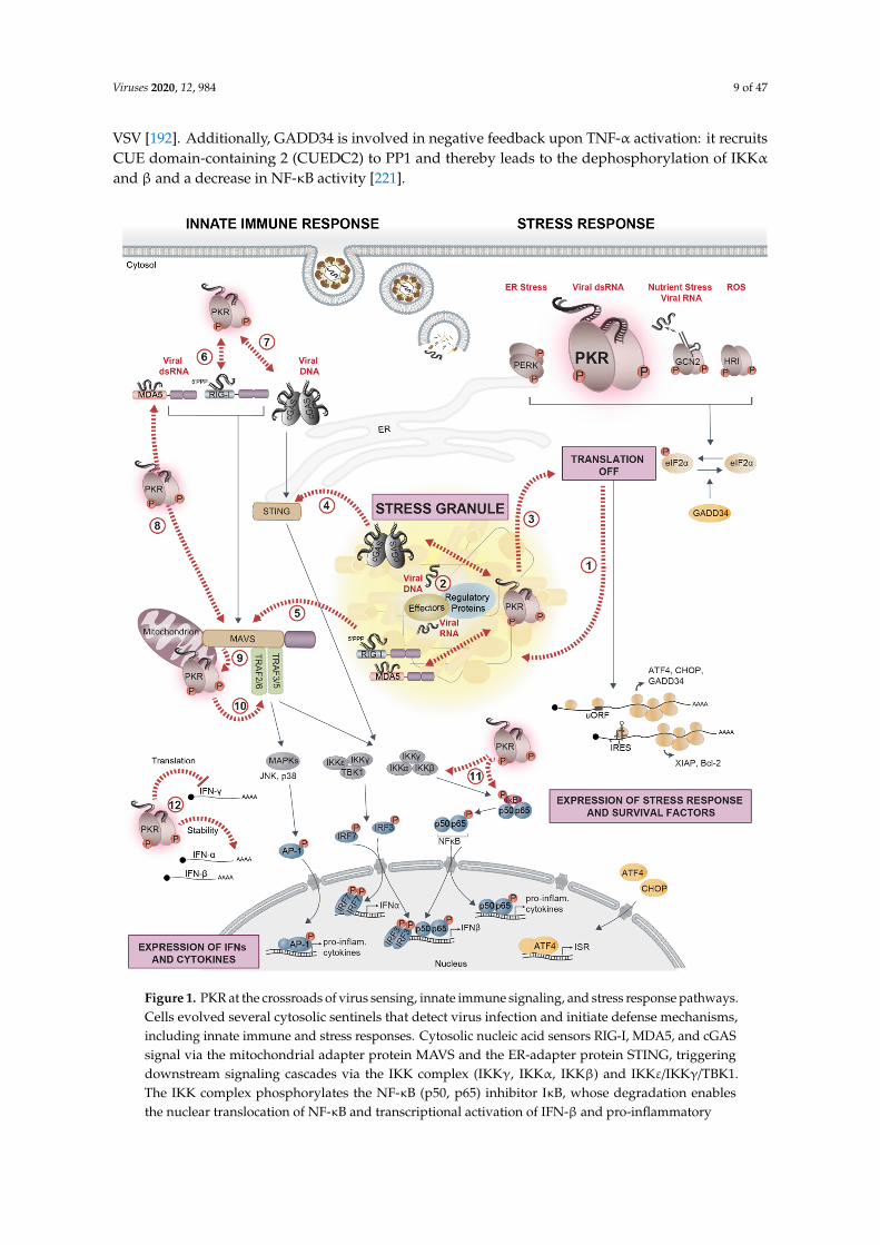
Figure 1. PKR at the crossroads of virus sensing, innate immune signaling, and stress response pathways. Cells evolved several cytosolic sentinels that detect virus infection and initiate defense mechanisms, including innate immune and stress responses. Cytosolic nucleic acid sensors RIG-I, MDA5, and cGAS signal via the mitochondrial adapter protein MAVS and the ER-adapter protein STING, triggering downstream signaling cascades via the IKK complex (IKKγ, IKKα, IKKβ) and IKKε/IKKγ/TBK1. The IKK complex phosphorylates the NF-κB (p50, p65) inhibitor IκB, whose degradation enables the nuclear translocation of NF-κB and transcriptional activation of IFN-β and pro-inflammatory cytokines. TBK1, on the other hand, phosphorylates IRF3/7, whose nuclear translocation mediates the transcriptional activation of IFN-α/β (left side). Cytosolic dsRNA that accumulates during viral replication is sensed by the stress kinase PKR. As a consequence, phosphorylation of eIF2α strongly represses the translation of most cellular mRNAs, while the translation of factors related to the stress response (ATF4, CHOP, and GADD34) or cell survival (XIAP and Bcl-2) is selectively favored (right side). The other eIF2α-kinases, GCN2, PERK, and HRI,
Figure 1. PKR at the crossroads of virus sensing, innate immune signaling, and stress response pathways.Cells evolved several cytosolic sentinels that detect virus infection and initiate defense mechanisms,including innate immune and stress responses. Cytosolic nucleic acid sensors RIG-I, MDA5, and cGASsignal via the mitochondrial adapter protein MAVS and the ER-adapter protein STING, triggeringdownstream signaling cascades via the IKK complex (IKKγ, IKKα, IKKβ) and IKKε/IKKγ/TBK1.The IKK complex phosphorylates the NF-κB (p50, p65) inhibitor IκB, whose degradation enablesthe nuclear translocation of NF-κB and transcriptional activation of IFN-β and pro-inflammatory
Viruses 2020, 12, 984 10 of 47
cytokines. TBK1, on the other hand, phosphorylates IRF3/7, whose nuclear translocation mediatesthe transcriptional activation of IFN-α/β (left side). Cytosolic dsRNA that accumulates during viralreplication is sensed by the stress kinase PKR. As a consequence, phosphorylation of eIF2α stronglyrepresses the translation of most cellular mRNAs, while the translation of factors related to the stressresponse (ATF4, CHOP, and GADD34) or cell survival (XIAP and Bcl-2) is selectively favored (right side).The other eIF2α-kinases, GCN2, PERK, and HRI, contribute to translation suppression by detectingvirus-induced changes in cellular homeostasis such as nutrient deprivation and accumulation of reactiveoxygen species (ROS) or unfolded proteins in the ER. In the cytosol, untranslated mRNAs condensetogether with numerous RBPs and form SGs (1). Upon infection with certain viruses, innate immunesensors, PKR, regulators of stress and immune sensors, and interferon (IFN)-induced effectors localize instress granules (SGs) together with viral RNA or DNA, forming a signaling platform (2) that coordinatesand potentiates the antiviral response (3,4,5). PKR is at the crossroads of stress and innate immunesignaling pathways: PKR interacts with RIG-I, promotes its activation, and amplifies the downstreamsignaling cascade (6). A similar interaction exists with cGAS (7). PKR promotes MDA5 filamentformation, is activated by MDA5 to enhance downstream MAVS signaling (8), and, in turn, can also beactivated by MAVS (9). PKR affects pro-inflammatory responses by interacting with TRAFs to activatethe NF-κB signaling and potentially the JNK/p38 MAPK pathway leading to AP-1 activation (10), ordirectly regulates NF-κB activity via phosphorylation of IκB and IKK (11). Finally, PKR is involved incontrolling the stability and translation of IFN mRNAs (12). Black arrows indicate signaling pathways;dashed red arrows indicate crossroads between stress response pathways, innate immune signaling,and SGs.
Altogether, the translational inhibition of unstable immune-regulatory proteins, as well asinteractions of stress kinases or downstream signal transducers with components of the immunesignaling cascade, promote the transcription of IFNs and pro-inflammatory cytokines. Hence, inductionof the stress response, especially the activation of PKR, upon viral infections strongly contributes toestablishing a pro-inflammatory and antiviral state. At the same time, PKR, PERK, ATF4, and GADD34,however, also initiate distinct negative feedback loops in order to fine-tune and possibly locallyand temporally restrict IFN production to prevent an overactivation of the innate immune system.
An unsolved question is how antiviral proteins are synthesized when global translation is repressed.While uORFs facilitate translation of mRNAs under such conditions, as exemplified by ATF4 [222],most mRNAs encoding antiviral proteins do not appear to harbor uORFs. GADD34 is induced asa negative feedback regulator upon translational inhibition, leading to dephosphorylation of eIF2αand resumption of translation. Mathematic modeling in combination with flow cytometry experimentssuggest that this negative feedback loop contributes to the stochastic expression of IFN in individualcells upon poly(I:C) treatment and VSV infection, whereby transcriptional induction of IFNs duringtranslation-off states is followed by synthesis of IFNs during translation-on states [192,223]. Recently,two different laboratories made another important contribution to the understanding of how antiviralfactors are preferentially produced in response to viral infections. They discovered that ribonuclease L(RNase L), which is activated in response to dsRNA, strongly depletes cellular mRNA pools throughwide-spread mRNA degradation, but specifically leaves mRNAs encoding IFNs, cytokines, and otherdefense proteins intact [224–226]. The mechanism by which these mRNAs are excluded from RNaseL-mediated cleavage remains unclear, but probably involves regulatory sequences, RNA secondarystructures, and RBPs that protect individual mRNAs from degradation. Hence, it appears thatthe combination of increased transcription and selective mRNA stabilization and translation permitsthe synthesis of antiviral factors under conditions of repressed global translation.
An early link between the ISR, in particular the formation of SGs, and the temporal control ofcytokine production comes from studies on the adaptive immune system. In naïve T helper cells, IL-4mRNA was found to accumulate in SG-like foci in the cytoplasm during T cell priming, concomitantwith elevated phosphorylation of eIF2α. The release of these mRNAs then allowed for the rapidproduction of cytokines during T cell restimulation [227], suggesting that SGs can exert storageand regulatory functions outside of classical stress conditions.
Viruses 2020, 12, 984 11 of 47
5. SGs as Immune Signaling Platforms in Antiviral Defense
While SGs assemble in response to translational shut-off, they are not necessary for translationsuppression under stress conditions. Rather, SGs were proposed to function as (i) hubs for modulatinglocal protein and mRNA concentrations; (ii) timers for the stress response, marking a “window ofopportunity” during which the stress can be resolved or an apoptotic program will be initiated; (iii) jointassemblies of unfolded proteins and translation complexes that coordinate the activities of the proteinsynthesis and folding machineries; (iv) storage sites for pre-assembled initiation complexes, allowingfor rapid resumption of protein synthesis when cells recover from stress; (v) signaling platformsthat connect stress sensors with effectors of immune responses, especially in the context of viralinfection [15,16,18,228,229].
Viruses deploy many different strategies to inhibit the formation of SGs, suggesting that SGsserve an antiviral function. In particular, flaviviruses have evolved numerous mechanisms to interferewith SG assembly. The core protein of JEV, for instance, interacts with Caprin1 to relocalize G3BP1and USP10 to the perinuclear region, thereby preventing SG formation [230]. Other SG componentsseem to be sequestered by viral RNAs, as reported for TIA1 and TIAR, which bind to the genomicRNA 3′ end of DENV, WNV, and tick-borne encephalitis virus [231,232]. At later stages of infection,flavivirus genomes are degraded by the cellular exoribonuclease XRN1 from the 5′ end up to a compactpseudoknot structure in the 3′ UTR, leading to the cytosolic accumulation of the remaining subgenomicflaviviral RNA (sfRNA) [233,234]. G3BP1, G3BP2, Caprin1, and USP10 are sequestered by sfRNAs,which leads to inhibition of ISG mRNA translation and thus, attenuation of the antiviral response [235].A similar sequestration strategy was observed for SeV infection, during which trailer RNAs, i.e., smallabortive products of viral genome replication, bind TIAR, and inhibit SG formation [236–238]. Ourown work showed that flaviviruses uncouple the stress response from translation control by blockingboth eIF2α phosphorylation and SG assembly, while protein synthesis is still suppressed [58]. In linewith this observation, other laboratories reported that the expression of single viral proteins suchas Zika virus (ZIKV) capsid, NS3, NS2B-3, or NS4A is sufficient to inhibit SG assembly [239–241].The precise mechanism behind this inhibition remains to be uncovered. In the following, we will focuson the role of SGs within the complex signaling network that controls cellular reprogramming towardsan antiviral state (Table 1 and Figure 2).
5.1. G3BP1 at the Interface between SGs and the IFN Response
G3BP1 turns out to be a target of particular importance for viruses, which aim to interfere with itsfunction as a SG nucleator and regulator of immune responses. Moreover, G3BP1 appears to facilitatereplication of many viruses. Several viral proteins were shown to directly recruit G3BP1 to sites of viralreplication, e.g., HCV polymerase NS5B [271,296–298], Chikungunya virus nsP3 [299,300], SemlikiForest virus and SINV Nsp3 [301,302], and Junín virus N protein [303]. Murine norovirus inhibitsthe formation of canonical SGs not only by redistributing G3BP1 together with the NS3 protein to sitesof viral replication, but also by modifying the interactome of G3BP1 [304]. Notably, G3BP1 is directlyinvolved in translation of the norovirus genome by associating with the VPg viral cap complex to helpribosome recruitment [305].
Viruses 2020, 12, 984 12 of 47
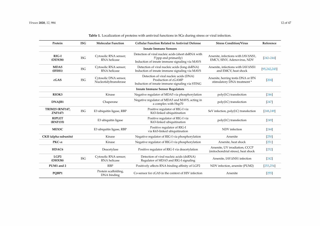
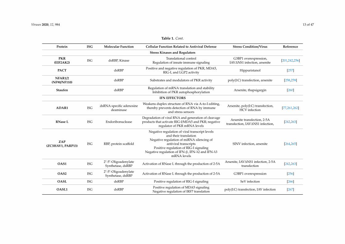
Table 1. Localization of proteins with antiviral functions in SGs during stress or viral infection.
Protein ISG Molecular Function Cellular Function Related to Antiviral Defense Stress Condition/Virus Reference
Innate Immune Sensors
RIG-I(DDX58) ISG Cytosolic RNA sensor;
RNA helicase
Detection of viral nucleic acids (short dsRNA with5′ppp end primarily)
Induction of innate immune signaling via MAVS
Arsenite, infections with IAV∆NS1,EMCV, SINV, Adenovirus, NDV [242–244]
MDA5(IFIH1) ISG Cytosolic RNA sensor;
RNA helicaseDetection of viral nucleic acids (long dsRNA)
Induction of innate immune signaling via MAVSArsenite, infections with IAV∆NS1
and EMCV, heat shock [95,242,245]
cGAS ISG Cytosolic DNA sensor,Nucleotidyltransferase
Detection of viral nucleic acids (DNA)Production of cGAMP
Induction of innate immune signaling via STING
Arsenite, herring testis DNA or IFNstimulatory DNA treatment * [204]
Innate Immune Sensor Regulators
RIOK3 Kinase Negative regulator of MDA5 via phosphorylation poly(I:C) transfection [246]
DNAJB1 Chaperone Negative regulator of MDA5 and MAVS, acting ina complex with Hsp70 poly(I:C) transfection [247]
TRIM25 (RNF147,ZNF147) ISG E3 ubiquitin ligase, RBP Positive regulator of RIG-I via
K63-linked ubiquitination SeV infection, poly(I:C) transfection [248,249]
RIPLET(RNF135) E3 ubiquitin ligase Positive regulator of RIG-I via
K63-linked ubiquitination poly(I:C) transfection [249]
MEX3C E3 ubiquitin ligase, RBP Positive regulator of RIG-Ivia K63-linked ubiquitination NDV infection [244]
CKII (alpha subunits) Kinase Negative regulator of RIG-I via phosphorylation Arsenite [250]
PKC-α Kinase Negative regulator of RIG-I via phosphorylation Arsenite, heat shock [251]
HDAC6 Deacetylase Positive regulator of RIG-I via deacetylation Arsenite, UV irradiation, CCCP(mitochondrial stress), heat shock [252]
LGP2(DHX58) ISG Cytosolic RNA sensor;
RNA helicaseDetection of viral nucleic acids (dsRNA)Regulator of MDA5 and RIG-I signaling Arsenite, IAV∆NS1 infection [242]
PUM1 and 2 RBP Positively affects RNA binding affinity of LGP2 NDV infection, arsenite (PUM2) [253,254]
PQBP1 Protein scaffolding,DNA binding Co-sensor for cGAS in the context of HIV infection Arsenite [255]
Viruses 2020, 12, 984 13 of 47
Table 1. Cont.
Protein ISG Molecular Function Cellular Function Related to Antiviral Defense Stress Condition/Virus Reference
Stress Kinases and Regulators
PKR(EIF2AK2) ISG dsRBP, Kinase Translational control
Regulation of innate immune signalingG3BP1 overexpression,
IAV∆NS1 infection, arsenite [201,242,256]
PACT dsRBP Positive and negative regulation of PKR, MDA5,RIG-I, and LGP2 activity Hippuristanol [257]
NFAR1/2(NF90/NF110) dsRBP Substrates and modulators of PKR activity poly(I:C) transfection, arsenite [258,259]
Staufen dsRBP Regulation of mRNA translation and stabilityInhibition of PKR autophosphorylation Arsenite, thapsigargin [260]
IFN EFFECTORS
ADAR1 ISG dsRNA-specific adenosinedeaminase
Weakens duplex structure of RNA via A-to-I editing,thereby prevents detection of RNA by immune
and stress sensors
Arsenite, poly(I:C) transfection,HCV infection [77,261,262]
RNase L ISG EndoribonucleaseDegradation of viral RNA and generation of cleavageproducts that activate RIG-I/MDA5 and PKR; negative
regulator of PKR mRNA levels
Arsenite transfection, 2-5Atransfection, IAV∆NS1 infection, [242,263]
ZAP(ZC3HAV1, PARP13) ISG RBP, protein scaffold
Negative regulation of viral transcript levelsand their translation
Negative regulation of miRNA silencing ofantiviral transcripts
Positive regulation of RIG-I signalingNegative regulation of IFN-β, IFN-λ2 and IFN-λ3
mRNA levels
SINV infection, arsenite [264,265]
OAS1 ISG 2′-5′-OligoadenylateSynthetase, dsRBP Activation of RNase L through the production of 2-5A Arsenite, IAV∆NS1 infection, 2-5A
transfection [242,263]
OAS2 ISG 2′-5′-OligoadenylateSynthetase, dsRBP Activation of RNase L through the production of 2-5A G3BP1 overexpression [256]
OASL ISG dsRBP Positive regulation of RIG-I signaling SeV infection [266]
OASL1 ISG dsRBP Positive regulation of MDA5 signalingNegative regulation of IRF7 translation poly(I:C) transfection, IAV infection [267]
Viruses 2020, 12, 984 14 of 47
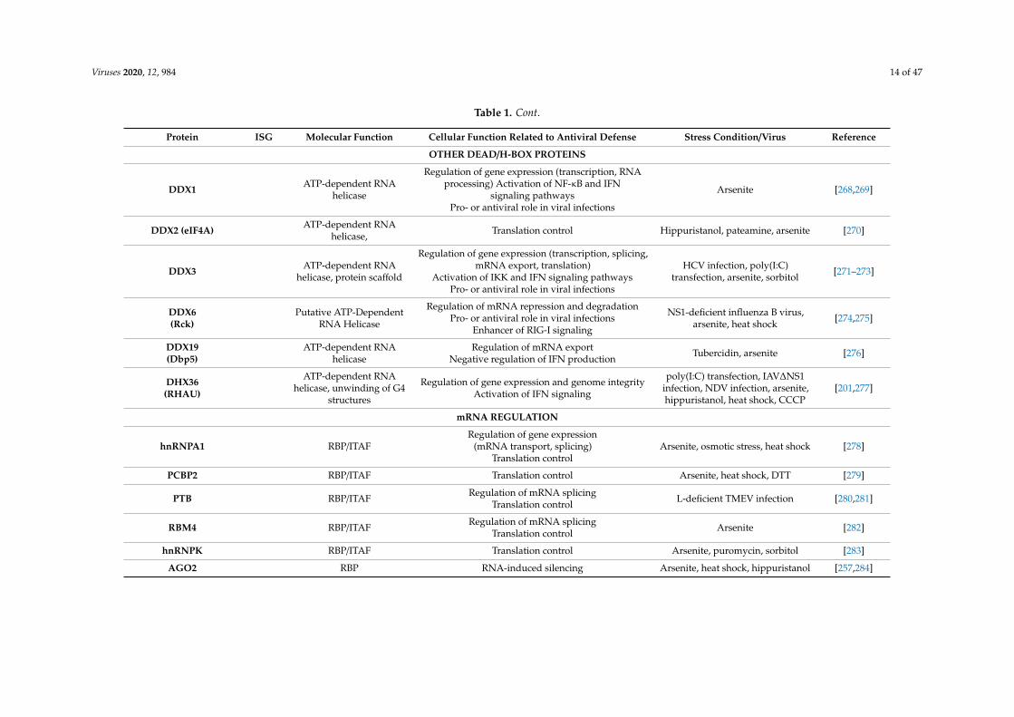
Table 1. Cont.
Protein ISG Molecular Function Cellular Function Related to Antiviral Defense Stress Condition/Virus Reference
OTHER DEAD/H-BOX PROTEINS
DDX1 ATP-dependent RNAhelicase
Regulation of gene expression (transcription, RNAprocessing) Activation of NF-κB and IFN
signaling pathwaysPro- or antiviral role in viral infections
Arsenite [268,269]
DDX2 (eIF4A) ATP-dependent RNAhelicase, Translation control Hippuristanol, pateamine, arsenite [270]
DDX3 ATP-dependent RNAhelicase, protein scaffold
Regulation of gene expression (transcription, splicing,mRNA export, translation)
Activation of IKK and IFN signaling pathwaysPro- or antiviral role in viral infections
HCV infection, poly(I:C)transfection, arsenite, sorbitol [271–273]
DDX6(Rck)
Putative ATP-DependentRNA Helicase
Regulation of mRNA repression and degradationPro- or antiviral role in viral infections
Enhancer of RIG-I signaling
NS1-deficient influenza B virus,arsenite, heat shock [274,275]
DDX19(Dbp5)
ATP-dependent RNAhelicase
Regulation of mRNA exportNegative regulation of IFN production Tubercidin, arsenite [276]
DHX36(RHAU)
ATP-dependent RNAhelicase, unwinding of G4
structures
Regulation of gene expression and genome integrityActivation of IFN signaling
poly(I:C) transfection, IAV∆NS1infection, NDV infection, arsenite,hippuristanol, heat shock, CCCP
[201,277]
mRNA REGULATION
hnRNPA1 RBP/ITAFRegulation of gene expression
(mRNA transport, splicing)Translation control
Arsenite, osmotic stress, heat shock [278]
PCBP2 RBP/ITAF Translation control Arsenite, heat shock, DTT [279]
PTB RBP/ITAF Regulation of mRNA splicingTranslation control L-deficient TMEV infection [280,281]
RBM4 RBP/ITAF Regulation of mRNA splicingTranslation control Arsenite [282]
hnRNPK RBP/ITAF Translation control Arsenite, puromycin, sorbitol [283]
AGO2 RBP RNA-induced silencing Arsenite, heat shock, hippuristanol [257,284]
Viruses 2020, 12, 984 15 of 47
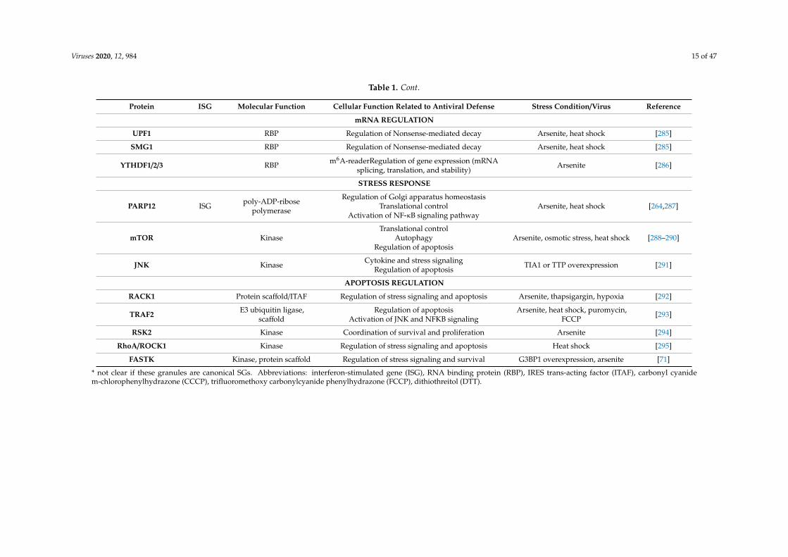
Table 1. Cont.
Protein ISG Molecular Function Cellular Function Related to Antiviral Defense Stress Condition/Virus Reference
mRNA REGULATION
UPF1 RBP Regulation of Nonsense-mediated decay Arsenite, heat shock [285]
SMG1 RBP Regulation of Nonsense-mediated decay Arsenite, heat shock [285]
YTHDF1/2/3 RBP m6A-readerRegulation of gene expression (mRNAsplicing, translation, and stability)
Arsenite [286]
STRESS RESPONSE
PARP12 ISG poly-ADP-ribosepolymerase
Regulation of Golgi apparatus homeostasisTranslational control
Activation of NF-κB signaling pathwayArsenite, heat shock [264,287]
mTOR KinaseTranslational control
AutophagyRegulation of apoptosis
Arsenite, osmotic stress, heat shock [288–290]
JNK Kinase Cytokine and stress signalingRegulation of apoptosis TIA1 or TTP overexpression [291]
APOPTOSIS REGULATION
RACK1 Protein scaffold/ITAF Regulation of stress signaling and apoptosis Arsenite, thapsigargin, hypoxia [292]
TRAF2 E3 ubiquitin ligase,scaffold
Regulation of apoptosisActivation of JNK and NFKB signaling
Arsenite, heat shock, puromycin,FCCP [293]
RSK2 Kinase Coordination of survival and proliferation Arsenite [294]
RhoA/ROCK1 Kinase Regulation of stress signaling and apoptosis Heat shock [295]
FASTK Kinase, protein scaffold Regulation of stress signaling and survival G3BP1 overexpression, arsenite [71]
* not clear if these granules are canonical SGs. Abbreviations: interferon-stimulated gene (ISG), RNA binding protein (RBP), IRES trans-acting factor (ITAF), carbonyl cyanidem-chlorophenylhydrazone (CCCP), trifluoromethoxy carbonylcyanide phenylhydrazone (FCCP), dithiothreitol (DTT).
Viruses 2020, 12, 984 16 of 47
Viruses 2020, 12, x 15 of 47
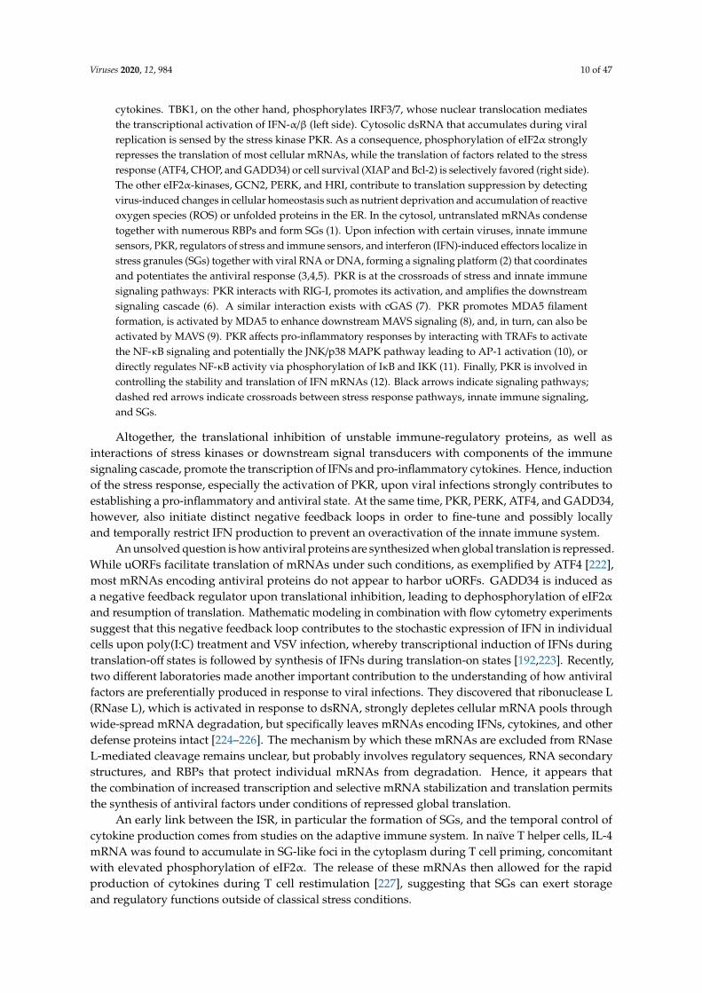
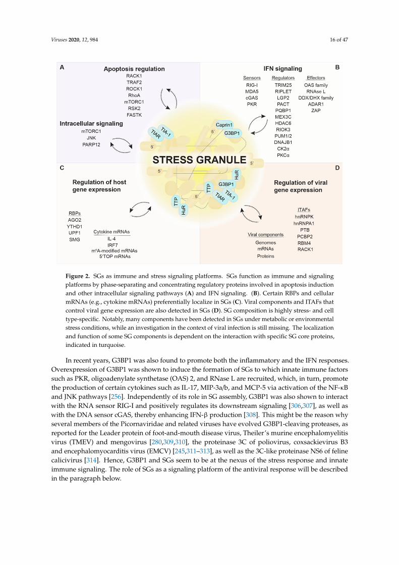
Figure 2. SGs as immune and stress signaling platforms. SGs function as immune and signaling platforms by phase-separating and concentrating regulatory proteins involved in apoptosis induction and other intracellular signaling pathways (A) and IFN signaling. (B). Certain RBPs and cellular mRNAs (e.g., cytokine mRNAs) preferentially localize in SGs (C). Viral components and ITAFs that control viral gene expression are also detected in SGs (D). SG composition is highly stress- and cell type-specific. Notably, many components have been detected in SGs under metabolic or environmental stress conditions, while an investigation in the context of viral infection is still missing. The localization and function of some SG components is dependent on the interaction with specific SG core proteins, indicated in turquoise.
5.1. G3BP1 at the Interface between SGs and the IFN Response
G3BP1 turns out to be a target of particular importance for viruses, which aim to interfere with its function as a SG nucleator and regulator of immune responses. Moreover, G3BP1 appears to facilitate replication of many viruses. Several viral proteins were shown to directly recruit G3BP1 to sites of viral replication, e.g., HCV polymerase NS5B [271,296–298], Chikungunya virus nsP3 [299,300], Semliki Forest virus and SINV Nsp3 [301,302], and Junín virus N protein [303]. Murine norovirus inhibits the formation of canonical SGs not only by redistributing G3BP1 together with the NS3 protein to sites of viral replication, but also by modifying the interactome of G3BP1 [304]. Notably, G3BP1 is directly involved in translation of the norovirus genome by associating with the VPg viral cap complex to help ribosome recruitment [305].
In recent years, G3BP1 was also found to promote both the inflammatory and the IFN responses. Overexpression of G3BP1 was shown to induce the formation of SGs to which innate immune factors such as PKR, oligoadenylate synthetase (OAS) 2, and RNase L are recruited, which, in turn, promote the production of certain cytokines such as IL-17, MIP-3a/b, and MCP-5 via activation of the NF-κB and JNK pathways [256]. Independently of its role in SG assembly, G3BP1 was also shown to interact with the RNA sensor RIG-I and positively regulates its downstream signaling [306,307], as well as with the DNA sensor cGAS, thereby enhancing IFN-β production [308]. This might be the reason why several members of the Picornaviridae and related viruses have evolved G3BP1-cleaving proteases, as reported for the Leader protein of foot-and-mouth disease
Figure 2. SGs as immune and stress signaling platforms. SGs function as immune and signalingplatforms by phase-separating and concentrating regulatory proteins involved in apoptosis inductionand other intracellular signaling pathways (A) and IFN signaling. (B). Certain RBPs and cellularmRNAs (e.g., cytokine mRNAs) preferentially localize in SGs (C). Viral components and ITAFs thatcontrol viral gene expression are also detected in SGs (D). SG composition is highly stress- and celltype-specific. Notably, many components have been detected in SGs under metabolic or environmentalstress conditions, while an investigation in the context of viral infection is still missing. The localizationand function of some SG components is dependent on the interaction with specific SG core proteins,indicated in turquoise.
In recent years, G3BP1 was also found to promote both the inflammatory and the IFN responses.Overexpression of G3BP1 was shown to induce the formation of SGs to which innate immune factorssuch as PKR, oligoadenylate synthetase (OAS) 2, and RNase L are recruited, which, in turn, promotethe production of certain cytokines such as IL-17, MIP-3a/b, and MCP-5 via activation of the NF-κBand JNK pathways [256]. Independently of its role in SG assembly, G3BP1 was also shown to interactwith the RNA sensor RIG-I and positively regulates its downstream signaling [306,307], as well aswith the DNA sensor cGAS, thereby enhancing IFN-β production [308]. This might be the reason whyseveral members of the Picornaviridae and related viruses have evolved G3BP1-cleaving proteases, asreported for the Leader protein of foot-and-mouth disease virus, Theiler’s murine encephalomyelitisvirus (TMEV) and mengovirus [280,309,310], the proteinase 3C of poliovirus, coxsackievirus B3and encephalomyocarditis virus (EMCV) [245,311–313], as well as the 3C-like proteinase NS6 of felinecalicivirus [314]. Hence, G3BP1 and SGs seem to be at the nexus of the stress response and innateimmune signaling. The role of SGs as a signaling platform of the antiviral response will be describedin the paragraph below.
Viruses 2020, 12, 984 17 of 47
5.2. SGs, a Platform to Initiate IFN Signaling?
In cells infected with IAV lacking the NS1 protein (IAV∆NS1) and thus, unable to counteract PKR,viral RNA was found to co-localize in SGs together with the immune sensors RIG-I and MDA5 as wellas other antiviral effectors such as OAS, RNase L, and PKR. A similar redistribution of RIG-I was alsoobserved upon infection with EMCV, adenovirus, and SINV. In these infection models, the inhibitionof SG assembly by depletion of essential SG components resulted in a strong attenuation of IFNproduction and an increase in viral replication. Based on these results, Onomoto et al. proposedthe term “antiviral SGs” (avSGs) to indicate that SGs may serve as a platform for the activationof innate immune sensors by viral RNA and the subsequent initiation of the IFN response [242].While most RNA viruses are sensed by RIG-I alone or by RIG-I and MDA5 together [315], MDA5 isessential for IFN-α/β induction in response to Picornaviridae infection [316,317]. In agreement withthe idea of avSGs, MDA5 and dsRNA were found to locate in EMCV-induced SGs at early times postinfection. At later stages of EMCV infection, cleavage of G3BP1 by the EMCV protease 3C resultedin SG dissolution concomitant with a reduced IFN-β and cytokine response, which could be rescuedexpressing a cleavage-resistant G3BP1 mutant [245]. The characterization of avSGs formed uponinfection with SeV, again revealed the presence of many antiviral components and SG formation wasshown to be important for IFN-β production and viral restriction [263]. Furthermore, late during NDVinfection, uncapped viral RNA(+) derived from read-through transcription was found to accumulatetogether with RIG-I in SGs and trigger RIG-I activation. Consistent with the idea of avSGs, IFN-βinduction could be reduced by the disruption of SGs in this model [243].
In contrast to these findings, some studies suggest that SG formation may not be necessary forIFN production upon virus infection. During infection with a mutant mengovirus lacking the Leaderprotein, for instance, localization of MDA5 to SGs was not a prerequisite for IFN induction. However,it may be important to note that viral dsRNA could not be detected within SGs in this infection model,which may indicate that SGs adopt an antiviral function as platforms for RIG-I or MDA5 activationonly when they accumulate viral RNA [95]. Another study showed that disruption of SGs in IAV∆NS1-and SeV-infected cells did not reduce but actually increased RIG-I-mediated IFN-β production [202],which is in direct contradiction with previous observations [242,263]. It is therefore still unclear whetherthe changes observed in the IFN response ensue from the disruption of SGs as a coordinating platformor are due to the immunomodulatory function of single SG components. Additional experimentsbased on genetic ablation of other SG components, interference with SG assembly by cycloheximide orISRIB treatments, or the use of eIF2α S51A mutant cells might be necessary to answer this question
Finally, MDA5 and RIG-I were also detected in SGs formed under other stress conditions suchas heat shock and arsenite treatment [95,242], which raises the possibility that MDA5/RIG-I can alsoassemble with endogenous (e.g., damaged) RNAs in SGs, or that these sensors passively phase separatein SGs.
Whether SGs contribute to the antiviral response upon infection with DNA viruses is even lessclear. Upon DNA virus infection, viral DNA is sensed by specialized DNA sensors, among thosecGAS. In addition, viral transcripts, cytosolic noncoding RNAs transcribed by the RNA polymeraseIII, as well as Alu-derived host RNAs can activate MDA5 or RIG-I [318]. In the case of VACV∆E3L,MDA5-mediated IFN-β production was unchanged upon SG dissolution, and rather dependent onthe immunomodulatory function of PKR [202]. HSV-1 infection induces formation of SGs, in whichdsRNA (whose origin is unclear) was detected. In this infection model, IFN production was inducedby the DNA sensing cGAS-STING pathway rather than by MDA5/RIG-I, and preceded PKR activationand SG formation, indicating that SGs are dispensable for cGAS-mediated IFN production uponHSV-1 infection [121]. Interestingly, cGAS was reported to associate with dsDNA, G3BP1, and PKR ina complex and one study claimed the presence in cytoplasmic foci upon dsDNA transfection [204,308].While it is not clear if these foci represent canonical SGs, they were proposed to be essential forcGAS activation and downstream IFN-β production [204]. Of note, polyglutamine binding protein 1(PQBP1), identified as a co-sensor of cGAS in the context of HIV infection [319], was shown to localize
Viruses 2020, 12, 984 18 of 47
to SGs [70,255]. Hence, SGs might support cGAS-mediated IFN production under specific conditionsor for particular virus types. However, this needs to be further investigated.
Given these discrepancies, the link between SGs and innate immune signaling appears to becomplex. Besides the co-localization of innate immune sensors with viral RNAs or DNAs within SGs,the type of virus and the presence or absence of regulatory factors are likely to affect the potential ofSGs to serve as platforms initiating the IFN response. It should further be noted that studies so far havemainly focused on the impact of SGs on the production of IFN-β or cytokines such as IL-6, RANTES,and CXCL10. Hence, it would be interesting to investigate how SGs might influence the expression ofother IFN types and subtypes, and how this could affect virus replication.
5.3. Regulators of the Innate Immune Sensors
RLR activity is tightly controlled by multiple posttranslational modifications and proteininteractions. Tripartite motif protein 25 (TRIM25) and Riplet are two examples of E3 ubiquitin ligases,which, according to a current model, mediate consecutive K63-linked ubiquitination of RIG-I. The firstubiquitination event mediated by Riplet leads to the exposure of the RIG-I CARD domains and theirsubsequent ubiquitination by TRIM25. Thereby RIG-I gets activated, oligomerizes, and initiatesdownstream MAVS signaling [320]. Both E3 ubiquitin ligases were found to be recruited together withRIG-I to SGs when cells were exposed to poly(I:C) [249]. It is not clear, however, whether co-localizationof these proteins within SGs is important for the sequential ubiquitination of RIG-I and downstreamsignaling events. Using a bimolecular fluorescence complementation assay and super-resolutionmicroscopy in SeV-infected cells, RIG-I was identified in two distinct complexes—RIG-I/TRIM25and RIG-I/MAVS—with different cellular localization [248]. While TRIM25/RIG-I complexes localizedin SGs, RIG-I/MAVS complexes remained attached to mitochondrial membranes. An interactionbetween MAVS and TRIM25 was barely observed [248], contrasting previous studies in which TRIM25was proposed to act as an ubiquitin ligase of MAVS [321,322]. RIG-I/TRIM25 complexes furtherappeared to be in close proximity with mitochondria upon virus stimulation [248], consistent withprevious reports about physical contacts between avSGs and MAVS [242,243]. These findings suggestthat RIG-I first needs to interact with TRIM25 within SGs to become ubiquitinated and activated beforeit is released from the complex to activate MAVS at mitochondria. It should be noted that TRIM25 alsohas a second function: when bound to FAT10, it stabilizes the protein, which acts as a negative regulatorof RIG-I and prevents the formation of avSGs under conditions of inflammation [323]. Recent in vivostudies suggest that Riplet alone is sufficient for RIG-I activation [324,325], and confirmed a role ofRiplet in modulating the kinetics of RIG-I recruitment into SGs. However, the ability of Riplet to activateRIG-I signaling was not dependent on SG formation [324]. Apart from Riplet and TRIM25, otherE3 ubiquitin ligases can mediate RIG-I ubiquitination including MEX3C, an RNA-binding E3 ligasethat was found to localize in NDV-induced avSGs and plays an essential role in RIG-I-mediated IFNsignaling [244]. Moreover, RIG-I activity is regulated by the activity of several kinases, phosphatases,and acetyl transferases, some of which localize to SGs such as histone deacetylase 6 (HDAC6) [252],PKC-α [251], and the α-subunits of CKII [250].
MDA5 activity is similarly regulated by posttranslational modifications [326]. RIO kinase 3(RIOK3) is involved in maintaining MDA5 inactive under normal conditions by phosphorylating itsC-terminal domain, thereby interfering with MDA5 filament formation on dsRNA and downstreamIFN signaling. Upon poly(I:C) exposure, RIOK3 was shown to be partially recruited to SGs, togetherwith MDA5 [246]. DNAJ heat shock protein family (Hsp40) member B1 (DNAJB1), a member ofthe Hsp40 family, was identified as an interactor of MDA5 that negatively regulates MDA5-MAVSsignaling. Upon poly(I:C) treatment, DNAJB1 in conjunction with Hsp70 was found to co-localize withSG markers and the mitochondrial membrane, negatively affecting MDA5 oligomerization and MAVSaggregation [247]. If and how the co-localization of MDA5 together with the DNAJB1-Hsp70 complexor RIOK3 within SGs affects MDA5-MAVS signaling still needs to be investigated.
Viruses 2020, 12, 984 19 of 47
Laboratory of genetics and physiology 2 (LGP2, DHX58) belongs to the RLR family and acts asa modulatory co-receptor of RIG-I and MDA5 [315]. LGP2 has been shown to localize in SGs uponIAV∆NS1 infection [242]. Moreover, the positive activator of PKR (PACT, PRKRA) was identified asan LGP2 interactor essential for RLR regulation [327], and also shown to localize in SGs upon treatmentwith hippuristanol [257], a steroid compound that interferes with cap-dependent translation [270].Interestingly, PACT was also found in a complex with RIG-I and MDA5, positively regulatingtheir downstream signaling [328,329]. In addition, two members of the pumilio RBP family, PUM1and PUM2, localized to SGs and were found to associate with LGP2 upon NDV infection, enhancing itsdsRNA binding ability through conformational changes and thereby increasing the IFN response [253].Whether co-localization of LGP2 with PACT, PUM1, and PUM2 in SGs is necessary for the regulatoryeffects on RIG-I and MDA5 activity is currently not clear.
5.4. Stress Kinase PKR and Its Regulators
Localization of the dsRNA-sensor PKR in SGs is a prime example of their functionalrelevance [121,256,330]. PKR is recruited to SGs through its direct interaction with the NTF2 and PXXPdomains of G3BP1 [330]. By complex formation with G3BP1 and Caprin1, PKR can be activated ina dsRNA-independent manner. The high local concentrations of all three components in SGs are likelywhat drive formation of the tripartite G3BP1/Caprin-1/PKR complex. Activated PKR is subsequentlyreleased from SGs into the cytosol, where it phosphorylates eIF2α and thereby promotes both translationrepression and SG persistence [330]. Hence, SGs are part of a feed-forward loop that amplifies PKRactivity and restricts viral replication. This amplification mechanism might explain why so manyviruses simultaneously target PKR and SG assembly. One may speculate that the ubiquitin-specificpeptidase USP10, which binds to G3BP1 and inhibits SG formation [331], could affect PKR activationand other antiviral signaling events in SGs.
In contrast, activation of PKR by MAVS was reported to occur outside of SGs prior to its recruitmentinto SGs [203]. This finding corroborates the idea that PKR is activated in sequential steps and that SGshelp to maintain PKR activity at later stages of the infection. Interestingly, PKR was also reported tolocalize both inside and at the outer membrane of mitochondria [99]. Since SGs and mitochondria areoften found in close proximity [248], it is tempting to speculate that PKR shuttles between the twocompartments. Accordingly, recruitment of PKR to SGs could affect not only the activation status ofPKR within protein complexes inside SGs, but also within protein complexes that specifically forminside or at mitochondria. This scenario is well in line with the tight collaboration between SGsand mitochondria in the RIG-I/MAVS activation pathway described above (Figure 3).
Besides G3BP1 and Caprin1, PACT, transactivation response element RNA-binding protein(TRBP), and P58IPK modulate PKR activity by directly targeting its kinase domain. It is generallythought that PACT stimulates PKR whereas TRBP and P58IPK inhibit PKR activity, although exceptionshave been reported and may be cell-context specific [332–336]. The interaction between PKR, PACT,and TRBP is especially important for cellular physiology during viral infections and has implicationsin stress recovery and the RNA-induced silencing pathway [335,337,338]. However, it is currentlyunclear whether the recruitment of PKR into SGs affects its interaction with PACT, TRBP, and P58IPK.Upon hippuristanol treatment, PACT localizes in SGs where it was proposed to form a complex withargonaute 2 (AGO2) [257]. Furthermore, the SG proteins TIA1 and TIAR were found to suppress PKRactivity, although they seem to exert this effect by controlling PACT pre-mRNA splicing, independentlyof their role in SG assembly [336]. In addition to PACT [257], several modulators of PKR activity,including NFAR1/2 [258,259], adenosine deaminase RNA-specific 1 (ADAR1) [77,262] and Staufen [260],were found to localize in SGs or modulate SGs formation. Further studies are needed to assess whetherthese PKR regulators relocalize into, or are specifically excluded from, SGs upon virus infection.The differential activation of PKR within different complexes and cellular compartments is likely todictate the spectrum of PKR targets and thereby decides whether PKR has pro-apoptotic, anti-apoptotic,inflammatory, or translational effects.
Viruses 2020, 12, 984 20 of 47
Viruses 2020, 12, x 19 of 47
(ADAR1) [77,262] and Staufen [260], were found to localize in SGs or modulate SGs formation. Further studies are needed to assess whether these PKR regulators relocalize into, or are specifically excluded from, SGs upon virus infection. The differential activation of PKR within different complexes and cellular compartments is likely to dictate the spectrum of PKR targets and thereby decides whether PKR has pro-apoptotic, anti-apoptotic, inflammatory, or translational effects.
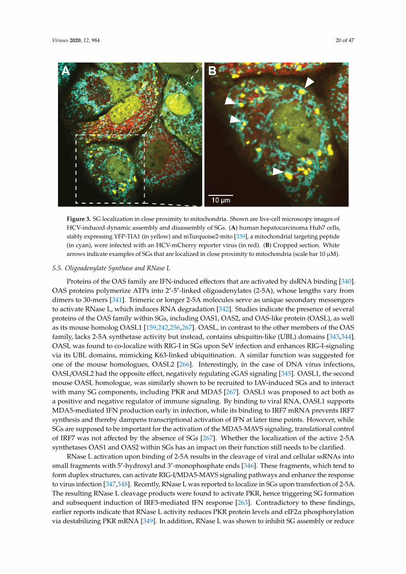
Figure 3. SG localization in close proximity to mitochondria. Shown are live-cell microscopy images of HCV-induced dynamic assembly and disassembly of SGs. (A) human hepatocarcinoma Huh7 cells, stably expressing YFP-TIA1 (in yellow) and mTurquoise2-mito [339], a mitochondrial targeting peptide (in cyan), were infected with an HCV-mCherry reporter virus (in red). (B) Cropped section. White arrows indicate examples of SGs that are localized in close proximity to mitochondria (scale bar 10 µM).
5.5. Oligoadenylate Synthase and RNase L
Proteins of the OAS family are IFN-induced effectors that are activated by dsRNA binding [340]. OAS proteins polymerize ATPs into 2′-5′-linked oligoadenylates (2-5A), whose lengths vary from dimers to 30-mers [341]. Trimeric or longer 2-5A molecules serve as unique secondary messengers to activate RNase L, which induces RNA degradation [342]. Studies indicate the presence of several proteins of the OAS family within SGs, including OAS1, OAS2, and OAS-like protein (OASL), as well as its mouse homolog OASL1 [159,242,256,267]. OASL, in contrast to the other members of the OAS family, lacks 2-5A synthetase activity but instead, contains ubiquitin-like (UBL) domains [343,344]. OASL was found to co-localize with RIG-I in SGs upon SeV infection and enhances RIG-I-signaling via its UBL domains, mimicking K63-linked ubiquitination. A similar function was suggested for one of the mouse homologues, OASL2 [266]. Interestingly, in the case of DNA virus infections, OASL/OASL2 had the opposite effect, negatively regulating cGAS signaling [345]. OASL1, the second mouse OASL homologue, was similarly shown to be recruited to IAV-induced SGs and to interact with many SG components, including PKR and MDA5 [267]. OASL1 was proposed to act both as a positive and negative regulator of immune signaling. By binding to viral RNA, OASL1 supports MDA5-mediated IFN production early in infection, while its binding to IRF7 mRNA prevents IRF7 synthesis and thereby dampens transcriptional activation of IFN at later time points. However, while SGs are supposed to be important for the activation of the MDA5-MAVS signaling, translational control of IRF7 was not affected by the absence of SGs [267]. Whether the localization of the active 2-5A synthetases OAS1 and OAS2 within SGs has an impact on their function still needs to be clarified.
RNase L activation upon binding of 2-5A results in the cleavage of viral and cellular ssRNAs into small fragments with 5′-hydroxyl and 3′-monophosphate ends [346]. These fragments, which
Figure 3. SG localization in close proximity to mitochondria. Shown are live-cell microscopy images ofHCV-induced dynamic assembly and disassembly of SGs. (A) human hepatocarcinoma Huh7 cells,stably expressing YFP-TIA1 (in yellow) and mTurquoise2-mito [339], a mitochondrial targeting peptide(in cyan), were infected with an HCV-mCherry reporter virus (in red). (B) Cropped section. Whitearrows indicate examples of SGs that are localized in close proximity to mitochondria (scale bar 10 µM).
5.5. Oligoadenylate Synthase and RNase L
Proteins of the OAS family are IFN-induced effectors that are activated by dsRNA binding [340].OAS proteins polymerize ATPs into 2′-5′-linked oligoadenylates (2-5A), whose lengths vary fromdimers to 30-mers [341]. Trimeric or longer 2-5A molecules serve as unique secondary messengersto activate RNase L, which induces RNA degradation [342]. Studies indicate the presence of severalproteins of the OAS family within SGs, including OAS1, OAS2, and OAS-like protein (OASL), as wellas its mouse homolog OASL1 [159,242,256,267]. OASL, in contrast to the other members of the OASfamily, lacks 2-5A synthetase activity but instead, contains ubiquitin-like (UBL) domains [343,344].OASL was found to co-localize with RIG-I in SGs upon SeV infection and enhances RIG-I-signalingvia its UBL domains, mimicking K63-linked ubiquitination. A similar function was suggested forone of the mouse homologues, OASL2 [266]. Interestingly, in the case of DNA virus infections,OASL/OASL2 had the opposite effect, negatively regulating cGAS signaling [345]. OASL1, the secondmouse OASL homologue, was similarly shown to be recruited to IAV-induced SGs and to interactwith many SG components, including PKR and MDA5 [267]. OASL1 was proposed to act both asa positive and negative regulator of immune signaling. By binding to viral RNA, OASL1 supportsMDA5-mediated IFN production early in infection, while its binding to IRF7 mRNA prevents IRF7synthesis and thereby dampens transcriptional activation of IFN at later time points. However, whileSGs are supposed to be important for the activation of the MDA5-MAVS signaling, translational controlof IRF7 was not affected by the absence of SGs [267]. Whether the localization of the active 2-5Asynthetases OAS1 and OAS2 within SGs has an impact on their function still needs to be clarified.
RNase L activation upon binding of 2-5A results in the cleavage of viral and cellular ssRNAs intosmall fragments with 5′-hydroxyl and 3′-monophosphate ends [346]. These fragments, which tend toform duplex structures, can activate RIG-I/MDA5-MAVS signaling pathways and enhance the responseto virus infection [347,348]. Recently, RNase L was reported to localize in SGs upon transfection of 2-5A.The resulting RNase L cleavage products were found to activate PKR, hence triggering SG formationand subsequent induction of IRF3-mediated IFN response [263]. Contradictory to these findings,earlier reports indicate that RNase L activity reduces PKR protein levels and eIF2α phosphorylationvia destabilizing PKR mRNA [349]. In addition, RNase L was shown to inhibit SG assembly or reduce
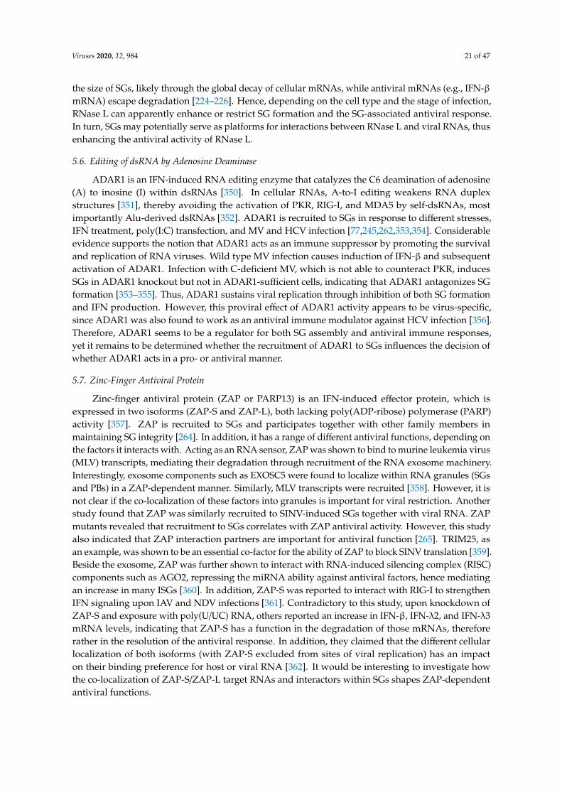
Viruses 2020, 12, 984 21 of 47
the size of SGs, likely through the global decay of cellular mRNAs, while antiviral mRNAs (e.g., IFN-βmRNA) escape degradation [224–226]. Hence, depending on the cell type and the stage of infection,RNase L can apparently enhance or restrict SG formation and the SG-associated antiviral response.In turn, SGs may potentially serve as platforms for interactions between RNase L and viral RNAs, thusenhancing the antiviral activity of RNase L.
5.6. Editing of dsRNA by Adenosine Deaminase
ADAR1 is an IFN-induced RNA editing enzyme that catalyzes the C6 deamination of adenosine(A) to inosine (I) within dsRNAs [350]. In cellular RNAs, A-to-I editing weakens RNA duplexstructures [351], thereby avoiding the activation of PKR, RIG-I, and MDA5 by self-dsRNAs, mostimportantly Alu-derived dsRNAs [352]. ADAR1 is recruited to SGs in response to different stresses,IFN treatment, poly(I:C) transfection, and MV and HCV infection [77,245,262,353,354]. Considerableevidence supports the notion that ADAR1 acts as an immune suppressor by promoting the survivaland replication of RNA viruses. Wild type MV infection causes induction of IFN-β and subsequentactivation of ADAR1. Infection with C-deficient MV, which is not able to counteract PKR, inducesSGs in ADAR1 knockout but not in ADAR1-sufficient cells, indicating that ADAR1 antagonizes SGformation [353–355]. Thus, ADAR1 sustains viral replication through inhibition of both SG formationand IFN production. However, this proviral effect of ADAR1 activity appears to be virus-specific,since ADAR1 was also found to work as an antiviral immune modulator against HCV infection [356].Therefore, ADAR1 seems to be a regulator for both SG assembly and antiviral immune responses,yet it remains to be determined whether the recruitment of ADAR1 to SGs influences the decision ofwhether ADAR1 acts in a pro- or antiviral manner.
5.7. Zinc-Finger Antiviral Protein
Zinc-finger antiviral protein (ZAP or PARP13) is an IFN-induced effector protein, which isexpressed in two isoforms (ZAP-S and ZAP-L), both lacking poly(ADP-ribose) polymerase (PARP)activity [357]. ZAP is recruited to SGs and participates together with other family members inmaintaining SG integrity [264]. In addition, it has a range of different antiviral functions, depending onthe factors it interacts with. Acting as an RNA sensor, ZAP was shown to bind to murine leukemia virus(MLV) transcripts, mediating their degradation through recruitment of the RNA exosome machinery.Interestingly, exosome components such as EXOSC5 were found to localize within RNA granules (SGsand PBs) in a ZAP-dependent manner. Similarly, MLV transcripts were recruited [358]. However, it isnot clear if the co-localization of these factors into granules is important for viral restriction. Anotherstudy found that ZAP was similarly recruited to SINV-induced SGs together with viral RNA. ZAPmutants revealed that recruitment to SGs correlates with ZAP antiviral activity. However, this studyalso indicated that ZAP interaction partners are important for antiviral function [265]. TRIM25, asan example, was shown to be an essential co-factor for the ability of ZAP to block SINV translation [359].Beside the exosome, ZAP was further shown to interact with RNA-induced silencing complex (RISC)components such as AGO2, repressing the miRNA ability against antiviral factors, hence mediatingan increase in many ISGs [360]. In addition, ZAP-S was reported to interact with RIG-I to strengthenIFN signaling upon IAV and NDV infections [361]. Contradictory to this study, upon knockdown ofZAP-S and exposure with poly(U/UC) RNA, others reported an increase in IFN-β, IFN-λ2, and IFN-λ3mRNA levels, indicating that ZAP-S has a function in the degradation of those mRNAs, thereforerather in the resolution of the antiviral response. In addition, they claimed that the different cellularlocalization of both isoforms (with ZAP-S excluded from sites of viral replication) has an impacton their binding preference for host or viral RNA [362]. It would be interesting to investigate howthe co-localization of ZAP-S/ZAP-L target RNAs and interactors within SGs shapes ZAP-dependentantiviral functions.
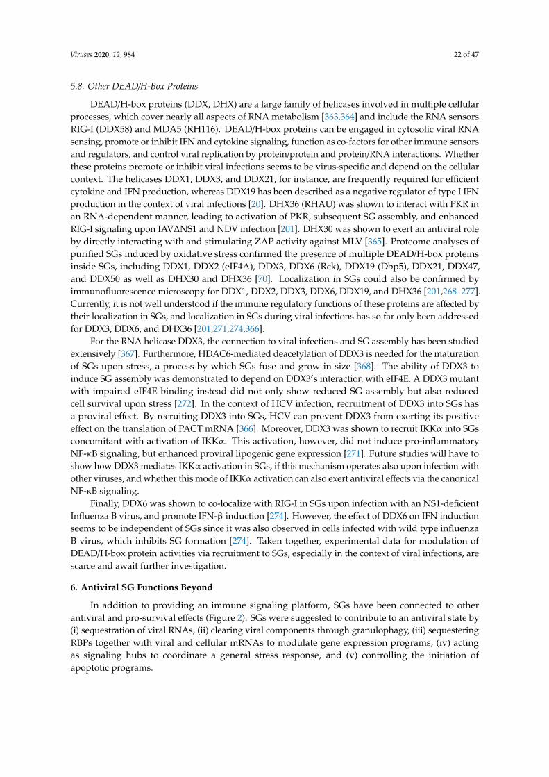
Viruses 2020, 12, 984 22 of 47
5.8. Other DEAD/H-Box Proteins
DEAD/H-box proteins (DDX, DHX) are a large family of helicases involved in multiple cellularprocesses, which cover nearly all aspects of RNA metabolism [363,364] and include the RNA sensorsRIG-I (DDX58) and MDA5 (RH116). DEAD/H-box proteins can be engaged in cytosolic viral RNAsensing, promote or inhibit IFN and cytokine signaling, function as co-factors for other immune sensorsand regulators, and control viral replication by protein/protein and protein/RNA interactions. Whetherthese proteins promote or inhibit viral infections seems to be virus-specific and depend on the cellularcontext. The helicases DDX1, DDX3, and DDX21, for instance, are frequently required for efficientcytokine and IFN production, whereas DDX19 has been described as a negative regulator of type I IFNproduction in the context of viral infections [20]. DHX36 (RHAU) was shown to interact with PKR inan RNA-dependent manner, leading to activation of PKR, subsequent SG assembly, and enhancedRIG-I signaling upon IAV∆NS1 and NDV infection [201]. DHX30 was shown to exert an antiviral roleby directly interacting with and stimulating ZAP activity against MLV [365]. Proteome analyses ofpurified SGs induced by oxidative stress confirmed the presence of multiple DEAD/H-box proteinsinside SGs, including DDX1, DDX2 (eIF4A), DDX3, DDX6 (Rck), DDX19 (Dbp5), DDX21, DDX47,and DDX50 as well as DHX30 and DHX36 [70]. Localization in SGs could also be confirmed byimmunofluorescence microscopy for DDX1, DDX2, DDX3, DDX6, DDX19, and DHX36 [201,268–277].Currently, it is not well understood if the immune regulatory functions of these proteins are affected bytheir localization in SGs, and localization in SGs during viral infections has so far only been addressedfor DDX3, DDX6, and DHX36 [201,271,274,366].
For the RNA helicase DDX3, the connection to viral infections and SG assembly has been studiedextensively [367]. Furthermore, HDAC6-mediated deacetylation of DDX3 is needed for the maturationof SGs upon stress, a process by which SGs fuse and grow in size [368]. The ability of DDX3 toinduce SG assembly was demonstrated to depend on DDX3′s interaction with eIF4E. A DDX3 mutantwith impaired eIF4E binding instead did not only show reduced SG assembly but also reducedcell survival upon stress [272]. In the context of HCV infection, recruitment of DDX3 into SGs hasa proviral effect. By recruiting DDX3 into SGs, HCV can prevent DDX3 from exerting its positiveeffect on the translation of PACT mRNA [366]. Moreover, DDX3 was shown to recruit IKKα into SGsconcomitant with activation of IKKα. This activation, however, did not induce pro-inflammatoryNF-κB signaling, but enhanced proviral lipogenic gene expression [271]. Future studies will have toshow how DDX3 mediates IKKα activation in SGs, if this mechanism operates also upon infection withother viruses, and whether this mode of IKKα activation can also exert antiviral effects via the canonicalNF-κB signaling.
Finally, DDX6 was shown to co-localize with RIG-I in SGs upon infection with an NS1-deficientInfluenza B virus, and promote IFN-β induction [274]. However, the effect of DDX6 on IFN inductionseems to be independent of SGs since it was also observed in cells infected with wild type influenzaB virus, which inhibits SG formation [274]. Taken together, experimental data for modulation ofDEAD/H-box protein activities via recruitment to SGs, especially in the context of viral infections, arescarce and await further investigation.
6. Antiviral SG Functions Beyond
In addition to providing an immune signaling platform, SGs have been connected to otherantiviral and pro-survival effects (Figure 2). SGs were suggested to contribute to an antiviral state by(i) sequestration of viral RNAs, (ii) clearing viral components through granulophagy, (iii) sequesteringRBPs together with viral and cellular mRNAs to modulate gene expression programs, (iv) actingas signaling hubs to coordinate a general stress response, and (v) controlling the initiation ofapoptotic programs.
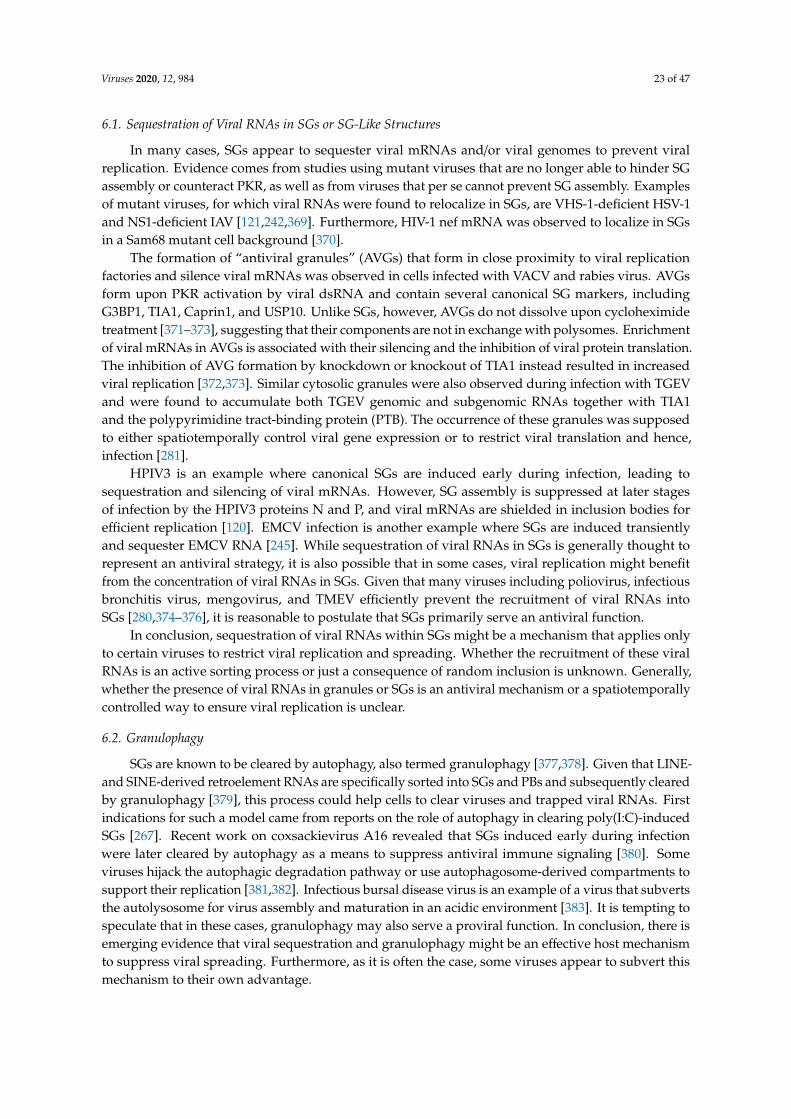
Viruses 2020, 12, 984 23 of 47
6.1. Sequestration of Viral RNAs in SGs or SG-Like Structures
In many cases, SGs appear to sequester viral mRNAs and/or viral genomes to prevent viralreplication. Evidence comes from studies using mutant viruses that are no longer able to hinder SGassembly or counteract PKR, as well as from viruses that per se cannot prevent SG assembly. Examplesof mutant viruses, for which viral RNAs were found to relocalize in SGs, are VHS-1-deficient HSV-1and NS1-deficient IAV [121,242,369]. Furthermore, HIV-1 nef mRNA was observed to localize in SGsin a Sam68 mutant cell background [370].
The formation of “antiviral granules” (AVGs) that form in close proximity to viral replicationfactories and silence viral mRNAs was observed in cells infected with VACV and rabies virus. AVGsform upon PKR activation by viral dsRNA and contain several canonical SG markers, includingG3BP1, TIA1, Caprin1, and USP10. Unlike SGs, however, AVGs do not dissolve upon cycloheximidetreatment [371–373], suggesting that their components are not in exchange with polysomes. Enrichmentof viral mRNAs in AVGs is associated with their silencing and the inhibition of viral protein translation.The inhibition of AVG formation by knockdown or knockout of TIA1 instead resulted in increasedviral replication [372,373]. Similar cytosolic granules were also observed during infection with TGEVand were found to accumulate both TGEV genomic and subgenomic RNAs together with TIA1and the polypyrimidine tract-binding protein (PTB). The occurrence of these granules was supposedto either spatiotemporally control viral gene expression or to restrict viral translation and hence,infection [281].
HPIV3 is an example where canonical SGs are induced early during infection, leading tosequestration and silencing of viral mRNAs. However, SG assembly is suppressed at later stagesof infection by the HPIV3 proteins N and P, and viral mRNAs are shielded in inclusion bodies forefficient replication [120]. EMCV infection is another example where SGs are induced transientlyand sequester EMCV RNA [245]. While sequestration of viral RNAs in SGs is generally thought torepresent an antiviral strategy, it is also possible that in some cases, viral replication might benefitfrom the concentration of viral RNAs in SGs. Given that many viruses including poliovirus, infectiousbronchitis virus, mengovirus, and TMEV efficiently prevent the recruitment of viral RNAs intoSGs [280,374–376], it is reasonable to postulate that SGs primarily serve an antiviral function.
In conclusion, sequestration of viral RNAs within SGs might be a mechanism that applies onlyto certain viruses to restrict viral replication and spreading. Whether the recruitment of these viralRNAs is an active sorting process or just a consequence of random inclusion is unknown. Generally,whether the presence of viral RNAs in granules or SGs is an antiviral mechanism or a spatiotemporallycontrolled way to ensure viral replication is unclear.
6.2. Granulophagy
SGs are known to be cleared by autophagy, also termed granulophagy [377,378]. Given that LINE-and SINE-derived retroelement RNAs are specifically sorted into SGs and PBs and subsequently clearedby granulophagy [379], this process could help cells to clear viruses and trapped viral RNAs. Firstindications for such a model came from reports on the role of autophagy in clearing poly(I:C)-inducedSGs [267]. Recent work on coxsackievirus A16 revealed that SGs induced early during infectionwere later cleared by autophagy as a means to suppress antiviral immune signaling [380]. Someviruses hijack the autophagic degradation pathway or use autophagosome-derived compartments tosupport their replication [381,382]. Infectious bursal disease virus is an example of a virus that subvertsthe autolysosome for virus assembly and maturation in an acidic environment [383]. It is tempting tospeculate that in these cases, granulophagy may also serve a proviral function. In conclusion, there isemerging evidence that viral sequestration and granulophagy might be an effective host mechanismto suppress viral spreading. Furthermore, as it is often the case, some viruses appear to subvert thismechanism to their own advantage.
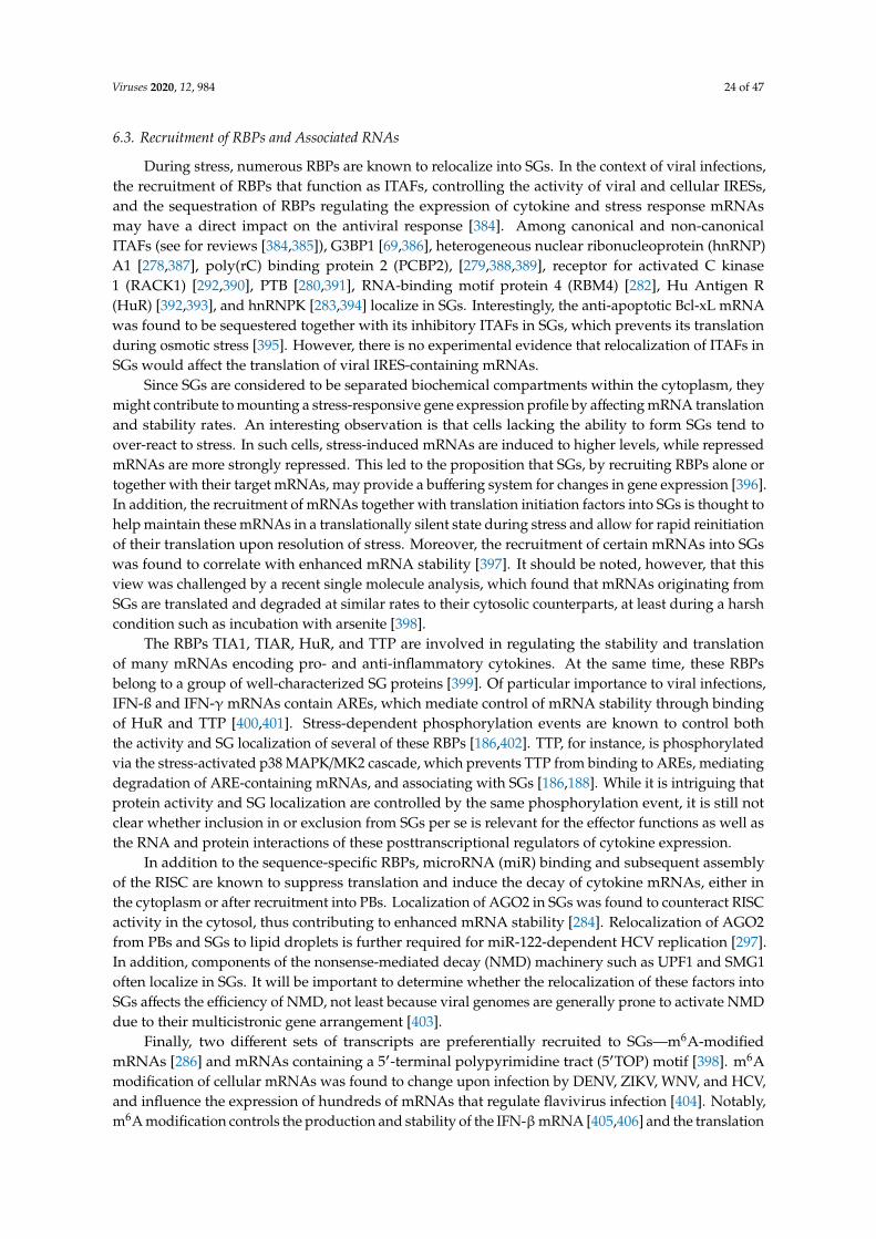
Viruses 2020, 12, 984 24 of 47
6.3. Recruitment of RBPs and Associated RNAs
During stress, numerous RBPs are known to relocalize into SGs. In the context of viral infections,the recruitment of RBPs that function as ITAFs, controlling the activity of viral and cellular IRESs,and the sequestration of RBPs regulating the expression of cytokine and stress response mRNAsmay have a direct impact on the antiviral response [384]. Among canonical and non-canonicalITAFs (see for reviews [384,385]), G3BP1 [69,386], heterogeneous nuclear ribonucleoprotein (hnRNP)A1 [278,387], poly(rC) binding protein 2 (PCBP2), [279,388,389], receptor for activated C kinase1 (RACK1) [292,390], PTB [280,391], RNA-binding motif protein 4 (RBM4) [282], Hu Antigen R(HuR) [392,393], and hnRNPK [283,394] localize in SGs. Interestingly, the anti-apoptotic Bcl-xL mRNAwas found to be sequestered together with its inhibitory ITAFs in SGs, which prevents its translationduring osmotic stress [395]. However, there is no experimental evidence that relocalization of ITAFs inSGs would affect the translation of viral IRES-containing mRNAs.
Since SGs are considered to be separated biochemical compartments within the cytoplasm, theymight contribute to mounting a stress-responsive gene expression profile by affecting mRNA translationand stability rates. An interesting observation is that cells lacking the ability to form SGs tend toover-react to stress. In such cells, stress-induced mRNAs are induced to higher levels, while repressedmRNAs are more strongly repressed. This led to the proposition that SGs, by recruiting RBPs alone ortogether with their target mRNAs, may provide a buffering system for changes in gene expression [396].In addition, the recruitment of mRNAs together with translation initiation factors into SGs is thought tohelp maintain these mRNAs in a translationally silent state during stress and allow for rapid reinitiationof their translation upon resolution of stress. Moreover, the recruitment of certain mRNAs into SGswas found to correlate with enhanced mRNA stability [397]. It should be noted, however, that thisview was challenged by a recent single molecule analysis, which found that mRNAs originating fromSGs are translated and degraded at similar rates to their cytosolic counterparts, at least during a harshcondition such as incubation with arsenite [398].
The RBPs TIA1, TIAR, HuR, and TTP are involved in regulating the stability and translationof many mRNAs encoding pro- and anti-inflammatory cytokines. At the same time, these RBPsbelong to a group of well-characterized SG proteins [399]. Of particular importance to viral infections,IFN-ß and IFN-γmRNAs contain AREs, which mediate control of mRNA stability through bindingof HuR and TTP [400,401]. Stress-dependent phosphorylation events are known to control boththe activity and SG localization of several of these RBPs [186,402]. TTP, for instance, is phosphorylatedvia the stress-activated p38 MAPK/MK2 cascade, which prevents TTP from binding to AREs, mediatingdegradation of ARE-containing mRNAs, and associating with SGs [186,188]. While it is intriguing thatprotein activity and SG localization are controlled by the same phosphorylation event, it is still notclear whether inclusion in or exclusion from SGs per se is relevant for the effector functions as well asthe RNA and protein interactions of these posttranscriptional regulators of cytokine expression.
In addition to the sequence-specific RBPs, microRNA (miR) binding and subsequent assemblyof the RISC are known to suppress translation and induce the decay of cytokine mRNAs, either inthe cytoplasm or after recruitment into PBs. Localization of AGO2 in SGs was found to counteract RISCactivity in the cytosol, thus contributing to enhanced mRNA stability [284]. Relocalization of AGO2from PBs and SGs to lipid droplets is further required for miR-122-dependent HCV replication [297].In addition, components of the nonsense-mediated decay (NMD) machinery such as UPF1 and SMG1often localize in SGs. It will be important to determine whether the relocalization of these factors intoSGs affects the efficiency of NMD, not least because viral genomes are generally prone to activate NMDdue to their multicistronic gene arrangement [403].
Finally, two different sets of transcripts are preferentially recruited to SGs—m6A-modifiedmRNAs [286] and mRNAs containing a 5′-terminal polypyrimidine tract (5′TOP) motif [398]. m6Amodification of cellular mRNAs was found to change upon infection by DENV, ZIKV, WNV, and HCV,and influence the expression of hundreds of mRNAs that regulate flavivirus infection [404]. Notably,m6A modification controls the production and stability of the IFN-βmRNA [405,406] and the translation
Viruses 2020, 12, 984 25 of 47
of the transcription factor Foxo3, an antagonist of ISG transcription [407]. YTHDF proteins, whichspecifically recognize m6A, were found to interact with m6A-containing mRNAs at the shell ofSGs [286]. Given the role of m6A in regulating the expression of innate immune factors, the enrichmentof m6A-containing mRNAs in SGs may contribute to the control of viral replication. The preferentialrecruitment of 5′TOP mRNAs in SGs is also interesting. Since these mRNAs encode all ribosomalproteins and several translation factors, their sequestration could exert a sustained suppressive effecton virus replication by reducing the long-term availability of the translation apparatus [26].
6.4. SGs as Signaling Hubs to Coordinate the General Stress Response
Approximately 50% of the proteins in SGs are not known to bind RNA but rather contribute to cellsignaling during stress. Thus, not only RBPs, but also various enzymes that catalyze posttranslationalmodifications localize in SGs. These include for example components of mTORC1 complex, JNK and itsscaffold protein WDR62, the ubiquitin peptidase USP10, the protein arginine methyltransferase (PRMT)1 and PRMT5, the glycosyltransferase OGG1, and the poly-ADP-ribose polymerase PARP12 [70,408].Accordingly, proteins within SGs were found to carry a plethora of posttranslational modificationsincluding phosphorylation [250,409], GlcNac glycosylation [408], sumoylation [410], neddylation [411],ubiquitination [380], and ADP-ribosylation [264].
The localization of enzymes within SGs can have different consequences. First, enzymes localize inSGs to modulate the assembly, disassembly, or composition of SGs. They are needed for SG biology, butSGs, in turn, are unlikely to influence the activity of these enzymes. Second, enzymes are sequesteredin SGs as a way of silencing their activity. This may be particularly relevant for low abundance proteinswith low SG shuttling rates, leading to efficient separation of the enzyme from its cytosolic or nucleartargets. Third, enzymes are recruited in their active form into SGs, or become activated inside SGs, toparticipate in signaling pathways. This mechanism is likely to benefit from the high concentration ofenzyme and substrate within SGs.
One interesting example in this context is the recruitment of PARP12 into SGs. While the variouscellular functions of PARP12 are not fully understood, PARP12 expression is induced by IFN and hasan antiviral role that can potentially be modulated in a SG-dependent manner. The antiviral functionsof PARP12 have been linked to its ability to bind viral and cellular RNAs, and a possible role intranslation control [412,413]. PARP12 might further be involved in ADP ribosylation of AGO proteinswithin SGs, which potentially results in reduced microRNA activity [264]. Relocalization of PARP12from the Golgi into SGs was further shown to result in the disassembly of the Golgi compartment and ina block of the anterograde membrane trafficking [287]—a route that many viruses rely on for particleassembly. In contrast, inflammation-induced association between PARP12 and p62/SQSTM1 outside ofSGs was found to increase NF-κB signaling [412]. Moreover, PARP12-mediated ADP-ribosylation ofZIKV proteins NS1 and NS3 in the cytosol leads to their degradation and restricts virus replication [414].Taken together, these findings point to a potential role of SG localization for PARP12 substrate selectionand its multiple effects on virus replication.
6.5. Apoptosis Control by SGs
Several reports indicate that SGs fulfill important anti-apoptotic functions. An attractiveconcept is that cells, upon damage or insult, assemble SGs while they attempt to resolvethe stressful conditions. Cytosolic phase separation could herein mark this “window of opportunity”,and only if the damage persists and stress cannot be resolved cells would undergo apoptosis [15].For several types of stress, SGs were found to efficiently sequester RACK1, thereby inhibitingRACK1-dependent activation of the pro-apoptotic mitogen-activated protein kinase MTK1-p38/JNKsignaling axis [292]. Similarly, the localization of the small GTPase RhoA and the Rho-associatedcoiled-coil containing protein kinase 1 (ROCK1) in SGs hinders the induction of JNK-mediatedapoptosis [295]. NF-κB-mediated pro-inflammatory signaling—which can also lead to apoptosis—ispartially suppressed by eIF4G-dependent recruitment of TRAF2 into SGs during heat stress
Viruses 2020, 12, 984 26 of 47
and arsenite exposure [293]. Furthermore, Astrin-dependent recruitment of mTORC1 into SGsalso prevents apoptosis, which may arise from mTOR hyperactivation, upon exposure to arsenite [289].An anti-apoptotic role was also revealed for the co-localization of TIA1 and the p90 ribosomal S6kinase 2 (RSK2) in SGs upon oxidative stress [294]. The Fas-activated serine/threonine kinase (FASTK)is a pro-survival factor that also localizes in SGs and controls protein synthesis of the anti-apoptoticfactors cIAP-1 and XIAP. Like RSK2, FASTK interacts with and mediates its anti-apoptotic effectsby binding to and regulating TIA1 [415], though it is not yet clear if the SG-resident pool of FASTKis critical to its anti-apoptotic function. Finally, DDX3-dependent assembly of SGs was found tonegatively correlate with DDX3-mediated induction of the NLP3 inflammasome and subsequentpyroptosis in LPS-challenged macrophages [416]. Notably, the above examples assign an anti-apoptoticrole to SGs, indicating that SGs indeed play an active role in opening a “window of opportunity” forcells in distress, by delaying the onset of apoptosis. Most lytic viruses induce premature death ofthe infected host cell. To prevent this from turning into a disadvantage by limiting viral spread, somehave evolved molecular strategies to counteract or delay apoptosis. One example is SeV, in whichencoded trailer RNA binds TIAR to limit apoptosis and SG formation [238]. On the other hand, virusescan also actively trigger apoptosis to release progenies during later stages of lytic infections [417].Whether SGs also serve as a timer function for the life span of virus-infected cells remains, however, tobe determined.
7. Conclusions
Knowing whether RBPs and cellular or viral RNAs are actively “recruited”, “sequestered”, or justpassively “phase separated” into SGs is much more than a matter of semantics—it is key to a mechanisticunderstanding of how SGs shape host–virus interactions and coordinate signaling events. Evidencefor the presence of a specific RNA or protein in SGs largely relies on microscopy analyses, whichhave recently been complemented by the development of labeling methods, purification protocols,and the use of high-resolution imaging. However, most SG studies are performed in cells exposed tooxidative stress or other harsh conditions, and many aspects of SG biology still need to be addressed invirus-infected cells.
The co-evolution of virus and host can be described as an arms race that needs to be carefullybalanced with the consequences of collateral damage. The dynamic nature of SGs gives testimonyto this contest: while some viruses effectively suppress SG assembly, others face an initial wave ofSGs that they can only curb during later stages of infection. The appearance of oscillating SGs duringchronic virus infections is maybe the most stunning strategy by which cells contain “the enemy within”without suffocating from the antiviral response.
It is tempting to speculate that the condensation state, composition, and function of SGs maychange over time as viral infections progress. Further studies building on the recent technical advanceswill be required to mechanistically dissect the antiviral role of SGs and provide a time-resolvedunderstanding of how stress response pathways and innate immune signaling intersect in the contextof viral infections. This may eventually allow us to harness the SGs’ “dance with the devil” for antiviraltherapeutic approaches against important human pathogens.
Author Contributions: Writing—original draft preparation, N.E., K.H. and Z.S. Writing—review and editing, G.S.and A.R. All authors have read and agreed to the published version of the manuscript.
Funding: This research was funded by the Deutsche Forschungsgemeinschaft (DFG, German Research Foundation)project number 278001972-TRR 186 to A.R. and G.S., project number 240245660-SFB 1129 to A.R., and projectnumber 201348542-SFB 1036 to G.S.
Conflicts of Interest: The authors declare no conflict of interest. The funders had no role in the design of the study;in the collection, analyses, or interpretation of data; in the writing of the manuscript, or in the decision to publishthe results.
Viruses 2020, 12, 984 27 of 47
References
1. Mir, M.A.; Panganiban, A.T. A protein that replaces the entire cellular eIF4F complex. EMBO J. 2008, 27,3129–3139. [CrossRef]
2. Schulz, F.; Yutin, N.; Ivanova, N.N.; Ortega, D.R.; Lee, T.K.; Vierheilig, J.; Daims, H.; Horn, M.; Wagner, M.;Jensen, G.J.; et al. Giant viruses with an expanded complement of translation system components. Science2017, 356, 82–85. [CrossRef] [PubMed]
3. Colson, P.; De Lamballerie, X.; Yutin, N.; Asgari, S.; Bigot, Y.; Bideshi, D.K.; Cheng, X.W.; Federici, B.A.;Van Etten, J.L.; Koonin, E.V.; et al. “Megavirales”, a proposed new order for eukaryotic nucleocytoplasmiclarge DNA viruses. Arch. Virol. 2013, 158, 2517–2521. [CrossRef]
4. Sullivan, M.B.; Huang, K.H.; Ignacio-Espinoza, J.C.; Berlin, A.M.; Kelly, L.; Weigele, P.R.; DeFrancesco, A.S.;Kern, S.E.; Thompson, L.R.; Young, S.; et al. Genomic analysis of oceanic cyanobacterial myovirusescompared with T4-like myoviruses from diverse hosts and environments. Environ. Microbiol. 2010, 12,3035–3056. [CrossRef] [PubMed]
5. Sencilo, A.; Jacobs-Sera, D.; Russell, D.A.; Ko, C.C.; Bowman, C.A.; Atanasova, N.S.; Osterlund, E.;Oksanen, H.M.; Bamford, D.H.; Hatfull, G.F.; et al. Snapshot of haloarchaeal tailed virus genomes. RNA Biol.2013, 10, 803–816. [CrossRef] [PubMed]
6. Abergel, C.; Rudinger-Thirion, J.; Giege, R.; Claverie, J.M. Virus-encoded aminoacyl-tRNA synthetases:Structural and functional characterization of mimivirus TyrRS and MetRS. J. Virol. 2007, 81, 12406–12417.[CrossRef]
7. Mizuno, C.M.; Guyomar, C.; Roux, S.; Lavigne, R.; Rodriguez-Valera, F.; Sullivan, M.B.; Gillet, R.; Forterre, P.;Krupovic, M. Numerous cultivated and uncultivated viruses encode ribosomal proteins. Nat. Commun. 2019,10, 752. [CrossRef]
8. Goubau, D.; Deddouche, S.; Reis e Sousa, C. Cytosolic sensing of viruses. Immunity 2013, 38, 855–869.[CrossRef]
9. Jensen, S.; Thomsen, A.R. Sensing of RNA viruses: A review of innate immune receptors involved inrecognizing RNA virus invasion. J. Virol. 2012, 86, 2900–2910. [CrossRef]
10. Ma, Z.; Ni, G.; Damania, B. Innate Sensing of DNA Virus Genomes. Annu. Rev. Virol. 2018, 5, 341–362.[CrossRef]
11. Donnelly, N.; Gorman, A.M.; Gupta, S.; Samali, A. The eIF2alpha kinases: Their structures and functions.Cell. Mol. Life Sci. 2013, 70, 3493–3511. [CrossRef] [PubMed]
12. Pakos-Zebrucka, K.; Koryga, I.; Mnich, K.; Ljujic, M.; Samali, A.; Gorman, A.M. The integrated stress response.EMBO Rep. 2016, 17, 1374–1395. [CrossRef] [PubMed]
13. Buchan, J.R.; Parker, R. Eukaryotic stress granules: The ins and outs of translation. Mol. Cell 2009, 36, 932–941.[CrossRef] [PubMed]
14. Kedersha, N.; Anderson, P. Stress granules: Sites of mRNA triage that regulate mRNA stabilityand translatability. Biochem. Soc. Trans. 2002, 30, 963–969. [CrossRef]
15. Kedersha, N.; Ivanov, P.; Anderson, P. Stress granules and cell signaling: More than just a passing phase?Trends Biochem. Sci. 2013, 38, 494–506. [CrossRef]
16. Mahboubi, H.; Stochaj, U. Cytoplasmic stress granules: Dynamic modulators of cell signaling and disease.Biochim. Biophys. Acta Mol. Basis Dis. 2017, 1863, 884–895. [CrossRef]
17. Onomoto, K.; Yoneyama, M.; Fung, G.; Kato, H.; Fujita, T. Antiviral innate immunity and stress granuleresponses. Trends Immunol. 2014, 35, 420–428. [CrossRef]
18. McCormick, C.; Khaperskyy, D.A. Translation inhibition and stress granules in the antiviral immune response.Nat. Rev. Immunol. 2017, 17, 647–660. [CrossRef]
19. Poblete-Duran, N.; Prades-Perez, Y.; Vera-Otarola, J.; Soto-Rifo, R.; Valiente-Echeverria, F. Who RegulatesWhom? An Overview of RNA Granules and Viral Infections. Viruses 2016, 8, 180. [CrossRef]
20. Zhang, Q.; Sharma, N.R.; Zheng, Z.M.; Chen, M. Viral Regulation of RNA Granules in Infected Cells.Virol. Sin. 2019, 34, 175–191. [CrossRef]
21. Montero, H.; Trujillo-Alonso, V. Stress granules in the viral replication cycle. Viruses 2011, 3, 2328–2338.[CrossRef] [PubMed]
22. Tsai, W.C.; Lloyd, R.E. Cytoplasmic RNA Granules and Viral Infection. Annu. Rev. Virol. 2014, 1, 147–170.[CrossRef] [PubMed]
Viruses 2020, 12, 984 28 of 47
23. Holcik, M.; Sonenberg, N. Translational control in stress and apoptosis. Nat. Rev. Mol. Cell Biol. 2005, 6,318–327. [CrossRef]
24. Spriggs, K.A.; Bushell, M.; Willis, A.E. Translational regulation of gene expression during conditions of cellstress. Mol. Cell 2010, 40, 228–237. [CrossRef] [PubMed]
25. Liu, B.; Qian, S.B. Translational reprogramming in cellular stress response. Wiley Interdiscip. Rev. RNA 2014,5, 301–315. [CrossRef] [PubMed]
26. Walsh, D.; Mathews, M.B.; Mohr, I. Tinkering with translation: Protein synthesis in virus-infected cells.Cold Spring Harb Perspect. Biol. 2013, 5, a012351. [CrossRef]
27. Stern-Ginossar, N.; Thompson, S.R.; Mathews, M.B.; Mohr, I. Translational Control in Virus-Infected Cells.Cold Spring Harb Perspect. Biol. 2019, 11. [CrossRef]
28. Hinnebusch, A.G. Molecular mechanism of scanning and start codon selection in eukaryotes. Microbiol. Mol.Biol. Rev. 2011, 75, 434–467. [CrossRef]
29. Harvey, R.F.; Smith, T.S.; Mulroney, T.; Queiroz, R.M.L.; Pizzinga, M.; Dezi, V.; Villenueva, E.; Ramakrishna, M.;Lilley, K.S.; Willis, A.E. Trans-acting translational regulatory RNA binding proteins. Wiley Interdiscip. Rev. RNA2018, 9, e1465. [CrossRef]
30. Leppek, K.; Das, R.; Barna, M. Functional 5′ UTR mRNA structures in eukaryotic translation regulationand how to find them. Nat. Rev. Mol. Cell Biol. 2018, 19, 158–174. [CrossRef]
31. Galloway, A.; Cowling, V.H. mRNA cap regulation in mammalian cell function and fate. Biochim. Biophys.Acta Gene Regul. Mech. 2019, 1862, 270–279. [CrossRef] [PubMed]
32. Hyde, J.L.; Diamond, M.S. Innate immune restriction and antagonism of viral RNA lacking 2-O methylation.Virology 2015, 479-480, 66–74. [CrossRef] [PubMed]
33. Aitken, C.E.; Lorsch, J.R. A mechanistic overview of translation initiation in eukaryotes. Nat. Struct. Mol. Biol.2012, 19, 568–576. [CrossRef]
34. Pavitt, G.D. Regulation of translation initiation factor eIF2B at the hub of the integrated stress response.Wiley Interdiscip. Rev. RNA 2018, 9, e1491. [CrossRef]
35. Jousse, C.; Oyadomari, S.; Novoa, I.; Lu, P.; Zhang, Y.; Harding, H.P.; Ron, D. Inhibition of a constitutivetranslation initiation factor 2alpha phosphatase, CReP, promotes survival of stressed cells. J. Cell Biol. 2003,163, 767–775. [CrossRef] [PubMed]
36. Kastan, J.P.; Dobrikova, E.Y.; Bryant, J.D.; Gromeier, M. CReP mediates selective translation initiation atthe endoplasmic reticulum. Sci. Adv. 2020, 6, eaba0745. [CrossRef]
37. Connor, J.H.; Weiser, D.C.; Li, S.; Hallenbeck, J.M.; Shenolikar, S. Growth arrest and DNA damage-inducibleprotein GADD34 assembles a novel signaling complex containing protein phosphatase 1 and inhibitor 1.Mol. Cell. Biol. 2001, 21, 6841–6850. [CrossRef]
38. Novoa, I.; Zeng, H.; Harding, H.P.; Ron, D. Feedback inhibition of the unfolded protein response byGADD34-mediated dephosphorylation of eIF2alpha. J. Cell Biol. 2001, 153, 1011–1022. [CrossRef]
39. Kojima, E.; Takeuchi, A.; Haneda, M.; Yagi, A.; Hasegawa, T.; Yamaki, K.; Takeda, K.; Akira, S.; Shimokata, K.;Isobe, K. The function of GADD34 is a recovery from a shutoff of protein synthesis induced by ER stress:Elucidation by GADD34-deficient mice. FASEB J. 2003, 17, 1573–1575. [CrossRef]
40. Novoa, I.; Zhang, Y.; Zeng, H.; Jungreis, R.; Harding, H.P.; Ron, D. Stress-induced gene expression requiresprogrammed recovery from translational repression. EMBO J. 2003, 22, 1180–1187. [CrossRef]
41. Haghighat, A.; Mader, S.; Pause, A.; Sonenberg, N. Repression of cap-dependent translation by 4E-bindingprotein 1: Competition with p220 for binding to eukaryotic initiation factor-4E. EMBO J. 1995, 14, 5701–5709.[CrossRef] [PubMed]
42. Young, S.K.; Wek, R.C. Upstream Open Reading Frames Differentially Regulate Gene-specific Translation inthe Integrated Stress Response. J. Biol. Chem. 2016, 291, 16927–16935. [CrossRef] [PubMed]
43. Weingarten-Gabbay, S.; Elias-Kirma, S.; Nir, R.; Gritsenko, A.A.; Stern-Ginossar, N.; Yakhini, Z.; Weinberger, A.;Segal, E. Comparative genetics. Systematic discovery of cap-independent translation sequences in humanand viral genomes. Science 2016, 351. [CrossRef] [PubMed]
44. Hellen, C.U.; Sarnow, P. Internal ribosome entry sites in eukaryotic mRNA molecules. Genes Dev. 2001,15, 1593–1612. [CrossRef] [PubMed]
45. Komar, A.A.; Hatzoglou, M. Cellular IRES-mediated translation: The war of ITAFs in pathophysiologicalstates. Cell Cycle 2011, 10, 229–240. [CrossRef] [PubMed]
Viruses 2020, 12, 984 29 of 47
46. Jang, S.K.; Krausslich, H.G.; Nicklin, M.J.; Duke, G.M.; Palmenberg, A.C.; Wimmer, E. A segment of the 5′
nontranslated region of encephalomyocarditis virus RNA directs internal entry of ribosomes during in vitrotranslation. J. Virol. 1988, 62, 2636–2643. [CrossRef]
47. Pelletier, J.; Sonenberg, N. Internal initiation of translation of eukaryotic mRNA directed by a sequencederived from poliovirus RNA. Nature 1988, 334, 320–325. [CrossRef]
48. Holcik, M.; Lefebvre, C.; Yeh, C.; Chow, T.; Korneluk, R.G. A new internal-ribosome-entry-site motifpotentiates XIAP-mediated cytoprotection. Nat. Cell Biol. 1999, 1, 190–192. [CrossRef]
49. Sherrill, K.W.; Byrd, M.P.; Van Eden, M.E.; Lloyd, R.E. BCL-2 translation is mediated via internal ribosomeentry during cell stress. J. Biol. Chem. 2004, 279, 29066–29074. [CrossRef]
50. Marques-Ramos, A.; Candeias, M.M.; Menezes, J.; Lacerda, R.; Willcocks, M.; Teixeira, A.; Locker, N.;Romao, L. Cap-independent translation ensures mTOR expression and function upon protein synthesisinhibition. RNA 2017, 23, 1712–1728. [CrossRef]
51. Kwon, O.S.; An, S.; Kim, E.; Yu, J.; Hong, K.Y.; Lee, J.S.; Jang, S.K. An mRNA-specific tRNAi carrier eIF2Aplays a pivotal role in cell proliferation under stress conditions: Stress-resistant translation of c-Src mRNA ismediated by eIF2A. Nucleic Acids Res. 2017, 45, 296–310. [CrossRef] [PubMed]
52. Meyer, K.D.; Patil, D.P.; Zhou, J.; Zinoviev, A.; Skabkin, M.A.; Elemento, O.; Pestova, T.V.; Qian, S.B.;Jaffrey, S.R. 5′ UTR m(6)A Promotes Cap-Independent Translation. Cell 2015, 163, 999–1010. [CrossRef]
53. Wang, X.; Zhao, B.S.; Roundtree, I.A.; Lu, Z.; Han, D.; Ma, H.; Weng, X.; Chen, K.; Shi, H.; He, C.N(6)-methyladenosine Modulates Messenger RNA Translation Efficiency. Cell 2015, 161, 1388–1399. [CrossRef][PubMed]
54. Coots, R.A.; Liu, X.M.; Mao, Y.; Dong, L.; Zhou, J.; Wan, J.; Zhang, X.; Qian, S.B. m(6)A FacilitateseIF4F-Independent mRNA Translation. Mol. Cell 2017, 68, 504–514.e7. [CrossRef] [PubMed]
55. Ho, J.J.D.; Lee, S. A Cap for Every Occasion: Alternative eIF4F Complexes. Trends Biochem. Sci. 2016,41, 821–823. [CrossRef] [PubMed]
56. Jaafar, Z.A.; Kieft, J.S. Viral RNA structure-based strategies to manipulate translation. Nat. Rev. Microbiol.2019, 17, 110–123. [CrossRef]
57. Edgil, D.; Polacek, C.; Harris, E. Dengue virus utilizes a novel strategy for translation initiation whencap-dependent translation is inhibited. J. Virol. 2006, 80, 2976–2986. [CrossRef]
58. Roth, H.; Magg, V.; Uch, F.; Mutz, P.; Klein, P.; Haneke, K.; Lohmann, V.; Bartenschlager, R.; Fackler, O.T.;Locker, N.; et al. Flavivirus infection uncouples translation suppression from cellular stress responses. mBio2017, 8. [CrossRef]
59. Song, Y.; Mugavero, J.; Stauft, C.B.; Wimmer, E. Dengue and Zika Virus 5′ Untranslated Regions HarborInternal Ribosomal Entry Site Functions. mBio 2019, 10. [CrossRef]
60. Wang, Q.S.; Jan, E. Switch from cap- to factorless IRES-dependent 0 and +1 frame translation during cellularstress and dicistrovirus infection. PLoS ONE 2014, 9, e103601. [CrossRef]
61. Panas, M.D.; Ivanov, P.; Anderson, P. Mechanistic insights into mammalian stress granule dynamics. J. CellBiol. 2016, 215, 313–323. [CrossRef] [PubMed]
62. Van Treeck, B.; Protter, D.S.W.; Matheny, T.; Khong, A.; Link, C.D.; Parker, R. RNA self-assembly contributesto stress granule formation and defining the stress granule transcriptome. Proc. Natl. Acad. Sci. USA 2018,115, 2734–2739. [CrossRef] [PubMed]
63. Ivanov, P.; Kedersha, N.; Anderson, P. Stress Granules and Processing Bodies in Translational Control.Cold Spring Harb Perspect. Biol. 2019, 11. [CrossRef] [PubMed]
64. Youn, J.Y.; Dunham, W.H.; Hong, S.J.; Knight, J.D.R.; Bashkurov, M.; Chen, G.I.; Bagci, H.; Rathod, B.;MacLeod, G.; Eng, S.W.M.; et al. High-Density Proximity Mapping Reveals the Subcellular Organization ofmRNA-Associated Granules and Bodies. Mol. Cell 2018, 69, 517–532.e11. [CrossRef] [PubMed]
65. Cirillo, L.; Cieren, A.; Barbieri, S.; Khong, A.; Schwager, F.; Parker, R.; Gotta, M. UBAP2L Forms DistinctCores that Act in Nucleating Stress Granules Upstream of G3BP1. Curr. Biol. 2020, 30, 698–707.e6. [CrossRef][PubMed]
66. Sanders, D.W.; Kedersha, N.; Lee, D.S.W.; Strom, A.R.; Drake, V.; Riback, J.A.; Bracha, D.; Eeftens, J.M.;Iwanicki, A.; Wang, A.; et al. Competing Protein-RNA Interaction Networks Control Multiphase IntracellularOrganization. Cell 2020, 181, 306–324.e28. [CrossRef]
Viruses 2020, 12, 984 30 of 47
67. Huang, C.; Chen, Y.; Dai, H.; Zhang, H.; Xie, M.; Zhang, H.; Chen, F.; Kang, X.; Bai, X.; Chen, Z. UBAP2Larginine methylation by PRMT1 modulates stress granule assembly. Cell Death Differ. 2020, 27, 227–241.[CrossRef]
68. Gilks, N.; Kedersha, N.; Ayodele, M.; Shen, L.; Stoecklin, G.; Dember, L.M.; Anderson, P. Stress granuleassembly is mediated by prion-like aggregation of TIA-1. Mol. Biol. Cell 2004, 15, 5383–5398. [CrossRef]
69. Tourriere, H.; Chebli, K.; Zekri, L.; Courselaud, B.; Blanchard, J.M.; Bertrand, E.; Tazi, J. The RasGAP-associatedendoribonuclease G3BP assembles stress granules. J. Cell Biol. 2003, 160, 823–831. [CrossRef]
70. Jain, S.; Wheeler, J.R.; Walters, R.W.; Agrawal, A.; Barsic, A.; Parker, R. ATPase-Modulated Stress GranulesContain a Diverse Proteome and Substructure. Cell 2016, 164, 487–498. [CrossRef]
71. Kedersha, N.; Stoecklin, G.; Ayodele, M.; Yacono, P.; Lykke-Andersen, J.; Fritzler, M.J.; Scheuner, D.;Kaufman, R.J.; Golan, D.E.; Anderson, P. Stress granules and processing bodies are dynamically linked sitesof mRNP remodeling. J. Cell Biol. 2005, 169, 871–884. [CrossRef] [PubMed]
72. Hofmann, S.; Cherkasova, V.; Bankhead, P.; Bukau, B.; Stoecklin, G. Translation suppression promotes stressgranule formation and cell survival in response to cold shock. Mol. Biol. Cell 2012, 23, 3786–3800. [CrossRef][PubMed]
73. Chernov, K.G.; Barbet, A.; Hamon, L.; Ovchinnikov, L.P.; Curmi, P.A.; Pastre, D. Role of microtubules in stressgranule assembly: Microtubule dynamical instability favors the formation of micrometric stress granules incells. J. Biol. Chem. 2009, 284, 36569–36580. [CrossRef] [PubMed]
74. Mollet, S.; Cougot, N.; Wilczynska, A.; Dautry, F.; Kress, M.; Bertrand, E.; Weil, D. Translationally repressedmRNA transiently cycles through stress granules during stress. Mol. Biol. Cell 2008, 19, 4469–4479. [CrossRef]
75. Zhang, J.; Okabe, K.; Tani, T.; Funatsu, T. Dynamic association-dissociation and harboring of endogenousmRNAs in stress granules. J. Cell Sci. 2011, 124, 4087–4095. [CrossRef]
76. Hubstenberger, A.; Courel, M.; Benard, M.; Souquere, S.; Ernoult-Lange, M.; Chouaib, R.; Yi, Z.; Morlot, J.B.;Munier, A.; Fradet, M.; et al. P-Body Purification Reveals the Condensation of Repressed mRNA Regulons.Mol. Cell 2017, 68, 144–157 e145. [CrossRef]
77. Ruggieri, A.; Dazert, E.; Metz, P.; Hofmann, S.; Bergeest, J.P.; Mazur, J.; Bankhead, P.; Hiet, M.S.; Kallis, S.;Alvisi, G.; et al. Dynamic Oscillation of Translation and Stress Granule Formation Mark the Cellular Responseto Virus Infection. Cell Host Microbe 2012. [CrossRef]
78. Garcia, M.A.; Meurs, E.F.; Esteban, M. The dsRNA protein kinase PKR: Virus and cell control. Biochimie 2007,89, 799–811. [CrossRef]
79. Bou-Nader, C.; Gordon, J.M.; Henderson, F.E.; Zhang, J. The search for a PKR code-differential regulation ofprotein kinase R activity by diverse RNA and protein regulators. RNA 2019, 25, 539–556. [CrossRef]
80. Pindel, A.; Sadler, A. The role of protein kinase R in the interferon response. J. Interferon Cytokine Res. 2011,31, 59–70. [CrossRef]
81. Meurs, E.; Chong, K.; Galabru, J.; Thomas, N.S.; Kerr, I.M.; Williams, B.R.; Hovanessian, A.G. Molecularcloning and characterization of the human double-stranded RNA-activated protein kinase induced byinterferon. Cell 1990, 62, 379–390. [CrossRef]
82. Cole, J.L. Activation of PKR: An open and shut case? Trends Biochem. Sci. 2007, 32, 57–62. [CrossRef][PubMed]
83. Dar, A.C.; Dever, T.E.; Sicheri, F. Higher-order substrate recognition of eIF2alpha by the RNA-dependentprotein kinase PKR. Cell 2005, 122, 887–900. [CrossRef]
84. Dey, M.; Cao, C.; Dar, A.C.; Tamura, T.; Ozato, K.; Sicheri, F.; Dever, T.E. Mechanistic link between PKRdimerization, autophosphorylation, and eIF2alpha substrate recognition. Cell 2005, 122, 901–913. [CrossRef][PubMed]
85. Lemaire, P.A.; Anderson, E.; Lary, J.; Cole, J.L. Mechanism of PKR Activation by dsRNA. J. Mol. Biol. 2008,381, 351–360. [CrossRef] [PubMed]
86. Robertson, H.D.; Mathews, M.B. The regulation of the protein kinase PKR by RNA. Biochimie 1996, 78,909–914. [CrossRef]
87. Circle, D.A.; Neel, O.D.; Robertson, H.D.; Clarke, P.A.; Mathews, M.B. Surprising specificity of PKR bindingto delta agent genomic RNA. RNA 1997, 3, 438–448.
88. Heinicke, L.A.; Bevilacqua, P.C. Activation of PKR by RNA misfolding: HDV ribozyme dimers activate PKR.RNA 2012, 18, 2157–2165. [CrossRef]
Viruses 2020, 12, 984 31 of 47
89. Heinicke, L.A.; Wong, C.J.; Lary, J.; Nallagatla, S.R.; Diegelman-Parente, A.; Zheng, X.; Cole, J.L.;Bevilacqua, P.C. RNA dimerization promotes PKR dimerization and activation. J. Mol. Biol. 2009,390, 319–338. [CrossRef]
90. Rojas, M.; Arias, C.F.; Lopez, S. Protein kinase R is responsible for the phosphorylation of eIF2alpha inrotavirus infection. J. Virol. 2010, 84, 10457–10466. [CrossRef]
91. Weber, F.; Wagner, V.; Rasmussen, S.B.; Hartmann, R.; Paludan, S.R. Double-stranded RNA is producedby positive-strand RNA viruses and DNA viruses but not in detectable amounts by negative-strand RNAviruses. J. Virol. 2006, 80, 5059–5064. [CrossRef] [PubMed]
92. Son, K.N.; Liang, Z.; Lipton, H.L. Double-Stranded RNA Is Detected by Immunofluorescence Analysis inRNA and DNA Virus Infections, Including Those by Negative-Stranded RNA Viruses. J. Virol. 2015, 89,9383–9392. [CrossRef] [PubMed]
93. Willis, K.L.; Langland, J.O.; Shisler, J.L. Viral double-stranded RNAs from vaccinia virus early or intermediategene transcripts possess PKR activating function, resulting in NF-kappaB activation, when the K1 protein isabsent or mutated. J. Biol. Chem. 2011, 286, 7765–7778. [CrossRef] [PubMed]
94. Nallagatla, S.R.; Hwang, J.; Toroney, R.; Zheng, X.; Cameron, C.E.; Bevilacqua, P.C. 5′-triphosphate-dependentactivation of PKR by RNAs with short stem-loops. Science 2007, 318, 1455–1458. [CrossRef] [PubMed]
95. Langereis, M.A.; Feng, Q.; van Kuppeveld, F.J. MDA5 localizes to stress granules, but this localization is notrequired for the induction of type I interferon. J. Virol. 2013, 87, 6314–6325. [CrossRef] [PubMed]
96. Mayo, C.B.; Wong, C.J.; Lopez, P.E.; Lary, J.W.; Cole, J.L. Activation of PKR by short stem-loop RNAscontaining single-stranded arms. RNA 2016, 22, 1065–1075. [CrossRef]
97. Safran, S.A.; Eckert, D.M.; Leslie, E.A.; Bass, B.L. PKR activation by noncanonical ligands: A 5′-triphosphaterequirement versus antisense contamination. RNA 2019, 25, 1192–1201. [CrossRef]
98. Kim, Y.; Lee, J.H.; Park, J.E.; Cho, J.; Yi, H.; Kim, V.N. PKR is activated by cellular dsRNAs during mitosisand acts as a mitotic regulator. Genes Dev. 2014, 28, 1310–1322. [CrossRef]
99. Kim, Y.; Park, J.; Kim, S.; Kim, M.; Kang, M.G.; Kwak, C.; Kang, M.; Kim, B.; Rhee, H.W.; Kim, V.N. PKRSenses Nuclear and Mitochondrial Signals by Interacting with Endogenous Double-Stranded RNAs. Mol. Cell2018, 71, 1051–1063 e1056. [CrossRef]
100. Youssef, O.A.; Safran, S.A.; Nakamura, T.; Nix, D.A.; Hotamisligil, G.S.; Bass, B.L. Potential role for snoRNAsin PKR activation during metabolic stress. Proc. Natl. Acad. Sci. USA 2015, 112, 5023–5028. [CrossRef]
101. Nakamura, T.; Furuhashi, M.; Li, P.; Cao, H.; Tuncman, G.; Sonenberg, N.; Gorgun, C.Z.; Hotamisligil, G.S.Double-stranded RNA-dependent protein kinase links pathogen sensing with stress and metabolichomeostasis. Cell 2010, 140, 338–348. [CrossRef] [PubMed]
102. Nakamura, T.; Kunz, R.C.; Zhang, C.; Kimura, T.; Yuan, C.L.; Baccaro, B.; Namiki, Y.; Gygi, S.P.;Hotamisligil, G.S. A critical role for PKR complexes with TRBP in Immunometabolic regulation and eIF2alphaphosphorylation in obesity. Cell Rep. 2015, 11, 295–307. [CrossRef] [PubMed]
103. Liu, C.X.; Li, X.; Nan, F.; Jiang, S.; Gao, X.; Guo, S.K.; Xue, W.; Cui, Y.; Dong, K.; Ding, H.; et al. Structureand Degradation of Circular RNAs Regulate PKR Activation in Innate Immunity. Cell 2019, 177, 865–880.e21.[CrossRef] [PubMed]
104. Hwang, K.D.; Bak, M.S.; Kim, S.J.; Rhee, S.; Lee, Y.S. Restoring synaptic plasticity and memory in mousemodels of Alzheimer’s disease by PKR inhibition. Mol. Brain 2017, 10, 57. [CrossRef] [PubMed]
105. Zhu, P.J.; Huang, W.; Kalikulov, D.; Yoo, J.W.; Placzek, A.N.; Stoica, L.; Zhou, H.; Bell, J.C.; Friedlander, M.J.;Krnjevic, K.; et al. Suppression of PKR promotes network excitability and enhanced cognition byinterferon-gamma-mediated disinhibition. Cell 2011, 147, 1384–1396. [CrossRef] [PubMed]
106. Gal-Ben-Ari, S.; Barrera, I.; Ehrlich, M.; Rosenblum, K. PKR: A Kinase to Remember. Front. Mol. Neurosci.2018, 11, 480. [CrossRef]
107. Chu, W.M.; Ballard, R.; Carpick, B.W.; Williams, B.R.; Schmid, C.W. Potential Alu function: Regulation ofthe activity of double-stranded RNA-activated kinase PKR. Mol. Cell. Biol. 1998, 18, 58–68. [CrossRef]
108. Zhou, H.R.; He, K.; Landgraf, J.; Pan, X.; Pestka, J.J. Direct activation of ribosome-associated double-strandedRNA-dependent protein kinase (PKR) by deoxynivalenol, anisomycin and ricin: A new model for ribotoxicstress response induction. Toxins 2014, 6, 3406–3425. [CrossRef]
109. Yuen, K.C.; Xu, B.; Krantz, I.D.; Gerton, J.L. NIPBL Controls RNA Biogenesis to Prevent Activation ofthe Stress Kinase PKR. Cell Rep. 2016, 14, 93–102. [CrossRef]
Viruses 2020, 12, 984 32 of 47
110. Nallagatla, S.R.; Jones, C.N.; Ghosh, S.K.; Sharma, S.D.; Cameron, C.E.; Spremulli, L.L.; Bevilacqua, P.C.Native tertiary structure and nucleoside modifications suppress tRNA’s intrinsic ability to activate the innateimmune sensor PKR. PLoS ONE 2013, 8, e57905. [CrossRef]
111. Lee, K.; Kunkeaw, N.; Jeon, S.H.; Lee, I.; Johnson, B.H.; Kang, G.Y.; Bang, J.Y.; Park, H.S.; Leelayuwat, C.;Lee, Y.S. Precursor miR-886, a novel noncoding RNA repressed in cancer, associates with PKR and modulatesits activity. RNA 2011, 17, 1076–1089. [CrossRef] [PubMed]
112. Calderon, B.M.; Conn, G.L. Human noncoding RNA 886 (nc886) adopts two structurally distinct conformersthat are functionally opposing regulators of PKR. RNA 2017, 23, 557–566. [CrossRef] [PubMed]
113. Davies, M.V.; Chang, H.W.; Jacobs, B.L.; Kaufman, R.J. The E3L and K3L vaccinia virus gene productsstimulate translation through inhibition of the double-stranded RNA-dependent protein kinase by differentmechanisms. J. Virol. 1993, 67, 1688–1692. [CrossRef] [PubMed]
114. Khaperskyy, D.A.; Hatchette, T.F.; McCormick, C. Influenza A virus inhibits cytoplasmic stress granuleformation. FASEB J. 2012, 26, 1629–1639. [CrossRef] [PubMed]
115. Nakagawa, K.; Narayanan, K.; Wada, M.; Makino, S. Inhibition of Stress Granule Formation by Middle EastRespiratory Syndrome Coronavirus 4a Accessory Protein Facilitates Viral Translation, Leading to EfficientVirus Replication. J. Virol. 2018, 92. [CrossRef] [PubMed]
116. Rabouw, H.H.; Langereis, M.A.; Knaap, R.C.; Dalebout, T.J.; Canton, J.; Sola, I.; Enjuanes, L.; Bredenbeek, P.J.;Kikkert, M.; de Groot, R.J.; et al. Middle East Respiratory Coronavirus Accessory Protein 4a InhibitsPKR-Mediated Antiviral Stress Responses. PLoS Pathog. 2016, 12, e1005982. [CrossRef] [PubMed]
117. Yue, Z.; Shatkin, A.J. Double-stranded RNA-dependent protein kinase (PKR) is regulated by reovirusstructural proteins. Virology 1997, 234, 364–371. [CrossRef]
118. Nelson, E.V.; Schmidt, K.M.; Deflube, L.R.; Doganay, S.; Banadyga, L.; Olejnik, J.; Hume, A.J.; Ryabchikova, E.;Ebihara, H.; Kedersha, N.; et al. Ebola Virus Does Not Induce Stress Granule Formation during Infectionand Sequesters Stress Granule Proteins within Viral Inclusions. J. Virol. 2016, 90, 7268–7284. [CrossRef]
119. Le Sage, V.; Cinti, A.; McCarthy, S.; Amorim, R.; Rao, S.; Daino, G.L.; Tramontano, E.; Branch, D.R.;Mouland, A.J. Ebola virus VP35 blocks stress granule assembly. Virology 2017, 502, 73–83. [CrossRef]
120. Hu, Z.; Wang, Y.; Tang, Q.; Yang, X.; Qin, Y.; Chen, M. Inclusion bodies of human parainfluenza virustype 3 inhibit antiviral stress granule formation by shielding viral RNAs. PLoS Pathog. 2018, 14, e1006948.[CrossRef]
121. Burgess, H.M.; Mohr, I. Defining the Role of Stress Granules in Innate Immune Suppression by the HerpesSimplex Virus 1 Endoribonuclease VHS. J. Virol. 2018, 92. [CrossRef]
122. Dauber, B.; Poon, D.; Dos Santos, T.; Duguay, B.A.; Mehta, N.; Saffran, H.A.; Smiley, J.R. The HerpesSimplex Virus Virion Host Shutoff Protein Enhances Translation of Viral True Late mRNAs Independentlyof Suppressing Protein Kinase R and Stress Granule Formation. J. Virol. 2016, 90, 6049–6057. [CrossRef][PubMed]
123. Finnen, R.L.; Zhu, M.; Li, J.; Romo, D.; Banfield, B.W. Herpes Simplex Virus 2 Virion Host Shutoff
Endoribonuclease Activity Is Required To Disrupt Stress Granule Formation. J. Virol. 2016, 90, 7943–7955.[CrossRef] [PubMed]
124. Takeuchi, K.; Komatsu, T.; Kitagawa, Y.; Sada, K.; Gotoh, B. Sendai virus C protein plays a role in restrictingPKR activation by limiting the generation of intracellular double-stranded RNA. J. Virol. 2008, 82, 10102–10110.[CrossRef]
125. Boonyaratanakornkit, J.; Bartlett, E.; Schomacker, H.; Surman, S.; Akira, S.; Bae, Y.S.; Collins, P.; Murphy, B.;Schmidt, A. The C proteins of human parainfluenza virus type 1 limit double-stranded RNA accumulationthat would otherwise trigger activation of MDA5 and protein kinase R. J. Virol. 2011, 85, 1495–1506. [CrossRef][PubMed]
126. Pfaller, C.K.; Radeke, M.J.; Cattaneo, R.; Samuel, C.E. Measles virus C protein impairs production of defectivecopyback double-stranded viral RNA and activation of protein kinase R. J. Virol. 2014, 88, 456–468. [CrossRef]
127. Toroney, R.; Nallagatla, S.R.; Boyer, J.A.; Cameron, C.E.; Bevilacqua, P.C. Regulation of PKR by HCV IRESRNA: Importance of domain II and NS5A. J. Mol. Biol. 2010, 400, 393–412. [CrossRef]
128. Tu, Y.C.; Yu, C.Y.; Liang, J.J.; Lin, E.; Liao, C.L.; Lin, Y.L. Blocking double-stranded RNA-activated proteinkinase PKR by Japanese encephalitis virus nonstructural protein 2A. J. Virol. 2012, 86, 10347–10358. [CrossRef]
129. Ziehr, B.; Vincent, H.A.; Moorman, N.J. Human Cytomegalovirus pTRS1 and pIRS1 Antagonize ProteinKinase R To Facilitate Virus Replication. J. Virol. 2016, 90, 3839–3848. [CrossRef]
Viruses 2020, 12, 984 33 of 47
130. Sharma, N.R.; Majerciak, V.; Kruhlak, M.J.; Zheng, Z.M. KSHV inhibits stress granule formation by viralORF57 blocking PKR activation. PLoS Pathog. 2017, 13, e1006677. [CrossRef]
131. Kitajewski, J.; Schneider, R.J.; Safer, B.; Munemitsu, S.M.; Samuel, C.E.; Thimmappaya, B.; Shenk, T.Adenovirus VAI RNA antagonizes the antiviral action of interferon by preventing activation ofthe interferon-induced eIF-2 alpha kinase. Cell 1986, 45, 195–200. [CrossRef]
132. Clarke, P.A.; Schwemmle, M.; Schickinger, J.; Hilse, K.; Clemens, M.J. Binding of Epstein-Barr virus smallRNA EBER-1 to the double-stranded RNA-activated protein kinase DAI. Nucleic Acids Res. 1991, 19, 243–248.[CrossRef] [PubMed]
133. Sharp, T.V.; Schwemmle, M.; Jeffrey, I.; Laing, K.; Mellor, H.; Proud, C.G.; Hilse, K.; Clemens, M.J. Comparativeanalysis of the regulation of the interferon-inducible protein kinase PKR by Epstein-Barr virus RNAs EBER-1and EBER-2 and adenovirus VAI RNA. Nucleic Acids Res. 1993, 21, 4483–4490. [CrossRef] [PubMed]
134. Dever, T.E.; Sripriya, R.; McLachlin, J.R.; Lu, J.; Fabian, J.R.; Kimball, S.R.; Miller, L.K. Disruption of cellulartranslational control by a viral truncated eukaryotic translation initiation factor 2alpha kinase homolog.Proc. Natl. Acad. Sci. USA 1998, 95, 4164–4169. [CrossRef]
135. Li, J.J.; Cao, C.; Fixsen, S.M.; Young, J.M.; Ono, C.; Bando, H.; Elde, N.C.; Katsuma, S.; Dever, T.E.; Sicheri, F.Baculovirus protein PK2 subverts eIF2alpha kinase function by mimicry of its kinase domain C-lobe.Proc. Natl. Acad. Sci. USA 2015, 112, E4364–E4373. [CrossRef]
136. Ikegami, T.; Narayanan, K.; Won, S.; Kamitani, W.; Peters, C.J.; Makino, S. Rift Valley fever virus NSs proteinpromotes post-transcriptional downregulation of protein kinase PKR and inhibits eIF2alpha phosphorylation.PLoS Pathog. 2009, 5, e1000287. [CrossRef]
137. Mudhasani, R.; Tran, J.P.; Retterer, C.; Kota, K.P.; Whitehouse, C.A.; Bavari, S. Protein Kinase R DegradationIs Essential for Rift Valley Fever Virus Infection and Is Regulated by SKP1-CUL1-F-box (SCF)FBXW11-NSsE3 Ligase. PLoS Pathog. 2016, 12, e1005437. [CrossRef]
138. Habjan, M.; Pichlmair, A.; Elliott, R.M.; Overby, A.K.; Glatter, T.; Gstaiger, M.; Superti-Furga, G.; Unger, H.;Weber, F. NSs protein of rift valley fever virus induces the specific degradation of the double-strandedRNA-dependent protein kinase. J. Virol. 2009, 83, 4365–4375. [CrossRef]
139. Romero-Brey, I.; Bartenschlager, R. Endoplasmic Reticulum: The Favorite Intracellular Niche for ViralReplication and Assembly. Viruses 2016, 8, 160. [CrossRef]
140. Tatu, U.; Hammond, C.; Helenius, A. Folding and oligomerization of influenza hemagglutinin in the ERand the intermediate compartment. EMBO J. 1995, 14, 1340–1348. [CrossRef]
141. Dubuisson, J.; Rice, C.M. Hepatitis C virus glycoprotein folding: Disulfide bond formation and associationwith calnexin. J. Virol. 1996, 70, 778–786. [CrossRef] [PubMed]
142. Scott, C.; Griffin, S. Viroporins: Structure, function and potential as antiviral targets. J. Gen. Virol. 2015, 96,2000–2027. [CrossRef] [PubMed]
143. Ron, D.; Walter, P. Signal integration in the endoplasmic reticulum unfolded protein response. Nat. Rev. Mol.Cell Biol. 2007, 8, 519–529. [CrossRef] [PubMed]
144. Cheng, J.H.; Sun, Y.J.; Zhang, F.Q.; Zhang, X.R.; Qiu, X.S.; Yu, L.P.; Wu, Y.T.; Ding, C. Newcastle diseasevirus NP and P proteins induce autophagy via the endoplasmic reticulum stress-related unfolded proteinresponse. Sci. Rep. 2016, 6, 24721. [CrossRef]
145. Hou, L.; Dong, J.; Zhu, S.; Yuan, F.; Wei, L.; Wang, J.; Quan, R.; Chu, J.; Wang, D.; Jiang, H.; et al. Senecavalley virus activates autophagy through the PERK and ATF6 UPR pathways. Virology 2019, 537, 254–263.[CrossRef]
146. Ambrose, R.L.; Mackenzie, J.M. West Nile virus differentially modulates the unfolded protein response tofacilitate replication and immune evasion. J. Virol. 2011, 85, 2723–2732. [CrossRef]
147. Lewy, T.G.; Offerdahl, D.K.; Grabowski, J.M.; Kellman, E.; Mlera, L.; Chiramel, A.; Bloom, M.E.PERK-Mediated Unfolded Protein Response Signaling Restricts Replication of the Tick-Borne FlavivirusLangat Virus. Viruses 2020, 12, 328. [CrossRef]
148. Xue, M.; Fu, F.; Ma, Y.; Zhang, X.; Li, L.; Feng, L.; Liu, P. The PERK Arm of the Unfolded Protein ResponseNegatively Regulates Transmissible Gastroenteritis Virus Replication by Suppressing Protein Translationand Promoting Type I Interferon Production. J. Virol. 2018, 92. [CrossRef]
149. Liao, Y.; Gu, F.; Mao, X.; Niu, Q.; Wang, H.; Sun, Y.; Song, C.; Qiu, X.; Tan, L.; Ding, C. Regulation of de novotranslation of host cells by manipulation of PERK/PKR and GADD34-PP1 activity during Newcastle diseasevirus infection. J. Gen. Virol. 2016, 97, 867–879. [CrossRef]
Viruses 2020, 12, 984 34 of 47
150. Pena, J.; Harris, E. Dengue virus modulates the unfolded protein response in a time-dependent manner.J. Biol. Chem. 2011, 286, 14226–14236. [CrossRef]
151. Sood, R.; Porter, A.C.; Ma, K.; Quilliam, L.A.; Wek, R.C. Pancreatic eukaryotic initiation factor-2alphakinase (PEK) homologues in humans, Drosophila melanogaster and Caenorhabditis elegans that mediatetranslational control in response to endoplasmic reticulum stress. Biochem. J. 2000, 346 Pt. 2, 281–293.[CrossRef]
152. Liberman, E.; Fong, Y.L.; Selby, M.J.; Choo, Q.L.; Cousens, L.; Houghton, M.; Yen, T.S. Activation of the grp78and grp94 promoters by hepatitis C virus E2 envelope protein. J. Virol. 1999, 73, 3718–3722. [CrossRef][PubMed]
153. Pavio, N.; Romano, P.R.; Graczyk, T.M.; Feinstone, S.M.; Taylor, D.R. Protein synthesis and endoplasmicreticulum stress can be modulated by the hepatitis C virus envelope protein E2 through the eukaryoticinitiation factor 2alpha kinase PERK. J. Virol. 2003, 77, 3578–3585. [CrossRef] [PubMed]
154. Wang, Q.; Xin, X.; Wang, T.; Wan, J.; Ou, Y.; Yang, Z.; Yu, Q.; Zhu, L.; Guo, Y.; Wu, Y.; et al. JapaneseEncephalitis Virus Induces Apoptosis and Encephalitis by Activating the PERK Pathway. J. Virol. 2019, 93.[CrossRef] [PubMed]
155. Wek, R.C.; Jackson, B.M.; Hinnebusch, A.G. Juxtaposition of domains homologous to protein kinasesand histidyl-tRNA synthetases in GCN2 protein suggests a mechanism for coupling GCN4 expression toamino acid availability. Proc. Natl. Acad. Sci. USA 1989, 86, 4579–4583. [CrossRef] [PubMed]
156. Berlanga, J.J.; Ventoso, I.; Harding, H.P.; Deng, J.; Ron, D.; Sonenberg, N.; Carrasco, L.; de Haro, C. Antiviraleffect of the mammalian translation initiation factor 2alpha kinase GCN2 against RNA viruses. EMBO J.2006, 25, 1730–1740. [CrossRef]
157. Del Pino, J.; Jimenez, J.L.; Ventoso, I.; Castello, A.; Munoz-Fernandez, M.A.; de Haro, C.; Berlanga, J.J.GCN2 has inhibitory effect on human immunodeficiency virus-1 protein synthesis and is cleaved upon viralinfection. PLoS ONE 2012, 7, e47272. [CrossRef] [PubMed]
158. Won, S.; Eidenschenk, C.; Arnold, C.N.; Siggs, O.M.; Sun, L.; Brandl, K.; Mullen, T.M.; Nemerow, G.R.;Moresco, E.M.; Beutler, B. Increased susceptibility to DNA virus infection in mice with a GCN2 mutation.J. Virol. 2012, 86, 1802–1808. [CrossRef]
159. Zhu, R.; Zhang, Y.B.; Chen, Y.D.; Dong, C.W.; Zhang, F.T.; Zhang, Q.Y.; Gui, J.F. Molecular cloningand stress-induced expression of paralichthys olivaceus heme-regulated initiation factor 2alpha kinase.Dev. Comp. Immunol. 2006, 30, 1047–1059. [CrossRef]
160. Zang, S.; Zhang, X.; Li, C.; Wang, L.; Wei, J.; Qin, Q. HRI of Epinephelus coioides is a critical factor inthe grouper immune response to RGNNV infection. Fish Shellfish Immunol. 2019, 87, 659–668. [CrossRef]
161. Lu, L.; Han, A.P.; Chen, J.J. Translation initiation control by heme-regulated eukaryotic initiation factor2alpha kinase in erythroid cells under cytoplasmic stresses. Mol. Cell. Biol. 2001, 21, 7971–7980. [CrossRef][PubMed]
162. Khomich, O.A.; Kochetkov, S.N.; Bartosch, B.; Ivanov, A.V. Redox Biology of Respiratory Viral Infections.Viruses 2018, 10, 392. [CrossRef] [PubMed]
163. Valadao, A.L.; Aguiar, R.S.; de Arruda, L.B. Interplay between Inflammation and Cellular Stress Triggered byFlaviviridae Viruses. Front. Microbiol. 2016, 7, 1233. [CrossRef] [PubMed]
164. Tanaka, H.; Fujita, N.; Sugimoto, R.; Urawa, N.; Horiike, S.; Kobayashi, Y.; Iwasa, M.; Ma, N.; Kawanishi, S.;Watanabe, S.; et al. Hepatic oxidative DNA damage is associated with increased risk for hepatocellularcarcinoma in chronic hepatitis C. Br. J. Cancer 2008, 98, 580–586. [CrossRef]
165. Vasallo, C.; Gastaminza, P. Cellular stress responses in hepatitis C virus infection: Mastering a two-edgedsword. Virus Res. 2015, 209, 100–117. [CrossRef] [PubMed]
166. Waris, G.; Siddiqui, A. Regulatory mechanisms of viral hepatitis B and C. J. Biosci. 2003, 28, 311–321.[CrossRef] [PubMed]
167. Ivanov, A.V.; Valuev-Elliston, V.T.; Tyurina, D.A.; Ivanova, O.N.; Kochetkov, S.N.; Bartosch, B.;Isaguliants, M.G. Oxidative stress, a trigger of hepatitis C and B virus-induced liver carcinogenesis.Oncotarget 2017, 8, 3895–3932. [CrossRef]
168. Ivanov, A.V.; Valuev-Elliston, V.T.; Ivanova, O.N.; Kochetkov, S.N.; Starodubova, E.S.; Bartosch, B.;Isaguliants, M.G. Oxidative Stress during HIV Infection: Mechanisms and Consequences. Oxid. Med.Cell. Longev. 2016, 2016, 8910396. [CrossRef]
169. Liem, J.; Liu, J. Stress Beyond Translation: Poxviruses and More. Viruses 2016, 8, 169. [CrossRef]
Viruses 2020, 12, 984 35 of 47
170. Gullberg, R.C.; Jordan Steel, J.; Moon, S.L.; Soltani, E.; Geiss, B.J. Oxidative stress influences positive strandRNA virus genome synthesis and capping. Virology 2015, 475, 219–229. [CrossRef]
171. Camini, F.C.; da Silva Caetano, C.C.; Almeida, L.T.; de Brito Magalhaes, C.L. Implications of oxidative stresson viral pathogenesis. Arch. Virol. 2017, 162, 907–917. [CrossRef]
172. Lee, C. Therapeutic Modulation of Virus-Induced Oxidative Stress via the Nrf2-Dependent AntioxidativePathway. Oxid. Med. Cell. Longev. 2018, 2018, 6208067. [CrossRef] [PubMed]
173. Kawai, T.; Takahashi, K.; Sato, S.; Coban, C.; Kumar, H.; Kato, H.; Ishii, K.J.; Takeuchi, O.; Akira, S. IPS-1,an adaptor triggering RIG-I- and Mda5-mediated type I interferon induction. Nat. Immunol. 2005, 6, 981–988.[CrossRef] [PubMed]
174. Seth, R.B.; Sun, L.; Ea, C.K.; Chen, Z.J. Identification and characterization of MAVS, a mitochondrial antiviralsignaling protein that activates NF-kappaB and IRF 3. Cell 2005, 122, 669–682. [CrossRef]
175. Liu, S.; Cai, X.; Wu, J.; Cong, Q.; Chen, X.; Li, T.; Du, F.; Ren, J.; Wu, Y.T.; Grishin, N.V.; et al. Phosphorylationof innate immune adaptor proteins MAVS, STING, and TRIF induces IRF3 activation. Science 2015, 347,aaa2630. [CrossRef] [PubMed]
176. Abe, T.; Barber, G.N. Cytosolic-DNA-mediated, STING-dependent proinflammatory gene inductionnecessitates canonical NF-kappaB activation through TBK1. J. Virol. 2014, 88, 5328–5341. [CrossRef][PubMed]
177. Dobbs, N.; Burnaevskiy, N.; Chen, D.; Gonugunta, V.K.; Alto, N.M.; Yan, N. STING Activation by Translocationfrom the ER Is Associated with Infection and Autoinflammatory Disease. Cell Host Microbe 2015, 18, 157–168.[CrossRef] [PubMed]
178. Majzoub, K.; Wrensch, F.; Baumert, T.F. The Innate Antiviral Response in Animals: An EvolutionaryPerspective from Flagellates to Humans. Viruses 2019, 11, 758. [CrossRef]
179. Tan, X.; Sun, L.; Chen, J.; Chen, Z.J. Detection of Microbial Infections Through Innate Immune Sensing ofNucleic Acids. Annu Rev. Microbiol. 2018, 72, 447–478. [CrossRef]
180. Ivashkiv, L.B.; Donlin, L.T. Regulation of type I interferon responses. Nat. Rev. Immunol. 2014, 14, 36–49.[CrossRef]
181. Schoggins, J.W.; Wilson, S.J.; Panis, M.; Murphy, M.Y.; Jones, C.T.; Bieniasz, P.; Rice, C.M. A diverse range ofgene products are effectors of the type I interferon antiviral response. Nature 2011, 472, 481–485. [CrossRef][PubMed]
182. Zhang, Q.; Lenardo, M.J.; Baltimore, D. 30 Years of NF-kappaB: A Blossoming of Relevance to HumanPathobiology. Cell 2017, 168, 37–57. [CrossRef] [PubMed]
183. Liu, T.; Zhang, L.; Joo, D.; Sun, S.C. NF-kappaB signaling in inflammation. Signal Transduct. Target. Ther.2017, 2. [CrossRef] [PubMed]
184. Joshi, S.; Platanias, L.C. Mnk Kinases in Cytokine Signaling and Regulation of Cytokine Responses. Biomol. Concepts2012, 3, 127–139. [CrossRef] [PubMed]
185. Herdy, B.; Jaramillo, M.; Svitkin, Y.V.; Rosenfeld, A.B.; Kobayashi, M.; Walsh, D.; Alain, T.; Sean, P.;Robichaud, N.; Topisirovic, I.; et al. Translational control of the activation of transcription factor NF-kappaBand production of type I interferon by phosphorylation of the translation factor eIF4E. Nat. Immunol. 2012,13, 543–550. [CrossRef] [PubMed]
186. Stoecklin, G.; Stubbs, T.; Kedersha, N.; Wax, S.; Rigby, W.F.; Blackwell, T.K.; Anderson, P. MK2-inducedtristetraprolin:14-3-3 complexes prevent stress granule association and ARE-mRNA decay. EMBO J. 2004, 23,1313–1324. [CrossRef]
187. Schott, J.; Stoecklin, G. Networks controlling mRNA decay in the immune system. Wiley Interdiscip. Rev. RNA2010, 1, 432–456. [CrossRef]
188. Tiedje, C.; Ronkina, N.; Tehrani, M.; Dhamija, S.; Laass, K.; Holtmann, H.; Kotlyarov, A.; Gaestel, M.The p38/MK2-driven exchange between tristetraprolin and HuR regulates AU-rich element-dependenttranslation. PLoS Genet. 2012, 8, e1002977. [CrossRef]
189. Deng, J.; Lu, P.D.; Zhang, Y.; Scheuner, D.; Kaufman, R.J.; Sonenberg, N.; Harding, H.P.; Ron, D. Translationalrepression mediates activation of nuclear factor kappa B by phosphorylated translation initiation factor 2.Mol. Cell. Biol. 2004, 24, 10161–10168. [CrossRef]
190. Jiang, H.Y.; Wek, R.C. GCN2 phosphorylation of eIF2alpha activates NF-kappaB in response to UV irradiation.Biochem. J. 2005, 385, 371–380. [CrossRef]
Viruses 2020, 12, 984 36 of 47
191. McAllister, C.S.; Taghavi, N.; Samuel, C.E. Protein kinase PKR amplification of interferon beta induction occursthrough initiation factor eIF-2alpha-mediated translational control. J. Biol. Chem. 2012, 287, 36384–36392.[CrossRef] [PubMed]
192. Dalet, A.; Arguello, R.J.; Combes, A.; Spinelli, L.; Jaeger, S.; Fallet, M.; Vu Manh, T.P.; Mendes, A.; Perego, J.;Reverendo, M.; et al. Protein synthesis inhibition and GADD34 control IFN-beta heterogeneous expressionin response to dsRNA. EMBO J. 2017, 36, 761–782. [CrossRef] [PubMed]
193. Park, S.H.; Choi, H.J.; Yang, H.; Do, K.H.; Kim, J.; Moon, Y. Repression of peroxisome proliferator-activatedreceptor gamma by mucosal ribotoxic insult-activated CCAAT/enhancer-binding protein homologous protein.J. Immunol. 2010, 185, 5522–5530. [CrossRef]
194. Iordanov, M.S.; Paranjape, J.M.; Zhou, A.; Wong, J.; Williams, B.R.; Meurs, E.F.; Silverman, R.H.; Magun, B.E.Activation of p38 mitogen-activated protein kinase and c-Jun NH(2)-terminal kinase by double-strandedRNA and encephalomyocarditis virus: Involvement of RNase L, protein kinase R, and alternative pathways.Mol. Cell. Biol. 2000, 20, 617–627. [CrossRef]
195. Kumar, A.; Haque, J.; Lacoste, J.; Hiscott, J.; Williams, B.R. Double-stranded RNA-dependent protein kinaseactivates transcription factor NF-kappa B by phosphorylating I kappa B. Proc. Natl. Acad. Sci. USA 1994, 91,6288–6292. [CrossRef] [PubMed]
196. Bonnet, M.C.; Weil, R.; Dam, E.; Hovanessian, A.G.; Meurs, E.F. PKR stimulates NF-kappaB irrespectiveof its kinase function by interacting with the IkappaB kinase complex. Mol. Cell. Biol. 2000, 20, 4532–4542.[CrossRef] [PubMed]
197. Bonnet, M.C.; Daurat, C.; Ottone, C.; Meurs, E.F. The N-terminus of PKR is responsible for the activation ofthe NF-kappaB signaling pathway by interacting with the IKK complex. Cell Signal. 2006, 18, 1865–1875.[CrossRef]
198. Zamanian-Daryoush, M.; Mogensen, T.H.; DiDonato, J.A.; Williams, B.R. NF-kappaB activation bydouble-stranded-RNA-activated protein kinase (PKR) is mediated through NF-kappaB-inducing kinaseand IkappaB kinase. Mol. Cell. Biol. 2000, 20, 1278–1290. [CrossRef]
199. Gil, J.; Garcia, M.A.; Gomez-Puertas, P.; Guerra, S.; Rullas, J.; Nakano, H.; Alcami, J.; Esteban, M. TRAFfamily proteins link PKR with NF-kappa B activation. Mol. Cell. Biol. 2004, 24, 4502–4512. [CrossRef]
200. Arnaud, N.; Dabo, S.; Akazawa, D.; Fukasawa, M.; Shinkai-Ouchi, F.; Hugon, J.; Wakita, T.; Meurs, E.F.Hepatitis C virus reveals a novel early control in acute immune response. PLoS Pathog. 2011, 7, e1002289.[CrossRef]
201. Yoo, J.S.; Takahasi, K.; Ng, C.S.; Ouda, R.; Onomoto, K.; Yoneyama, M.; Lai, J.C.; Lattmann, S.; Nagamine, Y.;Matsui, T.; et al. DHX36 Enhances RIG-I Signaling by Facilitating PKR-Mediated Antiviral Stress GranuleFormation. PLoS Pathog. 2014, 10. [CrossRef] [PubMed]
202. Pham, A.M.; Santa Maria, F.G.; Lahiri, T.; Friedman, E.; Marie, I.J.; Levy, D.E. PKR TransducesMDA5-Dependent Signals for Type I IFN Induction. PLoS Pathog. 2016, 12, e1005489. [CrossRef] [PubMed]
203. Zhang, P.; Li, Y.; Xia, J.; He, J.; Pu, J.; Xie, J.; Wu, S.; Feng, L.; Huang, X.; Zhang, P. IPS-1 plays an essential rolein dsRNA-induced stress granule formation by interacting with PKR and promoting its activation. J. Cell Sci.2014, 127, 2471–2482. [CrossRef] [PubMed]
204. Hu, S.; Sun, H.; Yin, L.; Li, J.; Mei, S.; Xu, F.; Wu, C.; Liu, X.; Zhao, F.; Zhang, D.; et al. PKR-dependentcytosolic cGAS foci are necessary for intracellular DNA sensing. Sci. Signal. 2019, 12. [CrossRef] [PubMed]
205. Wong, A.H.; Tam, N.W.; Yang, Y.L.; Cuddihy, A.R.; Li, S.; Kirchhoff, S.; Hauser, H.; Decker, T.; Koromilas, A.E.Physical association between STAT1 and the interferon-inducible protein kinase PKR and implications forinterferon and double-stranded RNA signaling pathways. EMBO J. 1997, 16, 1291–1304. [CrossRef]
206. Deb, A.; Zamanian-Daryoush, M.; Xu, Z.; Kadereit, S.; Williams, B.R. Protein kinase PKR is required forplatelet-derived growth factor signaling of c-fos gene expression via Erks and Stat3. EMBO J. 2001, 20,2487–2496. [CrossRef]
207. Takada, Y.; Ichikawa, H.; Pataer, A.; Swisher, S.; Aggarwal, B.B. Genetic deletion of PKR abrogatesTNF-induced activation of IkappaBalpha kinase, JNK, Akt and cell proliferation but potentiates p44/p42MAPK and p38 MAPK activation. Oncogene 2007, 26, 1201–1212. [CrossRef]
208. Williams, B.R. Signal integration via PKR. Sci. STKE 2001, 2001, re2. [CrossRef]209. Osman, F.; Jarrous, N.; Ben-Asouli, Y.; Kaempfer, R. A cis-acting element in the 3′-untranslated region of
human TNF-alpha mRNA renders splicing dependent on the activation of protein kinase PKR. Genes Dev.1999, 13, 3280–3293. [CrossRef]
Viruses 2020, 12, 984 37 of 47
210. Ben-Asouli, Y.; Banai, Y.; Pel-Or, Y.; Shir, A.; Kaempfer, R. Human interferon-gamma mRNA autoregulatesits translation through a pseudoknot that activates the interferon-inducible protein kinase PKR. Cell 2002,108, 221–232. [CrossRef]
211. Schulz, O.; Pichlmair, A.; Rehwinkel, J.; Rogers, N.C.; Scheuner, D.; Kato, H.; Takeuchi, O.; Akira, S.;Kaufman, R.J.; Reis e Sousa, C. Protein kinase R contributes to immunity against specific viruses by regulatinginterferon mRNA integrity. Cell Host Microbe 2010, 7, 354–361. [CrossRef] [PubMed]
212. Ghosh, S.; Baltimore, D. Activation in vitro of NF-kappa B by phosphorylation of its inhibitor I kappa B.Nature 1990, 344, 678–682. [CrossRef] [PubMed]
213. Fougeray, S.; Mami, I.; Bertho, G.; Beaune, P.; Thervet, E.; Pallet, N. Tryptophan depletion and the kinaseGCN2 mediate IFN-gamma-induced autophagy. J. Immunol. 2012, 189, 2954–2964. [CrossRef] [PubMed]
214. Ravindran, R.; Loebbermann, J.; Nakaya, H.I.; Khan, N.; Ma, H.; Gama, L.; Machiah, D.K.; Lawson, B.;Hakimpour, P.; Wang, Y.C.; et al. The amino acid sensor GCN2 controls gut inflammation by inhibitinginflammasome activation. Nature 2016, 531, 523–527. [CrossRef] [PubMed]
215. Ravindran, R.; Khan, N.; Nakaya, H.I.; Li, S.; Loebbermann, J.; Maddur, M.S.; Park, Y.; Jones, D.P.;Chappert, P.; Davoust, J.; et al. Vaccine activation of the nutrient sensor GCN2 in dendritic cells enhancesantigen presentation. Science 2014, 343, 313–317. [CrossRef]
216. Meares, G.P.; Liu, Y.; Rajbhandari, R.; Qin, H.; Nozell, S.E.; Mobley, J.A.; Corbett, J.A.; Benveniste, E.N.PERK-dependent activation of JAK1 and STAT3 contributes to endoplasmic reticulum stress-inducedinflammation. Mol. Cell. Biol. 2014, 34, 3911–3925. [CrossRef]
217. Mijosek, V.; Lasitschka, F.; Warth, A.; Zabeck, H.; Dalpke, A.H.; Weitnauer, M. Endoplasmic Reticulum StressIs a Danger Signal Promoting Innate Inflammatory Responses in Bronchial Epithelial Cells. J. Innate Immun.2016, 8, 464–478. [CrossRef]
218. Liu, J.; HuangFu, W.C.; Kumar, K.G.; Qian, J.; Casey, J.P.; Hamanaka, R.B.; Grigoriadou, C.; Aldabe, R.;Diehl, J.A.; Fuchs, S.Y. Virus-induced unfolded protein response attenuates antiviral defenses viaphosphorylation-dependent degradation of the type I interferon receptor. Cell Host Microbe 2009, 5,72–83. [CrossRef]
219. Chandra, P.K.; Bao, L.; Song, K.; Aboulnasr, F.M.; Baker, D.P.; Shores, N.; Wimley, W.C.; Liu, S.; Hagedorn, C.H.;Fuchs, S.Y.; et al. HCV infection selectively impairs type I but not type III IFN signaling. Am. J. Pathol. 2014,184, 214–229. [CrossRef]
220. Liang, Q.; Deng, H.; Sun, C.W.; Townes, T.M.; Zhu, F. Negative regulation of IRF7 activation by activatingtranscription factor 4 suggests a cross-regulation between the IFN responses and the cellular integratedstress responses. J. Immunol. 2011, 186, 1001–1010. [CrossRef]
221. Li, H.Y.; Liu, H.; Wang, C.H.; Zhang, J.Y.; Man, J.H.; Gao, Y.F.; Zhang, P.J.; Li, W.H.; Zhao, J.; Pan, X.; et al.Deactivation of the kinase IKK by CUEDC2 through recruitment of the phosphatase PP1. Nat. Immunol.2008, 9, 533–541. [CrossRef] [PubMed]
222. Vattem, K.M.; Wek, R.C. Reinitiation involving upstream ORFs regulates ATF4 mRNA translation inmammalian cells. Proc. Natl. Acad. Sci. USA 2004, 101, 11269–11274. [CrossRef] [PubMed]
223. Dalet, A.; Gatti, E.; Pierre, P. Integration of PKR-dependent translation inhibition with innate immunity isrequired for a coordinated anti-viral response. FEBS Lett. 2015, 589, 1539–1545. [CrossRef] [PubMed]
224. Chitrakar, A.; Rath, S.; Donovan, J.; Demarest, K.; Li, Y.; Sridhar, R.R.; Weiss, S.R.; Kotenko, S.V.; Wingreen, N.S.;Korennykh, A. Real-time 2-5A kinetics suggest that interferons beta and lambda evade global arrest oftranslation by RNase L. Proc. Natl. Acad. Sci. USA 2019, 116, 2103–2111. [CrossRef]
225. Rath, S.; Prangley, E.; Donovan, J.; Demarest, K.; Wingreen, N.S.; Meir, Y.; Korennykh, A. Concerted2-5A-Mediated mRNA Decay and Transcription Reprogram Protein Synthesis in the dsRNA Response.Mol. Cell 2019, 75, 1218–1228 e1216. [CrossRef]
226. Burke, J.M.; Moon, S.L.; Matheny, T.; Parker, R. RNase L Reprograms Translation by Widespread mRNATurnover Escaped by Antiviral mRNAs. Mol. Cell 2019, 75, 1203–1217.e5. [CrossRef]
227. Scheu, S.; Stetson, D.B.; Reinhardt, R.L.; Leber, J.H.; Mohrs, M.; Locksley, R.M. Activation of the integratedstress response during T helper cell differentiation. Nat. Immunol. 2006, 7, 644–651. [CrossRef]
228. Cherkasov, V.; Hofmann, S.; Druffel-Augustin, S.; Mogk, A.; Tyedmers, J.; Stoecklin, G.; Bukau, B. Coordinationof translational control and protein homeostasis during severe heat stress. Curr. Biol. 2013, 23, 2452–2462.[CrossRef]
Viruses 2020, 12, 984 38 of 47
229. Yoneyama, M.; Jogi, M.; Onomoto, K. Regulation of antiviral innate immune signaling by stress-inducedRNA granules. J. Biochem. 2016, 159, 279–286. [CrossRef]
230. Katoh, H.; Okamoto, T.; Fukuhara, T.; Kambara, H.; Morita, E.; Mori, Y.; Kamitani, W.; Matsuura, Y. JapaneseEncephalitis Virus Core Protein Inhibits Stress Granule Formation through an Interaction with Caprin-1and Facilitates Viral Propagation. J. Virol. 2013, 87, 489–502. [CrossRef]
231. Emara, M.M.; Brinton, M.A. Interaction of TIA-1/TIAR with West Nile and dengue virus products in infectedcells interferes with stress granule formation and processing body assembly. Proc. Natl. Acad. Sci. USA 2007,104, 9041–9046. [CrossRef] [PubMed]
232. Albornoz, A.; Carletti, T.; Corazza, G.; Marcello, A. The Stress Granule Component TIA-1 Binds Tick-BorneEncephalitis Virus RNA and Is Recruited to Perinuclear Sites of Viral Replication To Inhibit Viral Translation.J. Virol. 2014, 88, 6611–6622. [CrossRef]
233. Urosevic, N.; van Maanen, M.; Mansfield, J.P.; Mackenzie, J.S.; Shellam, G.R. Molecular characterization ofvirus-specific RNA produced in the brains of flavivirus-susceptible and -resistant mice after challenge withMurray Valley encephalitis virus. J. Gen. Virol. 1997, 78 Pt 1, 23–29. [CrossRef]
234. Pijlman, G.P.; Funk, A.; Kondratieva, N.; Leung, J.; Torres, S.; van der Aa, L.; Liu, W.J.; Palmenberg, A.C.;Shi, P.Y.; Hall, R.A.; et al. A highly structured, nuclease-resistant, noncoding RNA produced by flavivirusesis required for pathogenicity. Cell Host Microbe 2008, 4, 579–591. [CrossRef] [PubMed]
235. Bidet, K.; Dadlani, D.; Garcia-Blanco, M.A. G3BP1, G3BP2 and CAPRIN1 Are Required for Translation ofInterferon Stimulated mRNAs and Are Targeted by a Dengue Virus Non-coding RNA. PLoS Pathog. 2014,10, e1004242. [CrossRef] [PubMed]
236. Li, W.; Li, Y.; Kedersha, N.; Anderson, P.; Emara, M.; Swiderek, K.M.; Moreno, G.T.; Brinton, M.A. CellProteins TIA-1 and TIAR Interact with the 3′ Stem-Loop of the West Nile Virus Complementary Minus-StrandRNA and Facilitate Virus Replication. J. Virol. 2002, 76, 11989–12000. [CrossRef]
237. Ward, A.M.; Bidet, K.; Yinglin, A.; Ler, S.G.; Hogue, K.; Blackstock, W.; Gunaratne, J.; Garcia-Blanco, M.A.Quantitative mass spectrometry of DENV-2 RNA-interacting proteins reveals that the DEAD-box RNAhelicase DDX6 binds the DB1 and DB2 3′ UTR structures. RNA Biol. 2011, 8, 1173–1186. [CrossRef]
238. Iseni, F.; Garcin, D.; Nishio, M.; Kedersha, N.; Anderson, P.; Kolakofsky, D. Sendai virus trailer RNA bindsTIAR, a cellular protein involved in virus-induced apoptosis. EMBO J. 2002, 21, 5141–5150. [CrossRef]
239. Amorim, R.; Temzi, A.; Griffin, B.D.; Mouland, A.J. Zika virus inhibits eIF2α-dependent stress granuleassembly. PLoS Negl. Trop. Dis. 2017, 11. [CrossRef]
240. Bonenfant, G.; Williams, N.; Netzband, R.; Schwarz, M.C.; Evans, M.J.; Pager, C.T. Zika Virus Subverts StressGranules To Promote and Restrict Viral Gene Expression. J. Virol. 2019, 93. [CrossRef]
241. Hou, S.; Kumar, A.; Xu, Z.; Airo, A.M.; Stryapunina, I.; Wong, C.P.; Branton, W.; Tchesnokov, E.; Götte, M.;Power, C.; et al. Zika Virus Hijacks Stress Granule Proteins and Modulates the Host Stress Response. J. Virol. 2017.[CrossRef]
242. Onomoto, K.; Jogi, M.; Yoo, J.S.; Narita, R.; Morimoto, S.; Takemura, A.; Sambhara, S.; Kawaguchi, A.;Osari, S.; Nagata, K.; et al. Critical role of an antiviral stress granule containing RIG-I and PKR in viraldetection and innate immunity. PLoS ONE 2012, 7, e43031. [CrossRef]
243. Oh, S.W.; Onomoto, K.; Wakimoto, M.; Onoguchi, K.; Ishidate, F.; Fujiwara, T.; Yoneyama, M.; Kato, H.;Fujita, T. Leader-Containing Uncapped Viral Transcript Activates RIG-I in Antiviral Stress Granules.PLoS Pathog. 2016, 12, e1005444. [CrossRef] [PubMed]
244. Kuniyoshi, K.; Takeuchi, O.; Pandey, S.; Satoh, T.; Iwasaki, H.; Akira, S.; Kawai, T. Pivotal role of RNA-bindingE3 ubiquitin ligase MEX3C in RIG-I-mediated antiviral innate immunity. Proc. Natl. Acad. Sci. USA 2014,111, 5646–5651. [CrossRef] [PubMed]
245. Ng, C.S.; Jogi, M.; Yoo, J.S.; Onomoto, K.; Koike, S.; Iwasaki, T.; Yoneyama, M.; Kato, H.; Fujita, T.Encephalomyocarditis Virus Disrupts Stress Granules, the Critical Platform for Triggering Antiviral InnateImmune Responses. J. Virol. 2013, 87, 9511–9522. [CrossRef] [PubMed]
246. Takashima, K.; Oshiumi, H.; Takaki, H.; Matsumoto, M.; Seya, T. RIOK3-mediated phosphorylation ofMDA5 interferes with its assembly and attenuates the innate immune response. Cell Rep. 2015, 11, 192–200.[CrossRef]
247. Takashima, K.; Oshiumi, H.; Matsumoto, M.; Seya, T. DNAJB1/HSP40 Suppresses MelanomaDifferentiation-Associated Gene 5-Mitochondrial Antiviral Signaling Protein Function in Conjunctionwith HSP70. J. Innate Immun. 2018, 10, 44–55. [CrossRef]
Viruses 2020, 12, 984 39 of 47
248. Sánchez-Aparicio, M.T.; Ayllón, J.; Leo-Macias, A.; Wolff, T.; García-Sastre, A. Subcellular Localizations ofRIG-I, TRIM25, and MAVS Complexes. J. Virol. 2017, 91. [CrossRef]
249. Oshiumi, H.; Miyashita, M.; Matsumoto, M.; Seya, T. A distinct role of Riplet-mediated K63-Linkedpolyubiquitination of the RIG-I repressor domain in human antiviral innate immune responses. PLoS Pathog.2013, 9, e1003533. [CrossRef]
250. Reineke, L.C.; Tsai, W.C.; Jain, A.; Kaelber, J.T.; Jung, S.Y.; Lloyd, R.E. Casein Kinase 2 Is Linked to StressGranule Dynamics through Phosphorylation of the Stress Granule Nucleating Protein G3BP1. Mol. Cell. Biol.2017, 37. [CrossRef]
251. Kobayashi, T.; Winslow, S.; Sunesson, L.; Hellman, U.; Larsson, C. PKCalpha binds G3BP2 and regulatesstress granule formation following cellular stress. PLoS ONE 2012, 7, e35820. [CrossRef]
252. Kwon, S.; Zhang, Y.; Matthias, P. The deacetylase HDAC6 is a novel critical component of stress granulesinvolved in the stress response. Genes Dev. 2007, 21, 3381–3394. [CrossRef]
253. Narita, R.; Takahasi, K.; Murakami, E.; Hirano, E.; Yamamoto, S.P.; Yoneyama, M.; Kato, H.; Fujita, T. A NovelFunction of Human Pumilio Proteins in Cytoplasmic Sensing of Viral Infection. PLoS Pathog. 2014, 10.[CrossRef] [PubMed]
254. Vessey, J.P.; Vaccani, A.; Xie, Y.; Dahm, R.; Karra, D.; Kiebler, M.A.; Macchi, P. Dendritic localization ofthe translational repressor Pumilio 2 and its contribution to dendritic stress granules. J. Neurosci. 2006, 26,6496–6508. [CrossRef] [PubMed]
255. Kunde, S.A.; Musante, L.; Grimme, A.; Fischer, U.; Müller, E.; Wanker, E.E.; Kalscheuer, V.M.The X-chromosome-linked intellectual disability protein PQBP1 is a component of neuronal RNA granulesand regulates the appearance of stress granules. Hum. Mol. Genet. 2011, 20, 4916–4931. [CrossRef] [PubMed]
256. Reineke, L.C.; Lloyd, R.E. The stress granule protein G3BP1 recruits protein kinase R to promote multipleinnate immune antiviral responses. J. Virol. 2015, 89, 2575–2589. [CrossRef]
257. Pare, J.M.; Tahbaz, N.; Lopez-Orozco, J.; LaPointe, P.; Lasko, P.; Hobman, T.C. Hsp90 regulates the function ofargonaute 2 and its recruitment to stress granules and P-bodies. Mol. Biol. Cell 2009, 20, 3273–3284. [CrossRef]
258. Shiina, N.; Nakayama, K. RNA granule assembly and disassembly modulated by nuclear factor associatedwith double-stranded RNA 2 and nuclear factor 45. J. Biol. Chem. 2014, 289, 21163–21180. [CrossRef]
259. Wen, X.; Huang, X.; Mok, B.W.; Chen, Y.; Zheng, M.; Lau, S.Y.; Wang, P.; Song, W.; Jin, D.Y.; Yuen, K.Y.; et al.NF90 exerts antiviral activity through regulation of PKR phosphorylation and stress granules in infectedcells. J. Immunol. 2014, 192, 3753–3764. [CrossRef]
260. Thomas, M.G.; Martinez Tosar, L.J.; Desbats, M.A.; Leishman, C.C.; Boccaccio, G.L. Mammalian Staufen 1 isrecruited to stress granules and impairs their assembly. J. Cell Sci. 2009, 122, 563–573. [CrossRef]
261. Kit Ng, S.; Weissbach, R.; Ronson, G.E.; J Scadden, A.D. Proteins that contain a functional Z-DNA-bindingdomain localize to cytoplasmic stress granules. Nucleic Acids Res. 2013, 41, 9786–9799. [CrossRef]
262. Weissbach, R.; Scadden, A.D.J. Tudor-SN and ADAR1 are components of cytoplasmic stress granules. RNA2012, 18, 462–471. [CrossRef] [PubMed]
263. Manivannan, P.; Siddiqui, M.A.; Malathi, K. RNase L amplifies interferon signaling by inducing PKR-mediatedantiviral stress granules. J. Virol. 2020. [CrossRef] [PubMed]
264. Leung, A.K.; Vyas, S.; Rood, J.E.; Bhutkar, A.; Sharp, P.A.; Chang, P. Poly(ADP-ribose) regulates stressresponses and microRNA activity in the cytoplasm. Mol. Cell 2011, 42, 489–499. [CrossRef] [PubMed]
265. Law, L.M.J.; Razooky, B.S.; Li, M.M.H.; You, S.; Jurado, A.; Rice, C.M.; Macdonald, M.R. ZAP’s stress granulelocalization is correlated with its antiviral activity and induced by virus replication. PLoS Pathog. 2019, 15.[CrossRef] [PubMed]
266. Zhu, J.; Zhang, Y.; Ghosh, A.; Cuevas, R.A.; Forero, A.; Dhar, J.; Ibsen, M.S.; Schmid-Burgk, J.L.; Schmidt, T.;Ganapathiraju, M.K.; et al. Antiviral activity of human oligoadenylate synthetases-like (OASL) is mediatedby enhancing retinoic acid-inducible gene I (RIG-I) signaling. Immunity 2014, 40, 936–948. [CrossRef][PubMed]
267. Kang, J.S.; Hwang, Y.S.; Kim, L.K.; Lee, S.; Lee, W.B.; Kim-Ha, J.; Kim, Y.J. OASL1 traps viral RNAs in stressgranules to promote antiviral responses. Mol. Cells 2018, 41, 214–223. [CrossRef] [PubMed]
268. Onishi, H.; Kino, Y.; Morita, T.; Futai, E.; Sasagawa, N.; Ishiura, S. MBNL1 associates with YB-1 in cytoplasmicstress granules. J. Neurosci. Res. 2008, 86, 1994–2002. [CrossRef]
Viruses 2020, 12, 984 40 of 47
269. Ozeki, K.; Sugiyama, M.; Akter, K.A.; Nishiwaki, K.; Asano-Inami, E.; Senga, T. FAM98A is localized tostress granules and associates with multiple stress granule-localized proteins. Mol. Cell. Biochem. 2019, 451,107–115. [CrossRef]
270. Mazroui, R.; Sukarieh, R.; Bordeleau, M.E.; Kaufman, R.J.; Northcote, P.; Tanaka, J.; Gallouzi, I.; Pelletier, J.Inhibition of ribosome recruitment induces stress granule formation independently of eukaryotic initiationfactor 2alpha phosphorylation. Mol. Biol. Cell 2006, 17, 4212–4219. [CrossRef]
271. Pène, V.; Li, Q.; Sodroski, C.; Hsu, C.-S.; Liang, T.J. Dynamic Interaction of Stress Granules, DDX3X, and IKK-αMediates Multiple Functions in Hepatitis C Virus Infection. J. Virol. 2015, 89, 5462–5477. [CrossRef] [PubMed]
272. Shih, J.W.; Wang, W.T.; Tsai, T.Y.; Kuo, C.Y.; Li, H.K.; Wu Lee, Y.H. Critical roles of RNA helicase DDX3and its interactions with eIF4E/PABP1 in stress granule assembly and stress response. Biochem. J. 2012, 441,119–129. [CrossRef]
273. Lai, M.C.; Lee, Y.H.; Tarn, W.Y. The DEAD-box RNA helicase DDX3 associates with export messengerribonucleoproteins as well as tip-associated protein and participates in translational control. Mol. Biol. Cell2008, 19, 3847–3858. [CrossRef] [PubMed]
274. Núñez, R.D.; Budt, M.; Saenger, S.; Paki, K.; Arnold, U.; Sadewasser, A.; Wolff, T. The RNA helicase DDX6associates with RIG-I to augment induction of antiviral signaling. Int. J. Mol. Sci. 2018, 19, 1877. [CrossRef][PubMed]
275. Wilczynska, A.; Aigueperse, C.; Kress, M.; Dautry, F.; Weil, D. The translational regulator CPEB1 providesa link between dcp1 bodies and stress granules. J. Cell Sci. 2005, 118, 981–992. [CrossRef] [PubMed]
276. Hochberg-Laufer, H.; Schwed-Gross, A.; Neugebauer, K.M.; Shav-Tal, Y. Uncoupling of nucleo-cytoplasmicRNA export and localization during stress. Nucleic Acids Res. 2019, 47, 4778–4797. [CrossRef]
277. Chalupnikova, K.; Lattmann, S.; Selak, N.; Iwamoto, F.; Fujiki, Y.; Nagamine, Y. Recruitment of the RNAhelicase RHAU to stress granules via a unique RNA-binding domain. J. Biol. Chem. 2008, 283, 35186–35198.[CrossRef] [PubMed]
278. Guil, S.; Long, J.C.; Caceres, J.F. hnRNP A1 relocalization to the stress granules reflects a role in the stressresponse. Mol. Cell. Biol. 2006, 26, 5744–5758. [CrossRef]
279. Fujimura, K.; Kano, F.; Murata, M. Identification of PCBP2, a facilitator of IRES-mediated translation, asa novel constituent of stress granules and processing bodies. RNA 2008, 14, 425–431. [CrossRef]
280. Borghese, F.; Michiels, T. The Leader Protein of Cardioviruses Inhibits Stress Granule Assembly. J. Virol.2011, 85, 9614–9622. [CrossRef]
281. Sola, I.; Galan, C.; Mateos-Gomez, P.A.; Palacio, L.; Zuniga, S.; Cruz, J.L.; Almazan, F.; Enjuanes, L.The polypyrimidine tract-binding protein affects coronavirus RNA accumulation levels and relocalizesviral RNAs to novel cytoplasmic domains different from replication-transcription sites. J. Virol. 2011, 85,5136–5149. [CrossRef] [PubMed]
282. Lin, J.C.; Hsu, M.; Tarn, W.Y. Cell stress modulates the function of splicing regulatory protein RBM4 intranslation control. Proc. Natl. Acad. Sci. USA 2007, 104, 2235–2240. [CrossRef] [PubMed]
283. Fukuda, T.; Naiki, T.; Saito, M.; Irie, K. hnRNP K interacts with RNA binding motif protein 42 and functionsin the maintenance of cellular ATP level during stress conditions. Genes Cells 2009, 14, 113–128. [CrossRef][PubMed]
284. Detzer, A.; Engel, C.; Wunsche, W.; Sczakiel, G. Cell stress is related to re-localization of Argonaute 2 and todecreased RNA interference in human cells. Nucleic Acids Res. 2011, 39, 2727–2741. [CrossRef] [PubMed]
285. Brown, J.A.; Roberts, T.L.; Richards, R.; Woods, R.; Birrell, G.; Lim, Y.C.; Ohno, S.; Yamashita, A.; Abraham, R.T.;Gueven, N.; et al. A novel role for hSMG-1 in stress granule formation. Mol. Cell. Biol. 2011, 31, 4417–4429.[CrossRef]
286. Fu, Y.; Zhuang, X. m(6)A-binding YTHDF proteins promote stress granule formation. Nat. Chem. Biol. 2020.[CrossRef]
287. Catara, G.; Grimaldi, G.; Schembri, L.; Spano, D.; Turacchio, G.; Lo Monte, M.; Beccari, A.R.;Valente, C.; Corda, D. PARP1-produced poly-ADP-ribose causes the PARP12 translocation to stress granulesand impairment of Golgi complex functions. Sci. Rep. 2017, 7, 14035. [CrossRef]
288. Takahara, T.; Maeda, T. Transient sequestration of TORC1 into stress granules during heat stress. Mol. Cell2012, 47, 242–252. [CrossRef]
Viruses 2020, 12, 984 41 of 47
289. Thedieck, K.; Holzwarth, B.; Prentzell, M.T.; Boehlke, C.; Klasener, K.; Ruf, S.; Sonntag, A.G.; Maerz, L.;Grellscheid, S.N.; Kremmer, E.; et al. Inhibition of mTORC1 by astrin and stress granules prevents apoptosisin cancer cells. Cell 2013, 154, 859–874. [CrossRef]
290. Wippich, F.; Bodenmiller, B.; Trajkovska, M.G.; Wanka, S.; Aebersold, R.; Pelkmans, L. Dual specificity kinaseDYRK3 couples stress granule condensation/dissolution to mTORC1 signaling. Cell 2013, 152, 791–805.[CrossRef]
291. Wasserman, T.; Katsenelson, K.; Daniliuc, S.; Hasin, T.; Choder, M.; Aronheim, A. A novel c-Jun N-terminalkinase (JNK)-binding protein WDR62 is recruited to stress granules and mediates a nonclassical JNKactivation. Mol. Biol. Cell 2010, 21, 117–130. [CrossRef] [PubMed]
292. Arimoto, K.; Fukuda, H.; Imajoh-Ohmi, S.; Saito, H.; Takekawa, M. Formation of stress granules inhibitsapoptosis by suppressing stress-responsive MAPK pathways. Nat. Cell Biol. 2008, 10, 1324–1332. [CrossRef]
293. Kim, W.J.; Back, S.H.; Kim, V.; Ryu, I.; Jang, S.K. Sequestration of TRAF2 into stress granules interrupts tumornecrosis factor signaling under stress conditions. Mol. Cell. Biol. 2005, 25, 2450–2462. [CrossRef] [PubMed]
294. Eisinger-Mathason, T.S.; Andrade, J.; Groehler, A.L.; Clark, D.E.; Muratore-Schroeder, T.L.; Pasic, L.; Smith, J.A.;Shabanowitz, J.; Hunt, D.F.; Macara, I.G.; et al. Codependent functions of RSK2 and the apoptosis-promoting factorTIA-1 in stress granule assembly and cell survival. Mol. Cell 2008, 31, 722–736. [CrossRef] [PubMed]
295. Tsai, N.P.; Wei, L.N. RhoA/ROCK1 signaling regulates stress granule formation and apoptosis. Cell Signal.2010, 22, 668–675. [CrossRef] [PubMed]
296. Yi, Z.; Pan, T.; Wu, X.; Song, W.; Wang, S.; Xu, Y.; Rice, C.M.; Macdonald, M.R.; Yuan, Z. Hepatitis Cvirus co-opts Ras-GTPase-activating protein-binding protein 1 for its genome replication. J. Virol. 2011, 85,6996–7004. [CrossRef] [PubMed]
297. Ariumi, Y.; Kuroki, M.; Kushima, Y.; Osugi, K.; Hijikata, M.; Maki, M.; Ikeda, M.; Kato, N. Hepatitis CVirus Hijacks P-Body and Stress Granule Components around Lipid Droplets. J. Virol. 2011, 85, 6882–6892.[CrossRef]
298. Garaigorta, U.; Heim, M.H.; Boyd, B.; Wieland, S.; Chisari, F.V. Hepatitis C Virus (HCV) Induces Formationof Stress Granules Whose Proteins Regulate HCV RNA Replication and Virus Assembly and Egress. J. Virol.2012, 86, 11043–11056. [CrossRef]
299. Fros, J.J.; Domeradzka, N.E.; Baggen, J.; Geertsema, C.; Flipse, J.; Vlak, J.M.; Pijlman, G.P. Chikungunyavirus nsP3 blocks stress granule assembly by recruitment of G3BP into cytoplasmic foci. J. Virol. 2012, 86,10873–10879. [CrossRef]
300. Scholte, F.E.; Tas, A.; Albulescu, I.C.; Zusinaite, E.; Merits, A.; Snijder, E.J.; van Hemert, M.J. Stress granulecomponents G3BP1 and G3BP2 play a proviral role early in Chikungunya virus replication. J. Virol. 2015, 89,4457–4469. [CrossRef]
301. Panas, M.D.; Varjak, M.; Lulla, A.; Eng, K.E.; Merits, A.; Karlsson Hedestam, G.B.; McInerney, G.M.Sequestration of G3BP coupled with efficient translation inhibits stress granules in Semliki Forest virusinfection. Mol. Biol. Cell 2012, 23, 4701–4712. [CrossRef] [PubMed]
302. Frolova, E.; Gorchakov, R.; Garmashova, N.; Atasheva, S.; Vergara, L.A.; Frolov, I. Formation of nsP3-specificprotein complexes during Sindbis virus replication. J. Virol. 2006, 80, 4122–4134. [CrossRef] [PubMed]
303. Baird, N.L.; York, J.; Nunberg, J.H. Arenavirus infection induces discrete cytosolic structures for RNAreplication. J. Virol. 2012, 86, 11301–11310. [CrossRef] [PubMed]
304. Brocard, M.; Iadevaia, V.; Klein, P.; Hall, B.; Lewis, G.; Lu, J.; Burke, J.; Willcocks, M.M.; Parker, R.;Goodfellow, I.G.; et al. Norovirus infection results in eIF2α independent host translation shut-off and remodelsthe G3BP1 interactome evading stress granule formation. PLoS Pathog. 2020, 16. [CrossRef]
305. Hosmillo, M.; Lu, J.; McAllaster, M.R.; Eaglesham, J.B.; Wang, X.; Emmott, E.; Domingues, P.; Chaudhry, Y.;Fitzmaurice, T.J.; Tung, M.K.; et al. Noroviruses subvert the core stress granule component G3BP1 to promoteviral VPg-dependent translation. eLife 2019, 8. [CrossRef]
306. Kim, S.S.; Sze, L.; Liu, C.; Lam, K.P. The stress granule protein G3BP1 binds viral dsRNA and RIG-I toenhance interferon-beta response. J. Biol. Chem. 2019, 294, 6430–6438. [CrossRef]
307. Yang, W.; Ru, Y.; Ren, J.; Bai, J.; Wei, J.; Fu, S.; Liu, X.; Li, D.; Zheng, H. G3BP1 inhibits RNA virus replicationby positively regulating RIG-I-mediated cellular antiviral response. Cell Death Dis. 2019, 10, 1–15. [CrossRef]
308. Liu, Z.S.; Cai, H.; Xue, W.; Wang, M.; Xia, T.; Li, W.J.; Xing, J.Q.; Zhao, M.; Huang, Y.J.; Chen, S.; et al. G3BP1promotes DNA binding and activation of cGAS. Nat. Immunol. 2019, 20, 18–28. [CrossRef]
Viruses 2020, 12, 984 42 of 47
309. Visser, L.J.; Medina, G.N.; Rabouw, H.H.; de Groot, R.J.; Langereis, M.A.; de los Santos, T.;van Kuppeveld, F.J.M. Foot-and-Mouth Disease Virus Leader Protease Cleaves G3BP1 and G3BP2 and InhibitsStress Granule Formation. J. Virol. 2018, 93. [CrossRef]
310. Ye, X.; Pan, T.; Wang, D.; Fang, L.; Ma, J.; Zhu, X.; Shi, Y.; Zhang, K.; Zheng, H.; Chen, H.; et al. Foot-and-mouthdisease virus counteracts on internal ribosome entry site suppression by G3BP1 and inhibits G3BP1-mediatedstress granule assembly via post-translational mechanisms. Front. Immunol. 2018, 9, 1142. [CrossRef]
311. White, J.P.; Cardenas, A.M.; Marissen, W.E.; Lloyd, R.E. Inhibition of Cytoplasmic mRNA Stress GranuleFormation by a Viral Proteinase. Cell Host Microbe 2007, 2, 295–305. [CrossRef] [PubMed]
312. Fung, G.; Ng, C.S.; Zhang, J.; Shi, J.; Wong, J.; Piesik, P.; Han, L.; Chu, F.; Jagdeo, J.; Jan, E.; et al. Production ofa dominant-negative fragment due to G3BP1 cleavage contributes to the disruption of mitochondria-associatedprotective stress granules during CVB3 infection. PLoS ONE 2013, 8, e79546. [CrossRef]
313. Dougherty, J.D.; Tsai, W.C.; Lloyd, R.E. Multiple poliovirus proteins repress cytoplasmic RNA granules.Viruses 2015, 7, 6127–6140. [CrossRef]
314. Humoud, M.N.; Doyle, N.; Royall, E.; Willcocks, M.M.; Sorgeloos, F.; van Kuppeveld, F.; Roberts, L.O.;Goodfellow, I.G.; Langereis, M.A.; Locker, N. Feline Calicivirus Infection Disrupts Assembly of CytoplasmicStress Granules and Induces G3BP1 Cleavage. J. Virol. 2016, 90, 6489–6501. [CrossRef] [PubMed]
315. Rehwinkel, J.; Gack, M.U. RIG-I-like receptors: Their regulation and roles in RNA sensing. Nat. Rev. Immunol.2020. [CrossRef]
316. Feng, Q.; Hato, S.V.; Langereis, M.A.; Zoll, J.; Virgen-Slane, R.; Peisley, A.; Hur, S.; Semler, B.L.; van Rij, R.P.;van Kuppeveld, F.J. MDA5 detects the double-stranded RNA replicative form in picornavirus-infected cells.Cell Rep. 2012, 2, 1187–1196. [CrossRef] [PubMed]
317. Gitlin, L.; Barchet, W.; Gilfillan, S.; Cella, M.; Beutler, B.; Flavell, R.A.; Diamond, M.S.; Colonna, M. Essentialrole of mda-5 in type I IFN responses to polyriboinosinic:polyribocytidylic acid and encephalomyocarditispicornavirus. Proc. Natl. Acad. Sci. USA 2006, 103, 8459–8464. [CrossRef] [PubMed]
318. Zhao, Y.; Karijolich, J. Know Thyself: RIG-I-Like Receptor Sensing of DNA Virus Infection. J. Virol. 2019, 93.[CrossRef]
319. Yoh, S.M.; Schneider, M.; Seifried, J.; Soonthornvacharin, S.; Akleh, R.E.; Olivieri, K.C.; De Jesus, P.D.;Ruan, C.; De Castro, E.; Ruiz, P.A.; et al. PQBP1 is a proximal sensor of the cGAS-dependent innate responseto HIV-1. Cell 2015, 161, 1293–1305. [CrossRef]
320. Okamoto, M.; Kouwaki, T.; Fukushima, Y.; Oshiumi, H. Regulation of RIG-I Activation by K63-LinkedPolyubiquitination. Front. Immunol. 2017, 8, 1942. [CrossRef]
321. Castanier, C.; Zemirli, N.; Portier, A.; Garcin, D.; Bidère, N.; Vazquez, A.; Arnoult, D. MAVS ubiquitinationby the E3 ligase TRIM25 and degradation by the proteasome is involved in type I interferon production afteractivation of the antiviral RIG-I-like receptors. BMC Biol. 2012, 10, 44. [CrossRef] [PubMed]
322. Liu, W.; Li, J.; Zheng, W.; Shang, Y.; Zhao, Z.; Wang, S.; Bi, Y.; Zhang, S.; Xu, C.; Duan, Z.; et al. CyclophilinA-regulated ubiquitination is critical for RIG-I-mediated antiviral immune responses. eLife 2017, 6. [CrossRef][PubMed]
323. Nguyen, N.T.; Now, H.; Kim, W.J.; Kim, N.; Yoo, J.Y. Ubiquitin-like modifier FAT10 attenuates RIG-I mediatedantiviral signaling by segregating activated RIG-I from its signaling platform. Sci. Rep. 2016, 6, 23377.[CrossRef] [PubMed]
324. Cadena, C.; Ahmad, S.; Xavier, A.; Hou, F.; Binder, M.; Hur, S. Ubiquitin-Dependent and-Independent Rolesof E3 Ligase RIPLET in Innate Immunity The E3 ligase RIPLET activates RIG-I via dual ubiquitin-dependentand-independent mechanisms that together work to discriminate the length of dsRNA sensed by RIG-I. Cell2019, 177, 1187–1200.e16. [CrossRef] [PubMed]
325. Hayman, T.J.; Hsu, A.C.; Kolesnik, T.B.; Dagley, L.F.; Willemsen, J.; Tate, M.D.; Baker, P.J.; Kershaw, N.J.;Kedzierski, L.; Webb, A.I.; et al. RIPLET, and not TRIM25, is required for endogenous RIG-I-dependentantiviral responses. Immunol. Cell Biol. 2019, 97, 840–852. [CrossRef] [PubMed]
326. Brisse, M.; Ly, H. Comparative Structure and Function Analysis of the RIG-I-Like Receptors: RIG-I and MDA5.Front. Immunol. 2019, 10, 1586. [CrossRef]
327. Sanchez David, R.Y.; Combredet, C.; Najburg, V.; Millot, G.A.; Beauclair, G.; Schwikowski, B.; Leger, T.;Camadro, J.M.; Jacob, Y.; Bellalou, J.; et al. LGP2 binds to PACT to regulate RIG-I- and MDA5-mediatedantiviral responses. Sci. Signal. 2019, 12. [CrossRef]
Viruses 2020, 12, 984 43 of 47
328. Kok, K.H.; Lui, P.Y.; Ng, M.H.; Siu, K.L.; Au, S.W.; Jin, D.Y. The double-stranded RNA-binding protein PACTfunctions as a cellular activator of RIG-I to facilitate innate antiviral response. Cell Host Microbe 2011, 9,299–309. [CrossRef] [PubMed]
329. Lui, P.Y.; Wong, L.R.; Ho, T.H.; Au, S.W.N.; Chan, C.P.; Kok, K.H.; Jin, D.Y. PACT Facilitates RNA-InducedActivation of MDA5 by Promoting MDA5 Oligomerization. J. Immunol. 2017, 199, 1846–1855. [CrossRef]
330. Reineke, L.C.; Kedersha, N.; Langereis, M.A.; van Kuppeveld, F.J.; Lloyd, R.E. Stress granules regulatedouble-stranded RNA-dependent protein kinase activation through a complex containing G3BP1 and Caprin1.mBio 2015, 6, e02486. [CrossRef]
331. Kedersha, N.; Panas, M.D.; Achorn, C.A.; Lyons, S.; Tisdale, S.; Hickman, T.; Thomas, M.; Lieberman, J.;McInerney, G.M.; Ivanov, P.; et al. G3BP-Caprin1-USP10 complexes mediate stress granule condensationand associate with 40S subunits. J. Cell Biol. 2016, 212, 845–860. [CrossRef] [PubMed]
332. Patel, R.C.; Sen, G.C. PACT, a protein activator of the interferon-induced protein kinase, PKR. EMBO J. 1998,17, 4379–4390. [CrossRef] [PubMed]
333. Benkirane, M.; Neuveut, C.; Chun, R.F.; Smith, S.M.; Samuel, C.E.; Gatignol, A.; Jeang, K.T. Oncogenicpotential of TAR RNA binding protein TRBP and its regulatory interaction with RNA-dependent proteinkinase PKR. EMBO J. 1997, 16, 611–624. [CrossRef] [PubMed]
334. Lee, T.G.; Tang, N.; Thompson, S.; Miller, J.; Katze, M.G. The 58,000-dalton cellular inhibitorof the interferon-induced double-stranded RNA-activated protein kinase (PKR) is a member ofthe tetratricopeptide repeat family of proteins. Mol. Cell. Biol. 1994, 14, 2331–2342. [CrossRef]
335. Clerzius, G.; Shaw, E.; Daher, A.; Burugu, S.; Gelinas, J.F.; Ear, T.; Sinck, L.; Routy, J.P.; Mouland, A.J.;Patel, R.C.; et al. The PKR activator, PACT, becomes a PKR inhibitor during HIV-1 replication. Retrovirology2013, 10, 96. [CrossRef]
336. Meyer, C.; Garzia, A.; Mazzola, M.; Gerstberger, S.; Molina, H.; Tuschl, T. The TIA1 RNA-Binding ProteinFamily Regulates EIF2AK2-Mediated Stress Response and Cell Cycle Progression. Mol. Cell 2018, 69, 622–635e626. [CrossRef]
337. Chukwurah, E.; Patel, R.C. Stress-induced TRBP phosphorylation enhances its interaction with PKR toregulate cellular survival. Sci. Rep. 2018, 8, 1020. [CrossRef]
338. Haase, A.D.; Jaskiewicz, L.; Zhang, H.; Laine, S.; Sack, R.; Gatignol, A.; Filipowicz, W. TRBP, a regulator ofcellular PKR and HIV-1 virus expression, interacts with Dicer and functions in RNA silencing. EMBO Rep.2005, 6, 961–967. [CrossRef]
339. Goedhart, J.; von Stetten, D.; Noirclerc-Savoye, M.; Lelimousin, M.; Joosen, L.; Hink, M.A.; van Weeren, L.;Gadella, T.W., Jr.; Royant, A. Structure-guided evolution of cyan fluorescent proteins towards a quantumyield of 93%. Nat. Commun. 2012, 3, 751. [CrossRef]
340. Lohöfener, J.; Steinke, N.; Kay-Fedorov, P.; Baruch, P.; Nikulin, A.; Tishchenko, S.; Manstein, D.J.; Fedorov, R.The activation mechanism of 2′-5′-oligoadenylate synthetase gives new insights into OAS/cGAS triggers ofinnate immunity. Structure 2015, 23, 851–862. [CrossRef]
341. Sarkar, S.N.; Bandyopadhyay, S.; Ghosh, A.; Sen, G.C. Enzymatic characteristics of recombinant mediumisozyme of 2′-5′ oligoadenylate synthetase. J. Biol. Chem. 1999, 274, 1848–1855. [CrossRef] [PubMed]
342. Dong, B.; Xu, L.; Zhou, A.; Hassel, B.A.; Lee, X.; Torrence, P.F.; Silverman, R.H. Intrinsic molecular activitiesof the interferon-induced 2-5A-dependent RNase. J. Biol. Chem. 1994, 269, 14153–14158. [PubMed]
343. Hartmann, R.; Olsen, H.S.; Widder, S.; Jorgensen, R.; Justesen, J. p59OASL, a 2′-5′ oligoadenylate synthetaselike protein: A novel human gene related to the 2′-5′ oligoadenylate synthetase family. Nucleic Acids Res.1998, 26, 4121–4128. [CrossRef] [PubMed]
344. Rebouillat, D.; Marie, I.; Hovanessian, A.G. Molecular cloning and characterization of two relatedand interferon-induced 56-kDa and 30-kDa proteins highly similar to 2′-5′ oligoadenylate synthetase.Eur. J. Biochem. 1998, 257, 319–330. [CrossRef] [PubMed]
345. Ghosh, A.; Shao, L.; Sampath, P.; Zhao, B.; Patel, N.V.; Zhu, J.; Behl, B.; Parise, R.A.; Beumer, J.H.;O’Sullivan, R.J.; et al. Oligoadenylate-Synthetase-Family Protein OASL Inhibits Activity of the DNA SensorcGAS during DNA Virus Infection to Limit Interferon Production. Immunity 2019, 50, 51–63.e5. [CrossRef]
346. Chakrabarti, A.; Jha, B.K.; Silverman, R.H. New insights into the role of RNase L in innate immunity.J. Interferon Cytokine Res. 2011, 31, 49–57. [CrossRef]
347. Malathi, K.; Dong, B.; Gale, M.; Silverman, R.H. Small self-RNA generated by RNase L amplifies antiviralinnate immunity. Nature 2007, 448, 816–819. [CrossRef]
Viruses 2020, 12, 984 44 of 47
348. Malathi, K.; Saito, T.; Crochet, N.; Barton, D.J.; Gale, M., Jr.; Silverman, R.H. RNase L releases a small RNAfrom HCV RNA that refolds into a potent PAMP. RNA 2010, 16, 2108–2119. [CrossRef]
349. Khabar, K.S.A.; Siddiqui, Y.M.; Al-Zoghaibi, F.; Al-Haj, L.; Dhalla, M.; Zhou, A.; Dong, B.; Whitmore, M.;Paranjape, J.; Al-Ahdal, M.N.; et al. RNase L mediates transient control of the interferon response throughmodulation of the double-stranded RNA-dependent protein kinase PKR. J. Biol. Chem. 2003, 278, 20124–20132.[CrossRef]
350. Lamers, M.M.; van den Hoogen, B.G.; Haagmans, B.L. ADAR1: “Editor-in-Chief” of Cytoplasmic InnateImmunity. Front. Immunol. 2019, 10, 1763. [CrossRef]
351. Serra, M.J.; Smolter, P.E.; Westhof, E. Pronounced instability of tandem IU base pairs in RNA. Nucleic AcidsRes. 2004, 32, 1824–1828. [CrossRef]
352. Kim, D.D.Y.; Kim, T.T.Y.; Walsh, T.; Kobayashi, Y.; Matise, T.C.; Buyske, S.; Gabriel, A. Widespread RNAediting of embedded Alu elements in the human transcriptome. Genome Res. 2004, 14, 1719–1725. [CrossRef][PubMed]
353. John, L.; Samuel, C.E. Induction of stress granules by interferon and down-regulation by the cellular RNAadenosine deaminase ADAR1. Virology 2014, 454–455, 299–310. [CrossRef] [PubMed]
354. Okonski, K.M.; Samuel, C.E. Stress Granule Formation Induced by Measles Virus Is Protein Kinase PKRDependent and Impaired by RNA Adenosine Deaminase ADAR1. J. Virol. 2013, 87, 756–766. [CrossRef]
355. Toth, A.M.; Li, Z.; Cattaneo, R.; Samuel, C.E. RNA-specific adenosine deaminase ADAR1 suppressesmeasles virus-induced apoptosis and activation of protein kinase PKR. J. Biol. Chem. 2009, 284, 29350–29356.[CrossRef] [PubMed]
356. Pujantell, M.; Franco, S.; Galván-Femenía, I.; Badia, R.; Castellví, M.; Garcia-Vidal, E.; Clotet, B.; de Cid, R.;Tural, C.; Martínez, M.A.; et al. ADAR1 affects HCV infection by modulating innate immune response.Antivir. Res. 2018, 156, 116–127. [CrossRef]
357. Todorova, T.; Bock, F.J.; Chang, P. Poly(ADP-ribose) polymerase-13 and RNA regulation in immunityand cancer. Trends Mol. Med. 2015, 21, 373–384. [CrossRef]
358. Lee, H.; Komano, J.; Saitoh, Y.; Yamaoka, S.; Kozaki, T.; Misawa, T.; Takahama, M.; Satoh, T.;Takeuchi, O.; Yamamoto, N.; et al. Zinc-finger antiviral protein mediates retinoic acid inducible geneI-like receptor-independent antiviral response to murine leukemia virus. Proc. Natl. Acad. Sci. USA 2013, 110,12379–12384. [CrossRef]
359. Li, M.M.; Lau, Z.; Cheung, P.; Aguilar, E.G.; Schneider, W.M.; Bozzacco, L.; Molina, H.; Buehler, E.; Takaoka, A.;Rice, C.M.; et al. TRIM25 Enhances the Antiviral Action of Zinc-Finger Antiviral Protein (ZAP). PLoS Pathog.2017, 13, e1006145. [CrossRef]
360. Seo, G.J.; Kincaid, R.P.; Phanaksri, T.; Burke, J.M.; Pare, J.M.; Cox, J.E.; Hsiang, T.Y.; Krug, R.M.; Sullivan, C.S.Reciprocal inhibition between intracellular antiviral signaling and the RNAi machinery in mammalian cells.Cell Host Microbe 2013, 14, 435–445. [CrossRef]
361. Hayakawa, S.; Shiratori, S.; Yamato, H.; Kameyama, T.; Kitatsuji, C.; Kashigi, F.; Goto, S.; Kameoka, S.;Fujikura, D.; Yamada, T.; et al. ZAPS is a potent stimulator of signaling mediated by the RNA helicase RIG-Iduring antiviral responses. Nat. Immunol. 2011, 12, 37–44. [CrossRef] [PubMed]
362. Schwerk, J.; Soveg, F.W.; Ryan, A.P.; Thomas, K.R.; Hatfield, L.D.; Ozarkar, S.; Forero, A.; Kell, A.M.; Roby, J.A.;So, L.; et al. RNA-binding protein isoforms ZAP-S and ZAP-L have distinct antiviral and immune resolutionfunctions. Nat. Immunol. 2019, 20, 1610–1620. [CrossRef]
363. Jankowsky, E.; Jankowsky, A. The DExH/D protein family database. Nucleic Acids Res. 2000, 28, 333–334.[CrossRef] [PubMed]
364. Linder, P.; Jankowsky, E. From unwinding to clamping—The DEAD box RNA helicase family. Nat. Rev. Mol.Cell Biol. 2011, 12, 505–516. [CrossRef] [PubMed]
365. Ye, P.; Liu, S.; Zhu, Y.; Chen, G.; Gao, G. DEXH-Box protein DHX30 is required for optimal function ofthe zinc-finger antiviral protein. Protein Cell 2010, 1, 956–964. [CrossRef]
366. Lai, M.C.; Sun, H.S.; Wang, S.W.; Tarn, W.Y. DDX3 functions in antiviral innate immunity through translationalcontrol of PACT. FEBS J. 2016, 283, 88–101. [CrossRef]
367. Valiente-Echeverria, F.; Hermoso, M.A.; Soto-Rifo, R. RNA helicase DDX3: At the crossroad of viral replicationand antiviral immunity. Rev. Med. Virol. 2015, 25, 286–299. [CrossRef]
Viruses 2020, 12, 984 45 of 47
368. Saito, M.; Hess, D.; Eglinger, J.; Fritsch, A.W.; Kreysing, M.; Weinert, B.T.; Choudhary, C.; Matthias, P.Acetylation of intrinsically disordered regions regulates phase separation. Nat. Chem. Biol. 2019, 15, 51–61.[CrossRef]
369. Mok, B.W.; Song, W.; Wang, P.; Tai, H.; Chen, Y.; Zheng, M.; Wen, X.; Lau, S.Y.; Wu, W.L.; Matsumoto, K.; et al.The NS1 protein of influenza A virus interacts with cellular processing bodies and stress granules throughRNA-associated protein 55 (RAP55) during virus infection. J. Virol. 2012, 86, 12695–12707. [CrossRef]
370. Henao-Mejia, J.; Liu, Y.; Park, I.W.; Zhang, J.; Sanford, J.; He, J.J. Suppression of HIV-1 Nef translation bySam68 mutant-induced stress granules and nef mRNA sequestration. Mol. Cell 2009, 33, 87–96. [CrossRef]
371. Simpson-Holley, M.; Kedersha, N.; Dower, K.; Rubins, K.H.; Anderson, P.; Hensley, L.E.; Connor, J.H.Formation of antiviral cytoplasmic granules during orthopoxvirus infection. J. Virol. 2011, 85, 1581–1593.[CrossRef] [PubMed]
372. Rozelle, D.K.; Filone, C.M.; Kedersha, N.; Connor, J.H. Activation of stress response pathways promotesformation of antiviral granules and restricts virus replication. Mol. Cell. Biol. 2014, 34, 2003–2016. [CrossRef][PubMed]
373. Nikolic, J.; Civas, A.; Lama, Z.; Lagaudriere-Gesbert, C.; Blondel, D. Rabies Virus Infection Inducesthe Formation of Stress Granules Closely Connected to the Viral Factories. PLoS Pathog. 2016, 12, e1005942.[CrossRef]
374. Brownsword, M.J.; Doyle, N.; Brocard, M.; Locker, N.; Maier, H.J. Infectious Bronchitis Virus RegulatesCellular Stress Granule Signaling. Viruses 2020, 12, 536. [CrossRef] [PubMed]
375. Langland, J.O.; Cameron, J.M.; Heck, M.C.; Jancovich, J.K.; Jacobs, B.L. Inhibition of PKR by RNA and DNAviruses. Virus Res. 2006, 119, 100–110. [CrossRef] [PubMed]
376. Piotrowska, J.; Hansen, S.J.; Park, N.; Jamka, K.; Sarnow, P.; Gustin, K.E. Stable formation of compositionallyunique stress granules in virus-infected cells. J. Virol. 2010, 84, 3654–3665. [CrossRef]
377. Buchan, J.R.; Kolaitis, R.M.; Taylor, J.P.; Parker, R. Eukaryotic stress granules are cleared by autophagyand Cdc48/VCP function. Cell 2013, 153, 1461–1474. [CrossRef]
378. Krisenko, M.O.; Higgins, R.L.; Ghosh, S.; Zhou, Q.; Trybula, J.S.; Wang, W.H.; Geahlen, R.L. Syk Is Recruitedto Stress Granules and Promotes Their Clearance through Autophagy. J. Biol. Chem. 2015, 290, 27803–27815.[CrossRef]
379. Guo, H.; Chitiprolu, M.; Gagnon, D.; Meng, L.; Perez-Iratxeta, C.; Lagace, D.; Gibbings, D. Autophagysupports genomic stability by degrading retrotransposon RNA. Nat. Commun. 2014, 5, 5276. [CrossRef]
380. Zheng, Y.; Zhu, G.; Tang, Y.; Yan, J.; Han, S.; Yin, J.; Peng, B.; He, X.; Liu, W. HDAC6, A Novel Cargo forAutophagic Clearance of Stress Granules, Mediates the Repression of the Type I Interferon Response DuringCoxsackievirus A16 Infection. Front. Microbiol. 2020, 11, 78. [CrossRef]
381. Frankel, L.B.; Lubas, M.; Lund, A.H. Emerging connections between RNA and autophagy. Autophagy 2017,13, 3–23. [CrossRef] [PubMed]
382. Olasunkanmi, O.I.; Chen, S.; Mageto, J.; Zhong, Z. Virus-Induced Cytoplasmic Aggregates and Inclusionsare Critical Cellular Regulatory and Antiviral Factors. Viruses 2020, 12, 399. [CrossRef]
383. Wang, Y.; Duan, Y.; Han, C.; Yao, S.; Qi, X.; Gao, Y.; Maier, H.J.; Britton, P.; Chen, L.; Zhang, L.; et al. InfectiousBursal Disease Virus Subverts Autophagic Vacuoles To Promote Viral Maturation and Release. J. Virol. 2017,91. [CrossRef] [PubMed]
384. Lee, K.M.; Chen, C.J.; Shih, S.R. Regulation Mechanisms of Viral IRES-Driven Translation. Trends Microbiol.2017, 25, 546–561. [CrossRef] [PubMed]
385. Pacheco, A.; Martinez-Salas, E. Insights into the biology of IRES elements through riboproteomic approaches.J. Biomed. Biotechnol. 2010, 2010, 458927. [CrossRef]
386. Galan, A.; Lozano, G.; Pineiro, D.; Martinez-Salas, E. G3BP1 interacts directly with the FMDV IRESand negatively regulates translation. FEBS J. 2017, 284, 3202–3217. [CrossRef]
387. Cammas, A.; Pileur, F.; Bonnal, S.; Lewis, S.M.; Leveque, N.; Holcik, M.; Vagner, S. Cytoplasmic relocalizationof heterogeneous nuclear ribonucleoprotein A1 controls translation initiation of specific mRNAs. Mol. Biol.Cell 2007, 18, 5048–5059. [CrossRef]
388. Asnani, M.; Pestova, T.V.; Hellen, C.U. PCBP2 enables the cadicivirus IRES to exploit the function ofa conserved GRNA tetraloop to enhance ribosomal initiation complex formation. Nucleic Acids Res. 2016, 44,9902–9917. [CrossRef]
Viruses 2020, 12, 984 46 of 47
389. Blyn, L.B.; Towner, J.S.; Semler, B.L.; Ehrenfeld, E. Requirement of poly(rC) binding protein 2 for translationof poliovirus RNA. J. Virol. 1997, 71, 6243–6246. [CrossRef]
390. Majzoub, K.; Hafirassou, M.L.; Meignin, C.; Goto, A.; Marzi, S.; Fedorova, A.; Verdier, Y.; Vinh, J.;Hoffmann, J.A.; Martin, F.; et al. RACK1 controls IRES-mediated translation of viruses. Cell 2014, 159,1086–1095. [CrossRef]
391. Dave, P.; George, B.; Sharma, D.K.; Das, S. Polypyrimidine tract-binding protein (PTB) and PTB-associatedsplicing factor in CVB3 infection: An ITAF for an ITAF. Nucleic Acids Res. 2017, 45, 9068–9084. [CrossRef][PubMed]
392. Gallouzi, I.E.; Brennan, C.M.; Stenberg, M.G.; Swanson, M.S.; Eversole, A.; Maizels, N.; Steitz, J.A. HuRbinding to cytoplasmic mRNA is perturbed by heat shock. Proc. Natl. Acad. Sci. USA 2000, 97, 3073–3078.[CrossRef] [PubMed]
393. Rivas-Aravena, A.; Ramdohr, P.; Vallejos, M.; Valiente-Echeverria, F.; Dormoy-Raclet, V.; Rodriguez, F.;Pino, K.; Holzmann, C.; Huidobro-Toro, J.P.; Gallouzi, I.E.; et al. The Elav-like protein HuR exerts translationalcontrol of viral internal ribosome entry sites. Virology 2009, 392, 178–185. [CrossRef]
394. Liu, W.; Yang, D.; Sun, C.; Wang, H.; Zhao, B.; Zhou, G.; Yu, L. hnRNP K Is a Novel Internal Ribosomal EntrySite-Transacting Factor That Negatively Regulates Foot-and-Mouth Disease Virus Translation and Replicationand Is Antagonized by Viral 3C Protease. J. Virol. 2020, 94. [CrossRef]
395. Bevilacqua, E.; Wang, X.; Majumder, M.; Gaccioli, F.; Yuan, C.L.; Wang, C.; Zhu, X.; Jordan, L.E.; Scheuner, D.;Kaufman, R.J.; et al. eIF2alpha phosphorylation tips the balance to apoptosis during osmotic stress. J. Biol.Chem. 2010, 285, 17098–17111. [CrossRef] [PubMed]
396. Yang, X.; Shen, Y.; Garre, E.; Hao, X.; Krumlinde, D.; Cvijovic, M.; Arens, C.; Nystrom, T.; Liu, B.;Sunnerhagen, P. Stress granule-defective mutants deregulate stress responsive transcripts. PLoS Genet. 2014,10, e1004763. [CrossRef] [PubMed]
397. Stohr, N.; Lederer, M.; Reinke, C.; Meyer, S.; Hatzfeld, M.; Singer, R.H.; Huttelmaier, S. ZBP1 regulates mRNAstability during cellular stress. J. Cell Biol. 2006, 175, 527–534. [CrossRef] [PubMed]
398. Wilbertz, J.H.; Voigt, F.; Horvathova, I.; Roth, G.; Zhan, Y.; Chao, J.A. Single-Molecule Imaging of mRNALocalization and Regulation during the Integrated Stress Response. Mol. Cell 2019, 73, 946–958 e947.[CrossRef]
399. Anderson, P.; Kedersha, N. Stress granules. Curr. Biol. 2009, 19, R397–R398. [CrossRef]400. Herdy, B.; Karonitsch, T.; Vladimer, G.I.; Tan, C.S.; Stukalov, A.; Trefzer, C.; Bigenzahn, J.W.; Theil, T.;
Holinka, J.; Kiener, H.P.; et al. The RNA-binding protein HuR/ELAVL1 regulates IFN-beta mRNA abundanceand the type I IFN response. Eur. J. Immunol. 2015, 45, 1500–1511. [CrossRef] [PubMed]
401. Ogilvie, R.L.; Sternjohn, J.R.; Rattenbacher, B.; Vlasova, I.A.; Williams, D.A.; Hau, H.H.; Blackshear, P.J.;Bohjanen, P.R. Tristetraprolin mediates interferon-gamma mRNA decay. J. Biol. Chem. 2009, 284, 11216–11223.[CrossRef] [PubMed]
402. Yoon, J.H.; Abdelmohsen, K.; Srikantan, S.; Guo, R.; Yang, X.; Martindale, J.L.; Gorospe, M. Tyrosinephosphorylation of HuR by JAK3 triggers dissociation and degradation of HuR target mRNAs. Nucleic AcidsRes. 2014, 42, 1196–1208. [CrossRef]
403. Contu, L.; Steiner, S.; Thiel, V.; Muhlemann, O. The Role of Stress Granules and the Nonsense-mediatedmRNA Decay Pathway in Antiviral Defence. Chimia (Aarau) 2019, 73, 374–379. [CrossRef] [PubMed]
404. Gokhale, N.S.; McIntyre, A.B.R.; Mattocks, M.D.; Holley, C.L.; Lazear, H.M.; Mason, C.E.; Horner, S.M.Altered m(6)A Modification of Specific Cellular Transcripts Affects Flaviviridae Infection. Mol. Cell 2020, 77,542–555 e548. [CrossRef] [PubMed]
405. Rubio, R.M.; Depledge, D.P.; Bianco, C.; Thompson, L.; Mohr, I. RNA m(6) A modification enzymes shapeinnate responses to DNA by regulating interferon beta. Genes Dev. 2018, 32, 1472–1484. [CrossRef] [PubMed]
406. Winkler, R.; Gillis, E.; Lasman, L.; Safra, M.; Geula, S.; Soyris, C.; Nachshon, A.; Tai-Schmiedel, J.; Friedman, N.;Le-Trilling, V.T.K.; et al. m(6)A modification controls the innate immune response to infection by targetingtype I interferons. Nat. Immunol. 2019, 20, 173–182. [CrossRef] [PubMed]
407. Zhang, Y.; Wang, X.; Zhang, X.; Wang, J.; Ma, Y.; Zhang, L.; Cao, X. RNA-binding protein YTHDF3 suppressesinterferon-dependent antiviral responses by promoting FOXO3 translation. Proc. Natl. Acad. Sci. USA 2019,116, 976–981. [CrossRef] [PubMed]
Viruses 2020, 12, 984 47 of 47
408. Ohn, T.; Kedersha, N.; Hickman, T.; Tisdale, S.; Anderson, P. A functional RNAi screen links O-GlcNAcmodification of ribosomal proteins to stress granule and processing body assembly. Nat. Cell Biol. 2008, 10,1224–1231. [CrossRef]
409. Shattuck, J.E.; Paul, K.R.; Cascarina, S.M.; Ross, E.D. The prion-like protein kinase Sky1 is required forefficient stress granule disassembly. Nat. Commun. 2019, 10, 3614. [CrossRef]
410. Jongjitwimol, J.; Baldock, R.A.; Morley, S.J.; Watts, F.Z. Sumoylation of eIF4A2 affects stress granule formation.J. Cell Sci. 2016, 129, 2407–2415. [CrossRef]
411. Jayabalan, A.K.; Sanchez, A.; Park, R.Y.; Yoon, S.P.; Kang, G.Y.; Baek, J.H.; Anderson, P.; Kee, Y.; Ohn, T.NEDDylation promotes stress granule assembly. Nat. Commun. 2016, 7, 12125. [CrossRef] [PubMed]
412. Welsby, I.; Hutin, D.; Gueydan, C.; Kruys, V.; Rongvaux, A.; Leo, O. PARP12, an interferon-stimulatedgene involved in the control of protein translation and inflammation. J. Biol. Chem. 2014, 289, 26642–26657.[CrossRef] [PubMed]
413. Atasheva, S.; Frolova, E.I.; Frolov, I. Interferon-stimulated poly(ADP-Ribose) polymerases are potentinhibitors of cellular translation and virus replication. J. Virol. 2014, 88, 2116–2130. [CrossRef] [PubMed]
414. Li, L.; Zhao, H.; Liu, P.; Li, C.; Quanquin, N.; Ji, X.; Sun, N.; Du, P.; Qin, C.F.; Lu, N.; et al. PARP12 suppressesZika virus infection through PARP-dependent degradation of NS1 and NS3 viral proteins. Sci. Signal. 2018,11. [CrossRef]
415. Li, W.; Simarro, M.; Kedersha, N.; Anderson, P. FAST is a survival protein that senses mitochondrial stressand modulates TIA-1-regulated changes in protein expression. Mol. Cell. Biol. 2004, 24, 10718–10732.[CrossRef]
416. Samir, P.; Kesavardhana, S.; Patmore, D.M.; Gingras, S.; Malireddi, R.K.S.; Karki, R.; Guy, C.S.; Briard, B.;Place, D.E.; Bhattacharya, A.; et al. DDX3X acts as a live-or-die checkpoint in stressed cells by regulatingNLRP3 inflammasome. Nature 2019, 573, 590–594. [CrossRef]
417. Zhou, X.; Jiang, W.; Liu, Z.; Liu, S.; Liang, X. Virus Infection and Death Receptor-Mediated Apoptosis.Viruses 2017, 9, 316. [CrossRef]
© 2020 by the authors. Licensee MDPI, Basel, Switzerland. This article is an open accessarticle distributed under the terms and conditions of the Creative Commons Attribution(CC BY) license (http://creativecommons.org/licenses/by/4.0/).
Related Documents


![OPEN ACCESS viruses - Semantic Scholar · , viruses [1], the viral etiology of these previously recognized diseases such as infectious pancreatic necrosis, Oregon sockeye disease,](https://static.cupdf.com/doc/110x72/5e855f018b3d144fe76983d0/open-access-viruses-semantic-scholar-viruses-1-the-viral-etiology-of-these.jpg)

