ECTOPIC PREGNANCY RmG: FEb, 2016. @KIjOhs KIZZA jOhN KIjOhs

Welcome message from author
This document is posted to help you gain knowledge. Please leave a comment to let me know what you think about it! Share it to your friends and learn new things together.
Transcript

ECTOPIC PREGNANCY
RmG: FEb, 2016. @KIjOhs
KIZZA jOhN KIjOhs
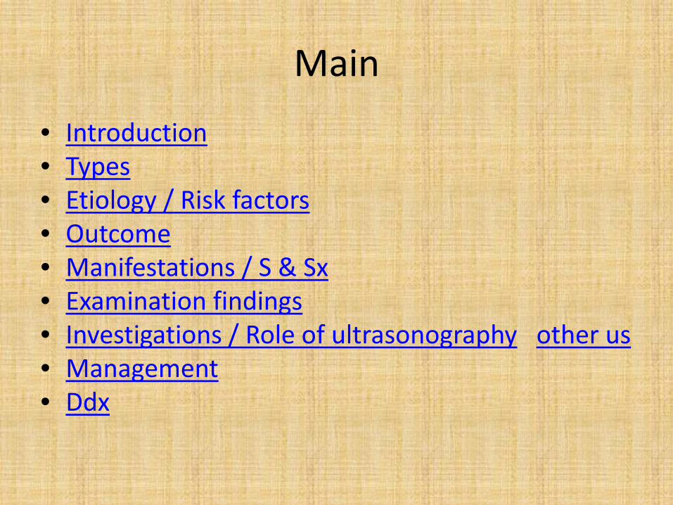
Main
• Introduction • Types • Etiology / Risk factors • Outcome • Manifestations / S & Sx • Examination findings • Investigations / Role of ultrasonography other us • Management • Ddx
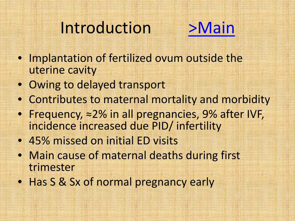
Introduction >Main
• Implantation of fertilized ovum outside the uterine cavity
• Owing to delayed transport • Contributes to maternal mortality and morbidity • Frequency, ≈2% in all pregnancies, 9% after IVF,
incidence increased due PID/ infertility • 45% missed on initial ED visits • Main cause of maternal deaths during first
trimester • Has S & Sx of normal pregnancy early
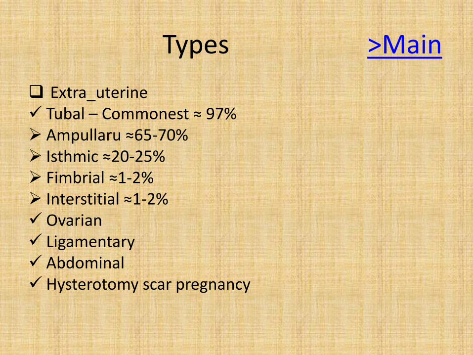
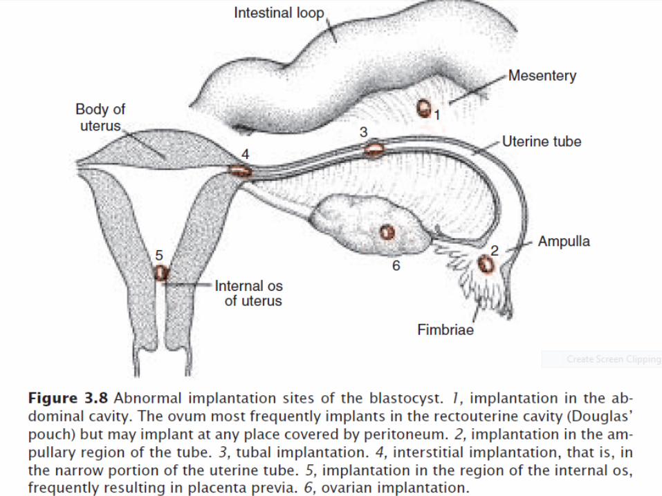
Types >Main Extra_uterine Tubal – Commonest ≈ 97% Ampullaru ≈65-70% Isthmic ≈20-25% Fimbrial ≈1-2% Interstitial ≈1-2% Ovarian Ligamentary Abdominal Hysterotomy scar pregnancy
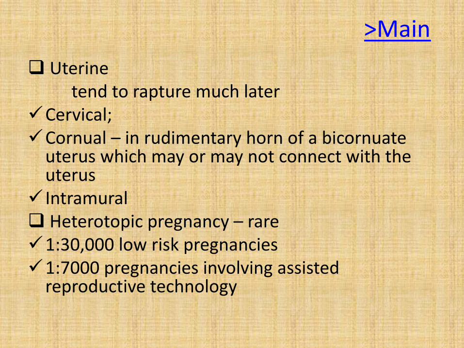
>Main Uterine tend to rapture much later Cervical; Cornual – in rudimentary horn of a bicornuate
uterus which may or may not connect with the uterus
Intramural Heterotopic pregnancy – rare 1:30,000 low risk pregnancies 1:7000 pregnancies involving assisted
reproductive technology
>Main Ovarian Pregnancy • Rare • Spiegelberg’s criteria for diagnosis (1) Tube on affected side must be intact. (2) Gestation sac must be in the position of the ovary. (3) Gestation sac is connected to the uterus by the ovarian
ligament. (4) Ovarian tissue must be found on the wall of the gestation
sac at histology. • Embedding may occur intra follicular or extra follicular. • In either type rupture is inevitable. • Salpingo-oophorectomy is the definitive surgery.
>Main Abdominal pregnancy • Primary or secondary • Primary – Rare; Studiford criteria for diagnosis. (1) Both tubes and ovaries are normal without
evidence of recent pregnancy. (2) Absence of utero-peritoneal fistula. (3) Presence of a pregnancy related exclusively to the
peritoneal surface and young enough to eliminate possibility of secondary implantation following primary nidation in the tube
Secondary –usual; primary sites being the tube, ovary, uterus; incidence increasing with the use of Assisted Reproductive Techniques
>Main
• H/O disturbed tubal pregnancy during early months (Lower abdominal pain & vaginal bleeding); minor ailments of normal pregnancy are often exaggerated e.g nausea, vomiting, constipation, abdominal pain, increased foetal movements.
• Signs of advanced pregnancy;
>Main – (1) uterine contour isn’t well defined even by palpating the
abdominal wall, as the braxton-Hicks contraction is absent in abdominal pregnancy.
– (2) foetal parts felt easily & persistent abnormal attitude & position of foetus on repeated exam is common. Abnormal high foetal position commonly found in intra-peritoneal pregnancy, the foetus is low lying in intra-ligamentary pregnancy.
• p/v –uterus difficult to separate from the abdominal mass. If separated, its enlarged(12-16wks) but cervix isn’t typically soft & usually displaced depending on position of sac.

>Main
• Sonograghy ;(1) absence wall around foetus – (2)abnormally high fetal position with abnormal
attitude – (3) foetal parts in close approximation to maternal
abdominal wall – (4) visualisation of uterus separately
• Mgnt: on diagnosis urgent laparotomy irrespective of gestational age.
>Main
• Laparotomy –ideal is to remove entire sac-foetus, placenta & membranes. Possible if placenta attached to removable organ like uterus or broad ligament.
• If placenta attached to vital organs , remove foetus, tie cord & leave placenta. Then placental activity monitored by quantitative serum hCG & scan. Complications are secondary haemorrhage, Intestinal obstruction, infection

>Main
Cervical pregnancy • Rare variant of ectopic pregnancy • Implantation in lining of endocervical canal • Incidence~ 1:9000 pregnancies • Cause unknown: local pathology related to previous
cervical/ uterine surgery may play a role. There is a given association with asherman’s syndrome & caesarean delivery. – Another is rapid transport of fertilised ovum into
endocervical canal before its capable of nidation or because of an unreceptive endometrium
– Common in pregnancies achieved by Assisted Reproductive Techniques
>Main
• Commonly have profuse painless per vaginal bleeding
• Lower abdominal pain or cramps occur in < 1/3 of patients
• Speculum exam- external os may be open, showing foetal membranes/ pregnancy tissues that appear blue/ purple
• Bimanual exam- soft cervix disproportionately enlarged compared to the uterus- an hour-glass shaped uterus
>Main
• Diagnosis –(1)thru Trans-vaginal intra-cervical localisation of a gestation sac surrounded by an echogenic rim – (2) closed internal os – (3) trophoblastic invasion of endocervical tissue
• Differential diagnosis – cervical abortion • Mgt –hysterectomy often needed; methotraxate may
be considered to avoid hysterectomy
Etiology /Risk factors >Main
Hight risk factors; • Previous ectopic ≈15% recurrence (related to
both underlying disorder that led to initial ectopic and to the choice of treatment procedure
• Tubal pahthology Disruption of normal tubal anatomy
>functional impairment due to damaged ciliary activity
>Main
Infections, congenital anomalies, tumors, surgery (Ifections:STDs_Chlamydia & gonorrhoea, sepsis_postoperative - post abortal – puerperal, TB_Salpingitis) Congenital anomalies eg, diverticula,
accessory ostia, duplication/ partial duplication, extremely lon/short tubes Tumour eg UT fibroids, Ovarian/ broad
ligament cyst
>Main
• Tubal surgery Reconstructive surgery done to re-anastomose
the tubes and reverse sterilisation or infertility surgery eg salpingostomy, salpingolysis etc Tubal surgery itself isn’t the culprit bt the
under lying tubal damage from the pior PID or ectopic pregnancy
>Main
Failure of sterilisation (BTL) attributed to; o Rare occurrence of a fertilised ovum being
trapped distal to the ligature at the time of operation
o Later re-canalisation allowing passage of sperm bt not the fertilized ovum
o Formation of tubal – peritoneal fistulas of sufficient size to allow for sperm passage but not the embryo
>Main
• In – utero DES exposure – 9fold increased risk • IUC – women with an IUC who become pregnant
are at high risk that pregnancy is ectopic (risk varies by method of contraception)
CuT 380A & levonorgesteral devices – lowest rate Progestasart – highest risk, may b coz its action is
limited to a local effec on endometrium Progestin only pill – increased risk – impaired
tubal motility IUD – increased risk of PID
>Main
Moderate risk factors Previous genital infections – pelvic infection eg
non specific salpingitis, Chlamydia, gonorrhoea esp recurrent infections Infertility – there is a suggested association
btn fertility drugs (ovulation induction) & ectopic pregnancy --- High oestrogen levels may alter ovum maturation & tubal transportation
>Main
• Multiple sexual partners – related to increased risk of PID
• Smoking –in peri-conception period increases risk of ectopic pregnancy in a dose dependent manner thus can be low/ moderate risk depending on patient’s habits
(Thought to result from impaired immunity in smokers predisposing them to PID; decreased ciliary action; abnormal blastocyst implantation related to decreased oestrogen levels found in smokers)

>Main
Low risk factors • (1) IVF- associated with increased risk of both ectopic &
heterotypic pregnancy • (2) vaginal douching- associated with increased risk of PID &
thus ectopic pregnancy • (3)Age- young age < 18 at first intercourse slightly increases
risk of ectopic pregnancy – Also increased proportion of ectopics in older age group; may be a
reflection of cumulative risk factors • (4)previous pelvic/ abdominal surgery; commonly an
appendicectomy-due to adhesions causing kinking or luminal compromise
• (5) an abnormal conceptus more likely to implant in the tube
>Main
Ovulation dysfunction • Described by Iffy(1961,1963) • Showed that in some cases embryo of an ectopic is
older than menstrual history suggested • He thought that mechanism was that of late ovulation
& inadequate steroid production by the corpus luteum producing a short luteal phase & withdrawal bleeding b4 the ovum can implant in the uterus.
• He suggests that the ovum is forced back up the tube by retrograde menstruation.
• Could account for the not uncommon finding of a tubal pregnancy & corpus luteum on contralateral sides
Outcome >Main
• Outcome • (1) absorption or mummification- pregnancy usually
dies before six weeks due to deficient placentation. – If small it may be absorbed in the tube / after abortion,
into the peritoneal cavity – Dead embryo may become mummified, infected or
calcified (lithopaedion)
• (2) Tubal mole – due to repeated small haemorrhages into the chorio-capsularis space, separating the villi from it’s attachments
>Main • (3)Tubal abortion – freq. depends in part on the
implantation site • Abortion common in ampullary tubal pregnancy • Conceptus separated from tubal wall by chorio-
decidual haemorrhage producing a haematosalpinx
• Complete tubal abortion- all POC extruded into peritoneal cavity
• Incomplete tubal abortion-POC partially extruded into peritoneal cavity
• Missed tubal abortion- pregnancy remains in haematosalpinx
>Main • (4) Tubal rupture –common in isthmic &
interstitial implantation • Isthmic rupture- occurs at 6-8 wks • Ampullary rupture- occurs at 8-12 wks • Interstitial rupture- occurs at ~ 4 months • Intraperitoneal rupture- common; rent in roof
or sides of tube; bleeding is intraperitoneal • Extraperitoneal (intraligamentary) rupture-
rare; occurs when rent lies in floor of tube where broad ligament attaches. Commonly met in isthmic implantation
Manifestations /S & Sx >Main >subacute manifestation
• Diverse & depend on whether rupture has occurred or not.
• Acute presentation: less common(~30%) & associated with tubal rupture/abortion with massive intraperitoneal haemorrhage
• Patient profile: -incidence maximum btn 20-30yrs & prevalance is mostly limited to nulliparity/following long periods of infertility
• Onset acute; about 1/3 of patients have persistent unilateral uneasiness before acute symptoms appear
>Main • Classic triad of symptoms;
amenorrhoea(75%),abdominal pain (100%) & appearance of vaginal bleeding(70%)
• Amenorrhoea of 6-8 wks/ a delayed period/ slight spotting on the expected date of the period. NB: amenorrhoea may be absent in a good number of cases
• Abdominal pain-most constant feature of triad – Pain is acute, agonising/colicky – Initially located in lower abdomen on one side
but gradually spreads all over the abdomen
>Main
• Vaginal bleeding- not an important feature in acute case – Bleeding slight, sanginous or dark coloured
& usually continuous • Nausea ,vomiting, fainting attacks (syncope)
may be present – Syncopal attack(10%) is peculiar to ectopic
& is probably due to reflex vasomotor disturbance caused by irritation of peritoneum by the blood.
Examination findings >Main >subacute findinds
• Pallor-usually severe & depends on amount of bleeding and is significantly out of proportion of vaginal bleeding if any
• Hypovolaemic shock (rapid, thin pulse, low BP, cold & clammy extremities)
• P/A- tense, tumid & tender – Tenderness usually in lower abdomen – No mass usually felt – Shifting dullness may be elicited
• P/V-pale vaginal mucosa – Uterus normal in size/ slightly bulky – Fornices extremely tender – Cervix excitable – No mass usually felt thru the fornix – Uterus floats as if in water
>Main Unruptured tubal ectopic • Need high index of suspicion • Often diagnosis is made accidentally during
laparoscopy / laparotomy • NB: Should always suspect an ectopic in a sexually
active female with abnormal bleeding &/or abdominal pain
• Symptoms : presence of delayed period with features suggestive of pregnancy – Discomfort on one side of the flank which is continuous or
at times colicky in nature.
>Main
• p/v – uterus slightly smaller than period of amenorrhoea showing evidence of early pregnancy (bimanually). – A pulsatile small, well circumscribed tender mass
may be felt thru one fornix separated from the uterus
>acute presentation >Main
Sub-acute/Chronic/Old tubal ectopic • Insidious onset • Amenorrhoea (6-8wks) usually present • Abdominal pain-usually starts as acute &
gradually becomes dull/ colicky in nature • Vaginal bleeding-scanty sanguineous or dark
coloured & continuous in nature

>Main
• Features of bladder irritation (dysuria, frequency, retention of urine) due to pelvic haematocoele causing pressure on adjacent organs
• Rectal tenesmus-due to the haematocoele • Rise in temp- due to infection or absorption of
products of degenerated blood accumulated in the abdomen
>Main >acute findings
Examination findings for sub acute • Patient looks ill • Varying degrees of pallor not proportionate to vaginal
bleeding • Persistent high pulse rate during rest is a conspicuous finding • Features of shock absent • Temp may be slightly elevated to 38 c • P/A –tenderness & guarding on lower abd esp on affected
side – An irregular tender mass may be felt in the lower abd – Cullen’s sign-bluish discolouration surrounding the umbilicus(umbilical
black eye) suggesting intraperitoneal bleeding
>Main
• P/V – Pale vaginal mucosa – Uterus seems normal in size or bulky, often
incorporated in a mass occupying the pelvis – Extreme tenderness on cervical motion – Chadwick’s sign (cervix and vaginal cyanosis) – An ill defined, boggy & extremely tender mass felt
thru posterolateral fornix extending to the pouch of Douglas
Investigations / Role of ultrasonograph >Main
• Acute ectopic-typical presentation • Chronic- high index of suspicion • Investigations(1)HCG; single value only confirms
pregnancy but does not determine its location – Suspicious findings are: (1) lower [HCG] compared to
normal I.U.P – (2) Doubling time in plasma fails to occur in 48 hours
• (2) blood- HB; ABO/RH grouping; WBC; ESR

>Main
• (3) Sonography-Trans-vaginal scan is the most informative – Absence of Intrauterine pregnancy with a positive
pregnancy test – Fluid in pouch of douglas – Adnexial mass clearly separated from ovary – Rarely cardiac activity may be seen in an unruptured tubal
ectopic – Colour Doppler can identify the placental shape (ring-of-
fire pattern) & blood flow pattern outside the uterine cavity

>Main
• (4) combination of HCG & Sonography-if HCG is > 1500IU/L with an empty uterus, ectopic is most likely – Failure to double HCG by 48 hrs along with an
empty uterus is very much suggestive • (5)serum progesterone- levels >25ng/ml
suggestive of a viable intrauterine pregnancy & levels <5ng/ml suggest an ectopic /abnormal Intrauterine pregnancy
>Main Serum hCG correlates with size and age, rise exponetially
and predictably during first 6-8weeks - increase by atleast 66%, multiplying 1.6 every 48hrs (doubles every 2-3days in normal pregnancy) Low hCG does not rule out EP - approx. 30-40% of EP with level <1000 mIU/mL will be ruptured at time of diagnosis Discriminatory zone. 1000 mIU/mL GS; >2000 mIU/mL YS
+/- embryo • (6) Laparoscopy-haemodynamically stable patient.
– Advantages are (1)confirmation of diagnosis; (2) removal of ectopic surgically at same setting; (3) direct injection of chemotheraputic agents into ectopic mass if medical management decided
>Main
• Culdocentesis- if Trans-vaginal sonography or laparoscopy not available. – Helps confirm presence of blood in the Pouch of Douglas – Thru speculum posterior lip of cervix grasped with a
tenaculum & drawn up under the symphysis – Lower part of post vaginal fornix steadied with Allis forceps
& an 18 gauge needle attached to a 10ml syringe thrust thru the dimple of the vagina between the utero sacral ligaments into the Pouch of Douglas
– Aspiration of non clotting blood signifies intraperitoneal blood
>Main
• Dilatation & curettage-identification of decidua without villi is very much suggestive of ectopic pregnancy. chorionic villi, that float in N/saline as lacy fronds, is diagnostic of an Intrauterine pregnancy
• Laparotomy-if in doubt & patient is haemodynamically unstable.

>Main
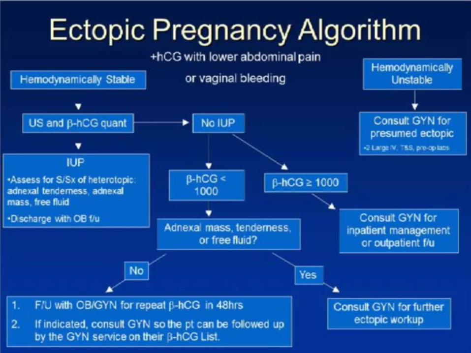
The other US • Rule out ectopic pregnancy by ruling in IUP • Perform a pelvic ultrasound for symptomatic
pregnant patients with a β-hCG below any discriminatory threshold
• Don’t use the β-hCG value to exclude the diagnis of ectopic pregnancy in patients who have an indeterminate ultrasound. Obtain special consultation or arrange close outpatient follow up for all patients with indeterminate pelvic ultrasound
Management >Main
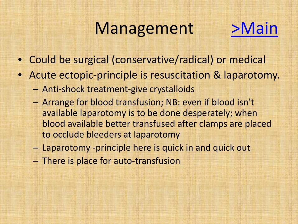
• Could be surgical (conservative/radical) or medical • Acute ectopic-principle is resuscitation & laparotomy.
– Anti-shock treatment-give crystalloids – Arrange for blood transfusion; NB: even if blood isn’t
available laparotomy is to be done desperately; when blood available better transfused after clamps are placed to occlude bleeders at laparotomy
– Laparotomy -principle here is quick in and quick out – There is place for auto-transfusion

>Main
• Chronic ectopic- admit pt & observe; do necessary investigations & Laparotomy at earliest convenient time – Usually a pelvic haematocele is found, blood clots
removed, affected tube identified & salpingectomy done
• NB: about 15% of women ovulate by 19 days & abt 25% ovulate by 30th postoperative day. Thus contraception should be started at time of hospital discharge
>Main
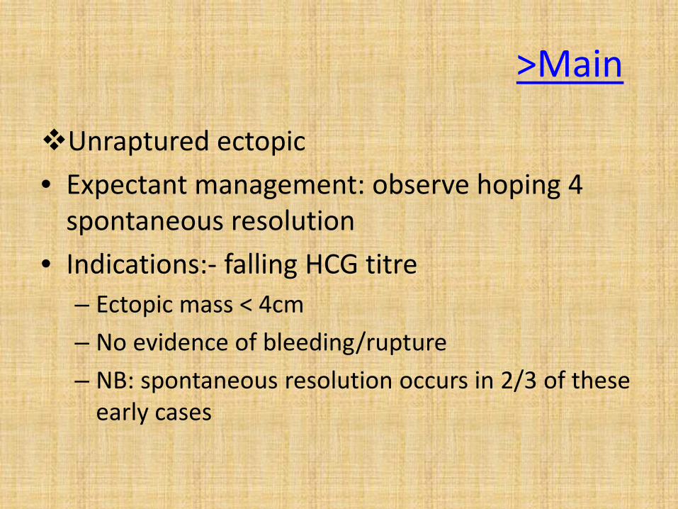
Unraptured ectopic • Expectant management: observe hoping 4
spontaneous resolution • Indications:- falling HCG titre
– Ectopic mass < 4cm – No evidence of bleeding/rupture – NB: spontaneous resolution occurs in 2/3 of these
early cases
>Main
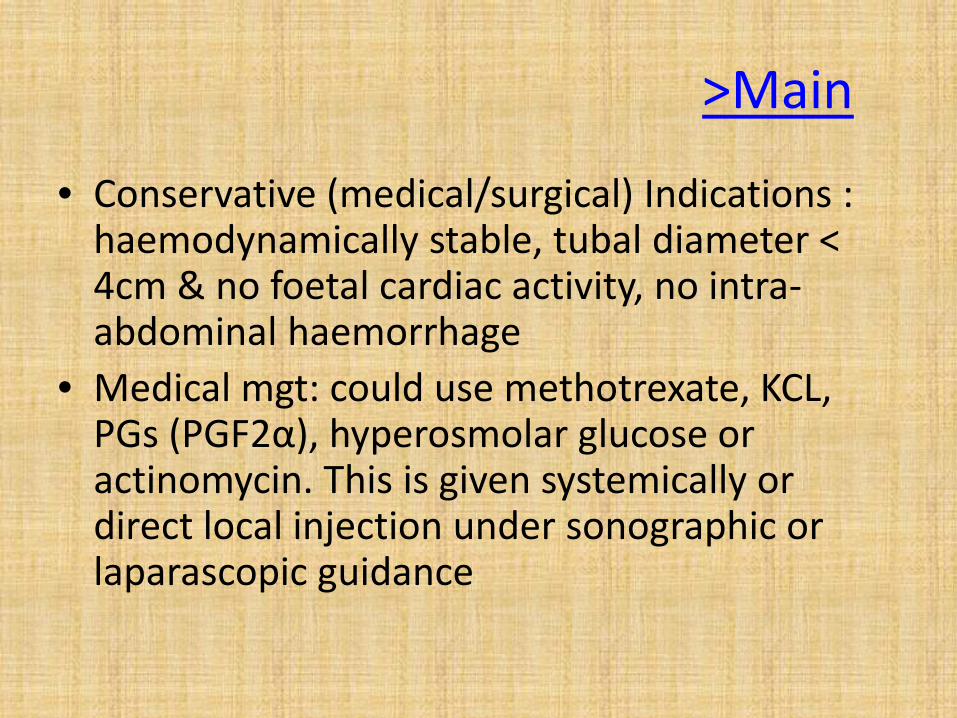
• Conservative (medical/surgical) Indications : haemodynamically stable, tubal diameter < 4cm & no foetal cardiac activity, no intra-abdominal haemorrhage
• Medical mgt: could use methotrexate, KCL, PGs (PGF2α), hyperosmolar glucose or actinomycin. This is given systemically or direct local injection under sonographic or laparascopic guidance

>Main
• Methotrexate –single dose 50mg/m2 I.M • Monitoring- done by measuring serum hCG on
day 4 & 7; if decline in hCG between day 4 & 7 is greater or equal to15%, patient is followed up weekly with serum hCG until hCG <10mIU/ml; if decline is < 15% second dose of Methotrexate 50mg/m2 is given on day 7
• Alternative dose: Methotrexate 1mg/kg I.M on days 1,3,5,7 & leukovorin 0.1mg/kg I.M on days 2,4,6,8. serum hCG is monitored weekly until <5.0 Miu/ML
Ddx >Main • Pelvic inflammatory disease • Threatened abortion • GTD • Ovarian torsion / raptured ovarian cyst • Appendicitis • Cholecystitis • Nephrolithiasis Alternate diagnoses • Dysmenorrhea • DUB • UTI • Mesenteric lymphadenitis
Remember >Main
• Ectopic pregnancy is a life threatening condition & on the increase
• Not all cases present with a classical picture • Always suspect ectopic pregnancy in a woman of a
child bearing age c/o abd pain / or p.v bleeding • Early diagnosis and management is feasible • Tailor ur management on the patient presentation. +/-
f/u • Routine Ultrasound for even asymptomatic pregnant
females, atleast once in early first trimester
Related Documents