Virtual Screening for HIV Protease Inhibitors: A Comparison of AutoDock 4 and Vina Max W. Chang 1 , Christian Ayeni 2 , Sebastian Breuer 1 , Bruce E. Torbett 1 * 1 Department of Molecular and Experimental Medicine, The Scripps Research Institute, La Jolla, California, United States of America, 2 Department of Bioengineering, University of California Merced, Merced, California, United States of America Abstract Background: The AutoDock family of software has been widely used in protein-ligand docking research. This study compares AutoDock 4 and AutoDock Vina in the context of virtual screening by using these programs to select compounds active against HIV protease. Methodology/Principal Findings: Both programs were used to rank the members of two chemical libraries, each containing experimentally verified binders to HIV protease. In the case of the NCI Diversity Set II, both AutoDock 4 and Vina were able to select active compounds significantly better than random (AUC = 0.69 and 0.68, respectively; p,0.001). The binding energy predictions were highly correlated in this case, with r = 0.63 and i = 0.82. For a set of larger, more flexible compounds from the Directory of Universal Decoys, the binding energy predictions were not correlated, and only Vina was able to rank compounds significantly better than random. Conclusions/Significance: In ranking smaller molecules with few rotatable bonds, AutoDock 4 and Vina were equally capable, though both exhibited a size-related bias in scoring. However, as Vina executes more quickly and is able to more accurately rank larger molecules, researchers should look to it first when undertaking a virtual screen. Citation: Chang MW, Ayeni C, Breuer S, Torbett BE (2010) Virtual Screening for HIV Protease Inhibitors: A Comparison of AutoDock 4 and Vina. PLoS ONE 5(8): e11955. doi:10.1371/journal.pone.0011955 Editor: Hendrik W. van Veen, University of Cambridge, United Kingdom Received March 1, 2010; Accepted June 18, 2010; Published August 4, 2010 Copyright: ß 2010 Chang et al. This is an open-access article distributed under the terms of the Creative Commons Attribution License, which permits unrestricted use, distribution, and reproduction in any medium, provided the original author and source are credited. Funding: Funding from the NIH to M.W.C (5T32NSO412119), B.E.T. (GM083658, GM48870 and AI40882) and CFAR support (3 P30 AI036214-13S1) is gratefully acknowledged. The funders had no role in study design, data collection and analysis, decision to publish, or preparation of the manuscript. Competing Interests: The authors have declared that no competing interests exist. * E-mail: [email protected] Introduction The use of virtual screening to discover new inhibitors is becoming a common practice in modern drug discovery [1]. Receptor-based virtual screens seek to ‘‘dock’’ members of a chemical library against a given protein structure, predicting the conformation and binding affinity of the small molecules [2]. A large number of programs are available for this purpose, such as DOCK [3], FlexX [4], GOLD [5], and AutoDock [6,7,8]. This study focuses on AutoDock 4 and AutoDock Vina (henceforth referred to as AD4 and Vina), both notable for being among the few docking programs that are freely available for academic and industrial use. The AutoDock programs are further unique in that they are some of the only widely-used docking programs released under open source licenses (GNU General Public License and Apache Open Source License). Both AD4 and Vina operate in a roughly similar manner, pairing an empirically-weighted scoring function with a global optimization algorithm. Key differences lie in the local search function (illustrated in Figure 1) and parameterization of the scoring function. In addition, Vina is designed to operate much more quickly and its authors have shown that its accuracy in re- docking protein-ligand complexes is greater than AD4 [8]. For 190 protein-ligand complexes, Vina was able to recapitulate the observed binding mode within 2 A ˚ RMSD in 78% of cases, while AD4 succeeded for only 49%. However, using AD4 and Vina to screen chemical libraries was not addressed. In this study, we compared the ability of AD4 and Vina to identify ligands by ranking the relative binding affinity of small molecules. For this task, the National Cancer Institute (NCI) Diversity Set II (DSII) was one of the chemical libraries used. DSII contains 1,364 compounds that tend to be small (the average molecular weight is less than 300 Daltons) and have few rotatable bonds. HIV protease was chosen as the protein target because it is a well- studied protein that has been a major focus for structure-based drug design [9]. As a complement to the relatively small DSII compounds, an additional collection of molecules was taken from the Directory of Universal Decoys (DUD) [10]. DUD contains known ligands for a variety of proteins, and provides accompa- nying ‘‘decoys’’ – molecules with composition similar to the known ligands, but with a different topology – that are assumed not to bind to the protein. There are 53 known HIV protease ligands in DUD, along with 1,885 decoys. Overall, these compounds tend to be appreciably larger than those from DSII, in terms of both molecular weight and number of rotatable bonds. Although DUD is already divided into known ‘‘active’’ and inactive compounds against HIV protease, that information is not available for DSII. A biophysical method, differential scanning fluorimetry (DSF) [11,12,13], was used to infer binding between HIV protease and the constituents of DSII. DSF functions by PLoS ONE | www.plosone.org 1 August 2010 | Volume 5 | Issue 8 | e11955

Welcome message from author
This document is posted to help you gain knowledge. Please leave a comment to let me know what you think about it! Share it to your friends and learn new things together.
Transcript
Virtual Screening for HIV Protease Inhibitors: AComparison of AutoDock 4 and VinaMax W. Chang1, Christian Ayeni2, Sebastian Breuer1, Bruce E. Torbett1*
1 Department of Molecular and Experimental Medicine, The Scripps Research Institute, La Jolla, California, United States of America, 2 Department of Bioengineering,
University of California Merced, Merced, California, United States of America
Abstract
Background: The AutoDock family of software has been widely used in protein-ligand docking research. This studycompares AutoDock 4 and AutoDock Vina in the context of virtual screening by using these programs to select compoundsactive against HIV protease.
Methodology/Principal Findings: Both programs were used to rank the members of two chemical libraries, each containingexperimentally verified binders to HIV protease. In the case of the NCI Diversity Set II, both AutoDock 4 and Vina were ableto select active compounds significantly better than random (AUC = 0.69 and 0.68, respectively; p,0.001). The bindingenergy predictions were highly correlated in this case, with r = 0.63 and i= 0.82. For a set of larger, more flexible compoundsfrom the Directory of Universal Decoys, the binding energy predictions were not correlated, and only Vina was able to rankcompounds significantly better than random.
Conclusions/Significance: In ranking smaller molecules with few rotatable bonds, AutoDock 4 and Vina were equallycapable, though both exhibited a size-related bias in scoring. However, as Vina executes more quickly and is able to moreaccurately rank larger molecules, researchers should look to it first when undertaking a virtual screen.
Citation: Chang MW, Ayeni C, Breuer S, Torbett BE (2010) Virtual Screening for HIV Protease Inhibitors: A Comparison of AutoDock 4 and Vina. PLoS ONE 5(8):e11955. doi:10.1371/journal.pone.0011955
Editor: Hendrik W. van Veen, University of Cambridge, United Kingdom
Received March 1, 2010; Accepted June 18, 2010; Published August 4, 2010
Copyright: � 2010 Chang et al. This is an open-access article distributed under the terms of the Creative Commons Attribution License, which permitsunrestricted use, distribution, and reproduction in any medium, provided the original author and source are credited.
Funding: Funding from the NIH to M.W.C (5T32NSO412119), B.E.T. (GM083658, GM48870 and AI40882) and CFAR support (3 P30 AI036214-13S1) is gratefullyacknowledged. The funders had no role in study design, data collection and analysis, decision to publish, or preparation of the manuscript.
Competing Interests: The authors have declared that no competing interests exist.
* E-mail: [email protected]
Introduction
The use of virtual screening to discover new inhibitors is
becoming a common practice in modern drug discovery [1].
Receptor-based virtual screens seek to ‘‘dock’’ members of a
chemical library against a given protein structure, predicting the
conformation and binding affinity of the small molecules [2]. A
large number of programs are available for this purpose, such as
DOCK [3], FlexX [4], GOLD [5], and AutoDock [6,7,8]. This
study focuses on AutoDock 4 and AutoDock Vina (henceforth
referred to as AD4 and Vina), both notable for being among the
few docking programs that are freely available for academic and
industrial use. The AutoDock programs are further unique in that
they are some of the only widely-used docking programs released
under open source licenses (GNU General Public License and
Apache Open Source License).
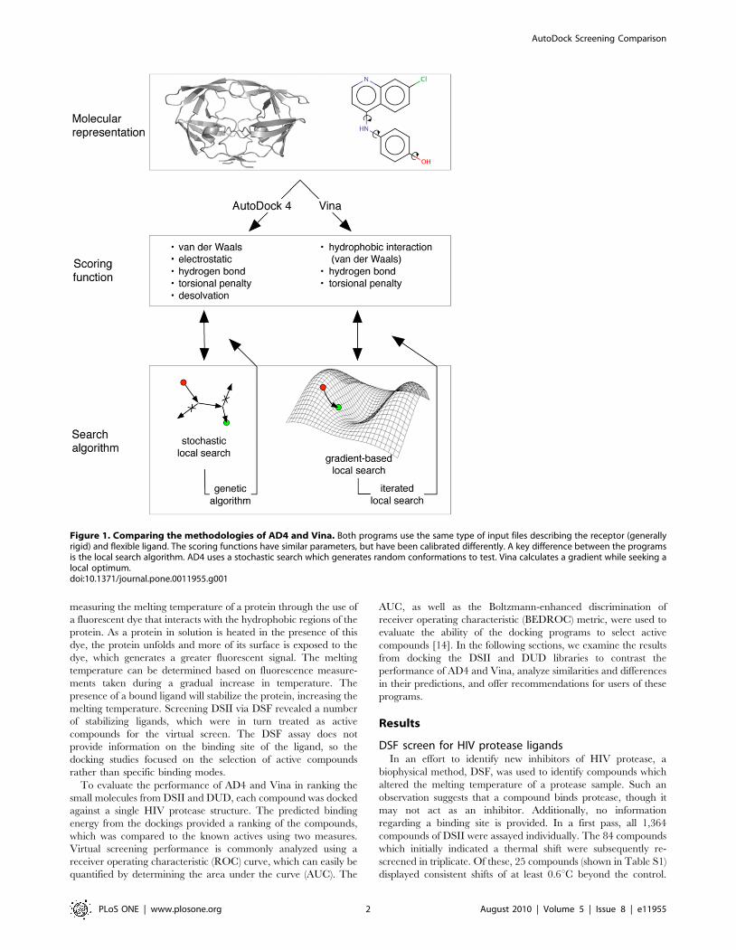
Both AD4 and Vina operate in a roughly similar manner,
pairing an empirically-weighted scoring function with a global
optimization algorithm. Key differences lie in the local search
function (illustrated in Figure 1) and parameterization of the
scoring function. In addition, Vina is designed to operate much
more quickly and its authors have shown that its accuracy in re-
docking protein-ligand complexes is greater than AD4 [8]. For 190
protein-ligand complexes, Vina was able to recapitulate the
observed binding mode within 2 A RMSD in 78% of cases, while
AD4 succeeded for only 49%. However, using AD4 and Vina to
screen chemical libraries was not addressed. In this study, we
compared the ability of AD4 and Vina to identify ligands by
ranking the relative binding affinity of small molecules.
For this task, the National Cancer Institute (NCI) Diversity Set
II (DSII) was one of the chemical libraries used. DSII contains
1,364 compounds that tend to be small (the average molecular
weight is less than 300 Daltons) and have few rotatable bonds.
HIV protease was chosen as the protein target because it is a well-
studied protein that has been a major focus for structure-based
drug design [9]. As a complement to the relatively small DSII
compounds, an additional collection of molecules was taken from
the Directory of Universal Decoys (DUD) [10]. DUD contains
known ligands for a variety of proteins, and provides accompa-
nying ‘‘decoys’’ – molecules with composition similar to the known
ligands, but with a different topology – that are assumed not to
bind to the protein. There are 53 known HIV protease ligands in
DUD, along with 1,885 decoys. Overall, these compounds tend to
be appreciably larger than those from DSII, in terms of both
molecular weight and number of rotatable bonds.
Although DUD is already divided into known ‘‘active’’ and
inactive compounds against HIV protease, that information is not
available for DSII. A biophysical method, differential scanning
fluorimetry (DSF) [11,12,13], was used to infer binding between
HIV protease and the constituents of DSII. DSF functions by
PLoS ONE | www.plosone.org 1 August 2010 | Volume 5 | Issue 8 | e11955
measuring the melting temperature of a protein through the use of
a fluorescent dye that interacts with the hydrophobic regions of the
protein. As a protein in solution is heated in the presence of this
dye, the protein unfolds and more of its surface is exposed to the
dye, which generates a greater fluorescent signal. The melting
temperature can be determined based on fluorescence measure-
ments taken during a gradual increase in temperature. The
presence of a bound ligand will stabilize the protein, increasing the
melting temperature. Screening DSII via DSF revealed a number
of stabilizing ligands, which were in turn treated as active
compounds for the virtual screen. The DSF assay does not
provide information on the binding site of the ligand, so the
docking studies focused on the selection of active compounds
rather than specific binding modes.
To evaluate the performance of AD4 and Vina in ranking the
small molecules from DSII and DUD, each compound was docked
against a single HIV protease structure. The predicted binding
energy from the dockings provided a ranking of the compounds,
which was compared to the known actives using two measures.
Virtual screening performance is commonly analyzed using a
receiver operating characteristic (ROC) curve, which can easily be
quantified by determining the area under the curve (AUC). The
AUC, as well as the Boltzmann-enhanced discrimination of
receiver operating characteristic (BEDROC) metric, were used to
evaluate the ability of the docking programs to select active
compounds [14]. In the following sections, we examine the results
from docking the DSII and DUD libraries to contrast the
performance of AD4 and Vina, analyze similarities and differences
in their predictions, and offer recommendations for users of these
programs.
Results
DSF screen for HIV protease ligandsIn an effort to identify new inhibitors of HIV protease, a
biophysical method, DSF, was used to identify compounds which
altered the melting temperature of a protease sample. Such an
observation suggests that a compound binds protease, though it
may not act as an inhibitor. Additionally, no information
regarding a binding site is provided. In a first pass, all 1,364
compounds of DSII were assayed individually. The 84 compounds
which initially indicated a thermal shift were subsequently re-
screened in triplicate. Of these, 25 compounds (shown in Table S1)
displayed consistent shifts of at least 0.6uC beyond the control.
Figure 1. Comparing the methodologies of AD4 and Vina. Both programs use the same type of input files describing the receptor (generallyrigid) and flexible ligand. The scoring functions have similar parameters, but have been calibrated differently. A key difference between the programsis the local search algorithm. AD4 uses a stochastic search which generates random conformations to test. Vina calculates a gradient while seeking alocal optimum.doi:10.1371/journal.pone.0011955.g001
AutoDock Screening Comparison
PLoS ONE | www.plosone.org 2 August 2010 | Volume 5 | Issue 8 | e11955
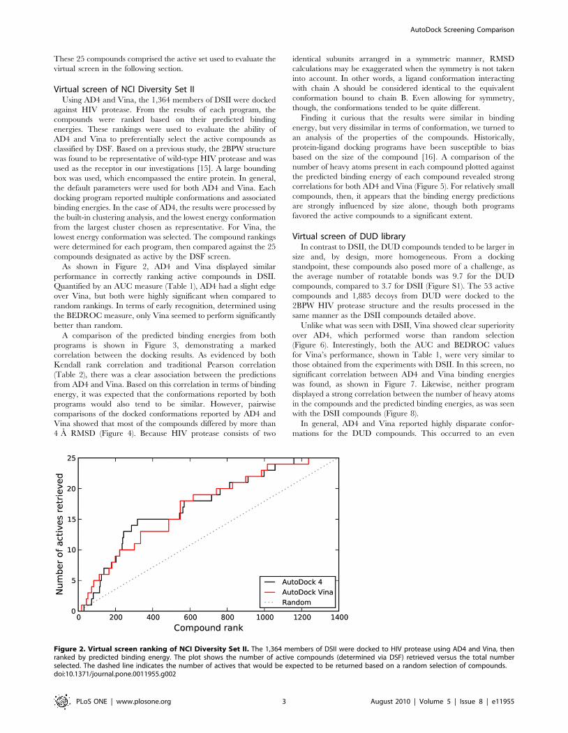
These 25 compounds comprised the active set used to evaluate the
virtual screen in the following section.
Virtual screen of NCI Diversity Set IIUsing AD4 and Vina, the 1,364 members of DSII were docked
against HIV protease. From the results of each program, the
compounds were ranked based on their predicted binding
energies. These rankings were used to evaluate the ability of
AD4 and Vina to preferentially select the active compounds as
classified by DSF. Based on a previous study, the 2BPW structure
was found to be representative of wild-type HIV protease and was
used as the receptor in our investigations [15]. A large bounding
box was used, which encompassed the entire protein. In general,
the default parameters were used for both AD4 and Vina. Each
docking program reported multiple conformations and associated
binding energies. In the case of AD4, the results were processed by
the built-in clustering analysis, and the lowest energy conformation
from the largest cluster chosen as representative. For Vina, the
lowest energy conformation was selected. The compound rankings
were determined for each program, then compared against the 25
compounds designated as active by the DSF screen.
As shown in Figure 2, AD4 and Vina displayed similar
performance in correctly ranking active compounds in DSII.
Quantified by an AUC measure (Table 1), AD4 had a slight edge
over Vina, but both were highly significant when compared to
random rankings. In terms of early recognition, determined using
the BEDROC measure, only Vina seemed to perform significantly
better than random.
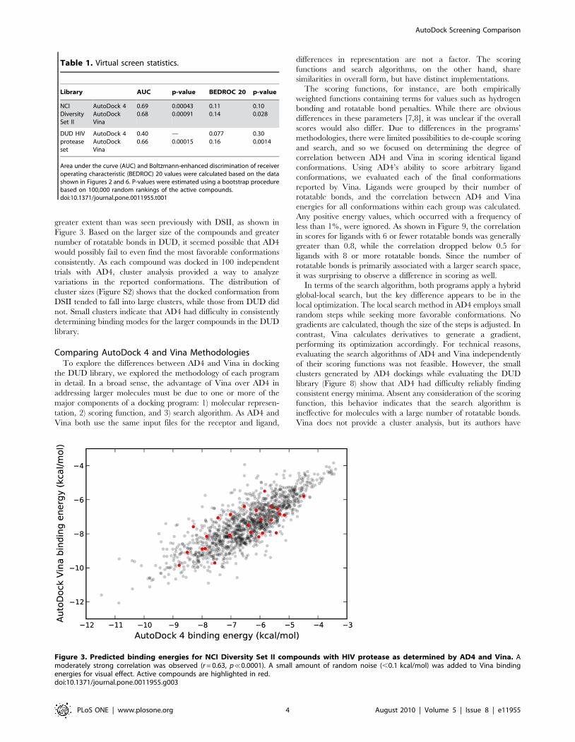
A comparison of the predicted binding energies from both
programs is shown in Figure 3, demonstrating a marked
correlation between the docking results. As evidenced by both
Kendall rank correlation and traditional Pearson correlation
(Table 2), there was a clear association between the predictions
from AD4 and Vina. Based on this correlation in terms of binding
energy, it was expected that the conformations reported by both
programs would also tend to be similar. However, pairwise
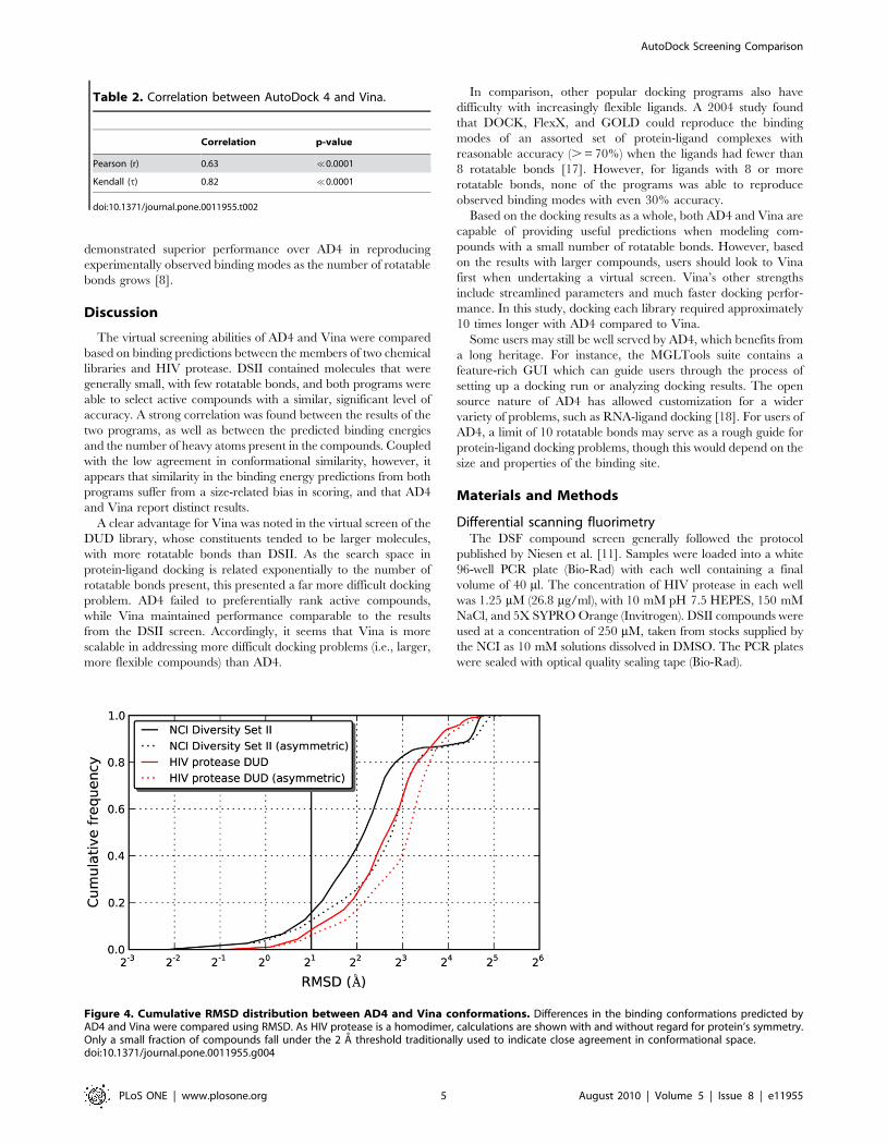
comparisons of the docked conformations reported by AD4 and
Vina showed that most of the compounds differed by more than
4 A RMSD (Figure 4). Because HIV protease consists of two
identical subunits arranged in a symmetric manner, RMSD
calculations may be exaggerated when the symmetry is not taken
into account. In other words, a ligand conformation interacting
with chain A should be considered identical to the equivalent
conformation bound to chain B. Even allowing for symmetry,
though, the conformations tended to be quite different.
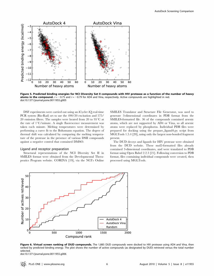
Finding it curious that the results were similar in binding
energy, but very dissimilar in terms of conformation, we turned to
an analysis of the properties of the compounds. Historically,
protein-ligand docking programs have been susceptible to bias
based on the size of the compound [16]. A comparison of the
number of heavy atoms present in each compound plotted against
the predicted binding energy of each compound revealed strong
correlations for both AD4 and Vina (Figure 5). For relatively small
compounds, then, it appears that the binding energy predictions
are strongly influenced by size alone, though both programs
favored the active compounds to a significant extent.
Virtual screen of DUD libraryIn contrast to DSII, the DUD compounds tended to be larger in
size and, by design, more homogeneous. From a docking
standpoint, these compounds also posed more of a challenge, as
the average number of rotatable bonds was 9.7 for the DUD
compounds, compared to 3.7 for DSII (Figure S1). The 53 active
compounds and 1,885 decoys from DUD were docked to the
2BPW HIV protease structure and the results processed in the
same manner as the DSII compounds detailed above.
Unlike what was seen with DSII, Vina showed clear superiority
over AD4, which performed worse than random selection
(Figure 6). Interestingly, both the AUC and BEDROC values
for Vina’s performance, shown in Table 1, were very similar to
those obtained from the experiments with DSII. In this screen, no
significant correlation between AD4 and Vina binding energies
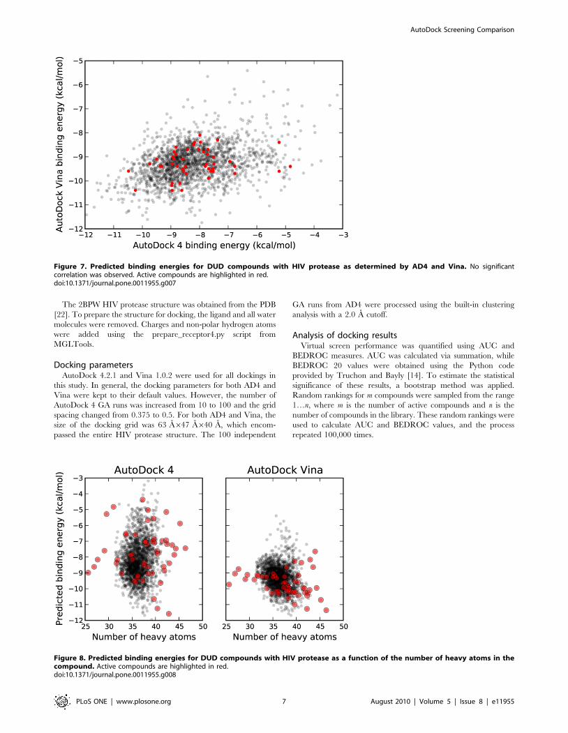
was found, as shown in Figure 7. Likewise, neither program
displayed a strong correlation between the number of heavy atoms
in the compounds and the predicted binding energies, as was seen
with the DSII compounds (Figure 8).
In general, AD4 and Vina reported highly disparate confor-
mations for the DUD compounds. This occurred to an even
Figure 2. Virtual screen ranking of NCI Diversity Set II. The 1,364 members of DSII were docked to HIV protease using AD4 and Vina, thenranked by predicted binding energy. The plot shows the number of active compounds (determined via DSF) retrieved versus the total numberselected. The dashed line indicates the number of actives that would be expected to be returned based on a random selection of compounds.doi:10.1371/journal.pone.0011955.g002
AutoDock Screening Comparison
PLoS ONE | www.plosone.org 3 August 2010 | Volume 5 | Issue 8 | e11955
greater extent than was seen previously with DSII, as shown in
Figure 3. Based on the larger size of the compounds and greater
number of rotatable bonds in DUD, it seemed possible that AD4
would possibly fail to even find the most favorable conformations
consistently. As each compound was docked in 100 independent
trials with AD4, cluster analysis provided a way to analyze
variations in the reported conformations. The distribution of
cluster sizes (Figure S2) shows that the docked conformation from
DSII tended to fall into large clusters, while those from DUD did
not. Small clusters indicate that AD4 had difficulty in consistently
determining binding modes for the larger compounds in the DUD
library.
Comparing AutoDock 4 and Vina MethodologiesTo explore the differences between AD4 and Vina in docking
the DUD library, we explored the methodology of each program
in detail. In a broad sense, the advantage of Vina over AD4 in
addressing larger molecules must be due to one or more of the
major components of a docking program: 1) molecular represen-
tation, 2) scoring function, and 3) search algorithm. As AD4 and
Vina both use the same input files for the receptor and ligand,
differences in representation are not a factor. The scoring
functions and search algorithms, on the other hand, share
similarities in overall form, but have distinct implementations.
The scoring functions, for instance, are both empirically
weighted functions containing terms for values such as hydrogen
bonding and rotatable bond penalties. While there are obvious
differences in these parameters [7,8], it was unclear if the overall
scores would also differ. Due to differences in the programs’
methodologies, there were limited possibilities to de-couple scoring
and search, and so we focused on determining the degree of
correlation between AD4 and Vina in scoring identical ligand
conformations. Using AD4’s ability to score arbitrary ligand
conformations, we evaluated each of the final conformations
reported by Vina. Ligands were grouped by their number of
rotatable bonds, and the correlation between AD4 and Vina
energies for all conformations within each group was calculated.
Any positive energy values, which occurred with a frequency of
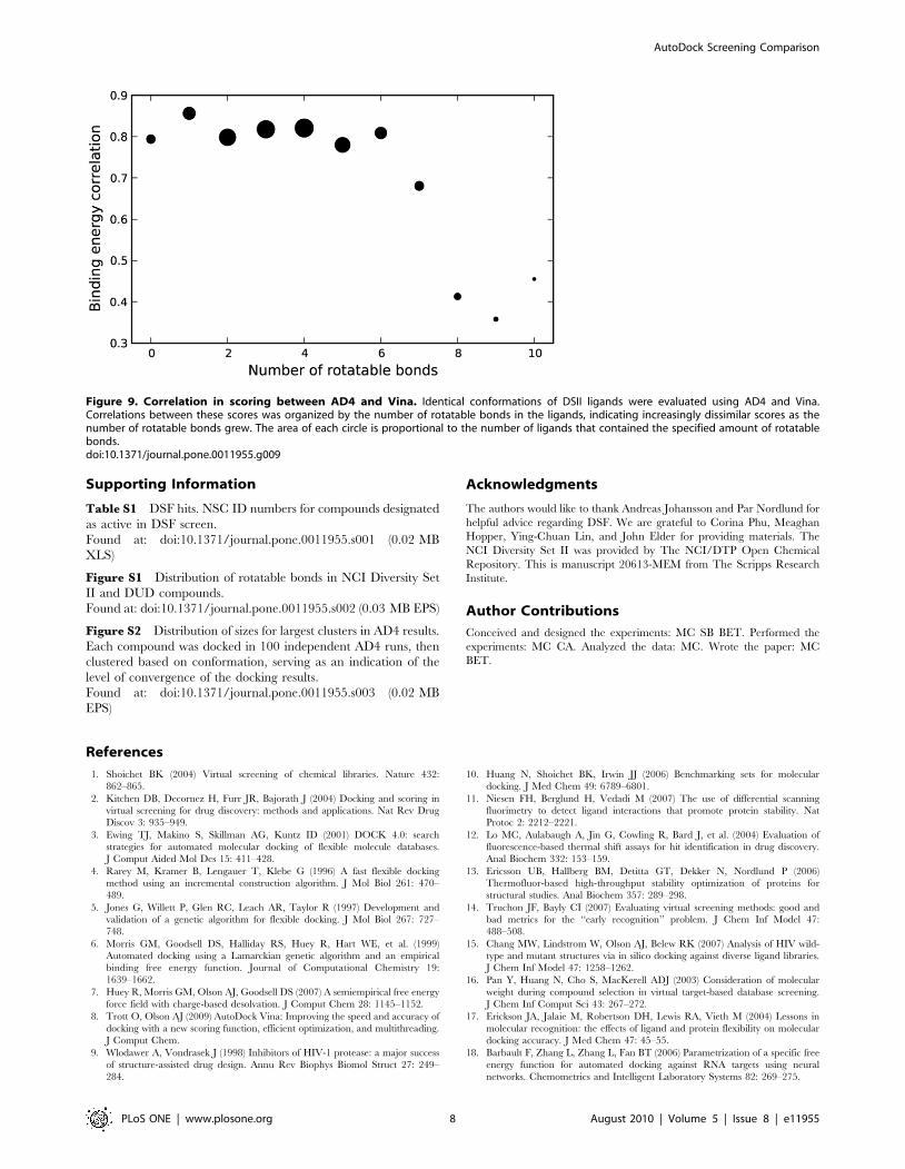
less than 1%, were ignored. As shown in Figure 9, the correlation
in scores for ligands with 6 or fewer rotatable bonds was generally
greater than 0.8, while the correlation dropped below 0.5 for
ligands with 8 or more rotatable bonds. Since the number of
rotatable bonds is primarily associated with a larger search space,
it was surprising to observe a difference in scoring as well.
In terms of the search algorithm, both programs apply a hybrid
global-local search, but the key difference appears to be in the
local optimization. The local search method in AD4 employs small
random steps while seeking more favorable conformations. No
gradients are calculated, though the size of the steps is adjusted. In
contrast, Vina calculates derivatives to generate a gradient,
performing its optimization accordingly. For technical reasons,
evaluating the search algorithms of AD4 and Vina independently
of their scoring functions was not feasible. However, the small
clusters generated by AD4 dockings while evaluating the DUD
library (Figure 8) show that AD4 had difficulty reliably finding
consistent energy minima. Absent any consideration of the scoring
function, this behavior indicates that the search algorithm is
ineffective for molecules with a large number of rotatable bonds.
Vina does not provide a cluster analysis, but its authors have
Figure 3. Predicted binding energies for NCI Diversity Set II compounds with HIV protease as determined by AD4 and Vina. Amoderately strong correlation was observed (r = 0.63, p%0.0001). A small amount of random noise (,0.1 kcal/mol) was added to Vina bindingenergies for visual effect. Active compounds are highlighted in red.doi:10.1371/journal.pone.0011955.g003
Table 1. Virtual screen statistics.
Library AUC p-value BEDROC 20 p-value
NCIDiversitySet II
AutoDock 4AutoDockVina
0.690.68
0.000430.00091
0.110.14
0.100.028
DUD HIVproteaseset
AutoDock 4AutoDockVina
0.400.66
—0.00015
0.0770.16
0.300.0014
Area under the curve (AUC) and Boltzmann-enhanced discrimination of receiveroperating characteristic (BEDROC) 20 values were calculated based on the datashown in Figures 2 and 6. P-values were estimated using a bootstrap procedurebased on 100,000 random rankings of the active compounds.doi:10.1371/journal.pone.0011955.t001
AutoDock Screening Comparison
PLoS ONE | www.plosone.org 4 August 2010 | Volume 5 | Issue 8 | e11955
demonstrated superior performance over AD4 in reproducing
experimentally observed binding modes as the number of rotatable
bonds grows [8].
Discussion
The virtual screening abilities of AD4 and Vina were compared
based on binding predictions between the members of two chemical
libraries and HIV protease. DSII contained molecules that were
generally small, with few rotatable bonds, and both programs were
able to select active compounds with a similar, significant level of
accuracy. A strong correlation was found between the results of the
two programs, as well as between the predicted binding energies
and the number of heavy atoms present in the compounds. Coupled
with the low agreement in conformational similarity, however, it
appears that similarity in the binding energy predictions from both
programs suffer from a size-related bias in scoring, and that AD4
and Vina report distinct results.
A clear advantage for Vina was noted in the virtual screen of the
DUD library, whose constituents tended to be larger molecules,
with more rotatable bonds than DSII. As the search space in
protein-ligand docking is related exponentially to the number of
rotatable bonds present, this presented a far more difficult docking
problem. AD4 failed to preferentially rank active compounds,
while Vina maintained performance comparable to the results
from the DSII screen. Accordingly, it seems that Vina is more
scalable in addressing more difficult docking problems (i.e., larger,
more flexible compounds) than AD4.
In comparison, other popular docking programs also have
difficulty with increasingly flexible ligands. A 2004 study found
that DOCK, FlexX, and GOLD could reproduce the binding
modes of an assorted set of protein-ligand complexes with
reasonable accuracy (. = 70%) when the ligands had fewer than
8 rotatable bonds [17]. However, for ligands with 8 or more
rotatable bonds, none of the programs was able to reproduce
observed binding modes with even 30% accuracy.
Based on the docking results as a whole, both AD4 and Vina are
capable of providing useful predictions when modeling com-
pounds with a small number of rotatable bonds. However, based
on the results with larger compounds, users should look to Vina
first when undertaking a virtual screen. Vina’s other strengths
include streamlined parameters and much faster docking perfor-
mance. In this study, docking each library required approximately
10 times longer with AD4 compared to Vina.
Some users may still be well served by AD4, which benefits from
a long heritage. For instance, the MGLTools suite contains a
feature-rich GUI which can guide users through the process of
setting up a docking run or analyzing docking results. The open
source nature of AD4 has allowed customization for a wider
variety of problems, such as RNA-ligand docking [18]. For users of
AD4, a limit of 10 rotatable bonds may serve as a rough guide for
protein-ligand docking problems, though this would depend on the
size and properties of the binding site.
Materials and Methods
Differential scanning fluorimetryThe DSF compound screen generally followed the protocol
published by Niesen et al. [11]. Samples were loaded into a white
96-well PCR plate (Bio-Rad) with each well containing a final
volume of 40 ml. The concentration of HIV protease in each well
was 1.25 mM (26.8 mg/ml), with 10 mM pH 7.5 HEPES, 150 mM
NaCl, and 5X SYPRO Orange (Invitrogen). DSII compounds were
used at a concentration of 250 mM, taken from stocks supplied by
the NCI as 10 mM solutions dissolved in DMSO. The PCR plates
were sealed with optical quality sealing tape (Bio-Rad).
Table 2. Correlation between AutoDock 4 and Vina.
Correlation p-value
Pearson (r) 0.63 %0.0001
Kendall (t) 0.82 %0.0001
doi:10.1371/journal.pone.0011955.t002
Figure 4. Cumulative RMSD distribution between AD4 and Vina conformations. Differences in the binding conformations predicted byAD4 and Vina were compared using RMSD. As HIV protease is a homodimer, calculations are shown with and without regard for protein’s symmetry.Only a small fraction of compounds fall under the 2 A threshold traditionally used to indicate close agreement in conformational space.doi:10.1371/journal.pone.0011955.g004
AutoDock Screening Comparison
PLoS ONE | www.plosone.org 5 August 2010 | Volume 5 | Issue 8 | e11955
DSF experiments were carried out using an iCycler iQ real-time
PCR system (Bio-Rad) set to use the 490/20 excitation and 575/
20 emission filters. The samples were heated from 20 to 95uC at
the rate of 1uC/minute. A single fluorescence measurement was
taken each minute. Melting temperatures were determined by
performing a curve fit to the Boltzmann equation. The degree of
thermal shift was calculated by comparing the melting tempera-
ture of the protease in the presence of various DSII compounds
against a negative control that contained DMSO.
Ligand and receptor preparationStructural representations of the NCI Diversity Set II in
SMILES format were obtained from the Developmental Thera-
peutics Program website. CORINA [19], via the NCI’s Online
SMILES Translator and Structure File Generator, was used to
generate 3-dimensional coordinates in PDB format from the
SMILES-formatted file. 36 of the compounds contained arsenic
atoms, which are not supported by AD4 or Vina, so all arsenic
atoms were replaced by phosphorus. Individual PDB files were
prepared for docking using the prepare_ligand4.py script from
MGLTools 1.5.4 [20], using only the largest non-bonded fragment
present.
The DUD decoys and ligands for HIV protease were obtained
from the DUD website. These mol2-formatted files already
contained 3-dimesional coordinates, and were translated to PDB
format using Open Babel 2.2.3 [21]. Following conversion to PDB
format, files containing individual compounds were created, then
processed using MGLTools.
Figure 6. Virtual screen ranking of DUD compounds. The 1,885 DUD compounds were docked to HIV protease using AD4 and Vina, thenranked by predicted binding energy. The plot shows the number of active compounds (as designated by DUD) retrieved versus the total numberselected.doi:10.1371/journal.pone.0011955.g006
Figure 5. Predicted binding energies for NCI Diversity Set II compounds with HIV protease as a function of the number of heavyatoms in the compound. r = 20.75 and r = 20.79 for AD4 and Vina, respectively. Active compounds are highlighted in red.doi:10.1371/journal.pone.0011955.g005
AutoDock Screening Comparison
PLoS ONE | www.plosone.org 6 August 2010 | Volume 5 | Issue 8 | e11955
The 2BPW HIV protease structure was obtained from the PDB
[22]. To prepare the structure for docking, the ligand and all water
molecules were removed. Charges and non-polar hydrogen atoms
were added using the prepare_receptor4.py script from
MGLTools.
Docking parametersAutoDock 4.2.1 and Vina 1.0.2 were used for all dockings in
this study. In general, the docking parameters for both AD4 and
Vina were kept to their default values. However, the number of
AutoDock 4 GA runs was increased from 10 to 100 and the grid
spacing changed from 0.375 to 0.5. For both AD4 and Vina, the
size of the docking grid was 63 A647 A640 A, which encom-
passed the entire HIV protease structure. The 100 independent
GA runs from AD4 were processed using the built-in clustering
analysis with a 2.0 A cutoff.
Analysis of docking resultsVirtual screen performance was quantified using AUC and
BEDROC measures. AUC was calculated via summation, while
BEDROC 20 values were obtained using the Python code
provided by Truchon and Bayly [14]. To estimate the statistical
significance of these results, a bootstrap method was applied.
Random rankings for m compounds were sampled from the range
1…n, where m is the number of active compounds and n is the
number of compounds in the library. These random rankings were
used to calculate AUC and BEDROC values, and the process
repeated 100,000 times.
Figure 7. Predicted binding energies for DUD compounds with HIV protease as determined by AD4 and Vina. No significantcorrelation was observed. Active compounds are highlighted in red.doi:10.1371/journal.pone.0011955.g007
Figure 8. Predicted binding energies for DUD compounds with HIV protease as a function of the number of heavy atoms in thecompound. Active compounds are highlighted in red.doi:10.1371/journal.pone.0011955.g008
AutoDock Screening Comparison
PLoS ONE | www.plosone.org 7 August 2010 | Volume 5 | Issue 8 | e11955
Supporting Information
Table S1 DSF hits. NSC ID numbers for compounds designated
as active in DSF screen.
Found at: doi:10.1371/journal.pone.0011955.s001 (0.02 MB
XLS)
Figure S1 Distribution of rotatable bonds in NCI Diversity Set
II and DUD compounds.
Found at: doi:10.1371/journal.pone.0011955.s002 (0.03 MB EPS)
Figure S2 Distribution of sizes for largest clusters in AD4 results.
Each compound was docked in 100 independent AD4 runs, then
clustered based on conformation, serving as an indication of the
level of convergence of the docking results.
Found at: doi:10.1371/journal.pone.0011955.s003 (0.02 MB
EPS)
Acknowledgments
The authors would like to thank Andreas Johansson and Par Nordlund for
helpful advice regarding DSF. We are grateful to Corina Phu, Meaghan
Hopper, Ying-Chuan Lin, and John Elder for providing materials. The
NCI Diversity Set II was provided by The NCI/DTP Open Chemical
Repository. This is manuscript 20613-MEM from The Scripps Research
Institute.
Author Contributions
Conceived and designed the experiments: MC SB BET. Performed the
experiments: MC CA. Analyzed the data: MC. Wrote the paper: MC
BET.
References
1. Shoichet BK (2004) Virtual screening of chemical libraries. Nature 432:
862–865.
2. Kitchen DB, Decornez H, Furr JR, Bajorath J (2004) Docking and scoring in
virtual screening for drug discovery: methods and applications. Nat Rev Drug
Discov 3: 935–949.
3. Ewing TJ, Makino S, Skillman AG, Kuntz ID (2001) DOCK 4.0: search
strategies for automated molecular docking of flexible molecule databases.
J Comput Aided Mol Des 15: 411–428.
4. Rarey M, Kramer B, Lengauer T, Klebe G (1996) A fast flexible docking
method using an incremental construction algorithm. J Mol Biol 261: 470–
489.
5. Jones G, Willett P, Glen RC, Leach AR, Taylor R (1997) Development and
validation of a genetic algorithm for flexible docking. J Mol Biol 267: 727–
748.
6. Morris GM, Goodsell DS, Halliday RS, Huey R, Hart WE, et al. (1999)
Automated docking using a Lamarckian genetic algorithm and an empirical
binding free energy function. Journal of Computational Chemistry 19:
1639–1662.
7. Huey R, Morris GM, Olson AJ, Goodsell DS (2007) A semiempirical free energy
force field with charge-based desolvation. J Comput Chem 28: 1145–1152.
8. Trott O, Olson AJ (2009) AutoDock Vina: Improving the speed and accuracy of
docking with a new scoring function, efficient optimization, and multithreading.
J Comput Chem.
9. Wlodawer A, Vondrasek J (1998) Inhibitors of HIV-1 protease: a major success
of structure-assisted drug design. Annu Rev Biophys Biomol Struct 27: 249–
284.
10. Huang N, Shoichet BK, Irwin JJ (2006) Benchmarking sets for molecular
docking. J Med Chem 49: 6789–6801.
11. Niesen FH, Berglund H, Vedadi M (2007) The use of differential scanning
fluorimetry to detect ligand interactions that promote protein stability. Nat
Protoc 2: 2212–2221.
12. Lo MC, Aulabaugh A, Jin G, Cowling R, Bard J, et al. (2004) Evaluation of
fluorescence-based thermal shift assays for hit identification in drug discovery.
Anal Biochem 332: 153–159.
13. Ericsson UB, Hallberg BM, Detitta GT, Dekker N, Nordlund P (2006)
Thermofluor-based high-throughput stability optimization of proteins for
structural studies. Anal Biochem 357: 289–298.
14. Truchon JF, Bayly CI (2007) Evaluating virtual screening methods: good and
bad metrics for the ‘‘early recognition’’ problem. J Chem Inf Model 47:
488–508.
15. Chang MW, Lindstrom W, Olson AJ, Belew RK (2007) Analysis of HIV wild-
type and mutant structures via in silico docking against diverse ligand libraries.
J Chem Inf Model 47: 1258–1262.
16. Pan Y, Huang N, Cho S, MacKerell ADJ (2003) Consideration of molecular
weight during compound selection in virtual target-based database screening.
J Chem Inf Comput Sci 43: 267–272.
17. Erickson JA, Jalaie M, Robertson DH, Lewis RA, Vieth M (2004) Lessons in
molecular recognition: the effects of ligand and protein flexibility on molecular
docking accuracy. J Med Chem 47: 45–55.
18. Barbault F, Zhang L, Zhang L, Fan BT (2006) Parametrization of a specific free
energy function for automated docking against RNA targets using neural
networks. Chemometrics and Intelligent Laboratory Systems 82: 269–275.
Figure 9. Correlation in scoring between AD4 and Vina. Identical conformations of DSII ligands were evaluated using AD4 and Vina.Correlations between these scores was organized by the number of rotatable bonds in the ligands, indicating increasingly dissimilar scores as thenumber of rotatable bonds grew. The area of each circle is proportional to the number of ligands that contained the specified amount of rotatablebonds.doi:10.1371/journal.pone.0011955.g009
AutoDock Screening Comparison
PLoS ONE | www.plosone.org 8 August 2010 | Volume 5 | Issue 8 | e11955
19. Gasteiger J, Rudolph C, Sadowski J (1990) Automatic generation of 3D-atomic
coordinates for organic molecules. Tetrahedron Comput Methodol 3: 3.
20. Sanner MF (1999) Python: a programming language for software integration
and development. J Mol Graph Model 17: 57–61.
21. Guha R, Howard MT, Hutchison GR, Murray-Rust P, Rzepa H, et al. (2006)
The Blue Obelisk-interoperability in chemical informatics. J Chem Inf Model46: 991–998.
22. Berman HM, Westbrook J, Feng Z, Gilliland G, Bhat TN, et al. (2000) The
Protein Data Bank. Nucleic Acids Res 28: 235–242.
AutoDock Screening Comparison
PLoS ONE | www.plosone.org 9 August 2010 | Volume 5 | Issue 8 | e11955
Related Documents