VILNIAUS UNIVERSITETAS VILNIAUS UNIVERSITETO ONKOLOGIJOS INSTITUTAS Aida Laurinavičienė DUKTALINĖS KRŪTIES KARCINOMOS BIOLOGINĖS ĮVAIROVĖS TYRIMAS MOLEKULINĖS IR SKAITMENINĖS PATOLOGIJOS METODAIS Daktaro disertacija Biomedicinos mokslai, medicina (06B), Citologija, onkologija, kancerologija (B200) Vilnius, 2012

Welcome message from author
This document is posted to help you gain knowledge. Please leave a comment to let me know what you think about it! Share it to your friends and learn new things together.
Transcript

VILNIAUS UNIVERSITETAS
VILNIAUS UNIVERSITETO ONKOLOGIJOS INSTITUTAS
Aida Laurinavičienė
DUKTALINĖS KRŪTIES KARCINOMOS BIOLOGINĖS
ĮVAIROVĖS TYRIMAS MOLEKULINĖS IR SKAITMENINĖS
PATOLOGIJOS METODAIS
Daktaro disertacija
Biomedicinos mokslai, medicina (06B),
Citologija, onkologija, kancerologija (B200)
Vilnius, 2012

2
Disertacija rengta 2006 – 2012 metais Vilniaus universiteto Onkologijos
institute
Moksliniai vadovai:
Prof. dr. Sonata Jarmalaitė (Vilniaus universitetas, biomedicinos
mokslai, biologija – 01B, citologija, onkologija, kancerologija – B200).
2006 – 2010 m.m.
Habil. dr. Valerijus Ostapenko (Vilniaus universiteto Onkologijos
institutas, biomedicinos mokslai, medicina – 06B, citologija, onkologija,
kancerologija – B200). 2011 – 2012 m.m.
Mokslinis konsultantas:
Prof. dr. Dainius Characiejus (Vilniaus universitetas, biomedicinos
mokslai, medicina – 06B, citologija, onkologija, kancerologija – B200).
2006 – 2008 m.m.

3
TURINYS
1. ĮVADAS .............................................................................................................8
2. LITERATŪROS APŽVALGA ......................................................................... 13
2.1. Krūties vėžio rizikos veiksniai ir formavimosi molekuliniai ........................ 15
mechanizmai ...................................................................................................... 15
2.2. Krūties vėžio DNR metilinimo pakitimai .................................................... 16
2.3. Pagrindiniai krūties vėžio žymenys ............................................................. 19
2.3.1. Hormonų receptoriai ............................................................................. 19
2.3.2. HER2 onkogenas .................................................................................. 21
2.3.3. Ląstelių proliferacijos ir kiti imunohistocheminiai žymenys ................. 22
2.2.4. Naviką slopinantys genai ...................................................................... 24
3. TIRIAMIEJI IR TYRIMO METODAI .............................................................. 29
3.1. Tiriamųjų atranka ir tyrimo eiga .................................................................. 29
3.2. Tyrimo metodai........................................................................................... 32
3.2.1. Mėginių paruošimas.............................................................................. 32
3.2.2. Imunohistocheminiai tyrimai ................................................................ 35
3.2.3. Vizualus patologo vertinimas ................................................................ 40
3.2.4. Skaitmeninė vaizdų analizė ................................................................... 41
3.2.5. Imunofluorescenciniai tyrimai .............................................................. 44
3.2.6. DNR išskyrimas, metilinimo ir geno mutacijų tyrimai .......................... 45
3.3. Statistinė analizė ......................................................................................... 50
3.4. Etiniai aspektai ............................................................................................ 51
4. REZULTATAI .................................................................................................. 52
4.1. Imunohistocheminių žymenų raiškos skaitmeninės analizės kalibravimas ... 52

4
4.1.1. Baltymų raiškos skaitmeninės analizės rezultatų palyginimas su
patologo vertinimo rezultatais ........................................................................ 52
4.1.2. Baltymų raiškos dviejų skaitmeninių analizių rezultatų palyginimas ..... 55
4.1.3. Baltymų raiškos skaitmeninės analizės rezultatų palyginimas su
analizės fluorescencinės in situ hibridizacijos rezultatais ............................... 56
4.1.4. Baltymų raiškos skaitmeninės analizės rezultatų palyginimas su
analizės stereologiniu metodu rezultatais ........................................................ 60
4.2. Krūties duktalinės karcinomos imunohistocheminio tyrimo skaitmeninė
analizė ............................................................................................................... 64
4.3. Duktalinės krūties karcinomos genetinė analizė .......................................... 69
4.4. Tiriamųjų mėginių analizė pagal naviko biologinį tipą ................................ 70
4.5. Baltymų raiškos krūties duktalinės karcinomos audinyje tarpusavio ............ 72
sąsajos ............................................................................................................... 72
4.6. Imunohistocheminių žymenų derinių sąsajos su krūties duktalinės
karcinomos charakteristikomis ........................................................................... 77
4.7. Krūties duktalinių karcinomų, turinčių hormonų receptorių raišką,
imunofenotipo faktorinė analizė ......................................................................... 81
4.8. Krūties duktalinių karcinomų, turinčių hormonų receptorių raišką,
atrinktų imunohistocheminių žymenų derinių sąsajos su krūties duktalinės
karcinomos charakteristikomis ........................................................................... 83
4.9. Krūties duktalinės karcinomos imunofenotipo klasterinė analizė ir
pagrindinių klasterių charakteristikos ................................................................. 84
5. REZULTATŲ APTARIMAS............................................................................ 86
6. IŠVADOS ......................................................................................................... 95
7. REKOMENDACIJOS ....................................................................................... 97
8. PUBLIKACIJOS IR DISERTACIJOS TEMA SKAITYTI PRANEŠIMAI ....... 98
9. CITUOJAMŲ LITERATŪROS ŠALTINIŲ SĄRAŠAS ................................. 101

5
10. PRIEDAI ...................................................................................................... 113
11. PADĖKA ...................................................................................................... 114

6
SANTRUMPOS
ASCO – Amerikos klinikinių onkologų draugija
AR – androgenų receptorius
BCL2 – baltymas, apoptozės supresorius
CAP – Amerikos patologų draugija
Cdk – nuo ciklinų priklausoma kinazė
DAPK1 – apoptozę aktyvinančios kinazės genas
DCIS – duktalinė in situ karcinoma
ER – estrogenų receptorius
FDA – Maisto ir vaistų administracija
FISH – fluorescencinė in situ hibridizacija
G – naviko diferenciacijos laipsnis
GSTP1 – glutationo S-transferazės pi genas 1
HER2 – žmogaus epidermio augimo faktoriaus receptorius 2
HER2+ – neluminalinis HER2+ krūties vėžio tipas
HIF1 – atsaką į hipoksiją kontroliuojantis transkripcijos veiksnys
HR – steroidinių hormonų receptoriai
IHC – imunohistochemija
Ki67 – ląstelių proliferacijos žymuo
LA – luminalinis A krūties vėžio tipas
LB – luminalinis B krūties vėžio tipas
LB HER2+ – luminalinis B HER2+ krūties vėžio tipas
M – atokiosios metastazės
MGMT – O6-metilguanino DNR metiltransferazės genas
MSP – metilinimui jautri polimerazės grandininė reakcija
N – sritinių limfmazgių būklė
NACB – Nacionalinė klinikinės biochemijos akademija
NCCN – Nacionalinis išsamių vėžio tyrimų centras
NSG – naviką slopinantys genai
p14/ARF – naviką slopinantis genas

7
p16 – baltymas, vėžio supresorius
p16/ INK4α/CDKN2A – naviką slopinantis genas
p53 – baltymas p53, vėžio supresorius
PR – progesteronų receptorius
PGR – polimerazinė grandininė reakcija
PSO – Pasaulinė sveikatos organizacija
RARB – retinoinės rūgšties receptoriaus β genas
RASSF1 – Ras sąveikos domeną turinčių baltymų šeimos genas 1
SA – skaitmeninė vaizdo analizė
SATB1 – transkripcijos veiksnys
SSCP –DNR grandinės konformacijos kitimų nustatymo metodas
T – naviko stadija
TP53 – p53 baltymą koduojantis genas
Tipas – biologinis naviko tipas
TMA – audinių mikrogardelės
TN – trejopai neigiamas krūties vėžio tipas
VPC – Valstybinis patologijos centras
VUOI – Vilniaus universiteto Onkologijos institutas
VV – vizualus (tyrėjo) skaitmeninio vaizdo vertinimas

8
1. ĮVADAS
Per pastarąjį dešimtmetį vykdyti intensyvūs molekuliniai krūties vėžio
tyrimai atskleidė didelę šių navikų biologinę įvairovę. Naujos žinios leido
apibrėžti krūties vėžio biologinius subtipus, sparčiau plėtoti genų raiškos
tyrimus, leidžiančius tiksliau prognozuoti ligos eigą bei individualizuoti
pacientų gydymą.
Krūties vėžio biologinius subtipus galima nustatyti atlikus genų rinkinių
[1-3] ir imunohistocheminius tyrimus [4-7]. Išskirtų subtipų krūties vėžio yra
skirtingi epidemiologiniai rizikos veiksniai [8, 9], skirtingos prigimtinės
charakteristikos [10-12] bei skirtingas atsakas į sisteminę ir vietinę terapiją
[13-17].
Nepaisant neginčijamos naujų tiriamųjų sistemų klinikinės naudos, šiuos
tyrimus atlikti kasdienėje praktikoje sunku dėl vis dar didelės tyrimų kainos bei
šių tyrimų prieinamumo (tyrimai atliekami centralizuotose laboratorijose,
dažniausiai naudojant šaldytus nefiksuotus mėginius). Dėl minėtų priežasčių
krūties vėžio gydymo klinikinėje praktikoje šiandien vis dar remiamasi
įprastais klinikiniais ir patologiniais kriterijais – naviko stadija (T), sritinių
limfmazgių būkle (N), histologiniu diferenciacijos laipsniu (G), hormonų
receptorių (estrogenų ir progesteronų) raiška ir epidermio augimo faktoriaus 2
hiperekspresija ar geno amplifikacija naviko audinyje. Atotrūkį tarp mokslo
sukauptų žinių apie krūties vėžio biologinį profilį ir kasdieninės klinikinės
praktikos iš dalies kompensuoja XII tarptautinėje St Gallen krūties vėžio
konferencijoje (2011) Ekspertų komisijos priimta nauja pacientų klasifikacija
sisteminei terapijai atlikti, paremta biologiniais krūties vėžio subtipais.
Biologiniai naviko subtipai (tipai) apibrėžiami imunohistocheminiu tyrimu
nustačius estrogenų receptorių (ER), progesteronų receptorių (PR), epidermio
augimo faktoriaus receptorių 2 (HER2) ir ląstelių proliferacijos žymens (Ki67)
raišką (1 lentelė). Luminalinio A iš dalies luminalinio B subtipo navikams
gydyti skiriama hormonų terapija. Chemoterapija skiriama daugumai pacientų,
kuriems nustatytas luminalinis B HER2+ ir trejopai neigiamo subtipo navikas.

9
Toms pacientėms, kurių naviko audinyje nustatyta HER2 baltymo raiška ar
geno amplifikacija, skiriama papildoma biologinė terapija trastuzumabu [18].
Skirtumas tarp luminalinio A ir luminalinio B tipų paremtas naviko
proliferacinio aktyvumo įvertinimu atlikus imunohistocheminį tyrimą ir
apskaičiavus biožymens Ki67 teigiamų naviko ląstelių procentą.
1 lentelė. Krūties vėžio tipai
Krūties vėžio tipas Biologinių žymenų raiška
Luminalinis A
(LA) ER(+) ir/arba PR(+); HER2(-); Ki67 (<14%)
Luminalinis B
(LB) ER(+) ir/arba PR(+); HER2(-); Ki67 (>14%)
Luminalinis B HER2+
(LB HER2+) ER(+) ir/arba PR(+); HER2(+); Ki67 (bet koks)
HER2+ HER2(+); ER(-); PR(-)
Trejopai neigiamas (TN) ER(-); PR(-); HER2(-)
ER – estrogenų receptorius, PR – progesteronų receptorius, HER2 – žmogaus epidermio augimo faktoriaus receptorius 2, Ki67 – ląstelių proliferacijos žymuo
XII St Galleno konferencijoje Ekspertų komisijos siūlomas „tiltas“ tarp
apibrėžtų naviko subtipų ir klinikinės praktikos iš esmės pagrįstas pusiau
kiekybiniu biožymenų raiškos vertinimu, todėl išlieka aktuali ribinių verčių
nustatymo problematika, paribiniais atvejais pasitaiko, kad navikai priskiriami
ne tai navikų klasifikacijos kategorijai. Esminiai pokyčiai imunohistocheminių
tyrimų srityje galimi atsiradus skaitmeninio vaizdinimo technologijoms,
padedančioms geriau suprasti navikinio audinio morfologiją bei leidžiančiomis
imunohistocheminių tyrimų (IHC) rezultatus analizuoti remiantis kiekybiniais
parametrais [19-21].
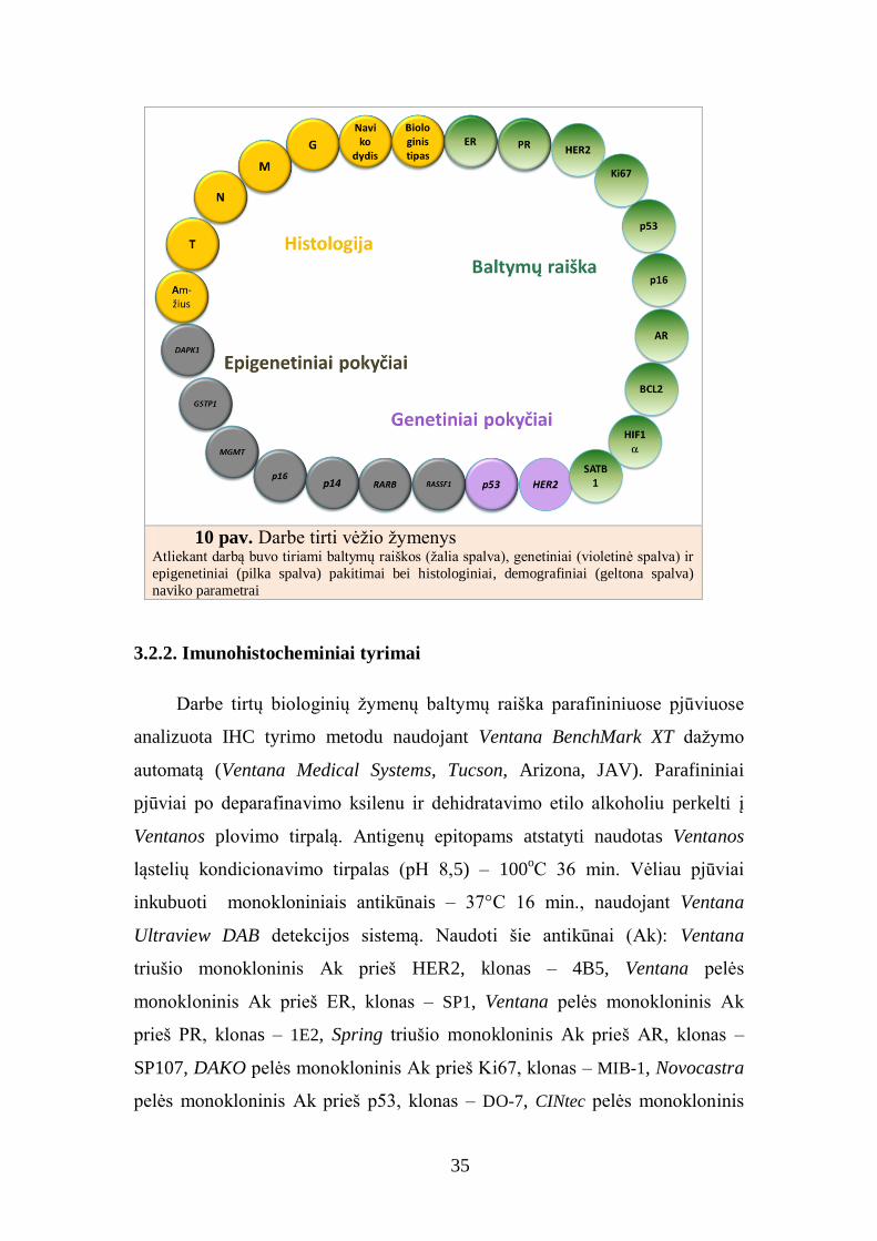
Darbe naudojant skaitmeninį vaizdo analizės metodą tirta 10 IHC
žymenų, apimant įprastinius, klinikinėje praktikoje naudojamus žymenis – ER,
PR, HER2 ir Ki67 bei mažiau tirtus – androgenų receptorių (AR), BCL2,
HIF1α, SATB1, p53 ir p16, žymenis. Siekiant geriau suprasti krūties navikų
molekulinius patogenezės mechanizmus darbe buvo tiriami ir genetiniai –
TP53, HER2 – bei epigenetiniai – p14, p16, ESR1, RARB, RASSF1, DAPK1,

10
GSTP1, MGMT – pakitimai genuose, susijusiuose su ląstelės atsparumu vėžiui,
su ląstelės ciklo ir kitų ląstelės atsakų valdymu.
Tyrimui atrinkti IHC, genetiniai ir epigenetiniai žymenys, kurių
padidėjusi raiška, DNR metilinimas ar mutacijos dažnai nustatomi krūties
navikuose, o genų koduojami baltymai atlieka svarbų vaidmenį sustiprinant
priešvėžinį atsaką ir valdant ląstelės dalijimąsi bei apoptozę.
MOKSLINIO DARBO AKTUALUMAS IR NAUJUMAS
Vykdant patikros/prevencijos programas bei diegiant naujas gydymo
strategijas, mažėja mirtingumas nuo krūties vėžio. Tačiau, apibendrintais
Jungtinių Amerikos Valstijų (JAV) ir Europos Sąjungos duomenimis, kasmet
JAV ir Europos Sąjungoje užregistruojama daugiau kaip 120 000 mirties atvejų
nuo krūties vėžio [22]. Ši situacija iš dalies paaiškinama tuo, kad vis dar
nepakanka informacijos apie krūties vėžio biologinį heterogeniškumą,
molekulinius pokyčius, jautrumą gydymui bei ląstelinę audinio sudėtį. Naviko
heterogeniškumo problematika ypač aktuali šiandien, siekiant individualizuoti
gydymą. Heterogeniškumo fenomenas iki šiol nepaaiškinamas svarbiais
klinikiniais parametrais (amžiumi, naviko dydžiu, limfmazgių būkle,
diferenciacijos laipsniu) ir biologiniais žymenimis (ER, PR, HER2), kurie
naudojami kasdieninėje praktikoje numatant ligos eigos prognozę bei
parenkant gydymą.
Per pastaruosius dešimt metų nustatyti ir analizuoti krūties vėžio tipai:
LA, LB, LB HER2+, HER2+ ir TN [1, 23]. Atlikti visuminiai genų raiškos
tyrimai parodė, kad praktikoje taikomų biologinių žymenų vertė ribota, kad yra
daugiau žymenų, svarbių krūties vėžio prognozei nustatyti bei atsakui į terapiją
numatyti [24-26]. Vienas pavyzdžių – naviko proliferacinio aktyvumo
įvertinimas. Nuo šių metų tarptautinė St Galleno Ekspertų kolegija prieš
parenkant sisteminę pirminio ankstyvojo krūties vėžio terapiją rekomenduoja
įvertinti naviko proliferacijos biologinius žymenis (pvz., Ki67 indeksą ar
mitozes) [18].

11
Mūsų darbe krūties naviko mėginiai pirmą kartą analizuoti naudojant
skirtingų tyrimo metodų kompleksą, apimantį genetinių bei epigenetinių
pažaidų ir baltymų raiškos analizės duomenis. Baltymų raiška tirta IHC tyrimo
metodu, genų raiška vertinta fluorescencinės in situ hibridizacijos (FISH)
metodu, genetinių ir epigenetinių pažaidų analizei pasirinkti šiuolaikiniai
tyrimo metodai – sekoskaitos, DNR grandinės konformacijos analizės,
metilinimui jautrios PGR ir bisulfitinės sekoskaitos. Atliktas išsamus
biologinių žymenų tyrimas leido palyginti svarbių, tačiau nepakankamai ištirtų
(p53, AR, p16, BCL2, SATB1, HIF1) IHC žymenų informatyvumą su esamų
prognozinių žymenų (ER, PR, HER2, Ki67) rodikliais. Ištirtas platus genetinių
ir epigenetinių krūties vėžio žymenų spektras.
Pirmą kartą IHC žymenų rinkinio, įvertinto skaitmeninės analizės būdu,
duomenys panaudoti faktorinės analizės metodu nustatyti jų variacijų vidinius
veiksnius, atskleidžiančius biologinius dėsningumus ir IHC žymenų bei jų
derinių informatyvumą. Šios analizės rezultatai leido naujai įvertinti
publikuotų krūties vėžio IHC žymenų bei jų derinių (p16, SATB1, HIF1,
Ki67/BCL2 ir kt.) informatyvumą.
Šio tyrimo metu mėginių atranka atlikta naudojant audinių mikrogardelių
technologiją. Ši technologija leidžia ne tik tiksliai atrinkti pažeistų žmogaus
audinių fragmentus, bet ir standartizuotai ištirti kelis šimtus mėginių, taupiai
naudojant tiriamuosius audinius ir reagentus.
Šis darbas taip pat yra vienas pirmųjų, kuriame IHC tyrimai atlikti
naudojant virtualiosios (skaitmeninės) mikroskopijos vaizdo analizės metodus.
Skaitmeninės vaizdų analizės technologijos pastaruoju metu atveria vis
daugiau pažangių galimybių kiekybiniais metodais išmatuoti biologinių
žymenų raišką tiriamuosiuose audiniuose [27-30]. Šios technologijos plačiai
pritaikomos ne tik mokslo tiriamuosiuose darbuose, bet ir atliekant
kasdieninius diagnostinius tyrimus. Skaitmeninės vaizdų analizės technologijos
pradedamos taikyti praktikoje standartizuojant diagnostinius testus (kokybės
kontrolės procedūros) ar vertinant tyrėjų variacijas (pagalba testui
interpretuoti). Pastaruoju metu siekiant individualizuoti gydymą, atsiranda vis
12
didesnis poreikis IHC biologinių žymenų raišką išmatuoti naudojant tikslius
kiekybinius analizės metodus ir gauti kiekybines vertes. Be skaitmeninės
patologijos vaizdų analizės algoritmų šio poreikio įgyvendinti nebuvo galima
[30-33]. Yra darbų, rodančių, kad patologo vertinimo rezultatai stipriai
koreliuoja su skaitmeninės analizės tyrimo rezultatais [34-36]. Nustatyta, kad
skaitmeninės mikroskopijos analizė leidžia ženkliai sumažinti tyrėjų variacijas
interpretuojant IHC tyrimų rezultatus [37]. Kitaip negu įprastas vizualus
audinių patologinis tyrimas, skaitmeninė tiriamųjų vaizdų analizė leidžia
objektyvizuoti ir kiekybiniais parametrais išmatuoti biologinių žymenų raišką
audinyje, nustatyti jų pasiskirstymą audinių bei ląstelių struktūrose.
Skaitmeninės analizės metodai leidžia įvertinti ir kitą svarbų biologinių
žymenų tyrimų aspektą – navikinio audinio heterogeniškumą. Navikinio
audinio heterogeniškumas skaitmeninės analizės bei stereologijos metodais
vertintas bendradarbiaujant su partneriais iš Prancūzijos (François Baclesse
Comprehensive Cancer Centre of Caen, Prancūzija). Disertacinis darbas
papildytas naujais duomenimis, išsamiai charakterizuojančiais krūties navikų
biologines savybes.
DARBO TIKSLAS
Taikant molekulinės ir skaitmeninės patologijos tyrimo metodus ištirti
pirminių krūties duktalinių karcinomų biologinių pokyčių spektrą, nustatytus
pokyčius susieti su klinikinėmis – patologinėmis ligos charakteristikomis.
DARBO UŽDAVINIAI
1. Pritaikyti skaitmeninės imunohistocheminių vaizdų analizės metodą
kiekybinei baltymų raiškos analizei atlikti ir įvertinti metodo patikimumą
rezultatus lyginant su patologo vizualaus vertinimo rezultatais.
2. Duktalinėse krūties karcinomose ištirti baltymų ER, PR, HER2, Ki67, p53,
AR, BCL2, p16, SATB1, HIF1 raišką, genų ESR1, DAPK1, GSTP1,

13
MGMT, p14, p16, RARB ir RASSF1 hipermetilinimo, geno HER2
amplifikacijos bei geno TP53 mutacijos dažnį.
3. Molekulinės ir skaitmeninės patologijos metodais nustatytą duktalinės
krūties karcinomos biologinius profilius susieti su klinikiniais rodikliais ir
nustatyti tirtų žymenų prognozinę vertę.
2. LITERATŪROS APŽVALGA
Krūties vėžys – heterogeniška liga, būdinga skirtingiems biologiniams
organizmams, pasireiškianti specifiniais morfologiniais ir
imunohistocheminiais radiniais bei įvairia klinikine eiga. Kovojant su šia liga
svarbų vaidmenį atlieka biologiniai žymenys, kurie padeda anksti aptikti vėžį,
stebėti ligos progresiją, numatyti prognozę bei individualizuoti sergančiųjų
krūties vėžiu gydymą. Biologiniai žymenys, susieti su klinikiniais-
patologiniais prognoziniais faktoriais, tokiais kaip naviko dydis,
diferenciacijos laipsnis, piktybinio proceso invazija į limfagysles bei
limfmazgių būklė, svarbūs ir skirstant ligonius į prognozines grupes. Iki šiol
pripažinti ir kasdieninėje praktikoje rekomenduojami tik keli ankstyvajam
krūties vėžiui gydyti svarbūs biologiniai žymenys, turintys patikimą
prognozinę vertę. Vienas svarbiausių predikcinių krūties vėžio žymenų – ER,
leidžiantys nustatyti ligos prognozę ir numatyti atsaką į hormonų terapiją. PR
taip pat plačiai praktikoje naudojamas žymuo, nors duomenų apie jo vertę
sukaupta mažiau. Neseniai šias gretas papildė HER2 biožymuo, leidžiantis
patikimai prognozuoti ligos eigą bei svarbus skiriant terapiją trastuzumabu
(HerceptinTM
).
Integruojant diagnostiką ir terapiją onkologijoje, taikinių terapija ir
individualizuota medicina šiomis dienomis išgyvena naują erą. Siekiant
pacientams parinkti tinkamiausią gydymą, farmacinių kompanijų kuriami
antikūnai prieš vėžį gydymui aprobuojami tik kartu naudojant klinikinį testą,
skirtą nustatyti konkretaus biologinio žymens raišką. Žinoma, kad Vakarų
pasaulyje taikomas sisteminis adjuvantinis gydymas leido sumažinti
14
mirtingumą nuo krūties vėžio. Tačiau taip pat žinoma, kad nemažai daliai
pacientų taikomas gydymas suteikia daugiau toksinio poveikio nei gydymo
naudos. Todėl labai svarbu turėti patikimus prognozinius veiksnius, kurie
padėtų pacientams parinkti tinkamiausią gydymą.
Visame pasaulyje krūties vėžys yra dažniausia moterų onkologinė liga ir
sudaro apie 23 procentus visų formų vėžio. Pasaulinės sveikatos organizacijos
(PSO) duomenimis, kasmet pasaulyje diagnozuojama daugiau nei 1 mln. naujų
krūties vėžio atvejų. Sergamumo krūties vėžiu vidurkis įvairiose pasaulio
šalyse yra 66,7 atvejo 100 000 moterų. Didžiausi sergamumo rodikliai būdingi
išsivysčiusioms šalims, mažiausi – Azijai ir Afrikai. Europoje šis skaičius
sudaro apie 350 000 naujų atvejų per metus. Pastarųjų metų Europos
sergamumo rodiklių analizė rodo, kad didžiausias sergamumas registruojamas
ekonomiškai išsivysčiusiose šalyse – Vakarų Europoje (Prancūzijoje,
Belgijoje, Šveicarijoje) ir Šiaurės Europoje (Danijoje, Švedijoje ir Jungtinėje
Karalystėje). Mažiausias sergamumas krūties vėžiu – Vidurio ir Rytų
Europoje. Lietuvoje sergamumo krūties vėžio rodikliai yra vieni mažiausių
Europoje [38], čia kasmet užregistruojama apie 1400 naujų krūties vėžio
atvejų [39, 40].
Dėl pradėtų taikyti atrankinių patikros programų, efektyvesnių
diagnostinių ir gydymo galimybių bei kintančių rizikos veiksnių pastaraisiais
metais gerokai padaugėjo diagnozuotų ankstyvosios stadijos krūties vėžio
atvejų [40]. Pažymima, kad daugumoje Europos šalių būdingas mirtingumo
nuo krūties vėžio augimo lėtėjimas. Šiaurės Europos šalyse sergamumo
didėjimas lydimas mirtingumo mažėjimo. Manoma, kad tai sietina su
efektyviai veikiančiomis mamografinės patikros programomis, efektyvesnėmis
diagnostinėmis galimybėmis, leidžančiomis identifikuoti mažiau agresyvių
formų bei neinvazinį vėžį, ir efektyvesniu gydymu.

15
2.1. Krūties vėžio rizikos veiksniai ir formavimosi molekuliniai
mechanizmai
Krūties vėžio rizikos veiksniai dažniausiai skirstomi į veiksnius, kuriuos
galima pakeisti (gyvensena, gimdymas, nežindymas, pakaitinė hormonų
terapija, estrogenų terapija, peroraliniai kontraceptikai) ir į veiksnius, kurių
pakeisti negalima (lytis, amžius, genetiniai rizikos veiksniai, ankstyvos
mėnesinės ir vėlyva menopauzė, patirtas vienos krūties vėžys) [41]. Bilimoria
savo darbe apibendrino svarbiausius krūties vėžio rizikos veiksnius [42].
Autorius pagal rizikos veiksnius išskyrė keturias krūties vėžio rizikos grupes:
krūties vėžys šeimoje (rizika tarp pirmo laipsnio giminių – 1,2–3,0),
menstruacinė istorija (santykinė rizika, kai menstruacijų pradžia <12 m. – 1,3,
menopauzės pradžia > 55 m. – 1,5–2,0), nėštumas (pirmasis gimdymas 25–29
m. amžiaus grupėje – 1,5, > 30 m. – 1,9, negimdžius – 3,0) ir gerybiniai krūtų
navikai (proliferuojantys – 1,9, proliferuojantys esant atipinei hiperplazijai –
4,4, lobulinė karcinoma in situ – 6,9–12,0).
Molekuliniai mechanizmai, sukeliantys krūties vėžio vystymąsi, nėra iki
galo suprasti, ypač susiję su estrogenų inicijuota kancerogeneze. Manoma, kad
krūties vėžiui būdinga nekontroliuojama ląstelių proliferacija ir/arba netipiniai
apoptozės reguliavimo mechanizmai. Vykstant minėtiems reiškiniams kaupiasi
genetinės pažaidos, aktyvinančios protoonkogenus bei gesinančios naviką
slopinančių genų raišką. Genetiniai pokyčiai gali būti įgimti (šeiminės
mutacijos) ar sąlygoti aplinkos kancerogenų (somatinės mutacijos). Pagal
klasikinį dviejų pakopų kancerogenezės modelį inicijuotose ląstelėse įvykusios
genomo pažaidos yra negrįžtamos, naviko progresavimas – kitas etapas, kurio
eiga priklauso nuo epigenetinių, potencialiai grįžtamų procesų.
Naujos tyrimų technologijos padėjo atskleisti vėžio formavimosi
mechanizmus. Šiandien žinoma, kad vėžinėms ląstelėms augant ir plintant į
gretimus audinius bei organus būdinga: savęs palaikymas augimo signalais,
nejautra ląstelės augimą slopinantiems signalams, nevaldomas dalijimasis,
apoptozės signalų blokavimas, ląstelės diferenciacijos susilpnėjimas,

16
angiogenezės stimuliavimas, gebėjimas plisti metastazėmis. Visos šios savybės
gali atsirasti ne tik dėl genetinių mutacijų (chromosomų aberacijų,
chromosomų skaičiaus pakitimų, taškinių mutacijų, delecijų), bet ir dėl
epigenetinių pakitimų – DNR ir histonų modifikacijos, keičiančios genų raišką
(hipermetilinimo ir hipometilinimo). Vykstant epigenetiniams
persitvarkymams ląstelėje, kai kurių genų sekose CpG salos tampa gausiai
metilintos, o kitos – demetilintos. Tokie epigenetinės pusiausvyros pakitimai
nulemia ląstelių piktybėjimą ir vėžio vystymąsi.
2.2. Krūties vėžio DNR metilinimo pakitimai
DNR metilinimas yra epigenetinis pakitimas, pakeičiantis genų raišką be
tiesioginių persitvarkymų DNR nukleotidų sekoje. Tai poreplikacinė,
kovalentinė DNR modifikacija, kurios metu metilo (CH3) grupė perkeliama
ant citozino C atomo penktoje pozicijoje. Reakciją katalizuoja DNR
metiltransferazės, metilo grupės donoras – S-adenozilmetioninas (1 pav.).
DNR metilinimas nėra atsitiktinis procesas. Žmogaus somatinėse
ląstelėse metilinti citozinai sudaro nedaug, apie 1 procentą visų DNR bazių
[43]. Dauguma metilintų citozinų yra 5‘-CG-3‘ sekoje arba CpG
dinukleotiduose. Maždaug pusė žmogaus genų promotorių sekų turi gausius
CpG regionus, kurie dar vadinami CpG salomis [44]. Dauguma šių genų,
1 pav. Citozino metilinimo reakcija

17
išskyrus imprintingo būdu valdomus ir neaktyvioje moterų X chromosomoje
esančius genus, išlieka nemetilinti. Už salos ribų esantys CpG dinukleotidai yra
gausiai metilinami [45]. Pažymima, kad metilinimas geno promotoriuje
visiškai užslopina geno raišką, o metilinimas transkribuojamame geno regione
gali veikti geno raišką įvairiai [46] (2 pav.).
Metilintos DNR sekos, sąveikaudamos su įvairiais baltymų kompleksais,
slopina genų raišką ir neleidžia prisijungti transkripcijos veiksniams prie
promotoriaus sekos. Baltymai, atrankiai besijungiantys su metilinta DNR,
skatina histonų deacetilazių ir histonų metiltransferazių jungimąsi, neaktyvaus
ir kompaktiško chromatino susidarymą.
Vienas dažniausių DNR metilinimo pakitimų, nustatomų navikuose, yra
CpG salų hipermetilinimas genų promotoriaus regione. Įvairių tipų navikų
tyrimai parodė, kad dažniausiai hipermetilinami naviką slopinančių genų
(NSG) promotoriai. Įvykus epigenetiniams pakitimams šiuose genuose
sutrikdoma ląstelės ciklo reguliacija, DNR reparacija, diferenciacija, apoptozė
ir kiti procesai, palaikantys normalų ląstelės funkcionavimą. Pagrindiniai NSG
grupės genai ir jų funkcijos ląstelėje išvardyti 2 lentelėje.
2 pav. CpG sekų pasiskirstymas
Apie pusę genų promotoriaus sekoje turi CpG salas, kurios paprastai yra
nemetilintos. Įvykus jų metilinimui užslopinama geno raiška [47]

18
2 lentelė. Pagrindiniai naviką slopinantys genai, inaktyvinami vykstant
hipermetilinimui
Funkcijos Genai, kurių promotoriai hipermetilinti
vėžinėse ląstelėse
Ląstelės ciklo kontrolė
DNR pažaidų reparacija
Kancerogenų metabolizmas
Apoptozė
Adhezija
Replikacinis senėjimas
Signalų perdavimas
Rb, p16, p15, p14, 14-3-3σ, Chfr
hMLH1, O6MGMT, GSTπ, BRCA1
GSTP1, CYP27B1
DAPK, casp-8, Apaf1, Xiap, p14
E-cad, APC, TIMP3
TERT
ER, PR, AR, RASSF1, RARB, VHL
Kai kurių NSG, pavyzdžiui, RASSF1 ir p16, promotorių hipermetilinimas
yra aptinkamas daugelyje įvairios lokalizacijos ir morfologijos navikų. Kitų
genų promotorių metilinimas vyksta specifiškai, tik tam tikrų tipų navikuose.
Plaučių karcinomose dažnai hipermetilinami DAPK1, MGMT, p16 genai,
storžarnės ir skrandžio navikuose – p14 ir APC genai, krūties ir kiaušidžių
karcinomose – GSTP1, BRCA1 ir p16 genai [48]. Pastebima, kad kiekvieno
tipo vėžys turi atrankiai hipermetilinamų NSG rinkinį arba DNR metilinimo
profilį. Pagal metilinimo profilį galima atskirti naviko histologinius tipus ir
apibūdinti turimą naviko mėginį. Taip pat pabrėžiama, kad metilinimo
žymenimis gali būti tik tokie genai, kurie normaliose ląstelėse yra nemetilinti
[49].
Hipometilinimas – tai DNR metilinimo praradimas. Šis metilinimo
pakitimas taip pat dažnas reiškinys navikinėse ląstelėse. Nustatyta, kad
navikinėje ląstelėje metilinto citozino kiekis yra 20–60 procentų mažesnis nei
nepakitusioje ląstelėje. Metilo grupės praradimas dažniausiai vyksta
hipometilinant geną koduojančias sekas ir intronus bei demetilinant
pasikartojančias DNR sekas, kurios sudaro 20–30 procentų žmogaus genomo.
Navikinėse ląstelėse globalus hipometilinimas gali sukelti chromosomų
nestabilumą, judriųjų genomo elementų reaktyvinimą ir imprintingo praradimą.
Taip pat navikuose dažnai aptinkamas hipometilinimas chromosomos
centromeros regione, ir tai gali sukelti aneuploidiją [49]. Yra duomenų, kad

19
hipometilinimas suaktyvina onkogenus cMYC ir HRAS [46]. Manoma, kad
DNR hipometilinimas vėžinėse ląstelėse skatina naviko invaziją ir metastazes
[50]. Svarbu tai, kad hipermetilinimo ir hipometilinimo reiškiniai vyksta kartu
toje pačioje navikinėje ląstelėje [50, 51].
2.3. Pagrindiniai krūties vėžio žymenys
2.3.1. Hormonų receptoriai
Hormonų receptorių (HR) svarba krūties vėžiui vystytis nustatyta daugiau
kaip prieš 40 metų. Šiandien žinoma, kad krūties vėžio vystymasis priklauso
nuo estrogenų ir/ar progesteronų kiekio ir šį kiekį reguliuoja estrogenų ir
progesteronų receptoriai.
ER ir PR priklauso branduolio receptorių šeimai, į kurią įeina ir
androgenų receptoriai (AR). Šie receptoriai yra ląstelių branduolyje, kur veikia
kaip transkripcijos veiksniai, atlikdami svarbų vaidmenį endokrininių signalų
sistemoje. Į ląstelės branduolį difuzijos būdu patekęs estrogenas susijungia su
ląstelės branduolyje esančiu ER suformuodamas ligando-receptoriaus
kompleksą, galintį sąveikauti su reguliacinėmis genų sekomis [52]. Estrogenas,
veikdamas per ER, reguliuoja epitelinio audinio augimą ir diferenciaciją,
aktyvina ląstelių proliferaciją bei reguliuoja kitų genų raišką (pS2, PR ir
BCL2) [53]. Progesteronui sąveikaujant su PR, sustiprinamas mitozinis
veikimas ir taip pat aktyvinama ląstelių proliferacija [54].
Estrogenų receptoriai, kaip branduolio transkripcijos veiksniai,
dalyvauja krūties audinio vystymosi, augimo, diferenciacijos ir navikų
formavimosi procesuose. Žinomos dvi ER izoformos: ER-alfa (ERα) ir ER-
beta (ER). Nors šie receptoriai panašūs daugeliu aspektų, tačiau skiriasi
biologinėmis funkcijomis ir raiškos pobūdžiu. ERα ir ERβ koduoja du skirtingi
genai, ESR1 ir ESR2 atitinkamai [55]. Literatūros duomenimis, ERα siejamas
su krūties vėžio vystymusi. Daugiau nei 70 procentų diagnozuojamų
ankstyvųjų krūties navikų yra ERα teigiami [56] ir vėžio progresijos metu ERα
raiška didėja [57]. ERβ vaidmuo krūties vėžiui išsivystyti kol kas nėra aiškus

20
[57, 58]. Padidėjusi ERα raiška pripažinta prognoziniu ir prediktyviniu krūties
vėžio žymeniu [59], siejama su aukštesniu vėžio diferenciacijos laipsniu ir
geresnėmis ligos prognozėmis [52, 60].
Progesteronų receptorių raiškos įtaka krūties vėžiui formuotis kol kas
diskutuotina [61]. Nustatyta, kad ER teigiami, tačiau PR neigiami krūties
navikai sąlygoja blogesnę ligos prognozę negu krūties navikai, kurie turi ER ir
PR raišką [62, 63]. Vis dėlto prognozinė PR vertė, literatūros duomenimis,
išlieka prieštaringa. Yra tyrimų, kuriuos atlikę tyrėjai teigia, kad prognozinė
PR vertė numatant krūties vėžio gydymą hormonų terapija mažesnė nei ER,
tačiau kiti tyrėjai teigia, kad PR suteikia papildomos informacijos,
nepriklausomos nuo ER prognozinių verčių [64-66]. Navike nustatyta PR
raiška informuoja, kad ER reguliuojami signalų perdavimo keliai
funkciuonuoja, veikla nesutrikusi. Taip pat nustatyta, kad tiek ER, tiek PR
raiška priešingai koreliuoja su histologiniu diferenciacijos laipsniu,
proliferacijos indeksu, HER2 ir p53.
Šiandien ER ir PR raiška krūties audiniuose nulemia pirminį krūties vėžio
gydymo taktikos parinkimą. Tik įvertinus hormonų receptorių raišką
parenkamas gydymas taikant adjuvantinę hormonų terapiją (tamoksifenas),
aromatazės inhibitorius bei terapiją selektyviais estrogenų receptorių
moduliatoriais. HR raiška – svarbus rodiklis, apibrėžiantis naviko atsaką į
hormonų terapiją, tai vienas svarbiausių krūties vėžio gydymo parametrų.
Metastazuojančio krūties vėžio atveju hormonų terapija turi nemažai
privalumų, palyginti su citotoksine chemoterapija. Esant naujai diagnozuotai
metastazuojančiai ligai, nuo 30 iki 40 procentų atvejų gaunamas teigiamas
atsakas į hormoninį gydymą, nemaža dalis ligonių turi kliniškai reikšmingą
ligos nesant recidyvo laikotarpį [20, 67-71].
Androgenų receptorių raiška dažnai nustatoma krūties navikuose ir yra
susijusi su mažesniu naviko piktybiškumu ir palankesniu naviko
diferenciacijos laipsniu. Atlikta nemažai tyrimų, nagrinėjančių AR įtaką krūties
vėžio genezėje. Manoma, kad AR gali būti nepriklausomas prognozinis
faktorius ar žymuo, svarbus parenkant trejopai neigiamų (ER–, PR–, HER2–)

21
navikų gydymą, tačiau kol kas svarių duomenų, pagrindžiančių šias hipotezes,
nėra [72-79].
Hormonų receptoriams nustatyti audiniuose taikomi du pagrindiniai
tyrimo metodai: biocheminis ligando prijungimo metodas – šaldytų mėginių
tyrimams ir IHC metodas – mėginių, paruoštų parafino metodu, tyrimams. Per
pastaruosius du dešimtmečius krūties vėžio diagnostikai pasirinktas IHC
tyrimo metodas, kaip patogesnis, jautresnis ir patikimesnis. Siekiant pagerinti
pacientų atranką hormonų terapijai, ASCO (angl. American Society of Clinical
Oncology) ir CAP (angl. College of American Pathologists) nesenai apibrėžė
ER/PR tyrimo gaires, kuriose rekomenduoja hormonų raišką tirti IHC tyrimo
metodu [80, 81].
2.3.2. HER2 onkogenas
HER2 – onkogenas, koduojantis ląstelės 185 kD transmembraninį
receptorių, turintį ir tirozino kinazės aktyvumą [82]. HER2 receptorius
priklauso epidermio augimo faktoriaus receptorių šeimai (EGFR), kuri
aktyvina tarpląstelinių signalų perdavimo kelius, kontroliuojančius epitelio
ląstelių augimą, diferenciaciją [83, 84] ir angiogenezę [46, 85]. Anksčiau
epidermio augimo faktoriaus receptorius 2 buvo vadinamas NEU, NGL,
HER2, TKR1, HER-2, HER2/neu arba cerbB-2. 2007 metais pasiūlyta apsistoti
ties dažniausiai vartotu epidermio augimo faktoriaus receptoriaus 2
pavadinimu – HER2 [86].
HER2 geno amplifikacija ar jo produkto baltymo hiperekspresija ląstelės
membranoje pasireiškia nuo 18 iki 20 procentų krūties vėžio atvejų [87-89] ir
tai susiję su bloga ligos prognoze bei padidėjusia rizika ligai atsinaujinti [87].
HER2 – ne tik prognozinis agresyvios ligos žymuo, bet ir svarbus veiksnys
kliniškai parenkant specifinę terapiją. HER2 receptorius – molekulinis taikinys
monokloniniam antikūnui trastuzumabui (Herceptin; Genentech, South San
Francisco, JAV), adjuvantui, kuris reikšmingai pagerina atsaką į ligos
progresavimą bei prailgina pacientų išgyvenimą, palyginti su išgyvenimu

22
pacientų, kuriems taikytas vien tik chemoterapinis gydymas [90, 91]. HER2
terapijai parenkami pacientai, kuriems nustatyta HER2 geno hiperekspresija
arba geno amplifikacija.
Nuo 2001 metų naujai diagnozuoto ir metastazuojančio krūties vėžio
HER2 raišką rutiniškai nustatyti pasiūlė net kelios organizacijos: ASCO, NCCN
(angl. Nacional Comprehensive Cancer Network) bei NACB (angl. Nacional
Academy of Clinical Biochemistry) [92-94]. 2007 metais jungtinis ekspertų
komitetas, atstovaujantis ASCO ir CAP, paskelbė rekomendacijas, kuriose
išanalizuoti konkretūs techniniai ir analitiniai HER2 tyrimo aspektai [86].
Vadovaujantis šiomis rekomendacijomis bei 2011 St. Galleno Ekspertų grupės
sutarimu [95], diagnostikoje rekomenduojamas IHC HER2 vertinimo metodas,
kuriuo nustatoma baltymo raiška ląstelės membranoje. IHC būdu nustatyti
paribiniai atvejai testuojami genų raiškos lygmeniu, atlikus FISH. FISH
tyrimas dažnai vadinamas „aukso standartu“, kuris leidžia kiekybiniais
parametrais išmatuoti analizuojamo geno raišką [86, 96, 97].
2.3.3. Ląstelių proliferacijos ir kiti imunohistocheminiai žymenys
Vėžinių ląstelių proliferacija yra svarbus navikinio audinio prognozinis
požymis. Padidėjusi naviko proliferacija koreliuoja su blogesne negydytų
pacientų ligos prognoze, aukštu histologiniu diferenciacijos laipsniu, dažnai su
hormonų receptorių raiškos nebuvimu, didesniu naviko dydžiu bei pažeistais
limfmazgiais [98].
Naviko proliferacija krūties navikuose gali būti vertinama keliais
skirtingais tyrimo metodais: mitozių skaičiavimu, timidino indekso ir S fazės
frakcijos nustatymu, tėkmės citometrijos bei IHC tyrimu, naudojant antikūnus
prieš proliferuojančių ląstelių antigenus. Geriausiai ląstelių proliferaciją
atspindi Ki67 žymuo, kuris išryškinamas IHC metodu su MIB-1 antikūnu
formaline fiksuotuose parafinu impregnuotuose mėginių pjūviuose.
Literatūroje pabrėžiama, kad Ki67 raiška ląstelės ciklo metu nevienoda [59,
99]. Nustatyta, kad raiška vyksta G1, S ir G2 fazių metu, tačiau nevyksta

23
ląstelės ciklo ramybės fazėje G0. Nepakitusiame krūties audinyje Ki67 raiška
yra labai silpna (< 3 % ląstelių) [99], vėžiniame audinyje labai padidėja.
Ki67 proliferacijos indeksas tiksliai išmatuojamas kiekybiniais
stereologijos ir skaitmeninės analizės tyrimo metodais, skaičiuojant ląsteles
nustatytame analizės plote [100]. Daugelis klinikinių tyrimų parodė, kad
egzistuoja stiprus Ki67 proliferacijos indekso ryšys su ligos prognoze,
koreliacija su išgyvenimo nesant recidyvo ir bendrojo išgyvenimo laikotarpiu
[101, 102]:
naudojant Ki67 slenkstines vertes nuo 3,5 iki 34 procentų nustatyta
Ki67 teigiama vertė susijusi su reikšmingai didesne recidyvo rizika
tiek esant invazijai į limfmazgius, tiek nesant invazijos;
taip pat nustatytas reikšmingas ryšys tarp Ki67 teigiamos vertės ir
trumpesnio išgyvenimo tiek esant naviko invazijai į limfmazgius, tiek
jo nesant.
2007 metais ASCO/CAP išleistose krūties vėžio tyrimo gairėse
rekomenduojama Ki67 indeksą nustatyti visais krūties vėžio tyrimo atvejais
[61].
Baltymas SATB1 (angl. special AT-rich sequence-binding protein-1)
dažnai vadinamas „genomo organizatoriumi“, galinčiu koordinuoti daugiau
kaip 10 procentų visų genų veiklą. Spėjama, kad šis baltymas gali atlikti
reikšmingą vaidmenį vystantis ir progresuojant krūties vėžiui. 2008 metais Han
su bendraautoriais teigė, kad SATB1 baltymo raiška padidėja naviko genezės
metu, keisdama krūties vėžio ląstelių genų ekspresijos profilį, kuris palaiko
agresyvių ląstelių fenotipą, skatinantį naviko augimą ir metastazavimą [103].
2010 metais Iorns su tyrėjų grupe, kitaip negu ankstesni tyrėjai, nustatė, kad
SATB1 raiška neskatina krūties vėžio progresavimo ir nėra susijusi su krūties
vėžio eiga [103]. Šiuo metu tyrėjų nuomonė apie SATB1 vaidmenį vystantis
krūties vėžiui gana kontroversiška.
HIF1 (angl. hypoxia-inducible factor 1, alpha subunit), transkripcijos
veiksnys, kontroliuojantis ląstelės atsaką į hipoksiją. Indukuojamo subvieneto
HIF1 raiška ženkliai padidėjusi daugelyje navikų, tarp jų ir krūties. Dėl

24
baltymo veiklos suntensyvėja vaskuliarizacija naviko zonoje, padidėjęs
baltymo kiekis koreliuoja su naviko agresyvumu, kontroliuoja ląstelės atsaką į
hipoksiją. Indukuojamo subvieneto HIF1 raiška ženkliai padidėjusi daugelyje
navikų.
BCL2 (angl. B-cell lymphoma 2) šeimos baltymai reguliuoja
programuotos ląstelių mirties arba apoptozės procesus. BCL2 genas koduoja
mitochondrijų membranos baltymą, kuris palaiko mitochondrijų membranos
integralumą ir slopina ląstelių (pvz., limfocitų) apoptozinę mirtį. Padidėjusi
baltymo raiška nustatoma daugelyje navikų, tarp jų ir krūties navikuose.
Baltymo kiekio padidėjimas slopina natūralų piktybėjančių ląstelių
pasišalinimą apoptozės būdu, skatina vėžio progresiją, naviko didėjimą. Yra
žinoma, kad BCL2 baltymo raiška susijusi su žemesniu naviko diferenciacijos
laipsniu bei lėčiau proliferuojančiais, didesnės ER raiškos navikais [104, 105].
Atliktuose tyrimuose nustatyta, kad BCL2 baltymo raiška koreliuoja su
ilgesniu pacientų, kuriems būdinga ER raiška, išgyvenimo nesant ligos
atkryčio laikotarpiu ir gali būti nepriklausomu ankstyvųjų krūties vėžio stadijų,
geresnės prognozės žymeniu [106].
2.2.4. Naviką slopinantys genai
Naviką slopinančio geno TP53 mutacijos ar pakitusi baltymo p53 (angl.
tumor protein 53) raiška aptinkama nuo 20 iki 50 procentų žmogaus krūties
vėžio atvejų [107, 108]. Prognozinė ir predikcinė p53 vertė šiandien tyrėjų dar
vertinama prieštaringai. Dalis tyrimų parodė, kad padidėjusi baltymo p53
raiška (išmatuota IHC tyrimo metodu), mutacijos ir delecijos TP53 gene, yra
stiprus nepriklausomas prognozinis veiksnys, reiškiantis trumpesnį išgyvenimo
be recidyvo bei bendrojo išgyvenimo laikotarpį. Manoma, kad IHC tyrimo
metodu nustatytos p53 baltymo sankaupos audinyje ne tik koreliuoja su geno
TP53 mutacija, bet ir yra patikimas nepriklausomas trumpesnio išgyvenimo
žymuo [109-115]. Tačiau yra tyrimų, kurių metu tokios sąsajos nerasta [109,
116, 117].

25
p53 baltymą koduoja TP53 genas, esantis 17-os chromosomos
trumpajame petyje (17p13.1). Tai transkripcijos faktorius, reguliuojantis
ląstelės ciklą, apoptozę ir DNR pažaidų taisymą. Baltymas р53 sintetinamas
visose organizmo ląstelėse. Nemutavusios būklės p53 veikia kaip ląstelės
piktybėjimo supresorius. p53 yra universalus natūralaus ląstelės sunykimo,
arba apoptozės, reguliatorius, be to, jis dalyvauja ir pažeistos DNR atkūrimo
procese. Normaliose, streso nepažeistose ląstelėse p53 baltymo kiekis ir
aktyvumas labai mažas. Tuo tarpu streso pažeistose ląstelėse p53 baltymas
suaktyvinamas.
ASCO tyrėjų grupė mano, jog mažai tikėtina, kad nustatant baltymo p53
raišką IHC tyrimo metodu bus gaunami pakankamai tikslūs rezultatai,
leidžiantys numatyti klinikos eigą. Patikimesnis būdas p53 būklei įvertinti yra
TP53 geno sekoskaita [61].
p16INK4a
/p14ARF
lokusas, esantis 9p21 chromosomoje, koduoja du
struktūriškai skirtingus baltymus, kurie susidaro dėl alternatyvaus rėmelio
nuskaitymo. Abu genai (p16INK4a
ir p14ARF
) koduoja naviką slopinančius
baltymus, dalyvaujančius valdant ląstelės ciklą ir reguliuojančius Rb bei p53
signalinius kelius.
Baltymas p14ARF
dalyvauja p53 signaliniame kelyje ir yra svarbus G1 ir
G2 ląstelės ciklo fazių eigai. p14ARF
batymas palaiko vėžio supresoriaus p53
stabilumą, kuris, esant onkogeniniams signalams ar kitoms pažaidoms, stabdo
ląstelės ciklą ir inicijuoja apoptozę [118]. Nesant aktyvaus p14, naviką
slopinančio baltymo p53 stabilumas smarkiai sumažėja.
Genas p16 koduoja baltymą, nuo ciklinų priklausomos kinazės (Cdk)
inhibitorių, kuris dalyvauja baltymo Rb atsako kelyje. p16INK4a
baltymas
jungiasi prie Cdk4 ir Cdk6 ir inhibuoja fermentinio komplekso Cdk4-
Cdk6/ciklino D susidarymą G1 ląstelės ciklo fazėje. Šis kompleksas svarbus
Rb fosforilinimo procesui. Nesusidarius šiam kompleksui, nevyksta Rb
fosforilinimas ir ląstelės ciklas sulaikomas G1/S fazėje. Ląstelėse, neturinčiose
aktyvaus p16, ląstelės dalijimosi ciklas nestabdomas G1/S fazėje ir tampa

26
nekontroliuojamas. Taigi p16 baltymas palaiko Rb baltymą nefosforilintos
būklės inhibuodamas ląstelės ciklo progresiją iš G1 į S stadiją.
Genai p14 ir p16 dažnai inaktyvinami įvairiausiuose žmogaus navikuose.
Vienas iš inaktyvinimo būdų yra promotoriaus regione esančių CpG salų
hipermetilinimas. Literatūroje pateikiami geno p14 hipermetilinimo dažniai
krūties karcinomose svyruoja nuo 28 iki 47 % [119, 120], tačiau yra autorių,
kurie geno p14 hipermetilinimo nenustatė [121]. Geno p16 raiškos
sumažėjimas koreliuoja su sunkia ligos eiga ir mažesniu išgyvenamumu. Geno
hipermetilinimo dažniai nustatomi krūties karcinomose, įvairiose publikacijose
svyruoja nuo 26 iki 44 % [122, 123].
RARB genas (angl. retinoic acid receptor beta) – tai retinoinės rūgšties
receptoriaus β genas, esantis 3p24 chromosomoje ir priklausantis branduolio
receptorių RAR klasei. RAR branduolio receptorių klasei priklauso trys
skirtingi genai (α, β ir γ). Tai transkripcijos veiksniai, kurių raiška vyksta
skirtingais organizmo vystymosi etapais. Manoma, kad RARB yra svarbus
epitelinių ląstelių augimo reguliavimui bei kancerogenezės procesui [124].
RAR šeimos receptoriai būna ląstelės citozolyje ir branduolyje. Šios šeimos
receptorių ligandas yra retinoinė rūgštis, vitamino A biologiškai aktyvi forma,
kuri svarbi ląstelei dalytis, diferencijuotis ir perduoti morfogenezės signalams
[125].
Susilpnėjusi RARB geno raiška vėžinėse ląstelėse yra siejama su
promotoriaus regione esančių CpG salų metilinimo pakitimais. Geno
hipermetilinimas aptinkamas krūties, plaučių, prostatos, gaubtinės žarnos,
skrandžio, gimdos kaklelio bei šlapimo pūslės navikuose [126]. Literatūros
duomenimis, geno RARB hipermetilinimo dažnis krūties navikuose svyruoja
nuo 10 iki 38 procentų [122, 127-129].
RASSF1 genas (angl. Ras association (RalGDS/AF-6) domain family
member 1) – RAS sąveikos domeną turinčių baltymų šeimos narys. RASSF1
genas yra 3p21.3 chromosomoje, jo produktas RASSF1A baltymas dalyvauja
ląstelės ciklo valdymo, apoptozės reguliacijos ir mikrovamzdelių stabilizacijos
procesuose.

27
RASSF1A yra mitozės inhibitorius, sustabdantis ląstelės ciklą
metafazėje. Tai svarbu tam, kad chromosomos taisyklingai išsirikiuotų
metafazinėje plokštelėje [55]. RASSF1A taip pat dalyvauja apoptozės
reguliacijoje, sąveikaudamas su vienu iš Ras (K-Ras) šeimos baltymų, kuris
svarbus inicijuojant apoptozę [130].
Epigenetinis geno RASSF1 nuslopinimas yra vienas iš dažniausių
molekulinių pakitimų navikuose. Šio geno promotoriuje esančių CpG salų
hipermetilinimas nustatytas įvairių tipų navikuose, tarp jų plaučių, prostatos,
inkstų, neuroblastomos, gliomos, krūties. Genų hipermetilinimo dažniai,
nustatomi krūties karcinomose, įvairiose publikacijose svyruoja nuo 42 iki
85% [127, 129, 131]. Siūloma metilintą RASSF1 geną laikyti biologiniu
žymeniu diagnozuojant ankstyvuosius vėžinius pakitimus žmogaus organizme
[132].
DAPK1 genas (angl. Death-associated protein kinase) koduoja
serino/treonino kinazę. Genas yra 9 chromosomos trumpajame petyje (9q
34.1), koduojamas produktas DAPK dalyvauja apoptozės procese. DAPK yra
kalcio/kalmodulino reguliuojama baltymų kinazė. Baltymo struktūroje yra
ankirino pasikartojantis domenas ir mirties domenas, kurie padeda DAPK
sąveikauti su kitais baltymais [133]. Cohen [134] parodė, kad DAPK dalyvauja
TNFα (angl. tumor necrosis factor) ir Fas receptorių indukuotoje ląstelių
apoptozėje. Nustatytas antimetastazinis [134] ir naviką slopinantis DAPK1
aktyvumas [135]. Hipermetilinimas DAPK1 geno promotoriaus sekoje
koreliuoja su blogomis prognozėmis daugelio tipų navikuose: plaučių, inkstų,
storžarnės, gimdos kaklelio. Literatūros duomenimis, krūties navikuose
hipermetilinimas varijuoja nuo 16 [136]) iki 50 procentų [137].
GSTP1 genas (angl. glutathione S-transferase pi 1) koduoja
kenksmingas medžiagas detoksikuojantį fermentą glutation-S-transferazę π
(GST-π). Genas yra 11 chromosomos trumpajame petyje (11q 13). GST-π
raiška nepakituosiuose audiniuose intensyviausiai vyksta šlapimtakių,
virškinimo ir kvėpavimo takuose. Padidėjęs GST-π kiekis siejamas su tam
tikro kancerogeno poveikiu [138], o geno GSTP1 raiškos nebuvimas padidina

28
vėžio išsivystymo tikimybę [139]. Be to, užslopinta geno raiška turi įtakos
vėžinių ląstelių jautrumui chemoterapiniams vaistams [140].
Metilinimas geno GSTP1 CpG salose nutatytas kelių tipų navikiniuose
audiniuose, tarp jų krūties (nuo 13 iki 31 procentų [128, 140, 141]) ir kepenų, o
prostatos navikuose nustatytas didžiausias šio geno hipermetilinimo dažnis
(apie 90 procentų).
MGMT genas (angl. O-6-methylguanine-DNA methyltransferase)
koduoja DNR reparacijos fermentą O6-metilguanino DNR metiltransferazę
(EC2.1.1.64). Genas yra 10 chromosomos trumpajame petyje (10q 26). MGMT
pašalina mutageninius ir citotoksinius aduktus nuo guanino nukleotido šeštoje
pozicijoje esančio deguonies ir apsaugo ląstelę nuo mutacijų kaupimosi.
MGMT raiška vyksta visuose nepakitusiuose žmogaus audiniuose, tačiau
baltymo lygis varijuoja tarp organų ir tarp individų.
Žmogaus navikuose MGMT geno mutacijos ar delecijos retos.
Dažniausiai MGMT genas užslopinamas dėl epigenetinių pakitimų
promotoriaus regione [142]. Dėl nuslopintos MGMT raiškos padidėja svarbių
genų (TP53, K-RAS) mutacijų tikimybė. Be to, navikai, kuriuose MGMT yra
metilintas, jautresni terapiniam alkilinančių priešvėžinių vaistų veikimui [143].
Krūties navikuose šio geno epigenetiniai pakitimai tyrinėti mažai. Literatūroje
pateikiami MGMT hipermetilinimo dažniai krūties navikuose – 8,4 % [121] ir
10 % [144].
Tyrimui atrinkti aukščiau aprašyti IHC, genetiniai ir epigenetiniai
žymenys, kurių padidėjusi raiška, mutacijos ar DNR metilinimas dažnai
nustatomi krūties navikuose. Darbe planuota naujai atsiradusiomis žymenų
raiškos analizės priemonėmis išanalizuoti plačiai klinikinėje praktikoje
naudojamus bei naujus, menkai tyrinėtus žymenis.
29
3. TIRIAMIEJI IR TYRIMO METODAI
3.1. Tiriamųjų atranka ir tyrimo eiga
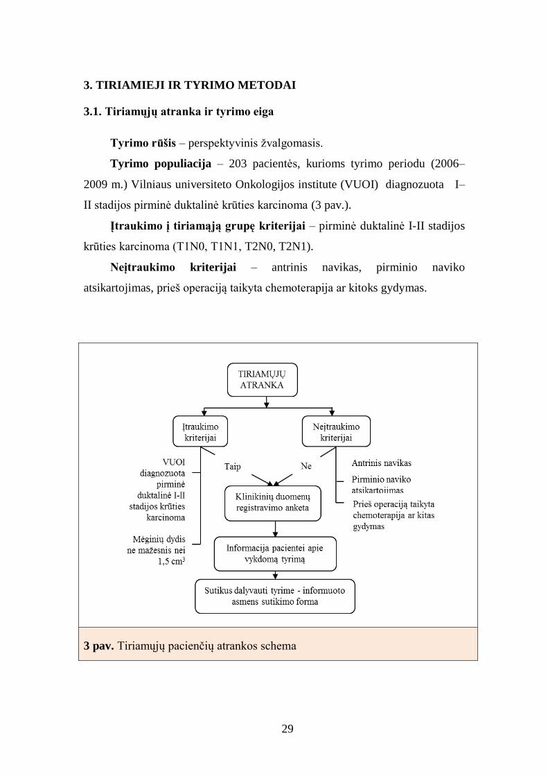
Tyrimo rūšis – perspektyvinis žvalgomasis.
Tyrimo populiacija – 203 pacientės, kurioms tyrimo periodu (2006–
2009 m.) Vilniaus universiteto Onkologijos institute (VUOI) diagnozuota I–
II stadijos pirminė duktalinė krūties karcinoma (3 pav.).
Įtraukimo į tiriamąją grupę kriterijai – pirminė duktalinė I-II stadijos
krūties karcinoma (T1N0, T1N1, T2N0, T2N1).
Neįtraukimo kriterijai – antrinis navikas, pirminio naviko
atsikartojimas, prieš operaciją taikyta chemoterapija ar kitoks gydymas.
3 pav. Tiriamųjų pacienčių atrankos schema

30
Kaip jau buvo minėta, tiriamąją grupę sudarė 203 pacientės, sergančios
ankstyvųjų stadijų (T1N0, T1N1, T2N0, T2N1) krūties duktaline karcinoma ir
2007–2009 metais gydytos VUOI klinikoje. Vidutinis pacienčių amžius –56
metai (±12,9), jauniausia pacientė 27 metų, vyriausia – 87 metų (4 pav.).
4 pav. Tiriamosios grupės pacienčių pasiskirstymas pagal amžių
Pagal medianą pacientės suskirstytos į dvi amžiaus grupes (27–55 m.,
56–87 m.). Tiriamosios grupės pacienčių pasiskirstymas pagal amžiaus grupes,
TNM klasifikaciją ir diferenciacijos laipsnį pateiktas 3 lentelėje. T1 ir T2
grupes sudarė panašus pacienčių skaičius: atitinkamai 107 (52,7%) ir 96
(47,3%). Pagal sritinių limfmazgių pažeidimo metastazėmis laipsnį pusė
pacienčių sudarė grupę be pažeidimų – N0 – 104 (52,5%), kita pusė – su
pažeidimais: N1 – 64 (32,3%), N2 – 21 (10,6%) ir N3 – 9 (4,6%) asmenys.
Pagal naviko diferenciacijos laipsnį daugumai pacienčių nustatyta vidutinė
(G2) ar bloga (G3) naviko diferenciacija (atitinkamai 38,9% ir 41,4%).
Pacienčių amžius
%

31
Tyrimo eiga
1. Pacienčių atranka (VUOI diagnozavus pirminę duktalinę krūties
karcinomą).
2. Mėginių atranka (Valstybiniame patologijos centre (VPC) įvertinus
esamą naviko dydį, histologiją).
3. Atrinktų mėginių šaldymas, fiksavimas, impregnavimas parafinu.
4. Audinių mikrogardelių (TMA) ruošimas.
5. IHC, FISH reakcijų atlikimas.
6. IHC mikropreparatų skenavimas.
7. Skenuotų vaizdų vizualus patologo vertinimas.
8. Skenuotų vaizdų skaitmeninio vaizdo analizė.
9. Naviko DNR išskyrimas. Genetinių ir epigenetinių pažaidų analizė.
10. Sukauptų duomenų analizė statistiniais tyrimo metodais (5 pav.).
3 lentelė. Tirtųjų pacienčių pasiskirstymas pagal amžių, TNM
klasifikaciją ir naviko diferenciacijos laipsnį
Amžiaus grupė Dažnis Procentas
1 (27-55 m.) 100 49,3
2 (56-87 m.) 103 50,7
T
1 107 52,7
2 96 47,3
N
0 104 52,5
1 64 32,3
2 21 10,6
3 9 4,6
M
0 203 100
G
1 40 19,7
2 79 38,9
3 84 41,4 T – pirminis navikas (T1 – navikas ≤ 2 cm, T2 - navikas > 2 cm, < 5 cm); N –
regioninės metastazės (N0 – regioniniai limfmazgiai nepažeisti, N1 - metastazės 1-3 regioniniuose limfmazgiuose, N2 - metastazės 4-9 regioniniuose limfmazgiuose, N3 -
metastazės 10 ir daugiau regioninių limfmazgių); M – atokiosios metastazės (M0 –
atokiųjų metastazių nėra); G – naviko diferenciacijos laipsnis (G1 – gerai diferencijuotas,
G2 – vidutiniškai diferencijuotas, G3 – blogai diferencijuotas).

32
3.2. Tyrimo metodai
3.2.1. Mėginių paruošimas
Naviko mėginiai atrinkti iš 203 pacienčių, VUOI diagnozavus ir VPC
morfologiškai patvirtinus ankstyvąją, t.y. I–II stadijos (T1N0, T1N1, T2N0,
T2N1) krūties duktalinę karcinomą. Mėginiai rinkti 2006–2009 metų
laikotarpiu.
Audinių mikrogardelės ruoštos iš 10 procentų buferinio formalino tirpale
fiksuotų, parafinu impregnuotų audinių blokų. Audinių blokus mikrogardelėms
atrinko gydytojas patologas, vertindamas hematoksilinu ir eozinu (H/E)
dažytus audinių pjūvius. Atrinktų mėginių H/E mikropreparatai skenuoti 20x
padidinti Aperio Scan Scope GL, skeneriu (Aperio Technologies, Vista, CA,
JAV). Skenuotame viso pjūvio audinio vaizde patologas pažymėjo navikinio
audinio vietas (6 pav). Žymėti vaizdai konvertuoti į Mirax MViewMRXS
5 pav. Tyrimo eiga

33
formatą, kuris naudotas konstruojant audinių mikrogardeles pusiau automatiniu
audinių mikrogardelių ruošimo aparatu (3DHISTECH, TMA Master,
Budapeštas, Vengrija). Iš žymėtų naviko zonų imti šeši 1 mm diametro
mėginiai: 4 – TMA IHC analizei, 2 – genetiniams ir epigenetiniams tyrimams
(7 pav.).
6 pav. Hematoksilino-eozino histocheminiu metodu dažyto mikropreparato
skenuotas vaizdas, padidintas 20 x Raudona spalva pažymėtos navikinio audinio zonos, iš kurių paimti mėginiai tyrimui
7 pav. Žymėtas parafino blokas audinių
mikrogardelių ruošimo automate Iš pažymėtų vietų konstruojant audinių mikrogardeles imti
tiriamieji naviko mėginiai

34
Iš 203 pacienčių mėginių paruošta 16 audinių mikrogardelių, kuriose
sudėti 812 tiriamieji naviko mėginiai. Tyrimui imti 6 kiekvienos pacientės
naviko mėginiai (8 pav.).
IHC analizei atlikti pjauti 3 mikronų storio pjūviai (9 pav.), FISH
tyrimams – 4 mikronų storio pjūviai. 105 pacienčių 1 mm išgręžti navikinio
audinio fragmentai atrinkti genetinei ir epigenetinei analizei. Darbe analizuoti
žymenys pateikti 10 paveiksle.
8 pav. Vienas audinių mikrogardelės blokas,
kuriame sudėti 34 pacienčių 136 (34×4) tiriamieji
naviko mėginiai
9 pav. Audinių mikrogardelės pjūvis Atlikta imunohistocheminė reakcija prieš žmogaus estrogenų receptorius
35
10 pav. Darbe tirti vėžio žymenys Atliekant darbą buvo tiriami baltymų raiškos (žalia spalva), genetiniai (violetinė spalva) ir
epigenetiniai (pilka spalva) pakitimai bei histologiniai, demografiniai (geltona spalva)
naviko parametrai
3.2.2. Imunohistocheminiai tyrimai
Darbe tirtų biologinių žymenų baltymų raiška parafininiuose pjūviuose
analizuota IHC tyrimo metodu naudojant Ventana BenchMark XT dažymo
automatą (Ventana Medical Systems, Tucson, Arizona, JAV). Parafininiai
pjūviai po deparafinavimo ksilenu ir dehidratavimo etilo alkoholiu perkelti į
Ventanos plovimo tirpalą. Antigenų epitopams atstatyti naudotas Ventanos
ląstelių kondicionavimo tirpalas (pH 8,5) – 100oC 36 min. Vėliau pjūviai
inkubuoti monokloniniais antikūnais – 37°C 16 min., naudojant Ventana
Ultraview DAB detekcijos sistemą. Naudoti šie antikūnai (Ak): Ventana
triušio monokloninis Ak prieš HER2, klonas – 4B5, Ventana pelės
monokloninis Ak prieš ER, klonas – SP1, Ventana pelės monokloninis Ak
prieš PR, klonas – 1E2, Spring triušio monokloninis Ak prieš AR, klonas –
SP107, DAKO pelės monokloninis Ak prieš Ki67, klonas – MIB-1, Novocastra
pelės monokloninis Ak prieš p53, klonas – DO-7, CINtec pelės monokloninis

36
Ak prieš p16, klonas – E6H4, DAKO pelės monokloninis Ak prieš BCL2
onkoproteiną, klonas – 124 , Epitomics triušio monokloninis Ak prieš SATB1,
klonas – EPR3895 ir Epitomics triušio monokloninis Ak prieš HIF1α, klonas –
EP1215Y (4 lentelė). IHC reakcijos pabaigoje pjūviai kontrastuoti Mayer’s
hematoksilinu, uždenti dengiamaisiais stikleliais. Teigiamai IHC testo
kontrolei naudoti viso pjūvio krūties naviko mėginiai, neigiama kontrolė atlikta
tuose pačiuose pjūviuose, praleidus IHC reakcijoje vykdomo pirminio antikūno
uždėjimo etapą. Skaitmeniniai vaizdai skenuoti Aperio ScanScope GL
objektinių stiklelių skeneriu (Aperio Technologies, Vista, CA, JAV) 20x
padidinimu (11–16 pav.).
4 lentelė. Imunohistocheminiam tyrimui naudoti antikūnai
Antikūnas Gamintojas Klonas Skiedimo
santykis
ER Ventana SP1 RTU
PR Ventana 1E2 RTU
HER2 Ventana 4B5 RTU
AR Spring SP107 1:100
Ki67 Dako MIB-1 1:200
p53 Novocastra DO-7 1:25
p16 CINtec E6H4 RTU
BCL2 Dako 124 1:50
SATB1 Epitomics EPR3895 1:75
HIF1α Epitomics EP1215Y 1500
ER – estrogenų receptorius, PR – progesteronų receptorius, HER2 – žmogaus epidermio
augimo faktoriaus receptorius 2, AR – androgenų receptorius, Ki67 – ląstelių proliferacijos
žymuo, p53, p16 – vėžio supresoriai, BCL2 – apoptozę slopinantis baltymas, SATB1 –
transkripcijos veiksnys, HIF1α – atsaką į hipoksiją kontroliuojantis transkripcijos veiksnys,
RTU – angl. paruoštas naudoti.

37
11 pav. Imunohistocheminė reakcija estrogenų receptoriams nustatyti
Baltymo raiška skaitmeninės analizės būdu įvertinta – ++
12 pav. Imunohistocheminė reakcija progesteronų receptoriams nustatyti
Baltymo raiška skaitmeninės analizės būdu įvertinta – ++

38
13 pav. Imunohistocheminė reakcija androgenų receptoriams nustatyti
Baltymo raiška skaitmeninės analizės būdu įvertinta – +
14 pav. Imunohistocheminė reakcija apoptozės slopinančiam baltymui BCL2
nustatyti. Baltymo raiška skaitmeninės analizės būdu įvertinta – +++
39
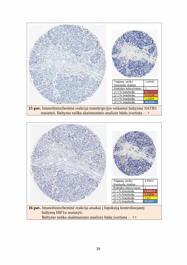
15 pav. Imunohistocheminė reakcija transkripcijos veiksniui baltymui SATB1
nustatyti. Baltymo raiška skaitmeninės analizės būdu įvertinta – +
16 pav. Imunohistocheminė reakcija atsakui į hipoksiją kontroliuojantį
baltymą HIF1α nustatyti.
Baltymo raiška skaitmeninės analizės būdu įvertinta – ++

40
3.2.3. Vizualus patologo vertinimas
Vizualus patologo IHC baltymų raiškos vertinimas atliktas skenuotame
vaizde kompiuterio monitoriuje (Acer AL2616W). Patologas kiekvieną audinių
mikrogardelės mėginį vertino individualiai. Nepakankamos kokybės mėginiai
(suardyta audinio struktūra, išlikęs nepakankamas mėginio kiekis, nekokybiška
IHC reakcija) toliau neanalizuoti. Toliau neanalizuoti ir tie mėginiai, kuriuose
nustatytas dultalinės in situ karcinomos komponentas.
IHC reakcijų rezultatus gydytojas patologas vertino atsižvelgdamas į
ASCO/CAP bei FDA Maisto ir vaistų administracijos (angl. Food and Drug
Administration) patvirtintas gaires. Biologinių žymenų IHC raiška vertinta
pagal skalę nuo 0 iki 3 (0, 1+, 2+ ar 3+). 0 ir 1+ neigiamos žymenų raiškos
kategorijos sujungtos į vieną neigiamos raiškos navikų grupę (0/1+).
HER2 IHC teigiama raiška (3+) konstatuota, kai matoma >10 procentų
invazinio naviko ląstelių intensyviai, nepertraukiamai nusidažiusi ląstelės
membrana arba nustatyta HER2 geno amplifikacija FISH tyrimo metodu
(nustatytas HER2 ir SEP17 santykis >2 ar HER2 geno kopijų skaičiaus
vidurkis ląstelės branduolyje >6). Ribinė HER2 geno vertė (2+) navike
konstatuota, kai HER2 IHC nustatyta raiška yra 2+ arba geno amplifikacija
FISH tyrimo metodu yra 1,8–2,0 ar geno kopijų skaičiaus vidurkis ląstelės
branduolyje yra nuo 4 iki 6 signalų. HER2 neigiamos raiškos navikas
konstatuotas, kai HER2 IHC nustatyta raiška – 0/1+ arba FISH tyrimo metodu
nenustatyta geno amplifikacijos (nustatytas HER2 ir SEP17 santykis < 1,8 ar
HER2 geno kopijų skaičiaus vidurkis ląstelės branduolyje < 4).
Kitų žymenų teigiama baltymų raiška vertinta, atsižvelgiant į nustatytą
baltymą ekspresuojančių naviko ląstelių skaičiaus tiriamajame audinyje
slenkstinę vertę: ER, PR, HIF1 ir SATB1 >10%, Ki67 ir p16 >14%, AR ir
BCL2 >30%, p53 >5%.

41
3.2.4. Skaitmeninė vaizdų analizė
Skaitmeninė vaizdų analizė atlikta naudojant tuos pačius audinių
mikrogardelių vaizdus, kuriuos vertino gydytojas patologas.
Skaitmeninės analizės objektyvumas priklauso nuo daugelio veiksnių
[34]. Vienas esminių reikalavimų – tiksliai atskirti tiriamo audinio navikines
struktūras. Jei skaitmeninė analizė atliekama ne tik navikiniame audinyje, bet ir
kitose jį supančiuose struktūrose (stromoje, leukocituose, duktalinėje in situ
karcinomoje), analizės rezultatai „praskiedžia“ analizuojamo žymens raiškos
procentą naviko ląstelėse. Savo darbe mes naudojome vieną iš pirmaujančių
mikroskopinio vaizdo skaitmeninės analizės platformų – AperioGenie. Šią
sistemą „apmokėme“ tiksliai atpažinti skirtingus krūties vėžio audinius
sukurdami audinių atpažinimo klasifikatorių. Toks automatizuotas audinio
atpažinimas ne tik paspartina analizės procesą, bet ir pašalina žmogaus tyrėjo
įtaką analizės rezultatams.
Tolesnei analizei naudoti skaitmeninės analizės parametrai: tolygaus
membraninio naviko ląstelių nusidažymo (HER2 ir BCL2) ir naviko ląstelių
branduolių (ER, PR, AR, Ki67, p53, p16, SATB1 ir HIF1α) nusidažymo
procentai. IHC ir skaitmeninės analizės (SA) rezultatai parodyti 17 pav.

42
1a,b
2 a,b
3 a,b
17 pav. Imunohistocheminė reakcija (a) ir skaitmeninės analizės rezultatai (b):
1 a,b – SATB1, 2 a,b – HIF1α, 3 a,b – BCL2 SATB1 – transkripcijos veiksnys, HIF1α – atsaką į hipoksiją kontroliuojantis transkripcijos veiksnys,
BCL2 – apoptozę slopinantis baltymas
Susumuoti kiekvienos pacientės atskirai gauti audinių mikrogardelių
analizės duomenys (susumuotos visos ir teigiamos konkrečiam IHC žymeniui
naviko ląstelės, apskaičiuotas teigiamų naviko ląstelių procentas). Pacientės,
kurių mėginiuose suskaičiuota <500 naviko ląstelių, į tolesnę analizę
neįtrauktos. Tolesnei visų 10 IHC žymenų skaitmeninei analizei atlikti atrinkti
109 pacienčių mėginiai, tenkinę nustatytus tiriamajam mėginiui reikalavimus.
Sukurto skaitmeninės analizės klasifikatoriaus tikslumas – 98,7 procentų,
jautrumas – 98,1 procentų ir specifiškumas – 99,3 procentų (18 pav.).
3
ba
3
a
2
a
1
b
1
a
2
b

43
18 pav. Sukurto AperioGenie klasifikatoriaus rezultatai
Klasifikatorius atskiria skirtingas audinio stuktūras: mėlyna spalva pažymėtas navikinis audinys, žalia – stroma, geltona – stiklas
Sukurtas klasifikatorius naudotas kartu su branduoline Nuclear v9 ir
membranine Membrane v9 Aperio analizėmis. Analizuoti pasirinktas tik
navikinio audinio komponentas. Skaitmeninis algoritmas nurodo, kokiu
intensyvumu ekspresuojamas tiriamasis žymuo (silpnu, vidutiniu ar stipriu)
bei suskaičiuoja, kiek ląstelių ekspresuoja analizuojamą baltymą (19, 20 pav.).
19 pav. Skaitmeninė imunohistocheminio Ki67
branduolinio žymens tyrimo analizė

44
20 pav. Skaitmeninė imunohistocheminio membraninio žymens –
žmogaus epidermio augimo faktoriaus receptoriaus 2, tyrimo analizė Raudona spalva pažymėtos intensyviai baltymą ekspresuojančios naviko ląstelės (HER2 3+), oranžine
spalva – paribinės ekspresijos ląstelės (HER2 2+), geltona spalva – silpnos ekspresijos ląstelės (HER2
1+). Mėlyna spalva pažymėtos HER2 baltymo neekspresuojančios ląstelės (HER2 0).
3.2.5. Imunofluorescenciniai tyrimai
HER2 geno amplifikacija buvo nustatyta dviejų zondų PathVysion HER2
FISH rinkiniu (Abbott-Vysis, Inc., Downers Grove, IL, JAV). 4 m parafinu
impregnuoti mėginių pjūviai perkelti ant objektinių stiklelių, turinčių
elektrostatinį krūvį, inkubuoti per naktį 56oC temperatūroje. Vėliau pjūviai
deparafinuoti ksilene, dehidratuoti etilo alkoholiu ir džiovinti ore. Tolesnio
reakcijos etapo metu po pjūvių paruošimo naudojant 0,2N HCl tirpalą 20 min.
ir specialų Path Vysion 80oC tirpalą 30 min., mėginiai buvo apdorojami
proteazės 37oC tirpalu 26 min. Hibridizavimo tirpalas, sudarytas iš tiesiogiai
žymėtų dviejų žymių – SpectrumGreen, 17 chromosomos centromeros
(CEP17) ir SpectrumOrange – HER2 geno lokusui žymėti buvo užlašintas ant
tiriamųjų mėginių. Tolesnio reakcijos etapo metu mėginių pjūviai denatūruoti
hibridizatoriuje (DAKO Diagnostics, Glostrupas, Danija) 5 min. 72oC
temperatūroje ir hibridizuoti 19 val. 37oC temperatūroje. Neprisijungę žymens
fragmentai du kartus plauti karštu 72oC SSC ir 0,3% NP-40 tirpalų mišiniu 2

45
min. Branduoliai kontrastuoti DAPI tirpalu, mėginių pjūviai uždengti
dengiamąja medžiaga (Invitrogen Corporaton, Carlsbadas, JAV). Kartu su
tiriamaisiai mėginiais buvo tiriami ir kontroliniai amplifikuoti bei
neamplifikuoti mėginiai. Tyrimo rezultatai vertinti fluorescenciniu Zeiss
mikroskopu (Zeiss, Axio Imager.Z2, Gottingenas, Vokietija).
Mikroskopavimui naudoti atskiri – žalias, oranžinis ir trigubas – Dapi-FITC-
Cy3 filtrai.
FISH analizė buvo atlikta tyrėjui nežinant IHC tyrimo rezultatų bei
vadovaujantis FDA vertinimo schemomis (8.2.3. skyrius, 21 pav.).
a) b)
21 pav. Žmogaus epidermio augimo faktoriaus receptoriaus 2 fluorescencinės
in situ hibridizacijos tyrimas a) geno amplifikacijos nenustatyta, b) nustatyta geno amplifikacija
3.2.6. DNR išskyrimas, metilinimo ir geno mutacijų tyrimai
Šaldytų ir parafininių audinių DNR buvo išskiriama naudojant organinį
(fenolio-chloroformo) metodą. Parafininiai audiniai (1 mm diametro stulpeliai)
dėti į 1,5 ml mėgintuvėlį. Parafinas pašalintas įpylus 1 ml ksilolo (Roth).
Mėginiai supurtyti ir inkubuoti 10 min. 55 ºC temperatūroje termopurtyklėje.
Po to mėginiai centrifuguoti 10 min. 13000 rpm jėga, supernatantas pašalintas.
Šis etapas kartotas. Ksilolas iš mėginių pašalintas įpylus 1 ml 96% etanolio,
mėginiai inkubuoti 10 min. 55 ºC temperatūroje termopurtyklėje. Vėliau

46
centrifuguota 10 min. 13000 rpm jėga, supernatantas pašalintas. Poveikis
etanoliu kartotas. Etanolio likutis išgarintas atvirus mėgintuvėlius laikant 37 ºC
temperatūroje.
Į mėgintuvėlį su audinių pjūviais įpilta 500 μl lizės buferinio tirpalo (50
mM Tris–HCl, Roth), pH 8,5, 1 mM EDTA (Roth) ir 0,5% Tween–20 (Roth),
sumaišyta ir inkubuota 1 val. 55 ºC temperatūroje. Po to įpilta 25 μl
proteinazės K (20 mg/ml, Fermentas) ir palikta per naktį purtyklėje 55 ºC
temperatūroje. Vėliau įpilta 500 μl fenolio/chloroformo/izoamilalkoholio
(25:24:1, Roth), mėginiai purtyti 10 min., centrifuguoti 10 min. 13500 rpm
jėga. Supernatantas perpiltas į naują 2 ml mėgintuvėlį, etapas su
fenolio/chloroformo/izoamilalkoholio mišiniu pakartotas. Fenolio ir
izoamilalkoholio likučiai pašalinti į supernatantą įpylus 500 μl chloroformo
(Roth). Mėginiai purtyti 10 min. ir centrifuguoti 10 min. 13500 rpm jėga.
Supernatantas surinktas ir perpiltas į naują mėgintuvėlį. DNR nusodinta
veikiant 40 μl 5 M amonio acetato (Fluca) ir 1 ml šalto (–20 ºC) 96% etanolio.
DNR laikoma 2–3 val. –20 ºC temperatūroje. Centrifuguojama 15 min. 13500
rpm jėga +4 ºC temperatūroje. Supernatantas pašalintas, o DNR perplauta šaltu
70% etanoliu ir centrifuguota 15 min. 13500 rpm jėga +4 ºC temperatūroje.
Supernatantas pašalintas. Mėgintuvėlis paliktas atviras 37 ºC temperatūroje,
kol išgaravo etanolio likučiai. DNR ištirpinta 20 μl benukleaziniame vandenyje
ir laikyta –20 °C.
Atlikti epigenetinei analizei naudota viena jautriausių technologijų –
metilinimui jautri PGR arba MSP. Prieš analizę DNR seka modifikuota:
veikiant natrio bisulfitu, nemetilintas citozinas virsta uracilu, o PGR metu
atpažįstamas kaip timinas; metilcitozinas yra nejautrus bisulfitui ir PGR metu
atpažįstamas kaip citozinas. Ši reakcija dar vadinama bisulfitiniu virsmu, jos
metu epigenetinė DNR modifikacija paverčiama genetiniu pokyčiu. Principinė
MSP schema pateikiama 22 pav.

47
22 pav. Metilinimui jautrios polimerazės
grandininės reakcijos metodo darbo schema
Audinių DNR modifikuoti 1 µg DNR 15 min. denatūruojamas 37°C
temperatūroje su 3 M NaOH (Sigma). Vėliau DNR 16 val. 50°C temperatūroje
veikiama natrio metabisulfitu (2,3 M 5pH, Sigma) kartu su hidrochinonu (10
mM, Fluka). Iš modifikuotos DNR išvalomos druskos naudojant DNR valymo
kolonėles Wizard DNA Clean-up System (Promega). Galutinei DNR
modifikacijai gauti DNR su 3 M NaOH inkubuojama 15 min. 37ºC
temperatūroje. Tirpalas neutralizuojamas 10 M amonio acetatu, DNR
nusodinama etanoliu. Paruošta DNR tirpinama benukleaziniame vandenyje ir
laikoma šaldytuve –20°C temperatūroje.
Metilinimui jautri PGR (MSP) vykdoma naudojant pradmenų poras,
atrankiai sąveikaujančias su metilinta (M reakcija) ir nemetilinta (U reakcija)
DNR seka geno promotoriuje. Dauguma MSP pradmenų pagausina CpG salas,
esančias tiriamų genų promotoriuje arba 1–ame egzone.
PGR reakcija vykdoma 25 µl reakcijos mišinio, kurio sudėtis: 1–2 µl
modifikuotos DNR, 1 x PGR buferio (Applied Biosystems), 0,4 mmol/L dNTP
(Fermentas), 2,5 mmol/L MgCl2 (Applied Biosystems), 1% dimetilsulfoksido
(Fluka), reikalingas pradmenų kiekis (galutinė koncentracija – 6 ng/µl) ir 1,25
vnt. AmpliTaq Gold polimerazės (Applied Biosystems). PGR vykdoma

48
termocikleryje (Mastercycler Epgradient S, Eppendorf) naudojant šį
temperatūrinį režimą: 95 °C – 10 min., 35-37 ciklai: 95 °C – 45 sek., pradmenų
jungimosi temperatūra – 45 sek., 72°C – 45 sek. ir 72°C – 10 min. Pradmenų
jungimosi temperatūra varijuoja nuo 58 iki 66°C priklausomai nuo geno. PGR
produktas analizuojamas vykdant elektroforezę 7,5% poliakrilamido gelyje
arba 2% agarozės gelyje (23 pav.). Mėginys yra metilintas, jei produktas
nustatomas MSP reakcijos su M pradmenimis arba su M ir U pradmenimis
būdu. Nustačius produktą vykstant reakcijai tik su U pradmeniu mėginys
laikomas nemetilintu.
23 pav. Poliakrilamido gelio nuotraukos Analizuotas RASSF1, RARB ir DAPK1 genų promotorių hipermetilinimas krūties
navikuose. Sm – ilgio žymuo, M – reakcija, kuria nustatoma metilinta geno promotoriaus seka, U – reakcija, kuria nustatoma nemetilinta promotoriaus seka,
KL - sveikų donorų leukocitų DNR, KM – in vitro metilinta sveikų donorų
leukocitų DNR, KxT – krūties karcinomos DNR, T24 – vėžinių ląstelių linija
MSP M ir U pradmenų atrankumas tikrintas vykdant reakciją su
nemodifikuota DNR ir naudojant kontrolinę in vitro metilintą DNR (CpG
Methylase; New England BioLabs), vėžinių ląstelių linijų DNR ir leukocitų
DNR kaip neigiamą kontrolę. Tarša kontroliuota atliekant kiekvieną tyrimą.

49
Geno TP53 mutacijos tirtos genetiniu analizatoriumi ABI 3130 (Applied
Biosystems), taikant DNR grandinės konformacijos kitimų nustatymo metodą
(SSCP). Šiuo metodu identifikavus pakitusį piką toliau vykdyta sekoskaita.
SSCP tyrimui atlikti geno TP53 egzonai (5-9) pagausinti PGR metodu,
naudojant skirtingomis fluorescencinėmis žymėmis (6-karboksifluoresceinu (6-
FAM) ir 4,7,2’,4’,5’,7’-heksachloro-6-karboksifluoresceinu, HEX) žymėtus
prasminius ir antiprasminius pradmenis. Reakcijai naudota: 2 µl GeneAmp 10 x
PGR buferio, 5 mM MgCl2, 0,4 mM dNTP mišinio, 1 vnt. AmpliTaqGold
polimerazės, po 0,4 µl (20 µM) 6-FAM žymėto prasminio pradmens ir HEX
žymėto antiprasminio pradmens, 0,5 µl DMSO, 100 ng tiriamosios DNR.
Bendrasis vieno mėginio reakcijos tūris neviršijo 20 µl. PGR reakcija vykdyta:
95C temperatūroje – 10 min., 95C temperatūroje – 30 sek., pradmenų
jungimo temperatūroje – 30 sek., 74C temperatūroje – 1 min., 74C
temperatūroje – 10 min. PGR ciklai kartoti 40 kartų. Pradmenų jungimo
temperatūra atitinkamų egzonų pradmenims: 5 – 58ºC; 6 – 60ºC; 7 – 60ºC; 8 –
58ºC ir 9 – 56ºC. Po pagausinimo PGR produktas analizuotas 7,5%
nedenatūruojančiame poliakrilamido gelyje norint įvertinti pagausinto DNR
fragmento kokybę ir kiekybę. Ruoštas SSCP analizės mišinys: 0,5 µl PGR
produkto, 0,5 µl GeneScan-500 LIZ arba ROX ilgio standarto (Applied
Biosystems) ir 15µl HiDi formamido. Mišinys perkeltas ant plokštelės ir
analizuoas 3130 genetiniu analizatoriumi (Applied Biosystems). Atskyrimo
terpei naudotas 3130 CAP polimeras, turintis 10% glicerolio ir 1 x EDTA
buferio. Elektroforezė vykdyta 43 min. 15 kV elektros lauke 18 °C ar 35 °C
temperatūroje. Rezultatai analizuoti naudojant GeneMapper® programą.
Sekoskaita vykdyta ABI PRISM 3130 (Applied Biosystems) genetiniu
analizatoriumi. Tiriamojo geno TP53 egzonai pagausinti PGR metodu.
Reakcijai naudotas 2µl GeneAmp 10 x PGR buferinis tirpalas, 3,2 µl 25 mM
MgCl2, 1 µl 4 mM dNTP mišinys, 0,1 µl 5 vnt./µl AmpliTaqGold polimerazės
(Applied Biosystems), po 0,5µl 20 µM prasminio ir antiprasminio pradmenų,
10,7 µl H2O, 2 µl 100 μg/ml vėžinės DNR. Naudotas vieno mėginio bendrasis
reakcijos tūris – 20 µl. PGR reakcija vykdyta: 95C temperatūroje – 10 min.,

50
95C – 30 sek., 60C – 30 sek., 74C – 1 min., 74C – 10 min. PGR ciklai
kartoti 36 kartus. PGR produktas analizuotas 1,5% agarozės arba 7,5%
nedenatūruojančiame poliakrilamido gelyje. Tolesniems etapams atrinkti tik
sėkmingai pagausinti PGR produktai.
PGR produktas (DNR konc. – 500 μg/ml) skiestas 100 kartų. Sekoskaitos
mišinį PGR reakcijai atlikti sudarė 8µl BigDye terminator v3,1 Ready Reaction
mix, 1,6 µl 2 pmol prasminio arba antiprasminio pradmens, 5,8 µl H2O ir 5 µl
PGR produkto. Naudotas bendras vieno mėginio reakcijos tūris – 20 µl.
Sekoskaitos PGR reakcija vykdyta optimaliomis sąlygomis: 96C
temperatūroje – 1 min., 96C – 10 sek., 50C – 5 sek., 60C – 4 min. PGR
ciklai katoti 25 kartus.
Iš pagausintos DNR išvalyti neįjungti žymėti nukleotidai, kurie trukdo
vykti sekoskaitos reakcijai. Į pagausintą DNR pridėta 1,25 µl 0,5 M EDTA ir
60 µl 100% etilo alkoholio. Mėginiai inkubuoti kambario temperatūroje 15
min., centrifuguoti 30 min. 13000 rpm jėga 4°C temperatūroje. Supernatantas
pašalintas. Įpylus 60 µl 70% etanolio, centrifuguota 30 min. 13000 rpm jėga,
4°C temperatūroje, supernatantas pašalintas. DNR džiovinta 37°C
temperatūroje.
Sekoskaitai paruošti DNR mėginiai ištirpinti 12 µl formamido tirpale ir
perkelti ant plokštelės, kur vėliau analizuoti 3130 genetiniu analizatoriumi.
Atskyrimo terpei naudotas 3130 POP-7 polimeras (Applied Biosystems), 1 x
EDTA buferis. Elektroforezė vykdyta 30 min. 15 kV elektros lauke. Gauta seka
analizuota maudojant SeqScape programą, palyginta su geno seka iš Genų
banko.
3.3. Statistinė analizė
Atlikta duomenų padėties ir sklaidos statistinė analizė, kiekybiniai
duomenys pateikti kaip aritmetinis vidurkis ± standartinis nuokrypis. Esant
duomenų sklaidos nuokrypiui į kairę (HER2, Ki67, HIF1α, SATB1, p53, p16),
tolesnei parametrinei statistikai naudotos šių kintamųjų logoritmuotos vertės.

51
Kiekybinėms kintamųjų variacijoms įvairiose veiksnių grupėse palyginti
naudota vienfaktorinė variacijų analizė (ANOVA) pagal Fisherio kriterijų,
pritaikant Bonferroni (Dunn) t testą poriniams palyginimams. Kokybinių bei
ranginių dydžių sąsajoms nustatyti naudota lentelių analizė, panaudojant χ2 ir
Fisherio tikslųjį kriterijus; kiekybiniai dydžiai šiai ir kitoms neparametrinėms
analizėms dichotomizuoti, atsižvelgiant į jų sklaidos medianas bei mokslinėje
literatūroje panaudotas ribines vertes. Kiekybinių dydžių porinėms sąsajoms
įvertinti naudota Pearsono koreliacijos analizė. Daugiamatei kiekybinių
kintamųjų sąsajų analizei atlikti taikyta faktorinė analizė, panaudojant
principinių komponenčių metodą ir bendrąją orthomax veiksnių rotaciją.
Faktorinės analizės būdu nustatytų veiksnių vertės panaudotos klasterinėje
analizėje, įvertinant tiriamųjų grupės heterogeniškumą. Kintamųjų nustatymo
(tyrėjų bei metodų) variacijoms įvertinti taikyta sutarimo analizė, naudojant κ
statistiką, κ vertes nuo 0,81 iki 1,00 vertinant kaip „beveik tobulą” sutarimą
[145], rezultatus pateikiant kaip pasvertąją κ esant 95% pasiklautinumo
intervalui. Išgyvenimo statistinei analizei taikyti Kaplano-Meierio
išgyvenamumo ir Coxo regresijos metodai. Taikytas statistinio patikimumo
lygmuo p<0,05. Statistinė analizė atlikta SAS 9,2 programiniu paketu.
3.4. Etiniai aspektai
Prieš pradedant tyrimą pacientės supažindintos su tyrimo tikslu ir eiga,
pateikiant informuoto asmens sutikimo formą. Savo sutikimą dalyvauti tyrime
pacientės patvirtino parašu.
Tyrimas pradėtas gavus Lietuvos bioetikos komiteto leidimą (2007-08-03
Nr.: 33), 1 priedas.

52
4. REZULTATAI
4.1. Imunohistocheminių žymenų raiškos skaitmeninės analizės
kalibravimas
Skaitmeninei IHC analizei atlikti naudotas AperioGenie membraninis ir
AperioGenie branduolinis algoritmai. Siekiant kalibruoti pasirinktus analizės
algoritmus, jie lyginti su kitomis to paties objekto matavimo sistemomis.
Skaitmeninės branduolinių IHC žymenų analizės rezultatai lyginti su
stereologinės skaitmeninio vaizdo analizės rezultatais, membraninių IHC
žymenų analizė – su kito gamintojo skaitmeninės analizės bei FISH rezultatais.
Visų analizių rezultatai palyginti tarpusavyje bei su gydytojo patologo
vizualaus vertinimo rezultatais.
4.1.1. Baltymų raiškos skaitmeninės analizės rezultatų palyginimas su
patologo vertinimo rezultatais
Baltymų raiškos IHC tyrimų rezultatus gydytojas patologas vertino
analizuodamas monitoriuje matomą skenuotą TMA mikropreparatų vaizdą.
Pirmame analizės etape gydytojas patologas vertino mėginių kokybę.
Nekokybiški mėginiai (suardytas audinio vientisumas, nepakankamas
navikinio audinio kiekis, dažymosi artefaktai) ir mėginiai, kuriuose nustatytas
in situ duktalinės karcinomos komponentas, nenaudoti tolesnei analizei. Toliau
analizuojamus mėginius TMA gydytojas patologas vertino individuliai, IHC
tyrimų rezultatai apskaičiuoti susumavus kiekvienos pacientės visų mėginių
analizės rezultatus, vėliau apskaičiuotos vidurkių vertės. IHC tyrimų rezultatai
apskaičiuoti vadovaujantis CAP gairėmis. Tų pačių TMA IHC vaizdų
skaitmeninė analizė atlikta naudojant tik kokybiškus TMA mėginius. Tolesnei
analizei naudotas skaitmeninės analizės būdu apskaičiuotų teigiamų naviko
ląstelių branduolių (ER, PR, AR, Ki67, p53, p16, SATB1 ir HIF1α) ir ląstelių
su nepertraukiamu membraniniu nusidažymu (HER2 ir BCL2) procentas
(kiekvienos pacientės visuose į analizę patekusiuose mėginiuose apskaičiuotas
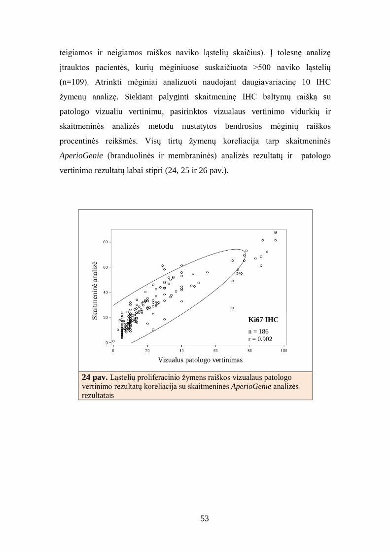
53
teigiamos ir neigiamos raiškos naviko ląstelių skaičius). Į tolesnę analizę
įtrauktos pacientės, kurių mėginiuose suskaičiuota >500 naviko ląstelių
(n=109). Atrinkti mėginiai analizuoti naudojant daugiavariacinę 10 IHC
žymenų analizę. Siekiant palyginti skaitmeninę IHC baltymų raišką su
patologo vizualiu vertinimu, pasirinktos vizualaus vertinimo vidurkių ir
skaitmeninės analizės metodu nustatytos bendrosios mėginių raiškos
procentinės reikšmės. Visų tirtų žymenų koreliacija tarp skaitmeninės
AperioGenie (branduolinės ir membraninės) analizės rezultatų ir patologo
vertinimo rezultatų labai stipri (24, 25 ir 26 pav.).
24 pav. Ląstelių proliferacinio žymens raiškos vizualaus patologo
vertinimo rezultatų koreliacija su skaitmeninės AperioGenie analizės
rezultatais
Ki67 IHC
n = 186
r = 0,902
Vizualus patologo vertinimas
Skai
tmen
inė
anal
izė

54
25 pav. Estrogenų receptorių raiškos vizualaus patologo vertinimo
rezultatų koreliacija su skaitmeninės AperioGenie analizės rezultatais
26 pav. Progesteronų receptorių raiškos vizualaus patologo rezultatų
koreliacija su skaitmeninės analizės vertinimo rezultatais
PR IHC
n = 184
r = 0,956
Vizualus patologo vertinimas
Sk
aitm
enin
ė an
aliz
ė
ER IHC
n = 183
r = 0,933
Vizualus patologo vertinimas
Sk
aitm
enin
ė an
aliz
ė
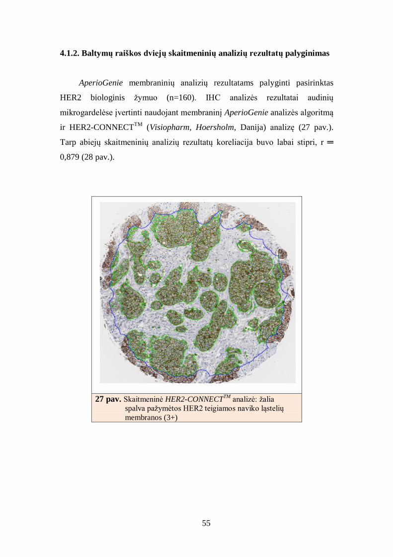
55
4.1.2. Baltymų raiškos dviejų skaitmeninių analizių rezultatų palyginimas
AperioGenie membraninių analizių rezultatams palyginti pasirinktas
HER2 biologinis žymuo (n=160). IHC analizės rezultatai audinių
mikrogardelėse įvertinti naudojant membraninį AperioGenie analizės algoritmą
ir HER2-CONNECTTM
(Visiopharm, Hoersholm, Danija) analizę (27 pav.).
Tarp abiejų skaitmeninių analizių rezultatų koreliacija buvo labai stipri, r ═
0,879 (28 pav.).
27 pav. Skaitmeninė HER2-CONNECTTM
analizė: žalia
spalva pažymėtos HER2 teigiamos naviko ląstelių
membranos (3+)

56
28 pav. Ištisinio ląstelių membranų dažymosi (Aperio) ir HER2-
CONNECTTM
membranų vientisumo indekso rezultatų palyginimas
4.1.3. Baltymų raiškos skaitmeninės analizės rezultatų palyginimas su
analizės fluorescencinės in situ hibridizacijos rezultatais
Membraninė skaitmeninė HER2 IHC analizė lyginta su vizualaus
patologo vertinimo rezultatais ir „aukso standarto“ - FISH tyrimo metodu
gautais rezultatais (n=152)[146]. Visos analizės atliktos naudojant tas pačias
audinių mikrogardeles. Patologas IHC tyrimo rezultatus vertino dviejų mėnesių
laikotarpiu du kartus (VV1 – pirmasis patologo vertinimas, VV2 – antrasis
patologo vertinimas). Kiekvieną naviko mėginį, esantį audinių mikrogardelėse,
patologas vertino individualiai, apskaičiuodamas HER2 IHC tyrimo reikšmę
(0, 1+, 2+ ir 3+). Atliekant tolesnę analizę navikai, kuriuose nustatyta neigiama
HER2 IHC raiška, sujungti į vieną grupę (0/1+). FISH tyrimas atliktas
rankiniu būdu, tyrėjui nežinant IHC HER2 tyrimo rezultatų. HER2 FISH
tyrimo duomenys interpretuoti vadovaujantis FDA patvirtintomis duomenų
skaičiavimo schemomis, kuriose teigiama HER2 geno amplifikacija nustatoma
HER2/CEP17 santykiui esant didesniam už 2. FISH analizei pasirinktas
HER2 IHC
n = 160
r = 0,879
Ląstelių su ištisiniu membranos nusidažymu procentas
CO
NN
EC
T H
ER
2 I
HC
mem
bra
nos
vie
nti
sum
o i
ndek
sas

57
tinkamiausias vienas iš keturių kiekvienos pacientės naviko mėginys, kuriame
įvertinta 20 naviko ląstelių branduolių.
Pradiniai HER2 IHC (vizualaus ir skaitmeninio) bei FISH tyrimo
vertinimo duomenys pateikti 6 lentelėje. Skaitmeninės analizės ir abiejų
patologo vertinimų reikšmės daugumos HER2 neigiamų (0/1+) ir teigiamų
(3+) navikų kategorijų grupėse sutapo su skaitmeninės analizės rezultatais
(kappa=0,86). Didžiausia vertinimo variacija matoma tik HER2 IHC tyrimo
2+ kategorijoje. Palyginus gautus rezultatus su FISH tyrimo reikšmėmis,
matyti, kad 5 FISH tyrimu nustatyti HER2 teigiami pacienčių mėginiai pateko į
sujungtą HER2 IHC tyrimu nustatytą neigiamų navikų kategoriją (0/1+), IHC
tyrimo rezultatus vertinant tiek vizualiai, tiek naudojant skaitmeninę vaizdo
analizę (5 lentelė). Į HER2 neigiamų navikų kategoriją pirmojo patologo
vertinimo metu pateko papildomi dar 3, tolesniu – 2 pacienčių mėginiai.
Atlikus jų FISH tyrimą, gauti teigiami HER2 geno amplifikacijos rezultatai.
Daugiausia teigiamų HER2 mėginių HER2 2+ ir 3+ grupėse nustatyta SA
(3+16=19), mažiau antruoju patologo vertinimu (VV2) (2+15=17) ir pirmuoju
patologo vertinimu (VV1) (2+14=16). Tačiau, skaitmeninės analizės
rezultatais, į 3+ navikų kategoriją pateko 4 mėginiai, kai, atlikus HER2 FISH
tyrimą, gauti neigiami HER2 rezultatai. Tie patys 4 mėginiai VV1 ir VV2
vertinimu pateko į HER2 3+ arba 2+ grupę (6 lentelė).
Panaudojus HER2 analizę kaip modelį skaitmeninės ląstelių plazminės
membranos baltymų raiškos analizės patikimumui įvertinti nustatyta, kad
HER2 raiškos krūties vėžio audinyje skaitmeninės vaizdo analizės rezultatai
atitiko patologo vertinimą (kappa=0,86) ir leidžia tiksliau nustatyti
amplifikuoto (HER2 FISH) krūties vėžio atvejus. Naudojant SA buvo tiksliau
atrinkta FISH tyrimu nustatytų HER2 teigiamų mėginių grupė. Keli
nesutapimo atvejai traktuotini kaip biologinio naviko netolygumo
(heterogeniškumo) ar testo variacijų pasekmė.

58
5 lentelė. HER2 teigiamų mėginių skaičiaus santykis skirtingose baltymų
raiškos kategorijose lyginant patologo vertinimo ir skaitmeninės analizės
rezultatus IHC metodu
nustatyta HER2
raiška
FISH metodu nustatyti HER2 teigiami navikai (%)
VV1 VV2 SA
0/+1 8/127 (6,3) 7/120 (5,8) 5/113 (4.4)
2+ 2/8 (25,0) 2/15 (13,3) 3/19 (15,8)
3+ 14/17 (82,4) 15/17 (88,2) 16/20 (80,0)
Iš viso 152 152 152 VV1 ir VV2 – pirmasis ir antrasis patologo vertinimai, SA – skaitmeninė analizė, HER2 – žmogaus
epidermio augimo faktoriaus receptorius 2, FISH – fluorescencinis in situ hibridizacijos tyrimas

59
6 lentelė. Pradiniai HER2 baltymo raiškos vizualaus ir
skaitmeninio bei FISH HER2/CEP17 vertinimo duomenys
Eilė # HER2/
CEP17 HER2 CEP17
VV1
VV2
SA
1 0,4 1,8 4,2 1 1 1
2 0,5 2,1 4,4 1 1 1
3 0,5 1,9 3,5 1 1 1
4 0,9 3,0 3,4 1 1 1
5 1,1 3,7 3,5 1 1 1
6 1,3 4,2 3,3 1 1 1
7 1,7 5,4 3,1 1 1 1
8 1,8 5,7 3,2 1 1 1
9 1,8 7,1 3,9 1 1 1
10 2,1 2,7 1,3 1 1 1
11 2,3 4,2 1,9 1 1 1
12 2,4 3,2 1,3 1 1 1
13 2,7 3,8 1,4 1 1 1
14 4,6 12,6 2,8 1 1 1
15 0,5 1,5 2,9 1 2 1
16 1,2 1,8 1,6 1 1 2
17 1,3 2,3 1,8 1 1 2
18 1,4 5,7 3,9 1 1 2
19 1,6 4,3 2,7 1 1 2
20 4,5 4,5 1,0 1 1 2
21 1,0 4,9 4,7 2 2 2
22 1,5 6,7 4,5 1 1 2
23 1,6 5,3 3,3 1 1 2
24 2,1 3,8 1,8 1 1 2
25 1,0 2,1 2,1 1 2 2
26 1,3 4,7 3,7 1 2 2
27 1,4 4,1 2,8 1 2 2
28 1,7 5,8 3,5 1 2 2
29 2,0 5,4 2,8 1 2 2
30 2,2 4,3 1,9 1 2 2
31 1,1 4,1 3,7 2 2 2
32 1,2 3,0 2,4 2 2 2
33 1,3 5,0 3,8 2 2 2
34 1,4 3,3 2,4 2 2 2
35 1,5 3,3 2,2 2 2 3
36 3,4 11,3 3,4 2 2 3
37 6,5 12,4 1,9 2 3 3
38 2,3 4,3 1,9 3 3 3
39 1,7 9,0 5,2 3 2 3
40 1,4 2,6 1,8 3 3 3
41 1,5 4,6 3,1 3 3 3

60
VV1 ir VV2 – pirmasis ir antrasis patologo vertinimai, SA – skaitmeninė
analizė, HER2 – žmogaus epidermio augimo faktoriaus receptorius 2, CEP 17
– 17 chromosomos centromera, FISH – fluorescencinis in situ hibridizacijos
tyrimas.
4.1.4. Baltymų raiškos skaitmeninės analizės rezultatų palyginimas su
analizės stereologiniu metodu rezultatais
Skaitmeninė analizė pasižymi geresniu atkartojamumu (angl.
reproducibility), t.y., pakartotinai atlikus to paties objekto analizę tomis
pačiomis sąlygomis, matavimo rezultatas praktiškai nesikeičia. Tai yra
neabejotinas skaitmeninės analizės pranašumas, palyginti su žmogaus-tyrėjo
vertinimu. Tačiau svarbu įvertinti ir skaitmeninės analizės tikslumą (angl.
accuracy), tai yra sisteminį nuokrypį nuo tikrųjų (referentinių) matuojamojo
dydžio verčių. Ir skaitmeninė analizė, ir žmogaus–tyrėjo vertinimas gali būti
nukrypęs nuo tikrųjų verčių – tiesos kriterijaus, kitaip „aukso standarto“. Nors
patologijos ir skaitmeninės analizės tyrimų praktikoje dažnai tiesos kriterijumi
pasirenkamas kvalifikuoto tyrėjo-gydytojo patologo vertinimas, šis taip pat
varijuoja ir gali būti sistemiškai nukrypęs nuo referentinių verčių. Pavyzdžiui,
vertindamas žymenį ekspresuojančių ląstelių procentą, patologas paprastai
remiasi vizualiu įspūdžiu ir pusiau kiekybiniu dydžio įvertinimu. Todėl
skaitmeninė analizė (potencialiai tikslesnis įrankis) neturėtų būti kalibruojama
pagal pusiau kiekybinį blogiau atkartojamą žmogaus–tyrėjo vizualų vertinimą.
42 4,5 4,5 1,0 3 3 3
43 4,7 10,1 2,2 3 3 3
44 4,9 11,2 2,3 3 3 3
45 4,9 15,5 3,2 3 3 3
46 5,4 13,8 2,6 3 3 3
47 5,9 16,8 2,9 3 3 3
48 6,5 20,4 3,2 3 3 3
49 6,8 13,6 2,0 3 3 3
50 7,2 22,7 3,2 3 3 3
51 7,2 23,2 3,2 3 3 3
52 8,3 22,0 2,7 3 3 3
53 13,1 28,2 2,2 3 3 3
54 13,3 38,5 2,9 3 3 3

61
Mūsų darbe skaitmeninės analizės algoritmams kalibruoti naudotas
stereologijos metodu nustatytas tiesos kriterijus [100]. Trys tyrėjai
nepriklausomai įvertino 30 tų pačių navikinių mėginių skaitmeninius vaizdus,
stereologinėse gardelėse pažymėdami Ki67 teigiamus ir neigiamus vėžio
ląstelių branduolius (29 pav.). Nepaisant to, kad tai yra žmogaus–tyrėjo
vertinimas, jis pagrįstas ne vizualiu įspūdžiu, bet individualiu ląstelių
įvertinimu ir tiksliu suskaičiavimu.
29 pav. Stereologijos metodas. Pasirinkto dydžio stereologiame tinklelyje
Ki67 žymeniui teigiami naviko ląstelių branduoliai pažymėti oranžine,
neigiami – žalia spalvomis
Trijų tyrėjų stereologinio vertinimo rezultatai buvo panašūs (pirminiai
duomenys pateikti 30 pav.), skirtingi tyrėjai nebuvo reikšmingas variacijų
šaltinis (ANOVA analizės duomenimis p=0,955, 31 ir 32 pav.).

62
30 pav. Stereologijos metodas. Trijų tyrėjų Ki67 raiškos vertinimo palyginimas
31 pav. Stereologijos metodas. Trijų tyrėjų stereologinių verčių
rezultatų palyginimas, ANOVA analizės duomenimis p>0,955.
N = 30
1 Tyrėjas 2 Tyrėjas 3 Tyrėjas
1 Tyrėjas
1 Tyrėjas
Tirti mėginiai (n=30)
Ki6
7te
igia
mų l
ąste
lių %
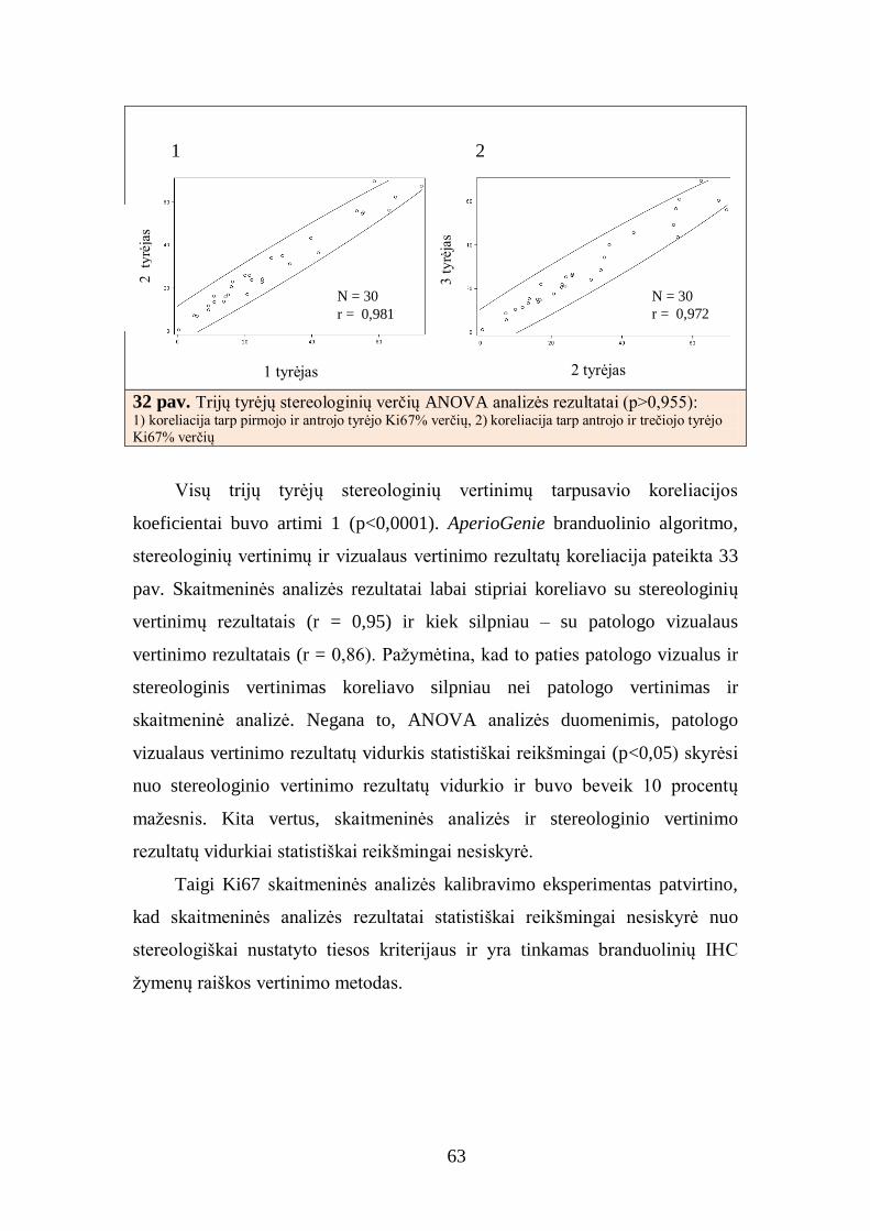
63
1 2
32 pav. Trijų tyrėjų stereologinių verčių ANOVA analizės rezultatai (p>0,955): 1) koreliacija tarp pirmojo ir antrojo tyrėjo Ki67% verčių, 2) koreliacija tarp antrojo ir trečiojo tyrėjo
Ki67% verčių
Visų trijų tyrėjų stereologinių vertinimų tarpusavio koreliacijos
koeficientai buvo artimi 1 (p<0,0001). AperioGenie branduolinio algoritmo,
stereologinių vertinimų ir vizualaus vertinimo rezultatų koreliacija pateikta 33
pav. Skaitmeninės analizės rezultatai labai stipriai koreliavo su stereologinių
vertinimų rezultatais (r = 0,95) ir kiek silpniau – su patologo vizualaus
vertinimo rezultatais (r = 0,86). Pažymėtina, kad to paties patologo vizualus ir
stereologinis vertinimas koreliavo silpniau nei patologo vertinimas ir
skaitmeninė analizė. Negana to, ANOVA analizės duomenimis, patologo
vizualaus vertinimo rezultatų vidurkis statistiškai reikšmingai (p<0,05) skyrėsi
nuo stereologinio vertinimo rezultatų vidurkio ir buvo beveik 10 procentų
mažesnis. Kita vertus, skaitmeninės analizės ir stereologinio vertinimo
rezultatų vidurkiai statistiškai reikšmingai nesiskyrė.
Taigi Ki67 skaitmeninės analizės kalibravimo eksperimentas patvirtino,
kad skaitmeninės analizės rezultatai statistiškai reikšmingai nesiskyrė nuo
stereologiškai nustatyto tiesos kriterijaus ir yra tinkamas branduolinių IHC
žymenų raiškos vertinimo metodas.
2 tyrėjas 1 tyrėjas
2 ty
rėja
s
3 t
yrė
jas
N = 30
r = 0,981
N = 30
r = 0,972

64
33 pav. Ki67 IHC vizualaus vertinimo (VV), skaitmeninės analizės (SA) ir
stereologinio vertinimo skirtumų analizė (Stereo, ANOVA, p<0,01)
* nustatytas patikimas skirtumas tarp VV ir stereologinio vertinimo rezultatų (p<0,05)
4.2. Krūties duktalinės karcinomos imunohistocheminio tyrimo
skaitmeninė analizė
IHC naviko charakteristikos/kiekybiniai skaitmeninio vertinimo rezultatai
pateikti 7 lentelėje.
7 lentelė. Skaitmeninės baltymų raiškos analizės rodikliai duktalinėse krūties
karcinomose
Žymuo
Teigiamų
naviko ląstelių
vidurkis, %
Standartinis nuokrypis
Min Max Mediana n
ER 54,8 36,4 0,3 97,7 72,3 190
PR 33,4 34,6 0,01 94,6 13,3 190
HER2 12,1 19,7 0 82,7 1,6 175
Ki67 27,8 18,7 1,2 88,2 22,2 192
BCL2 41,7 23,4 0,5 75,1 47,9 171
p53 21,3 26,0 0 94,2 10,2 193
p16 17,0 15,2 0,5 75,9 12,3 193
AR 32,2 24,1 0,4 89,2 33,6 175
HIF1α 9,1 12,0 0,2 62,8 4,8 173
SATB1 8,7 7,5 0,8 56,9 6,8 165
ER – estrogenų receptorius, PR – progesteronų receptorius, HER2 – žmogaus epidermio augimo
faktoriaus receptorius 2, AR – androgenų receptorius, Ki67 – ląstelių proliferacijos žymuo, p53,
p16 – vėžio supresoriai, BCL2 – apoptozę slopinantis baltymas, SATB1 – transkripcijos veiksnys,
HIF1α – atsaką į hipoksiją kontroliuojantis transkripcijos veiksnys.
* *
VV SA Stereo

65
BCL2
IHC žymenų raiškos (teigiamų naviko ląstelių %) pasiskirstymas
pateiktas histogramose. Nustatytas ER, PR ir BCL2 raiškos bimodalinis
pasiskirstymas (34 ir 35 pav.), o AR raiškos – bimodalinis su kairiuoju
nuokrypiu (35 pav.). Pagal HER2, Ki67, p16, p53, SATB1 ir HIF žymenų
raišką atvejai buvo pasiskirstę su nuokrypiu į kairę. Siekiant toliau analizuoti
IHC žymenis atlikta šių duomenų lognormalinė transformacija, kurios išraiška
kartu su išvardytų žymenų raiškos pasiskirstymu pateikta 36 ir 37 pav.
34 pav. Tiriamųjų mėginių pasiskirstymas pagal estrogenų (1) ir progesteronų (2)
receptorių raišką naviko audinyje
35 pav. Tiriamųjų mėginių pasiskirstymas pagal androgenų receptorių (1) ir
apoptozę slopinančio baltymo BCL2 (2) raišką naviko audinyje
n = 190
Vidurkis 55±36
Mediana 72
n = 190
Vidurkis 33±35
Mediana 13
n = 171
Vidurkis 41±23
Mediana 48
n = 175
Vidurkis 32±24
Mediana 33
1 2
2
1
2
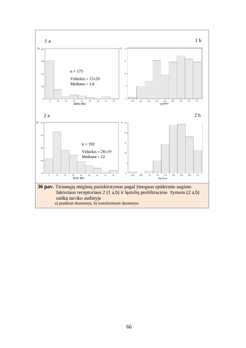
66
36 pav. Tiriamųjų mėginių pasiskirstymas pagal žmogaus epidermio augimo
faktoriaus receptoriaus 2 (1 a,b) ir ląstelių proliferacinio žymens (2 a,b)
raišką naviko audinyje a) pradiniai duomenys, b) transformuoti duomenys
n = 175
Vidurkis 12±20
Mediana 1,6
1 a 1 b
2 a
n = 192
Vidurkis 28±19
Mediana 22
2 b

67
37 pav. Tiriamųjų mėginių pasiskirstymas pagal vėžio supresorių p16 (1 a,b) ir p53
(2 a,b), transkripcijos veiksnio (3 a,b) ir hipoksiją aktyvinančio baltymo (4
a,b) raišką naviko audinyje a) pradiniai duomenys, b) transformuoti duomenys
n = 193
Vidurkis 21±26
Mediana 10
2 a
n = 193
Vidurkis 17±15
Mediana 12
1 a 1 b
2 b
n = 165
Vidurkis 9±8
Mediana 7
3 a
n = 173
Vidurkis 9±12
Mediana 5
4 a
3 b
4 b

68
Luminalinis A ir luminalinis B tipai išsiskyrė dažna BCL2 (95% ir 73%,
9 lentelė) bei reta HIF1 (8% ir 20%) žymenų raiška (p<0,0001), o luminalinis
A tipas dar pasižymėjo ir dažna AR raiška (72%, p<0,0001). HER2 teigiamų
navikų grupėje (LB HER2+ ir HER2+) p53 baltymo raiška konstatuota
atitinkamai 74% ir 64%, tačiau statistiškai reikšmingo p53 baltymo raiškos
skirtumo pagal naviko biologinius tipus nenustatyta. Pažymėtina, kad HER2+
navikų grupėje skirtinga BCL2 ir HIF1 žymenų raiška: luminalinio B HER2+
navikų grupėje BCL2 raiška yra dažna (50%), HIF1 - reta (13%), HER2+
grupėje BCL2 raiška – reta (8%), HIF1 – dažna (73%). Nustatyti skirtumai
yra statistiškai reikšmingi (p<0,0001). Trejopai neigiamų navikų grupėje
nustatyta dažna p16 (79%, p<0,0001) žymens raiška. Šioje navikų kategorijoje
AR (10%) ir BCL2 (23%) žymenų raiška yra reta.
* Žymuo ir žymens teigiamos raiškos naviko ląstelėse slenkstinė vertė.
ER – estrogenų receptorius, PR – progesteronų receptorius, HER2 – žmogaus epidermio augimo
faktoriaus receptorius 2, AR – androgenų receptorius, Ki67 – ląstelių proliferacijos žymuo, p53 ir
p16 – vėžio supresoriai, BCL2 – apoptozę slopinantis baltymas, SATB1 – transkripcijos veiksnys,
HIF1α – atsaką į hipoksiją kontroliuojantis transkripcijos veiksnys.
8 lentelė. Baltymų raiškos dažnis duktalinėse krūties karcinomose
Žymuo* Dažnis Procentas n
Tiriamieji mėginiai (n=203)
ER >10% 141 73,0 193 PR >10% 105 54,4 193 HER2 >10% 37 18,2 203 Ki67 >14% 143 74,0 193 AR >30% 89 50,6 176 BCL2 >30% 116 67,4 172
HIF1 >10% 40 22,9 175
p16 >14% 76 40,6 187 p53 >5% 125 64,8 193 SATB1 >10% 57 33,9 168

69
9 lentelė. Baltymų raiškos dažniai įvairiose biologinio tipo kategorijose
Žymuo* LA
n (%) LB
n (%) LB HER2+
n (%) HER2+ n (%)
TN n (%)
n p
Tiriamieji mėginiai 89 (45) 41 (20) 25 (12) 13 (6) 35 (17) 203 ER >10% 84 (100) 32 (80) 21 (91) 0 (0) 4 (12) 193 <0,0001 PR >10% 70 (83) 23 (58) 10 (44) 0 (0) 2 (6) 193 <0,0001
HER2 >10% 0 (0) 0 (0) 24 (96) 13 (35) 0 (0) 203 <0,0001 Ki67 >14% 42 (50) 39 (98) 17 (74) 12 (100) 33 (97) 193 <0,0001 AR >30% 53 (72) 20 (54) 8 (35) 5 (45) 3 (10) 176 <0,0001 BCL2 >30% 71 (95) 27 (73) 11 (50) 1 (8) 6 (23) 172 <0,0001
HIF1 >10% 6 (8) 7 (20) 3 (13) 8 (73) 16 (50) 175 <0,0001
p16 >14% 25 (31) 13 (33) 8 (38) 3 (27) 27 (79) 187 <0,0001 p53 >5% 47 (56) 30 (75) 17 (74) 8 (67) 23 (68) 193 SATB1 >10% 16 (23) 12 (34) 9 (41) 4 (44) 16 (52) 168 0,0142
* Žymuo ir žymens teigiamos raiškos naviko ląstelėse slenkstinė vertė.
Lentelėje pateiktos tik reikšmingos (p<0,05) vertės.
ER – estrogenų receptorius, PR – progesteronų receptorius, HER2 – žmogaus epidermio augimo
faktoriaus receptorius 2, AR – androgenų receptorius, Ki67 – ląstelių proliferacijos žymuo, p53 ir p16
– vėžio supresoriai, BCL2 – apoptozę slopinantis baltymas, SATB1 – transkripcijos veiksnys, HIF1α –
atsaką į hipoksiją kontroliuojantis transkripcijos veiksnys, naviko biologinis tipas: LA – luminalinis A,
LB – luminalinis B, LB HER2+ – luminalinis B HER2+, HER2+ – ne luminalinis HER2+, TN –
trejopai neigiamas.
4.3. Duktalinės krūties karcinomos genetinė analizė
Ištyrus 106 mėginius nustatėme, kad dažniausiai hipermetilinami genai
duktalinėse krūties karcinomose buvo RASSF1 (69%) ir ESR1 (pirmas
fragmentas – 50%, antras – 64%). Kiek rečiau hipermetilinami buvo genų
MGMT (25%), RARB (20%), GSTP1 (19%) ir DAPK1 (15%) promotoriai.
Ypač retai epigenetinės pažaidos vyko genų p16 (5%) ir p14 (2%)
promotoriuose. Nustatytos 28% navikų geno TP53 mutacijos.
Dažniausiai hipermetilintų genų ESR1 ir RASSF1 pakitimų dažnis siekė
86%, o kartu su geno RARB hipermetilinimu – 90%.
Epigenetinių ir genetinių pakitimų dažniai pagal biologinį krūties naviko
subtipą pateikti 11 lentelėje.
RASSF1 ir ESR1 (ESR1-1 ir ESR1-4) genų hipermetilinimo dažniai buvo
panašūs visuose luminalinio tipo navikuose: luminalinio A tipo navikuose
78%, 52% ir 75%; luminalinio B tipo navikuose 62%, 52% ir 67%, o
luminalinio B HER2+ tipo navikuose atitinkamai 71%, 47% ir 92%. HER2+
tipo navikuose dažnas hipermetilinimas nustatytas ne tik genų RASSF1 (67%),

70
ESR1-1 (60%) promotoriuose, bet ir geno MGMT sekose (67% (4/106),
santykinai dažnai (33%) buvo hipermetilinamas p16 genas. Geno TP53
mutacijų dažniai didžiausi (57%) buvo TN navikų grupėje.
Nustatyta statistiškai reikšminga sąsaja tarp naviko diferenciacijos
laipsnio ir genų p14 (p=0,012), RASSF1 (p=0,014) hipermetilinimo bei geno
TP53 (p=0,00001) mutacijų. Metastazių plitimas į limfmazgius siejosi su geno
GSTP1 hipermetilinimu (p=0,053).
Nustatytos reikšmingos sąsajos tarp epigenetinių/genetinių ir IHC
žymenų. Estrogenų receptorių raiška, nustatyta skaitmeninės analizės būdu ir
suskirstyta į teigiamus ir neigiamus atvejus, siejosi su genų RASSF1 (p=0,023),
ESR1 (p=0,008) hipermetilinimu ir geno TP53 mutacijomis (p=0,006).
Progesteronų receptorių raiška siejosi su genų p16 (p=0,038) ir ESR1-4
(p=0,033) hipermetilinimu. Nustatyta reikšminga sąsaja tarp geno TP53
mutacijų ir ląstelių proliferacijos žymens Ki67 (p=0,039). Svarbu pažymėti,
kad geno TP53 mutacijos, tirtos genetiniais metodais, statistiškai reikšmingai
(p=0,011) siejosi su baltymo p53 raiška, nustatyta IHC tyrimu. Panašus ryšys
nustatytas tiriant ESR1 geną, kur promotoriaus ketvirtojo fragmento
hipermetilinimo statusas statistiškai reikšmingai (p=0,008) koreliavo su
baltymo raiška.
4.4. Tiriamųjų mėginių analizė pagal naviko biologinį tipą
Atsižvelgiant į patologo ER, PR, HER2 ir Ki67 IHC vizualaus vertinimo
rezultatus (8 lentelė), tiriamieji mėginiai suskirstyti į biologinius tipus. Apie
du trečdaliai navikų buvo luminalinio A (LA; 44%) ir luminalinio B (LB;
20%) tipo navikai. Kitą duktalinių karcinomų grupę sudarė HER2 teigiami
navikai: luminalinio B tipo (LB HER2+; 12%) ir ne luminalinio tipo (HER2+;
6%) navikai. Trejopai neigiamų (TN; ER-, PR-, HER2-) navikų kategoriją
sudarė 35 (17%) pacienčių mėginiai. Nustatytas statistiškai reikšmingas
skirtumas tarp biologinio naviko tipo ir ligos stadijos (p=0,0001). Blogai
diferencijuotų navikų (G3) daugiausia nustatyta tarp trejopai neigiamų navikų

71
(86%). G3 dominuoja ir tarp ne luminalinio HER2+ (69%) bei luminalinio B
tipo navikų (64%). Tarp luminalinio A tipo navikų G3 nustatytas tik 6 (7%)
atvejais.
TN ir HER2+ navikai dažniau buvo T2 stadijos (atitinkamai 63% ir
62%), o luminalinio A ir luminalinio B HER2+ tipo navikai, turintys hormonų
receptoriaus raišką – T1 stadijos (atitinkamai 61% ir 60%). Nustatytas
statistiškai reikšmingas skirtumas tarp naviko biologinio tipo ir T (p=0,00002).
10 lentelė. Tiriamųjų mėginių pasiskirstymas pagal biologinį naviko subtipą
Parametras LA
N (%)
LB
N (%)
LB HER2+
N (%)
HER2+
N (%)
TN
N (%) n p
Tiriamieji
mėginiai
89 (44) 41 (20) 25 (12) 13 (6) 35 (17) 203
Amžius, m±SD 56,4±13,2 54,6±13,5 55,3±12,2 59,8±12,5 55,7±12,5
T 0,00002
T1 54 (61) 20 (49) 15 (60) 5 (38) 13 (37) 107
T2 35 (39) 21 (51) 10 (40) 8 (62) 22 (63) 96
N
N0 45 (52) 18 (45) 13 (52) 7 (54) 21 (64) 104 0,0002
>N0 42 (48) 22 (55) 12 (48) 6 (46) 12 (36) 94
G
G1 32 (36) 5 (12) 2 (8) 0 (0) 1 (3) 40 0,0001
G2 51 (57) 10 (24) 10 (40) 4 (31) 4 (11) 79
G3 6 (7) 26 (64) 13 (52) 9 (69) 30 (86) 84
Lentelėje pateiktos tik statistiškai reikšmingos (p<0,05) vertės.
T – pirminis navikas (T1 – navikas ≤ 2 cm, T2 - navikas > 2 cm, < 5 cm); N – regioninės metastazės
(N0 – regioniniai limfmazgiai nepažeisti, N1 – metastazės 1–3 regioniniuose limfmazgiuose, N2 –
metastazės 4–9 regioniniuose limfmazgiuose, N3 – metastazės 10 ir daugiau regioninių limfmazgių);
G – naviko diferenciacijos laipsnis (G1 – gerai diferencijuotas, G2 – vidutiniškai diferencijuotas, G3 –
blogai diferencijuotas) naviko biologinis tipas: LA – luminalinis A, LB – luminalinis B, LB HER2+ –
luminalinis B HER2+, HER2+ – ne luminalinis HER2+, TN – trejopai neigiamas.
Vertinant genetinių žymenų sąsajas su biologiniu naviko tipu nustatyta,
kad geno ESR1-4 hipermetilinimas buvo statistiškai reikšmingai susijęs su
naviko biologiniu tipu ir dažniausiai vyko luminaliniuose navikuose. Su naviko
biologiniu tipu taip pat siejosi geno TP53 mutacijos ir geno p16
hipermetilinimas.

72
11 Lentelė. Epigenetinių ir genetinių pažaidų dažnis pagal biologinį naviko subtipą
Parametras LA
n (%)
LB
n (%)
LB HER2+
n (%)
HER2+
n (%)
TN
n (%) n p
DAPK 8/47 (17) 2/22 (9) 4/14 (29) 0/6 (0) 1/16 (6) 105 ESR1-1 24/46 (52) 11/21 (52) 7/15 (47) 3/5 (60) 7/16 (44) 103 ESR1-4 24/32 (75) 12/18 (67) 11/12 (92) 1/4 (25) 4/13 (31) 79 0,0045 GSTP1 8/47 (17) 5/21 (24) 5/15 (33) 2/5 (40) 0/16 (0) 104 MGMT 10/47 (21) 6/22 (27) 3/15 (20) 4/6 (67) 3/16 (19) 106 p16 1/47 (2) 2/22 (9) 0/15 (0) 2/6 (33) 0/16 (0) 106 0,03 p14 2/46 (4) 0/21 (0) 0/14 (0) 0/5 (0) 0/16 (0) 102 p53 5/44 (11) 9/21 (43) 4/15 (27) 2/5 (40) 8/14 (57) 99 0,003 RARB 10/47 (21) 6/22 (27) 0/15 (0) 2/5 (40) 3/21 (19) 105 RASSF1 36/46 (78) 13/21 (62) 10/14 (71) 4/6 (67) 8/16 (50) 103
Naviko biologinis tipas: LA – luminalinis A, LB – luminalinis B, LB HER2+ – luminalinis B HER2+,
HER2+ – ne luminalinis HER2+, TN – trejopai neigiamas; DAPK1: apoptozę aktyvinančios kinazės
genas, ESR1, ESR2 – estrogenų receptoriaus genai, GSTP1: glutationo S-transferazės pi genas 1,
MGMT: O6-metilguanino DNR metiltransferazės genas, p16, p14, p53: naviką slopinantys genai,
RASSF1: Ras sąveikos domeną turinčių baltymų šeimos genas 1, RARB: retinoinės rūgšties
receptoriaus β genas.
4.5. Baltymų raiškos krūties duktalinės karcinomos audinyje tarpusavio
sąsajos
IHC žymenų spektro analizei (ER, PR, AR, HER2, BCL2, Ki67, HIFα,
SATB1, p53, ir p16) atrinkti 109 pacienčių naviko mėginiai, kuriuose, atlikus
skaitmeninę analizę nustatytas didesnis nei 500 naviko ląstelių skaičius. Šios
pacientų grupės navikų charakteristikos pateiktos 12 lentelėje ir iš esmės
atitinka visos pacientų grupės charakteristikas (10 lentelė). Tirtų žymenų raiška
įvertinta faktorinės analizės būdu. Prieš faktorinę duomenų analizę atlikta
porinės koreliacijos analizė. IHC žymenų pirminių verčių koreliacijos
rezultatai pateikti 13 lentelėje.

73
12 lentelė. Pagrindinės tiriamosios grupės charakteristikos
Parametras LA
n (%)
LB
n (%)
LB HER2+
n (%)
HER2+
n (%)
TN
n (%) p
Amžiaus grupė
1 < 55 m. (n=52) 17 (40) 14 (52) 8 (53) 1 (17) 12 (67)
2 > 55 m. (n=57) 26 (60) 13 (48) 7 (47) 5 (83) 6 (33)
G <0,0001
1 16 (37) 2 (8) 1 (7) 0 (0) 0 (0)
2 26 (61) 6 (22) 6 (40) 3 (50) 3 (17)
3 1 (2) 19 (70) 8 (53) 3 (50) 15 (83)
T 1 28 (65) 13 (48) 8 (53) 3 (50) 6 (33)
2 15 (35) 14 (52) 7 (47) 3 (50) 12 (67)
N
0 27 (63) 13 (48) 8 (53) 4 (67) 14 (78)
1 16 (37) 14 (52) 7 (47) 2 (33) 4 (22)
Naviko ląstelių, kuriose nustatyta baltymų raiška, procentai
skaitmeninės analizės duomenimis (vidurkis ± standartinis nuokrypis)
ER 80 ± 13 62 ± 33 52 ± 26 2 ± 1 4 ± 7 <0,0001
PR 53 ± 31 38 ± 36 19 ± 29 2 ± 2 3 ± 4 <0,0001
AR 47 ± 20 32 ± 23 28 ± 21 33 ± 15 10 ± 15 <0,0001 BCL2 56 ± 11 46 ± 24 33 ± 27 7 ± 6 18 ± 15 <0,0001
HER2 7 ± 11 7 ± 12 37 ± 25 64 ± 19 1 ± 3 <0,0001
Ki67 14 ± 7 40 ± 17 22 ± 12 31 ± 15 53 ± 16 <0,0001 p53 13 ± 16 34 ± 32 19 ± 18 17 ± 26 44 ± 35
p16 14 ± 8 14 ± 12 14 ± 7 10 ± 4 40 ± 21 <0,0001
HIF1α 9 ± 6 12 ± 10 12 ± 10 18 ± 13 18 ± 10 <0,005 SATB1 12 ± 7 14 ± 10 13 ± 10 10 ± 4 19 ± 18
T – pirminis navikas (T1 – navikas ≤ 2 cm, T2 – navikas > 2 cm, < 5 cm); N – regioninės metastazės
(N0 – regioniniai limfmazgiai nepažeisti, N1 – metastazės 1-3 regioniniuose limfmazgiuose, N2 –
metastazės 4–9 regioniniuose limfmazgiuose, N3 – metastazės 10 ir daugiau regioninių limfmazgių); G
– naviko diferenciacijos laipsnis (G1 – gerai diferencijuotas, G2 – vidutiniškai diferencijuotas, G3 –
blogai diferencijuotas) naviko biologinis tipas: LA – luminalinis A, LB – luminalinis B, LB HER2+ –
luminalinis B HER2+, HER2+ – ne luminalinis HER2+, TN – trejopai neigiamas, ER – estrogenų
receptorius, PR – progesteronų receptorius, HER2 – žmogaus epidermio augimo faktoriaus receptorius
2, AR – androgenų receptorius, Ki67 – ląstelių proliferacijos žymuo, p53, p16 – vėžio supresoriai,
BCL2 – apoptozę slopinantis baltymas, SATB1 – transkripcijos veiksnys, HIF1α – atsaką į hipoksiją
kontroliuojantis transkripcijos veiksnys.

74
ER – estrogenų receptorius, PR – progesteronų receptorius, HER2 – žmogaus epidermio augimo faktoriaus receptorius 2, AR – androgenų receptorius, Ki67 – ląstelių proliferacijos žymuo, p53 ir p16 –
vėžio supresoriai, BCL2 – apoptozę slopinantis baltymas, SATB1 – transkripcijos veiksnys, HIF1α –
atsaką į hipoksiją kontroliuojantis transkripcijos veiksnys.
Remiantis minimalia savąja verte 0,84, išskirti 5 veiksniai, atlikta šių
veiksnių rotacija ortomax metodu. Rotuotų veiksnių struktūra ir kiekvieno
veiksnio įtaka variacijoms pateikta 14 lentelėje. Šie penki veiksniai visi kartu
paaiškino 80% duomenų rinkinio variacijų.
14 lentelė. Rotuotų veiksnių imunofenotipas krūties duktalinėje karcinomoje
Pirmasis veiksnys
Antrasis veiksnys
Trečiasis veiksnys
Ketvirtasis veiksnys
Penktasis veiksnys
ER 0,83941 -0,01623 0,06273 -0,10947 -0,13607
PR 0,78891 -0,03484 -0,28622 0,17900 -0,09682 AR 0,77439 -0,22905 0,06032 0,10351 0,00616
BCL2 0,76608 -0,06227 -0,21251 -0,21011 0,03420
HER2 -0,08421 -0,06469 0,93842 0,05171 -0,05224
Ki67 -0,70211 -0,23907 -0,38056 0,18840 -0,04474 p53 -0,11614 0,05720 0,03361 0,95603 0,00840
p16 -0,18339 0,01554 -0,04829 0,00668 0,97677
HIF1α -0,61104 0,57686 -0,04245 -0,11539 0,02041 SATB1 -0,25600 0,89064 -0,03403 0,11291 0,00695
ER – estrogenų receptorius, PR – progesteronų receptorius, HER2 – žmogaus epidermio augimo
faktoriaus receptorius 2, AR – androgenų receptorius, Ki67 – ląstelių proliferacijos žymuo, p53 ir p16
– vėžio supresoriai, BCL2 – apoptozę slopinantis baltymas, SATB1 – transkripcijos veiksnys, HIF1α
– atsaką į hipoksiją kontroliuojantis transkripcijos veiksnys.
13 lentelė. Baltymų raiškos pirminių verčių koreliacijos rezultatai
ER PR AR BCL2 HER2 Ki67 p53 p16 HIF1α SATB1
ER 1,0000
PR 0,5562 1,0000
AR 0,5651 0,6199 1,0000
BCL2 0,6950 0,5159 0,4403 1,0000
HER2 0,0148 -,2411 -,0097 -,1898 1,0000
Ki67 -,5022 -,3644 -,4497 -,3804 -,1313 1,0000
p53 -,1546 -,0204 -,0731 -,2154 0,0602 0,1930 1,0000
p16 -,2583 -,2016 -,1513 -,1058 -,0619 0,1363 0,0264 1,0000
HIF1α -,4606 -,4763 -,5118 -,4665 0,0130 0,3136 0,0245 0,1499 1,0000
SATB1 -,2651 -,2065 -,3727 -,2572 -,0253 0,0877 0,1345 0,0749 0,5159 1,0000

75
Pirmasis ir antrasis veiksniai paaiškino didžiąją dalį penkių veiksnių
variacijų (atitinkamai 43,9% ir 15,7%). Pirmojo ir antrojo veiksnio krūviai
(pirminių duomenų įtaka) pateikti 38 paveiksle. Pirmąjį veiksnį apibūdina
stiprūs teigiami hormonų receptorių (ER, PR, AR) ir BCL2 bei stiprūs
neigiami – Ki67 ir HIF1α krūviai. Remiantis biologine ir prognozine šių
žymenų informacija, gauta tiriant krūties karcinomų mėginius, bei mūsų tyrime
nustatytu statistiškai reikšmingu ryšiu su naviko diferenciacijos laipsniu, šis
pirmasis veiksnys interpretuotas kaip naviko „IHC metodu nustatyta
diferenciacijos laipsnis“ (i-G): navikai, kuriuose vyrauja hormonų receptorių ir
BCL2 raiška – mažo IHC diferenciacijos laipsnio (i-G-mažas) ir navikai,
kuriuose vyrauja Ki67 ir HIF1α raiška – didelio IHC diferenciacijos laipsnio (i-
G-didelis). Antrasis veiksnys, teigiamai susijęs su SATB1 ir HIF1α raiška
(veiksnio krūviai atitinkamai 0,89 ir 0,58), pavadintas SATB1/HIF1α (38
pav.).
38 pav. Pirmojo ir antrojo veiksnių krūviai
ER – estrogenų receptorius, PR – progesteronų receptorius, HER2 – žmogaus
epidermio augimo faktoriaus receptorius 2, AR – androgenų receptorius, Ki67 –
ląstelių proliferacijos žymuo, p53, p16 – vėžio supresoriai, BCL2 – apoptozę
slopinantis baltymas, SATB1 – transkripcijos veiksnys, HIF1α – atsaką į hipoksiją kontroliuojantis transkripcijos veiksnys.
Antr
asis
vei
ksn
ys
(15,6
5%
)
Pirmasis veiksnys (43,85%)

76
Pirmojo ir antrojo veiksnių vertės (39a pav.) pateiktos atitinkamose
histogramose (39b ir 39c pav.). Histogramose matomas bimodalinis pirmojo
veiksnio (i-G) ir normalus antrojo veiksnio (SATB1/HIF1α) verčių
pasiskirstymas. Atitinkamai mėginių grupės heterogeniškumas pasireiškia ir
išsibarstymo diagramoje (39a pav.). Atsižvelgus į klinikines ir patologines
navikų charakteristikas, tiriamieji mėginiai padalyti į kelias i-G grupes: mažo
IHC diferenciacijos laipsnio – i-G-mažas (pirmojo veiksnio vertė > -0,5) ir
didelio IHC diferenciacijos laipsnio – i-G-didelis (pirmojo veiksnio vertė ≤ -
0,5) ir kelias SATB1/HIF1α grupes: SATB1/HIF1α-mažas (2–ojo veiksnio
vertė ≤0) ir SATB1/HIF1α-didelis (antrojo veiksnio vertė >0).
39 pav. Pirmojo ir antrojo veiksnių pasiskirstymas (a,b,c)
% %
a) Pirmojo ir antrojo veiksnių išsibarstymo diagrama
Antr
asis
vei
ksn
ys
Pirmasis veiksnys
b) Pirmojo veiksnio verčių histograma c) Antrojo veiksnio verčių histograma
Pirmasis veiksnys Antrasis veiksnys

77
Trečiasis, ketvirtasis ir penktasis veiksniai kartu sudarė likusią dalį
(40,5%) variacijų (atitinkamai 14,6%, 13,5% ir 12,4%). Šie veiksniai išsiskyrė
stipriais teigiamais vieno žymens krūviais ir atitinkamai pavadinti: trečiasis
veiksnys – HER2, ketvirtasis veiksnys – p53 ir penktasis veiksnys – p16. Šių
veiksnių verčių pasiskirstymas buvo normalus.
4.6. Imunohistocheminių žymenų derinių sąsajos su krūties duktalinės
karcinomos charakteristikomis
Ištirtos penkių veiksnių, išskirtų iš atrinktų IHC žymenų derinių sąsajos
su kitais įprastiniais krūties duktalinės karcinomos parametrais: biologiniu
subtipu, diferenciacijos laipsniu (G), naviko stadija (T), limfmazgių būkle (N)
ir HER2 statusu (15 lentelė).
15 lentelė. Penkių veiksnių sąsajos su įprastinėmis krūties duktalinės
karcinomos charakteristikomis
n
i-G -didelis
SATB1/HIF1α
-didelis
HER2 -didelis
p53 -didelis
p16 -didelis
Tiriamoji grupė 109
Amžiaus grupė p<0,03 p<0,003
1 < 55 m. (n=52) 52 19 (37) 28 (54) 20 (38) 39 (75) 28 (54)
2 > 55 m. (n=57) 57 17 (30) 29 (51) 34 (60) 27 (47) 30 (53)
G p<0,00001 1 19 0 (0) 12 (63) 10 (53) 7 (37) 12 (63)
2 44 9 (21) 26 (59) 27 (61) 30 (68) 22 (50)
3 46 27 (59) 19 (41) 17 (37) 29 (63) 24 (52)
T 1 58 16 (28) 31 (53) 33 (57) 37 (64) 27 (47)
2 51 20 (44) 26 (51) 21 (41) 29 (57) 31 (61)
N
0 66 25 (38) 36 (55) 30 (45) 40 (61) 35 (53)
1 43 11 (26) 21 (49) 24 (56) 26 (60) 23 (53)
Biologinis naviko
subtipas
p<0,0001 p<0,00001 p<0,002
LA 43 0 (0) 29 (67) 25 (58) 23 (53) 26 (60) LB 27 7 (26) 10 (37) 10 (37) 19 (70) 8 (30)
LB HER2+ 15 6 (40) 7 (47) 12 (80) 10 (67) 8 (53)
HER2+ 6 6 (100) 2 (33) 6 (100) 2 (33) 1 (17) TN 18 17 (94) 9 (50) 1 (6) 12 (67) 15 (83)

78
T – pirminis navikas (T1 – navikas ≤ 2 cm, T2 – navikas > 2 cm, < 5 cm); N – regioninės metastazės (N0
– regioniniai limfmazgiai nepažeisti, N1 – metastazės 1–3 regioniniuose limfmazgiuose, N2 – metastazės
4–9 regioniniuose limfmazgiuose, N3 – metastazės 10 ir daugiau regioninių limfmazgių); G – naviko
diferenciacijos laipsnis (G1 – gerai diferencijuotas, G2 – vidutiniškai diferencijuotas, G3 – blogai
diferencijuotas) naviko biologinis tipas: LA – luminalinis A, LB – luminalinis B, LB HER2+ – luminalinis
B HER2+ , HER2+ – ne luminalinis HER2+, TN – trejopai neigiamas.
Pirmasis naviko veiksnys i-G buvo susijęs su naviko biologiniu tipu
(p<0,0001): visi HER2 teigiami (n=6) ir 94 % (n=17) TN krūties duktalinės
karcinomos atvejai bei didelė luminalinio B (11/31, 35,5%) ir luminalinio B
HER2+ (10/17, 58,8%) dalis pateko į i-G-didelis navikų kategoriją. Tuo tarpu
luminalinio A tipo navikai (n=43) į šią navikų kategoriją nepateko.
Pirmasis naviko veiksnys i-G buvo glaudžiai susijęs su diferenciacijos
laipsniu (p<0,0001, 40 pav.): visi G1 atvejai buvo i-G-mažas (n=19), dalis G2
ir didžioji dalis G3 atvejų buvo i-G-didelis (atitinkamai 21% ir 58%). i-G
naviko veiksnys nebuvo statistiškai reikšmingai susijęs su pacientų amžiumi,
naviko stadija bei limfmazgių būkle.
40 pav. Pirmojo veiksnio i-G sąsajos su naviko diferenciacijos laipsniu
Trečiasis veiksnys (HER2) buvo statistiškai patikimai susijęs su vyresnio
amžiaus pacientų grupe ir atitinkamai su naviko biologiniu tipu (15 lentelė).
G
Pirmasis naviko veiksnys i-G:
i-G-mažas i-G-didelis
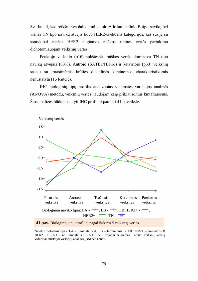
79
Svarbu tai, kad reikšminga dalis luminalinio A ir luminalinio B tipo navikų bei
vienas TN tipo navikų atvejis buvo HER2-G-didelis kategorijos, kas susiję su
santykinai mažos HER2 teigiamos raiškos ribinės vertės parinkimu
dichotomizuojant veiksnių vertes.
Penktojo veiksnio (p16) aukštesnės raiškos vertės dominavo TN tipo
navikų atvejais (83%). Antrojo (SATB1/HIF1α) ir ketvirtojo (p53) veiksnių
sąsajų su įprastinėmis krūties duktalinės karcinomos charakteristikomis
nenustatyta (15 lentelė).
IHC biologinių tipų profilis analizuotas vienmatės variacijos analizės
(ANOVA) metodu, veiksnių vertes naudojant kaip priklausomus kintamuosius.
Šios analizės būdu nustatyti IHC profiliai pateikti 41 paveiksle.
Biologiniai naviko tipai: LA - , LB - , LB HER2+ - ,
HER2+ - , TN -
41 pav. Biologinių tipų profiliai pagal išskirtų 5 veiksnių vertes
Naviko biologinis tipas: LA – luminalinis A, LB – luminalinis B, LB HER2+ –luminalinis B
HER2+, HER2+ – ne luminalinis HER2+, TN – trejopai neigiamas. Pateikti veiksnių verčių
vidurkiai, nustatyti variacijų analizės (ANOVA) būdu.
Veiksnių vertės
Pirmasis Antrasis Trečiasis Ketvirtasis Penktasis
veiksnys veiksnys veiksnys veiksnys veiksnys
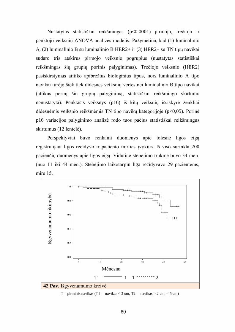
80
Nustatytas statistiškai reikšmingas (p<0.0001) pirmojo, trečiojo ir
penktojo veiksnių ANOVA analizės modelis. Pažymėtina, kad (1) luminalinio
A, (2) luminalinio B su luminalinio B HER2+ ir (3) HER2+ su TN tipų navikai
sudaro tris atskirus pirmojo veiksnio pogrupius (nustatytas statistiškai
reikšmingas šių grupių porinis palyginimas). Trečiojo veiksnio (HER2)
pasiskirstymas atitiko apibrėžtus biologinius tipus, nors luminalinio A tipo
navikai turėjo šiek tiek didesnes veiksnių vertes nei luminalinio B tipo navikai
(atlikus porinį šių grupių palyginimą, statistiškai reikšmingo skirtumo
nenustatyta). Penktasis veiksnys (p16) iš kitų veiksnių išsiskyrė ženkliai
didesnėmis veiksnio reikšmėmis TN tipo navikų kategorijoje (p<0,05). Porinė
p16 variacijos palyginimo analizė rodo tuos pačius statistiškai reikšmingus
skirtumus (12 lentelė).
Perspektyviai buvo renkami duomenys apie tolesnę ligos eigą
registruojant ligos recidyvo ir paciento mirties įvykius. Iš viso surinkta 200
pacienčių duomenys apie ligos eigą. Vidutinė stebėjimo trukmė buvo 34 mėn.
(nuo 11 iki 44 mėn.). Stebėjimo laikotarpiu liga recidyvavo 29 pacientėms,
mirė 15.
42 Pav. Išgyvenamumo kreivė
T – pirminis navikas (T1 – navikas ≤ 2 cm, T2 – navikas > 2 cm, < 5 cm)
Išg
yv
enam
um
o t
ikim
ybė
Mėnesiai
T 1 T 2

81
Atlikta Kaplano-Meierio ir Coxo regresijos analizė naudojant klinikos,
patologijos, IHC ir molekulinių žymenų rinkinius, leido nustatyti tik patikimą
naviko stadijos (T) įtaką išgyvenamumui nesant recidyvo: T2 stadija buvo
susijusi su blogesne prognoze (42 pav., p<0,05). Kitų patikimų prognozinių
požymių nustatyti nepavyko dėl santykinai trumpos stebėjimo trukmės ir
nedidelio recidyvų dažnio ankstyvosios duktalinės krūties karcinomos
pacienčių grupėje. Tirtų pacienčių stebėsena yra tęsiama.
4.7. Krūties duktalinių karcinomų, turinčių hormonų receptorių raišką,
imunofenotipo faktorinė analizė
HR+ navikų imunofenotipo analizė atlikta analizuojant 85 pacienčių
luminalinio A, luminalinio B ir luminalinio B HER2+ tipo navikų mėginius.
Rotuotų veiksnių struktūra pateikta 15 lentelėje.
15 lentelė. Krūties duktalinės karcinomos grupės, turinčios hormonų receptorių
raišką, rotuotų veiksnių struktūra
Pirmasis
veiksnys
Antrasis
veiksnys
Trečiasis
veiksnys
Ketvirtasis
veiksnys
Penktasis
veiksnys
ER -0,47598 -0,10090 -0,60245 -0,10337 -0,16151 PR -0,35541 -0,67714 -0,31955 0,29194 0,00229
AR -0,55499 -0,39729 -0,39127 0,24653 0,27274
BCL2 -0,48636 -0,34242 -0,31599 -0,38459 -0,01865 HER2 0,00251 0,87192 -0,15417 0,25187 0,08731
Ki67 -0,01734 -0,04969 0,82934 0,10271 -0,15723
p53 0,04843 0,05677 0,12929 0,88141 -0,12610
p16 -0,03786 0,06404 -0,09880 -0,12972 0,95100 HIF1α 0,84950 0,06950 0,12918 -0,09585 0,05170
SATB1 0,87715 -0,08281 -0,20321 0,16228 -0,07999
ER – estrogenų receptorius, PR – progesteronų receptorius, HER2 – žmogaus epidermio augimo
faktoriaus receptorius 2, AR – androgenų receptorius, Ki67 – ląstelių proliferacijos žymuo, p53 ir p16
– vėžio supresoriai, BCL2 – apoptozę slopinantis baltymas, SATB1 – transkripcijos veiksnys, HIF1α
– atsaką į hipoksiją kontroliuojantis transkripcijos veiksnys. Pateikta nustatytų veiksnių krūvių struktūra.
Pirmojo ir antrojo veiksnio krūviai pateikti 43 ir 44 paveiksluose. Visi
penki šie veiksniai kartu paaiškino 77% duomenų rinkinio variacijų.

82
43 pav. Krūties duktalinių karcinomų, turinčių hormonų
receptorių raišką, rotuotų veiksnių struktūra: pirmojo ir
antrojo veiksnių krūviai
ER – estrogenų receptorius, PR – progesteronų receptorius, HER2 – žmogaus epidermio augimo faktoriaus receptorius 2, AR – androgenų receptorius, Ki67 –
ląstelių proliferacijos žymuo, p53 ir p16 – vėžio supresoriai, BCL2 – apoptozę
slopinantis baltymas, SATB1 – transkripcijos veiksnys, HIF1α – atsaką į hipoksiją
kontroliuojantis transkripcijos veiksnys.
44 pav. Krūties duktalinių karcinomų, turinčių hormonų
receptorių raišką, rotuotų veiksnių struktūra: pirmojo ir
trečiojo veiksnių krūviai
Tre
čias
is v
eiksn
ys
(19.6
6.%
)
Pirmasis veiksnys (31.06%)
Pirmasis veiksnys (31,06%)
Antr
asis
vei
ksn
ys
(19,8
1%
)

83
ER – estrogenų receptorius, PR – progesteronų receptorius, HER2 – žmogaus
epidermio augimo faktoriaus receptorius 2, AR – androgenų receptorius, Ki67 –
ląstelių proliferacijos žymuo, p53 ir p16 – vėžio supresoriai, BCL2 – apoptozę
slopinantis baltymas, SATB1 – transkripcijos veiksnys, HIF1α – atsaką į hipoksiją
kontroliuojantis transkripcijos veiksnys.
Pirmasis veiksnys (31,1% variacijų) buvo panašus į antrąjį veiksnį
(SATB1/HIF1α) visoje tirtų pacientų grupėje, tačiau veiksnio struktūra -
skirtinga: šį veiksnį charakterizuoja stiprūs teigiami HIF1α krūviai, identiški
SATB1 ir vidutiškai neigiami AR, ER ir BCL2 krūviai. Ši veiksnio struktūra
rodo atvirkštinį ryšį tarp SATB1 ir HIF1α bendrosios raiškos ir AR, ER ir
BCL2 bendrosios raiškos HR+ krūties duktalinės karcinomos mėginiuose.
Kadangi biologinė šio reiškinio prasmė nėra aiški, veiksnys pavadintas
“SATB1/HIF1α–AR/ER/BCL2”.
Antrasis ir trečiasis veiksniai vienodomis dalimis paaiškino turimo
duomenų rinkinio variacijas (atitinkamai 19,8 ir 19,7%). Antrasis veiksnys
charakterizuojamas teigiamu HER2 (0,87) ir neigiamu PR (-0,68) krūviais, o
trečiasis veiksnys – teigiamu Ki67 (0,83) ir neigiamu ER (-0,60) krūviais.
Atitinkamai šie veiksniai buvo pavadinti HER2-PR ir Ki67-ER.
Ketvirtasis (p53) ir penktasis (p16) veiksniai buvo panašūs į anksčiau
nustatytus veiksnius visoje tirtų mėginių grupėje ir paaiškino atitinkamai 15,7
ir 13,8% duomenų rinkinio skirtumus.
Visų veiksnių vertės turėjo normalų pasiskirstymą ir buvo
dichotomizuotos ties 0 ribine verte.
4.8. Krūties duktalinių karcinomų, turinčių hormonų receptorių raišką,
atrinktų imunohistocheminių žymenų derinių sąsajos su krūties
duktalinės karcinomos charakteristikomis
Pirmasis naviko veiksnys (SATB1/HIF1α–AR/ER/BCL2) nebuvo susijęs
nei su pacientų amžiaus grupėmis, nei su T, N, G, nei su biologiniais naviko
tipais (14 lentelė). Antrasis veiksnys (HER2-PR-didelis) verčių kategorijoje
buvo susijęs su vyresnių pacienčių amžiaus grupe (p<0,002) ir biologiniu
naviko tipu (p<0,05).

84
Trečiasis veiksnys (Ki67-ER) buvo panašus į pirmąjį veiksnį (i-G) visoje
tirtų mėginių: didelės vertės buvo susijusios su naviko diferenciacijos laipsniu
(p<0,0001, 45 pav.) ir dažniau nustatytas luminalinio B navikų tipo mėginiuose
(p<0,0001). Pažymėtina, kad kai kurie Ki67-ER-didelis grupės navikai buvo
aptinkami ir G1 (3/19, 16%), ir luminalinio A (9/43, 21%) navikų
kategorijose.
45 pav. Pirmojo veiksnio - Ki67-ER sąsajos su naviko diferenciacijos
laipsniu
4.9. Krūties duktalinės karcinomos imunofenotipo klasterinė analizė ir
pagrindinių klasterių charakteristikos
Klasterinė analizė buvo atlikta vidurkio sąsajų metodu klasterizuojant
faktorinės analizės metu išskirtų penkių veiksnių verčių erdvėje.
Klasterizacijos medžio diagrama pateikta 46 pav.
Išskyrus 10 klasterių, didžioji dalis navikų pateko į 3 pagrindinius
klasterius. Išskirtų klasterių palyginimas su biologiniais tipais (16 lentelė)
parodė, kad 1-ame klasteryje (n=37) dominavo luminalinio A tipo navikai
(15/37, 41%), tačiau buvo nemažai ir luminalinio B HER2+ tipo atvejų (10/37,
G
Pirmasis naviko veiksnys Ki67-ER:
Ki67-ER-mažas Ki67-ER-didelis

85
27%). 2-ąjį klasterį (n=35) sudarė daugiausia luminalinio A (22/35, 61%) ir
luminalinio B tipo navikai (10/35, 29%). 3-asis klasteris (n=18) buvo beveik
išimtinai iš TN navikų (16/18, 89%). Likusius negausius 7 klasterius sudarė
daugiausia luminalinio B ir luminalinio A tipų navikai (19/109, 17%).
46 pav. Klasterizacijos medžio diagrama
16 lentelė. Klasterių palyginimas su naviko biologiniais tipais
Klasteris LA
n (%)
LB
n (%)
LB HER2+
n (%)
HER2+
n (%)
TN
n (%)
n
1 15 (40) 6 (16) 10 (27) 5 (14) 1 (3) 37
2 22 (63) 10 (28) 3 (9) 0 (0) 0 (0) 35
3 0 (0) 2 (11) 0 (0) 0 (0) 16 (89) 18 4 3 (75) 0 (0) 1 (25) 0 (0) 0 (0) 4
5 2 (25) 4 (50) 1 (12) 0 (0) 1 (13) 8
6 0 (0) 2 (100) 0 (0) 0 (0) 0 (0) 2 7 0 (0) 2 (100) 0 (0) 0 (0) 0 (0) 2
8 0 (0) 0 (0) 0 (0) 1 (100) 0 (0) 1
9 0 (0) 1 (100) 0 (0) 0 (0) 0 (0) 1 10 1 (100) 0 (0) 0 (0) 0 (0) 0 (0) 1
Naviko biologinis tipas: LA – luminalinis A, LB – luminalinis B, LB HER2+ – luminalinis B
HER2+, HER2+ – ne luminalinis HER2+, TN – trejopai neigiamas.
Tirti mėginiai

86
Pagrindinių klasterių ir biologinių tipų atitikimas buvo esminis, nors ir
neabsoliutus. Ši sąsaja tarp skaitmeninės IHC analizės duomenų ir krūties
karcinomos biologinių tipų patvirtina daugiamatės IHC analizės galimybes
krūties vėžio heterogeniškumui tyrinėti. Tačiau kadangi didžioji mėginių dalis
yra HR+ navikų (luminalinio A ir luminalinio B tipų) mėginiai, matome, kad
atlikti išsamiai analizei reikalinga didesnė mėginių imtis. Dėl šios priežasties
turima mėginių imtis toliau analizuota nebuvo.
5. REZULTATŲ APTARIMAS
Krūties duktalinėje karcinomoje tyrėme baltymų raišką (ER, PR, AR,
HER2, Ki67, SATB1, BCL2, HIF, p16, p53) IHC metodu, HER2 geno raišką
vertinome FISH metodu, genetines (TP53, HER2) ir epigenetines (ESR-1, p14,
p16, RARB, RASSF1, DAPK1, GSTP1, MGMT) genų pažaidas analizavome
sekoskaitos, DNR grandinės konformacijos, metilinimui jautrios PGR ir
bisulfitinės sekoskaitos tyrimo metodais. Baltymų raiška vertinta patologo
vizualiai ir skaitmeninės vaizdo analizės metodu, o gauti kiekybiniai IHC
žymenų duomenys analizuoti daugiamatės statistinės analizės metodais. Visų
atliktų analizių rezultatai palyginti atsižvelgiant į atskirus krūties vėžio
biologinius tipus. Nustatytos statistiškai reikšmingos sąsajos tarp biologinio
naviko tipo ir ligos stadijos (p<0,00002), naviko diferenciacijos laipsnio
(p<0,0001) ir metastazių sritiniuose limfmazgiuose (p<0,0002), IHC žymenų,
išskyrus p53, raiška buvo statistiškai reikšmingai susijusi su biologiniu krūties
naviko tipu (p<0,05). Darbe nustatyti statistiškai reikšmingi biologinių žymenų
tarpusavio ryšiai, atskleistos naujos įžvalgos apie krūties vėžio biologiją bei
naviko heterogeniškumą.
Iki šiol pripažinti ir kasdieninėje praktikoje rekomenduojami tik keli
ankstyvajam krūties vėžiui gydyti svarbūs biologiniai žymenys, turintys
patikimą prognozinę vertę – tai ER, PR ir HER2. Nuo 2011 metų, parenkant

87
individualizuotą krūties vėžio gydymą, rekomenduojama atsižvelgti į naviko
proliferacijos laipsnį (Ki67) [18]. Šiuo metu jau aišku, kad, siekiant anksti
diagnozuoti onkologinę ligą, parinkti tinkamas gydymo strategijas ir
prognozuoti ligos eigą, būtini nauji molekuliniai krūties vėžio biologiniai
žymenys [25, 72, 147-150].
Pastaruoju metu atlikta daug tyrimų, kuriuose analizuojamos sąsajos tarp
krūties vėžio biologinių tipų ir ligos prognozės [23, 151, 152], genetinių
pokyčių [153] bei atsako į terapiją [154, 155]. Tyrimai, kuriuose detaliai
analizuotas biologinis navikinio audinio profilis, išimtinai atlikti naudojant
DNR/RNR mikrogardelių (angl. microarrays) technologiją, kuri iki šiol dėl
tiriamojo mėginio paruošimo sudėtingumo vis dar retai naudojama klinikinėje
praktikoje [156, 157]. Savo darbe navikinio audinio profilį analizavome
derindami plačiai klinikinėje praktikoje taikomas technologijas (IHC, FISH
tyrimai) su naujais tyrimo metodais (audinių mikrogardelių, skaitmeninio
vaizdo, genetinių ir epigenetinių pažaidų analizės).
Mažesnėje duktalinių karcinomų grupėje (n=106) tirti DNR metilinimo
pakitimai genų promotorinėse sekose, analizuoti dažniausiai mutuojantys TP53
egzonai (nuo penkto iki devinto imtinai). Dažniausiai hipermetilinami genai
duktalinėse krūties karcinomose buvo RASSF1 ir ESR1, nustatytas jų
hipermetilinimo dažnis didesnis kaip 50 procentų. Kiek rečiau buvo
hipermetilinami genų MGMT, RARB, GSTP1 ir DAPK1 promotoriai, pakitimų
dažnis buvo apie 20% atvejų. Epigenetinis geno RASSF1 nutildymas yra vienas
iš dažniausių molekulinių pakitimų krūties karcinomose. Geno hipermetilinimo
dažniai, apie kurių nustatymą skelbta įvairiose publikacijose, svyruoja nuo 42
iki 85 procentų [127, 128, 131, 158, 159]. Mūsų tyrime RASSF1
hipermetilinimas statistiškai reikšmingai koreliavo su naviko ląstelių
diferenciacija. Estrogenų receptoriaus geno (ESR1) raiškos pakitimas krūties
karcinomose – taip pat dažnas molekulinis pakitimas. Raiškos variacijas
sąlygoja daugelis veiksnių, tarp jų ir promotorinių sekų metilinimas. ESR1
promotorius – sudėtinga struktūra, o geno raiškai reikšmingiausios CpG salos
iki šiol nėra išsamiai ištirtos. Atliekant mūsų tyrimą analizuoti du ESR1

88
promotoriaus fragmentai, tačiau tik ESR1-4 sekos hipermetilinimas statistiškai
reikšmingai koreliavo su estrogenų receptoriaus raiška ir buvo būdingas
luminalinio tipo navikams. Su biologiniais krūties navikų tipais statistiškai
reikšmingai koreliavo genų ESR1-4 ir p16 hipermetilinimas bei geno TP53
mutacijos. Geno TP53 mutacijų dažniai buvo didžiausi tarp TN kategorijos
navikų. Epigenetinių pažaidų analizė atskleidė naujas biologinių krūties navikų
tipų charakteristikas ir parodė epigenetinių pažaidų svarbą vertinant ligos eigą.
Įvertinome geno TP53 mutacijų krūties duktalinės karcinomos
mėginiuose dažnį. Nors mutacijų dažnis nebuvo ypač didelis (28%), tačiau
mutacijos statistiškai reikšmingai koreliavo su IHC tyrimu nustatyta baltymo
p53 raiška, siejosi su ER ir Ki67 baltymų raiška bei su naviko diferenciacijos
laipsniu.
Analizuodami krūties duktalinės karcinomos mėginius, sudėtus į audinių
mikrogardeles tam, kad įvertintume dešimties IHC žymenų raišką, atlikome
skaitmeninių vaizdų analizę, kuri leido patikimai išskirti navikų biologinius
tipus ir suteikė papildomos informacijos apie šių tipų navikų biologinę elgseną.
Skaitmeninė analizė, kitaip nei įprastinis patologo vizualus vertinimas, leidžia
IHC žymenų raišką išmatuoti kiekybiniais parametrais. Tai iš esmės nauja
technologija, suteikianti papildomų galimybių detaliau apibūdinti konkrečių
žymenų raišką ir įvertinti jų tarpusavio sąsajas. Objektyvus žymenų kiekybinis
įvertinimas skaitmeninės vaizdų analizės būdu yra esminis žingsnis
individualizuojant krūties vėžio gydymą.
Gautas kiekybines skaitmeninės analizės vertes lyginome su įprastiniu
patologo vertinimu bei stereologiniu ir FISH tyrimo metodu nustatytu tiesos
kriterijumi, kitaip „aukso standartu“. Skaitmeninė analizė labai stipriai
koreliavo su stereologiniame tyrime dalyvavusių visų trijų tyrėjų vertinimais
(r=0,95) ir kiek silpniau su patologo vertinimu (r=0,86). Visų tirtų IHC
žymenų skaitmeninės analizės ir įprastinio patologo vertinimo koreliacija labai
stipri. Iš esmės mūsų tyrime atliktos skaitmeninės analizės rezultatų sutapimas
su įprastiniu vizualiu patologo vertinimu vienas didžiausių, palyginti su kitų
tyrėjų publikuotais duomenimis [31, 32, 35, 36, 160-163].

89
Kaip kituose tyrimuose [32, 160, 161], taip ir mūsų darbe HER2 žymens
skaitmeninės IHC analizės ir FISH tyrimo didžiausios vertinimo rezultatų
variacijos nustatytos HER2 IHC 2+ kategorijoje. Kitaip nei kiti tyrimai [164,
165], kurie rodo, kad skaitmeninė analizė dėl didesnio šio testo specifiškumo
leidžia atsisakyti dalies FISH tyrimų, mūsų duomenimis, atlikta skaitmeninė
analizė pareikalavo daugiau FISH tyrimų. Tačiau atlikus skaitmeninę analizę
daugiau naviko mėginių pateko ne tik į HER2 IHC 3+ kategoriją (sutaupyti
FISH tyrimai), bet ir iš HER2 IHC 0/1+ navikų kategorijos keli mėginiai
perkelti į HER2 IHC 2+ navikų kategoriją. Atlikus papildomus FISH tyrimus
nustatyta HER2 geno amplifikacija. Todėl apibendrinant galima pasakyti, kad
skaitmeninė analizė ne tik naudinga IHC žymenų raiškos vertinimo, bet ir
svarbi IHC tyrimų kokybės kontrolės priemonė, kuri leidžia ne tik tiksliai,
atkartojamai, bet ir kiekybiškai įvertinti IHC žymenų raišką naviko audiniuose.
Daugiamatė IHC vaizdų skaitmeninė analizė leido nustatyti svarbius
biologinių žymenų ryšius navikiniame audinyje. Atlikę išsamų duktalinės
krūties karcinomos mėginių tyrimą mažesnėje pacienčių grupėje (n=109) –
išanalizavę dešimties IHC žymenų raišką – nustatėme reikšmingus biologinių
žymenų tarpusavio ryšius ir heterogenišką jų pasiskirstymą krūties navikiniame
audinyje. Nustatyti ryšiai atitinka ir papildo kitų tyrėjų sukauptą informaciją
apie krūties vėžio biologinius tipus, padeda geriau suvokti ligos kilmę bei
suteikia naujų duomenų ir įžvalgų apie krūties vėžio biologinius procesus.
IHC tyrimo skaitmeninės analizės rezultatus analizavome naudodami
faktorinę ir klasterinę analizę. Faktorinė analizė kiekviename tirtame navike
leido atskleisti vidinius netiesioginius imunofenotipą nulemiančius
dėsningumus, kuriuos galima vadinti nepriklausomais biologiniais procesais,
darančiais įtaką tolesnei ligos eigai. Klasterinė analizė – natūraliai klasifikuoja
tiriamuosius mėginius daugiamatėje erdvėje. Atliekant mūsų tyrimą nustatyti
imunofenotipo įvairovę nulemiantys veiksniai ne tik atitinka kitų tyrėjų
radinius, bet ir suteikia papildomai naujos informacijos. Mūsų tyrimo metu
nustatyti veiksniai, kurie nulemia imunofenotipo variacijas, suteikia naujos

90
informacijos apie navikinį audinį, tačiau taip pat yra neatsiejami nuo dabartinių
žinių apie krūties vėžio biologinius procesus.
Savo tyrimais nustatėme, kad svarbiausias IHC žymenų profilio variacijų
veiksnys krūties duktalinėje karcinomoje – stiprus atvirkštinis ryšys tarp
hormonų receptorių (ER, PR, AR) raiškos ir antiapoptozinių žymenų:
vienavertus – BCL2, kitavertus – Ki67 (proliferacijos žymuo) ir HIF1α (atsaką
į hipoksiją kontroliuojantis transkripcijos veiksnys). Nors atliekant porines
duomenų koreliacijas buvo aptiktos atitinkamos koreliacijos, žymenų
tarpusavio ryšiai buvo silpni ir dėl daugybinių tarpusavio sąveikų – sunkiai
interpretuojami. Pirmąjį vidinį veiksnį, kuris paaiškina didžiąją dalį IHC
duomenų variacijų, mes pavadinome – i-G kadangi šio veiksnio struktūra
atitinka žinomas IHC žymenų išsidėstymo variacijas nuo naviko agresyvumą
sąlygojančių (Ki67, HIF1α) iki mažesnį agresyvumą sąlygojančių (HR, BCL2)
[166]. Iš dalies biologinė i-G reikšmė apibūdinama anti-
apoptozine←→proliferacine naviko elgsena. Svarbu tai, kad antiapoptozinis
naviko poveikis yra glaudžiai susijęs su HR raiška, o naviko proliferacija – su
padidėjusio hipoksinio streso ir angiogenezės rodikliu (HIF1α). Pirmojo
veiksnio (i-G) biologinė prigimtis pasitvirtino nustačius bimodalinį šio
veiksnio verčių pasiskirstymą, stiprią koreliaciją su geresniu naviko
diferenciacijos laipsniu ir su agresyvesniais biologiniais naviko tipais (HER2+
ir TN). i-G veiksnys gali būti integruojams į esamas krūties vėžio žymenų
sistemas kaip žymuo, leidžiantis išskirti agresyvias duktalinės karcinomos
subgrupes iš jau esamų grupių.
Pirmojo veiksnio (i-G) struktūra atskleidžia svarbias sąveikas tarp HR,
BCL2, Ki67 ir HIF1α. Mūsų duomenys patvirtina, kad BCL2 gali būti
naudingas žymuo, papildantis navikų vertinimo schemas, apie ką neseniai
paskelbė Dawson su bendraautoriais [106]. Taip pat sudėtinis mitozių/BCL2 ar
Ki67/BCL2 indeksas atspindi tikrąją krūties vėžio biologinę variaciją ir gali
suteikti reikšmingos prognozinės informacijos [152, 167]. Šie neseni
publikuoti pastebėjimai pagrįsti pusiau kiekybinėmis dviejų biologinių žymenų
raiškos vertinimo reikšmėmis; Ki67/BCL2 indeksas buvo apskaičiuotas iš Ki67

91
atėmus BCL2 raiškos vertinimo vertes. Mūsų duomenimis, pirmojo veiksnio
(i-G) vertės labai stipriai koreliavo (r=0,89, p<0,0001) su Ki67 ir BCL2
baltymų raiškos skirtumu (procentas ląstelių, kuriose nustatyta Ki67 baltymo
raiška, minus procentas ląstelių, kuriose nustatyta BCL2 baltymo raiška).
Kitaip negu kiti autoriai [152], Ki67/BCL2 indeksui apskaičiuoti naudojome
kiekybinius analizės duomenis ir daugiamatę statistiką. Papildomai mūsų
duomenys parodė, kad įtrauktas į šią analizę HIF1α žymuo suteikia naujos
informacijos apie naviko vystymosi eigą.
Grupėje navikų, kuriuose nustatyta padidėjusi hormonų receptorių raiška
– HR+ (LA, LB ir LB HER2+) – išskirtas trečiasis veiksnys (Ki67–ER), savo
struktūra panašus į pirmąjį i-G veiksnį. Trečiasis veiksnys, kaip ir pirmasis,
statistiškai patikimai koreliuoja su naviko diferenciacijos laipsniu ir biologiniu
tipu. Vertindami “agresyvumo indeksą” HR+ navikų grupėje, nustatėme, kad
trečiasis veiksnys (Ki67-ER) nebebuvo pagrindinis variacijų šaltinis – veiksnio
vertės nebuvo pasiskirsčiusios bimodališkai. Šis pokytis galėtų būti
aiškinamas sumažėjusios Ki67 raiškos variacija tiriamojoje navikų grupėje, iš
kurios pašalinti labiau proliferuojančių tipų navikai (TN ir HER2+). Antra
vertus, trečiojo veiksnio struktūra leidžia manyti, kad naviko agresyvumą HR+
navikų grupėje tiksliau apibūdina Ki67/ER indeksas nei Ki67/BCL2 indeksas.
Kitaip sakant, ER raiška yra glaudžiai susijusi su BCL2 žymens raiška, todėl
kartu su Ki67 žymeniu tinkamai charakterizuoja HR+ navikus. Nors klinikinei
praktikai BCL2, kaip prognozinis žymuo, buvo pasiūlytas anksčiau [106, 168,
169], vertindami savo duomenis, matome, kad šio žymens įtaka navikui
formuotis turėtų būti detaliau išanalizuota atlikus didesnės apimties tyrimus
naudojant daugiamatę duomenų analizę. Trečia, nepaisant to, kad Ki67-ER
veiksnys stipriai koreliuoja su naviko diferenciacijos laipsniu ir biologiniu tipu,
dalis geros diferenciacijos (G1) navikų ir luminalinio A tipo grupės navikai
pateko į Ki67-ER-G2 navikų kategoriją (atitinkamai, 16, 21% pacienčių
mėginiai). Tai leidžia manyti, kad naudojamas sudėtinis žymenų indeksas
suteikia papildomos informacijos, apsaugančios nuo klaidingo baltymų raiškos
interpretavimo, ypač paribinės raiškos vertinimo atvejais. Yra paskelbta, kad

92
pagal Ki67 indeksą vidutinio diferenciacijos laipsnio (G2) navikus galima
suskirstyti į dvi atskiras žemos ir aukštos rizikos grupes [170]. Taip pat
manome, kad sudėtinio indekso naudojimas klinikinėje praktikoje leistų
tiksliau išskirti luminalinio A ir luminalinio B tipo navikų grupes prieš
priimant sprendimą dėl pacienčių gydymo.
HER2 baltymo raiška krūties duktalinės karcinomos mėginių grupėje
buvo nepriklausoma nuo kitų IHC parametrų, tačiau HR+ mėginių grupėje
HER2 raiška dažniausiai konkuravo ne su ER, o su PR. Šiuos intriguojančius
radinius kol kas sunku interpretuoti, kadangi nėra pakankamai informacijos
apie nepriklausomą ER ir PR (taip pat ir BCL2) žymenų reikšmę bei šių
žymenų tarpusavio koreliacijas. PR raiškos nebuvimas rodo blogesnę
prognozę, palyginti su prognoze navikų, kuriuose nustatyta padidėjusi ER/PR
receptorių raiška, tuotarpu tirdami mėginius įvairiais BCL2, p53, HER2
žymenų deriniais gauname papildomos prognozinės informacijos, apie ką
nesenai užsiminė Rakha su bendraautoriais [166]. Atlikdami savo tyrimą
nustatėme porines koreliacijas tarp ER, PR ir BCL2, tačiau atlikta faktorinė
analizė atskleidė tikruosius vidinius tarpusavio žymenų ryšius: krūties
duktalinės karcinomos mėginiuose patvirtinome, kad HR ir BCL2 žymenys
pasižymi stipriais tarpusavio ryšiais, valdomais tų pačių vidinių veiksnių ir
kertu būdingu priešingu – Ki67 ir HIF1α žymenų raiškos ryšiu. Didelės ER
raiškos navikai HR+ navikų grupėje rasti mažiau priliferuojačiuose (Ki67)
navikuose, o maža PR raiška gali žymėti didesnę HER2 baltymo raišką. Todėl
ER ir PR raiškos skirtumai gali atspindėti proliferacinio aktyvumo bei HER2
baltymo raiškos skirtumus HR+ navikų grupėje, lydimus susijusios
prognozinės informacijos (jei didėjanti HER2 baltymo raiška vertintina kaip
didėjantis naviko agresyvumo veiksnys). Kaip pažymėta anksčiau, mūsų
tyrime iškelta prielaida, kad BCL2 raiška patikimai koreliuoja su HR raiška
(visoje tirtoje ir HR+ navikų grupėse) pasitvirtino turimame nepriklausomų
duomenų rinkinyje.
Mūsų tyrimas parodė galimą naujų žymenų reikšmę krūties vėžio
diagnostikoje. SATB1 – genomo organizatorius, kuris reguliuoja fermentų

93
veiklą chromatino struktūros persitvarkymo ir genų raiškos pokyčių procesų
metu. Yra nuomonių, kad šie pokyčiai krūties vėžio atvejais skatina metastazių
atsiradimą ir rodo blogesnę pronozę [171, 172]. Kitų tyrėjų darbais tai nebuvo
patvirtinta, todėl SATB1 žymens prognozinė reikšmė ir pritaikymas klinikinėje
praktikoje išlieka kontroversiškas [103, 173-175]. Negana to, išanalizavus
2058 krūties vėžio mėginių SATB1 žymens mRNR raišką, šios raiškos sąsajos
su pacienčių išgyvenimu iki prasidedant ligos atkryčiui ER– navikų grupėje
nenustatytos, kita vertus, didelė SATB1 raiška ER+ navikų grupėje buvo
susijusi su geresne ligos prognoze; vis dėlto net ir ER+ navikų grupėje
naudojant standartinius analizės daugiavariacinius parametrus nebuvo
nenustatyta nepriklausoma šio žymens prognozinė vertė [174]. Patani ir
bendraautorių atliktame tyrime nustatyta, kad didesnė SATB1 raiška dažniau
randama ER- navikų grupėje [172]. Mūsų tyrimas pirmą kartą parodo
akivaizdžią SATB1 baltymo raiškos, nustatytos IHC metodu, koreliaciją su
ER ir kitais žymenimis. Atliktame darbe išskirto pirmojo veiksnio
(SATB1/HIF-1α-AR/ER/BCL2) struktūra HR+ navikų grupėje atskleidė
priešingus ryšius tarp dviejų skirtingų žymenų grupių. Iš tiesų tikėtina, kad
prognozinis SATB1 žymens poveikis gali būti apibūdinamas priešingu ER
raiškos ryšiu [174]. Negana to, mes neradome jokių asociacijų tarp SATB1
raiškos ir naviko diferenciacijos laipsnio ar kitų tirtų naviką apibūdinančių
charakteristikų. Nepaisant to, įdomu, kad SATB1 raiška yra glaudžiai susijusi
su HIF1α tiek HR+, tiek visoje krūties duktalinės karcinomos navikų grupėje;
veiksniai, kuriuose dalyvauja SATB1 ir HIF1α žymenys, nulemia didžiąją dalį
variacijų abiejose tiriamosiose grupėse, ypač HR+ navikų grupėje.
HIF1α baltymo raiška yra nustatoma daugelyje žmogaus vėžio rūšių ir
koreliuoja su blogesne prognoze. Šis baltymas daro įtaką daugeliui procesų –
naviko iniciacijai, progresijai, invazijai, uždegiminių ląstelių telkimuisi,
metastazavimui ir gali būti krūties vėžio gydymo taikiniu [176]. Yamamoto su
bendraautoriais [177], išanalizavę 171 invazinės krūties karcinomos mėginį, 37
procentuose atvejų rado HIF1α baltymo raišką (šiame, kaip ir ankstesniame
tyrime buvo naudota 5 procentų teigiamos raiškos ribinė vertė [178]). HIF1α
94
baltymo raiška labai susijusi su agresyvesniu naviko fenotipu, taip kaip su
aukštesniu naviko diferenciacijos laipsniu, metastazėmis limfmazgiuose,
dideliu naviko dydžiu, dideliu proliferacijos laipsniu, neigiama hormonų
receptorių raiška, padidėjusia HER2 ir VEGF baltymo raiška; padidėjusi
HIF1α baltymo raiška koreliuoja su trumpesniu pacienčių išgyvenimu nesant
recidyvo ir bendruoju jų išgyvenimu [177]. Sąsajos tarp HIF1α baltymo raiškos
ir nepalankios ligos prognozės esant krūties vėžiui, atskleistos ir kituose
darbuose [178-180]. Savo darbe naudodami kelis tyrimo metodus patvirtinome
sąsajas tarp HIF1α ir Ki67 baltymo raiškos krūties duktalinėje karcinomoje:
porinės koreliacijos ir pirmojo veiksnio (i-G) struktūra. Taip pat i-G veiksnio
vertės stipriai koreliavo su naviko diferenciacijos laipsniu, patvirtinančiu
agresyvesnę HIFα ir Ki67 baltymų prigimtį (patvirtinta vienfaktorine ANOVA
naudojant HIF1α ir Ki67 kaip priklausomus kintamuosius). Visgi mūsų atlikta
faktorinė analizė atskleidžia kai kuriuos ypatumus. Pirma, krūties duktalinės
karcinomos grupėje nustatėme, kad HIF1α baltymo raiška padidėjusi dviejuose
veiksniuose: pirmąjame veiksnyje (i-G) kartu su Ki67 žymeniu ir antrajame
veiksnyje – kartu su SATB1. Antra, HR+ duktalinės karcinomos navikų
grupėje HIF1α padidėjusi raiška nustatyta reikšmingiausiame pirmajame
veiksnyje kartu su SATB1 suteikdama krūvius, priešingus AR, ER ir BCL2,
nepriklausomai nuo Ki67 raiškos (trečiojo veiksnio). Tai leidžia daryti
prielaidą, kad HIF1α ir SATB1 gali atspindėti svarbią biologinę informaciją,
kitokią nei ligos agresyvumas, atspindimas Ki67 baltymo raiškos ir naviko
diferenciacijos laipsnio, bent jau HR+ navikų grupėje.
p16 baltymo raiška mūsų analizėje buvo valdoma nepriklausomo
veiksnio ir išsiskyrė didele raiška TN tipo navikuose, palyginti su kitų
biologinių tipų navikais. Taigi mes patvirtiname skelbtus duomenis [181-184],
kad padidėjusi p16 baltymo raiška TN navikų grupėje taip pat galbūt susijusi
su retinoblastomos naviko supresoriumi (Rb) ir nuo ciklinų priklausomos
kinazės inhibitoriumi. Negana to, p16 baltymo raiškos slopinimas nustatytas
kai kuriuose TN krūties vėžio ląstelių linijose, manoma, kad šias ląsteles
galima padalyti į dvi grupes pagal Rb ir p16 statusą [185]. Įdomu tai, kad

95
atlikta faktorinė analizė 17 TN navikų grupėje atskleidė stiprų p16 ryšį su Ki67
ir stiprų atvirkštinį ryšį su AR (tačiau ne su ER ir PR) raiška. Šiuos duomenis
reiktų patvirtinti ištyrus didesnę navikinių mėginių grupę.
Klasterinė duktalinės karcinomos analizė penkių variacijos veiksnių,
nustatytų IHC žymenų faktorinės analizės būdu erdvėje, leido išskirti tris
pagrindines viena nuo kitos nutolusias navikų grupes, gerai atitinkančias
biologinius subtipus: luminalinį (A ir B), HER2+ ir TN. Šie rezultatai iš esmės
atitinka anksčiau publikuotus duomenis apie klasterinės analizės galimybes
žymenų IHC analize nustatyti heterogeniškas krūties vėžio grupes. Kitaip nei
cituojamame darbe, kuriame buvo naudojami pusiau kiekybiniai parametrai,
mes naudojome kiekybinius duomenis. Kadangi mūsų tirta pacienčių grupė
buvo santykinai nedidelė ir didžioji atvejų dalis pateko į luminalinį A ir B
klasterį, manome, kad tolesniam šios navikų grupės heterogeniškumo tyrimui
reikalinga didesnė pacienčių grupė ir/arba parametrų rinkinys.
6. IŠVADOS
1. Skaitmeninė vaizdo analizė yra patikimas būdas žymenų raiškos, analizuotos
imunohistocheminiu tyrimo metodu, įvertinimui krūties vėžio audinyje. Ki67
raiškos skaitmeninės vaizdo analizės rezultatai stipriai koreliavo (r=0,86) su
patologo vertinimu ir turėjo mažesnį sisteminį nuokrypį nuo stereologiškai
nustatytų referentinių verčių. HER2 raiškos skaitmeninės vaizdo analizės
rezultatai atitiko patologo vertinimą (kappa=0,86) ir leido tiksliau nustatyti
amplifikuotus (HER2 FISH) krūties vėžio atvejus.
2. Biologinių žymenų – ER, PR, HER2, Ki67 – imunohistocheminio tyrimo
analize pagrįstas krūties duktalinių karcinomų skirstymas į biologinius tipus
padeda atskleisti krūties vėžio biologinę įvairovę:
2.1. duktalinių krūties karcinomų grupėje (n=203) nustatytos statistiškai
patikimos sąsajos tarp biologinio naviko tipo ir ligos stadijos (p<0,00002),

96
naviko diferenciacijos laipsnio (p<0,0001), metastazių sritiniuose
limfmazgiuose (p<0,0002);
2.2. genų ESR1-4 ir p16 hipermetilinimas bei geno TP53 mutacijos susijęs
(p<0,05) su biologiniu krūties vėžio tipu;
2.3. biologiniams krūties vėžio tipams būdingas savitas baltymų ER, PR, AR,
HER2, Ki67, BCL2, HIF1, SATB1, p53, p16 raiškos intensyvumo profilis:
visų, išskyrus p53, imunohistocheminių žymenų raiškos intensyvumas buvo
susijęs (p<0,05) su biologiniu krūties vėžio tipu.
3. Skaitmeninės vaizdo analizės būdu įvertinto imunohistocheminio žymenų
rinkinio daugiamatė analizė yra efektyvus tyrimo metodas, leidžiantis
atskleisti baltymų raiškos profilių dėsningumus:
3.1. duktalinių krūties karcinomų grupėje (n=109) faktorinės daugiamatės
analizės būdu identifikuotas ligos agresyvumą atitinkantis veiksnys (i-G)
pasireiškia priešingos krypties ER/PR/AR/BCL2 ir Ki67/HIF1α raiška. Šio
veiksnio vertės stipriai susijusios su naviko diferenciacijos laipsniu ir
agresyviais biologiniais tipais;
3.2. SATB1 kartu su HIF1α formuoja antrą reikšmingą variacijos veiksnį
krūties duktalinės karcinomos grupėje. Šie žymenys gali suteikti svarbios
biologinės informacijos apie krūties vėžio navikus nepriklausomai nuo Ki67
raiškos ir naviko diferenciacijos laipsnio;
3.3. hormonų receptoriams teigiamų navikų grupėje (n=85) naviko
agresyvumą geriausiai atspindi Ki67 ir ER žymenų derinys. Šis indeksas gali
suteikti papildomos prognostinės informacijos greta jau naudojamų
prognostinių grupių;
3.4. hormonų receptoriams teigiamų navikų grupėje nustatytas atvirkštinis
ryšys tarp HER2 ir PR raiškos bei tarp Ki67 ir ER raiškos atskleidžia ER ir PR
disocijuotos raiškos biologinę reikšmę.
97
7. REKOMENDACIJOS
1. Mūsų darbo rezultatai rodo, kad biologinių žymenų nustatymas audinių
mikrogardelėse, panaudojant skaitmeninės vaizdo analizės technologijas,
leidžia padidinti šių tyrimų tikslumą ir efektyvumą. Siūlome skaitmeninio
vaizdo IHC analizę naudoti kokybei užtikrinti bei sprendimams palaikyti
klinikinėje praktikoje, šias priemones validuojant ne tik pagal patologo
vertinimo rezultatus, bet ir stereologiniais tyrimo metodais.
2. Siūlome įdiegti į klinikinę praktiką integruotus IHC žymenų rinkinius
(kompleksinius žymenis), kurie atskleidžia vidinius biologinių žymenų
tarpusavio sąsajų dėsningumus ir atveria naujas galimybes prognozuoti krūties
duktalinės karcinomos ligos eigą.

98
8. PUBLIKACIJOS IR DISERTACIJOS TEMA SKAITYTI
PRANEŠIMAI
DISERTACIJOS TEMA
ISI sąrašo žurnalai
1. Laurinavicius A., Laurinaviciene A, Ostapenko V, Dasevicius D,
Jarmalaite S, Lazutka J. Immunohistochemistry profiles of breast ductal
carcinoma: factor analysis of digital image analysis data. Diagn Pathol 2012,
priimta spaudai.
2. Laurinavicius A, Laurinaviciene A, Dasevicius D, Elie N, Plancoulaine B,
Bor C, Herlin P. Digital image analysis in pathology: Benefits and obligations.
Anal Cell Pathol 34 (2011) 1–4.
3. Laurinaviciene A, Dasevicius D, Ostapenko V, Jarmalaite S, Lazutka J,
Laurinavicius A. Membrane connectivity estimated by digital image analysis
of HER2 immunohistochemistry is concordant with visual scoring and
fluorescence in situ hybridization results: algorithm evaluation on breast cancer
tissue microarrays. Diagn Pathol 2011, 6(1):87.
KITOS PUBLIKACIJOS
ISI sąrašo žurnalai
1. Jarmalaite S., A. Laurinaviciene, J. Tverkuviene, N. Kalinauskaite, D.
Petroska, T. Bohling, K. Husgafvel-Pursiainen: Tumor suppressor gene
ZAC/PLAGL1: altered expression and loss of the nonimprinted allele in
pheochromocytomas. Cancer Genetics 2011, 204(7):398-404.
Kiti referuojami leidiniai
1. J. Tverkuviene, A. Laurinaviciene, K. Daniunaitė, A. Scesnaite, S.
Jarmalaite: Frequent aberrant DNA methylation of CDKN2A locus in capillary
hemangioblastomas, pheochromocytomas and gliomas. Acta Medica
Lithuanica, 2011. Vol. 18. No. 1. P. 4–11.

99
2. A. Laurinaviciene, D. Petroska, J. Tverkuviene, K. Daniunaite, A.
Scesnaite, S. Jarmalaite: Frequent aberrant expression of p53 protein in
gliomas, but not in capillary hemangioblastomas and pheochromocytomas.
Acta Medica Lithuanica, 2009 Vol. 16. No. 3–4. P. 130–138.
3. Česas A., Stratilatovas E., Laurinavičienė A. Biožymenų svarba gydant
kolorektalinį vėžį biologine terapija cetuksimabu. Onkologija: žurnalo
Internistas priedas. 2009; 4:58-59.
4. V.Žalgevičienė, A.Laurinavičienė. Bendrosios histologijos pradmenys.
Mokomoji priemonė. Vilnius: Vaistų žinios, 2009.
5. A.Laurinavičienė, R.Smaliukienė. Histologinių technologijų vadovas.
Mokomoji priemonė. Vilnius, Eugrimas, 2007.
SKAITYTI PRANEŠIMAI
1. Doktorantų stažuotės, projektas „Studentų mokslinės veiklos skatinimas“:
Ankstyvo krūties vėžio heterogeniškumo tyrimai audiniuose, 2011-03-17.
2. Molecular pathology for diagnosis and treatment: The use of tissue
microarrays for breast cancer research – experience at the National Center of
Pathology, 2010-02-26.
3. Pathology diagnosis and treatment of Early breast cancer: Molecular
characterization of early breast cancer: preliminary results of ongoing
collaborative research project, 2009-11-27.
4. Tirozinkinazės receptoriaus ErbB-2 (HER2) teigiamo krūties vėžio gydymo
aktualijos: ErbB receptorių ir intraląstelinio signalo perdavimo biologija,
2009-02-06.
5. Tiksli krūties vėžio diagnozė iki gydymo pradžios: prognostiniai ir
predikciniai veiksniai: Molekulinių žymenų nustatymas krūties vėžio
audinyje, 2009-04-08.

100
KONFERENCIJŲ MEDŽIAGA (TEZĖS)
1. S. Jarmalaite, A. Laurinaviciene, J. Dimonaite, A. Scesnaite, D. Dasevicius,
V. Ostapenko, J. Lazutka: Epigenetic and genetic biomarkers, of early-stage
breast cancer. Cellular Oncology, 30 (3): 259-260, 2008.
2. Dimonaite, A. Laurinaviciene, D. Petroska, N. Kalinauskaite, A. Scesnaite,
S. Jarmalaite: Epigenetic changes in pathogenesis of VHL-related tumours and
primary gliomas. Cellular Oncology, 30 (3): 254-255, 2008.

101
9. CITUOJAMŲ LITERATŪROS ŠALTINIŲ SĄRAŠAS
1. Perou CM, Sorlie T, Eisen MB, van de Rijn M, Jeffrey SS, Rees CA, Pollack JR, Ross DT, Johnsen H, Akslen LA et al: Molecular portraits of human breast tumours. Nature 2000, 406(6797):747-752.
2. Prat A, Perou CM: Deconstructing the molecular portraits of breast cancer. Mol Oncol 2011, 5(1):5-23.
3. Parker JS, Mullins M, Cheang MC, Leung S, Voduc D, Vickery T, Davies S, Fauron C, He X, Hu Z et al: Supervised risk predictor of breast cancer based on intrinsic subtypes. J Clin Oncol 2009, 27(8):1160-1167.
4. Nielsen TO, Hsu FD, Jensen K, Cheang M, Karaca G, Hu Z, Hernandez-Boussard T, Livasy C, Cowan D, Dressler L et al: Immunohistochemical and clinical characterization of the basal-like subtype of invasive breast carcinoma. Clin Cancer Res 2004, 10(16):5367-5374.
5. Blows FM, Driver KE, Schmidt MK, Broeks A, van Leeuwen FE, Wesseling J, Cheang MC, Gelmon K, Nielsen TO, Blomqvist C et al: Subtyping of breast cancer by immunohistochemistry to investigate a relationship between subtype and short and long term survival: a collaborative analysis of data for 10,159 cases from 12 studies. PLoS Med 2010, 7(5):e1000279.
6. Hugh J, Hanson J, Cheang MC, Nielsen TO, Perou CM, Dumontet C, Reed J, Krajewska M, Treilleux I, Rupin M et al: Breast cancer subtypes and response to docetaxel in node-positive breast cancer: use of an immunohistochemical definition in the BCIRG 001 trial. J Clin Oncol 2009, 27(8):1168-1176.
7. Cheang MC, Chia SK, Voduc D, Gao D, Leung S, Snider J, Watson M, Davies S, Bernard PS, Parker JS et al: Ki67 index, HER2 status, and prognosis of patients with luminal B breast cancer. J Natl Cancer Inst 2009, 101(10):736-750.
8. Millikan RC, Newman B, Tse CK, Moorman PG, Conway K, Dressler LG, Smith LV, Labbok MH, Geradts J, Bensen JT et al: Epidemiology of basal-like breast cancer. Breast Cancer Res Treat 2008, 109(1):123-139.
9. Phipps AI, Chlebowski RT, Prentice R, McTiernan A, Stefanick ML, Wactawski-Wende J, Kuller LH, Adams-Campbell LL, Lane D, Vitolins M et al: Body size, physical activity, and risk of triple-negative and estrogen receptor-positive breast cancer. Cancer Epidemiol Biomarkers Prev 2011, 20(3):454-463.
10. Phipps AI, Buist DS, Malone KE, Barlow WE, Porter PL, Kerlikowske K, Li CI: Reproductive history and risk of three breast cancer subtypes defined by three biomarkers. Cancer Causes Control 2011, 22(3):399-405.
11. Liedtke C, Mazouni C, Hess KR, Andre F, Tordai A, Mejia JA, Symmans WF, Gonzalez-Angulo AM, Hennessy B, Green M et al: Response to neoadjuvant therapy and long-term survival in patients with triple-negative breast cancer. J Clin Oncol 2008, 26(8):1275-1281.
12. Dignam JJ, Dukic V, Anderson SJ, Mamounas EP, Wickerham DL, Wolmark N: Hazard of recurrence and adjuvant treatment effects over time in lymph node-negative breast cancer. Breast Cancer Res Treat 2009, 116(3):595-602.
13. Aebi S, Sun Z, Braun D, Price KN, Castiglione-Gertsch M, Rabaglio M, Gelber RD, Crivellari D, Lindtner J, Snyder R et al: Differential efficacy of three cycles of CMF followed by tamoxifen in patients with ER-positive and ER-negative tumors: Long-term follow up on IBCSG Trial IX. Ann Oncol 2011.
14. Albain KS, Barlow WE, Shak S, Hortobagyi GN, Livingston RB, Yeh IT, Ravdin P, Bugarini R, Baehner FL, Davidson NE et al: Prognostic and predictive value of the 21-gene recurrence score assay in postmenopausal women with node-positive,

102
oestrogen-receptor-positive breast cancer on chemotherapy: a retrospective analysis of a randomised trial. Lancet Oncol 2010, 11(1):55-65.
15. Nguyen PL, Taghian AG, Katz MS, Niemierko A, Abi Raad RF, Boon WL, Bellon JR, Wong JS, Smith BL, Harris JR: Breast cancer subtype approximated by estrogen receptor, progesterone receptor, and HER-2 is associated with local and distant recurrence after breast-conserving therapy. J Clin Oncol 2008, 26(14):2373-2378.
16. Tang G, Shak S, Paik S, Anderson SJ, Costantino JP, Geyer CE, Jr., Mamounas EP, Wickerham DL, Wolmark N: Comparison of the prognostic and predictive utilities of the 21-gene Recurrence Score assay and Adjuvant! for women with node-negative, ER-positive breast cancer: results from NSABP B-14 and NSABP B-20. Breast Cancer Res Treat 2011, 127(1):133-142.
17. Wo JY, Taghian AG, Nguyen PL, Raad RA, Sreedhara M, Bellon JR, Wong JS, Gadd MA, Smith BL, Harris JR: The association between biological subtype and isolated regional nodal failure after breast-conserving therapy. Int J Radiat Oncol Biol Phys 2010, 77(1):188-196.
18. Goldhirsch A, Wood WC, Coates AS, Gelber RD, Thurlimann B, Senn HJ: Strategies for subtypes--dealing with the diversity of breast cancer: highlights of the St Gallen International Expert Consensus on the Primary Therapy of Early Breast Cancer 2011. Ann Oncol 2011, 22(8):1736-1747.
19. Basavanhally AN, Ganesan S, Agner S, Monaco JP, Feldman MD, Tomaszewski JE, Bhanot G, Madabhushi A: Computerized image-based detection and grading of lymphocytic infiltration in HER2+ breast cancer histopathology. IEEE transactions on bio-medical engineering 2010, 57(3):642-653.
20. Ali S, Veltri R, Epstein JI, Christudass C, Madabhushi A: Adaptive energy selective active contour with shape priors for nuclear segmentation and gleason grading of prostate cancer. Med Image Comput Comput Assist Interv 2011, 14(Pt 1):661-669.
21. Hipp J, Flotte T, Monaco J, Cheng J, Madabhushi A, Yagi Y, Rodriguez-Canales J, Emmert-Buck M, Dugan MC, Hewitt S et al: Computer aided diagnostic tools aim to empower rather than replace pathologists: Lessons learned from computational chess. J Pathol Inform 2011, 2:25.
22. Jemal A, Siegel R, Ward E, Hao Y, Xu J, Thun MJ: Cancer statistics, 2009. CA Cancer J Clin 2009, 59(4):225-249.
23. Sorlie T, Perou CM, Tibshirani R, Aas T, Geisler S, Johnsen H, Hastie T, Eisen MB, van de Rijn M, Jeffrey SS et al: Gene expression patterns of breast carcinomas distinguish tumor subclasses with clinical implications. Proc Natl Acad Sci U S A 2001, 98(19):10869-10874.
24. Reis-Filho JS, Simpson PT, Gale T, Lakhani SR: The molecular genetics of breast cancer: the contribution of comparative genomic hybridization. Pathol Res Pract 2005, 201(11):713-725.
25. Rakha EA, Reis-Filho JS, Baehner F, Dabbs DJ, Decker T, Eusebi V, Fox SB, Ichihara S, Jacquemier J, Lakhani SR et al: Breast cancer prognostic classification in the molecular era: the role of histological grade. Breast Cancer Res 2010, 12(4):207.
26. Simpson PT, Reis-Filho JS, Gale T, Lakhani SR: Molecular evolution of breast cancer. J Pathol 2005, 205(2):248-254.
27. Kayser K: Quantification of virtual slides: Approaches to analysis of content-based image information. J Pathol Inform 2011, 2:2.
28. Mulrane L, Rexhepaj E, Penney S, Callanan JJ, Gallagher WM: Automated image analysis in histopathology: a valuable tool in medical diagnostics. ExpertRevMolDiagn 2008, 8(6):707-725.

103
29. Soenksen D: Digital pathology at the crossroads of major health care trends: corporate innovation as an engine for change. ArchPatholLab Med 2009, 133(4):555-559.
30. Cregger M, Berger AJ, Rimm DL: Immunohistochemistry and quantitative analysis of protein expression. Archives of pathology & laboratory medicine 2006, 130(7):1026-1030.
31. Joshi AS, Sharangpani GM, Porter K, Keyhani S, Morrison C, Basu AS, Gholap GA, Gholap AS, Barsky SH: Semi-automated imaging system to quantitate Her-2/neu membrane receptor immunoreactivity in human breast cancer. Cytometry A 2007, 71(5):273-285.
32. Hall BH, Ianosi-Irimie M, Javidian P, Chen W, Ganesan S, Foran DJ: Computer-assisted assessment of the Human Epidermal Growth Factor Receptor 2 immunohistochemical assay in imaged histologic sections using a membrane isolation algorithm and quantitative analysis of positive controls. BMC Medical Imaging 2008, 8(1):11.
33. Gustavson MD, Bourke-Martin B, Reilly D, Cregger M, Williams C, Mayotte J, Zerkowski M, Tedeschi G, Pinard R, Christiansen J: Standardization of HER2 immunohistochemistry in breast cancer by automated quantitative analysis. Arch Pathol Lab Med 2009, 133(9):1413-1419.
34. Tadrous PJ: On the concept of objectivity in digital image analysis in pathology. Pathology 2010, 42(3):207-211.
35. Lloyd MC, Allam-Nandyala P, Purohit CN, Burke N, Coppola D, Bui MM: Using image analysis as a tool for assessment of prognostic and predictive biomarkers for breast cancer: How reliable is it? J Pathol Inform 2010, 1:29.
36. Slodkowska J, Filas V, Buszkiewicz E, Trzeciak P, Wojciechowski M, Koktysz R, Staniszewski W, Breborowicz J, Rojo MG: Study on breast carcinoma Her2/neu and hormonal receptors status assessed by automated images analysis systems: ACIS III (Dako) and ScanScope (Aperio). Folia Histochem Cytobiol 2010, 48(1):19-25.
37. Gavrielides MA, Gallas BD, Lenz P, Badano A, Hewitt SM: Observer variability in the interpretation of HER2/neu immunohistochemical expression with unaided and computer-aided digital microscopy. Arch Pathol Lab Med 2011, 135(2):233-242.
38. Ferlay J, Shin HR, Bray F, Forman D, Mathers C, Parkin DM: Estimates of worldwide burden of cancer in 2008: GLOBOCAN 2008. Int J Cancer 2010, 127(12):2893-2917.
39. Lietuvos vėžio registras. http:/www.vuoi.lt. 40. Botha JL, Bray F, Sankila R, Parkin DM: Breast cancer incidence and mortality trends
in 16 European countries. Eur J Cancer 2003, 39(12):1718-1729. 41. Sant M, Francisci S, Capocaccia R, Verdecchia A, Allemani C, Berrino F: Time trends
of breast cancer survival in Europe in relation to incidence and mortality. Int J Cancer 2006, 119(10):2417-2422.
42. Bilimoria MM, Morrow M: The woman at increased risk for breast cancer: evaluation and management strategies. CA Cancer J Clin 1995, 45(5):263-278.
43. Bird A: DNA methylation patterns and epigenetic memory. Genes & development 2002, 16(1):6-21.
44. Esteller M: Epigenetic gene silencing in cancer: the DNA hypermethylome. Hum Mol Genet 2007, 16 Spec No 1:R50-59.
45. Scarano MI, Strazzullo M, Matarazzo MR, D'Esposito M: DNA methylation 40 years later: Its role in human health and disease. Journal of cellular physiology 2005, 204(1):21-35.
46. Das PM, Singal R: DNA methylation and cancer. J Clin Oncol 2004, 22(22):4632-4642.

104
47. Chuang JC, Jones PA: Epigenetics and microRNAs. Pediatr Res 2007, 61(5 Pt 2):24R-29R.
48. Esteller M, Corn PG, Baylin SB, Herman JG: A gene hypermethylation profile of human cancer. Cancer Res 2001, 61(8):3225-3229.
49. Esteller M: Relevance of DNA methylation in the management of cancer. Lancet Oncol 2003, 4(6):351-358.
50. Szyf M: Targeting DNA methylation in cancer. Ageing research reviews 2003, 2(3):299-328.
51. Robertson KD: DNA methylation, methyltransferases, and cancer. Oncogene 2001, 20(24):3139-3155.
52. Urruticoechea A, Dowsett M, Mackay A, Dexter T, Young O, Miller WR, Evans DB, Dixon GM: Molecular characterisation of ER plus breast cancer before and during treatment with the aromatase inhibitors, letrozole and anastrozole. Journal of Clinical Oncology 2005, 23(16):850s-850s.
53. Jimenez Ruiz CA, Barrueco Ferrero M, Solano Reina S, Torrecilla Garcia M, Dominguez Grandal F, Diaz-Maroto Munoz JL, Alonso Moreno J, La Cruz Amoros Ed E, Abengozar Muela R: [Guidelines for a diagnostic and therapeutic approach to smoking addiction. A consensus report]. Archivos de bronconeumologia 2003, 39(1):35-41.
54. Kendall A, Anderson H, Dunbier AK, Mackay A, Dexter T, Urruticoechea A, Harper-Wynne C, Dowsett M: Impact of estrogen deprivation on gene expression profiles of normal postmenopausal breast tissue in vivo. Cancer Epidemiol Biomarkers Prev 2008, 17(4):855-863.
55. Song MS, Song SJ, Ayad NG, Chang JS, Lee JH, Hong HK, Lee H, Choi N, Kim J, Kim H et al: The tumour suppressor RASSF1A regulates mitosis by inhibiting the APC-Cdc20 complex. Nature cell biology 2004, 6(2):129-137.
56. Bird AW, Yu DY, Pray-Grant MG, Qiu Q, Harmon KE, Megee PC, Grant PA, Smith MM, Christman MF: Acetylation of histone H4 by Esa1 is required for DNA double-strand break repair. Nature 2002, 419(6905):411-415.
57. Bird A: The essentials of DNA methylation. Cell 1992, 70(1):5-8. 58. Fleisher AS, Esteller M, Tamura G, Rashid A, Stine OC, Yin J, Zou TT, Abraham JM,
Kong D, Nishizuka S et al: Hypermethylation of the hMLH1 gene promoter is associated with microsatellite instability in early human gastric neoplasia. Oncogene 2001, 20(3):329-335.
59. Esteva FJ, Hortobagyi GN: Prognostic molecular markers in early breast cancer. Breast Cancer Res 2004, 6(3):109-118.
60. Thomson CS, Twelves CJ, Mallon EA, Leake RE: Adjuvant ovarian ablation vs CMF chemotherapy in premenopausal breast cancer patients: trial update and impact of immunohistochemical assessment of ER status. Breast 2002, 11(5):419-429.
61. Harris LN, Hayes DF, Bast RC: Inconsistent criteria used in American Society of Clinical Oncology 2007 update of recommendations for the use of tumor markers in breast cancer - Reply. Journal of Clinical Oncology 2008, 26(12):2060-2061.
62. Clark GM, McGuire WL, Hubay CA, Pearson OH, Marshall JS: Progesterone receptors as a prognostic factor in Stage II breast cancer. The New England journal of medicine 1983, 309(22):1343-1347.
63. Thakkar JP, Mehta DG: A review of an unfavorable subset of breast cancer: estrogen receptor positive progesterone receptor negative. Oncologist 2011, 16(3):276-285.
64. Mohsin SK, Weiss H, Havighurst T, Clark GM, Berardo M, Roanh le D, To TV, Qian Z, Love RR, Allred DC: Progesterone receptor by immunohistochemistry and clinical outcome in breast cancer: a validation study. Mod Pathol 2004, 17(12):1545-1554.

105
65. Yamashita H, Yando Y, Nishio M, Zhang Z, Hamaguchi M, Mita K, Kobayashi S, Fujii Y, Iwase H: Immunohistochemical evaluation of hormone receptor status for predicting response to endocrine therapy in metastatic breast cancer. Breast Cancer 2006, 13(1):74-83.
66. Ogawa Y, Moriya T, Kato Y, Oguma M, Ikeda K, Takashima T, Nakata B, Ishikawa T, Hirakawa K: Immunohistochemical assessment for estrogen receptor and progesterone receptor status in breast cancer: analysis for a cut-off point as the predictor for endocrine therapy. Breast Cancer 2004, 11(3):267-275.
67. Turner NC, Lord CJ, Iorns E, Brough R, Swift S, Elliott R, Rayter S, Tutt AN, Ashworth A: A synthetic lethal siRNA screen identifying genes mediating sensitivity to a PARP inhibitor. The EMBO journal 2008, 27(9):1368-1377.
68. Iorns E, Lord CJ, Turner N, Ashworth A: Utilizing RNA interference to enhance cancer drug discovery. Nature reviews Drug discovery 2007, 6(7):556-568.
69. Xiao G, Madabhushi A: Aggregated distance metric learning (ADM) for image classification in presence of limited training data. Med Image Comput Comput Assist Interv 2011, 14(Pt 3):33-40.
70. Hipp J, Smith SC, Cheng J, Tomlins SA, Monaco J, Madabhushi A, Kunju LP, Balis UJ: Optimization of complex cancer morphology detection using the SIVQ pattern recognition algorithm. Anal Cell Pathol (Amst) 2011.
71. Tiwari P, Viswanath S, Kurhanewicz J, Sridhar A, Madabhushi A: Multimodal wavelet embedding representation for data combination (MaWERiC): integrating magnetic resonance imaging and spectroscopy for prostate cancer detection. NMR Biomed 2011.
72. Park S, Koo J, Park HS, Kim JH, Choi SY, Lee JH, Park BW, Lee KS: Expression of androgen receptors in primary breast cancer. Ann Oncol 2010, 21(3):488-492.
73. Tsezou A, Tzetis M, Gennatas C, Giannatou E, Pampanos A, Malamis G, Kanavakis E, Kitsiou S: Association of repeat polymorphisms in the estrogen receptors alpha, beta (ESR1, ESR2) and androgen receptor (AR) genes with the occurrence of breast cancer. Breast 2008, 17(2):159-166.
74. Agrawal AK, Jelen M, Grzebieniak Z, Zukrowski P, Rudnicki J, Nienartowicz E: Androgen receptors as a prognostic and predictive factor in breast cancer. Folia Histochem Cytobiol 2008, 46(3):269-276.
75. Nicolas Diaz-Chico B, German Rodriguez F, Gonzalez A, Ramirez R, Bilbao C, Cabrera de Leon A, Aguirre Jaime A, Chirino R, Navarro D, Diaz-Chico JC: Androgens and androgen receptors in breast cancer. J Steroid Biochem Mol Biol 2007, 105(1-5):1-15.
76. Moe RE, Anderson BO: Androgens and androgen receptors: a clinically neglected sector in breast cancer biology. J Surg Oncol 2007, 95(6):437-439.
77. Moinfar F, Okcu M, Tsybrovskyy O, Regitnig P, Lax SF, Weybora W, Ratschek M, Tavassoli FA, Denk H: Androgen receptors frequently are expressed in breast carcinomas: potential relevance to new therapeutic strategies. Cancer 2003, 98(4):703-711.
78. Bayer-Garner IB, Smoller B: Androgen receptors: a marker to increase sensitivity for identifying breast cancer in skin metastasis of unknown primary site. Mod Pathol 2000, 13(2):119-122.
79. Selim AG, Wells CA: Immunohistochemical localisation of androgen receptor in apocrine metaplasia and apocrine adenosis of the breast: relation to oestrogen and progesterone receptors. J Clin Pathol 1999, 52(11):838-841.
80. Hammond ME, Hayes DF, Wolff AC: Clinical Notice for American Society of Clinical Oncology-College of American Pathologists guideline recommendations on ER/PgR and HER2 testing in breast cancer. J Clin Oncol 2011, 29(15):e458.

106
81. Hammond ME, Hayes DF, Wolff AC, Mangu PB, Temin S: American society of clinical oncology/college of american pathologists guideline recommendations for immunohistochemical testing of estrogen and progesterone receptors in breast cancer. J Oncol Pract 2010, 6(4):195-197.
82. Patrono C, Scarano V, Cricchi F, Melone MA, Chiriaco M, Napolitano A, Malandrini A, De Michele G, Petrozzi L, Giraldi C et al: Autosomal dominant hereditary spastic paraplegia: DHPLC-based mutation analysis of SPG4 reveals eleven novel mutations. Hum Mutat 2005, 25(5):506.
83. Sagnelli E, Pasquale G, Coppola N, Marrocco C, Scarano F, Imparato M, Sagnelli C, Scolastico C, Piccinino F: Liver histology in patients with HBsAg negative anti-HBc and anti-HCV positive chronic hepatitis. J Med Virol 2005, 75(2):222-226.
84. Thami GP, Garg G, Singal A: Surgical pearl: gauzabrasion--abrading superficial slough with rough dressing gauze. J Am Acad Dermatol 2004, 51(6):977-978.
85. Khanna S, Vij JC, Kumar A, Singal D, Tandon R: Dengue fever is a differential diagnosis in patients with fever and abdominal pain in an endemic area. Ann Trop Med Parasitol 2004, 98(7):757-760.
86. Wolff AC, Hammond ME, Schwartz JN, Hagerty KL, Allred DC, Cote RJ, Dowsett M, Fitzgibbons PL, Hanna WM, Langer A et al: American Society of Clinical Oncology/College of American Pathologists guideline recommendations for human epidermal growth factor receptor 2 testing in breast cancer. Arch Pathol Lab Med 2007, 131(1):18-43.
87. Slamon DJ, Clark GM, Wong SG, Levin WJ, Ullrich A, McGuire WL: Human breast cancer: correlation of relapse and survival with amplification of the HER-2/neu oncogene. Science 1987, 235(4785):177-182.
88. Kim Y, Zhou P, Qian L, Chuang JZ, Lee J, Li C, Iadecola C, Nathan C, Ding A: MyD88-5 links mitochondria, microtubules, and JNK3 in neurons and regulates neuronal survival. J Exp Med 2007, 204(9):2063-2074.
89. Owens MA, Horten BC, Da Silva MM: HER2 amplification ratios by fluorescence in situ hybridization and correlation with immunohistochemistry in a cohort of 6556 breast cancer tissues. Clin Breast Cancer 2004, 5(1):63-69.
90. Martinez R, Schackert G, Esteller M: Hypermethylation of the proapoptotic gene TMS1/ASC: prognostic importance in glioblastoma multiforme. Journal of neuro-oncology 2007, 82(2):133-139.
91. Malheiros JA, Camargos ST, Oliveira JT, Cardoso FE: A Brazilian family with Brown-Vialetto-van Laere syndrome with autosomal recessive inheritance. Arquivos de neuro-psiquiatria 2007, 65(1):32-35.
92. Harris L, Fritsche H, Mennel R, Norton L, Ravdin P, Taube S, Somerfield MR, Hayes DF, Bast RC, Jr.: American Society of Clinical Oncology 2007 update of recommendations for the use of tumor markers in breast cancer. J Clin Oncol 2007, 25(33):5287-5312.
93. Li J, Machius M, Chuang JL, Wynn RM, Chuang DT: The two active sites in human branched-chain alpha-keto acid dehydrogenase operate independently without an obligatory alternating-site mechanism. J Biol Chem 2007, 282(16):11904-11913.
94. Matarazzo MR, De Bonis ML, Strazzullo M, Cerase A, Ferraro M, Vastarelli P, Ballestar E, Esteller M, Kudo S, D'Esposito M: Multiple binding of methyl-CpG and polycomb proteins in long-term gene silencing events. Journal of cellular physiology 2007, 210(3):711-719.
95. Pernas S, Gil M, Benitez A, Bajen MT, Climent F, Pla MJ, Benito E, Guma A, Gutierrez C, Pisa A et al: Avoiding Axillary Treatment in Sentinel Lymph Node Micrometastases of Breast Cancer: A Prospective Analysis of Axillary or Distant Recurrence. Ann Surg Oncol 2009.

107
96. Egervari K, Szollosi Z, Nemes Z: Tissue microarray technology in breast cancer HER2 diagnostics. Pathol Res Pract 2007, 203(3):169-177.
97. Walker RA, Bartlett JM, Dowsett M, Ellis IO, Hanby AM, Jasani B, Miller K, Pinder SE: HER2 testing in the UK: further update to recommendations. J Clin Pathol 2008, 61(7):818-824.
98. Colozza M, Azambuja E, Cardoso F, Sotiriou C, Larsimont D, Piccart MJ: Proliferative markers as prognostic and predictive tools in early breast cancer: where are we now? Ann Oncol 2005, 16(11):1723-1739.
99. Dowsett M, Urruticoechea A, Smith IE: Proliferation marker Ki-67 in early breast cancer. Journal of Clinical Oncology 2005, 23(28):7212-7220.
100. Laurinavicius A, Laurinaviciene A, Dasevicius D, Elie N, Plancoulaine B, Bor C, Herlin P: Digital image analysis in pathology: Benefits and obligation. Anal Cell Pathol (Amst) 2011.
101. Urruticoechea A, Smith IE, Dowsett M: Proliferation marker Ki-67 in early breast cancer. J Clin Oncol 2005, 23(28):7212-7220.
102. de Azambuja E, Cardoso F, de Castro G, Jr., Colozza M, Mano MS, Durbecq V, Sotiriou C, Larsimont D, Piccart-Gebhart MJ, Paesmans M: Ki-67 as prognostic marker in early breast cancer: a meta-analysis of published studies involving 12,155 patients. Br J Cancer 2007, 96(10):1504-1513.
103. Iorns E, Hnatyszyn HJ, Seo P, Clarke J, Ward T, Lippman M: The role of SATB1 in breast cancer pathogenesis. J Natl Cancer Inst 2010, 102(16):1284-1296.
104. Bilalovic N, Vranic S, Hasanagic S, Basic H, Tatarevic A, Beslija S, Selak I: The Bcl-2 protein: a prognostic indicator strongly related to ER and PR in breast cancer. Bosnian journal of basic medical sciences / Udruzenje basicnih mediciniskih znanosti = Association of Basic Medical Sciences 2004, 4(4):5-12.
105. Silvestrini R, Veneroni S, Daidone MG, Benini E, Boracchi P, Mezzetti M, Di Fronzo G, Rilke F, Veronesi U: The Bcl-2 protein: a prognostic indicator strongly related to p53 protein in lymph node-negative breast cancer patients. J Natl Cancer Inst 1994, 86(7):499-504.
106. Dawson SJ, Makretsov N, Blows FM, Driver KE, Provenzano E, Le Quesne J, Baglietto L, Severi G, Giles GG, McLean CA et al: BCL2 in breast cancer: a favourable prognostic marker across molecular subtypes and independent of adjuvant therapy received. Br J Cancer 2010, 103(5):668-675.
107. Jung SY, Jeong J, Shin SH, Kwon Y, Kim EA, Ko KL, Shin KH, Ro J, Lee KS, Park IH et al: Accumulation of p53 determined by immunohistochemistry as a prognostic marker in node negative breast cancer; analysis according to st gallen consensus and intrinsic subtypes. J SurgOncol 2010.
108. Lacroix M, Toillon RA, Leclercq G: p53 and breast cancer, an update. Endocr Relat Cancer 2006, 13(2):293-325.
109. Mansour EG, Ravdin PM, Dressler L: Prognostic factors in early breast carcinoma. Cancer 1994, 74(1 Suppl):381-400.
110. Thor AD, Moore DH, II, Edgerton SM, Kawasaki ES, Reihsaus E, Lynch HT, Marcus JN, Schwartz L, Chen LC, Mayall BH et al: Accumulation of p53 tumor suppressor gene protein: an independent marker of prognosis in breast cancers. J Natl Cancer Inst 1992, 84(11):845-855.
111. Allred DC, Clark GM, Elledge R, Fuqua SA, Brown RW, Chamness GC, Osborne CK, McGuire WL: Association of p53 protein expression with tumor cell proliferation rate and clinical outcome in node-negative breast cancer. J Natl Cancer Inst 1993, 85(3):200-206.

108
112. Isola J, Visakorpi T, Holli K, Kallioniemi OP: Association of overexpression of tumor suppressor protein p53 with rapid cell proliferation and poor prognosis in node-negative breast cancer patients. J Natl Cancer Inst 1992, 84(14):1109-1114.
113. Olivier M, Langerod A, Carrieri P, Bergh J, Klaar S, Eyfjord J, Theillet C, Rodriguez C, Lidereau R, Bieche I et al: The clinical value of somatic TP53 gene mutations in 1,794 patients with breast cancer. Clin Cancer Res 2006, 12(4):1157-1167.
114. Cuny M, Kramar A, Courjal F, Johannsdottir V, Iacopetta B, Fontaine H, Grenier J, Culine S, Theillet C: Relating genotype and phenotype in breast cancer: an analysis of the prognostic significance of amplification at eight different genes or loci and of p53 mutations. Cancer Res 2000, 60(4):1077-1083.
115. Pharoah PD, Day NE, Caldas C: Somatic mutations in the p53 gene and prognosis in breast cancer: a meta-analysis. Br J Cancer 1999, 80(12):1968-1973.
116. Rosen PP, Lesser ML, Arroyo CD, Cranor M, Borgen P, Norton L: p53 in node-negative breast carcinoma: an immunohistochemical study of epidemiologic risk factors, histologic features, and prognosis. J Clin Oncol 1995, 13(4):821-830.
117. Bull SB, Ozcelik H, Pinnaduwage D, Blackstein ME, Sutherland DA, Pritchard KI, Tzontcheva AT, Sidlofsky S, Hanna WM, Qizilbash AH et al: The combination of p53 mutation and neu/erbB-2 amplification is associated with poor survival in node-negative breast cancer. J Clin Oncol 2004, 22(1):86-96.
118. Dominguez G, Silva J, Garcia JM, Silva JM, Rodriguez R, Munoz C, Chacon I, Sanchez R, Carballido J, Colas A et al: Prevalence of aberrant methylation of p14ARF over p16INK4a in some human primary tumors. Mutat Res 2003, 530(1-2):9-17.
119. Sinha S, Chunder N, Mukherjee N, Alam N, Roy A, Roychoudhury S, Kumar Panda C: Frequent deletion and methylation in SH3GL2 and CDKN2A loci are associated with early- and late-onset breast carcinoma. Ann Surg Oncol 2008, 15(4):1070-1080.
120. Roa JC, Anabalon L, Tapia O, Martinez J, Araya JC, Villaseca M, Guzman P, Roa I: [Promoter methylation profile in breast cancer]. Revista medica de Chile 2004, 132(9):1069-1077.
121. Zemliakova VV, Zhevlova AI, Strel'nikov VV, Liubchenko LN, Vishnevskaia Ia V, Tret'iakova VA, Zaletaev DV, Nemtsova MV: [Abnormal methylation of several tumor suppressor genes in sporadic breast cancer]. Molekuliarnaia biologiia 2003, 37(4):696-703.
122. Tao MH, Shields PG, Nie J, Millen A, Ambrosone CB, Edge SB, Krishnan SS, Marian C, Xie B, Winston J et al: DNA hypermethylation and clinicopathological features in breast cancer: the Western New York Exposures and Breast Cancer (WEB) Study. Breast Cancer Res Treat 2009, 114(3):559-568.
123. Jing F, Zhang J, Tao J, Zhou Y, Jun L, Tang X, Wang Y, Hai H: Hypermethylation of tumor suppressor genes BRCA1, p16 and 14-3-3sigma in serum of sporadic breast cancer patients. Onkologie 2007, 30(1-2):14-19.
124. Swisshelm K, Ryan K, Lee X, Tsou HC, Peacocke M, Sager R: Down-regulation of retinoic acid receptor beta in mammary carcinoma cell lines and its up-regulation in senescing normal mammary epithelial cells. Cell growth & differentiation : the molecular biology journal of the American Association for Cancer Research 1994, 5(2):133-141.
125. Dietze EC, Caldwell LE, Marcom K, Collins SJ, Yee L, Swisshelm K, Hobbs KB, Bean GR, Seewaldt VL: Retinoids and retinoic acid receptors regulate growth arrest and apoptosis in human mammary epithelial cells and modulate expression of CBP/p300. Microsc Res Tech 2002, 59(1):23-40.

109
126. Wang Y, Fang MZ, Liao J, Yang GY, Nie Y, Song Y, So C, Xu X, Wang LD, Yang CS: Hypermethylation-associated inactivation of retinoic acid receptor beta in human esophageal squamous cell carcinoma. Clin Cancer Res 2003, 9(14):5257-5263.
127. Bagadi SA, Prasad CP, Kaur J, Srivastava A, Prashad R, Gupta SD, Ralhan R: Clinical significance of promoter hypermethylation of RASSF1A, RARbeta2, BRCA1 and HOXA5 in breast cancers of Indian patients. Life Sci 2008, 82(25-26):1288-1292.
128. Krassenstein R, Sauter E, Dulaimi E, Battagli C, Ehya H, Klein-Szanto A, Cairns P: Detection of breast cancer in nipple aspirate fluid by CpG island hypermethylation. Clin Cancer Res 2004, 10(1 Pt 1):28-32.
129. Shukla S, Mirza S, Sharma G, Parshad R, Gupta SD, Ralhan R: Detection of RASSF1A and RARbeta hypermethylation in serum DNA from breast cancer patients. Epigenetics : official journal of the DNA Methylation Society 2006, 1(2):88-93.
130. Khokhlatchev A, Rabizadeh S, Xavier R, Nedwidek M, Chen T, Zhang XF, Seed B, Avruch J: Identification of a novel Ras-regulated proapoptotic pathway. Current biology : CB 2002, 12(4):253-265.
131. Feng W, Shen L, Wen S, Rosen DG, Jelinek J, Hu X, Huan S, Huang M, Liu J, Sahin AA et al: Correlation between CpG methylation profiles and hormone receptor status in breast cancers. Breast Cancer Res 2007, 9(4):R57.
132. Hesson LB, Cooper WN, Latif F: The role of RASSF1A methylation in cancer. Dis Markers 2007, 23(1-2):73-87.
133. Cohen O, Feinstein E, Kimchi A: DAP-kinase is a Ca2+/calmodulin-dependent, cytoskeletal-associated protein kinase, with cell death-inducing functions that depend on its catalytic activity. The EMBO journal 1997, 16(5):998-1008.
134. Cohen O, Inbal B, Kissil JL, Raveh T, Berissi H, Spivak-Kroizaman T, Feinstein E, Kimchi A: DAP-kinase participates in TNF-alpha- and Fas-induced apoptosis and its function requires the death domain. J Cell Biol 1999, 146(1):141-148.
135. Levy-Strumpf N, Kimchi A: Death associated proteins (DAPs): from gene identification to the analysis of their apoptotic and tumor suppressive functions. Oncogene 1998, 17(25):3331-3340.
136. Lehmann U, Celikkaya G, Hasemeier B, Langer F, Kreipe H: Promoter hypermethylation of the death-associated protein kinase gene in breast cancer is associated with the invasive lobular subtype. Cancer Res 2002, 62(22):6634-6638.
137. Dulaimi E, Hillinck J, Ibanez de Caceres I, Al-Saleem T, Cairns P: Tumor suppressor gene promoter hypermethylation in serum of breast cancer patients. Clin Cancer Res 2004, 10(18 Pt 1):6189-6193.
138. Hopkins TG, Burns PA, Routledge MN: DNA methylation of GSTP1 as biomarker in diagnosis of prostate cancer. Urology 2007, 69(1):11-16.
139. Ketterer B: Glutathione S-transferases and prevention of cellular free radical damage. Free radical research 1998, 28(6):647-658.
140. Esteller M, Corn PG, Urena JM, Gabrielson E, Baylin SB, Herman JG: Inactivation of glutathione S-transferase P1 gene by promoter hypermethylation in human neoplasia. Cancer Res 1998, 58(20):4515-4518.
141. Shinozaki M, Hoon DS, Giuliano AE, Hansen NM, Wang HJ, Turner R, Taback B: Distinct hypermethylation profile of primary breast cancer is associated with sentinel lymph node metastasis. Clin Cancer Res 2005, 11(6):2156-2162.
142. Esteller M, Paz MF, Yaya-Tur R, Rojas-Marcos I, Reynes G, Pollan M, Aguirre-Cruz L, Garcia-Lopez JL, Piquer J, Safont MJ et al: CpG island hypermethylation of the DNA repair enzyme methyltransferase predicts response to temozolomide in primary gliomas. Clinical Cancer Research 2004, 10(15):4933-4938.

110
143. Esteller M, Herman JG: Generating mutations but providing chemosensitivity: the role of O-6-methylguanine DNA methyltransferase in human cancer. Oncogene 2004, 23(1):1-8.
144. Munot K, Bell SM, Lane S, Horgan K, Hanby AM, Speirs V: Pattern of expression of genes linked to epigenetic silencing in human breast cancer. Hum Pathol 2006, 37(8):989-999.
145. Landis JR, Koch GG: The measurement of observer agreement for categorical data. Biometrics 1977, 33(1):159-174.
146. Aida Laurinaviciene, Darius Dasevicius, Valerijus Ostapenko, Sonata Jarmalaite, Juozas Lazutka, Arvydas Laurinavicius: Membrane connectivity estimated by digital image analysis of HER2 immunohistochemistry is concordant with visual scoring and fluorescence in situ hybridization results: algorithm evaluation on breast cancer tissue microarrays. Diagnostic Pathology 2011.
147. Weigelt B, Mackay A, A'Hern R, Natrajan R, Tan DS, Dowsett M, Ashworth A, Reis-Filho JS: Breast cancer molecular profiling with single sample predictors: a retrospective analysis. Lancet Oncol 2010, 11(4):339-349.
148. Subik K, Lee JF, Baxter L, Strzepek T, Costello D, Crowley P, Xing L, Hung MC, Bonfiglio T, Hicks DG et al: The Expression Patterns of ER, PR, HER2, CK5/6, EGFR, Ki-67 and AR by Immunohistochemical Analysis in Breast Cancer Cell Lines. Breast Cancer (Auckl) 2010, 4:35-41.
149. Nassar A, Radhakrishnan A, Cabrero IA, Cotsonis GA, Cohen C: Intratumoral heterogeneity of immunohistochemical marker expression in breast carcinoma: a tissue microarray-based study. ApplImmunohistochemMolMorphol 2010, 18(5):433-441.
150. Parisi F, González AM, Nadler Y, Camp RL, Rimm DL, Kluger HM, Kluger Y: Benefits of biomarker selection and clinico-pathological covariate inclusion in breast cancer prognostic models. Breast Cancer Research 2010, 12(5):R66.
151. Santisteban M, Reynolds C, Barr Fritcher EG, Frost MH, Vierkant RA, Anderson SS, Degnim AC, Visscher DW, Pankratz VS, Hartmann LC: Ki67: a time-varying biomarker of risk of breast cancer in atypical hyperplasia. Breast Cancer Res Treat 2010, 121(2):431-437.
152. Ali HR, Dawson SJ, Blows FM, Provenzano E, Leung S, Nielsen T, Pharoah PD, Caldas C: A Ki67/BCL2 index based on immunohistochemistry is highly prognostic in ER-positive breast cancer. J Pathol 2011.
153. Mollerstrom E, Kovacs A, Lovgren K, Nemes S, Delle U, Danielsson A, Parris T, Brennan DJ, Jirstrom K, Karlsson P et al: Up-regulation of cell cycle arrest protein BTG2 correlates with increased overall survival in breast cancer, as detected by immunohistochemistry using tissue microarray. BMC Cancer 2010, 10:296.
154. Pierceall WE, Wolfe M, Suschak J, Chang H, Chen Y, Sprott KM, Kutok JL, Quan S, Weaver DT, Ward BE: Strategies for H-score normalization of preanalytical technical variables with potential utility to immunohistochemical-based biomarker quantitation in therapeutic response diagnostics. Anal Cell Pathol (Amst) 2011, 34(3):159-168.
155. Jalava P, Kuopio T, Juntti-Patinen L, Kotkansalo T, Kronqvist P, Collan Y: Ki67 immunohistochemistry: a valuable marker in prognostication but with a risk of misclassification: proliferation subgroups formed based on Ki67 immunoreactivity and standardized mitotic index. Histopathology 2006, 48(6):674-682.
156. Cobleigh MA, Tabesh B, Bitterman P, Baker J, Cronin M, Liu ML, Borchik R, Mosquera JM, Walker MG, Shak S: Tumor gene expression and prognosis in breast cancer patients with 10 or more positive lymph nodes. Clin Cancer Res 2005, 11(24 Pt 1):8623-8631.

111
157. Pandya AY, Talley LI, Frost AR, Fitzgerald TJ, Trivedi V, Chakravarthy M, Chhieng DC, Grizzle WE, Engler JA, Krontiras H et al: Nuclear localization of KLF4 is associated with an aggressive phenotype in early-stage breast cancer. Clin Cancer Res 2004, 10(8):2709-2719.
158. Habashy HO, Powe DG, Abdel-Fatah TM, Gee JM, Nicholson RI, Green AR, Rakha EA, Ellis IO: A review of the biological and clinical characteristics of luminal-like oestrogen receptor-positive breast cancer. Histopathology 2011.
159. Khoshnaw SM, Powe DG, Ellis IO, Green AR: Detection and quantification of microRNAs in laser-microdissected formalin-fixed paraffin-embedded breast cancer tissues. Methods Mol Biol 2011, 755:119-142.
160. Masmoudi H, Hewitt SM, Petrick N, Myers KJ, Gavrielides MA: Automated quantitative assessment of HER-2/neu immunohistochemical expression in breast cancer. IEEE Trans Med Imaging 2009, 28(6):916-925.
161. Turashvili G, Leung S, Turbin D, Montgomery K, Gilks B, West R, Carrier M, Huntsman D, Aparicio S: Inter-observer reproducibility of HER2 immunohistochemical assessment and concordance with fluorescent in situ hybridization (FISH): pathologist assessment compared to quantitative image analysis. BMC Cancer 2009, 9:165.
162. Bolton KL, Garcia-Closas M, Pfeiffer RM, Duggan MA, Howat WJ, Hewitt SM, Yang XR, Cornelison R, Anzick SL, Meltzer P et al: Assessment of automated image analysis of breast cancer tissue microarrays for epidemiologic studies. Cancer Epidemiol Biomarkers Prev 2010, 19(4):992-999.
163. Dobson L, Conway C, Hanley A, Johnson A, Costello S, O'Grady A, Connolly Y, Magee H, O'Shea D, Jeffers M et al: Image analysis as an adjunct to manual HER-2 immunohistochemical review: a diagnostic tool to standardize interpretation. Histopathology 2010, 57(1):27-38.
164. Minot DM, Kipp BR, Root RM, Meyer RG, Reynolds CA, Nassar A, Henry MR, Clayton AC: Automated cellular imaging system III for assessing HER2 status in breast cancer specimens: development of a standardized scoring method that correlates with FISH. Am J Clin Pathol 2009, 132(1):133-138.
165. Skaland I, Ovestad I, Janssen EA, Klos J, Kjellevold KH, Helliesen T, Baak JP: Digital image analysis improves the quality of subjective HER-2 expression scoring in breast cancer. Appl Immunohistochem Mol Morphol 2008, 16(2):185-190.
166. Rakha EA, Reis-Filho JS, Ellis IO: Combinatorial biomarker expression in breast cancer. Breast Cancer Res Treat 2010, 120(2):293-308.
167. Abdel-Fatah TM, Powe DG, Ball G, Lopez-Garcia MA, Habashy HO, Green AR, Reis-Filho JS, Ellis IO: Proposal for a modified grading system based on mitotic index and Bcl2 provides objective determination of clinical outcome for patients with breast cancer. J Pathol 2010, 222(4):388-399.
168. Kallel-Bayoudh I, Hassen HB, Khabir A, Boujelbene N, Daoud J, Frikha M, Sallemi-Boudawara T, Aifa S, Rebai A: Bcl-2 expression and triple negative profile in breast carcinoma. Med Oncol 2010.
169. Callagy GM, Webber MJ, Pharoah PD, Caldas C: Meta-analysis confirms BCL2 is an independent prognostic marker in breast cancer. BMC Cancer 2008, 8:153.
170. Aleskandarany MA, Rakha EA, Macmillan RD, Powe DG, Ellis IO, Green AR: MIB1/Ki-67 labelling index can classify grade 2 breast cancer into two clinically distinct subgroups. Breast Cancer Res Treat 2011, 127(3):591-599.
171. Han HJ, Russo J, Kohwi Y, Kohwi-Shigematsu T: SATB1 reprogrammes gene expression to promote breast tumour growth and metastasis. Nature 2008, 452(7184):187-193.

112
172. Patani N, Jiang W, Mansel R, Newbold R, Mokbel K: The mRNA expression of SATB1 and SATB2 in human breast cancer. Cancer Cell Int 2009, 9:18.
173. Yamayoshi A, Yasuhara M, Galande S, Kobori A, Murakami A: Decoy-DNA against special AT-rich sequence binding protein 1 inhibits the growth and invasive ability of human breast cancer. Oligonucleotides 2011, 21(2):115-121.
174. Hanker LC, Karn T, Mavrova-Risteska L, Ruckhaberle E, Gaetje R, Holtrich U, Kaufmann M, Rody A, Wiegratz I: SATB1 gene expression and breast cancer prognosis. Breast 2011, 20(4):309-313.
175. Kohwi-Shigematsu T, Han HJ, Russo J, Kohwi Y: Re: The role of SATB1 in breast cancer pathogenesis. J Natl Cancer Inst 2010, 102(24):1879-1880; author reply 1880-1871.
176. Keith B, Johnson RS, Simon MC: HIF1alpha and HIF2alpha: sibling rivalry in hypoxic tumour growth and progression. Nature reviews Cancer 2011, 12(1):9-22.
177. Yamamoto Y, Ibusuki M, Okumura Y, Kawasoe T, Kai K, Iyama K, Iwase H: Hypoxia-inducible factor 1alpha is closely linked to an aggressive phenotype in breast cancer. Breast Cancer Res Treat 2008, 110(3):465-475.
178. Bos R, van der Groep P, Greijer AE, Shvarts A, Meijer S, Pinedo HM, Semenza GL, van Diest PJ, van der Wall E: Levels of hypoxia-inducible factor-1alpha independently predict prognosis in patients with lymph node negative breast carcinoma. Cancer 2003, 97(6):1573-1581.
179. Schindl M, Schoppmann SF, Samonigg H, Hausmaninger H, Kwasny W, Gnant M, Jakesz R, Kubista E, Birner P, Oberhuber G: Overexpression of hypoxia-inducible factor 1alpha is associated with an unfavorable prognosis in lymph node-positive breast cancer. Clin Cancer Res 2002, 8(6):1831-1837.
180. Dales JP, Beaufils N, Silvy M, Picard C, Pauly V, Pradel V, Formisano-Treziny C, Bonnier P, Giusiano S, Charpin C et al: Hypoxia inducible factor 1alpha gene (HIF-1alpha) splice variants: potential prognostic biomarkers in breast cancer. BMC medicine 2010, 8:44.
181. Subhawong AP, Subhawong T, Nassar H, Kouprina N, Begum S, Vang R, Westra WH, Argani P: Most basal-like breast carcinomas demonstrate the same Rb-/p16+ immunophenotype as the HPV-related poorly differentiated squamous cell carcinomas which they resemble morphologically. Am J Surg Pathol 2009, 33(2):163-175.
182. Stefansson OA, Jonasson JG, Olafsdottir K, Hilmarsdottir H, Olafsdottir G, Esteller M, Johannsson OT, Eyfjord JE: CpG island hypermethylation of BRCA1 and loss of pRb as co-occurring events in basal/triple-negative breast cancer. Epigenetics : official journal of the DNA Methylation Society 2011, 6(5):638-649.
183. Stefansson OA, Jonasson JG, Olafsdottir K, Bjarnason H, Th Johannsson O, Bodvarsdottir SK, Valgeirsdottir S, Eyfjord JE: Genomic and phenotypic analysis of BRCA2 mutated breast cancers reveals co-occurring changes linked to progression. Breast Cancer Res 2011, 13(5):R95.
184. Karray-Chouayekh S, Baccouche S, Khabir A, Sellami-Boudawara T, Daoud J, Frikha M, Jlidi R, Gargouri A, Mokdad-Gargouri R: Prognostic significance of p16INK4a/p53 in Tunisian patients with breast carcinoma. Acta Histochem 2011, 113(5):508-513.
185. Arima Y, Hayashi N, Hayashi H, Sasaki M, Kai K, Sugihara E, Abe E, Yoshida A, Mikami S, Nakamura S et al: Loss of p16 expression is associated with the stem cell characteristics of surface markers and therapeutic resistance in estrogen receptor-negative breast cancer. Int J Cancer 2011.

113
10. PRIEDAI
Priedas Nr. 1. Lietuvos biomedicininių tyrimų etikos komiteto leidimas.

114
11. PADĖKA
Esu dėkinga už paramą ir pagalbą savo nuostabiai šeimai:
Vyrui, Arvydui, kuris paskatino mane atlikti šį darbą – dėkinga už tikėjimą,
kad galiu padaryti daugiau nei pati manau, už paskatinimą ir padrąsinimą
žvelgti giliau, matyti plačiau, eiti toliau. Dėkinga už suteiktas naujas žinias,
ilgas diskusijas ir kantrų bendrą darbą;
Mamai, be kurios paramos, rūpesčio, šilumos ir kantrybės sunku būtų pradėti
bet kokį naują darbą. Esu laiminga jausdama ją šalia; tėčiui, kurio šviesus
atminimas skatina nuolat mane tobulėti; seseriai, Ingai. Mylimiems vaikams –
Ugnei, Guodai, Ievutei, Kristutei ir Rokui, kurie kantriai laukė laiko, kada
mama bus laisvesnė.
Nuoširdžiai dėkoju:
Visiems Valstybinio patologijos centro Patologinių tyrimų laboratorijos
darbuotojams už techninę pagalbą ir moralinį palaikymą visų doktorantūros
studijų metu;
Dariui Dasevičiui, Valstybinio patologijos centro patologui – už išsamias
navikinio audinio pažinimo pamokas;
Evaldui Burbuliui – už įsimintinas audinių mikrogardelių ruošimo pamokas;
Prof. P.K.Valuckui, Vilniaus universiteto Onkologijos instituto direktoriui ir
instituto darbuotojų kolektyvui už suteiktas žinias, vertingas pastabas ir paramą
rengiant šį darbą. Ypatingus padėkos žodžius noriu tarti prof. Janinai
Didžiapetrienei, kuri įžvelgė manyje tyrėją ir nuo pirmųjų žingsnių šioje darbų
kryptyje mane globojo, sunkesniais momentais padrąsino, mokė ir patarė;
115
Gražinai Pruskuvienei, lietuvių kalbos redaktorei – už taisyklingos lietuvių
kalbos pamokas;
Prof. Paulett Herlin, Nicolas Elie, Benoit Plancoulaine, dr. Catherine Bor,
Prancūzijos Kanų vėžio tyrimų centras (François Baclesse Cancer Centre,
Kanai, Prancūzija) – už nuoširdžias, didžiuliu užsidegimu, entuziazmu bei
šiluma sklidinas skaitmeninių vaizdų analizės pamokas.
Prof. Juozui Lazutkai, Vilniaus universiteto Botanikos ir genetikos katedra – už
bendradarbiavimą projektiniuose darbuose;
Prof. Sonatai Jarmalaitei, Vilniaus universiteto Botanikos ir genetikos
kaltedra;
Prof. Dainiui Characiejui ir habil dr. Valerijui Ostapenko, Vilniaus universiteto
Onkologijos institutas.
Bendravimą su visais šiais ir kitais žmonėmis prisimenu su dėkingumu.
Related Documents