PINEAL REGION TUMORS Onc28 (1) Pineal Region Tumors, Pineal Parenchymal Tumors Last updated: April 12, 2019 TERMINOLOGY.......................................................... 1 EPIDEMIOLOGY..........................................................1 ETIOLOGY.............................................................1 CLASSIFICATION, PATHOLOGY...............................................1 PINEAL PARENCHYMAL TUMORS............................................. 1 Pineocytoma..................................................... 1 Pineoblastoma................................................... 3 Pineal parenchymal tumour of intermediate differentiation.......5 Papillary tumour of pineal region...............................6 GLIOMAS............................................................ 7 MISCELLANEOUS....................................................... 7 Benign pineal cysts............................................. 7 CLINICAL FEATURES..................................................... 8 DIAGNOSIS............................................................8 TREATMENT...........................................................10 BIOPSY............................................................ 10 SURGERY........................................................... 10 Approaches..................................................... 10 Patient positions.............................................. 13 Complications.................................................. 14 Postoperatively................................................ 14 RADIOTHERAPY....................................................... 15 CHEMOTHERAPY....................................................... 15 RADIOSURGERY....................................................... 15 PROGNOSIS...........................................................15 Pineocytomas................................................... 15 Pineoblastoma.................................................. 15 PPTID.......................................................... 15 Papillary tumor of pineal region...............................16 GERM CELL TUMORS – see p. Onc29 >> Pineal gland region has greatest variety of tumor types among CNS! pineal gland is neuroendocrine transducer that synchronizes hormonal release (LH, FSH) with light-dark cycle by means of its sympathetic input from retina. PINEALOCYTE - pineal parenchymal cell (specialized neuron); pinealocyte receives direct inervation by peripheral sympathetic nervous fibers! basics of pineal gland → further see p. 2717-2718 (ENDOCRINE SYSTEM) >> anatomy and embryology of pineal region → see p. A110 >> TERMINOLOGY PINEAL REGION TUMOR is preferred general term; individual tumor's histology is used for specificity (e.g. astrocytoma of pineal region). term PINEALOMA was originally used by Krabbe but is now obsolete.

Welcome message from author
This document is posted to help you gain knowledge. Please leave a comment to let me know what you think about it! Share it to your friends and learn new things together.
Transcript
PINEAL REGION TUMORS Onc28 (1)
Pineal Region Tumors, Pineal Parenchymal TumorsLast updated: April 13, 2019
TERMINOLOGY.........................................................................................................................................1
EPIDEMIOLOGY........................................................................................................................................1
ETIOLOGY.................................................................................................................................................1
CLASSIFICATION, PATHOLOGY...............................................................................................................1
PINEAL PARENCHYMAL TUMORS...........................................................................................................1
Pineocytoma......................................................................................................................................1
Pineoblastoma...................................................................................................................................3
Pineal parenchymal tumour of intermediate differentiation.............................................................5
Papillary tumour of pineal region.....................................................................................................6
GLIOMAS................................................................................................................................................7
MISCELLANEOUS....................................................................................................................................7
Benign pineal cysts...........................................................................................................................7
CLINICAL FEATURES................................................................................................................................8
DIAGNOSIS................................................................................................................................................8
TREATMENT............................................................................................................................................10
BIOPSY.................................................................................................................................................10
SURGERY..............................................................................................................................................10
Approaches......................................................................................................................................10
Patient positions..............................................................................................................................13
Complications.................................................................................................................................14
Postoperatively................................................................................................................................14
RADIOTHERAPY....................................................................................................................................15
CHEMOTHERAPY...................................................................................................................................15
RADIOSURGERY....................................................................................................................................15
PROGNOSIS.............................................................................................................................................15
Pineocytomas..................................................................................................................................15
Pineoblastoma.................................................................................................................................15
PPTID..............................................................................................................................................15
Papillary tumor of pineal region.....................................................................................................16
GERM CELL TUMORS – see p. Onc29 >>
Pineal gland region has greatest variety of tumor types among CNS!
pineal gland is neuroendocrine transducer that synchronizes hormonal release (LH, FSH) with light-dark cycle by means of its sympathetic input from retina.
PINEALOCYTE - pineal parenchymal cell (specialized neuron); pinealocyte receives direct inervation by peripheral sympathetic nervous fibers!
basics of pineal gland → further see p. 2717-2718 (ENDOCRINE SYSTEM) >>
anatomy and embryology of pineal region → see p. A110 >>
TERMINOLOGYPINEAL REGION TUMOR is preferred general term; individual tumor's histology is used for specificity (e.g. astrocytoma of pineal region).
term PINEALOMA was originally used by Krabbe but is now obsolete.
EPIDEMIOLOGYsee also individual tumors below
≤ 1.0% of intracranial tumors in adults (3.0-8.0% in children)
ETIOLOGYA) transformation of pinealocytes
B) transformation of pineal astroglia.
PINEAL REGION TUMORS Onc28 (2)
no specific genetic mutations have been associated.
CLASSIFICATION, PATHOLOGY1. Germ cell tumors (40-65%)
2. Pineal parenchymal tumors (17%)
3. Glial cell tumors (15-25%)
4. Miscellaneous tumors and cysts:
metastasis, meningioma, hemangioblastoma, choroid plexus papilloma, chemodectoma, adenocarcinoma, lymphoma, vascular lesions (AVMs, cavernous malformations, vein of Galen malformation), benign pineal cysts.
each category has continuum from benign to malignant; mixed tumors (of more than one cell type) also occur.
PINEAL PARENCHYMAL TUMORS
14-27% of pineal region tumors
term PINEALOMAS is obsolete;
from pinealocytes - cells with photosensory and neuroendocrine functions
males = females
most frequent in 1st decade
PINEOCYTOMA
(WHO grade I)
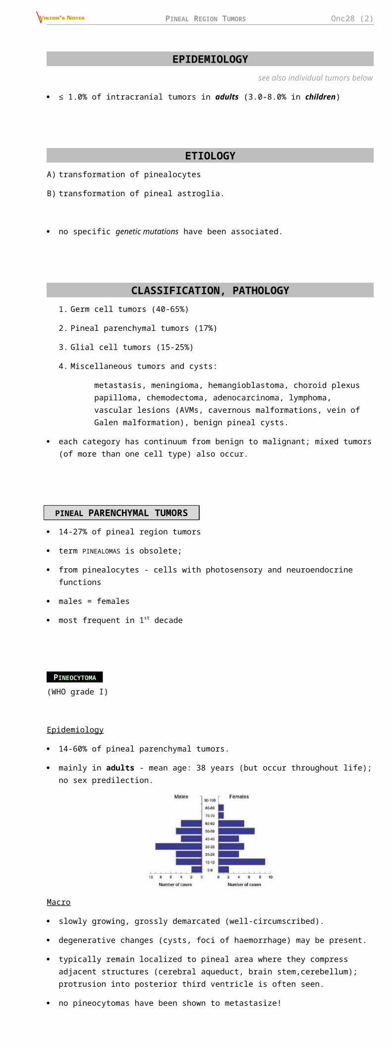
Epidemiology
14-60% of pineal parenchymal tumors.
mainly in adults - mean age: 38 years (but occur throughout life); no sex predilection.
Macro
slowly growing, grossly demarcated (well-circumscribed).
degenerative changes (cysts, foci of haemorrhage) may be present.
typically remain localized to pineal area where they compress adjacent structures (cerebral aqueduct, brain stem,cerebellum); protrusion into posterior third ventricle is often seen.
no pineocytomas have been shown to metastasize!
Micro
moderately cellular.
relatively small, uniform, homogeneously eosinophilic, mature cells resembling pineocytes.
grows in sheets or ill-defined lobules.
conspicous PROCESSES - short, often ending in club-shaped expansions.
mitotic figures are lacking in all but occasional large specimens (< 1/10 HPF)
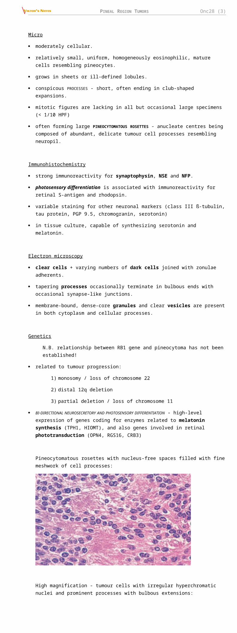
often forming large PINEOCYTOMATOUS ROSETTES - anucleate centres being composed of abundant, delicate tumour cell processes resembling neuropil.
Immunohistochemistry
strong immunoreactivity for synaptophysin, NSE and NFP.
photosensory differentiation is associated with immunoreactivity for retinal S-antigen and rhodopsin.
variable staining for other neuronal markers (class III ß-tubulin, tau protein, PGP 9.5, chromogranin, serotonin)
in tissue culture, capable of synthesizing serotonin and melatonin.
Electron microscopy
PINEAL REGION TUMORS Onc28 (3)
clear cells + varying numbers of dark cells joined with zonulae adherents.
tapering processes occasionally terminate in bulbous ends with occasional synapse-like junctions.
membrane-bound, dense-core granules and clear vesicles are present in both cytoplasm and cellular processes.
Genetics
N.B. relationship between RB1 gene and pineocytoma has not been established!
related to tumour progression:
1) monosomy / loss of chromosome 22
2) distal 12q deletion
3) partial deletion / loss of chromosome 11
BI-DIRECTIONAL NEUROSECRETORY AND PHOTOSENSORY DIFFERENTIATION - high-level expression of genes coding for enzymes related to melatonin synthesis (TPH1, HIOMT), and also genes involved in retinal phototransduction (OPN4, RGS16, CRB3)
Pineocytomatous rosettes with nucleus-free spaces filled with fine meshwork of cell processes:
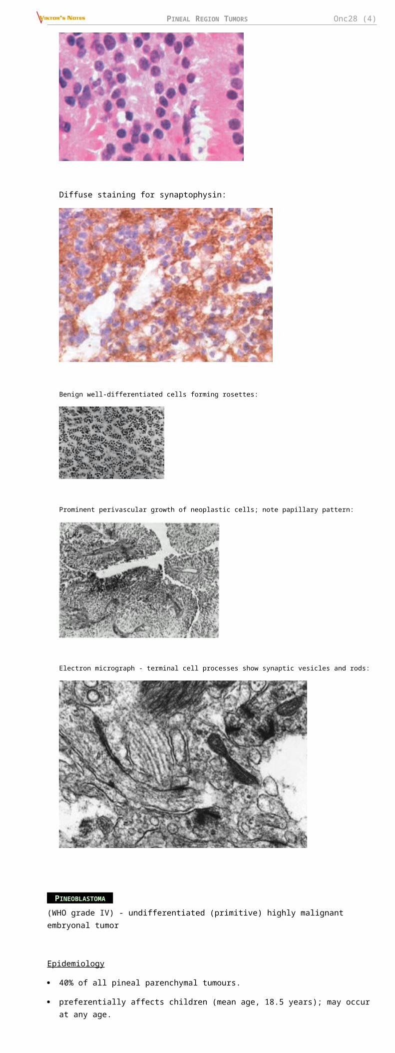
High magnification - tumour cells with irregular hyperchromatic nuclei and prominent processes with bulbous extensions:
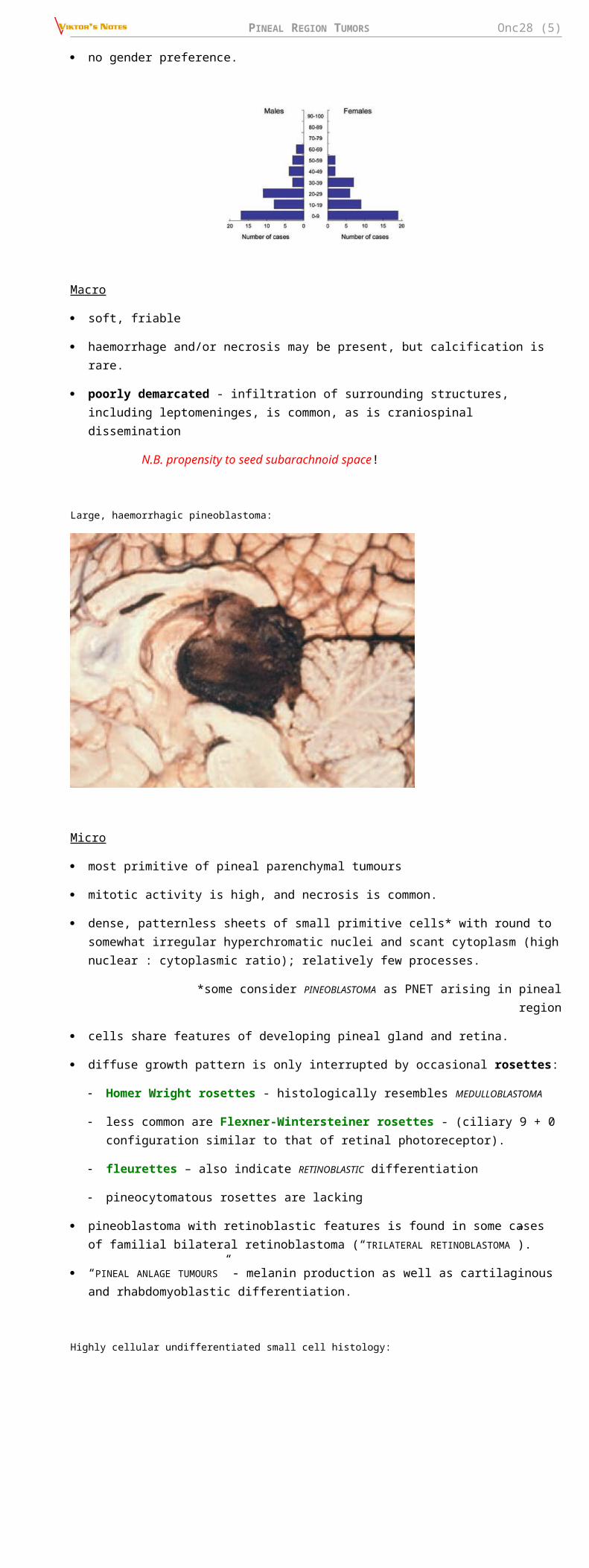
Diffuse staining for synaptophysin:
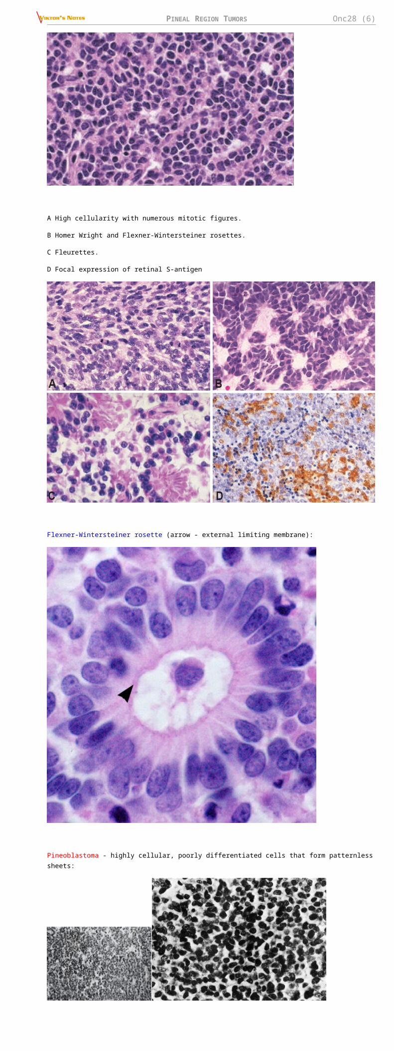
Benign well-differentiated cells forming rosettes:
Prominent perivascular growth of neoplastic cells; note papillary pattern:
PINEAL REGION TUMORS Onc28 (4)
Electron micrograph - terminal cell processes show synaptic vesicles and rods:
PINEOBLASTOMA (WHO grade IV) - undifferentiated (primitive) highly malignant embryonal tumor
Epidemiology
40% of all pineal parenchymal tumours.
preferentially affects children (mean age, 18.5 years); may occur at any age.
no gender preference.
Macro
soft, friable
haemorrhage and/or necrosis may be present, but calcification is rare.
poorly demarcated - infiltration of surrounding structures, including leptomeninges, is common, as is craniospinal dissemination
N.B. propensity to seed subarachnoid space!
Large, haemorrhagic pineoblastoma:
Micro
most primitive of pineal parenchymal tumours
mitotic activity is high, and necrosis is common.
PINEAL REGION TUMORS Onc28 (5)
dense, patternless sheets of small primitive cells* with round to somewhat irregular hyperchromatic nuclei and scant cytoplasm (high nuclear : cytoplasmic ratio); relatively few processes.
*some consider PINEOBLASTOMA as PNET arising in pineal region
cells share features of developing pineal gland and retina.
diffuse growth pattern is only interrupted by occasional rosettes:
Homer Wright rosettes - histologically resembles MEDULLOBLASTOMA
less common are Flexner-Wintersteiner rosettes - (ciliary 9 + 0 configuration similar to that of retinal photoreceptor).
fleurettes – also indicate RETINOBLASTIC differentiation
pineocytomatous rosettes are lacking
pineoblastoma with retinoblastic features is found in some cases of familial bilateral retinoblastoma (“TRILATERAL RETINOBLASTOMA”).
“PINEAL ANLAGE TUMOURS” - melanin production as well as cartilaginous and rhabdomyoblastic differentiation.
Highly cellular undifferentiated small cell histology:
A High cellularity with numerous mitotic figures.
B Homer Wright and Flexner-Wintersteiner rosettes.
C Fleurettes.
D Focal expression of retinal S-antigen
Flexner-Wintersteiner rosette (arrow - external limiting membrane):
PINEAL REGION TUMORS Onc28 (6)
Pineoblastoma - highly cellular, poorly differentiated cells that form patternless sheets:
Immunohistochemistry
- similar to pineocytomas - reactivity for neuronal, glial and photoreceptor markers (synaptophysin, NSE, NFP, class III ß-tubulin, chromogranin A, retinal S-antigen)
Electron microscopy
lack of significant differentiation (i.e. fine structure is similar to that of any poorly differentiated neuroectodermal neoplasm).
dense core granules are rarely seen.
cell processes, poorly formed and short; bulbous endings are not identified
synapses are absent
cilia with 9+0 microtubular pattern are occasionally seen
Genetic susceptibility
pineoblastomas may be seen in patients with familial (bilateral) retinoblastoma - termed “TRILATERAL RETINOBLASTOMA SYNDROME”; reported in familial adenomatous polyposis.
Genetics
- different studies, different results:
a) no consistent cytogenetic changes
b) monosomy 22 and missense INI1 mutation
pineoblastomas amy occur in patients with RB1 gene abnormalities - significantly worse prognosis.
PINEAL PARENCHYMAL TUMOUR OF INTERMEDIATE DIFFERENTIATION
(WHO grade II-III) - potentially aggressive neoplasm; part of spectrum from pineocytoma to pineoblastoma.
historical synonims: mixed pineocytoma/pineoblastoma, “malignant pineocytomas”, “pineoblastomas with lobules”
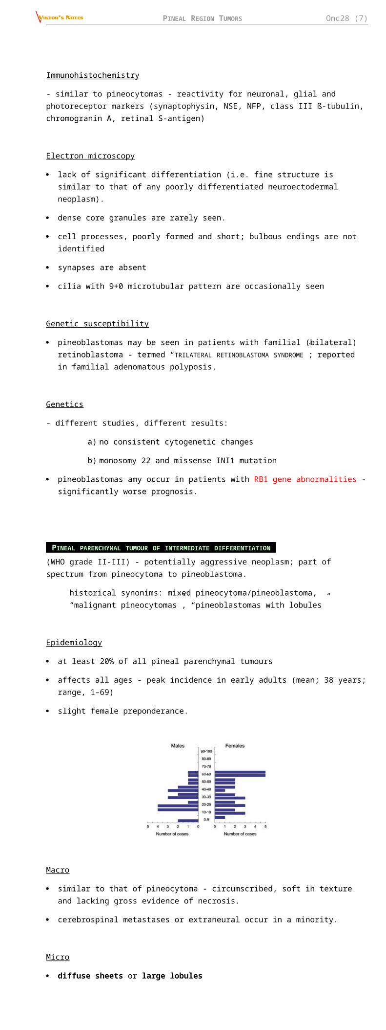
Epidemiology
at least 20% of all pineal parenchymal tumours
PINEAL REGION TUMORS Onc28 (7)
affects all ages - peak incidence in early adults (mean; 38 years; range, 1–69)
slight female preponderance.
Macro
similar to that of pineocytoma - circumscribed, soft in texture and lacking gross evidence of necrosis.
cerebrospinal metastases or extraneural occur in a minority.
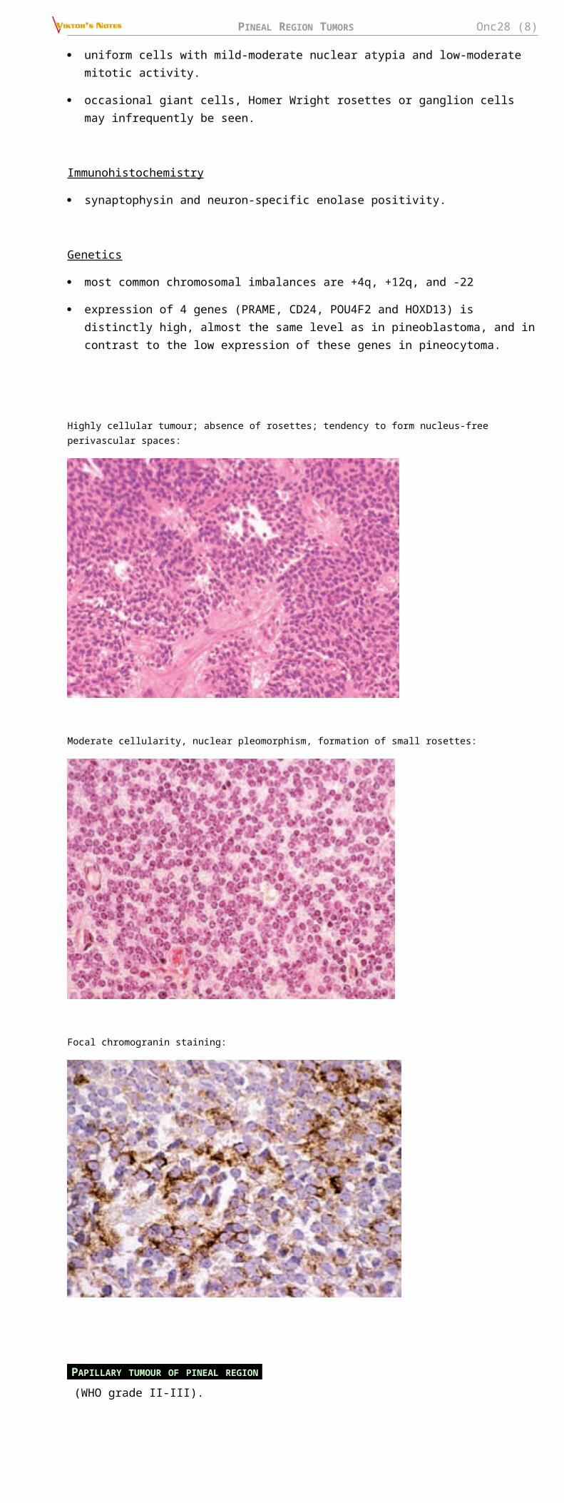
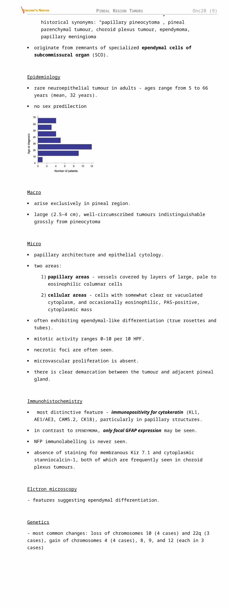
Micro
diffuse sheets or large lobules
uniform cells with mild-moderate nuclear atypia and low-moderate mitotic activity.
occasional giant cells, Homer Wright rosettes or ganglion cells may infrequently be seen.
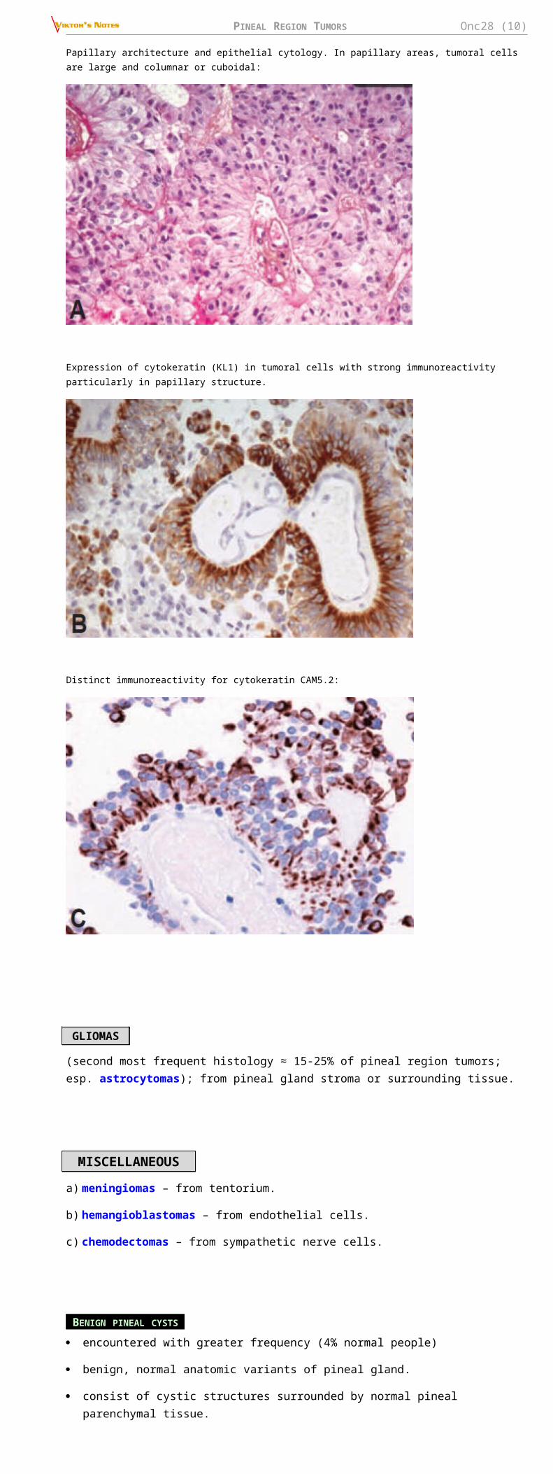
Immunohistochemistry
synaptophysin and neuron-specific enolase positivity.
Genetics
most common chromosomal imbalances are +4q, +12q, and -22
expression of 4 genes (PRAME, CD24, POU4F2 and HOXD13) is distinctly high, almost the same level as in pineoblastoma, and in contrast to the low expression of these genes in pineocytoma.
Highly cellular tumour; absence of rosettes; tendency to form nucleus-free perivascular spaces:
Moderate cellularity, nuclear pleomorphism, formation of small rosettes:
Focal chromogranin staining:
PINEAL REGION TUMORS Onc28 (8)
PAPILLARY TUMOUR OF PINEAL REGION
(WHO grade II-III).
historical synonyms: “papillary pineocytoma”, pineal parenchymal tumour, choroid plexus tumour, ependymoma, papillary meningioma
originate from remnants of specialized ependymal cells of subcommissural organ (SCO).
Epidemiology
rare neuroepithelial tumour in adults - ages range from 5 to 66 years (mean, 32 years).
no sex predilection
Macro
arise exclusively in pineal region.
large (2.5–4 cm), well-circumscribed tumours indistinguishable grossly from pineocytoma
Micro
papillary architecture and epithelial cytology.
two areas:
1) papillary areas - vessels covered by layers of large, pale to eosinophilic columnar cells
2) cellular areas - cells with somewhat clear or vacuolated cytoplasm, and occasionally eosinophilic, PAS-positive, cytoplasmic mass
often exhibiting ependymal-like differentiation (true rosettes and tubes).
mitotic activity ranges 0–10 per 10 HPF.
necrotic foci are often seen.
microvascular proliferation is absent.
there is clear demarcation between the tumour and adjacent pineal gland.
Immunohistochemistry
most distinctive feature - immunopositivity for cytokeratin (KL1, AE1/AE3, CAM5.2, CK18), particularly in papillary structures.
in contrast to EPENDYMOMA, only focal GFAP expression may be seen.
NFP immunolabelling is never seen.
absence of staining for membranous Kir 7.1 and cytoplasmic stanniocalcin-1, both of which are frequently seen in choroid plexus tumours.
Elctron microscopy
- features suggesting ependymal differentiation.
Genetics
- most common changes: loss of chromosomes 10 (4 cases) and 22q (3 cases), gain of chromosomes 4 (4 cases), 8, 9, and 12 (each in 3 cases)
PINEAL REGION TUMORS Onc28 (9)
Papillary architecture and epithelial cytology. In papillary areas, tumoral cells are large and columnar or cuboidal:
Expression of cytokeratin (KL1) in tumoral cells with strong immunoreactivity particularly in papillary structure.
Distinct immunoreactivity for cytokeratin CAM5.2:
GLIOMAS
(second most frequent histology ≈ 15-25% of pineal region tumors; esp. astrocytomas); from pineal gland stroma or surrounding tissue.
MISCELLANEOUS
a) meningiomas – from tentorium.
b) hemangioblastomas – from endothelial cells.
c) chemodectomas – from sympathetic nerve cells.
BENIGN PINEAL CYSTS
encountered with greater frequency (4% normal people)
benign, normal anatomic variants of pineal gland.
consist of cystic structures surrounded by normal pineal parenchymal tissue.
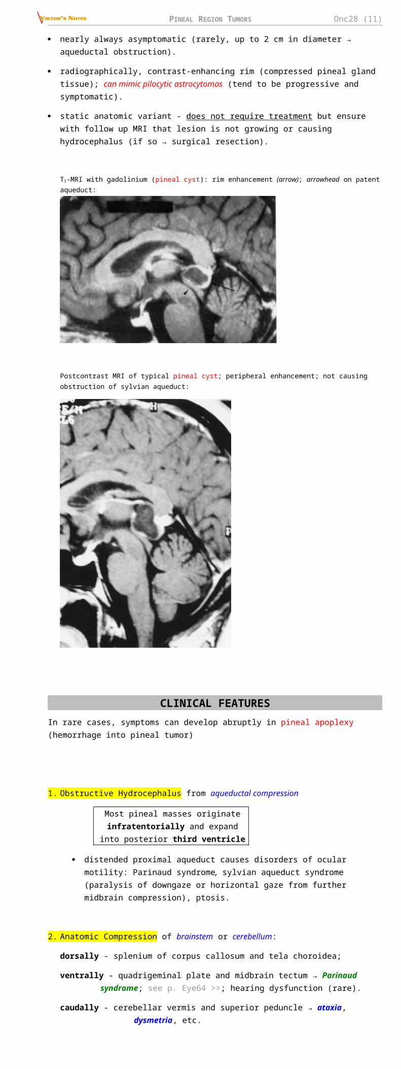
nearly always asymptomatic (rarely, up to 2 cm in diameter → aqueductal obstruction).
radiographically, contrast-enhancing rim (compressed pineal gland tissue); can mimic pilocytic astrocytomas (tend to be progressive and symptomatic).
PINEAL REGION TUMORS Onc28 (10)
static anatomic variant - does not require treatment but ensure with follow up MRI that lesion is not growing or causing hydrocephalus (if so → surgical resection).
T1-MRI with gadolinium (pineal cyst): rim enhancement (arrow); arrowhead on patent aqueduct:
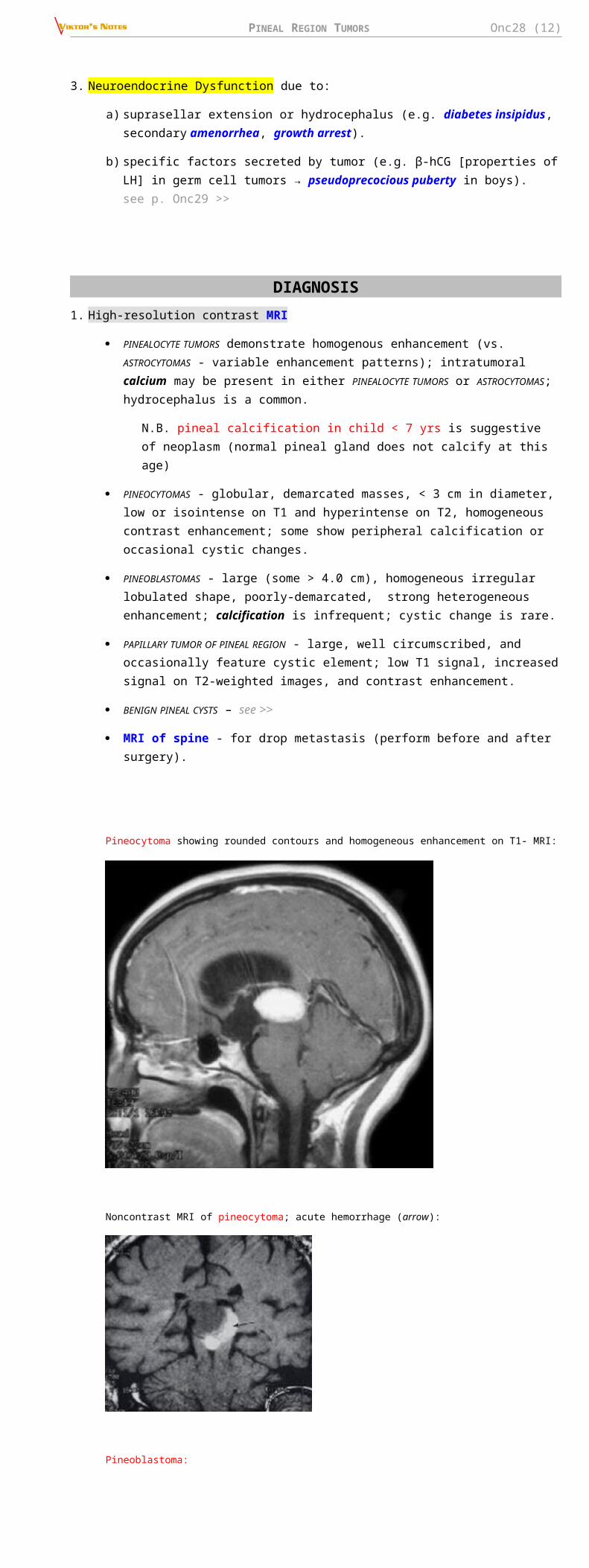
Postcontrast MRI of typical pineal cyst; peripheral enhancement; not causing obstruction of sylvian aqueduct:
CLINICAL FEATURESIn rare cases, symptoms can develop abruptly in pineal apoplexy (hemorrhage into pineal tumor)
1. Obstructive Hydrocephalus from aqueductal compression
Most pineal masses originate infratentorially and expand into posterior third ventricle
distended proximal aqueduct causes disorders of ocular motility: Parinaud syndrome, sylvian aqueduct syndrome (paralysis of downgaze or horizontal gaze from further midbrain compression), ptosis.
2. Anatomic Compression of brainstem or cerebellum:
dorsally - splenium of corpus callosum and tela choroidea;
ventrally - quadrigeminal plate and midbrain tectum → Parinaud syndrome; see p. Eye64 >>; hearing dysfunction (rare).
caudally - cerebellar vermis and superior peduncle → ataxia, dysmetria, etc.
3. Neuroendocrine Dysfunction due to:
a) suprasellar extension or hydrocephalus (e.g. diabetes insipidus, secondary amenorrhea, growth arrest).
b) specific factors secreted by tumor (e.g. β-hCG [properties of LH] in germ cell tumors → pseudoprecocious puberty in boys). see p. Onc29 >>
DIAGNOSIS1. High-resolution contrast MRI
PINEALOCYTE TUMORS demonstrate homogenous enhancement (vs. ASTROCYTOMAS - variable enhancement patterns); intratumoral calcium may be present in either PINEALOCYTE TUMORS or ASTROCYTOMAS; hydrocephalus is a common.
PINEAL REGION TUMORS Onc28 (11)
N.B. pineal calcification in child < 7 yrs is suggestive of neoplasm (normal pineal gland does not calcify at this age)
PINEOCYTOMAS - globular, demarcated masses, < 3 cm in diameter, low or isointense on T1 and hyperintense on T2, homogeneous contrast enhancement; some show peripheral calcification or occasional cystic changes.
PINEOBLASTOMAS - large (some > 4.0 cm), homogeneous irregular lobulated shape, poorly-demarcated, strong heterogeneous enhancement; calcification is infrequent; cystic change is rare.
PAPILLARY TUMOR OF PINEAL REGION - large, well circumscribed, and occasionally feature cystic element; low T1 signal, increased signal on T2-weighted images, and contrast enhancement.
BENIGN PINEAL CYSTS – see >>
MRI of spine - for drop metastasis (perform before and after surgery).
Pineocytoma showing rounded contours and homogeneous enhancement on T1- MRI:
Noncontrast MRI of pineocytoma; acute hemorrhage (arrow):
Pineoblastoma:
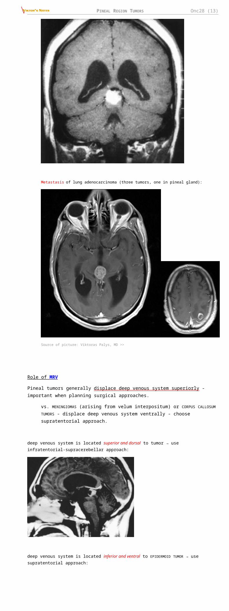
Metastasis of lung adenocarcinoma (three tumors, one in pineal gland):
PINEAL REGION TUMORS Onc28 (12)
Source of picture: Viktoras Palys, MD >>
Role of MRV
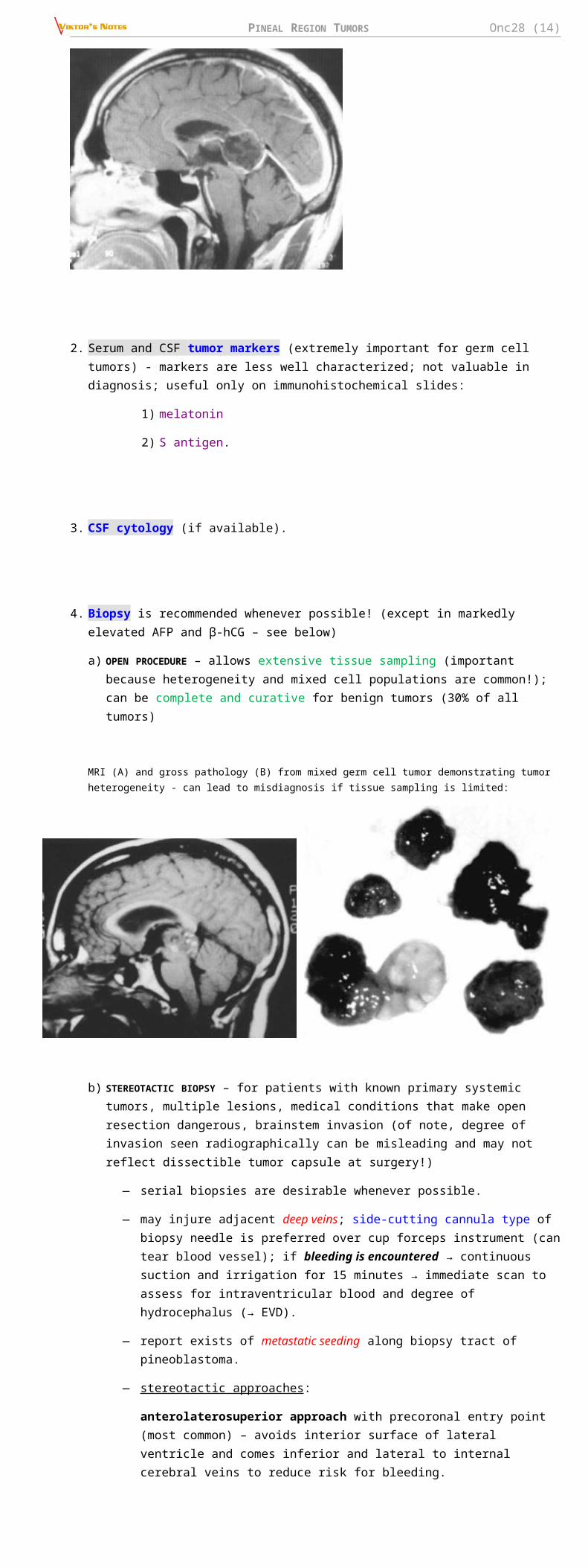
Pineal tumors generally displace deep venous system superiorly - important when planning surgical approaches.
vs. MENINGIOMAS (arising from velum interpositum) or CORPUS CALLOSUM TUMORS - displace deep venous system ventrally - choose supratentorial approach.
deep venous system is located superior and dorsal to tumor → use infratentorial-supracerebellar approach:
deep venous system is located inferior and ventral to EPIDERMOID TUMOR → use supratentorial approach:
2. Serum and CSF tumor markers (extremely important for germ cell tumors) - markers are less well characterized; not valuable in diagnosis; useful only on immunohistochemical slides:
1) melatonin
2) S antigen.
3. CSF cytology (if available).
4. Biopsy is recommended whenever possible! (except in markedly elevated AFP and β-hCG – see below)
PINEAL REGION TUMORS Onc28 (13)
a) OPEN PROCEDURE – allows extensive tissue sampling (important because heterogeneity and mixed cell populations are common!); can be complete and curative for benign tumors (30% of all tumors)
MRI (A) and gross pathology (B) from mixed germ cell tumor demonstrating tumor heterogeneity - can lead to misdiagnosis if tissue sampling is limited:
b) STEREOTACTIC BIOPSY – for patients with known primary systemic tumors, multiple lesions, medical conditions that make open resection dangerous, brainstem invasion (of note, degree of invasion seen radiographically can be misleading and may not reflect dissectible tumor capsule at surgery!)
— serial biopsies are desirable whenever possible.
— may injure adjacent deep veins; side-cutting cannula type of biopsy needle is preferred over cup forceps instrument (can tear blood vessel); if bleeding is encountered → continuous suction and irrigation for 15 minutes → immediate scan to assess for intraventricular blood and degree of hydrocephalus (→ EVD).
— report exists of metastatic seeding along biopsy tract of pineoblastoma.
— stereotactic approaches :
anterolaterosuperior approach with precoronal entry point (most common) – avoids interior surface of lateral ventricle and comes inferior and lateral to internal cerebral veins to reduce risk for bleeding.
posterolaterosuperior approach near parieto-occipital junction - useful for tumors that extend laterally or superiorly.
c) ENDOSCOPIC BIOPSY
— drawbacks : sampling error, no tissue turgor along ventricular surface to tamponade bleeding (typically, biopsy is combined with difficult to perform ventriculostomy)
— suitable entry point through forehead might allow use of rigid scope, but this offers no advantage over simple stereotactic biopsy.
— more typically, endoscopes have been used to aspirate pineal cysts; however, benefits are equivocal.
TREATMENTTherapy is based on tumor pathology;
PINEOCYTOMA, MATURE TERATOMA – surgery.
PINEOBLASTOMA – surgery → radiation ± chemotherapy.
PINEAL CYSTS – do not require treatment.
BIOPSY
See p. Op310 >>
SURGERY
See p. Op340 >>
Treat hydrocephalus prior to biopsy or resection!
endoscopic third ventriculostomy is better than ventriculoperitoneal shunt (peritoneal seeding is rare, but well-documented, complication!)
Except for well-encapsulated teratomas, few pineal region tumors are amenable to complete resection!
APPROACHES
A. Infratentorial approach - direct midline approach (originally described by Krause and popularized by Stein in 1971); trajectory between tentorium and cerebellum; tumor is encountered below deep venous system. see MRV above
PINEAL REGION TUMORS Onc28 (14)
perform in sitting slouch position - cerebellum falls away, exposing pineal region while minimizing pooling of venous blood in operative field.
limited access to tumors that extend above deep venous complex and anteriorly into 3rd ventricle*; lateral exposure is also restricted.
*with extra-long instruments, even tumors extending anteriorly into 3rd ventricle can be removed
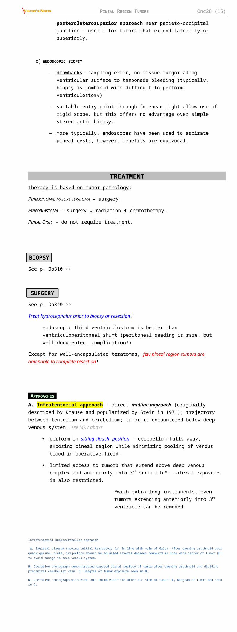
Infratentorial supracerebellar approach
A, Sagittal diagram showing initial trajectory (A) in line with vein of Galen. After opening arachnoid over quadrigeminal plate, trajectory should be adjusted several degrees downward in line with center of tumor (B) to avoid damage to deep venous system.
B, Operative photograph demonstrating exposed dorsal surface of tumor after opening arachnoid and dividing precentral cerebellar vein. C, Diagram of tumor exposure seen in B.
D, Operative photograph with view into third ventricle after excision of tumor. E, Diagram of tumor bed seen in D.
From Youmans:
The infratentorial supracerebellar approach is usually performed with patient in sitting position.[42,92,122] If necessary, ventricular drain can be placed in trigone of lateral ventricle through bur hole in midpupillary line at lambdoid suture. A suboccipital exposure is begun through linear midline incision extending from just above torcular and external occipital protuberance down to level of C4 spinous process. The incision is brought through nuchal ligament of suboccipital musculature. It is not necessary to detach muscles from spinous processes of C1 and C2, and foramen magnum does not need to be exposed. A single low-profile, self-retaining retractor is used to retract muscles and fascia of suboccipital region for exposure of suboccipital bone. The craniotomy is centered just below torcular. The bony opening must be sufficient to provide access for surgical instruments and adequate light from operating microscope. A craniotomy is preferred over craniectomy because it reduces incidence of postoperative aseptic meningitis, fluid collections, and discomfort. Slots are drilled over sagittal sinus, above torcular, and over both lateral sinuses. A final slot is drilled approximately 1 or 2 cm above foramen magnum in midline. A craniotome is used to connect slots, which allows bone flap to be elevated. Sufficient bone should be removed above transverse sinus to ensure that view along tentorium is not obscured. Any bone edges should be carefully waxed, and all venous bleeding should be controlled to avoid air emboli. The dura is opened in gentle semilunar curve that extends from lateral aspects of exposure. The dural flap is reflected upward and placed on slight tension with tenting sutures and rubber bands. Excess retraction obstructs sinuses and should be avoided. The inferior dura acts as sling to support cerebellar hemispheres. If posterior fossa is tight, fluid can be removed from ventricular drain or by opening cisterna magna. To open infratentorial corridor, arachnoidal adhesions and midline bridging veins between dorsal surface of cerebellum and tentorium are cauterized and carefully divided. The presence of extensive collateral circulation minimizes complications from venous sacrifice; however, to minimize risk, it is desirable to preserve any veins found laterally.[127] Cauterizing bridging veins and dividing them midway can minimize nuisance of bleeding from sinus. When these attachments are divided, cerebellum drops away from tentorium to provide excellent corridor with minimal brain retraction. The dorsal surface of cerebellum can be protected with padding such as Telfa, and small brain retractor can be used to provide additional cerebellar retraction in posterior and inferior direction. Additional adhesions and bridging veins can be divided when they become visible near anterior vermis as cerebellum is retracted. With retractor in place, opalescent arachnoid covering pineal region can be seen. The operating microscope is brought in at this time. Because of range of angles and different depths encountered along operative trajectory, microscope with variable objective is desirable to facilitate use of long instruments. Inclined eyepieces with wide degree of rotation accommodate different operative angles. A freestanding armrest assists surgeon and prevents fatigue. Under microscope, arachnoid overlying quadrigeminal plate is sharply opened. This is generally avascular plane, and minimal cautery is necessary. The precentral cerebellar vein is
PINEAL REGION TUMORS Onc28 (15)
identified as it courses from anterior vermis to vein of Galen and should be carefully dissected, cauterized, and divided. Although this vein can be taken without difficulty, it is not advisable to cauterize any other veins of deep venous system. The retractor can then be adjusted to visualize inferior portion of tumor. The trajectory of microscope is adjusted downward along central axis of tumor away from initial plane parallel to tentorium, where it would otherwise lead to direct encounter with vein of Galen (see Fig. 125-6A). With posterior surface of tumor exposed, central portion is cauterized and opened with long-handled knife or bayonet scissors (Fig. 125-6B and C). Specimens can be taken from within capsule and sent for frozen diagnosis. The accuracy of frozen tissue diagnosis is low, however, and this should be taken into consideration during intraoperative decision making. The tumor is then internally debulked with variety of instruments such as suction, cautery, tumor forceps, and Cavitron ultrasonic aspirator if necessary. Most tumors are soft and can generally be suctioned with large-bore Japanese-style suction device with variable control. As tumor is decompressed, capsule can be separated from surrounding thalamus. Most of vessels along wall of capsule are choroidal vessels and need not be preserved. The dissection continues until third ventricle is encountered. The tumor is then carefully dissected inferiorly off brainstem. This is often most difficult portion of tumor dissection and can be facilitated by retracting tumor superiorly and dissecting it bluntly off brainstem under direct vision. Finally, tumor is removed superiorly after separating attachments along velum interpositum and deep venous system. These attachments can be carefully cauterized and sharply dissected, although rent in deep venous system can be difficult to control and must be avoided. Intraoperative decision making regarding extent of tumor resection depends on degree of tumor invasion. Some tumors directly invade brainstem, and degree of resection is matter of judgment. Although there is evidence that more radical resection of even malignant tumors improves efficacy of adjuvant therapy and reduces risk for postoperative hemorrhage, this is still matter of judgment. Once tumor removal is completed, surgeon should have comprehensive view into third ventricle (Fig. 125-6D and E). Flexible mirrors can be useful for examining inferior portion of tumor bed to verify extent of resection and to avoid leaving any blood clots. Careful attention must be given to hemostasis.[92,128] Generally, direct but careful cautery is preferable. It is advisable to avoid extensive use of hemostatic agents, which can float into ventricle and obstruct shunt or aqueduct. If absolutely necessary, long strips of Surgicel draped over surface of cerebellum and covering tumor bed can provide hemostasis with small risk of floating into ventricle. Once hemostasis is obtained and retractors are removed, dura is closed in as watertight manner as possible. The bone flap is plated into place to reduce postoperative pain and inflammation. The patient should be extubated with reasonable degree of head elevation to avoid shifting decompressed brain within cranial vault.
B. Supratentorial approach - best for large tumors extending supratentorially or laterally into trigone of lateral ventricle; wide exposure can be obtained; difficult removal of tumor that lies below convergence of deep venous system. see MRV above
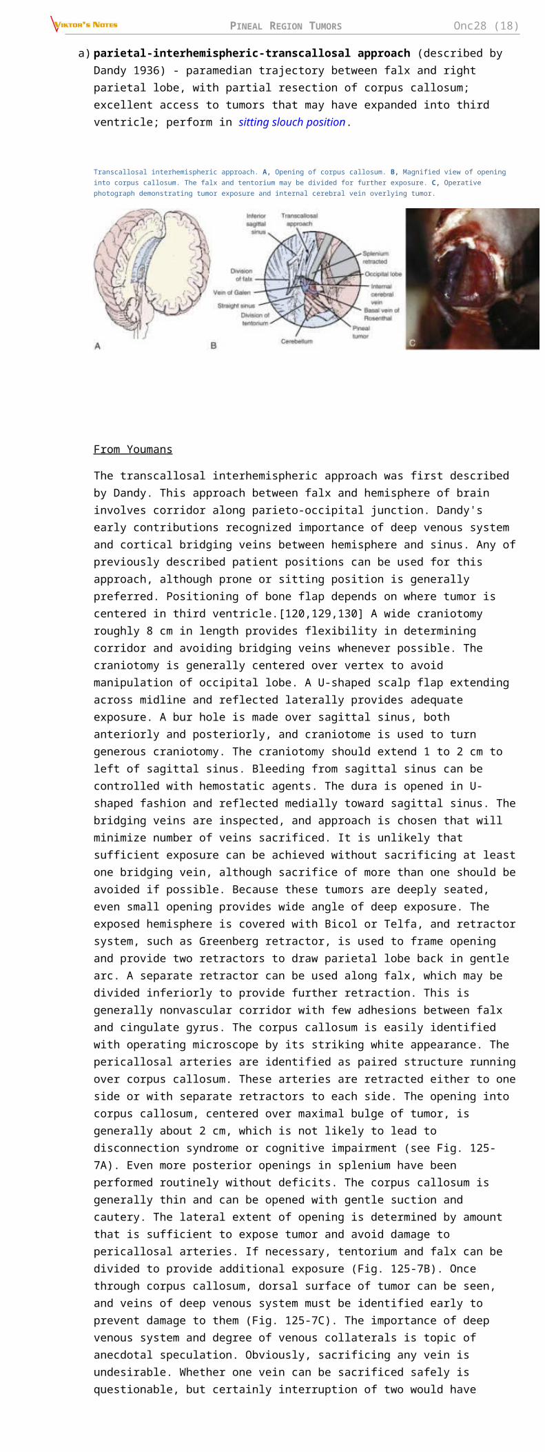
a) parietal-interhemispheric-transcallosal approach (described by Dandy 1936) - paramedian trajectory between falx and right parietal lobe, with partial resection of corpus callosum; excellent access to tumors that may have expanded into third ventricle; perform in sitting slouch position.
Transcallosal interhemispheric approach. A, Opening of corpus callosum. B, Magnified view of opening into corpus callosum. The falx and tentorium may be divided for further exposure. C, Operative photograph demonstrating tumor exposure and internal cerebral vein overlying tumor.
From Youmans
The transcallosal interhemispheric approach was first described by Dandy. This approach between falx and hemisphere of brain involves corridor along parieto-occipital junction. Dandy's early contributions recognized importance of deep venous system and cortical bridging veins between hemisphere and sinus. Any of previously described patient positions can be used for this approach, although prone or sitting position is generally preferred. Positioning of bone flap depends on where tumor is centered in third ventricle.[120,129,130] A wide craniotomy roughly 8 cm in length provides flexibility in determining corridor and avoiding bridging veins whenever possible. The craniotomy is generally centered over vertex to avoid manipulation of occipital lobe. A U-shaped scalp flap extending across midline and reflected laterally provides adequate exposure. A bur hole is made over sagittal sinus, both anteriorly and posteriorly, and craniotome is used to turn generous craniotomy. The craniotomy should extend 1 to 2 cm to left of sagittal sinus. Bleeding from sagittal sinus can be controlled with hemostatic agents. The dura is opened in U-shaped fashion and reflected medially toward sagittal sinus. The bridging veins are inspected, and approach is chosen that will minimize number of veins sacrificed. It is unlikely that sufficient exposure can be achieved without sacrificing at least one bridging vein, although sacrifice of more than one should be avoided if possible. Because these tumors are deeply seated, even small opening provides wide angle of deep exposure. The exposed hemisphere is covered with Bicol or Telfa, and retractor system, such as Greenberg retractor, is used to frame opening and provide two retractors to draw parietal lobe back in gentle arc. A separate retractor can be used along falx, which may be divided inferiorly to provide further retraction. This is generally nonvascular corridor with few adhesions between falx and cingulate gyrus. The corpus callosum is easily identified with operating microscope by its striking white appearance. The pericallosal arteries are identified as paired structure running over corpus
PINEAL REGION TUMORS Onc28 (16)
callosum. These arteries are retracted either to one side or with separate retractors to each side. The opening into corpus callosum, centered over maximal bulge of tumor, is generally about 2 cm, which is not likely to lead to disconnection syndrome or cognitive impairment (see Fig. 125-7A). Even more posterior openings in splenium have been performed routinely without deficits. The corpus callosum is generally thin and can be opened with gentle suction and cautery. The lateral extent of opening is determined by amount that is sufficient to expose tumor and avoid damage to pericallosal arteries. If necessary, tentorium and falx can be divided to provide additional exposure (Fig. 125-7B). Once through corpus callosum, dorsal surface of tumor can be seen, and veins of deep venous system must be identified early to prevent damage to them (Fig. 125-7C). The importance of deep venous system and degree of venous collaterals is topic of anecdotal speculation. Obviously, sacrificing any vein is undesirable. Whether one vein can be sacrificed safely is questionable, but certainly interruption of two would have devastating result. Once tumor is exposed, it is debulked and then dissected as described previously. Leaving ventricular drain in place for 1 or 2 days is optional.
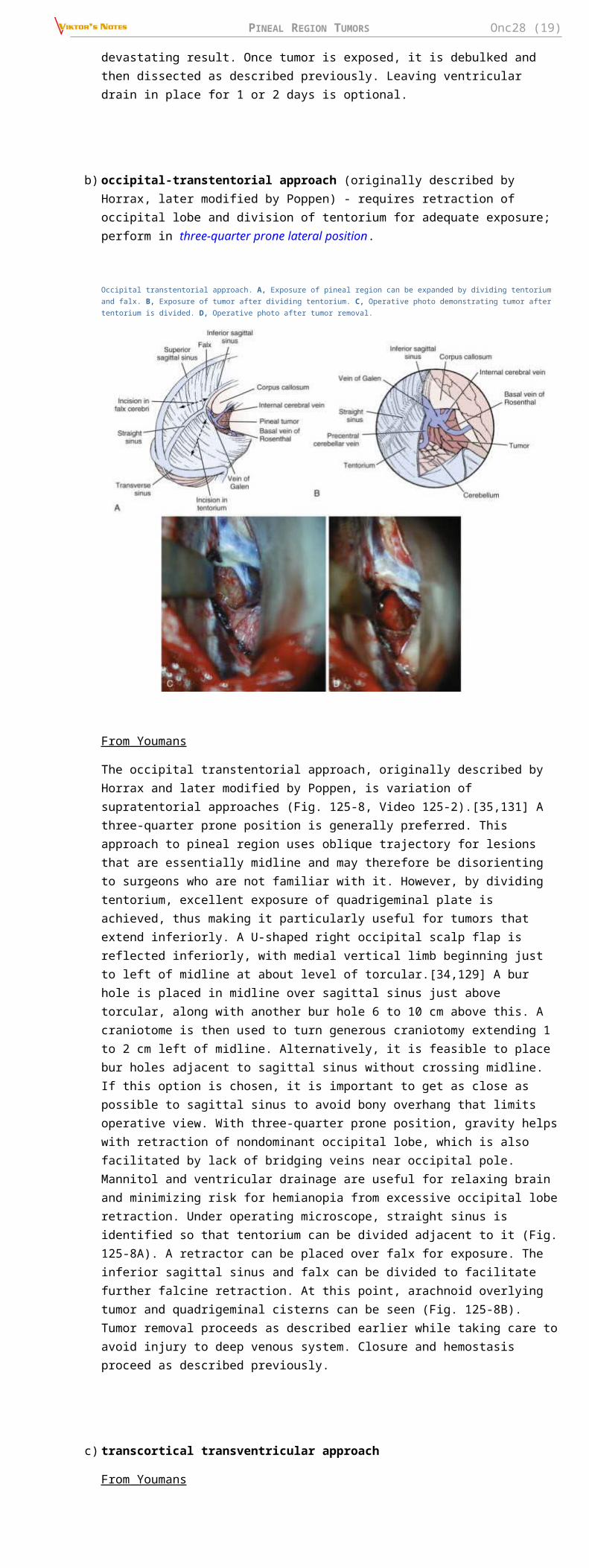
b) occipital-transtentorial approach (originally described by Horrax, later modified by Poppen) - requires retraction of occipital lobe and division of tentorium for adequate exposure; perform in three-quarter prone lateral position.
Occipital transtentorial approach. A, Exposure of pineal region can be expanded by dividing tentorium and falx. B, Exposure of tumor after dividing tentorium. C, Operative photo demonstrating tumor after tentorium is divided. D, Operative photo after tumor removal.
From Youmans
The occipital transtentorial approach, originally described by Horrax and later modified by Poppen, is variation of supratentorial approaches (Fig. 125-8, Video 125-2).[35,131] A three-quarter prone position is generally preferred. This approach to pineal region uses oblique trajectory for lesions that are essentially midline and may therefore be disorienting to surgeons who are not familiar with it. However, by dividing tentorium, excellent exposure of quadrigeminal plate is achieved, thus making it particularly useful for tumors that extend inferiorly. A U-shaped right occipital scalp flap is reflected inferiorly, with medial vertical limb beginning just to left of midline at about level of torcular.[34,129] A bur hole is placed in midline over sagittal sinus just above torcular, along with another bur hole 6 to 10 cm above this. A craniotome is then used to turn generous craniotomy extending 1 to 2 cm left of midline. Alternatively, it is feasible to place bur holes adjacent to sagittal sinus without crossing midline. If this option is chosen, it is important to get as close as possible to sagittal sinus to avoid bony overhang that limits operative view. With three-quarter prone position, gravity helps with retraction of nondominant occipital lobe, which is also facilitated by lack of bridging veins near occipital pole. Mannitol and ventricular drainage are useful for relaxing brain and minimizing risk for hemianopia from excessive occipital lobe retraction. Under operating microscope, straight sinus is identified so that tentorium can be divided adjacent to it (Fig. 125-8A). A retractor can be placed over falx for exposure. The inferior sagittal sinus and falx can be divided to facilitate further falcine retraction. At this point, arachnoid overlying tumor and quadrigeminal cisterns can be seen (Fig. 125-8B). Tumor removal proceeds as described earlier while taking care to avoid injury to deep venous system. Closure and hemostasis proceed as described previously.
c) transcortical transventricular approach
From Youmans
The transcortical transventricular approach was developed by Van Wagenen, who used trajectory through right lateral ventricle via transcortical incision.[13] This approach is rarely used because exposure is limited and need for cortical incision is undesirable. Obviously, entry point should be chosen in noneloquent cortex. Stereotactic guidance is often useful with this approach and may be desirable for tumor that extends into lateral ventricle.
C. Combined supratentorial-infratentorial approach - for large pineal region tumors.
in semiprone position.
PINEAL REGION TUMORS Onc28 (17)
advantage - wide exposure laterally, superiorly (to posterior 3rd ventricle), and inferiorly (to superior medullary velum), safe visualization of venous structures and minimal retraction of cerebellum and occipital lobe.
disadvantage - very extensive operation, including sacrifice of nondominant transverse sinus (only suitable for exceptional cases).
PATIENT POSITIONS
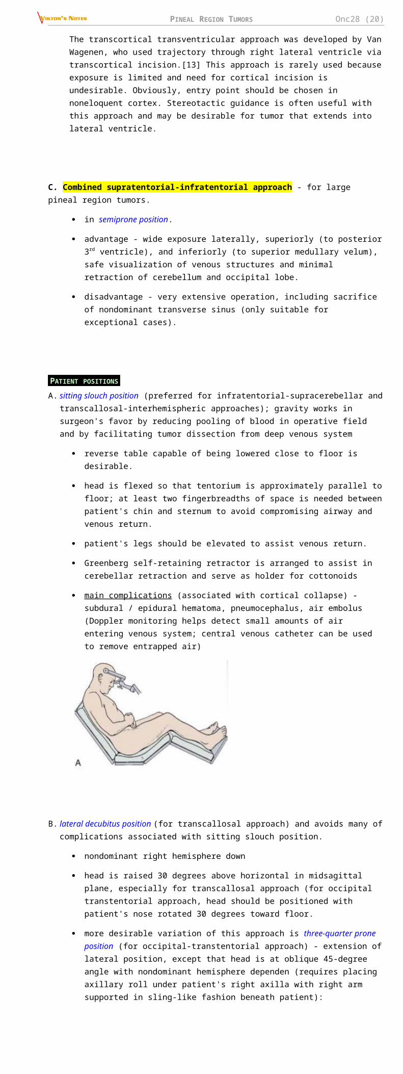
A. sitting slouch position (preferred for infratentorial-supracerebellar and transcallosal-interhemispheric approaches); gravity works in surgeon's favor by reducing pooling of blood in operative field and by facilitating tumor dissection from deep venous system
reverse table capable of being lowered close to floor is desirable.
head is flexed so that tentorium is approximately parallel to floor; at least two fingerbreadths of space is needed between patient's chin and sternum to avoid compromising airway and venous return.
patient's legs should be elevated to assist venous return.
Greenberg self-retaining retractor is arranged to assist in cerebellar retraction and serve as holder for cottonoids
main complications (associated with cortical collapse) - subdural / epidural hematoma, pneumocephalus, air embolus (Doppler monitoring helps detect small amounts of air entering venous system; central venous catheter can be used to remove entrapped air)
B. lateral decubitus position (for transcallosal approach) and avoids many of complications associated with sitting slouch position.
nondominant right hemisphere down
head is raised 30 degrees above horizontal in midsagittal plane, especially for transcallosal approach (for occipital transtentorial approach, head should be positioned with patient's nose rotated 30 degrees toward floor.
more desirable variation of this approach is three-quarter prone position (for occipital-transtentorial approach) - extension of lateral position, except that head is at oblique 45-degree angle with nondominant hemisphere dependen (requires placing axillary roll under patient's right axilla with right arm supported in sling-like fashion beneath patient):
legs are flexed with pillow between them and elevated to facilitate venous return
patient is strapped down so that table can be rotated during procedure to improve exposure
supporting roll is placed under left thorax.
C. prone position (for supratentorial approaches, esp. in pediatric population) - elevation of shoulders and head tilted to right - combined advantages of sitting slouch and three-quarter prone lateral positions; steep angle of tentorium, makes prone position impractical for infratentorial approach.
D. Concorde position – as prone position but head rotated 15 degrees away from craniotomy; more comfortable for surgeon and reduces risk of air embolism; desirable for preadolescent patients; more cumbersome in larger patients.
PINEAL REGION TUMORS Onc28 (18)
1 - supracerebellar-infratentorial approach.
2 - occipital-transtentorial approach.
3 - parietal-interhemispheric approach.
COMPLICATIONS
Overall MORTALITY of pineal region surgery is 0-8% and MORBIDITY 0-12% (vs. 90% mortality in early part of 20th century).
Most common complications - extraocular movement dysfunction (particularly limited upgaze and convergence)*, ataxia*, altered mental status (up to akinetic mutism from overzealous brainstem manipulation)
* may be present preoperatively and become transiently worse postoperatively before significantly improving or resolving completely within first few days (but can persist for several months).
Most devastating complications:
1) pineal apoplexy (bleeding into vascular-rich subtotally resected tumor bed); may be delayed for several days; large hemorrhage may require immediate evacuation.
2) venous infarction (with or without hemorrhage) - from brain retraction or from sacrifice of bridging veins.
parietal lobe retraction can cause sensory or stereognostic deficits on the opposite side.
occipital lobe retraction during transtentorial approach can cause visual field defects.
disconnection syndromes with corpus callosum incisions.
complications related to sitting position: subdural hematoma, hygroma, ventricular collapse.
POSTOPERATIVELY
high-dose steroids for first few days → taper as patient's condition improves.
seizure prophylaxis is desirable in immediate postoperative period.
careful and frequent neurological examinations; any changes → CT to rule out hydrocephalus, hemorrhage, residual air.
— lethargy and mild cognitive impairment are common (difficult to evaluate neurological status) in immediate postoperative period, particularly with extensive subdural air as result of sitting position
— shunt malfunction is frequent immediate problem (caused by air, blood, or operative debris) - deterioration can occur rapidly.
ambulate as early as possible in postoperative period.
drain should be removed or converted to shunt within first 72 hours to minimize risk for infection.
tumor markers, if present preoperatively, should be measured in postoperative period to serve as baseline.
MRI with gadolinium within 72 hours! see p. Onc3 >>
MRI before and after resection of well-differentiated pineocytoma:
PINEAL REGION TUMORS Onc28 (19)
CSF cytology (not particularly helpful) should be evaluated by lumbar puncture.
spinal MRI to look for spinal seeding is required for pineal cell tumors, malignant germ cell tumors, and ependymomas
N.B. blood clots or operative debris can sometimes mimic spinal metastasis (H: serial images before instituting spinal irradiation) - incidence of spinal seeding is low*, and prophylactic spinal irradiation is not recommended unless there is clear radiographic evidence of metastasis
*exception - highly malignant PINEOBLASTOMAS
Drop metastases in a patient with a malignant germ cell tumor:
RADIOTHERAPY
a) low-grade pineocytomas – cured with surgery alone (no adjuvant radiation) → follow with serial MRIs.
b) malignant pineal cell tumors (i.e. blastoma vs. cytoma): 40 Gy to ventricular system → 15 Gy to tumor bed (in 1.8 Gy daily fractions).
prophylactic spinal irradiation is controversial (current trend is to administer spinal irradiation 35 Gy only for documented seeding); rate of drop metastases is highest for PINEOBLASTOMAS.
CHEMOTHERAPY
- means of minimizing amount of radiation for children; tumors are not very chemosensitive (although pineal area is outside BBB)
used mostly for recurrent or disseminated tumors.
PINEOBLASTOMAS - high-dose CYCLOPHOSPHAMIDECYCLOPHOSPHAMIDE as single-agent.
RADIOSURGERY
for tumors < 3 cm
major problem - recurrence of tumor outside treatment volume (esp. pineal cell and germ cell tumors - particularly vulnerable to ventricular recurrence).
possible indications :
a) local boost to tumor bed so that exposure of ventricles and surrounding brain to radiation can be reduced.
b) tumors that recur locally.
PROGNOSISCan recur locally / distally as late as 5 yrs after diagnosis! – regular MRI follow-up
PINEAL REGION TUMORS Onc28 (20)
With the exception of PINEOCYTOMAS, all other pineal parenchymal tumours are potentially aggressive - craniospinal seeding and, rarely, extracranial metastases.
Metastases within CNS and vertebral column are most common causes of death!
PINEOCYTOMAS
lengthy interval (4 years in one series) between onset of symptoms and surgery.
5-year survival rate 86-100%
no relapses following gross total resection.
prognosis of pineocytomas with divergent differentiation (glial, neuronal, or retinoblastic) is similar to conventional pineocytomas.
PINEOBLASTOMA
median postsurgical survival 24-30 months.
In one series, projected 1-, 3- and 5-year survival rates of pineoblastoma patients treated by various modalities are 88%, 78% and 58%, respectively.
extent of disease (CSF examination and spine MRI) directly affects survival.
resection extent and radiotherapy also affect prognosis.
TRILATERAL RETINOBLASTOMA - survival < 1 year after diagnosis.
PPTID five-year survival is 39-74%
first relapse is local in 22% and spinal-leptomeningeal in 4%
PAPILLARY TUMOR OF PINEAL REGION
progression occurs in 72% of cases
5-year overall survival 73%
5-year progression-free survival 27%
BIBLIOGRAPHY for ch. “Neuro-Oncology” → follow this LINK >>
Viktor’s Notes℠ for the Neurosurgery Resident
Please visit website at www.NeurosurgeryResident.net
Related Documents