VGT voorjaar 2010 Door: N.H.J. Prakken

Welcome message from author
This document is posted to help you gain knowledge. Please leave a comment to let me know what you think about it! Share it to your friends and learn new things together.
Transcript

VGT voorjaar 2010
Door: N.H.J. Prakken
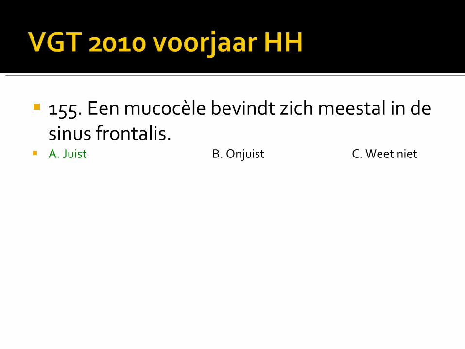
155. Een mucocèle bevindt zich meestal in de sinus frontalis.
A. Juist B. Onjuist C. Weet niet
155. Een mucocèle bevindt zich meestal in de sinus frontalis.
A. Juist B. Onjuist C. Weet niet

The frontal sinus is particularly prone to developing a mucocoeles, and up to two thirds of all mucocoeles occur there. The ethmoidal sinuses are also common, whereas maxillary and sphenoidal sinuses are infrequently involved.
Mucocoeles are best imaged with a combination of CT (to assess bony changes) and MRI (to assess any extension into the orbit or intracranial compartment).

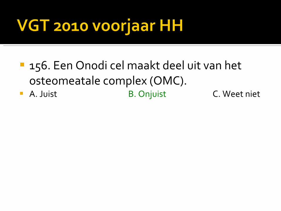
156. Een Onodi cel maakt deel uit van het osteomeatale complex (OMC).
A. Juist B. Onjuist C. Weet niet
156. Een Onodi cel maakt deel uit van het osteomeatale complex (OMC).
A. Juist B. Onjuist C. Weet niet

An Onodi cell ( or sphenoethmoidal air cell) is defined as an ethmoidal air cell that lies posteriorly, and sometimes, superiorly to the sphenoidal sinus. As a result of its location the optic nerve, and less commonly, the internal carotid artery, are very closely related with as little a 0.03mm (median 0.08mm) of bone seperating them. The incidence is variable, usually the figures quoted in western populations are between 8 and 13%.
Potential damage to the optic nerve and ICA occurs when attempts to enter the sphenoid sinus endoscopically by passing through what is thought to be the posterior most ethmoidal air cells is instead an Onodi cell.
Additionally, isolated mucocoeles of Onodi cells, squameous cell carcinoma and sinusitis have all been reported, leading to early optic nerve involvement.

157. Antrochoanale poliepen ontstaan in de sinus maxillaris.
A. Juist B. Onjuist C. Weet niet

157. Antrochoanale poliepen ontstaan in de sinus maxillaris.
A. Juist B. Onjuist C. Weet niet
An antrochoanal polyp (ACP) is a solitary polyp that arises within the maxillary sinus but passes through and enlarges the sinus ostium or more commonly an accessory ostium. The nasal cavity is therefore extended posteriorly into the nasopharynx through the posterior chonae.
Similar polyps can arise in the sphenoid sinus and extend in to nasopharynx: these are termed sphenochoanal polyps.

158. U verricht een CT-scan bij een patiënt met een larynxcarcinoom. U ziet sclerose van het aan de tumor grenzende thyroïd.
De kans op tumorinvasie van het thyroïd is meer dan 90%.
A. Juist B. Onjuist C. Weet niet

159. U verricht bij een 52-jarige man een echogeleide naaldbiopsie uit een tumor in de glandula parotis. De pathologieuitslag is een pleiomorf adenoom.
Chirurgische resectie is geïndiceerd. A. Juist B. Onjuist C. Weet niet

159. U verricht bij een 52-jarige man een echogeleide naaldbiopsie uit een tumor in de glandula parotis. De pathologieuitslag is een pleiomorf adenoom.
Chirurgische resectie is geïndiceerd. A. Juist B. Onjuist C. Weet niet

Patients are typically in their middle age.
Pleomorphic adenomas account for 70 - 80% of benign salivary gland tumours and are especially common in the parotid gland.
Prior head and neck irradiation is a demonstrable risk factor for the development of these tumours.
Distribution among the salivary glands is as follows: parotid gland : 84%: commoner in the superficial lobe submandibular gland : 8% minor salivary glands : 6.5%▪ widely distributed including the nasal cavity, pharynx, larynx,
trachea sublingual glands : 0.5%
Although they are less commonly seen in salivary glands other than the parotid, even then they remain the most common benign tumour in those locations.

160. De lamina papyracea bevindt zich tussen de orbita en het sphenoid.
A. Juist B. Onjuist C. Weet niet

160. De lamina papyracea bevindt zich tussen de orbita en het sphenoid.
A. Juist B. Onjuist C. Weet niet

161. Differentiatie tussen een opticusglioom en een opticusmeningioom kan moeilijk zijn.
Wanneer de oogzenuw op een MRI in de tumormassa te onderscheiden is, pleit dit sterk voor een opticusglioom.
A. Juist B. Onjuist C. Weet niet

161. Differentiatie tussen een opticusglioom en een opticusmeningioom kan moeilijk zijn.
Wanneer de oogzenuw op een MRI in de tumormassa te onderscheiden is, pleit dit sterk voor een opticusglioom.
A. Juist B. Onjuist C. Weet niet
An optic nerve meningioma is a benign tumour arising from the optic nerve sheath, and represents ~ 10 - 30% of all orbital meningiomas, the majority of which are direct extensions from intracranial meningiomas.
On axial or oblique sagittal imaging the enhancing tumour surrounding the non-enhancing optic nerve results in the so-called tram-track sign. This is most evident in tumours with tubular growth pattern.

162. Carotico-caverneuze fistels in de sinus cavernosus zijn geassocieerd met een gedilateerde vena ophthalmica superior.
A. Juist B. Onjuist C. Weet niet
162. Carotico-caverneuze fistels in de sinus cavernosus zijn geassocieerd met een gedilateerde vena ophthalmica superior.
A. Juist B. Onjuist C. Weet niet

Direct CCF, as they are usually secondary to trauma have demographics that reflect the distribution of head trauma.
pulsatile exophthalmos / proptosis : ~75% chemosis and subconjunctival haemorrhage progressive visual loss : 25 – 32% pulsatile tinnitus (usually objective) raised intracranial pressure subarachnoid haemorrhage, intracerebral haemorrhage, otorrhagia,
epistaxis : ~ 2.5 - 8.5%

Classification Direct : direct communication between intra-cavernous ICA and
cavernous sinus. Indirect : communication exists via branches of the carotid circulation
(ICA or ECA) type A : direct connection between the intracavernous ICA and CS type B : dural shunt between intracavernous branches of the ICA and
CS type C : dural shunt between meningeal branches of the ECA and CS type D : B + C Direct : type A
163. Bij Graves orbitopathie ziet u op een CT-scan bilateraal verdikte extraoculaire oogspieren.
Typisch voor Graves is dat de peesinserties niet verdikt zijn.
A. Juist B. Onjuist C. Weet niet

163. Bij Graves orbitopathie ziet u op een CT-scan bilateraal verdikte extraoculaire oogspieren.
Typisch voor Graves is dat de peesinserties niet verdikt zijn.
A. Juist B. Onjuist C. Weet niet
The order of extraocular muscle involvement can be remembered by the mnemonic I'M SLOW, and bilateral (76 - 90%) and symmetric (70%) involvement is typical.
The tendon is typically spared (although it can be involved in acute cases) with the swelling largely confined to the muscle belly.
164. De ruimte van Prussak bevindt zich mediaal van de gehoorbeenketen.
A. Juist B. Onjuist C. Weet niet
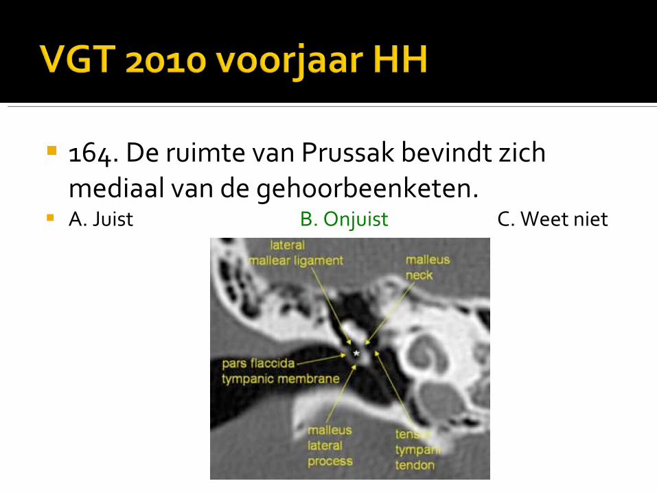
164. De ruimte van Prussak bevindt zich mediaal van de gehoorbeenketen.
A. Juist B. Onjuist C. Weet niet

165. Aankleuring van de nervus facialis in de inwendige gehoorgang is een pathologische bevinding.
A. Juist B. Onjuist C. Weet niet
Related Documents


![Nuirooefenen vgt[1]](https://static.cupdf.com/doc/110x72/556372cad8b42ae6088b55bd/nuirooefenen-vgt1.jpg)