Vertebrate Developmen t Biology II: Form and Function

Vertebrate Development Biology II: Form and Function.
Dec 16, 2015
Welcome message from author
This document is posted to help you gain knowledge. Please leave a comment to let me know what you think about it! Share it to your friends and learn new things together.
Transcript

Vertebrate Development
Biology II:
Form and Function
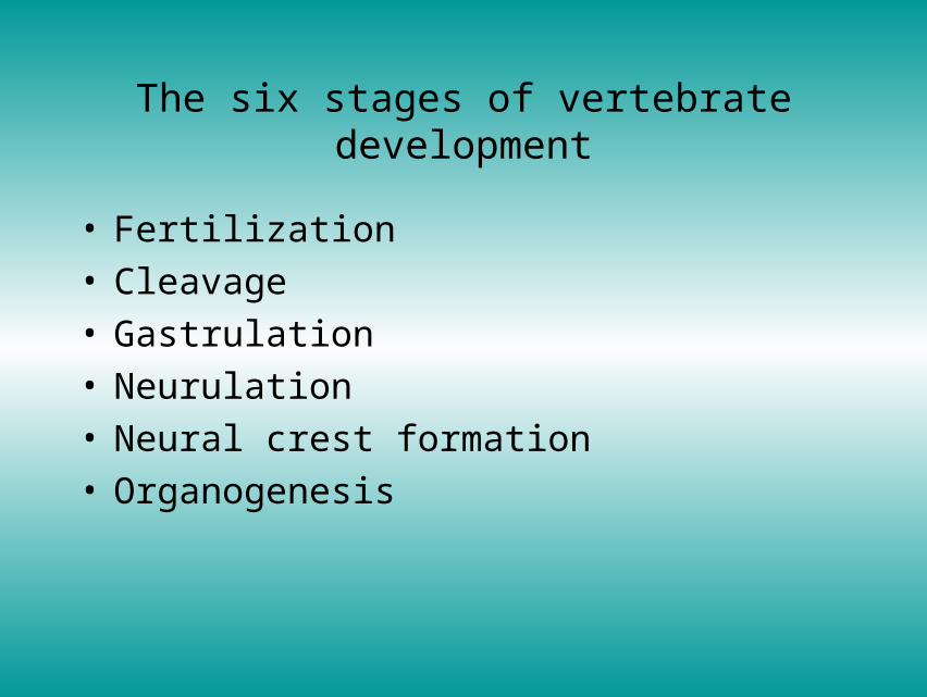
The six stages of vertebrate development
• Fertilization• Cleavage• Gastrulation• Neurulation• Neural crest formation• Organogenesis

Stage of vertebrate development (I)
Stage of vertebrate development (II)

Fertilization (I)
• Entry of sperm cell induces activation– prevents other
sperm from entering
– Intitiates second meiotic division of egg nucleus
– Induces polarity

Fertilization (II)

Fertilization in sea urchins

Sperm penetration

Polarity in early embryos

• Division of first cell to many within ball of same volume (morula) is followed by hollowing of that ball to a blastula. Form of cleavage and blastulation depends on orientation of yolk and nucleus– In primitive chordates, division is even, towards a
symmetrical blastula composed of cells of equal size– In amphibians, holoblastic cleavage leads to
assymetrical blastula– In reptiles and birds, meroblastic cleavage occurs,
resulting in a cap of cells on top of the yolk– In mammals, holoblastic cleavage occurs, creating a
trophoblast containing a blastocoel, with inner disc of cells equivalent to a blastodisc
Cleavage

Yolk distribution in amniotic eggs affects blastula development

Holoblastic cleavage
• Cells with little yolk, and central nucleus, develop evenly

Uneven cleavage• In frog cells, there is more
yolk, and nucleus of fertilized egg is to one side:– Yolk slows division, so
areas of low yolk content divide quicker, and create smaller cells (see here, front)
– Areas of high yolk content divide more slowly, and give rise to larger cells

Meroblastic cleavage
• Occurring in reptiles, birds and mammals, an uneven division of cells causes a cap of cells on top of the yolk

Blastula of mammals and birds
• Cap of cells develops into a blastodisc• Blastocoel develops in mammals, surrounded by trophoblast

Gastrulation
• Invagination of outer layer of cells to inside of the blastula is known as gastrulation, resulting in the formation of the gastrula
• Type of gastrulation is a function of type of blastula…
• End result is three types of germ layer tissue - endoderm, mesoderm and ectoderm

Gastrulation in the lancelet

Gastrulation in the frog

Gastrulation in birds

Gastrulation in mammals

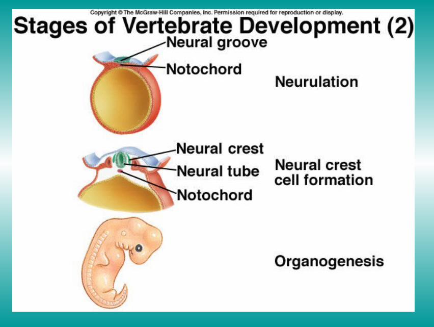
Neurulation and neural crest formation
• Formation of neural fold (primitive streak) above notocord, begins a channel that eventually seals on the dorsal surface, forming neural groove– Mesoderm derived tissue close to notocord develop into
somites, giving rise to muscles, connective tissue and vertebrae
• Layer of cells on dorsal surface of groove form neural crest, responsible for formation of several important organs– Associated patches of ectoderm tissue derive into
placodes, which evetually result in important neurally related organs

Neural tube formation (I)

Neural tube formation (II)
Induction
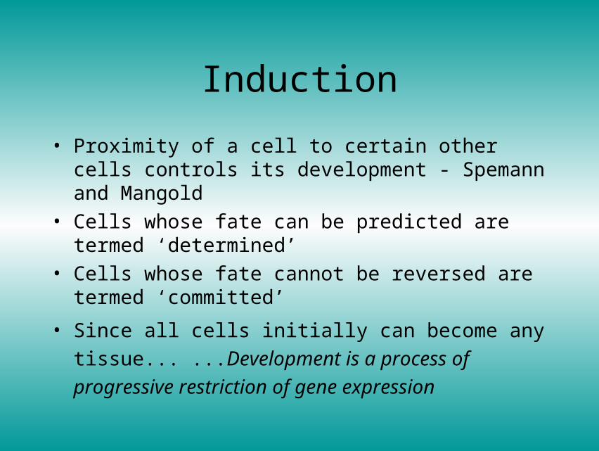
• Proximity of a cell to certain other cells controls its development - Spemann and Mangold
• Cells whose fate can be predicted are termed ‘determined’
• Cells whose fate cannot be reversed are termed ‘committed’
• Since all cells initially can become any
tissue... ...Development is a process of progressive
restriction of gene expression

Spemann and Mangold’s dorsal lip transplant experiment

Induction of the vertebrate eye

Organogenesis
• Ontogeny recapitulates phylogeny
• (and a quick word about extraembryonic membranes)

Derivation of major tissue types

Embryonic development of vertebrates (I)

Embryonic development of vertebrates (II)

Extraembryonic membranes - Chick embryo
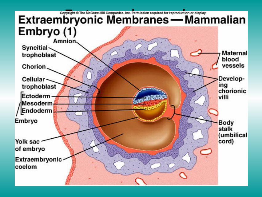
Extraembryonic membranes - mammalian embryo (I)

Extraembryonic membranes - mammalian embryo (II)

The placenta

Human development

Developing human at 4 weeks

Developing human at 7 weeks

Developing human at 3 months

Developing human at 4 months
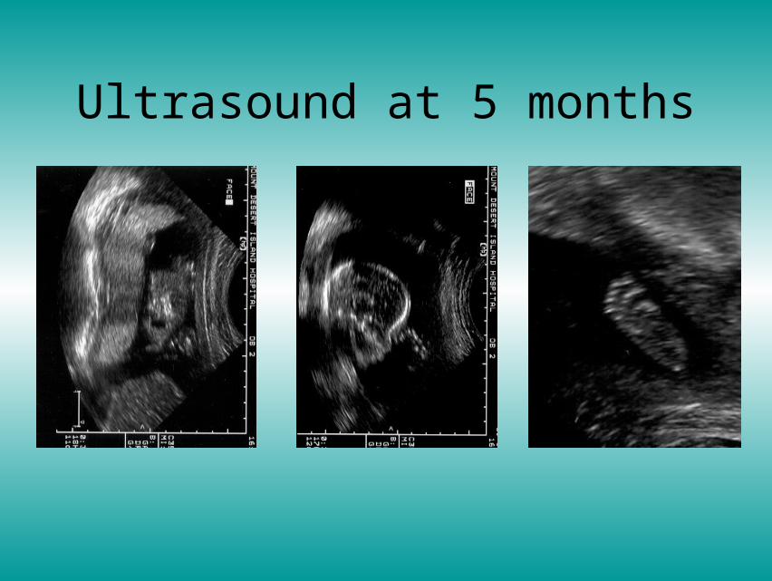
Ultrasound at 5 months

Delivery position of foetus
Related Documents