Ventromedial Prefrontal Cortex and Amygdala Dysfunction During an Anger Induction Positron Emission Tomography Study in Patients With Major Depressive Disorder With Anger Attacks Darin D. Dougherty, MD, MSc; Scott L. Rauch, MD; Thilo Deckersbach, PhD; Carl Marci, MD; Rebecca Loh, BS; Lisa M. Shin, PhD; Nathaniel M. Alpert, PhD; Alan J. Fischman, MD, PhD; Maurizio Fava, MD Context: Although a variety of functional neuroimag- ing studies have used emotion induction paradigms to investigate the neural basis of anger in control subjects, no functional neuroimaging studies using anger induc- tion have been conducted in patient populations. Objective: To study the neural basis of anger in un- medicated patients with major depressive disorder with anger attacks (MDD + A), unmedicated patients with MDD without anger attacks (MDD - A), and controls. Design: We used positron emission tomography, psycho- physiologic measures, and autobiographical narrative scripts in the context of an anger induction paradigm. Setting: Academic medical center. Participants: Thirty individuals, evenly divided among the 3 study groups. Interventions: In separate conditions, participants were exposed to anger and neutral autobiographical scripts during the positron emission tomography study. Subjective self-report and psychophysiologic data were also collected. Main Outcome Measures: Voxelwise methods were used for analyses of regional cerebral blood flow changes for the anger vs neutral contrast within and between groups. Results: Controls showed significantly (P.001) greater regional cerebral blood flow increases in the left ventro- medial prefrontal cortex during anger induction than pa- tients with MDD + A, whereas these differences were not present in other between-group analyses. Also, in con- trols, an inverse relationship was demonstrated be- tween regional cerebral blood flow changes during an- ger induction in the left ventromedial prefrontal cortex and left amygdala, whereas in patients with MDD + A there was a positive correlation between these brain regions during anger induction. There was no significant rela- tionship between these brain regions during anger in- duction in patients with MDD - A. Conclusion: These results suggest a pathophysiology of MDD+A that is distinct from that of MDD-A and that may be responsible for the unique clinical presentation of patients with MDD + A. Arch Gen Psychiatry. 2004;61:795-804 M AJOR DEPRESSIVE DISOR- der (MDD) is a clini- cal syndrome charac- terized by affect dysregulation, neuro- vegetative symptoms, autonomic distur- bances, and endocrine abnormalities. Freud 1 theorized that depression re- sulted from anger turned inward. In fact, numerous studies 2-8 have demonstrated that depressed patients have higher rates of anger and aggression than controls. One study 9 even found that the degree of an- ger expressed inward in depressed pa- tients correlated with the severity of de- pressive symptoms. The concept of “anger attacks” in pa- tients with MDD was introduced by Fava and colleagues 10 in 1990. These anger at- tacks are characterized by sudden spells of anger that are inappropriate to the situ- ation in which they occur and are unchar- acteristic of the patient’s usual behavior. In addition, clinical characterization of this depressive subtype reveals that patients with MDD with anger attacks (MDD + A) have higher scores on measures of hostil- ity, anxiety, and somatization than pa- tients with MDD without anger attacks (MDD-A). 11 The prevalence of anger at- tacks in depressed patients is approxi- mately 30% to 40%, 4,5,11-14 and the attacks resolve after successful treatment of the de- pressive episode. 5,11,12 Studies 15 have demonstrated that symptoms of anger or aggression may have a prevalence as high as 50% in outpatient psychiatric populations, indicating that an- ORIGINAL ARTICLE From the Departments of Psychiatry (Drs Dougherty, Rauch, Deckersbach, Marci, Shin, and Fava and Ms Loh) and Radiology (Drs Dougherty, Rauch, Alpert, and Fischman), Massachusetts General Hospital and Harvard Medical School, Boston, Mass; and the Department of Psychology, Tufts University, Medford, Mass (Dr Shin). (REPRINTED) ARCH GEN PSYCHIATRY/ VOL 61, AUG 2004 WWW.ARCHGENPSYCHIATRY.COM 795 ©2004 American Medical Association. All rights reserved. Downloaded From: http://archpsyc.jamanetwork.com/ by a Harvard University User on 09/01/2014

Welcome message from author
This document is posted to help you gain knowledge. Please leave a comment to let me know what you think about it! Share it to your friends and learn new things together.
Transcript
Ventromedial Prefrontal Cortex and AmygdalaDysfunction During an Anger Induction PositronEmission Tomography Study in Patients WithMajor Depressive Disorder With Anger AttacksDarin D. Dougherty, MD, MSc; Scott L. Rauch, MD; Thilo Deckersbach, PhD; Carl Marci, MD; Rebecca Loh, BS;Lisa M. Shin, PhD; Nathaniel M. Alpert, PhD; Alan J. Fischman, MD, PhD; Maurizio Fava, MD
Context: Although a variety of functional neuroimag-ing studies have used emotion induction paradigms toinvestigate the neural basis of anger in control subjects,no functional neuroimaging studies using anger induc-tion have been conducted in patient populations.
Objective: To study the neural basis of anger in un-medicated patients with major depressive disorder withanger attacks (MDD+A), unmedicated patients with MDDwithout anger attacks (MDD−A), and controls.
Design: We used positron emission tomography, psycho-physiologic measures, and autobiographical narrative scriptsin the context of an anger induction paradigm.
Setting: Academic medical center.
Participants: Thirty individuals, evenly divided amongthe 3 study groups.
Interventions: In separate conditions, participantswere exposed to anger and neutral autobiographicalscripts during the positron emission tomography study.Subjective self-report and psychophysiologic data werealso collected.
Main Outcome Measures: Voxelwise methods wereused for analyses of regional cerebral blood flow changesfor the anger vs neutral contrast within and betweengroups.
Results: Controls showed significantly (P�.001) greaterregional cerebral blood flow increases in the left ventro-medial prefrontal cortex during anger induction than pa-tients with MDD+A, whereas these differences were notpresent in other between-group analyses. Also, in con-trols, an inverse relationship was demonstrated be-tween regional cerebral blood flow changes during an-ger induction in the left ventromedial prefrontal cortexand left amygdala, whereas in patients with MDD+A therewas a positive correlation between these brain regionsduring anger induction. There was no significant rela-tionship between these brain regions during anger in-duction in patients with MDD−A.
Conclusion: These results suggest a pathophysiology ofMDD+A that is distinct from that of MDD−A and thatmay be responsible for the unique clinical presentationof patients with MDD+A.
Arch Gen Psychiatry. 2004;61:795-804
M AJOR DEPRESSIVE DISOR-der (MDD) is a clini-cal syndrome charac-t e r i zed by a f f ec tdysregulation, neuro-
vegetative symptoms, autonomic distur-bances, and endocrine abnormalities.Freud1 theorized that depression re-sulted from anger turned inward. In fact,numerous studies2-8 have demonstratedthat depressed patients have higher ratesof anger and aggression than controls. Onestudy9 even found that the degree of an-ger expressed inward in depressed pa-tients correlated with the severity of de-pressive symptoms.
The concept of “anger attacks” in pa-tients with MDD was introduced by Favaand colleagues10 in 1990. These anger at-
tacks are characterized by sudden spellsof anger that are inappropriate to the situ-ation in which they occur and are unchar-acteristic of the patient’s usual behavior.In addition, clinical characterization of thisdepressive subtype reveals that patientswith MDD with anger attacks (MDD+A)have higher scores on measures of hostil-ity, anxiety, and somatization than pa-tients with MDD without anger attacks(MDD−A).11 The prevalence of anger at-tacks in depressed patients is approxi-mately 30% to 40%,4,5,11-14 and the attacksresolve after successful treatment of the de-pressive episode.5,11,12
Studies15 have demonstrated thatsymptoms of anger or aggression may havea prevalence as high as 50% in outpatientpsychiatric populations, indicating that an-
ORIGINAL ARTICLE
From the Departments ofPsychiatry (Drs Dougherty,Rauch, Deckersbach, Marci,Shin, and Fava and Ms Loh)and Radiology (Drs Dougherty,Rauch, Alpert, and Fischman),Massachusetts General Hospitaland Harvard Medical School,Boston, Mass; and theDepartment of Psychology,Tufts University, Medford,Mass (Dr Shin).
(REPRINTED) ARCH GEN PSYCHIATRY/ VOL 61, AUG 2004 WWW.ARCHGENPSYCHIATRY.COM795
©2004 American Medical Association. All rights reserved.Downloaded From: http://archpsyc.jamanetwork.com/ by a Harvard University User on 09/01/2014
ger or aggression may be as common as symptoms of de-pression and anxiety in this population. Anger and ag-gression are especially common in patients with diagnosesof MDD, bipolar disorder, intermittent explosive disor-der (IED), and cluster B personality disorders. Despitethe high prevalence of anger and aggression in psychi-atric populations and the obvious public health impactof these symptoms, the role of anger and aggression inpsychiatric illness has been understudied. In addition,anger and hostility are associated with higher rates of coro-nary artery disease,16-18 myocardial infarction,19-22 and ab-normal glucose metabolism.23-27 Given that depression it-self is a significant risk factor for heart disease28-30 anddiabetes mellitus,31,32 it would seem that individualswith MDD+A would be especially at risk for medicalconsequences. Thus, the societal costs of anger and ag-gression in psychiatric illness stem from the conse-quences of violence and the medical sequelae associatedwith anger and hostility. In patients with MDD+A,these costs to society are above and beyond the alreadystaggering financial burden of depression.33 These find-ings underscore the importance of elucidating thepathophysiology of anger and aggression in psychiatricillness and of conducting clinical trials in the search foreffective treatments.
Converging data implicate a network of brain re-gions in the pathophysiology of MDD. These regions in-clude, but are not limited to, territories of the prefrontalcortex (PFC), anterior cingulate cortex, and medial tem-poral lobe structures, including the amygdala and hip-pocampus.34-38 Current conceptualization of the under-lying neuroanatomy of anger and aggression implicatesthe amygdala and related temporolimbic structures, thehypothalamus, the anterior cingulate cortex, and the PFC(most notably the ventral PFC) as brain regions in-volved in mediating aggression.39-41 Although a varietyof functional neuroimaging studies42-46 have used emo-tion induction paradigms to investigate the neural basisof anger in control subjects, no functional neuroimag-ing studies have been conducted in individuals who arediagnosed as having MDD and who have a predilectionfor anger, aggressive behavior, or both. The present studyuses positron emission tomography (PET) and autobio-graphical narrative scripts to study the neural basis of an-ger, particularly the relationship between the ventral PFCand the amygdala, in unmedicated patients with MDD+A,unmedicated patients with MDD−A, and control sub-
jects. There is especially strong evidence that anger andaggression seen in a multitude of diagnoses are associ-ated with hypofunctionality of the ventral PFC and amyg-dala.47-52 In contrast, numerous studies36,53,54 have dem-onstrated greater activity in the ventral PFC and amygdalain patients with MDD compared with control subjects.These results strongly suggest that ventral PFC and amyg-dala function should differentiate patients with MDD+Aand patients with MDD−A from each other and from con-trol subjects and form the basis for our a priori hypoth-eses. Specifically, we predicted (1) that, based on previ-ous results from our laboratory44 and others,42,43,45,46 thecontrol group would exhibit activation of the ventral PFCwith corresponding deactivation of the amygdala dur-ing anger induction, (2) that the MDD+A group wouldexhibit diminished activation in the ventral PFC andamygdala during anger induction relative to the controlgroup and the MDD−A group, and (3) that the MDD−Agroup would have greater activation in these same brainregions during anger induction relative to the controlgroup and the MDD+A group.
METHODS
PARTICIPANTS
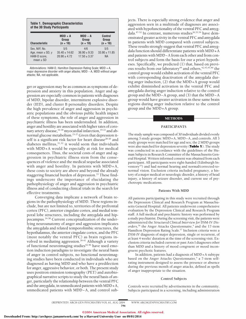
The study sample was composed of 30 individuals divided evenlyamong 3 study groups: MDD+A, MDD−A, and controls. All 3study groups were matched for age and sex; the 2 MDD groupswere also matched for depression severity (Table 1). The studywas conducted in accordance with the guidelines of the Hu-man Subjects in Research Committee of the Massachusetts Gen-eral Hospital. Written informed consent was obtained from eachparticipant. All participants were right-handed (Edinburgh In-ventory55) and had normal hearing and normal/corrected-to-normal vision. Exclusion criteria included pregnancy, a his-tory of a major medical or neurologic disorder, a history of headinjury, a history of seizure disorder, and current use of psy-chotropic medications.
Patients With MDD
All patients participating in this study were recruited throughthe Depression Clinical and Research Program at Massachu-setts General Hospital. All patients underwent comprehensiveevaluation by the Depression Clinical and Research Programstaff. A full medical and psychiatric history was performed bya study psychiatrist. During the screening visit, the patients wereadministered the Structured Clinical Interview for DSM-IV Dis-orders,56 the Anger Attacks Questionnaire,4 and the 17-itemHamilton Depression Rating Scale.57 Inclusion criteria were aDSM-IV diagnosis of major depression, single or recurrent, ofat least 4 weeks’ duration at the time of the screening visit. Ex-clusion criteria included current or past Axis I diagnoses otherthan MDD and a history of mood congruent or mood incon-gruent psychotic features.
In addition, patients had a diagnosis of MDD+A subtypebased on the Anger Attacks Questionnaire,4 a 7-item self-rating instrument designed to assess the presence or absenceduring the previous month of anger attacks, defined as spellsof anger inappropriate to the situation.
Control Subjects
Controls were recruited by advertisements in the community.Subjects participated in a screening, including administration
Table 1. Demographic Characteristicsof the 30 Study Participants
Characteristic
MDD + AGroup
(n = 10)
MDD − AGroup
(n = 10)
ControlGroup
(n = 10)
Sex, M/F, No. 5/5 4/6 5/5Age, mean ± SD, y 35.40 ± 14.62 36.90 ± 9.33 33.90 ± 11.85HAM-D score,
mean ± SD20.90 ± 4.72 17.50 ± 3.37 NA
Abbreviations: HAM-D, Hamilton Depression Rating Scale; MDD + A,major depressive disorder with anger attacks; MDD − A, MDD without angerattacks; NA, not applicable.
(REPRINTED) ARCH GEN PSYCHIATRY/ VOL 61, AUG 2004 WWW.ARCHGENPSYCHIATRY.COM796
©2004 American Medical Association. All rights reserved.Downloaded From: http://archpsyc.jamanetwork.com/ by a Harvard University User on 09/01/2014

of the Structured Clinical Interview for DSM-IV Disorders,56 toascertain their relevant psychiatric, medical, and neurologic his-tory. None of the controls had a history of major neurologic,medical, or psychiatric disorders.
SCRIPTS
Scripts of participants’ past personal events were prepared ac-cording to a previously published procedure.44,58-61 Each par-ticipant provided a written description of the 2 life events cor-responding to when they were the most and second most angry.Two autobiographical neutral scripts (eg, going for a walk andcooking dinner) were likewise developed. After describing eachevent, the participant examined a list of bodily responses (eg,“heart racing” and “labored breathing”) and circled those re-sponses (if any) that they experienced at the time. Based onthe material furnished by the participants, an investigator(D.D.D.) composed a script in the second person, present tenseand then audiotaped it in a neutral voice for playback in thelaboratory. All scripts were 30 to 40 seconds in duration.
PET STATE INDUCTION PARADIGM
After habituation to the PET suite environment, participants werescanned 8 times as part of a larger study. Two scans corre-sponded to the anger condition, 2 scans corresponded to the neu-tral condition, and 4 scans corresponded to other script-induced emotions. Neutral conditions were performed first andlast, whereas the order of the remaining 3 conditions (anger andthe other induced emotions) was counterbalanced across par-ticipants. Before each scan, the participant was instructed as fol-lows: “Close your eyes, listen carefully to the script, and imag-ine the event portrayed as vividly as possible, as if you are actuallyparticipating in the event rather than just ‘watching yourself ’in it.” Then the audiotape was played. During the 60 secondsimmediately after the script audiotape, as per instructions, par-ticipants continued to recall and imagine the event while PETdata were acquired. The 15O–carbon dioxide administration andPET data acquisition were then terminated, and the participantwas instructed to stop imagining the event. Positron emissiontomographic scans were separated by at least 10 minutes to al-low for radiation decay to negligible levels. In addition, psycho-physiologic measures were required to return to within 10% ofbaseline values before beginning the next PET scan.
EMOTIONAL STATE ANALOG SCALES
After scanning, the participants rated their emotional re-sponses (ie, happiness, sadness, anger, fear, disgust, surprise,guilt, and shame) to each script on separate subjective 0- to 10-point analog scales,44,59-61 where 0 indicated the “complete ab-sence of a response” and 10 indicated the “maximum possibleresponse” for the specified emotion. The participants also com-pleted similar analog scales for difficulty recalling the event,vividness of imagery, and strength of visual, auditory, tactile,olfactory, and gustatory imagery. Paired t tests were used to com-pare differences in analog scale scores between conditions.
PSYCHOPHYSIOLOGIC ASSESSMENT
Psychophysiologic assessment was performed during the PETstudy using equipment from ADInstruments (Sydney, Australia).Measured parameters included heart rate and galvanic skin re-sponse (GSR). Psychophysiologic parameters were recorded con-tinuously during the PET study. For purposes of data analyses,data were calculated during 2 epochs associated with each scan:30 seconds before the reading of the script (baseline) and 1 minuteduring each scan (imagery). Within the baseline and imagery pe-
riods (within each scan), heart rate values were averaged, whereasGSR values were calculated using area under the curve (AUC)methods. For each scan, the values of the baseline period weresubtracted from the values of the imagery period. Paired t tests,analyses of variance, and independent t tests were used, whereappropriate, for psychophysiologic data analyses.
PET FACILITIES AND PROCEDURES
PET Camera
A 15-slice whole-body tomograph (model PC4096; Scanditro-nix/General Electric Medical Systems, Milwaukee, Wis) wasused in its stationary mode to acquire the PET data.62 The slicegeometry consists of contiguous slices with center-to-centerdistance of 6.5 mm (axial field equal to 97.5 mm) and axialresolution of 6.0-mm full width at half maximum. Image re-construction was performed using a computed attenuationcorrection and a Hanning-weighted reconstruction filter set toyield 8.0-mm in-plane spatial resolution full width at halfmaximum. Additional corrections were made in the recon-struction process to account for scattered radiation, randomcoincidences, and counting losses due to dead time in thecamera electronics.
Participant Positioning
Head alignment was made relative to the canthomeatal line us-ing projected laser lines whose positions were known with re-spect to the slice positions of the scanner. An individually moldedthermoplastic mask was used to minimize head motion. Oncethe head was in place, the patient was fitted with a pair of na-sal cannulae and an overlying face mask, which were attachedto radiolabeled gas inflow and vacuum, respectively.
Image Acquisition
The participants were studied while continuously inhaling tracerquantities of 15O–carbon dioxide mixed with room air. The con-centration of the delivered gas was 2960 MBq/L (80 mCi/L),with a flow rate of 2 L/min, further diluted by free mixture withroom air within the face mask, resulting in a rapidly rising countrate in the brain, reaching terminal count rates of 100000 to200000 events per second. Previous work at Massachusetts Gen-eral Hospital using radial artery cannulation has demon-strated that the integrated counts over inhalation periods upto 90 seconds are a linear function over the flow range of 0 to130 mL/min per 100 g (N.M.A., unpublished data, 1991). There-fore, for data to be produced with units of flow relative to thewhole brain, no arterial access was necessary.
PET Data Analysis
Statistical analysis of the PET data was conducted following thetheory of statistical parametric mapping.63,64 Data were ana-lyzed using a software package (SPM99; Wellcome Depart-ment of Cognitive Neurology, London, England). Positron emis-sion tomographic images were motion corrected, spatiallynormalized to the standardized normalized space establishedby the Montreal Neurological Institute (MNI) (available at: http://www.bic.mni.mcgill.ca), and smoothed to 10-mm full widthat half maximum. At each voxel, the PET data were normal-ized by the global mean and fit to a linear statistical model bythe method of least squares. Planned contrasts at each voxelwere conducted; this method fits a linear statistical model, voxelby voxel, to the data, and hypotheses were tested as contrastsin which linear compounds of the model parameters were evalu-ated using t statistics, which were then transformed to z scores.
(REPRINTED) ARCH GEN PSYCHIATRY/ VOL 61, AUG 2004 WWW.ARCHGENPSYCHIATRY.COM797
©2004 American Medical Association. All rights reserved.Downloaded From: http://archpsyc.jamanetwork.com/ by a Harvard University User on 09/01/2014
Region of interest (ROI) definition for interregional correla-tion analyses (described in the “VMPFC ROI-Based Interre-gional Correlation Analyses” subsection) was conducted us-ing MarsBaR software.65
We report regions containing foci of activation with z scores�3.09 (corresponding to P�.001 [1-tailed], uncorrected formultiple comparisons). Note that the data were inspected in ahierarchical manner: first, regions from the a priori hypoth-eses were inspected, then the entire brain volume was in-spected, and post hoc findings are reported using a compa-rable threshold to obviate bias.
RESULTS
All values are reported as mean±SD.
SUBJECTIVE SELF-REPORT DATA
Analyses of the self-report data revealed that comparedwith the neutral condition, the anger condition was as-sociated with a higher rating of anger in all 3 groups. Themean difference in anger between the anger and neutralconditions was 7.06±1.85 (t17=16.18) for patients withMDD + A, 7.11 ± 2.32 (t19= 13.70) for patients withMDD−A, and 6.91±2.57 (t19=12.04) for control sub-jects (P�.001 for all). In addition, the anger self-reportscore difference between the anger and neutral condi-tions was significantly larger than any of the other emo-tional self-report score differences (t55=3.36; P=.02 com-pared with disgust, the self-report score with the nextlargest difference between the anger and neutralconditions). All patients confirmed that the emotionalstate achieved was reflective of an anger state and thatvisual and auditory modes represented its most promi-nent imagery components.
PSYCHOPHYSIOLOGIC DATA
Psychophysiologic data were successfully collected in 7patients with MDD+A, 7 patients with MDD−A, and 10control subjects; missing data were attributable to tech-nical difficulties.
Within-Group Observations
The average change in heart rate for patients with MDD+Afrom the neutral condition to the anger condition was3.53 ± 5.72 bpm, which was a significant increase(t12=2.23; P=.046). These patients also experienced anincrease in GSR AUC of 72.69±112.75 microsiemens dur-ing the anger condition, which was significant (t12=2.32;P=.04).
In patients with MDD−A, the average change in heartrate from the neutral condition to the anger conditionwas 0.50±8.24 bpm, which was not significant (t13=0.23;P=.82). However, these patients experienced a signifi-cant decrease in GSR AUC of −55.27±62.71 micro-siemens during the anger condition (t13=−3.30; P=.006).
The average change in heart rate for control sub-jects from the neutral condition to the anger conditionwas 5.83±7.57 bpm, which was a significant increase(t19=3.44; P=.003). These subjects also experienced anincrease in GSR AUC of 81.80±96.99 microsiemens dur-
ing the anger condition, which was significant (t19=3.77;P�.001).
Between-Group Observations
Analysis of variance revealed that a significant differ-ence exists between groups in GSR AUC (F2,44=10.114;P�.001) but not in heart rate (F2,44=2.173; P=.13). Fur-ther analyses confirmed that although no difference ex-isted between the control and MDD+A groups in GSRresponse (t31=0.27; P=.96), a significant difference wasdetected between patients with MDD−A and control sub-jects (t32=−4.22; P�.001) and between patients withMDD−A and those with MDD+A (t25=−3.56; P=.003).
PET DATA
Within-Group Analyses
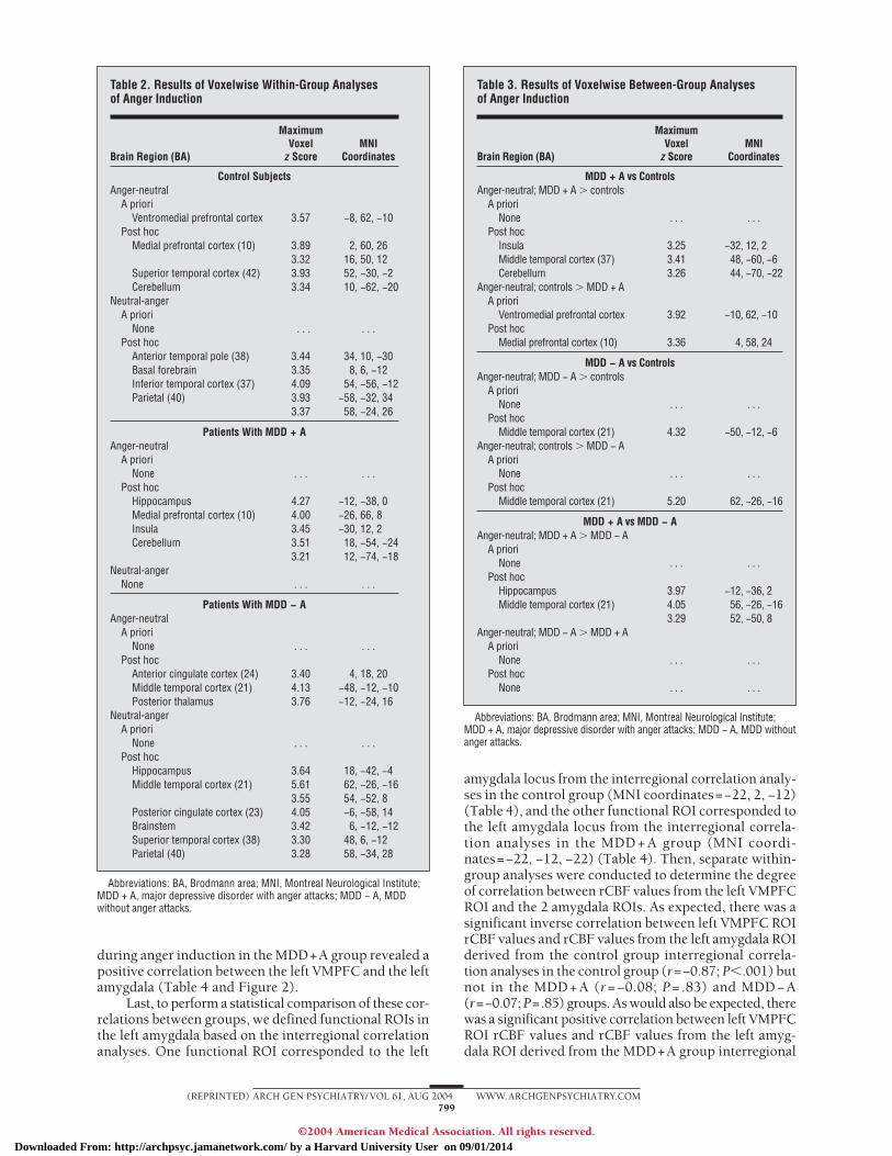
In the control group, the anger vs neutral comparisondemonstrated increased regional cerebral blood flow(rCBF) in the left ventromedial PFC (VMPFC) (Table2).Regarding the a priori territories of interest, no signifi-cant activations were found in within-group analyses in-volving the MDD+A and MDD−A groups.
Between-Group Analyses
Regarding a priori hypotheses, the between-group analy-ses revealed greater rCBF increases in the control groupthan in the MDD+A group in the left VMPFC during theanger vs neutral comparison (Table 3 and Figure 1).These differences were not present in other between-group analyses. Last, there were no between-group dif-ferences in rCBF changes in the amygdala during the an-ger vs neutral comparison.
VMPFC ROI-Based InterregionalCorrelation Analyses
Interregional correlation analyses examining the rela-tionship between rCBF responses in the left VMPFCand those in the rest of the brain were conducted in eachgroup for the anger vs neutral comparison (Table 4 andFigure 2). We defined a functional ROI in the leftVMPFC (MNI coordinates=−8, 62, −10) based on the an-ger vs neutral comparison in the control group. We thenextracted rCBF values from the ROI and conducted a cor-relational analysis between these ROI values and whole-brain, voxelwise rCBF changes in the anger vs neutralcomparison for each group. Based on known bidirec-tional connections between the PFC and the amygdalaand evidence that these 2 structures are mutually inhibi-tory, we hypothesized that these interregional correla-tion analyses would demonstrate an inverse correlationof rCBF changes during anger induction between the leftVMPFC and the left amygdala in the control group. Theanalysis confirmed this hypothesis (Table 4 and Figure2). Identical interregional correlation analyses of rCBFchanges during anger induction did not demonstrate anysignificant relationship between the left VMPFC and theleft amygdala in the MDD−A group (Table 4). How-ever, interregional correlation analyses of rCBF changes
(REPRINTED) ARCH GEN PSYCHIATRY/ VOL 61, AUG 2004 WWW.ARCHGENPSYCHIATRY.COM798
©2004 American Medical Association. All rights reserved.Downloaded From: http://archpsyc.jamanetwork.com/ by a Harvard University User on 09/01/2014
during anger induction in the MDD+A group revealed apositive correlation between the left VMPFC and the leftamygdala (Table 4 and Figure 2).
Last, to perform a statistical comparison of these cor-relations between groups, we defined functional ROIs inthe left amygdala based on the interregional correlationanalyses. One functional ROI corresponded to the left
amygdala locus from the interregional correlation analy-ses in the control group (MNI coordinates=−22, 2, −12)(Table 4), and the other functional ROI corresponded tothe left amygdala locus from the interregional correla-tion analyses in the MDD + A group (MNI coordi-nates=−22, −12, −22) (Table 4). Then, separate within-group analyses were conducted to determine the degreeof correlation between rCBF values from the left VMPFCROI and the 2 amygdala ROIs. As expected, there was asignificant inverse correlation between left VMPFC ROIrCBF values and rCBF values from the left amygdala ROIderived from the control group interregional correla-tion analyses in the control group (r=−0.87; P�.001) butnot in the MDD+A (r =−0.08; P = .83) and MDD−A(r=−0.07; P=.85) groups. As would also be expected, therewas a significant positive correlation between left VMPFCROI rCBF values and rCBF values from the left amyg-dala ROI derived from the MDD+A group interregional
Table 2. Results of Voxelwise Within-Group Analysesof Anger Induction
Brain Region (BA)
MaximumVoxel
z ScoreMNI
Coordinates
Control SubjectsAnger-neutral
A prioriVentromedial prefrontal cortex 3.57 −8, 62, −10
Post hocMedial prefrontal cortex (10) 3.89 2, 60, 26
3.32 16, 50, 12Superior temporal cortex (42) 3.93 52, −30, −2Cerebellum 3.34 10, −62, −20
Neutral-angerA priori
None . . . . . .Post hoc
Anterior temporal pole (38) 3.44 34, 10, −30Basal forebrain 3.35 8, 6, −12Inferior temporal cortex (37) 4.09 54, −56, −12Parietal (40) 3.93 −58, −32, 34
3.37 58, −24, 26
Patients With MDD + AAnger-neutral
A prioriNone . . . . . .
Post hocHippocampus 4.27 −12, −38, 0Medial prefrontal cortex (10) 4.00 −26, 66, 8Insula 3.45 −30, 12, 2Cerebellum 3.51 18, −54, −24
3.21 12, −74, −18Neutral-anger
None . . . . . .
Patients With MDD − AAnger-neutral
A prioriNone . . . . . .
Post hocAnterior cingulate cortex (24) 3.40 4, 18, 20Middle temporal cortex (21) 4.13 −48, −12, −10Posterior thalamus 3.76 −12, −24, 16
Neutral-angerA priori
None . . . . . .Post hoc
Hippocampus 3.64 18, −42, −4Middle temporal cortex (21) 5.61 62, −26, −16
3.55 54, −52, 8Posterior cingulate cortex (23) 4.05 −6, −58, 14Brainstem 3.42 6, −12, −12Superior temporal cortex (38) 3.30 48, 6, −12Parietal (40) 3.28 58, −34, 28
Abbreviations: BA, Brodmann area; MNI, Montreal Neurological Institute;MDD + A, major depressive disorder with anger attacks; MDD − A, MDDwithout anger attacks.
Table 3. Results of Voxelwise Between-Group Analysesof Anger Induction
Brain Region (BA)
MaximumVoxel
z ScoreMNI
Coordinates
MDD + A vs ControlsAnger-neutral; MDD + A � controls
A prioriNone . . . . . .
Post hocInsula 3.25 −32, 12, 2Middle temporal cortex (37) 3.41 48, −60, −6Cerebellum 3.26 44, −70, −22
Anger-neutral; controls � MDD + AA priori
Ventromedial prefrontal cortex 3.92 −10, 62, −10Post hoc
Medial prefrontal cortex (10) 3.36 4, 58, 24
MDD − A vs ControlsAnger-neutral; MDD − A � controls
A prioriNone . . . . . .
Post hocMiddle temporal cortex (21) 4.32 −50, −12, −6
Anger-neutral; controls � MDD − AA priori
None . . . . . .Post hoc
Middle temporal cortex (21) 5.20 62, −26, −16
MDD + A vs MDD − AAnger-neutral; MDD + A � MDD − A
A prioriNone . . . . . .
Post hocHippocampus 3.97 −12, −36, 2Middle temporal cortex (21) 4.05 56, −26, −16
3.29 52, −50, 8Anger-neutral; MDD − A � MDD + A
A prioriNone . . . . . .
Post hocNone . . . . . .
Abbreviations: BA, Brodmann area; MNI, Montreal Neurological Institute;MDD + A, major depressive disorder with anger attacks; MDD − A, MDD withoutanger attacks.
(REPRINTED) ARCH GEN PSYCHIATRY/ VOL 61, AUG 2004 WWW.ARCHGENPSYCHIATRY.COM799
©2004 American Medical Association. All rights reserved.Downloaded From: http://archpsyc.jamanetwork.com/ by a Harvard University User on 09/01/2014

correlation analyses in the MDD+A group (r=0.90;P�.001) but not in the MDD−A (r=−0.14; P=.69) andcontrol (r=0.31; P=.38) groups. Fisher z transforma-tion of the within-group correlation coefficients was usedto perform a statistical comparison of these correlationsbetween groups. The correlation coefficient arising fromthe comparison of left VMPFC ROI rCBF values and rCBF
values from the left amygdala ROI derived from the con-trol group interregional correlation analyses in the con-trol group (r=−0.87) differed significantly from the iden-tical comparisons in the MDD−A and MDD+A groups(P=.02 for both), whereas the MDD−A and MDD+Agroups did not differ significantly from one another(P=.99). The correlation coefficient arising from the com-parison of left VMPFC ROI rCBF values and rCBF val-ues from the left amygdala ROI derived from the MDD+Agroup interregional correlation analyses in the MDD+Agroup (r=0.90) differed significantly from the identicalcomparisons in the MDD − A (P = .002) and control(P=.03) groups, whereas the MDD−A and control groupsdid not differ significantly from one another (P=.38).
COMMENT
Previous functional neuroimaging studies conducted withindividuals predisposed to anger or aggression have prin-cipally used neutral-state or pharmacologic challenge stud-ies. In contrast, the present study represents an initialsymptom provocation PET study in patients with MDDpredisposed to anger or aggression and yields several im-portant findings. First, this study replicated findings fromour laboratory44 and others42,43,45,46 of increased ventralPFC (specifically, the left VMPFC in this study) rCBFduring anger induction in controls. Second, the controlsubjects demonstrated statistically significantly greaterleft VMPFC rCBF increases than the patients withMDD+A during anger induction. There was no corre-sponding difference in left VMPFC rCBF during angerinduction when comparing the patients with MDD−Awith either the patients with MDD+A or the control sub-
Figure 1. A statistical parametric map of positron emission tomography datacorresponding to a between-group comparison of the anger vs neutralconditions is superimposed over a nominally normal magnetic resonanceimage in Montreal Neurological Institute space for gross anatomic reference.Voxels exceeding the z score threshold of 3.09 are shown in yellow. Thisimage is an axial section demonstrating that control subjects have asignificantly greater regional cerebral blood flow increase in the leftventromedial prefrontal cortex than patients with major depressive disorderwith anger attacks.
Table 4. Results of Interregional Correlation Analyses of Left VMPFC rCBF Changes and rCBF Changesin the Rest of the Brain During Anger Induction*
Control Group MDD + A Group MDD − A Group
Brain Region (BA)z
ScoreMNI
Coordinates Brain Region (BA)z
ScoreMNI
Coordinates Brain Region (BA)z
ScoreMNI
Coordinates
Negative CorrelationAmygdala 3.29 −22, 2, −12 Insula 4.66 44, −16, 8 Lingual gyrus 3.99 −18, −52, −6Anterior temporal pole (38) 3.97 −30, 6, −20 Orbitofrontal cortex (11) 4.04 34, 56, −16 Parietal cortex 3.68 66, −28, 32Inferior temporal cortex (20) 3.97 −40, −16, −36 Occipital cortex 3.99 26, −80, 8 3.44 44, −46, 38Orbitofrontal cortex (11/47) 3.61 −42, 28, −12 Parietal/occipital cortex 3.34 20, −66, 14
3.38 −16, 20, −18 Cerebellum 3.23 26, −78, −22Brainstem 3.33 2, −20, −16
Positive CorrelationVentromedial prefrontal cortex 4.01 16, 60, −16 Putamen 4.18 14, 8, −4 Inferior frontal cortex (44) 4.60 −58, 18, 20Insula 3.96 40, −14, −2 4.09 18, −4, 8 Lingual gyrus 4.37 14, −60, −12Superior temporal cortex (22) 3.51 62, −16, 6 Orbitofrontal cortex (11/47) 4.11 −14, 46, −26 Insula 4.19 −38, −10, −6
3.59 −14, 32, −14 Superior temporal cortex 4.04 −58, −18, 63.26 8, 24, −22 Middle temporal cortex 3.71 −66, −52, −12
Insula 3.68 −32, 18, 10 3.25 −58, −32, 6Amygdala 3.59 −22, −12, −22 Ventromedial prefrontal
cortex3.41 12, 54, −16
Anterior cingulate cortex(24/32)
3.54 4, 12, 38 Occipital cortex 3.38 −24, −98, 12
Hippocampus 3.44 26, −32, −10Occipital cortex 3.26 −44, −78, 24
Abbreviations: MDD + A, major depressive disorder with anger attacks; MDD − A, MDD without anger attacks; BA, Brodmann area; MNI, Montreal NeurologicalInstitute; rCBF, regional cerebral blood flow; VMPFC, ventromedial prefrontal cortex.
*Boldfaced entries indicate a priori regions; all other reported regions represent post hoc findings.
(REPRINTED) ARCH GEN PSYCHIATRY/ VOL 61, AUG 2004 WWW.ARCHGENPSYCHIATRY.COM800
©2004 American Medical Association. All rights reserved.Downloaded From: http://archpsyc.jamanetwork.com/ by a Harvard University User on 09/01/2014
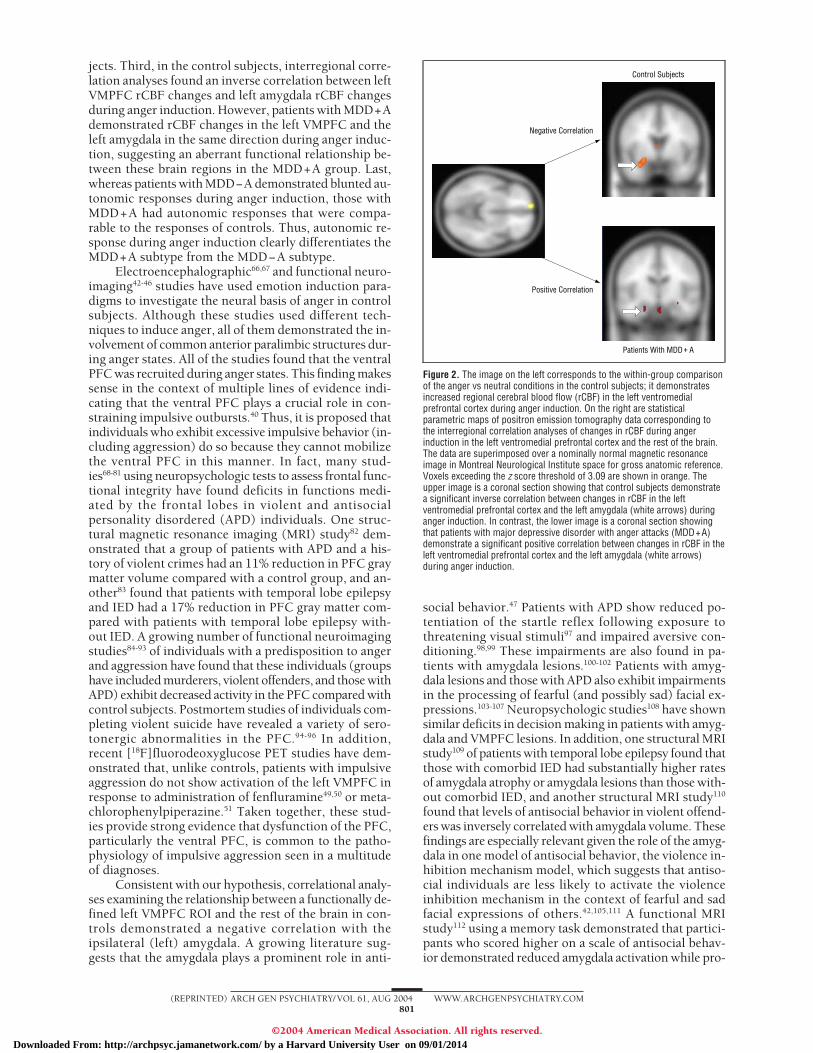
jects. Third, in the control subjects, interregional corre-lation analyses found an inverse correlation between leftVMPFC rCBF changes and left amygdala rCBF changesduring anger induction. However, patients with MDD+Ademonstrated rCBF changes in the left VMPFC and theleft amygdala in the same direction during anger induc-tion, suggesting an aberrant functional relationship be-tween these brain regions in the MDD+A group. Last,whereas patients with MDD−A demonstrated blunted au-tonomic responses during anger induction, those withMDD+A had autonomic responses that were compa-rable to the responses of controls. Thus, autonomic re-sponse during anger induction clearly differentiates theMDD+A subtype from the MDD−A subtype.
Electroencephalographic66,67 and functional neuro-imaging42-46 studies have used emotion induction para-digms to investigate the neural basis of anger in controlsubjects. Although these studies used different tech-niques to induce anger, all of them demonstrated the in-volvement of common anterior paralimbic structures dur-ing anger states. All of the studies found that the ventralPFC was recruited during anger states. This finding makessense in the context of multiple lines of evidence indi-cating that the ventral PFC plays a crucial role in con-straining impulsive outbursts.40 Thus, it is proposed thatindividuals who exhibit excessive impulsive behavior (in-cluding aggression) do so because they cannot mobilizethe ventral PFC in this manner. In fact, many stud-ies68-81 using neuropsychologic tests to assess frontal func-tional integrity have found deficits in functions medi-ated by the frontal lobes in violent and antisocialpersonality disordered (APD) individuals. One struc-tural magnetic resonance imaging (MRI) study82 dem-onstrated that a group of patients with APD and a his-tory of violent crimes had an 11% reduction in PFC graymatter volume compared with a control group, and an-other83 found that patients with temporal lobe epilepsyand IED had a 17% reduction in PFC gray matter com-pared with patients with temporal lobe epilepsy with-out IED. A growing number of functional neuroimagingstudies84-93 of individuals with a predisposition to angerand aggression have found that these individuals (groupshave included murderers, violent offenders, and those withAPD) exhibit decreased activity in the PFC compared withcontrol subjects. Postmortem studies of individuals com-pleting violent suicide have revealed a variety of sero-tonergic abnormalities in the PFC.94-96 In addition,recent [18F]fluorodeoxyglucose PET studies have dem-onstrated that, unlike controls, patients with impulsiveaggression do not show activation of the left VMPFC inresponse to administration of fenfluramine49,50 or meta-chlorophenylpiperazine.51 Taken together, these stud-ies provide strong evidence that dysfunction of the PFC,particularly the ventral PFC, is common to the patho-physiology of impulsive aggression seen in a multitudeof diagnoses.
Consistent with our hypothesis, correlational analy-ses examining the relationship between a functionally de-fined left VMPFC ROI and the rest of the brain in con-trols demonstrated a negative correlation with theipsilateral (left) amygdala. A growing literature sug-gests that the amygdala plays a prominent role in anti-
social behavior.47 Patients with APD show reduced po-tentiation of the startle reflex following exposure tothreatening visual stimuli97 and impaired aversive con-ditioning.98,99 These impairments are also found in pa-tients with amygdala lesions.100-102 Patients with amyg-dala lesions and those with APD also exhibit impairmentsin the processing of fearful (and possibly sad) facial ex-pressions.103-107 Neuropsychologic studies108 have shownsimilar deficits in decision making in patients with amyg-dala and VMPFC lesions. In addition, one structural MRIstudy109 of patients with temporal lobe epilepsy found thatthose with comorbid IED had substantially higher ratesof amygdala atrophy or amygdala lesions than those with-out comorbid IED, and another structural MRI study110
found that levels of antisocial behavior in violent offend-ers was inversely correlated with amygdala volume. Thesefindings are especially relevant given the role of the amyg-dala in one model of antisocial behavior, the violence in-hibition mechanism model, which suggests that antiso-cial individuals are less likely to activate the violenceinhibition mechanism in the context of fearful and sadfacial expressions of others.42,105,111 A functional MRIstudy112 using a memory task demonstrated that partici-pants who scored higher on a scale of antisocial behav-ior demonstrated reduced amygdala activation while pro-
Negative Correlation
Control Subjects
Patients With MDD + A
Positive Correlation
Figure 2. The image on the left corresponds to the within-group comparisonof the anger vs neutral conditions in the control subjects; it demonstratesincreased regional cerebral blood flow (rCBF) in the left ventromedialprefrontal cortex during anger induction. On the right are statisticalparametric maps of positron emission tomography data corresponding tothe interregional correlation analyses of changes in rCBF during angerinduction in the left ventromedial prefrontal cortex and the rest of the brain.The data are superimposed over a nominally normal magnetic resonanceimage in Montreal Neurological Institute space for gross anatomic reference.Voxels exceeding the z score threshold of 3.09 are shown in orange. Theupper image is a coronal section showing that control subjects demonstratea significant inverse correlation between changes in rCBF in the leftventromedial prefrontal cortex and the left amygdala (white arrows) duringanger induction. In contrast, the lower image is a coronal section showingthat patients with major depressive disorder with anger attacks (MDD+A)demonstrate a significant positive correlation between changes in rCBF in theleft ventromedial prefrontal cortex and the left amygdala (white arrows)during anger induction.
(REPRINTED) ARCH GEN PSYCHIATRY/ VOL 61, AUG 2004 WWW.ARCHGENPSYCHIATRY.COM801
©2004 American Medical Association. All rights reserved.Downloaded From: http://archpsyc.jamanetwork.com/ by a Harvard University User on 09/01/2014

cessing negatively valenced words compared withindividuals who scored lower on the scale. Another re-cent functional MRI study113 of patients with APD usinga differential aversive-delay conditioning task foundblunted activation of the orbitofrontal cortex, anteriorcingulate cortex, insula, and amygdala compared withcontrol subjects. Thus, multiple lines of evidence sug-gest that in addition to dysfunction of the PFC, amyg-dala dysfunction is also common to the pathophysiol-ogy of impulsive aggression seen in a variety of diagnoses.
It has been suggested that APD and IED may be as-sociated with “dual brain pathology” in which abnormali-ties in the amygdala result in dysfunctional arousal statesand those in the PFC result in dyscontrol states.114 Thereare known bidirectional connections between the PFC (es-pecially the medial PFC) and the amygdala,115-120 and thereis evidence121,122 that in control subjects these 2 struc-tures are mutually inhibitory in that increased activity inone structure inhibits activity in the other structure. Be-cause the controls in the present study demonstrated a re-ciprocal (or inverse) relationship between left VMPFC andleft amygdala rCBF during the anger vs neutral compari-son, we examined this relationship in the MDD groups.We did not demonstrate any statistically significant rela-tionship for rCBF changes during anger induction be-tween the left VMPFC and left amygdala in the MDD−Agroup. In contrast, for the MDD+A group the rCBF changesduring anger induction in the left VMPFC and the leftamygdala were in the same direction (ie, they demon-strated a positive correlation). This suggests that, at leastin the context of anger induction, the normal (inverse)functional relationship between the VMPFC and the amyg-dala is absent in the MDD−A group and is reversed in theMDD+A group. Therefore, this profile of VMPFC andamygdala activity and their interactions may distinguishMDD+A and may be responsible for the unique clinicalpresentation of patients with this subtype of MDD. Last,the differential abnormalities in VMPFC and amygdalafunction in the MDD+A group are consistent with the dys-function of both of these regions found in other impul-sively aggressive patient populations.
There are some limitations of the present study. First,this study was not designed to assess sex differences dur-ing anger induction. Second, formal assessment for AxisII diagnoses was not performed in the study popula-tions. Future studies that address these issues would bedesirable. Last, structural MRIs were not used for ana-tomic localization of significant rCBF changes. Instead,the MNI atlas, a spatially normalized composite of 152MRIs of a healthy brain, was used for localization pur-poses. Concerns regarding anatomic localization were fur-ther mitigated by the fact that we had concise, evidence-based, a priori hypotheses for the present study involvingthe ventral PFC and amygdala.
Patients with MDD+A experience a remission ofanger attacks in concert with remission of their depres-sive symptoms after successful treatment, whereasother patient populations that frequently exhibit impul-sive aggression typically exhibit a more chronic, treat-ment-refractory clinical course. For these reasons, fu-ture studies that include assessments of MDD+Apatients before and after treatment may provide valu-
able insight into the brain mechanisms underlying theresolution of these symptoms.
Submitted for publication July 7, 2003; final revision re-ceived January 9, 2004; accepted February 17, 2004.
This study was supported by Mentored Patient-Oriented Research Career Development Award MH01735from the National Institute of Mental Health, Bethesda, Md(Dr Dougherty).
We thank the individuals who served as research par-ticipants and Sandra Barrow, BS, and Steve Weise, BS, fortechnical assistance.
Correspondence: Darin D. Dougherty, MD, MSc, Mas-sachusetts General Hospital–East, CNY-2612, Bldg 149, 13thStreet, Charlestown, MA 02129 ([email protected]).
REFERENCES
1. Freud S. Mourning and Melancholia. Vol 4. London, England: Hogarth Press;1917.
2. Bland RC, Orn J. Family violence and psychiatric disorder. Can J Psychiatry.1986;31:129-137.
3. Eronen M, Hakola P, Tiihonen J. Mental disorders and homicidal behavior inFinland. Arch Gen Psychiatry. 1996;53:497-501.
4. Fava M, Rosenbaum JF, McCarthy MK, Pava JA, Steingard RJ, Bless E. Angerattacks in depressed outpatients and their response to fluoxetine. Psychophar-macol Bull. 1991;27:275-279.
5. Fava M, Nierenberg AA, Quitkin FM, Zisook S, Pearlstein T, Stone A, Rosen-baum JF. A preliminary study on the efficacy of sertraline and imipramine onanger attacks in atypical depression and dysthymia. Psychopharmacol Bull. 1997;33:101-103.
6. Koh KB, Kim CH, Park JK. Predominance of anger in depressive disorders com-pared with anxiety disorders and somatoform disorders. J Clin Psychiatry. 2002;63:486-492.
7. Swanson JW, Holzer CE, Ganju VK, Jono RT. Violence and psychiatric disorderin the community: evidence from the Epidemiological Catchment Area sur-veys. Hosp Community Psychiatry. 1990;41:761-770.
8. Weissman M, Klerman GL, Paykel ES. Clinical evaluation of hostility in depres-sion. Am J Psychiatry. 1971;128:41-46.
9. Schless AP, Mendels J, Kipperman A, Cochrane C. Depression and hostility.J Nerv Ment Dis. 1974;159:91-100.
10. Fava M, Anderson K, Rosenbaum JF. “Anger attacks”: possible variants of panicand major depressive disorders. Am J Psychiatry. 1990;147:867-870.
11. Fava M, Rosenbaum JF, Pava JA, McCarthy MK, Steingard RJ, Bouffides E. An-ger attacks in unipolar depression, part 1: clinical correlates and response tofluoxetine treatment. Am J Psychiatry. 1993;150:1158-1163.
12. Fava M, Alpert J, Nierenberg AA, Ghaemi N, O’Sullivan R, Tedlow J, Worthing-ton J, Rosenbaum JF. Fluoxetine treatment of anger attacks: a replication study.Ann Clin Psychiatry. 1996;8:7-10.
13. Morand P, Thomas G, Bungener C, Ferreri M, Jouvent R. Fava’s Anger AttacksQuestionnaire: the evaluation of the French version in depressed patients. EurPsychiatry. 1998;13:41-45.
14. Sayar K, Guzelhan Y, Solmaz M, Ozer OA, Ozturk M, Acar B, Arikan M. Anger at-tacks in depressed Turkish outpatients. Ann Clin Psychiatry. 2000;12:213-218.
15. Posternak MA, Zimmerman M. Anger and aggression in psychiatric outpa-tients. J Clin Psychiatry. 2002;63:665-672.
16. Iribarren C, Sidney S, Bild DE, Liu K, Markovitz JH, Roseman JM, Matthews K.Association of hostility with coronary artery calcification in young adults: theCARDIA study: Coronary Artery Risk Development in Young Adults. JAMA. 2000;283:2546-2551.
17. Julkunen J, Salonen R, Kaplan GA, Chesney MA, Salonen JT. Hostility and theprogression of carotid atherosclerosis. Psychosom Med. 1994;56:519-525.
18. Matthews KA, Owens JF, Kuller LH, Sutton-Tyrell K, Jansen-McWilliams L. Arehostility and anxiety associated with carotid atherosclerosis in healthy post-menopausal women? Psychosom Med. 1998;60:633-638.
19. Barefoot JC, Dahlstrom WG, Williams RB. Hostility, CHD incidence, and totalmortality: a 25-year follow-up study of 255 physicians. Psychosom Med. 1983;45:59-63.
20. Barefoot JC, Larsen S, von der Leith L, Schroll M. Hostility, incidence of myo-cardial infarction, and mortality in a sample of older Danish men and women.Am J Epidemiol. 1995;142:477-484.
(REPRINTED) ARCH GEN PSYCHIATRY/ VOL 61, AUG 2004 WWW.ARCHGENPSYCHIATRY.COM802
©2004 American Medical Association. All rights reserved.Downloaded From: http://archpsyc.jamanetwork.com/ by a Harvard University User on 09/01/2014

21. Hecker M, Chesney MA, Black GW, Frautschi N. Coronary-prone behaviors inthe Western Collaborative Group Study. Psychosom Med. 1988;50:153-164.
22. Shekelle RB, Gale M, Ostfeld AM, Paul O. Hostility, risk of coronary heart dis-ease, and mortality. Psychosom Med. 1983;45:109-114.
23. Kawakami N, Akari S, Ohtsu H, Hayashi T, Masumoto T, Yokohama K. Effectsof mood states, smoking, and urinary catecholamine excretion on hemoglobinA1C in male Japanese workers. Ind Health. 1995;33:153-162.
24. Niaura R, Banks SM, Ward KD, Stoney CM, Spiro A, Aldwin CM, Landsberg L,Weiss ST. Hostility and the metabolic syndrome in older males: the NormativeAging Study. Psychosom Med. 2000;62:7-16.
25. Raikkonen K, Keltikangas-Jarvinen L, Hautanen A. The role of psychological coro-nary risk factors in insulin and glucose metabolism. J Psychosom Res. 1994;38:705-713.
26. Raikkonen K, Matthews KA, Kuller LH, Reiber C, Bunker CH. Anger, hostility,and visceral adipose tissue in healthy postmenopausal women. Metabolism.1999;48:1146-1151.
27. Surwit RS, Williams RB, Siegler IC, Lane JD, Helms M, Applegate KL, ZuckerN, Feinglos MN, McCaskill CM, Barefoot JC. Hostility, race, and glucose me-tabolism in nondiabetic individuals. Diabetes Care. 2002;25:835-839.
28. Ariyo A, Haan M, Tangen C, Rutledge JC, Cushman M, Dobs A, Furberg CD.Depressive symptoms and risks of coronary artery disease and mortality in el-derly Americans. Circulation. 2000;102:1773-1779.
29. Frasure-Smith N, Lesperance F, Talajic M. Depression and 18-month progno-sis after myocardial infarction. Circulation. 1995;91:999-1005.
30. Pratt LA, Ford DE, Crum RM, Armenian HK, Gallo JJ, Eaton WW. Depression,psychotropic medication, and risk of myocardial infarction: prospective datafrom the Baltimore ECA follow-up. Circulation. 1996;94:3123-3129.
31. Hamilton J, Daneman D. Deteriorating diabetes control during adolescence: physi-ological or psychosocial? J Pediatr Endocrinol Metab. 2002;15:115-126.
32. Lernmark B, Persson B, Fisher L, Rydelius P-A. Symptoms of depression areimportant to psychological adaptation and metabolic control in children withdiabetes mellitus. Diabet Med. 1999;16:14-22.
33. World Health Organization. The World Health Report: Mental Health: New Un-derstanding, New Hope. Geneva, Switzerland: World Health Organization; 2001.
34. Davidson RJ, Pizzagalli D, Nitschke JB, Putnam K. Depression: perspectivesfrom affective neuroscience. Annu Rev Psychol. 2002;53:545-574.
35. Dougherty DD, Rauch SL. Neuroimaging and neurobiological models of de-pression. Harv Rev Psychiatry. 1997;5:138-159.
36. Drevets WC. Neuroimaging studies of mood disorders. Biol Psychiatry. 2000;48:813-829.
37. Drevets WC. Neuroimaging and neuropathological studies of depression: im-plications for the cognitive-emotional features of mood disorders. Curr OpinNeurobiol. 2001;11:240-249.
38. Mayberg HS. Limbic-cortical dysregulation: a proposed model of depression.J Neuropsychiatry Clin Neurosci. 1997;9:471-481.
39. Brower MC, Price BH. Neuropsychiatry of frontal lobe dysfunction in violentand criminal behavior: a critical review. J Neurol Neurosurg Psychiatry. 2001;71:720-726.
40. Davidson RJ, Putnam KM, Larson CL. Dysfunction in the neural circuitry of emo-tion regulation: a possible prelude to violence. Science. 2000;289:591-594.
41. Kavoussi R, Armstead P, Coccaro E. The neurobiology of impulsive aggres-sion. Psychiatr Clin North Am. 1997;20:395-403.
42. Blair RJR, Morris JS, Frith CD, Perrett DI, Dolan RJ. Dissociable neural re-sponses to facial expressions of sadness and anger. Brain. 1999;122:883-893.
43. Damasio AR, Grabowski TJ, Bechara A, Damasio H, Ponto LLB, Parvizi J, HichwaRD. Subcortical and cortical brain activity during the feelings of self-generatedemotions. Nat Neurosci. 2000;3:1049-1056.
44. Dougherty DD, Shin LM, Alpert NM, Pitman RK, Orr SP, Lasko M, Macklin ML,Fischman AJ, Rauch SL. Anger in healthy men: a PET study using script-drivenimagery. Biol Psychiatry. 1999;46:466-472.
45. Kimbrell TA, George MS, Parekh PI, Ketter TA, Podell DM, Danielson AL, Re-pella JD, Benson BE, Willis MW, Herscovitch P, Post RM. Regional brain ac-tivity during transiently self-induced anxiety and anger in healthy adults. BiolPsychiatry. 1999;46:454-465.
46. Pietrini P, Guazzelli M, Basso G, Jaffe K, Grafman J. Neural correlates of imagi-nal aggressive behavior assessed by positron emission tomography in healthysubjects. Am J Psychiatry. 2000;157:1772-1781.
47. Blair RJR. Neurobiological basis of psychopathy. Br J Psychiatry. 2003;182:5-7.48. McKay KE, Halperin JM. ADHD, aggression, and antisocial behavior across the
lifespan: interactions with neurochemical and cognitive function. Ann N Y AcadSci. 2001;931:84-96.
49. Siever LJ, Buchsbaum MS, New AS, Spiegel-Cohen J, Wei T, Hazlett EA, SevinE, Nunn M, Mitropoulou V. d,I-Fenfluramine response in impulsive personalitydisorder assessed with [18F]fluorodeoxyglucose positron emission tomogra-phy. Neuropsychopharmacology. 1999;20:413-423.
50. Soloff PH, Meltzer CC, Greer PJ, Constantine D, Kelly TM. A fenfluramine-activated FDG-PET study of borderline personality disorder. Biol Psychiatry. 2000;47:540-547.
51. New AS, Hazlett EA, Buchsbaum MS, Goodman M, Reynolds D, MitropoulouV, Sprung L, Shaw RB, Koenigsberg H, Platholi J, Silverman J, Siever LJ. Bluntedprefrontal cortical 18fluorodeoxyglucose positron emission tomography re-sponse to meta-chlorophenylpiperazine in impulsive aggression. Arch Gen Psy-chiatry. 2002;59:621-629.
52. Tebartz van Elst L, Hesslinger B, Thiel T, Geiger E, Haegele K, Lemieux L, LiebK, Bohus M, Hennig J, Ebert D. Frontolimbic brain abnormalities in patients withborderline personality disorder: a volumetric magnetic resonance imaging study.Biol Psychiatry. 2003;54:163-171.
53. Drevets WC, Price JL, Bardgett ME, Reich T, Todd RD, Raichle ME. Glucosemetabolism in the amygdala in depression: relationship to diagnostic subtypeand plasma cortisol levels. Pharmacol Biochem Behav. 2002;71:431-447.
54. Whalen PJ, Shin LM, Somerville LH, McLean AA, Kim H. Functional neuroim-aging studies of the amygdala in depression. Semin Clin Neuropsychiatry. 2002;7:234-242.
55. Oldfield RC. The assessment and analysis of handedness: the Edinburgh In-ventory. Neuropsychologia. 1971;9:97-113.
56. First MB, Spitzer RL, Gibbon M, Williams JBW. Structured Clinical Interviewfor Axis I DSM-IV Disorders–Patient Edition (With Psychotic Screen), Version2.0. New York: Biometrics Research Department, New York State PsychiatricInstitute; 1995.
57. Hamilton M. A rating scale for depression. J Neurol Neurosurg Psychiatry. 1960;23:56-62.
58. Pitman RK, Orr SP, Forgue DF, de Jong JB, Claiborn JM. Psychophysiologicassessment of posttraumatic stress disorder imagery in Vietnam combat vet-erans. Arch Gen Psychiatry. 1987;44:970-975.
59. Pitman RK, Orr SP, Forgue DF, Altman B, de Jong JB. Psychophysiologic re-sponses to combat imagery of Vietnam veterans with posttraumatic stress dis-order versus other anxiety disorders. J Abnorm Psychol. 1990;99:49-54.
60. Orr SP, Pitman RK, Lasko NB, Herz LR. Psychophysiological assessment ofposttraumatic stress disorder imagery in World War II and Korean combat vet-erans. J Abnorm Psychol. 1993;102:152-159.
61. Rauch SL, van der Kolk BA, Fisler RE, Alpert NM, Orr SP, Savage CR, FischmanAJ, Jenike MA, Pitman RK. A symptom provocation study of posttraumatic stressdisorder using positron emission tomography and script-driven imagery. ArchGen Psychiatry. 1996;53:380-387.
62. Kops ER, Herzog H, Schmid A, Holte S, Feinendegen LE. Performance charac-teristics of an eight-ring whole body PET scanner. J Comput Assist Tomogr.1990;14:437-445.
63. Friston KJ, Frith CD, Liddle PF, Frackowiak RSJ. Comparing functional (PET)images: the assessment of significant change. J Cereb Blood Flow Metab. 1991;11:690-699.
64. Friston KJ, Holmes AP, Worsley KJ, Poline JP, Frith CD, Frackowiak RSJ. Sta-tistical parametric maps in functional imaging: a general approach. Hum BrainMapp. 1995;2:189-210.
65. Brett M, Anton J-L, Valabregue R, Poline J-B. Region of interest analysis usingan SPM toolbox. Abstract presented at: 8th International Conference of Func-tional Mapping of the Human Brain; June 2-6, 2002; Sendai, Japan.
66. Harmon-Jones E, Sigelman J. State anger and prefrontal brain activity: evi-dence that insult-related relative left-prefrontal activation is associated with ex-perienced anger and aggression. J Pers Soc Psychol. 2001;80:797-803.
67. Harmon-Jones E, Allen JJ. Anger and frontal brain activity: EEG asymmetry con-sistent with approach motivation despite negative affective valence. J Pers SocPsychol. 1998;74:1310-1316.
68. Best M, Williams JM, Coccaro EF. Evidence for a dysfunctional prefrontalcircuit in patients with an impulsive aggressive disorder. Proc Natl Acad SciU S A. 2002;99:8448-8453.
69. Bergvall AH, Wessely H, Forsman A, Hansen S. A deficit in attentional set-shifting of violent offenders. Psychol Med. 2001;31:1095-1105.
70. Blair RJR, Colledge E, Mitchell DGV. Somatic markers and response reversal:is there orbitofrontal cortex dysfunction in boys with psychopathic tenden-cies? J Abnorm Child Psychol. 2001;29:499-511.
71. Deckel AW, Hesselbrock V, Bauer L. Antisocial personality disorder, childhooddelinquency, and frontal brain functioning: EEG and neuropsychological find-ings. J Clin Psychol. 1996;52:639-650.
72. Dinn WM, Harris CL. Neurocognitive function in antisocial personality disor-der. Psychiatry Res. 2000;97:173-190.
73. Dolan M, Park I. The neuropsychology of antisocial personality disorder. Psy-chol Med. 2002;32:417-427.
74. Foster HG, Hillbrand M, Silverstein M. Neuropsychological deficit and aggres-sive behavior: a prospective study. Prog Neuropsychopharmacol Biol Psychia-try. 1993;17:939-946.
(REPRINTED) ARCH GEN PSYCHIATRY/ VOL 61, AUG 2004 WWW.ARCHGENPSYCHIATRY.COM803
©2004 American Medical Association. All rights reserved.Downloaded From: http://archpsyc.jamanetwork.com/ by a Harvard University User on 09/01/2014
75. Giancola PR, Zeichner A. Neuropsychological performance on tests of frontal-lobe functioning and aggressive behavior in men. J Abnorm Psychol. 1994;103:832-835.
76. Giancola PR, Moss HB, Martin CS, Kirisci L, Tarter RE. Executive cognitive func-tioning predicts reactive aggression in boys at high risk for substance abuse: aprospective study. Alcohol Clin Exp Res. 1996;20:740-744.
77. Giancola PR, Mezzich AC, Tarter RE. Executive cognitive functioning, temper-ment, and antisocial behavior in conduct-disordered adolescent females. J Ab-norm Psychol. 1998;107:629-641.
78. LaPierre D, Braun CMJ, Hodgins S. Ventral frontal deficits in psychopathy: neu-ropsychological test findings. Neuropsychologia. 1995;33:139-151.
79. Lau MA, Pihl RO, Peterson JB. Provocation, acute alcohol intoxication, cogni-tive performance, and aggression. J Abnorm Psychol. 1995;104:150-155.
80. Mitchell DGV, Colledge E, Leonard A, Blair RJR. Risky decisions and responsereversal: is there evidence of orbitofrontal cortex dysfunction in psychopathicindividuals? Neuropsychologia. 2002;40:2013-2022.
81. Rosse RB, Miller MW, Deutsch SI. Violent antisocial behavior and WisconsinCard Sorting Test performance in cocaine addicts [letter]. Am J Psychol. 1993;150:170-171.
82. Raine A, Lencz T, Bihrle S, LaCasse L, Colletti P. Reduced prefrontal gray mat-ter volume and reduced autonomic activity in antisocial personality disorder.Arch Gen Psychiatry. 2000;57:119-127.
83. Woermann FG, Tebartz van Elst L, Koepp MJ, Free SL, Thompson PJ, TrimbleMR, Duncan JS. Reduction of frontal neocortical grey matter associated withaffective aggression in patients with temporal lobe epilepsy: an objective voxelby voxel analysis of automatically segmented MRI. J Neurol Neurosurg Psy-chiatry. 2000;68:162-169.
84. Amen DG, Stubblefield M, Carmichael B, Thisted R. Brain SPECT findings andaggressiveness. Ann Clin Psychiatry. 1996;8:129-137.
85. Goyer PF, Andreason PJ, Semple WE, Clayton AH, King AC, Compton-Toth BA,Schulz SC, Cohen RM. Positron-emission tomography and personality disor-ders. Neuropsychopharmacology. 1994;10:21-28.
86. Kuruoglu AC, Arikan Z, Vural G, Karatas M, Arac M, Isik E. Single photon emis-sion computerised tomography in chronic alcoholism: antisocial personality dis-order may be associated with decreased frontal perfusion. Br J Psychiatry. 1996;169:348-354.
87. Raine A, Buchsbaum M, LaCasse L. Brain abnormalities in murderers indi-cated by positron emission tomography. Biol Psychiatry. 1997;42:495-508.
88. Raine A, Meloy JR, Bihrle S, Stoddard J, LaCasse L, Buchsbaum MS. Reducedprefrontal and increased subcortical brain functioning assessed using posi-tron emission tomography in predatory and affective murderers. Behav Sci Law.1998;16:319-332.
89. Raine A, Phil D, Stoddard J, Bihrle S, Buchsbaum M. Prefrontal glucose defi-cits in murderers lacking psychosocial deprivation. Neuropsychiatry Neuro-psychol Behav Neurol. 1998;11:1-7.
90. Soderstrom H, Tullberg M, Wikkelso C, Ekholm S, Forsman A. Reduced re-gional cerebral blood flow in non-psychotic violent offenders. Psychiatry Res.2000;98:29-41.
91. Soderstrom H, Hultin L, Tullberg M, Wikkelso C, Ekholm S, Forsman A. Re-duced frontotemporal perfusion in psychopathic personality. Psychiatry Res.2002;114:81-94.
92. Volkow ND, Tancredi L. Neural substrates of violent behavior: a preliminary studywith positron emission tomography. Br J Psychiatry. 1987;151:668-673.
93. Volkow ND, Tancredi LR, Grant C, Gillespie H, Valentine A, Mullani N, WangGJ, Hollister L. Brain glucose metabolism in violent psychiatric patients: a pre-liminary study. Psychiatry Res. 1995;61:243-253.
94. Arango V, Underwood MD, Gubbi AV, Mann JJ. Localized alterations in pre-and postsynaptic serotonin binding sites in the ventrolateral prefrontal cortexof suicide victims. Brain Res. 1995;688:121-133.
95. Mann JJ, Huang YY, Underwood MD, Kassir SA, Oppenheim S, Kelly TM, DworkAJ, Arango V. A serotonin transporter gene promoter polymorphism(5-HTTLPR) and prefrontal cortical binding in major depression and suicide.Arch Gen Psychiatry. 2000;57:729-738.
96. Pandey GN, Dwivedi Y, Rizavi HS, Ren X, Pandey SC, Pesold C, Roberts RC,Conley RR, Taminga CA. Higher expression of serotonin 5-HT(2A) receptors inthe postmortem brains of teenage suicide victims. Am J Psychiatry. 2002;159:419-429.
97. Patrick CJ, Bradley MM, Lang PJ. Emotion in the criminal psychopath: startlereflex modulation. J Abnorm Psychol. 1993;102:82-92.
98. Hare RD. Psychopathy, affect and behavior. In: Van Dusen KT, Mednick SA, eds.Prospective Studies of Crime and Delinquency. Boston, Mass: Kluwer-Nijhoff;1998:225-236.
99. Lykken DT. The Antisocial Personalities. Hillsdale, NJ: Lawrence Erlbaum As-sociates Inc; 1995.
100. Angrilli A, Mauri A, Palomba D, Flor H, Birhaumer N, Sartori G, di Paola F. Startlereflex and emotion modulation impairment after a right amygdala lesion. Brain.1996;119:1991-2000.
101. Bechara A, Tranel D, Damasio H, Adolphs R, Rockland C, Damasio AR. Doubledissociation of conditioning and declarative knowledge relative to the amyg-dala and hippocampus in humans. Science. 1995;269:1115-1118.
102. LaBar KS, LeDoux JE, Spencer DD, Phelps EA. Impaired fear conditioned be-havior following unilateral temporal lobectomy in humans. J Neurosci. 1995;15:6846-6855.
103. Adolphs R, Tranel D, Young AW, Calder AJ, Phelps EA, Anderson AK, Lee GP,Damasio AR. Recognition of facial emotion in nine individuals with bilateral amyg-dala damage. Neuropsychologia. 1999;37:1111-1117.
104. Blair RJR, Coles M. Expression recognition and behavioural problems in earlyadolescence. Cogn Dev. 2000;15:421-434.
105. Blair RJR, Colledge E, Murray L, Mitchell DGV. A selective impairment in theprocessing of sad and fearful expressions in children with psychopathic ten-dencies. J Abnorm Child Psychol. 2001;29:491-498.
106. Fine C, Blair RJR. The cognitive and emotional effects of amygdala damage.Neurocase. 2000;6:435-450.
107. Stevens D, Charman T, Blair RJR. Recognition of emotion in expression andvocal tones in children with psychopathic tendencies. J Genet Psychol. 2001;162:201-211.
108. Bechara A, Damasio H, Damasio AR. Role of the amygdala in decision-making.Ann N Y Acad Sci. 2003;985:356-369.
109. Tebartz van Elst L, Woermann F, Lemieux L, Thompson PJ, Trimble MR. Af-fective aggression in patients with temporal lobe epilepsy: a quantitative mag-netic resonance study of the amygdala. Brain. 2000;123:234-243.
110. Tiihonen J, Hodgins S, Vaurio O, Laakso M, Repo E, Soininen H, Aronen HJ,Nieminen P, Savolainen L. Amygdaloid volume loss in psychopathy. In: Ab-stracts: Society for Neuroscience. Vol 26. Washington, DC: Society for Neuro-science; 2000:2017.
111. Blair RJR, Frith U. Neuro-cognitive explanations of the antisocial personalitydisorders. Crim Behav Ment Health. 2000;10(suppl):S66-S82.
112. Kiehl KA, Smith AM, Hare RD, Mendrek A, Forster BB, Brink J, Liddle PF. Lim-bic abnormalities in affective processing by criminal psychopaths revealed byfunctional magnetic imaging. Biol Psychiatry. 2001;50:677-684.
113. Veit R, Flor H, Erb M, Hermann C, Lotze M, Grodd W, Birbaumer N. Brain cir-cuits involved in emotional learning in antisocial behavior and social phobia inhumans. Neurosci Lett. 2002;328:233-236.
114. Tebartz van Elst L, Trimble MR, Ebert D. Dual brain pathology in patients withaffective aggressive episodes [letter]. Arch Gen Psychiatry. 2001;58:1187-1188.
115. Amaral DG, Price JL. Amygdalo-cortical projections in the monkey (Macacafascicularis). J Comp Neurol. 1984;230:465-496.
116. Amaral DG, Insuasti R. Retrograde transport of D-[H]-aspartate injected intothe monkey amygdaloid complex. Exp Brain Res. 1992;88:375-388.
117. Chiba T, Kayahara T, Nakano K. Efferent projections of infralimbic and prelim-bic areas of the medial prefrontal cortex in the Japanese monkey, Macaca fus-cata. Brain Res. 2001;888:83-101.
118. Stefanacci L, Amaral DG. Some observations on cortical inputs to the macaquemonkey amygdala: an anterograde tracing study. J Comp Neurol. 2002;451:301-323.
119. Price JL. Comparative aspects of amygdala connectivity. Ann N Y Acad Sci. 2003;985:50-58.
120. Carmichael ST, Price JL. Limbic connections of the orbital and medial prefron-tal cortex in macaque monkeys. J Comp Neurol. 1995;363:615-641.
121. Milad MR, Quirk GJ. Neurons in medial prefrontal cortex signal memory for fearextinction. Nature. 2002;420:70-74.
122. Hariri AR, Bookheimer SY, Mazziotta JC. Modulating emotional responses: ef-fects of a neocortical network on the limbic system. Neuroreport. 2000;11:43-48.
(REPRINTED) ARCH GEN PSYCHIATRY/ VOL 61, AUG 2004 WWW.ARCHGENPSYCHIATRY.COM804
©2004 American Medical Association. All rights reserved.Downloaded From: http://archpsyc.jamanetwork.com/ by a Harvard University User on 09/01/2014
Related Documents