British Journal of Ophthalmology, 1979, 63, 188-190 Vasculature of the optic nerve in anencephaly JACK ROOTMAN AND EUTHYMIOS P. CARVOUNIS From the Department of Ophthalmology, University of British Columbia, and Vancouver General Hospital, Vancouver, BC, Canada SUMMARY The optic nerves and globes obtained from 6 anencephalics were studied histologically and compared to normal specimens obtained from 4 stillborn infants as well as 1 case of septo-optic dysplasia. Special emphasis was placed on examination of the optic nerve, and it was found that an average of 48 vessels per high-power field were seen posterior to the lamina cribrosa in anencephalics. In contrast, control globes had an average of 12 vessels per high-power field. This suggested that an increase in the vasculature of the hypoplastic optic nerve is a characteristic feature of anencephalics. Anencephaly is a striking congenital malformation incompatible with life. We wish to report on the unusual and previously undescribed appearance of the vasculature of the optic nerve in anencephaly. We have been impressed by the multiplicity of vessels in the optic nerve proximal to the lamina cribrosa. This observation prompted us to review the optic nerves of all the anencephalics that have come to necropsy at the Vancouver General Hospital since 1968. Materials and methods Material was obtained from 6 necropsies of anence- phalics (9 eyes) done at the Vancouver General Hospital from 1968 to 1977. Seven eyes from 4 stillborn babies were randomly collected from the Ophthalmic Pathology Registry of the University of British Columbia to serve as controls. One case of septo-optic dysplasia was also included for comparison. Sections from all the eyes were stained with haematoxylin-eosin and Masson Trichrom. Vessel counts per high-power field were performed at 1 mm proximal to the lamina cribrosa. Results All the anencephalic eyes showed marked atrophy and/or hypoplasia of the ganglion cells, the nerve fibre layer, and the optic nerve (Fig 1). One globe showed proliferative vasculopathy similar to retino- pathy of prematurity; another eye had a coloboma Address for reprints: J. Rootman, MD, Department of Ophthalmology, 2550 Willow Street, Vancouver, BC, Canada V5Z 3N9 .¢ ~ ~~~~~~~~~~~~~~~~~~~~~~~~~~~~~~~~~~.. .... .:..- ..:.. !L%' -:7 N Fig. 1 Photomicrograph of the macular area of an anencephalic showing the lack of ganglion cells. Masson, x 75 adjacent to the optic nerve head. A most interesting finding was an increase in the number of vessels proximal to the lamina cribrosa (Fig. 2) in all the anencephalic optic nerves. Vessel counts 1 mm proximal to the lamina cribrosa gave 48 vessels on average per high-power field, with a range of 32 to 60 (Fig. 3). In contrast the control globes had an average of 12 vessels (from 6 to 20) per high power of field (Fig. 4). The case of the septo-optic dysplasia had retinal changes that were indistinguishable from anence- phaly, but the appearance of the vasculature of the 188 on 18 May 2018 by guest. Protected by copyright. http://bjo.bmj.com/ Br J Ophthalmol: first published as 10.1136/bjo.63.3.188 on 1 March 1979. Downloaded from

Welcome message from author
This document is posted to help you gain knowledge. Please leave a comment to let me know what you think about it! Share it to your friends and learn new things together.
Transcript

British Journal of Ophthalmology, 1979, 63, 188-190
Vasculature of the optic nerve in anencephalyJACK ROOTMAN AND EUTHYMIOS P. CARVOUNISFrom the Department of Ophthalmology, University of British Columbia, andVancouver General Hospital, Vancouver, BC, Canada
SUMMARY The optic nerves and globes obtained from 6 anencephalics were studied histologicallyand compared to normal specimens obtained from 4 stillborn infants as well as 1 case of septo-opticdysplasia. Special emphasis was placed on examination of the optic nerve, and it was found thatan average of 48 vessels per high-power field were seen posterior to the lamina cribrosa inanencephalics. In contrast, control globes had an average of 12 vessels per high-power field. Thissuggested that an increase in the vasculature of the hypoplastic optic nerve is a characteristicfeature of anencephalics.
Anencephaly is a striking congenital malformationincompatible with life. We wish to report on theunusual and previously undescribed appearance ofthe vasculature of the optic nerve in anencephaly.We have been impressed by the multiplicity ofvessels in the optic nerve proximal to the laminacribrosa. This observation prompted us to review theoptic nerves of all the anencephalics that have cometo necropsy at the Vancouver General Hospitalsince 1968.
Materials and methods
Material was obtained from 6 necropsies of anence-phalics (9 eyes) done at the Vancouver GeneralHospital from 1968 to 1977.
Seven eyes from 4 stillborn babies were randomlycollected from the Ophthalmic Pathology Registryof the University of British Columbia to serve ascontrols. One case of septo-optic dysplasia was alsoincluded for comparison.
Sections from all the eyes were stained withhaematoxylin-eosin and Masson Trichrom. Vesselcounts per high-power field were performed at 1 mmproximal to the lamina cribrosa.
Results
All the anencephalic eyes showed marked atrophyand/or hypoplasia of the ganglion cells, the nervefibre layer, and the optic nerve (Fig 1). One globeshowed proliferative vasculopathy similar to retino-pathy of prematurity; another eye had a coloboma
Address for reprints: J. Rootman, MD, Department ofOphthalmology, 2550 Willow Street, Vancouver, BC, CanadaV5Z 3N9
.¢~~~~~~~~~~~~~~~~~~~~~~~~~~~~~~~~~~~.......:..- ..:..
!L%'-:7
N
Fig. 1 Photomicrograph of the macular area of ananencephalic showing the lack ofganglion cells.Masson, x 75
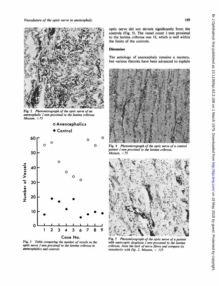
adjacent to the optic nerve head. A most interestingfinding was an increase in the number of vesselsproximal to the lamina cribrosa (Fig. 2) in all theanencephalic optic nerves. Vessel counts 1 mmproximal to the lamina cribrosa gave 48 vessels onaverage per high-power field, with a range of 32 to60 (Fig. 3). In contrast the control globes had anaverage of 12 vessels (from 6 to 20) per high powerof field (Fig. 4).The case of the septo-optic dysplasia had retinal
changes that were indistinguishable from anence-phaly, but the appearance of the vasculature of the
188
on 18 May 2018 by guest. P
rotected by copyright.http://bjo.bm
j.com/
Br J O
phthalmol: first published as 10.1136/bjo.63.3.188 on 1 M
arch 1979. Dow
nloaded from
Vasculature of the optic nerve in anencephaly
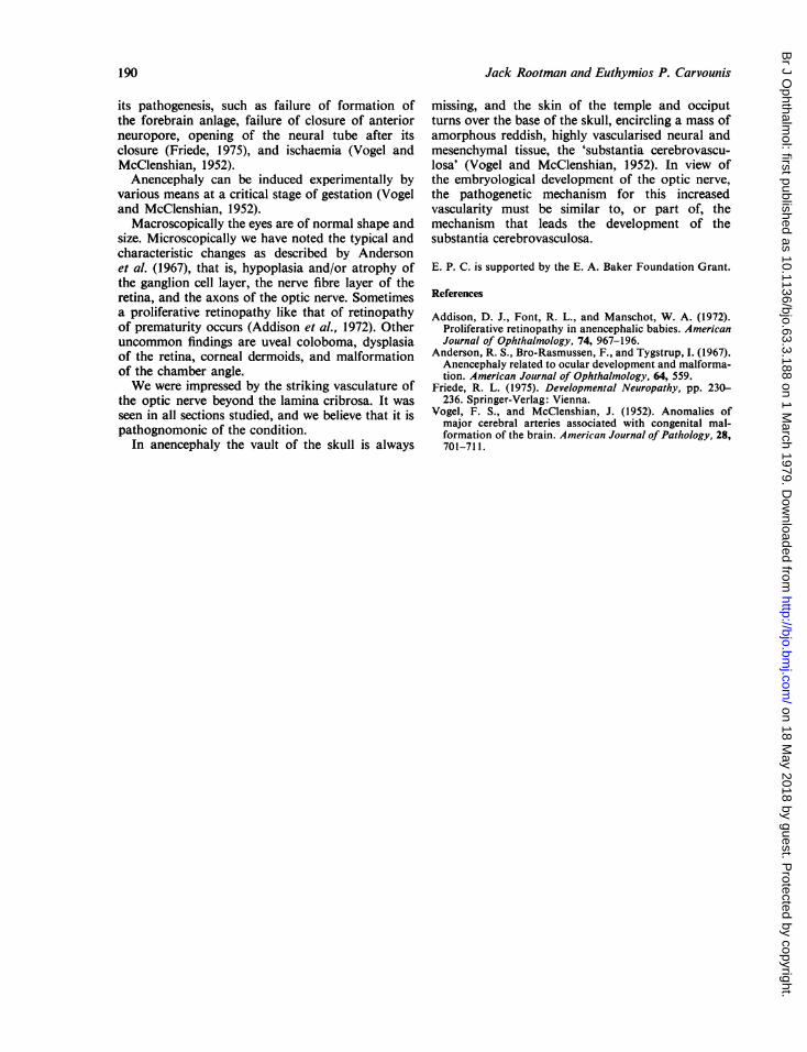
optic nerve did not deviate significantly from thecontrols (Fig. 5). The vessel count 1 mm proximalto the lamina cribrosa was 18, which is well withinthe limits of the controls.
Discussion
The aetiology of anencephaly remains a mystery,but various theories have been advanced to explain
Fig. 2 Photomicrograph of the optic nerve of ananencephalic I mm proximal to the lamina cribrosa.Masson, x 75
o Anencephalics* Control
60r0
00
0
0 Fig. 4 Photomicrograph of the optic nerve ofa controlpatient I mm proximal to the lamina cribrosa.Masson, x 75501-
0
0
0
E
z
401F
301-
20
10
0
Fig. 3 Taoptic nerveanencephali
_ O A **m ¢''RSW a }44w*+e At n '} W,, S4-S.%~~4~& *.{..a $* " 2l*.. ..? ;t
41~ ~ ~ #
0
0 j
1 2 3 4 5 6 7 8 9 tt,>e'l,ei&f .............
0~ ~
CaseNo. ~~~~Fig. 5 Photomicrograph of the optic nerve ofSa patientrble comparing the number of vessels in the with septo-optic dysplasia 1 mm proximal to the lamina1 mm proximal to the lamina cribrosa in cribrosa. Note the lack of nerve fibres and compare itsics and controls vascularit,v with Fig. 2. Masson, x 125
189
on 18 May 2018 by guest. P
rotected by copyright.http://bjo.bm
j.com/
Br J O
phthalmol: first published as 10.1136/bjo.63.3.188 on 1 M
arch 1979. Dow
nloaded from
Jack Rootman and Euthymios P. Carvounis
its pathogenesis, such as failure of formation ofthe forebrain anlage, failure of closure of anteriorneuropore, opening of the neural tube after itsclosure (Friede, 1975), and ischaemia (Vogel andMcClenshian, 1952).Anencephaly can be induced experimentally by
various means at a critical stage of gestation (Vogeland McClenshian, 1952).
Macroscopically the eyes are of normal shape andsize. Microscopically we have noted the typical andcharacteristic changes as described by Andersonet al. (1967), that is, hypoplasia and/or atrophy ofthe ganglion cell layer, the nerve fibre layer of theretina, and the axons of the optic nerve. Sometimesa proliferative retinopathy like that of retinopathyof prematurity occurs (Addison et al., 1972). Otheruncommon findings are uveal coloboma, dysplasiaof the retina, corneal dermoids, and malformationof the chamber angle.We were impressed by the striking vasculature of
the optic nerve beyond the lamina cribrosa. It wasseen in all sections studied, and we believe that it ispathognomonic of the condition.
In anencephaly the vault of the skull is always
missing, and the skin of the temple and occiputturns over the base of the skull, encircling a mass ofamorphous reddish, highly vascularised neural andmesenchymal tissue, the 'substantia cerebrovascu-losa' (Vogel and McClenshian, 1952). In view ofthe embryological development of the optic nerve,the pathogenetic mechanism for this increasedvascularity must be similar to, or part of, themechanism that leads the development of thesubstantia cerebrovasculosa.
E. P. C. is supported by the E. A. Baker Foundation Grant.
References
Addison, D. J., Font, R. L., and Manschot, W. A. (1972).Proliferative retinopathy in anencephalic babies. AmericanJournal of Ophthalmology, 74, 967-196.
Anderson, R. S., Bro-Rasmussen, F., and Tygstrup, I. (1967).Anencephaly related to ocular development and malforma-tion. American Journal of Ophthalmology, 64, 559.
Friede, R. L. (1975). Developmental Neuropathy, pp. 230-236. Springer-Verlag: Vienna.
Vogel, F. S., and McClenshian, J. (1952). Anomalies ofmajor cerebral arteries associated with congenital mal-formation of the brain. American Journal of Pathology, 28,701-711.
190
on 18 May 2018 by guest. P
rotected by copyright.http://bjo.bm
j.com/
Br J O
phthalmol: first published as 10.1136/bjo.63.3.188 on 1 M
arch 1979. Dow
nloaded from
Related Documents