GIE Ò GUIDELINE Self-expandable metal stents for obstructing colonic and extracolonic cancer: European Society of Gastrointestinal Endoscopy (ESGE) Clinical Guideline This Guideline is an official statement of the European Society of Gastrointestinal Endoscopy (ESGE). This Guide- line was also reviewed and endorsed by the Governing Board of the American Society for Gastrointestinal Endos- copy (ASGE). The Grading of Recommendations Assess- ment, Development, and Evaluation (GRADE) system was adopted to define the strength of recommendations and the quality of evidence. ESGE guidelines represent a consensus of best practice based on the available evidence at the time of preparation. They may not apply in all situations and should be inter- preted in the light of specific clinical situations and resource availability. Further controlled clinical studies may be needed to clarify aspects of these statements, and revision may be necessary as new data appear. Clinical consideration may justify a course of action at variance to these recommendations. ESGE guidelines are intended to be an educational device to provide information that may assist endoscopists in providing care to patients. They are not rules and should not be construed as establish- ing a legal standard of care or as encouraging, advocating, requiring, or discouraging any particular treatment. MAIN RECOMMENDATIONS The following recommendations should only be applied after a thorough diagnostic evaluation including a contrast- enhanced computed tomography (CT) scan. 1. Prophylactic colonic stent placement is not recommen- ded. Colonic stenting should be reserved for patients with clinical symptoms and imaging evidence of malig- nant large-bowel obstruction, without signs of perfora- tion (strong recommendation, low quality evidence). 2. Colonic self-expandable metal stent (SEMS) placement as a bridge to elective surgery is not recommended as a standard treatment of symptomatic left-sided malig- nant colonic obstruction (strong recommendation, high quality evidence). 3. For patients with potentially curable but obstructing left-sided colonic cancer, stent placement may be considered as an alternative to emergency surgery in those who have an increased risk of postoperative mor- tality, i.e. American Society of Anesthesiologists (ASA) Physical Status RIII and/or age O70 years (weak recom- mendation, low quality evidence). 4. SEMS placement is recommended as the preferred treatment for palliation of malignant colonic obstruction (strong recommendation, high quality evidence), except in patients treated or considered for treatment with antiangiogenic drugs (e.g. bevacizumab) (strong recom- mendation, low quality evidence). INTRODUCTION Colorectal cancer is one of the most common cancers worldwide, particularly in the economically developed world. 1 Large-bowel obstruction caused by advanced colonic cancer occurs in 8%–13% of colonic cancer patients. 2–4 The management of this severe clinical condi- tion remains controversial. 5 Over the last decade many articles have been published on the subject of colonic stenting for malignant colonic obstruction, including ran- domized controlled trials (RCTs) and systematic reviews. However, the definitive role of self-expandable metal stents (SEMSs) in the treatment of malignant colonic obstruction has not yet been clarified. This evidence- and consensus-based clinical guideline has been developed by the European Society of Gastrointestinal Endoscopy (ESGE) and endorsed by the American Society for Gastro- intestinal Endoscopy (ASGE) to provide practical guidance regarding the use of SEMS in the treatment of malignant colonic obstruction. With the exception of one trial, 6 all published RCTs on colonic stenting for malignant obstruction excluded rectal cancers, which were usually defined as within 8 to 10 cm of the anal verge, and colonic cancers proximal to the splenic flexure. Rectal stenting is often avoided because of the presumed association with complications such as pain, tenesmus, incontinence, and stent migration. Prox- imal colonic obstruction is generally managed with primary surgery, although there are no RCTs to support this assumption. Because of the aforementioned limitations, unless indicated otherwise the recommendations in this Guideline only apply to left-sided colon cancer arising from the rectosigmoid colon, sigmoid colon, descending Copyright ª 2014 by the American Society for Gastrointestinal Endoscopy and the European Society of Gastrointestinal Endoscopy 0016-5107/$36.00 http://dx.doi.org/10.1016/j.gie.2014.09.018 www.giejournal.org Volume 80, No. 5 : 2014 GASTROINTESTINAL ENDOSCOPY 747

Welcome message from author
This document is posted to help you gain knowledge. Please leave a comment to let me know what you think about it! Share it to your friends and learn new things together.
Transcript

Can00h
w
GIE�
GUIDELINE
opyright ª 2014 by thed the European Societ16-5107/$36.00ttp://dx.doi.org/10.1016
ww.giejournal.org
Self-expandable metal stents for obstructing colonic andextracolonic cancer: European Society of GastrointestinalEndoscopy (ESGE) Clinical Guideline
This Guideline is an official statement of the EuropeanSociety of Gastrointestinal Endoscopy (ESGE). This Guide-line was also reviewed and endorsed by the GoverningBoard of the American Society for Gastrointestinal Endos-copy (ASGE). The Grading of Recommendations Assess-ment, Development, and Evaluation (GRADE) systemwas adopted to define the strength of recommendationsand the quality of evidence.
ESGE guidelines represent a consensus of best practicebased on the available evidence at the time of preparation.They may not apply in all situations and should be inter-preted in the light of specific clinical situations andresource availability. Further controlled clinical studiesmay be needed to clarify aspects of these statements, andrevision may be necessary as new data appear. Clinicalconsideration may justify a course of action at varianceto these recommendations. ESGE guidelines are intendedto be an educational device to provide information thatmay assist endoscopists in providing care to patients.They are not rules and should not be construed as establish-ing a legal standard of care or as encouraging, advocating,requiring, or discouraging any particular treatment.
MAIN RECOMMENDATIONS
The following recommendations should only be appliedafter a thorough diagnostic evaluation including a contrast-enhanced computed tomography (CT) scan.1. Prophylactic colonic stent placement is not recommen-
ded. Colonic stenting should be reserved for patientswith clinical symptoms and imaging evidence of malig-nant large-bowel obstruction, without signs of perfora-tion (strong recommendation, low quality evidence).
2. Colonic self-expandable metal stent (SEMS) placementas a bridge to elective surgery is not recommended asa standard treatment of symptomatic left-sided malig-nant colonic obstruction (strong recommendation,high quality evidence).
3. For patients with potentially curable but obstructingleft-sided colonic cancer, stent placement may be
American Society for Gastrointestinal Endoscopyy of Gastrointestinal Endoscopy
/j.gie.2014.09.018
considered as an alternative to emergency surgery inthose who have an increased risk of postoperative mor-tality, i.e. American Society of Anesthesiologists (ASA)Physical StatusRIII and/or ageO70 years (weak recom-mendation, low quality evidence).
4. SEMS placement is recommended as the preferredtreatment for palliation of malignant colonic obstruction(strong recommendation, high quality evidence), exceptin patients treated or considered for treatment withantiangiogenic drugs (e.g. bevacizumab) (strong recom-mendation, low quality evidence).
INTRODUCTION
Colorectal cancer is one of the most common cancersworldwide, particularly in the economically developedworld.1 Large-bowel obstruction caused by advancedcolonic cancer occurs in 8%–13% of colonic cancerpatients.2–4 The management of this severe clinical condi-tion remains controversial.5 Over the last decade manyarticles have been published on the subject of colonicstenting for malignant colonic obstruction, including ran-domized controlled trials (RCTs) and systematic reviews.However, the definitive role of self-expandable metalstents (SEMSs) in the treatment of malignant colonicobstruction has not yet been clarified. This evidence- andconsensus-based clinical guideline has been developedby the European Society of Gastrointestinal Endoscopy(ESGE) and endorsed by the American Society for Gastro-intestinal Endoscopy (ASGE) to provide practical guidanceregarding the use of SEMS in the treatment of malignantcolonic obstruction.
With the exception of one trial,6 all published RCTs oncolonic stenting for malignant obstruction excluded rectalcancers, which were usually defined as within 8 to 10 cmof the anal verge, and colonic cancers proximal to thesplenic flexure. Rectal stenting is often avoided becauseof the presumed association with complications such aspain, tenesmus, incontinence, and stent migration. Prox-imal colonic obstruction is generally managed with primarysurgery, although there are no RCTs to support thisassumption. Because of the aforementioned limitations,unless indicated otherwise the recommendations in thisGuideline only apply to left-sided colon cancer arisingfrom the rectosigmoid colon, sigmoid colon, descending
Volume 80, No. 5 : 2014 GASTROINTESTINAL ENDOSCOPY 747

SEMSs for obstructing colonic and extracolonic cancer: ESGE Clinical Guideline
colon, and splenic flexure, while excluding rectal cancersand those proximal to the splenic flexure, and other causesof colonic obstruction including extracolonic obstruction.
METHODS
The ESGE commissioned this Guideline (chairs C.H.and J.-M.D.) and appointed a guideline leader (J.v.H.)who invited the listed authors to participate in the projectdevelopment. The key questions were prepared by thecoordinating team (E.v.H. and J.v.H.) and then approvedby the other members. The coordinating team formedtask force subgroups, each with its own leader, and dividedthe key topics among these task forces (see Appendix e1,available online at www.giejournal.org).
Each task force performed a systematic literature searchto prepare evidence-based and well-balanced statementson their assigned key questions. The coordinating teamindependently performed systematic literature searcheswith the assistance of a librarian. The Medline, EMBASEand Trip databases were searched including at minimumthe following key words: colon, cancer, malignancy orneoplasm, obstruction and stents. All articles studying theuse of SEMS for malignant large-bowel obstruction wereselected by title or abstract. After further exploration ofthe content, the article was then included and summarizedin the literature tables of the key topics when it containedrelevant data (see Appendix e2, Tables e1–e5, availableonline at www.giejournal.org). All selected articles weregraded by the level of evidence and strength of recommen-dation according to the GRADE system.7 The literaturesearches were updated until January 2014.
Each task force proposed statements on their assignedkey questions which were discussed and voted on duringthe plenary meeting held in February 2014, Düsseldorf,Germany. In March 2014, a draft prepared by the coordi-nating team was sent to all group members. After agree-ment on a final version, the manuscript was submitted toEndoscopy for publication. The journal subjected themanuscript to peer review and the manuscript wasamended to take into account the reviewers’ comments.All authors agreed on the final revised manuscript. The finalrevised manuscript was then reviewed and approved by theGoverning Board of ASGE. This Guideline was issued in2014 and will be considered for review in 2019 or soonerif new and relevant evidence becomes available. Any up-dates to the Guideline in the interim will be noted on theESGE website: http://www.esge.com/esge-guidelines.html.
RECOMMENDATIONS AND STATEMENTS
Evidence statements and recommendations are statedin bold italics.
General considerations before stent placement(Table e1, available online at www.giejournal.org)
748 GASTROINTESTINAL ENDOSCOPY Volume 80, No. 5 : 2014
Prophylactic colonic stent placement is not rec-ommended. Colonic stenting should be reservedfor patients with clinical symptoms and imaging ev-idence of malignant large-bowel obstruction,without signs of perforation (strong recommenda-tion, low quality evidence).
Colonic stenting is indicated only in those patients withboth obstructive symptoms and radiological or endoscopicfindings suspicious of malignant large-bowel obstruction.Prophylactic stenting for patients with colonic malignancybut no evidence of symptomatic obstruction is stronglydiscouraged because of the potential risks associated withcolonic SEMS placement. The only absolute contraindica-tion for colonic stenting is perforation. In addition, colonicstenting is less successful in patients with peritoneal carci-nomatosis and tumors close to the anal verge (!5 cm).8–10
Increasing age and American Society of Anesthesiolo-gists (ASA) classification RIII do not affect stent outcome(i.e. clinical success and complications) in several observa-tional studies,11–16 although these are well-known risk fac-tors for postoperative mortality after surgical treatment oflarge-bowel obstruction (Table 6).17–19
A contrast-enhanced computed tomography (CT)scan is recommended as the primary diagnostictool when malignant colonic obstruction is sus-pected (strong recommendation, low qualityevidence).
When malignant colonic obstruction is suspected,contrast-enhanced CT is recommended because it can di-agnose obstruction (sensitivity 96%, specificity 93%),define the level of the stenosis in 94% of cases, accuratelyidentify the etiology in 81% of cases, and provide correctlocal and distal staging in the majority of patients.5,20
When CT is inconclusive about the etiology of the obstruct-ing lesion, colonoscopy may be helpful to evaluate theexact cause of the stenosis.
Examination of the remaining colon with colo-noscopy or CT colonography (CTC) is recommendedin patients with potentially curable obstructingcolonic cancer, preferably within 3 months afteralleviation of the obstruction (strong recommenda-tion, low quality evidence).
European studies, including three that are population-based, show that synchronous colorectal tumors occur in3%–4% of patients diagnosed with colorectal cancer.21–24
Therefore, imaging of the remaining colon after potentiallycurative resection is recommended in patients with malig-nant colonic obstruction. Current evidence does not justifyroutine preoperative assessment for synchronous tumorsin obstructed patients by CTC or colonoscopy throughthe stent. However, preoperative CTC and colonoscopythrough the stent appear feasible and safe in these patientsand there are presently no data to discourage their use inthis population.25–28 The role of positron emission tomo-graphy (PET)/CT in the diagnosis of synchronous lesionsremains to be elucidated.29
www.giejournal.org

TABLE 6. Outcome of surgery according to age and American Society of Anesthesiologists (ASA) classification
First author, year Study population ResultsStudy design Level
of evidence
Tekkis, 200418 Patients undergoing surgeryfor acute colorectal cancerobstruction (n Z 1046)
Multivariate analysis of in-hospitalpostoperative mortality:- Age!65 years: 5.4%- Age 65-67 years: 13.1%;OR 2.97 (95%CI 1.26-7.08)
- Age 75-84 years: 21.9%;OR 4.31 (95%CI 1.83-10.05)
- Age R 85 years: 27.0%;OR 5.87 (95%CI 2.27-15.14)
- ASA I: 2.6%- ASA II: 7.6%; OR 3.32(95%CI 0.73-15.18)
- ASA III: 23.9%; OR 11.73(95%CI 2.58-53.36)
- ASA IV-V: 42.9%; OR 22.33(95%CI 4.58-109.68)
Nonrandomized prospectiveUK multicenter study
High quality evidence
Biondo, 200417 Patients undergoing emergencysurgery for acute large-bowelobstruction (n Z 234)
Colorectal cancer 82.1%Extracolonic cancer 4.7%Benign lesions 13.2%
Univariate analysis of 30-daypostoperative mortality:- Age %70 years: 10.7% (14/131)- Age O70 years: 29.1%(30/103); P! 0.001
- ASA I-II: 8.1% (9/111)- ASA III-IV: 28.5% (35/123);P! 0.001
Multivariate analysis of 30-daypostoperative mortality:- Age O70 years: OR 2.05(95%CI 0.92-4.60)
- ASA III-IV: OR 2.86(95%CI 1.15-7.11)
No description of study design,most likely retrospective
Moderate quality evidence
Tan, 201019 Patients who underwentoperativeintervention for acuteobstructionfrom colorectal malignancy(n Z 134)
Perioperative morbidity rate: 77.6%Perioperative mortality rate: 11.9%Multivariate analysis of worseoutcome (grade III-V complications,including death):- Age O60 years: OR 4.67(95%CI 1.78-12.25)
- ASA III-IV: OR 8.36(95%CI 3.58-19.48)
Retrospective analysisLow quality evidence
CI, Confidence interval; OR, odds ratio.
SEMSs for obstructing colonic and extracolonic cancer: ESGE Clinical Guideline
Colonic stenting should be avoided for divertic-ular strictures or when diverticular disease is sus-pected during endoscopy and/or CT scan (strongrecommendation, low quality evidence). Patholog-ical confirmation of malignancy by endoscopicbiopsy and/or brush cytology is not necessary inan urgent setting, such as before stent placement.However, pathology results may help to modifyfurther management of the stented patient (strongrecommendation, low quality evidence).
When malignancy is suspected after diagnostic studies, asmall number of patients will have a benign cause ofobstruction. Two RCTs comparing SEMS as a bridge tosurgery versus emergency surgery in patients with left-
www.giejournal.org
sided malignant obstruction reported benign obstructive le-sions in 4.6% (3/65)30 and 8.2% (8/98)31 of the randomizedpatients. These benign colonic lesions that mimic malig-nancy are usually due to diverticular disease. Further evi-dence of the difficulty of this distinction is also reflected bya systematic review showing a 2.1%prevalence of underlyingadenocarcinoma of the colon in 771 patients in whom acutediverticulitis was diagnosed via CT scan.32 Stent placementin active diverticular inflammation is associated with a riskof perforation and should therefore be avoided.33 Further-more, pathological confirmation ofmalignancy before emer-gency stent placement is often not feasible and is notrequired prior to colonic stent placement. Endoscopic bi-opsy and/or brush cytology for confirmation of malignancy
Volume 80, No. 5 : 2014 GASTROINTESTINAL ENDOSCOPY 749

SEMSs for obstructing colonic and extracolonic cancer: ESGE Clinical Guideline
should be obtained during the stent placement procedure,because it may be helpful in modifying the further manage-ment of the stented patient.34–36
Preparation of obstructed patients with anenema to clean the colon distal to the stenosis issuggested to facilitate the stent placement proce-dure (weak recommendation, low quality evi-dence). Antibiotic prophylaxis in obstructedpatients undergoing colon stenting is not indicatedbecause the risk of post-procedural infections isvery low (strong recommendation, moderate qual-ity evidence).
There are no studies to date that have focused on bowelpreparation before stent placement in obstructed patients.Symptomatic bowel obstruction is a relative contraindica-tion to oral bowel cleansing. An enema is advisable to facil-itate the stent placement procedure by cleaning the boweldistal to the stenosis.
Antibiotic prophylaxis before stent placement in pa-tients with malignant colonic obstruction is not indicatedbecause the risk of fever and bacteremia after stent inser-tion is very low. One prospective study analyzed 64 pa-tients with colorectal cancer who underwent a stentprocedure. Four of 64 patients (6.3%) had a positivepost-stenting blood culture and none of the patients devel-oped symptoms of infection within 48 hours followingstent placement. Prolonged procedure time was associatedwith transient bacteremia (36 vs. 16 minutes, P! 0.01).37
One other retrospective series of 233 patients undergoingcolonic stent placement for malignant obstructiondescribed that blood cultures had been drawn for unspec-ified reasons in 30 patients within 2 weeks after stent place-ment, showing bacteremia/fever in 7 patients (3%), whichwas reported as a minor complication.15
Colonic stent placement should be performed ordirectly supervised by an experienced operatorwho has performed at least 20 colonic stent place-ment procedures (strong recommendation, lowquality evidence).
Two noncomparative studies addressed the learningcurve of a single endoscopist performing colonic stentplacement. Both showed an increase in technical successand a decrease in the number of stents used per procedureafter performance of at least 20 procedures.38,39 Two otherretrospective series have shown that operator experienceaffects stenting outcome. The first reported significantlyhigher technical and clinical success rates when the stentwas inserted by an operator who had performed at least10 SEMS procedures.16 The second showed a significantlyincreased immediate perforation rate when colonic stentplacement was performed by endoscopists inexperiencedin pancreaticobiliary endoscopy.15 The authors of the latterarticle explained the lower immediate perforation rate bythe skills that therapeutic ERCP endoscopists have intraversing complex strictures, understanding fluoroscopy,and deploying stents.15
750 GASTROINTESTINAL ENDOSCOPY Volume 80, No. 5 : 2014
Technical considerations of stent placement(Table e2, available online at www.giejournal.org)
Colonic stent placement is recommended withthe combined use of endoscopy and fluoroscopy(weak recommendation, low quality evidence).
SEMS placement can be performed by using either thethrough-the-scope (TTS) or the over-the-guidewire(OTW) technique. The majority of SEMS are insertedthrough the endoscope with the use of fluoroscopic guid-ance. The OTW technique is performed using fluoroscopicguidance with or without tandem endoscopic monitoring.Purely radiologic stent placement is performed byadvancing the stent deployment system over a stiff guide-wire, and technical and clinical success rates of 83%–
100% and 77%–98%, respectively, have been reported inobservational studies.40–45 Retrospective studies thatcompared endoscopy combined with fluoroscopic guid-ance versus solely radiography for stent placement showcomparable success rates, although with a trend towardshigher technical success when the combined techniqueis used.16,46–48
Stricture dilation either before or after stentplacement is discouraged in the setting of obstruct-ing colorectal cancer (strong recommendation, lowquality evidence).
Although based on low quality evidence with small pa-tient numbers, there are strong indications to believethat stricture dilation either just before or after colonicstent placement adversely affects the clinical outcome ofstenting and particularly increases the risk of colonic perfo-ration.8,12,15,49 Pooled analyses, mainly based on retrospec-tive data, also show increased risk of perforation afterstricture dilation.47,50,51
Covered and uncovered SEMS are equally effec-tive and safe (high quality evidence). The stentshould have a body diameter R24 mm (strongrecommendation, low quality evidence) and alength suitable to extend at least 2 cm on eachside of the lesion after stent deployment (weakrecommendation, low quality evidence).
The clinician should be aware of specific features of thechosen stent that may affect the patient after insertion.Two meta-analyses comparing covered and uncoveredSEMS for malignant colonic obstruction found similar tech-nical success, clinical success, and overall complicationrates. Uncovered SEMS showed significantly higher tumoringrowth rates (11.4% vs. 0.9%) but were less prone tomigrate than covered SEMS (5.5% vs. 21.3%).52,53
The diameter of the stent also seems to influence stentoutcome. In mainly retrospective analyses, the use ofsmall-diameter stents with a body diameter!24 mm wasassociated with the occurrence of complications, in partic-ular stent migration.15,54–56 Stent length was not identifiedin observational studies as a risk factor for adverse stentoutcome.8,11,16,45 It is recommended to use a stent thatis long enough to bridge the stenosis and to extend at least
www.giejournal.org

TABLE 7. Short-term outcomes of self-expandable metal stent (SEMS) placement as a bridge to elective surgery
First author, year Study population ResultsStudy design Level of
evidence
Huang, 201481 Patients with acute left-sidedmalignant colonic obstruction
7 RCTs
Preoperative SEMS (n Z 195)Emergency surgery (n Z 187)
Mean success rate of colonic stentplacement: 76.9% (46.7%-100%)Permanent stoma rate (P Z 0.002):- SEMS as bridge to surgery: 9% (9/100)- Emergency surgery: 27.4% (26/95)- OR 0.28 (95%CI 0.12-0.62); I2 Z 36%Primary anastomosis rate (P Z 0.007):- SEMS as bridge to surgery: 67.2% (131/195)- Emergency surgery: 55.1% (103/187)- OR 2.01 (95%CI 1.21-3.31); I2 Z 0%Mortality rate (P Z 0.76):- SEMS as bridge to surgery: 10.7% (12/112)- Emergency surgery: 12.4% (14/113)- OR 0.88 (95%CI 0.40-1.96); I2 Z 17%Overall complication rate (P Z 0.03):- SEMS as bridge to surgery: 33.1% (55/166)- Emergency surgery: 53.9% (90/167)- OR 0.30 (95%CI 0.11-0.86); I2 Z 77%Anastomotic leakage rate (P Z 0.47):- SEMS as bridge to surgery: 4.1% (8/195)- Emergency surgery: 5.9% (11/187)- OR 0.74 (95%CI 0.33-1.67); I2 Z 27%Wound infection rate (P Z 0.004):- SEMS as bridge to surgery: 6.7% (10/150)- Emergency surgery: 18.1% (26/144)- OR 0.31 (95%CI 0.14-0.68); I2 Z 0%Intra-abdominal infection rate(P Z 0.57):- SEMS as bridge to surgery: 1.4% (1/73)- Emergency surgery: 3.2% (2/63)- OR 0.62 (95%CI 0.12-3.19); I2 Z 0%
Meta-analysis of RCTs
High quality evidence
Guo, 2011100 Patients aged R70 yearsdiagnosed with acuteleft-sided colonic obstruction
SEMS (n Z 34)Surgery (n Z 58)
SEMS versus surgeryOverall rate of successful bridging with SEMS: 79%Mean time to elective surgery: 9 days (range 4-16)Successful relief of obstruction: 91% vs. 100%(P Z 0.09)Primary anastomosis rate: 79% vs. 47% (PZ 0.002)Temporary stoma rate: 9% vs. 53% (P! 0.001)Permanent stoma rate: 6% vs. 12% (P Z 0.34)Median length of hospital stay: 19 vs. 14 days(P Z 0.06)Acute mortality rate: 3% vs. 19% (P Z 0.03)Acute complication rate: 24% vs. 40% (P Z 0.11)
Retrospective comparison
Low quality evidence
CI, Confidence interval; OR, odds ratio; RCT, randomized controlled trial; SEMS, self-expandable metal stent.
SEMSs for obstructing colonic and extracolonic cancer: ESGE Clinical Guideline
2 cm on each side of the lesion, taking into account the de-gree of shortening after stent deployment.57 Severalstudies, including one RCT, have shown no differencein outcomes (efficacy and safety) based on different stentdesigns.8,43,58–61
Surgical resection is suggested as the preferredtreatment for malignant obstruction of theproximal colon in patients with potentially curabledisease (weak recommendation, low qualityevidence). In a palliative setting, SEMS can be analternative to emergency surgery (weak recom-mendation, low quality evidence).
www.giejournal.org
Retrospective series have shown that SEMS may be suc-cessfully placed in malignant strictures located in the prox-imal colon (i.e. proximal to the splenic flexure).8,16,62–64
However, these data show conflicting results regardingSEMS outcome compared with stent placement in theleft-sided colon.8,11,15,16,45,62,65,66 Emergency resection isgenerally considered to be the treatment of choice forright-sided obstructing colon cancer. In this setting, pri-mary ileocolonic anastomosis or ileostomy can be per-formed depending on the surgical risk of the patient.5,67,68
SEMS placement is a valid alternative to surgeryfor the palliation of malignant extracolonic
Volume 80, No. 5 : 2014 GASTROINTESTINAL ENDOSCOPY 751

SEMSs for obstructing colonic and extracolonic cancer: ESGE Clinical Guideline
obstruction (weak recommendation, low qualityevidence). The technical and clinical success ratesof stenting for extracolonic malignancies are infe-rior to those reported in stenting of primary coloniccancer (low quality evidence).
Large-bowel obstruction caused by extracolonic malig-nancies is a different entity within colonic stenting andhas been studied mainly retrospectively. Technical andclinical success rates of stenting extracolonic malignancieshave been reported to range from 67% to 96% and from20% to 96%, respectively,65,69–75 and are considered infe-rior to those reported in stenting of primary colonic can-cer.8,55,70,74 One retrospective comparison of SEMS forextracolonic versus primary colonic malignancy showedan increased complication rate in the extracolonic malig-nancy group (33% vs. 9%, PZ 0.046), although this findingwas not statistically significant in the multivariate analysis.74
However, several larger series did not identify obstructionby extrinsic compression as a risk factor for complica-tions.8,11,15,70 It is generally advisable to attempt palliativestenting of extracolonic malignancies in order to avoid sur-gery in these patients who have a relatively short survival(median survival 30–141 days).69,70,72,73
There is insufficient evidence to discouragecolonic stenting based on the length of the stenosis(weak recommendation, low quality evidence) orthe degree of obstruction (strong recommendation,low quality evidence).
Few studies investigated the “stentability” of long ob-structed segments.58,76,77 However, in two retrospectivestudies that included a total of 240 patients, a betteroutcome was observed when SEMS were inserted in shortobstructed segments.55,78 One identified statistically signif-icantly more technical failures (odds ratio [OR] 5.33) andclinical failures (OR 2.40) in stenoses O4 cm.55
The outcomes of SEMS placement for complete obstruc-tion comparedwith subtotal obstruction are reported incon-sistently in the literature. One comparative prospectivestudy that specifically focused on this topic found similartechnical and clinical success rates between both groups.79
This was confirmed by more recently published large retro-spective series.8,55 However, in two observational studiessignificantly more complications were observed in the com-plete occlusion group (35% and 38% vs. 20% and 22%).13,15
Furthermore, multivariate analysis in one prospective multi-center study,which reported an11%overall perforation rate,identified complete obstruction as a risk factor for perfora-tion (OR 6.88).80
Clinical indication: SEMS placement as a bridge toelective surgery (Table e3, available online at www.giejournal.org)
ColonicSEMSplacementasabridge to elective sur-gery is not recommended as a standard treatment ofsymptomatic left-sided malignant colonic obstruc-tion (strong recommendation, high quality evi-dence). For patients with potentially curable
752 GASTROINTESTINAL ENDOSCOPY Volume 80, No. 5 : 2014
left-sidedobstructingcolonic cancer, stentplacementmay be considered as an alternative to emergencysurgery in thosewhohaveanincreasedriskofpostop-erativemortality, i.e. ASARIII and/or ageO70 years(weak recommendation, low quality evidence).
Eight systematic reviews with meta-analysis have beenpublished in the last decade that compared preoperativestenting with emergency resection for acute malignant left-sided colonic obstruction.81–88 Three of the sevenRCTs pub-lished to date on this subject 30,31,89–93 were prematurelyclosed, including two because of adverse outcomes in thestent group 30,31 andone because of a high incidence of anas-tomotic leakage in the primary surgery group.92
The most recent systematic review and meta-analysisevaluated the efficacy and safety of colonic stenting as abridge to surgery (nZ 195) compared with emergency sur-gery (n Z 187) and considered only RCTs for inclusion(Table 7).81 All seven RCTs that focused on the postopera-tive outcomeof SEMS and emergency surgerywere includedin this meta-analysis. The mean technical success rate ofcolonic stent placement was 76.9% (range 46.7%–100%).81
Therewas no statistically significant difference in thepostop-erative mortality comparing SEMS as bridge to surgery(10.7%) and emergency surgery (12.4%).81 The meta-analysis showed the SEMSgrouphad lower overallmorbidity(33.1% vs. 53.9%, P Z 0.03), a higher successful primaryanastomosis rate (67.2% vs. 55.1%, P! 0.01), and lower per-manent stoma rate (9% vs. 27.4%, P! 0.01).81
No clear conclusions may be drawn about differences incosts between the two procedures. In the two RCTs thatcompared costs between SEMS as bridge to surgery andemergency surgery, stenting seems to be the more costlystrategy.91,92 Cost–effectiveness depends on the rate of stentcomplications, in particular perforation, and a greaterbenefit of stenting is expected inhigh risk surgical patients.94
From the above data, some advantages of SEMS as abridge to surgery can be extracted. However, this has tobe balanced with the oncological outcomes in patientswith a curable colonic cancer. Potential concerns havebeen raised about impaired oncological outcome afterSEMS placement in the patient with potentially curable co-lon cancer, particularly following stent perforation. Long-term oncological outcome comparing SEMS as a bridgeto elective surgery versus acute resection was analyzedby three RCTs (Table 8).90,92,95 Although the study groupswere small, with 15 to 26 patients in the stent arms, allthree report higher disease recurrence rates in the SEMSgroup. This did not translate into a worse overall survivalin any of these RCTs, but this may be related to shortfollow-up and small sample sizes.90,92,95 These results arefurther supported by a larger comparative prospectivecohort study showing significantly more local disease re-currences in the stent group compared with the primarysurgery group in patients %75 years of age.96 However,no difference in survival was seen between the two groups.One retrospective analysis reported a significantly lower 5-
www.giejournal.org
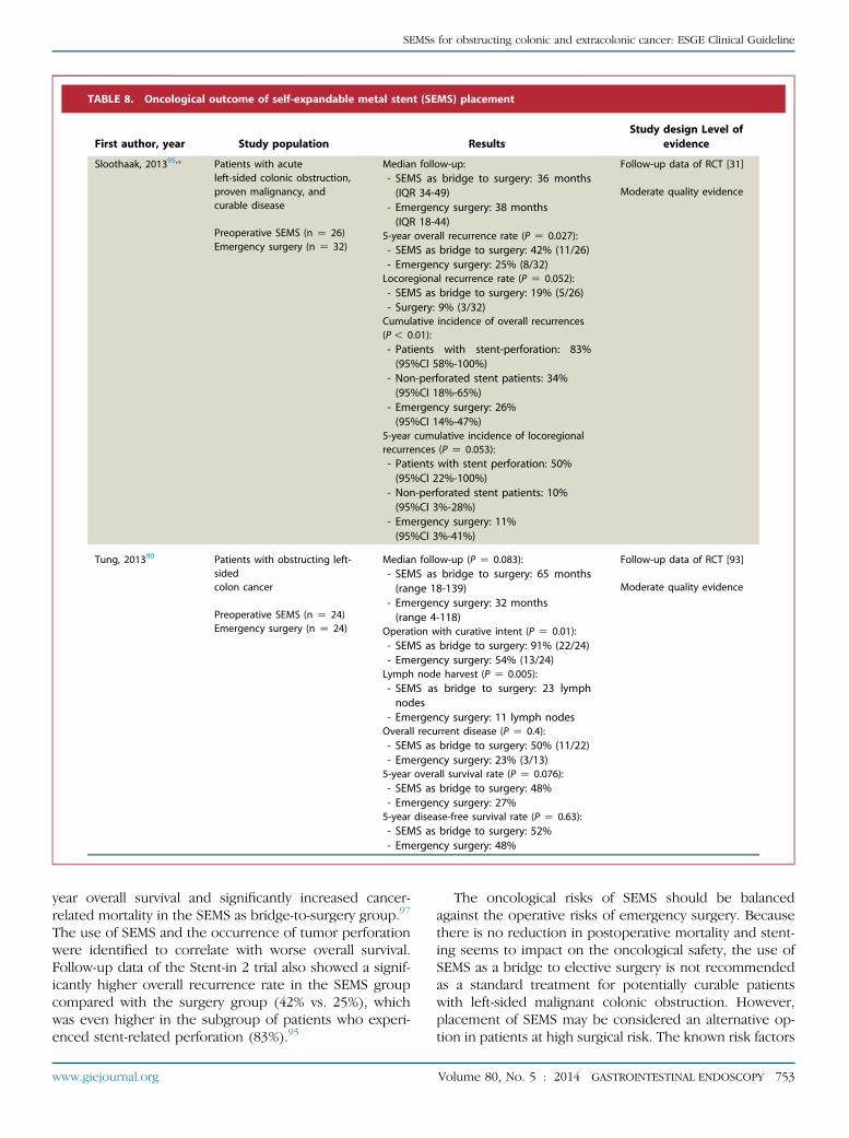
TABLE 8. Oncological outcome of self-expandable metal stent (SEMS) placement
First author, year Study population ResultsStudy design Level of
evidence
Sloothaak, 201395,* Patients with acuteleft-sided colonic obstruction,proven malignancy, andcurable disease
Preoperative SEMS (n Z 26)Emergency surgery (n Z 32)
Median follow-up:- SEMS as bridge to surgery: 36 months(IQR 34-49)
- Emergency surgery: 38 months(IQR 18-44)
5-year overall recurrence rate (P Z 0.027):- SEMS as bridge to surgery: 42% (11/26)- Emergency surgery: 25% (8/32)Locoregional recurrence rate (P Z 0.052):- SEMS as bridge to surgery: 19% (5/26)- Surgery: 9% (3/32)Cumulative incidence of overall recurrences(P! 0.01):- Patients with stent-perforation: 83%(95%CI 58%-100%)
- Non-perforated stent patients: 34%(95%CI 18%-65%)
- Emergency surgery: 26%(95%CI 14%-47%)
5-year cumulative incidence of locoregionalrecurrences (P Z 0.053):- Patients with stent perforation: 50%(95%CI 22%-100%)
- Non-perforated stent patients: 10%(95%CI 3%-28%)
- Emergency surgery: 11%(95%CI 3%-41%)
Follow-up data of RCT [31]
Moderate quality evidence
Tung, 201390 Patients with obstructing left-sidedcolon cancer
Preoperative SEMS (n Z 24)Emergency surgery (n Z 24)
Median follow-up (P Z 0.083):- SEMS as bridge to surgery: 65 months(range 18-139)
- Emergency surgery: 32 months(range 4-118)
Operation with curative intent (P Z 0.01):- SEMS as bridge to surgery: 91% (22/24)- Emergency surgery: 54% (13/24)Lymph node harvest (P Z 0.005):- SEMS as bridge to surgery: 23 lymphnodes
- Emergency surgery: 11 lymph nodesOverall recurrent disease (P Z 0.4):- SEMS as bridge to surgery: 50% (11/22)- Emergency surgery: 23% (3/13)5-year overall survival rate (P Z 0.076):- SEMS as bridge to surgery: 48%- Emergency surgery: 27%5-year disease-free survival rate (P Z 0.63):- SEMS as bridge to surgery: 52%- Emergency surgery: 48%
Follow-up data of RCT [93]
Moderate quality evidence
SEMSs for obstructing colonic and extracolonic cancer: ESGE Clinical Guideline
year overall survival and significantly increased cancer-related mortality in the SEMS as bridge-to-surgery group.97
The use of SEMS and the occurrence of tumor perforationwere identified to correlate with worse overall survival.Follow-up data of the Stent-in 2 trial also showed a signif-icantly higher overall recurrence rate in the SEMS groupcompared with the surgery group (42% vs. 25%), whichwas even higher in the subgroup of patients who experi-enced stent-related perforation (83%).95
www.giejournal.org
The oncological risks of SEMS should be balancedagainst the operative risks of emergency surgery. Becausethere is no reduction in postoperative mortality and stent-ing seems to impact on the oncological safety, the use ofSEMS as a bridge to elective surgery is not recommendedas a standard treatment for potentially curable patientswith left-sided malignant colonic obstruction. However,placement of SEMS may be considered an alternative op-tion in patients at high surgical risk. The known risk factors
Volume 80, No. 5 : 2014 GASTROINTESTINAL ENDOSCOPY 753

TABLE 8. Continued
First author, year Study population ResultsStudy design Level of
evidence
Alcantara, 201192 Patients with completeintestinal obstruction dueto tumor in the left colonSEMS as bridge to surgery(n Z 15)
Intraoperative colonic lavagewith primary anastomosis(n Z 13)
Overall mean follow-up: 37.6 monthsNo difference in overall survival(P Z 0.843)Disease-free period (P Z 0.096):- SEMS as bridge to surgery: 25.5 months- Emergency surgery: 27.1 monthsTumor reappearance (P Z 0.055):- SEMS as bridge to surgery: 53% (8/15)- Emergency surgery: 15% (2/13)
RCTModerate quality evidence
Gorissen, 201396 Patients with obstructing left-sided colonic cancer
Preoperative SEMS (n Z 62)Emergency surgery (n Z 43)
Median follow-up (P Z 0.294)- SEMS as bridge to surgery: 2.7 years- Emergency surgery: 2.8 yearsLocal recurrence rate (P Z 0.443):- SEMS as bridge to surgery: 23% (14/60)- Emergency surgery: 15% (6/39)Distant metastasis (P Z 1.000):- SEMS as bridge to surgery: 27% (16/60)- Emergency surgery: 26% (10/39)Overall recurrence (P Z 0.824):- SEMS as bridge to surgery: 32% (19/60)- Emergency surgery: 28% (11/39)Overall mortality (P Z 0.215):- SEMS as bridge to surgery: 29% (18/62)- Emergency surgery: 44% (19/43)Cancer-specific mortality (P Z 0.180):- SEMS as bridge to surgery: 24% (15/62)- Emergency surgery: 37% (16/43)Local recurrence rate in patients % 75 years(P Z 0.038):- SEMS as bridge to surgery: 32%- Emergency surgery: 8%
Prospective cohort studyModerate quality evidence
Sabbagh, 201397 Patients operated on forleft-sided malignant colonicobstruction with curativeintent
Preoperative SEMS (n Z 48)Emergency surgery (n Z 39)
Mean follow-up (P Z 0.21):- SEMS as bridge to surgery: 28 months- Emergency surgery: 32 months
5-year overall survival rate (P! 0.001):- SEMS as bridge to surgery: 25%- Emergency surgery: 62%
5-year cancer-specific mortality (P Z 0.02):- SEMS as bridge to surgery: 48%- Emergency surgery: 21%
5-year disease-free survival (P Z 0.24):- SEMS as bridge to surgery: 22%- Emergency surgery: 32%
Overall recurrence rate (P Z 0.18):- SEMS as bridge to surgery: 33%- Emergency surgery: 20%
Mean time to recurrence (P Z 0.92):- SEMS as bridge to surgery: 16 months- Emergency surgery: 23 months
In multivariate analysis SEMS (HR 2.42, 95%CI1.13-5.18) and tumor perforation(HR 5.96, 95%CI 1.70-20.95) wereassociated with overall survival
Retrospectiveintention-to-treat analysisLow quality evidence
CI, Confidence interval; HR, hazard ratio; IQR, interquartile range; RCT, randomized controlled trial.*Published in abstract form.
754 GASTROINTESTINAL ENDOSCOPY Volume 80, No. 5 : 2014 www.giejournal.org
SEMSs for obstructing colonic and extracolonic cancer: ESGE Clinical Guideline

TABLE 9. Meta-analyses of palliative self-expandable metal stent (SEMS) placement
First author, year Study population ResultsStudy type Level of
evidence
Liang, 2014104 Patients with malignantcolorectal obstruction causedby advanced malignancy
3 RCTs2 Prospective4 Retrospective
Palliative SEMS (n Z 195)Emergency surgery (n Z 215)
Major stent-related complications:- Short-term (!30 days) perforationrate: 3.7%
- Long-term (R30 days) perforationrate: 7.6%
- Overall stent migration rate: 8.9%- Re-obstruction: not analyzed.Successful relief of obstruction:- Palliative SEMS: 94%- Surgery: 100%Short-term (!30 days) complicationrate (P Z 0.22):- Palliative SEMS: 26.2% (51/195)- Surgery: 34.5% (74/215)- OR 0.83 (95%CI 0.39-1.79)Long-term (R30 days) complicationrate (P Z 0.03):- Palliative SEMS: 16.1% (25/155)- Surgery: 8.1% (14/173)- OR 2.34 (95%CI 1.07-5.14)Overall complication rate (P Z 0.56):- Palliative SEMS: 43.9% (68/155)- Surgery: 45.1% (78/173)- OR 1.27 (95%CI 0.58-2.77)Overall mortality rate (P Z 0.22):- Palliative SEMS: 7.1% (12/169)- Surgery: 11.6% (22/189)- OR 0.60 (95%CI 0.27-1.34)SEMS required significantly shorterhospitalization: weighted meandifference -6.07 days(95%CL -8.40, -3.74); P! 0.01
Systematic reviewsand meta-analysis ofcomparative studies
High quality evidence
SEMSs for obstructing colonic and extracolonic cancer: ESGE Clinical Guideline
associated with adverse outcomes following elective as wellas emergency surgery in colorectal cancer are increasingage and an ASA score RIII.3,17–19,98,99 Therefore, the useof SEMS as a bridge to elective surgery may be consideredan acceptable alternative treatment option in patients olderthan 70 years and/or with an ASA score RIII.100
A time interval to operation of 5–10 days is sug-gested when SEMS is used as a bridge to electivesurgery in patients with potentially curable left-sided colon cancer (weak recommendation, lowquality evidence).
There are limited data to determine an optimal time in-terval to operation following stent placement as a bridge tosurgery. Theoretically, a longer interval (O1 week) willallow for better recovery and more nearly optimal nutri-tional status, but this may increase the risk of stent-related complications and may compromise surgery bymore local tumor infiltration and fibrosis. Therefore wesuggest a 5- to 10-day interval between SEMS and electiveresection. Data from the abstract of one RCT (n Z 49)published in Chinese, which compared laparoscopic resec-tion 3 and 10 days after stent placement, reported a signif-
www.giejournal.org
icantly higher primary anastomosis rate and a lowerconversion rate to open procedure when surgery was de-ferred until 10 days after stenting.101 A retrospective anal-ysis revealed an anastomotic leakage rate of 20% (3/15)for an interval of 1 to 9 days and 0% (0/28) when surgerywas delayed for 10 days or longer (P Z 0.037).102 A pub-lished abstract comparing resection within 7 days (n Z26) and after 7 days (n Z 30) of stent placement, foundno differences in the postoperative morbidity and mortal-ity.103 In the literature, a median time interval to surgeryof 10 days is a common practice considering the patient’sclinical condition, potential risk of stent-related complica-tions, and impact on oncological outcomes.84
Clinical indication: palliative SEMS placement(Table e4, available online at www.giejournal.org)
SEMS placement is the preferred treatment forpalliation of malignant colonic obstruction (strongrecommendation, high quality evidence).
Two meta-analyses, including randomized and non-randomized comparative studies, have compared SEMS(n Z 195 and n Z 404) and surgery (n Z 215 andn Z 433) for palliation of malignant colonic obstruction
Volume 80, No. 5 : 2014 GASTROINTESTINAL ENDOSCOPY 755

TABLE 9. Continued
First author, year Study population ResultsStudy type Level of
evidence
Zhao, 2013105 Patients with malignantcolorectal obstruction thatwas unresectable
3 RCTs5 Prospective4 Retrospective1 Case-matched
Palliative SEMS (n Z 404)Palliative surgery (n Z 433)
Mean length of hospital stay (P! 0.001):- Palliative SEMS: 9.6 days- Surgery: 18.8 days,ICU admission rate (P Z 0.001):- Palliative SEMS: 0.8% (1/119)- Surgery: 18.0% (22/122)- RR 0.09 (95%CI 0.02-0.38); I2 Z 0%Mean interval to chemotherapy:- Palliative SEMS: 15.5 days- Surgery: 33.4 daysClinical relief of obstruction (P! 0.001):- Palliative SEMS: 93.1% (375/403)- Surgery: 99.8% (433/434)- RR 0.96 (95%CI 0.93-0.98); I2 Z 3%In-hospital mortality rate (P Z 0.01):- Palliative SEMS: 4.2% (14/334)- Surgery: 10.5% (37/354)- RR 0.46 (95%CI 0.25-0.85); I2 Z 0%Overall complication rate (P Z 0.60):- Palliative SEMS: 34.0% (137/403)- Surgery: 38.1% (172/452)- RR 0.91 (95%CI 0.64-1.29); I2 Z 66%Early complication rate (P Z 0.03):- Palliative SEMS: 13.7% (41/300)- Surgery: 33.7% (110/326)- RR 0.45 (95%CI 0.22-0.92); I2 Z 66%Late complication rate (P! 0.001):- Palliative SEMS: 32.3% (60/186)- Surgery: 12.7% (27/213)- RR 2.33 (95%CI 1.55-3.50); I2 Z 0%Stent complications:- Perforation rate: 10.1%- Stent migration: 9.2%- Stent obstruction: 18.3%Overall survival time (P Z n.s.):- Palliative SEMS: 7.6 months- Surgery: 7.9 monthsStoma formation rate (P! 0.001):- Palliative SEMS: 12.7% (38/299)- Surgery: 54.0% (170/315)- RR 0.26 (95%CI 0.18-0.37); I2 Z 18%
Systematic review andmeta-analysis ofcomparative studies
High quality evidence
CI, Confidence interval; CL, confidence limits; ICU, intensive care unit; n.s., not significant; OR, odds ratio; RCT, randomized controlled trial; RR, risk ratio.
SEMSs for obstructing colonic and extracolonic cancer: ESGE Clinical Guideline
(Table 9).104,105 The technical success of stent placementin the studies included ranged from 88% to 100%,6,106
while the initial clinical relief of obstruction was signifi-cantly higher after palliative surgery (100%) comparedwith stent placement (93%; P! 0.001).104,105
Both meta-analyses showed a lower 30-day mortalityrate for SEMS, but it was significant only in the largermeta-analysis (4% vs. 11%, SEMS vs. surgery, respec-tively).105 Placement of a SEMS was significantly associ-ated with a shorter hospitalization (10 vs. 19 days) anda lower intensive care unit (ICU) admission rate (0.8%vs. 18.0%),104,105 while permitting a shorter time to
756 GASTROINTESTINAL ENDOSCOPY Volume 80, No. 5 : 2014
initiation of chemotherapy (16 vs. 33 days).105,107 Sur-gical stoma formation was significantly lower afterpalliative SEMS compared with emergency surgery(13% vs. 54%).105
The larger meta-analysis showed no significant differ-ence in overall morbidity between the stent group(34%) and the surgery group (38%).105 Short-term com-plications did occur more often in the palliative surgerygroup, while late complications were more frequent inthe SEMS group. Stent-related complications mainlyincluded colonic perforation (10%), stent migration(9%) and re-obstruction (18%).105
www.giejournal.org

SEMSs for obstructing colonic and extracolonic cancer: ESGE Clinical Guideline
The aforementioned results are supported by otherrecently published literature, including one RCT that wasnot included in the meta-analyses.11,55,108–114
There are insufficient data regarding the outcome ofstent placement in patients with peritoneal carcinomatosis(Table e1(a), available online at www.giejournal.org). Onelarge retrospective study showed a significantly lower tech-nical success rate in patients with carcinomatosiscompared with patients without carcinomatosis (83% vs.93%).8 Another series, that focused on the outcomes ofsecondary SEMS insertion after initial stent failure, re-ported a significantly decreased stent patency in the settingof carcinomatosis (118 days vs. 361 days).115 Despite thelower probability of success, SEMS placement may be analternative to surgical decompression in the setting of peri-toneal carcinomatosis. However, there is a lack of evidenceto underpin a definite recommendation on this topic.
Patients who have undergone palliative stentingcan be safely treated with chemotherapy withoutantiangiogenic agents (strong recommendation,low quality evidence). Given the high risk of colonicperforation, it is not recommended to use SEMS aspalliative decompression if a patient is beingtreated or considered for treatment with antian-giogenic therapy (e.g. bevacizumab) (strongrecommendation, low quality evidence).
It has been speculated that chemotherapy during stent-ing might induce stent-related complications, in particularperforation. Several retrospective series reported anincreased risk of stent perforation (17%–50%) in patientstreated with bevacizumab, an angiogenesis inhibitor.15,55,116
Ameta-analysis, searching for risk factors of stent perforationin a heterogeneous population, found a significantlyincreased perforation rate in patients receiving bevacizumab(12.5%) compared with patients who received no concomi-tant therapy during colorectal stenting (9.0%), while chemo-therapy without bevacizumab was not associated with anincreased risk of stent perforation (7.0%).51 Despite thelack of evidence, an increased perforation risk can reason-ably be extrapolated to the newer antiangiogenic agents, afli-bercept and regorafenib, because of the similar therapeuticmechanism. Therefore, SEMS placement is strongly discour-aged for patients who are being treated or considered forfurther treatment with antiangiogenic drugs.
Low quality published evidence showed contradictory re-sults regarding the outcome of stenting during chemo-therapy.8,11,117 Nevertheless, no clear increase in adverseevents has been observed with colonic stenting. Palliativechemotherapy in patients with a colonic stent is associatedwith prolonged survival,76,118 and might therefore result inmore patients being exposed to the risk of late stent compli-cations. Suspicion of an association between chemotherapyand theoccurrenceof stentmigration due to tumor shrinkageis prompted by several retrospective series. 43,119,120
Long-term stent complications are not automatically anargument in favor of palliative surgery. The lower short-
www.giejournal.org
term mortality and the early start of chemotherapy becauseof SEMS should not be disregarded.
Adverse events related to colonic stenting(Table e5, available online at www.giejournal.org)
When stent obstruction or migration occurs inthe palliative setting, endoscopic re-interventionby stent-in-stent placement or SEMS replacementis suggested (weak recommendation, low qualityevidence). Surgery should always be consideredin patients with stent-related perforation (strongrecommendation, low quality evidence).
Colonic SEMS placement in patients with malignantlarge-bowel obstruction is associated with potentialadverse events. However, the 30-day stent-related mortalityrate is less than 4%.11,12,105 Median stent patency in thepalliative setting ranges widely between 55 days and 343days.58,59 One systematic review published in 2007 founda median stent patency of 106 days (range 68–288 days)in the palliative stent population.121 Around 80% (range53%–90%) of patients maintain stent patency until deathor end of follow-up.48,55,109,113,117,122 In the bridge-to-surgery setting, stent patency is maintained until surgeryin the large majority of patients.
Adverse events related to colonic stent placement areusually divided into early (%30 days) and late (O30days). The main early complications are perforation (range0%–12.8%), stent failure after technically successful stentdeployment (range 0%–11.7%), stent migration (range0%–4.9%), re-obstruction (range 0%–4.9%), pain (range0%–7.4%), and bleeding (range 0%–3.7%).8,12,31,109 Lateadverse events related to SEMS mainly include re-obstruction (range 4.0%–22.9%) and stent migration(range 1.0%–12.5%), and more rarely perforation (range0%–4.0%),8,11,105,109,113,117,122 although one RCT reportedlate perforations in 4 out of 10 stent patients.123 OtherSEMS complications reported less frequently in the litera-ture are tenesmus (up to 22%, related to rectal SEMS), in-continence, and fistula.16,109,112,122
Stent-related perforation may result from differentcauses which can be classified as proposed by Baronet al.: (i) guidewire or catheter malpositioning; (ii) dilationof the stricture before or after stent placement; (iii) stent-induced perforation (tumor and nontumor local perfora-tion); and (iv) proximal colonic distension because ofinadequate colonic decompression or excessive air insuf-flation.57 The final outcome of stent perforationhas been inconsistently reported in the literature,although a perforation-related mortality rate of 50% isobserved in a number of prospective and retrospectivestudies.11,55,120,123 Furthermore, there are strong indica-tions that perforation compromises the oncologicaloutcome in patients with colorectal cancer.95,97,124 Concur-rent bevacizumab therapy, intraprocedural and post-stenting stricture dilation, and diverticular strictures wereidentified by several studies as risk factors for stent-related perforation.12,15,33,47,51,55
Volume 80, No. 5 : 2014 GASTROINTESTINAL ENDOSCOPY 757

SEMSs for obstructing colonic and extracolonic cancer: ESGE Clinical Guideline
Stent migration can occur at any time following colonicstenting. Factors that have been identified to correlate withthe occurrence of migration are use of covered SEMS andof small-diameter (!24 mm) stents,15,52,54,55 and there issome evidence that chemotherapy may also be associatedwith stent migration by the mechanism of tumorshrinkage.43,119,120
Tumor ingrowth/overgrowth is the main cause of stentre-obstruction and usually occurs during the long-termcourse of stent therapy. The use of uncovered SEMS is arisk factor for tumor ingrowth.52 One retrospective seriesfocusing on predictive factors of stent occlusion foundthat !70% stent expansion within the first 48 hours isalso predictive for the occurrence of re-obstruction.125
Both migration and re-obstruction can be managedendoscopically. Stent replacement and stent reopeningby a stent-in-stent have been reported as first choice inthe majority of papers, with satisfactory results (clinicalsuccess 75%–86%),114,115 even though the long-termoutcome of second stenting or other endoscopic maneu-vers is rarely and poorly reported.11,15,48,76,109,110,112
DISCLOSURES: J.E. van Hooft: consultancy work for Cook Medical, BostonScientific, Abbott, and Covidien. J.M. Dewitt: consultant for BostonScientific, Olympus America, and Apollo Endosurgery without grant norhonoria. S. Meisner: consultancy work for Coloplast Denmark, OlympusDenmark, Olympus Europa, and Boston Scientific. V. Muthusami:consultant for Boston Scientific. A. Repici received a consulting fee andspeech fee from Boston Scientific and research grants from Fujifilm,Covidien GI Solutions, and Merit Medical. G. Webster: Advisory Board forCook Medical and Boston Scientific. All other authors disclosed nofinancial relationships relevant to this publication.
Abbreviations: ASA, American Society of Anesthesiologists; ASGE,American Society for Gastrointestinal Endoscopy; CT, computedtomography; CTC, computed tomography colonoscopy; ESGE,European Society of Gastrointestinal Endoscopy; GRADE, Grading ofRecommendations Assessment, Development, and Evaluation system;ICU, intensive care unit; OR, odds ratio; OTW, over-the-guidewiretechnique; RCT, randomized controlled trial; SEMS, self-expandablemetal stents..
REFERENCES
1. Jemal A, Bray F, Center MM, et al. Global cancer statistics. CA Cancer JClin 2011;61:69-90.
2. Winner M, Mooney SJ, Hershman DL, et al. Incidence and predictorsof bowel obstruction in elderly patients with stage IV colon cancer: apopulation-based cohort study. JAMA Surg 2013;148:715-22.
3. Jullumstro E, Wibe A, Lydersen S, et al. Colon cancer incidence, pre-sentation, treatment and outcomes over 25 years. Colorectal Dis2011;13:512-8.
4. Cheynel N, Cortet M, Lepage C, et al. Trends in frequency and man-agement of obstructing colorectal cancers in a well-defined popula-tion. Dis Colon Rectum 2007;50:1568-75.
5. Frago R, Ramirez E, Millan M, et al. Current management of acute ma-lignant large bowel obstruction: a systematic review. Am J Surg2014;207:127-38.
6. Fiori E, Lamazza A, De Cesare A, et al. Palliative management of ma-lignant rectosigmoidal obstruction. Colostomy vs. endoscopic stent-ing: a randomized prospective trial. Anticancer Res 2004;24:265-8.
758 GASTROINTESTINAL ENDOSCOPY Volume 80, No. 5 : 2014
7. Dumonceau JM, Hassan C, Riphaus A, et al. European Society ofGastrointestinal Endoscopy (ESGE) Guideline Development Policy.Endoscopy 2012;44:626-9.
8. Yoon JY, Jung YS, Hong SP, et al. Clinical outcomes and risk factors fortechnical and clinical failures of self-expandable metal stent insertionfor malignant colorectal obstruction. Gastrointest Endosc 2011;74:858-68.
9. Kim JH, Ku YS, Jeon TJ, et al. The efficacy of self-expanding metalstents for malignant colorectal obstruction by noncolonic malig-nancy with peritoneal carcinomatosis. Dis Colon Rectum 2013;56:1228-32.
10. Song HY, Kim JH, Kim KR, et al. Malignant rectal obstruction within 5cm of the anal verge: Is there a role for expandable metallic stentplacement? Gastrointest Endosc 2008;68:713-20.
11. Abbott S, Eglinton TW, Ma Y, et al. Predictors of outcome in palliativecolonic stent placement for malignant obstruction. Br J Surg2014;101:121-6.
12. Meisner S, Gonzalez-Huix F, Vandervoort JG, et al. Self-expandablemetal stents for relieving malignant colorectal obstruction: short-term safety and efficacy within 30 days of stent procedure in 447 pa-tients. Gastrointest Endosc 2011;74:876-84.
13. Choi JH, Lee YJ, Kim ES, et al. Covered self-expandable metal stentsare more associated with complications in the management of malig-nant colorectal obstruction. Surg Endosc 2013;27:3220-7.
14. Donnellan F, Cullen G, Cagney D, et al. Efficacy and safety of colonicstenting for malignant disease in the elderly. Int J Colorectal Dis2010;25:747-50.
15. Small AJ, Coelho-Prabhu N, Baron TH. Endoscopic placement of self-expandable metal stents for malignant colonic obstruction: long-termoutcomes and complication factors. Gastrointest Endosc 2010;71:560-72.
16. Geraghty J, Sarkar S, Cox T, et al. Management of large bowelobstruction with self-expanding metal stents: a multicentre retro-spective study of factors determining outcome. Colorectal Dis2014;16:476-83.
17. Biondo S, Pares D, Frago R, et al. Large bowel obstruction: predictivefactors for postoperative mortality. Dis Colon Rectum 2004;47:1889-97.
18. Tekkis PP, Kinsman R, Thompson MR, et al. The Association of Colo-proctology of Great Britain and Ireland study of large bowel obstruc-tion caused by colorectal cancer. Ann Surg 2004;240:76-81.
19. Tan KK, Sim R. Surgery for obstructed colorectal malignancy in anAsian population: predictors of morbidity and comparison betweenleft- and right-sided cancers. J Gastrointest Surg 2010;14:295-302.
20. Frager D, Rovno HD, Baer JW, et al. Prospective evaluation of colonicobstruction with computed tomography. Abdom Imaging 1998;23:141-6.
21. Kodeda K, Nathanaelsson L, Jung B, et al. Population-based data fromthe Swedish Colon Cancer Registry. Br J Surg 2013;100:1100-7.
22. Mulder SA, Kranse R, Damhuis RA, et al. Prevalence and prognosis ofsynchronous colorectal cancer: a Dutch population-based study. Can-cer Epidemiol 2011;35:442-7.
23. Latournerie M, Jooste V, Cottet V, et al. Epidemiology and prognosisof synchronous colorectal cancers. Br J Surg 2008;95:1528-33.
24. Papadopoulos V, Michalopoulos A, Basdanis G, et al. Synchronousand metachronous colorectal carcinoma. Tech Coloproctol(8 suppl1) 2004:S97-100.
25. Park SH, Lee JH, Lee SS, et al. CT colonography for detection and char-acterisation of synchronous proximal colonic lesions in patients withstenosing colorectal cancer. Gut 2012;61:1716-22.
26. Cha EY, Park SH, Lee SS, et al. CT colonography after metallic stentplacement for acute malignant colonic obstruction. Radiology2010;254:774-82.
27. Lim SG, Lee KJ, Suh KW, et al. Preoperative colonoscopy for detectionof synchronous neoplasms after insertion of self-expandable metalstents in occlusive colorectal cancer: comparison of covered and un-covered stents. Gut Liver 2013;7:311-6.
www.giejournal.org

SEMSs for obstructing colonic and extracolonic cancer: ESGE Clinical Guideline
28. Vitale MA, Villotti G, d’Alba L, et al. Preoperative colonoscopy afterself-expandable metallic stent placement in patients with acuteneoplastic colon obstruction. Gastrointest Endosc 2006;63:814-9.
29. Nagata K, Ota Y, Okawa T, et al. PET/CT colonography for the preop-erative evaluation of the colon proximal to the obstructive colorectalcancer. Dis Colon Rectum 2008;51:882-90.
30. Pirlet IA, Slim K, Kwiatkowski F, et al. Emergency preoperative stent-ing versus surgery for acute left-sided malignant colonic obstruction:a multicenter randomized controlled trial. Surg Endosc 2011;25:1814-21.
31. van Hooft JE, Bemelman WA, Oldenburg B, et al. Colonic stenting versusemergency surgery for acute left-sided malignant colonic obstruction: amulticentre randomised trial. Lancet Oncol 2011;12:344-52.
32. Sai VF, Velayos F, Neuhaus J, et al. Colonoscopy after CT diagnosis ofdiverticulitis to exclude colon cancer: a systematic literature review.Radiology 2012;263:383-90.
33. Currie A, Christmas C, Aldean H, et al. Systematic review of self-expanding stents in the management of benign colorectal obstruc-tion. Colorectal Dis 2014;16:239-45.
34. Brouwer R, MacDonald A, Matthews R, et al. Brush cytology for thediagnosis of colorectal cancer. Dis Colon Rectum 2009;52:598-601.
35. Geramizadeh B, Hooshmand F, Kumar PV. Brush cytology of colo-rectal malignancies. Acta Cytol 2003;47:431-44.
36. Farouk R, Edwards J, Thorne M, et al. Brush cytology for the diagnosisof rectal carcinoma. Br J Surg 1996;83:1456-8.
37. Chun YJ, Yoon NR, Park JM, et al. Prospective assessment of risk ofbacteremia following colorectal stent placement. Dig Dis Sci2012;57:1045-9.
38. Williams D, Law R, Pullyblank AM. Colorectal stenting in malignantlarge bowel obstruction: the learning curve. Int J Surg Oncol. Epub2011 Oct 11.
39. Lee JH, Yoon JY, Park SJ, et al. The learning curve for colorectal stentinsertion for the treatment of malignant colorectal obstruction. GutLiver 2012;6:328-33.
40. Kim SY, Kwon SH, Oh JH. Radiologic placement of uncovered stentsfor the treatment of malignant colorectal obstruction. J Vasc IntervRadiol 2010;21:1244-9.
41. Kim H, Kim SH, Choi SY, et al. Fluoroscopically guided placement ofself-expandable metallic stents and stent-grafts in the treatment ofacute malignant colorectal obstruction. J Vasc Interv Radiol 2008;19:1709-16.
42. Shrivastava V, Tariq O, Tiam R, et al. Palliation of obstructing malig-nant colonic lesions using self-expanding metal stents: a single-center experience. Cardiovasc Intervent Radiol 2008;31:931-6.
43. Kim JH, Song HY, Li YD, et al. Dual-design expandable colorectal stentfor malignant colorectal obstruction: comparison of flared ends andbent ends. AJR Am J Roentgenol 2009;193:248-54.
44. Alcantara M, Serra X, Bombardo J, et al. Colorectal stenting as an effec-tive therapy for preoperative and palliative treatment of large bowelobstruction: 9 years’ experience. Tech Coloproctol 2007;11:316-22.
45. Selinger CP, Ramesh J, Martin DF. Long-term success of colonic stentinsertion is influenced by indication but not by length of stent or siteof obstruction. Int J Colorectal Dis 2011;26:215-8.
46. Kim JW, Jeong JB, Lee KL, et al. Comparison of clinical outcomes be-tween endoscopic and radiologic placement of self-expandablemetal stent in patients with malignant colorectal obstruction. KoreanJ Gastroenterol 2013;61:22-9.
47. Sebastian S, Johnston S, Geoghegan T, et al. Pooled analysis of theefficacy and safety of self-expanding metal stenting in malignantcolorectal obstruction. Am J Gastroenterol 2004;99:2051-7.
48. de Gregorio MA, Laborda A, Tejero E, et al. Ten-year retrospectivestudy of treatment of malignant colonic obstructions with self-expandable stents. J Vasc Interv Radiol 2011;22:870-8.
49. Tanaka A, Sadahiro S, Yasuda M, et al. Endoscopic balloon dilation forobstructive colorectal cancer: a basic study on morphologic andpathologic features associated with perforation. Gastrointest Endosc2010;71:799-805.
www.giejournal.org
50. Khot UP, Lang AW, Murali K, et al. Systematic review of the efficacyand safety of colorectal stents. Br J Surg 2002;89:1096-102.
51. van Halsema EE, van Hooft JE, Small AJ, et al. Perforation in colorectalstenting: a meta-analysis and a search for risk factors. Gastrointest En-dosc 2014;79:970-982 e7.
52. Zhang Y, Shi J, Shi B, et al. Comparison of efficacy between uncov-ered and covered self-expanding metallic stents in malignant largebowel obstruction: a systematic review and meta-analysis. ColorectalDis 2012;14:e367-74.
53. Yang Z, Wu Q, Wang F, et al. A systematic review and meta-analysis ofrandomized trials and prospective studies comparing covered andbare self-expandable metal stents for the treatment of malignantobstruction in the digestive tract. Int J Med Sci 2013;10:825-35.
54. Kim BC, Han KS, Hong CW, et al. Clinical outcomes of palliative self-expanding metallic stents in patients with malignant colorectalobstruction. J Dig Dis 2012;13:258-66.
55. Manes G, de Bellis M, Fuccio L, et al. Endoscopic palliation in patientswith incurable malignant colorectal obstruction by means of self-expanding metal stent: analysis of results and predictors of outcomesin a large multicenter series. Arch Surg 2011;146:1157-62.
56. Im JP, Kim SG, Kang HW, et al. Clinical outcomes and patency of self-expanding metal stents in patients with malignant colorectal obstruc-tion: a prospective single center study. Int J Colorectal Dis 2008;23:789-94.
57. Baron TH, Wong Kee Song LM, Repici A. Role of self-expandablestents for patients with colon cancer (with videos). Gastrointest En-dosc 2012;75:653-62.
58. Cheung DY, Kim JY, Hong SP, et al. Outcome and safety of self-expandable metallic stents for malignant colon obstruction: a Koreanmulticenter randomized prospective study. Surg Endosc 2012;26:3106-13.
59. Park JK, Lee MS, Ko BM, et al. Outcome of palliative self-expanding metal stent placement in malignant colorectalobstruction according to stent type and manufacturer. Surg En-dosc 2011;25:1293-9.
60. Small AJ, Baron TH. Comparison of Wallstent and Ultraflex stents forpalliation of malignant left-sided colon obstruction: a retrospective,case-matched analysis. Gastrointest Endosc 2008;67:478-88.
61. Garcia-Cano J, Gonzalez-Huix F, Juzgado D, et al. Use of self-expanding metal stents to treat malignant colorectal obstruction ingeneral endoscopic practice (with videos). Gastrointest Endosc2006;64:914-20.
62. Cho YK, Kim SW, Lee BI, et al. Clinical outcome of self-expandablemetal stent placement in the management of malignant proximal co-lon obstruction. Gut Liver 2011;5:165-710.
63. Yao LQ, Zhong YS, Xu MD, et al. Self-expanding metallic stentsdrainage for acute proximal colon obstruction. World J Gastroenterol2011;17:3342-6.
64. Repici A, Adler DG, Gibbs CM, et al. Stenting of the proximal colon inpatients with malignant large bowel obstruction: techniques and out-comes. Gastrointest Endosc 2007;66:940-4.
65. Kim JY, Kim SG, Im JP, et al. Comparison of treatment outcomes ofendoscopic stenting for colonic and extracolonic malignant obstruc-tion. Surg Endosc 2013;27:272-7.
66. Dronamraju SS, Ramamurthy S, Kelly SB, et al. Role of self-expandingmetallic stents in the management of malignant obstruction of theproximal colon. Dis Colon Rectum 2009;52:1657-61.
67. Gainant A. Emergency management of acute colonic cancer obstruc-tion. J Visc Surg 2012;149:e3-10.
68. Cuffy M, Abir F, Audisio RA, et al. Colorectal cancer presenting as sur-gical emergencies. Surg Oncol 2004;13:149-57.
69. Moon SJ, Kim SW, Lee BI, et al. Palliative stent for malignant colonicobstruction by extracolonic malignancy: a comparison with colorectalcancer. Dig Dis Sci. Epub 2013 Sep 29.
70. Keranen I, Lepisto A, Udd M, et al. Stenting for malignant colorectalobstruction: a single-center experience with 101 patients. Surg En-dosc 2012;26:423-30.
Volume 80, No. 5 : 2014 GASTROINTESTINAL ENDOSCOPY 759

SEMSs for obstructing colonic and extracolonic cancer: ESGE Clinical Guideline
71. Kim BK, Hong SP, Heo HM, et al. Endoscopic stenting is not as effec-tive for palliation of colorectal obstruction in patients with advancedgastric cancer as emergency surgery. Gastrointest Endosc 2012;75:294-301.
72. Kim JH, SongHY, Park JH, et al. Metallic stent placement in the palliativetreatment of malignant colonic obstructions: primary colonic versusextracolonic malignancies. J Vasc Interv Radiol 2011;22:1727-32.
73. Trompetas V, Saunders M, Gossage J, et al. Shortcomings in colonicstenting to palliate large bowel obstruction from extracolonic malig-nancies. Int J Colorectal Dis 2010;25:851-4.
74. Keswani RN, Azar RR, Edmundowicz SA, et al. Stenting for malignantcolonic obstruction: a comparison of efficacy and complications incolonic versus extracolonic malignancy. Gastrointest Endosc2009;69:675-80.
75. Shin SJ, Kim TI, Kim BC, et al. Clinical application of self-expandablemetallic stent for treatment of colorectal obstruction caused byextrinsic invasive tumors. Dis Colon Rectum 2008;51:578-83.
76. Luigiano C, Ferrara F, Fabbri C, et al. Through-the-scope large diam-eter self-expanding metal stent placement as a safe and effectivetechnique for palliation of malignant colorectal obstruction: a singlecenter experience with a long-term follow-up. Scand J Gastroenterol2011;46:591-6.
77. Almadi MA, Azzam N, Alharbi O, et al. Complications and survival inpatients undergoing colonic stenting for malignant obstruction.World J Gastroenterol 2013;19:7138-45.
78. Jung MK, Park SY, Jeon SW, et al. Factors associated with the long-term outcome of a self-expandable colon stent used for palliationof malignant colorectal obstruction. Surg Endosc 2010;24:525-30.
79. Stenhouse A, Page B, Rowan A, et al. Self expanding wall stents in ma-lignant colorectal cancer: Is complete obstruction a contraindicationto stent placement? Colorectal Dis 2009;11:854-8.
80. Song HY, Kim JH, Shin JH, et al. A dual-design expandable colorectalstent for malignant colorectal obstruction: results of a multicenterstudy. Endoscopy 2007;39:448-54.
81. Huang X, Lv B, Zhang S, et al. Preoperative colonic stents versusemergency surgery for acute left-sided malignant colonic obstruc-tion: a meta-analysis. J Gastrointest Surg 2014;18:584-91.
82. Cennamo V, Luigiano C, Coccolini F, et al. Meta-analysis of random-ized trials comparing endoscopic stenting and surgical decompres-sion for colorectal cancer obstruction. Int J Colorectal Dis 2013;28:855-63.
83. Cirocchi R, Farinella E, Trastulli S, et al. Safety and efficacy of endo-scopic colonic stenting as a bridge to surgery in the managementof intestinal obstruction due to left colon and rectal cancer: a system-atic review and meta-analysis. Surg Oncol 2013;22:14-21.
84. De Ceglie A, Filiberti R, Baron TH, et al. A meta-analysis of endoscopicstenting as bridge to surgery versus emergency surgery for left-sidedcolorectal cancer obstruction. Crit Rev Oncol Hematol 2013;88:387-403.
85. Tan CJ, Dasari BV, Gardiner K. Systematic review and meta-analysis ofrandomized clinical trials of self-expanding metallic stents as a bridgeto surgery versus emergency surgery for malignant left-sided largebowel obstruction. Br J Surg 2012;99:469-76.
86. Ye GY, Cui Z, Chen L, et al. Colonic stenting vs emergent surgery foracute left-sided malignant colonic obstruction: a systematic reviewand meta-analysis. World J Gastroenterol 2012;18:5608-15.
87. Zhang Y, Shi J, Shi B, et al. Self-expanding metallic stent as a bridge tosurgery versus emergency surgery for obstructive colorectal cancer: ameta-analysis. Surg Endosc 2012;26:110-9.
88. Sagar J. Colorectal stents for the management of malignant colonicobstructions. Cochrane Database Syst Rev 2011:CD007378.
89. Ghazal AH, El-Shazly WG, Bessa SS, et al. Colonic endolumenal stent-ing devices and elective surgery versus emergency subtotal/total co-lectomy in the management of malignant obstructed left coloncarcinoma. J Gastrointest Surg 2013;17:1123-9.
90. Tung KL, Cheung HY, Ng LW, et al. Endo-laparoscopic approachversus conventional open surgery in the treatment of obstructing
760 GASTROINTESTINAL ENDOSCOPY Volume 80, No. 5 : 2014
left-sided colon cancer: long-term follow-up of a randomized trial.Asian J Endosc Surg 2013;6:78-81.
91. Ho KS, Quah HM, Lim JF, et al. Endoscopic stenting and elective sur-gery versus emergency surgery for left-sided malignant colonicobstruction: a prospective randomized trial. Int J Colorectal Dis2012;27:355-62.
92. Alcantara M, Serra-Aracil X, Falco J, et al. Prospective, controlled, ran-domized study of intraoperative colonic lavage versus stent place-ment in obstructive left-sided colonic cancer. World J Surg 2011;35:1904-10.
93. Cheung HY, Chung CC, Tsang WW, et al. Endolaparoscopic approachvs conventional open surgery in the treatment of obstructing left-sided colon cancer: a randomized controlled trial. Arch Surg2009;144:1127-32.
94. Govindarajan A, Naimark D, Coburn NG, et al. Use of colonic stents inemergent malignant left colonic obstruction: a Markov chain MonteCarlo decision analysis. Dis Colon Rectum 2007;50:1811-24.
95. Sloothaak D, van den Berg M, Dijkgraaf M, et al. Recurrences afterendoscopic stenting as treatment for acute malignant colonicobstruction in the Dutch Stent-In 2 trial. Conference: 21st United Eu-ropean Gastroenterology Week, October 12-16, 2013, Berlin.
96. Gorissen KJ, Tuynman JB, Fryer E, et al. Local recurrence after stent-ing for obstructing left-sided colonic cancer. Br J Surg 2013;100:1805-9.
97. Sabbagh C, Browet F, Diouf M, et al. Is stenting as “a bridge to sur-gery” an oncologically safe strategy for the management of acute,left-sided, malignant, colonic obstruction? A comparative studywith a propensity score analysis. Ann Surg 2013;258:107-15.
98. Iversen LH. Aspects of survival from colorectal cancer in Denmark.Dan Med J 2012;59:B4428.
99. Symeonidis D, Christodoulidis G, Koukoulis G, et al. Colorectal cancersurgery in the elderly: limitations and drawbacks. Tech Coloproctol(15suppl 1) 2011:S47-50.
100. Guo MG, Feng Y, Zheng Q, et al. Comparison of self-expanding metalstents and urgent surgery for left-sided malignant colonic obstructionin elderly patients. Dig Dis Sci 2011;56:2706-10.
101. Cui J, Zhang JL, Wang S, et al. A preliminary study of stenting fol-lowed by laparoscopic surgery for obstructing left-sided colon cancer[Chinese]. Zhonghua Wei Chang Wai Ke Za Zhi 2011;14:40-3.
102. Lee GJ, Kim HJ, Baek JH, et al. Comparison of short-term outcomesafter elective surgery following endoscopic stent insertion and emer-gency surgery for obstructive colorectal cancer. Int J Surg 2013;11:442-6.
103. Kim S, Park Y, Lee K, et al. Optimal time of surgery after preoperativeself-expandable metalic stent insertion for obstructive colorectal can-cer. Dis Colon Rectum 2009;52:853.
104. Liang TW, Sun Y, Wei YC, et al. Palliative treatment of malignant colo-rectal obstruction caused by advanced malignancy: a self-expandingmetallic stent or surgery? A system review and meta-analysis. SurgToday 2014;44:22-33.
105. Zhao XD, Cai BB, Cao RS, et al. Palliative treatment for incurable ma-lignant colorectal obstructions: a meta-analysis. World J Gastroenterol2013;19:5565-74.
106. Carne PW, Frye JN, Robertson GM, et al. Stents or open operation forpalliation of colorectal cancer: a retrospective, cohort study of periop-erative outcome and long-term survival. Dis Colon Rectum 2004;47:1455-61.
107. Karoui M, Charachon A, Delbaldo C, et al. Stents for palliation ofobstructive metastatic colon cancer: impact on management andchemotherapy administration. Arch Surg 2007;142:619-23; discussion623.
108. Fiori E, Lamazza A, Schillaci A, et al. Palliative management for pa-tients with subacute obstruction and stage IV unresectable rectosig-moid cancer: colostomy versus endoscopic stenting: final results of aprospective randomized trial. Am J Surg 2012;204:321-6.
109. Gianotti L, Tamini N, Nespoli L, et al. A prospective evaluation ofshort-term and long-term results from colonic stenting for palliation
www.giejournal.org

SEMSs
or as a bridge to elective operation versus immediate surgery forlarge-bowel obstruction. Surg Endosc 2013;27:832-42.
110. Yoshida S, Watabe H, Isayama H, et al. Feasibility of a new self-expandable metallic stent for patients with malignant colorectalobstruction. Dig Endosc 2013;25:160-6.
111. Huhtinen H, Varpe P, Karvonen J, et al. Late complications related topalliative stenting in patients with obstructing colorectal cancer.Minim Invasive Ther Allied Technol 2013;22:352-8.
112. Angenete E, Asplund D, Bergstrom M, et al. Stenting for colorectalcancer obstruction compared to surgeryda study of consecutive pa-tients in a single institution. Int J Colorectal Dis 2012;27:665-70.
113. Meisner S, Gonzalez-Huix F, Vandervoort JG, et al. Self-expandingmetal stenting for palliation of patients with malignant colonicobstruction: effectiveness and efficacy on 255 patients with 12-month’s follow-up. Gastroenterol Res Pract. Epub 2012 Jun 11.
114. Yoon JY, Park SJ, Hong SP, et al. Outcomes of secondary self-expandable metal stents versus surgery after delayed initial palliativestent failure in malignant colorectal obstruction. Digestion 2013;88:46-55.
115. Yoon JY, Jung YS, Hong SP, et al. Outcomes of secondary stent-in-stent self-expandable metal stent insertion for malignant colorectalobstruction. Gastrointest Endosc 2011;74:625-33.
116. Cennamo V, Fuccio L, Mutri V, et al. Does stent placement foradvanced colon cancer increase the risk of perforation duringbevacizumab-based therapy? Clin Gastroenterol Hepatol 2009;7:1174-6.
117. Di Mitri R, Mocciaro F, Traina M, et al. Self-expandable metal stents formalignant colonic obstruction: Data from a retrospective regionalSIED-AIGO study. Dig Liver Dis 2014;46:279-82.
118. Lee HJ, Hong SP, Cheon JH, et al. Long-term outcome of palliativetherapy for malignant colorectal obstruction in patients with unre-sectable metastatic colorectal cancers: endoscopic stenting versussurgery. Gastrointest Endosc 2011;73:535-42.
119. Canena JM, Liberato M, Marques I, et al. Sustained relief of obstructivesymptoms for the remaining life of patients following placement ofan expandable metal stent for malignant colorectal obstruction.Rev Esp Enferm Dig 2012;104:418-25.
120. Fernandez-Esparrach G, Bordas JM, Giraldez MD, et al. Severe compli-cations limit long-term clinical success of self-expanding metal stentsin patients with obstructive colorectal cancer. Am J Gastroenterol2010;105:1087-93.
121. Watt AM, Faragher IG, Griffin TT, et al. Self-expanding metallic stentsfor relieving malignant colorectal obstruction: a systematic review.Ann Surg 2007;246:24-30.
122. Young CJ, Suen MK, Young J, et al. Stenting large bowel obstructionavoids a stoma: consecutive series of 100 patients. Colorectal Dis2011;13:1138-41.
123. van Hooft JE, Fockens P, Marinelli AW, et al. Early closure of a multi-center randomized clinical trial of endoscopic stenting versus surgeryfor stage IV left-sided colorectal cancer. Endoscopy 2008;40:184-91.
124. Ho YH, Siu SK, Buttner P, et al. The effect of obstruction and perfora-tion on colorectal cancer disease-free survival. World J Surg 2010;34:1091-101.
125. Suh JP, Kim SW, Cho YK, et al. Effectiveness of stent placement forpalliative treatment in malignant colorectal obstruction and predic-tive factors for stent occlusion. Surg Endosc 2010;24:400-6.
126. Cennamo V, Luigiano C, Manes G, et al. Colorectal stenting as abridge to surgery reduces morbidity and mortality in left-sided malig-nant obstruction: a predictive risk score-based comparative study.Dig Liver Dis 2012;44:508-14.
127. Jimenez-Perez J, Casellas J, Garcia-Cano J, et al. Colonic stenting as abridge to surgery in malignant large-bowel obstruction: a report fromtwo large multinational registries. Am J Gastroenterol 2011;106:2174-80.
www.giejournal.org
128. Xinopoulos D, Dimitroulopoulos D, Theodosopoulos T, et al. Stentingor stoma creation for patients with inoperable malignant colonicobstructions? Results of a study and cost-effectiveness analysis.Surg Endosc 2004;18:421-6.
Jeanin E. van Hooft1
Emo E. van Halsema1
Geoffroy Vanbiervliet2
Regina G. H. Beets-Tan3
John M. DeWitt4
Fergal Donnellan5
Jean-Marc Dumonceau6
Robert G. T. Glynne-Jones7
Cesare Hassan8
Javier Jiménez-Perez9
Søren Meisner10
V. Raman Muthusamy11
Michael C. Parker12
Jean-Marc Regimbeau13
Charles Sabbagh13
Jayesh Sagar14
Pieter J. Tanis15
Jo Vandervoort16
George J. Webster17
Gianpiero Manes18
Marc A. Barthet19
Alessandro Repici20
Received August 19, 2014. Accepted August 25, 2014.
Current affiliations: Department of Gastroenterology and Hepatology,Academic Medical Center, Amsterdam, The Netherlands (1), CentreHospitalier Universitaire de l’Archet, Pôle digestif, Nice, France (2),Department of Radiology, Maastricht University Medical Center, TheNetherlands (3), Department of Gastroenterology and Hepatology,Indiana University Medical Center, Indianapolis, Indiana, USA (4), UBCDivision of Gastroenterology, Vancouver General Hospital, Vancouver,Canada (5), Gedyt Endoscopy Center, Buenos Aires, Argentina (6), MountVernon Cancer Centre, Northwood, Middlesex, United Kingdom (7),Digestive Endoscopy Unit, Catholic University, Rome, Italy (8), EndoscopyUnit, Gastroenterology Department, Complejo Hospitalario de Navarra,Pamplona, Spain (9), Endoscopy Unit, Digestive Disease Center,Bispebjerg University Hospital, Copenhagen, Denmark (10), Division ofGastroenterology and Hepatology, David Geffen School of Medicine atUniversity of California Los Angeles, Los Angeles, California, USA (11),Royal College of Surgeons of England, London, United Kingdom (12),Department of Digestive and Oncological Surgery, University Hospital ofAmiens, France (13), Department of Colorectal Surgery, Royal SurreyCounty Hospital, Guildford, United Kingdom (14), Department ofSurgery, Academic Medical Center, Amsterdam, The Netherlands (15),Department of Gastroenterology, Onze-Lieve-Vrouwziekenhuis, Aalst,Belgium (16), Department of Gastroenterology, University CollegeHospital, London, United Kingdom (17), Department of Gastroenterologyand Endoscopy, Guido Salvini Hospital, Garbagnate Milanese/Rho, Milan,Italy (18), Department of Gastroenterology, Hôpital Nord, Aix MarseilleUniversité, Marseille, France (19), Digestive Endoscopy Unit, IstitutoClinico Humanitas, Milan, Italy (20).
Reprint requests: Jeanin E. van Hooft, MD, PhD, Department ofGastroenterology and Hepatology, C2-116, Academic Medical Center,Meibergdreef 9, 1105 AZ, Amsterdam, The Netherlands.
for obstructing colonic and extracolonic cancer: ESGE Clinical Guideline
Volume 80, No. 5 : 2014 GASTROINTESTINAL ENDOSCOPY 761

Appendix e1 Self-expandable metal stents (SEMSs) for obstructing colonic and extracolonic cancer: key questions and task forcesubgroups.
Topics and key questionsTask forces
(leads in bold)
1. The stent placement procedure: general considerations
1a What are the radiographic, endoscopic, and clinical criteria of a colorectal obstruction suitable for stent placement?1b Pathological confirmation required?
– Is there a difference in safety and efficacy between colorectal stent placement in malignant versus benign strictures?– How many patients presenting with acute colorectal obstruction have a benign disease?– How to diagnose a malignancy in patients presenting with acute colorectal obstruction?
1c Patient characteristics– Is stent placement in the elderly associated with a worse outcome? Is there a difference in outcome between a palliative or curative intent in the elderly?– Does patient’s performance status (WHO) influence the outcome of stent therapy? Is there a difference in outcome between a palliative or curative intent withregard to performance status?
1d Preferred preparation?– What is the optimal workup of patients undergoing colorectal stent placement: radiographic imaging, bowel preparation?– Is antiobiotic prophylaxis indicated? In other words, what is the risk of post-procedural infections?
1e By whom?– Is there a difference in technical and clinical success rate between purely fluoroscopic, purely endoscopic, or combined stent placement?– Does operator experience influence the success rate of stent placement? If relevant, what should be the level of experience of an operator for performingcolorectal stent placement?
– Can the effect of a learning curve be observed in terms of a better technical and clinical outcome of stent placement?– Should a training be followed before an operator is allowed to perform colorectal stent placement? What should this training look like?
1f Patient monitoring?– How should patients be monitored during and post stent placement?– Is there an increased risk of aspiration in patients presenting with an ileus?
1g Synchronous strictures?– What is the incidence of a synchronous, second stricture which causes the primary stent placement to be ineffective?– How to check for synchronous lesions?
Regina Beets-TanFergal DonnellanGianpiero ManesMichael ParkerJo Vandervoort
Regina Beets-TanFergal DonnellanGianpiero ManesMichael ParkerAlessandro Repici
2. The stent placement procedure: technical considerations
2a Stent choice?– Is there a difference in technical and clinical success rate between stent placement over the wire or through the scope?– How to determine optimal stent length and diameter?– Are there stents specifically designed for certain characteristics of the stenosis: tortuous anatomy, proximal colon?– Is there a difference in technical and clinical outcome between the use of covered vs. uncovered stents?– Is there a difference in outcome (safety and efficacy) between the available stent designs?
2b Stricture dilation?– Is stricture dilation during the stent placement procedure contraindicated?– When to consider stricture dilation?
2c Stricture characteristics– Is there a difference in safety and efficacy between colorectal stent placement in malignant versus benign strictures?– Is there a difference in outcome (safety and efficacy) between stenting the right versus left colon?– Does an extracolonic malignancy influence the outcome (safety and efficacy) of stent placement?– Does stricture length influence the technical and clinical outcome of stent placement?– Is stenting of incomplete strictures as effective as stenting complete obstructions?
Marc BarthetGianpiero ManesSøren MeisnerGeoffroyVanbiervlietGeorge Webster
Marc BarthetGianpiero ManesSøren MeisnerJean-Marc RegimbeauCharles SabbaghGeoffroyVanbiervliet
761.e1GAST
ROINTEST
INALEN
DOSC
OPY
Volume80,
No.
5:2014
www.giejournal.org
SEMSs
forobstructing
colonicand
extracoloniccancer:
ESGEClinical
Guideline

Topics and key questionsTask forces
(leads in bold)
3. Clinical indications
3a Does stent placement followed by elective surgery (stent as bridge to surgery) improve clinical outcome measurements compared with emergency surgery:– clinical and technical success?– morbidity (including anastomotic leaks, wound infections) and mortality?– survival?– hospital stay?– one-stage surgery/stoma rate?– quality of life?– costs?– What should be the interval between stent placement and resection regarding patient’s clinical condition or oncological outcome?– Are there advantages for a subgroup of patients, e.g. poor performance status, high age?
3b Does colonic stenting in palliation of malignant colonic obstruction improve clinical outcome measurements compared with palliative surgery:– clinical and technical success?– morbidity (including anastomotic leaks, wound infections) and mortality?– survival?– hospital stay?– one-stage surgery/stoma rate?– quality of life?– costs?– Are there advantages for a subgroup of patients, e.g. poor performance status, high age?
Robert Glynne-JonesJavier Jiménez-PérezGianpiero ManesJayesh SagarPieter Tanis
Marc BarthetRobert Glynne-JonesMichael ParkerJayesh SagarGeoffroy VanbiervlietJo Vandervoort
4. Oncological perspective
4a Do stents influence the oncological outcome (local recurrence rate, metastatic disease) in a curative setting?4b Does stent perforation influence the oncological outcome (local recurrence rate, metastatic disease)?
– Are these consequences different for guidewire perforations, clinical perforations during stent placement, clinical perforations afterstent placement, and occult perforations?
4c What is the safety of chemotherapy during stent therapy?4d What is the safety of bevacizumab-based chemotherapy during stent therapy?
Robert Glynne-JonesJean-Marc RegimbeauCharles SabbaghPieter TanisJo VandervoortGeorge Webster
5. Adverse events related to colonic stenting
5a What are the adverse events related to colorectal stenting and what is their incidence?5b What is the mean/median stent patency?5c How should adverse events (migration, occlusion, malfunction, perforation) be treated?5d What factors influence the occurrence of adverse events?
Fergal DonnellanJean-Marc RegimbeauAlessandro RepiciCharles SabbaghGeorge Webster
WHO, World Health Organization
www.giejournal.org
Volume80,
No.
5:2014
GAST
ROINTEST
INALEN
DOSC
OPY
761.e2
SEMSs
forobstructing
colonicand
extracoloniccancer:
ESGEClinical
Guideline

TABLE E1. (a–h)General considerations before stent placement
First author,year
Studydesign Intervention Participants Outcomes Results
Level ofevidence
(a) Outcomes of SEMS placement in patients with peritoneal carcinomatosis and in the rectum.
Choi, 2013[13]
Retrospective Colorectal SEMS placement
Niti-S,Hanarostent,Choostent,Bonastent
Covered 27%Uncovered 73%
Patients with malignantcolorectal obstruction (n Z 152)– Palliative SEMS placement(n Z 83)
– SEMS as bridge to surgery(n Z 69)
Clinical effectiveness, complications,and risk factors associated with thecomplications of SEMS placement
Univariate analysis of risk factorsfor complications:– Carcinomatosis peritonei(P Z 0.009):Yes: 56.5%No: 27.9%
Multivariate analysis of risk factors forcomplications:– Carcinomatosis peritonei: OR 2.0(95%CI 0.70–5.72); P Z 0.198
Low
Kim JH, 2013[9]
Retrospective Palliative SEMS placement
Comvi stent,Niti-S stent
Patients with malignant colorectalobstruction by a noncolonicmalignancy with peritonealcarcinomatosis (n Z 20)
Survival and long-term clinicaloutcome of SEMS
Technical success rate: 90%Clinical success rate: 85%Overall clinical success: 50%Surgical intervention: 45%Mean event-free survival: 119 daysOverall survival: 156 days
Low
Yoon, 2011 [8] Retrospective Colorectal SEMS insertion
Niti-S covered,Comvi stent,WallFlex,Niti-S D-type
Patients with malignant colorectalobstruction (n Z 412)– Palliative SEMS placement(n Z 276)
– SEMS as bridge to surgery(n Z 136)
Rates and factors predictive oftechnical and clinical failureof SEMS
Technical success rate for:– Carcinomatosis, no vs. yes: 93%vs. 83% (OR 2.83; P Z 0.019)
– Age,!70 vs. R70 years: 86% vs.89.2% (P Z 0.477)
Immediate clinical success rate for:– Carcinomatosis, no vs. yes:84.5% vs. 83.2% (P Z 0.986)
– Age,!60 vs. R60 years: 83% vs.84.3% (P Z 0.790)
Low
Yoon, 2011[115]
Retrospective Secondary SEMS placementas stent-in-stentNiti-S covered,Comvi covered,WallFlex uncovered,Niti-S D-type uncovered
Patients who underwent secondarySEMS because of the recurrence ofobstructive symptoms (n Z 36)
Immediate and long-term clinicalsuccess and complications
Immediate clinical success rate for:– Carcinomatosis(P Z 0.062):Presence: 63.6%Absence: 92.9%
Median duration of stent patency for:– Carcinomatosis(P Z 0.004):Presence: 118 day-sAbsence: 361 days
Predictive factors for complications:– Carcinomatosis(P Z 0.467):Presence: 21.4%Absence: 36.4%
Low
Appendix e2 Evidence tables. Self-expandable metal stents (SEMSs) for obstructing colonic and extracolonic cancer: EuropeanSociety of Gastrointestinal Endoscopy (ESGE) Guideline
761.e3GAST
ROINTEST
INALEN
DOSC
OPY
Volume80,
No.
5:2014
www.giejournal.org
SEMSs
forobstructing
colonicand
extracoloniccancer:
ESGEClinical
Guideline

TABLE E1. Continued
First author,year
Studydesign Intervention Participants Outcomes Results
Level ofevidence
Song, 2008[10]
Retrospective SEMS placementFully covered Stentech,Fully covered Taewoong,Dual stent
Patients with malignant rectalobstruction–!5 cm from anal verge(n Z 16)
– O5 cm from anal verge(n Z 14)
Technical feasibility, clinicaleffectiveness and safety of SEMS
Overall technical success rate: 100%Complications of SEMS!5 cmvs. O5 cm of the anal verge:– Pain: 63% vs. 14%(P Z 0.011)
– Incontinence: 13% vs. 0%(P Z 0.485)
– Migration: 6% vs. 21%(P Z 0.315)
– Incomplete stent expansion:13% vs. 7% (P Z 1.0)
– Perforation: 6% vs. 7%(P Z 1.0)
– Tumor ingrowth: 0% vs. 7%(P Z 0.467)
– Hematochezia: 6% vs. 0%(P Z 1.0)
Multivariate analysis:– Clinical success of SEMS!5 cmof anal verge: OR 0.54; PZ 0.641
– Pain of SEMS!5 cm: OR 24.30;P Z 0.008
Low
(b) Outcome of SEMS placement according to age and American Society of Anesthesiologists (ASA) classification.
Abbott, 2014[11]
Retrospective Palliative endoscopic SEMS insertionWallstent,Taewoong,Schneider
Patients with colonic obstructiondue to colorectal cancer ormetastatic extracolonicdisease (n Z 146)
Technical success and complicationrates of SEMS, and identifying anypredictors of stent-relatedcomplications and re-intervention
Technical success rate: 97.3%Clinical success rate: 95.8%Overall complication rate: 39.7%Overall re-intervention rate: 30.8%– Endoscopic: 18.5%– Surgical: 14.4%Predictors of early complications:– Age: OR 1.03; P Z 0.545– ASA III–IV: OR 0.88; P Z 0.834Predictors of late complications:– Age: OR 1.01; P Z 0.972– ASA III–IV: OR 0.94; P Z 0.906Predictors of endoscopic re-intervention:– Age: OR 1.02; P Z 0.075– ASA III–IV: OR 1.29; P Z 0.628Predictors of surgical treatment:– Age: OR 0.98; P Z 0.543– ASA III–IV: OR 1.13; P Z 0.847
Low
www.giejournal.org
Volume80,
No.
5:2014
GAST
ROINTEST
INALEN
DOSC
OPY
761.e4
SEMSs
forobstructing
colonicand
extracoloniccancer:
ESGEClinical
Guideline

TABLE E1. Continued
First author,year
Studydesign Intervention Participants Outcomes Results
Level ofevidence
Geraghty,2014 [16]
Retrospective Colonic stenting for large-bowelobstruction
Patients in whom SEMS placementwas attempted for large-bowelobstruction(n Z 334)– CRC palliation (n Z 264)– CRC bridge to surgery(n Z 52)
– Benign (n Z 9)– Extrinsic (n Z 9)
Outcome of colonic stenting andfactors associated with successfulintervention
Factors related to technical success:– Age !70 vs. R70 years: 89.2%vs. 86.3% (P Z 0.428)
– ASA I–II vs. ASA III–V: 85% vs.87.8% (P Z 0.491)
Factors related to clinical success:– Age !70 vs. R70 years: 88.9%vs. 82.7% (OR 1.83; P Z 0.098)
– ASA III–V vs. ASA I–II: 80% vs.90.3% (OR 0.43; P Z 0.041)
Low
Choi, 2013[13]
Retrospective Colorectal SEMS placementNiti-SHanarostentChoostentBonastentCovered 27%Uncovered 73%
Patients with malignant colorectalobstruction (n Z 152)– Palliative SEMS placement(n Z 83)
– SEMS as bridge to surgery(n Z 69)
Clinical effectiveness, complicationsand risk factors associated with thecomplications of SEMS placement
Univariate analysis of risk factors forcomplications:– Mean age for complications vs.no complications: 66 vs. 70years (P Z 0.235)
– Complication rate for ASA I, IIand III: 24.2%, 34.3% and34.8%, respectively (P Z 0.556)
Low
Meisner, 2011[12]
Prospectivecohort
WallFlex Colonic stent placement Patients with malignant colonicobstruction (n Z 463)– Palliative SEMS placement(n Z 255)
– SEMS as bridge to surgery(n Z 182)
– Indication not specified(n Z 10)
Performance, safety andeffectiveness of colorectal stents
Procedural success rate for ASA I–IIvs. ASA RIII: 98.1% vs. 98.1% (P Z1.000)30-day clinical success rate for ASA I–II vs. ASA RIII: 92.1% vs. 87.4% (P Z0.162)Overall complication rate for ASA I–IIvs. ASA RIII: 11.4% vs. 11.5% (P Z0.987)
Moderate
Donnellan,2010 [14]
Retrospective Uncovered Wallstent insertion Patients with malignant colorectalobstruction– R70 years (n Z 24)–!70 years (n Z 19)
Success rate, complications andmortality of SEMS insertion
Older versus younger group– Successful stent placement: 88%vs. 100% (P Z n.s.)
– Overall complications: 12.5% vs.26% (P Z n.s.)
– 30-day mortality: 22% vs. 13%(P Z n.s.)
– Median survival: 112.5 vs. 134.5days (P Z 0.09)
Low
Small, 2010[15]
Retrospective Colonic SEMS placementUltraflex,Wallstent,WallFlex
Patients with malignant colorectalobstruction (n Z 233)– Palliative SEMS placement(n Z 168)
– SEMS as bridge to surgery(n Z 65)
Long-term efficacy, incidence ofcomplications, and risk factors ofSEMS placement
Major complication rate for age %65vs. O65 years: 25.9% vs. 22.2% (P Z0.259)
Low
761.e5GAST
ROINTEST
INALEN
DOSC
OPY
Volume80,
No.
5:2014
www.giejournal.org
SEMSs
forobstructing
colonicand
extracoloniccancer:
ESGEClinical
Guideline

TABLE E1. Continued
First author,year
Studydesign Intervention Participants Outcomes Results
Level ofevidence
(c) Computed tomography (CT) scan for the diagnosis of colonic obstruction.
Frager, 1998[20]
Prospective CT scan with oral contrast;Dynamic/spiral technique (n Z 40)Intravenous contrast (n Z 56)Rectal air insufflation (n Z 2)
Patients with suspected colonicobstruction (n Z 75)Outcomes of CT were compared with:– Surgery/endoscopy (n Z 65)– Clinical course (n Z 9)– Contrast enema (n Z 1)
Diagnostic capabilities andlimitations of CT in diagnosingcolonic obstruction
Sensitivity: 96% (45/47)Specificity: 93% (26/28)Correct pathologic diagnosis: 81%(38/47)Correct localization of obstruction:94% (44/47)CT was more sensitive (P Z 0.045),more accurate (P Z 0.047), and had abetter negative predictive value (P Z0.0004) than contrast enema
Low
(d) Preoperative detection of synchronous colorectal cancer.
Lim, 2013 [27] Prospective Preoperative colonoscopy afterSEMS insertion in patients witha resectable cancerBonastent covered,Niti-S uncovered
Patients with malignant colorectalobstruction (n Z 73)SEMS as bridge to surgery(n Z 45)
Success rate of completecolonoscopy after stentplacement
Complete preoperative colonoscopy:88.9% (40/45)Complete colonoscopy for uncoveredvs. covered SEMS: 96% vs. 80% (P Z0.154)The colonoscope was not damagedmechanically by passage through thestentBowel preparation:– Excellent 17.8%– Good 55.5%– Fair 26.7%Synchronous lesions:– Adenomas 42.2%– Intramucosal carcinoma 2.2%Stent migration due to:– Colonoscopy 7% (3/45)– Bowel preparation 2% (1/45)
Low
www.giejournal.org
Volume80,
No.
5:2014
GAST
ROINTEST
INALEN
DOSC
OPY
761.e6
SEMSs
forobstructing
colonicand
extracoloniccancer:
ESGEClinical
Guideline

TABLE E1. Continued
First author,year
Studydesign Intervention Participants Outcomes Results
Level ofevidence
Park SH, 2012[25]
Retrospective CT colonography (CTC) withintravenous contrast after failedcolonoscopy of the proximal colonpast a newly diagnosed advancedcolorectal cancer
Patients with advanced colorectalcancer without an acutely severecolonic obstruction requiringimmediate colonic decompression(n Z 411)Pathological specimen and/orpostoperative colonoscopy withpathological confirmation ofproximal lesions as reference (n Z284)
Performance measures of CTC fordetecting and characterizingsynchronous lesions proximal to astenosing colorectal cancer
Patients with positive findings onCTC (lesion R6 mm in proximalcolon): 31.7%Per-patient sensitivity for detectionin the proximal colon:– Cancer 100% (6/6)– Advanced neoplasia 88.6%(39/44)
Per-patient negative predictivevalue (NPV)– Cancer 100% (194/194) and– Advanced neoplasia 97.4%(189/194)
Per-lesion sensitivity for detectingcancer 100% (8/8)CTC missed:– Advanced adenomas 22.8%(13/57)
– Non-advanced adenomas 34.2%(25/73)
– Non-neoplastic lesions 57.1%(8/14)
False-positive lesions found by CTC:32.5% (51/157)Per-lesion positive predictive value(PPV) for all histological types oflesion: 67.5% (106/157)Per-lesion, for cancer with lesionsize criterion of R15 mm on CTC:– Sensitivity 87.5% (7/8)– PPV 70% (7/10)
Moderate
761.e7GAST
ROINTEST
INALEN
DOSC
OPY
Volume80,
No.
5:2014
www.giejournal.org
SEMSs
forobstructing
colonicand
extracoloniccancer:
ESGEClinical
Guideline

TABLE E1. Continued
First author,year
Studydesign Intervention Participants Outcomes Results
Level ofevidence
Cha, 2010 [26] Retrospective CT colonography (CTC) after SEMSplacementHercules SP colorectal stent,Niti-S D-type,WallFlex,Comvi stent,Bonastent
Patients successfully treated withSEMS placement for acute colonobstruction caused by pathologicallyproven colorectal cancer (n Z 50)Surgical specimen findings and/orpostoperative colonoscopy as areference standard (n Z 31)
Diagnostic performance of CTC forpreoperative examination of theproximal colon after metallic stentplacement
Examination quality inadequate: 6%14 lesions R6 mm were foundproximal to the stent in 10 patientsSynchronous lesions:– Cancers 6.5% (2/31)– Adenomatous lesions 29% (9/31)
Per-lesion sensitivity forlesions R6 mm proximal to stent:85.7% (12/14)CTC detection of synchronouslesions:– Cancers 100% (2/2)– Advanced adenomas 100% (5/5)CTC missed two sessile tubularadenomasPer-patient sensitivity forlesions R6 mm: 90% (9/10)Per-patient specificity forlesions R6 mm: 85.7% (18/21); falsepositive findings n Z 3CTC did not generate any falsediagnosis of synchronous cancerNo perforation or stent migration wasnoted in any of the 50 patients
Low
Vitale, 2006[28]
Prospective Preoperative colonoscopy aftereffective stent placementEnteral Wallstent,Ultraflex Precision
Patients with acute neoplastic colonobstruction (n Z 57)SEMS as bridge to surgery (n Z 31)
Feasibility of a preoperativecolonoscopy after stent placement
Complete colonoscopy in resectablepatients: 93.5% (29/31)Complications related tocolonoscopy:– Minor bleeding at stent site:16% (5/31)
No endoscope mechanical damagewas detectedBowel preparation:– Excellent: 35.5%– Good: 48.4%– Fair: 16.1%Results of preoperative colonoscopy:– Synchronous cancer 9.6% (3/31)– Adenomas 25.8% (8/31)
Low
www.giejournal.org
Volume80,
No.
5:2014
GAST
ROINTEST
INALEN
DOSC
OPY
761.e8
SEMSs
forobstructing
colonicand
extracoloniccancer:
ESGEClinical
Guideline

TABLE E1. Continued
First author,year
Studydesign Intervention Participants Outcomes Results
Level ofevidence
(e) Stenting for benign colorectal obstruction.
Currie, 2014[33]
Systematicreview
Endoscopic or fluoroscopicplacement of a self-expanding stent
Patients with benign colorectalobstruction (n Z 122)21 case seriesDiverticulitis 54%Anastomotic stricture 33%
Efficacy and safety of self-expandablestents
Overall success rate:– Technical 94%– Clinical 80%Perforation rate: 12%– In case of diverticulitis: 17%Re-obstruction rate 14%– Stent collapse (n Z 10)– Stool impaction (n Z 5)Stent migration rate: 20%Diverticulitis patients:– Bridge to surgery: 61%– Stoma avoidance: 42%– Complications: 52%
Moderate
(f) Brush cytology and biopsy for the diagnosis of colorectal cancer.
Brouwer, 2009[34]
Retrospective Brush cytology, endoscopic biopsy,and definitive resection of thecolorectal lesion
Patients who had both cytology andbiopsy of a colorectal lesion that wasthen resected and submitted toformal histologic analysis (n Z 918)
Results of brush cytology for thediagnosis of colorectal cancer
Brush cytology versus biopsy:– Sensitivity: 88.2% vs. 86.9% (P Z0.485)
– Specificity: 94.1% vs. 98.1% (P Z0.065)
– PPV: 98.6% vs. 99.5%(P Z 0.159)
– NPV: 61.9% vs. 60.3%(P Z 0.797)
– False-positive rate: 0.06% vs.0.02% (P Z 0.160)
– False-negative rate: 0.12% vs.0.13% (P Z 0.543)
Histology versus combined cytology/biopsy:– Sensitivity: 86.9% vs. 97.4% (P!0.001)
– Specificity: 98.1% vs. 98.7% (P Z1.000)
– PPV: 99.5% vs. 99.7%(P Z 0.900)
– NPV: 60.3% vs. 88.4%(P!0.001)
– False-positive rate: 0.02% vs.0.01% (P Z 1.000)
– False-negative rate: 0.13% vs.0.03% (P! 0.001)
Moderate
761.e9GAST
ROINTEST
INALEN
DOSC
OPY
Volume80,
No.
5:2014
www.giejournal.org
SEMSs
forobstructing
colonicand
extracoloniccancer:
ESGEClinical
Guideline

TABLE E1. Continued
First author,year
Studydesign Intervention Participants Outcomes Results
Level ofevidence
Geramizadeh,2003 [35] *
Descriptionof designmissing
Brush cytology and biopsy at thesame time
Patients with any colorectal lesion oncolonoscopy (n Z 72)
Specificity and sensitivity of brushcytology and biopsy
Sensitivity and specificity:Brush cytology: 88% and 98%Biopsy: 96% and 100%Combined cytology and biopsy:sensitivity 100%
Low
Farouk, 1996[36]
Prospective Brush biopsy with a cervical smearbrush followed by conventionalforceps biopsy
Patients seen at rectal outpatientclinic with rectal lesions suspicious ofcarcinoma (n Z 289)Subsequent surgical resection (n Z249)
Assessment of brush cytology asan aid for the diagnosis ofrectal cancer
Biopsy versus brush cytology:– True-positive: 88.1%vs. 90.6%
– True-negative: 100%vs. 97.8%
– False-positive: 0% vs. 0.3%– False-negative: 13.9%vs. 9.4%
– Sensitivity: 81%vs. 83% (combined 98%)
– Specificity: 100% vs. 90%
Moderate
(g) Risk of bacteremia following colorectal stent placement.
Chun, 2012[37]
Prospective Colorectal stent placementHanarostent,Bonastent
Patients who underwent colorectalstent insertion (n Z 125)Patients analyzed (n Z 64)– Colorectal cancer (n Z 62)– Metastatic origin (n Z 2)
Risk of bacteremia and infectiouscomplications within 48 h afterstent insertion
Blood cultures at baseline: allnegativePost-procedural positive bloodcultures: 6.3%Considered contaminants: 3.1%– Bacteroides fragilis: 1.6%– Escherichia coli: 3.1%– Klebsiella spp 1.6%None of the study subjectsdeveloped fever in the 48 h afterstent placementMedian time required for stentplacement in patients with transientbacteremia vs. negative bloodcultures: 35.5 vs. 16.0 min (P Z 0.006)
Moderate
www.giejournal.org
Volume80,
No.
5:2014
GAST
ROINTEST
INALEN
DOSC
OPY
761.e10
SEMSs
forobstructing
colonicand
extracoloniccancer:
ESGEClinical
Guideline

TABLE E1. Continued
First author,year
Studydesign Intervention Participants Outcomes Results
Level ofevidence
Small, 2010[15]
Retrospective Colonic SEMS placementUltraflex,Wallstent,WallFlex
Patients with malignant colorectalobstruction(n Z 233)– Palliative SEMS placement(n Z 168)
– SEMS as bridge to surgery(n Z 65)
Long-term efficacy, incidence ofcomplications, and risk factors ofSEMS placement
Blood cultures drawn %2 weeks afterstent placement: 13% (30/224)Positive cultures: 3% (7/224)– Escherichia coli (n Z 4)– Bacteroides fragilis (n Z 1)– Clostridium sordellii (n Z 1)– Staphylococcus aureus(n Z 2)
Bacteremia incidence for degree ofobstruction (P Z 0.38):– Complete obstruction: 20% (2/10)
– Subtotal obstruction: 25% (5/20)Minor complications:– Hematochezia: 0.9% (2/224)– Bacteremia/fever: 3.1%(7/224)
– Tenesmus: 2.2% (5/224)
Low
(h) Operator experience in colorectal stenting.
Geraghty,2014 [16]
Retrospective Colonic stenting for large-bowelobstruction
Patients in whom SEMS placementwas attempted for large-bowelobstruction (n Z 334)– CRC palliation (n Z 264)– CRC bridge to surgery(n Z 52)
– Benign (n Z 9)– Extrinsic (n Z 9)
Outcome of colonic stenting andfactors associated with successfulintervention
Multivariate analysis of factors relatedto technical success:– Experience O10 vs. %10 proce-dures: 88.2% vs. 85.8%; OR 3.34(95%CI 1.24–9.02); P Z 0.001
Multivariate analysis of factors relatedto clinical success:– Experience O10 vs. %10 proce-dures: 85.7% vs. 83.8%; OR 5.95(95%CI 1.66–21.28); P Z 0.006
Low
761.e11GAST
ROINTEST
INALEN
DOSC
OPY
Volume80,
No.
5:2014
www.giejournal.org
SEMSs
forobstructing
colonicand
extracoloniccancer:
ESGEClinical
Guideline

TABLE E1. Continued
First author,year
Studydesign Intervention Participants Outcomes Results
Level ofevidence
Lee JH, 2012[39]
Retrospective SEMS insertion by one experiencedcolonoscopist with no experience inERCPNiti-S covered,Comvi stent,WallFlex,Niti-S D-type
Patients with malignantcolorectal obstruction(n Z 120)
Assessment of the effectiveness ofSEMS insertion by evaluating thelearning curve an endoscopist
Outcomes from first to last quartileTechnical success rate: 90.0%, 96.7%,96.7%, and 96.7%(P Z 0.263)Clinical success rate: 90.0%, 90.0%,96.7%, and 83.3%(P Z 0.588)Complication rate: 26.7%, 23.3%,10.0%, and 33.3%(P Z 0.184)Number of stents per procedure:1.13, 1.03, 1.00, and 1.00 (P Z 0.029)Median procedure durationsignificantly decreased from 20.9 to14.8 minutes after the first 30procedures (P Z 0.005)
Low
Williams, 2011[38]
Prospective SEMS placement performed by asingle surgeon endoscopist and aconsultant radiographer
Patients with acute or subacutelarge-bowel obstruction(n Z 37)
Change in practice over timeand the learning curve of asingle surgeon endoscopist
Chronological outcome of stentingprocedures:Technical success rate:– Procedure 1–11: 82%– Procedure 12–21: 90%– Procedure 22–37: 94%Number of stents per procedure:– Procedure 1–11: 1.7 stents– Procedure 12–21: 1.1 stents– Procedure 22–37: 1.0 stents
Low
Small, 2010[15]
Retrospective Colonic SEMS placementUltraflex,Wallstent,WallFlex
Patients with malignant colorectalobstruction(n Z 233)– Palliative SEMS placement(n Z 168)
– SEMS as bridge to surgery(n Z 65)
Long-term efficacy, incidence ofcomplications, and risk factors ofSEMS placement
Major complication rate for ERCPistvs. non-ERCPist: 21.0% vs. 33.3% (P Z0.030)Immediate perforation rate forERCPist vs. non-ERCPist: 1.7% (3/176)vs. 7.0% (4/57) (P Z 0.021)
Low
ASA, American Society of Anesthesiologists; CRC, colorectal cancer; CTC, CT colonography; CI, confidence interval; ERCP, endoscopic retrograde cholangiopancreatography; n.s., not significant; NPV, negativepredictive value; PPV, positive predictive value; OR, odds ratio; SEMS, self-expandable metal stent.*Data extracted from abstract because of no access to the journal.
www.giejournal.org
Volume80,
No.
5:2014
GAST
ROINTEST
INALEN
DOSC
OPY
761.e12
SEMSs
forobstructing
colonicand
extracoloniccancer:
ESGEClinical
Guideline

TABLE E2. (a–i) Technical considerations of stent placement.
First author,year Study design Intervention Participants Outcomes Results
Level ofevidence
(a) Method of self-expandable metal stent (SEMS) placement.
Geraghty,2014 [16]
Retrospective Radiologic over-the-wire (OTW)or endoscopic through-the-scope (TTS) colonic stentplacement
Patients in whom SEMSplacement was attempted forlarge-bowel obstruction(n Z 334)– CRC palliation (n Z 264)– CRC bridge to surgery(n Z 52)
– Benign (n Z 9)– Extrinsic (n Z 9)
Outcome of colonic stentingand factors associated withsuccessful intervention
Overall success rate:– Technical 87.4%– Clinical 83.5%
Multivariate analysis of factorsrelated to technical success:– TTS vs. OTW technique:90.9% vs. 80.8%; OR 4.87(95%CI 1.89–12.78);P Z 0.017
Multivariate analysis of factorsrelated to clinical success:– TTS vs. OTW technique:90.3% vs. 74.8%; OR 7.93(95%CI 2.25–27.97);P Z 0.001
Low
Kim JW, 2013[46]
Retrospective Colorectal SEMS placement
Niti-S D-type uncoveredNiti-S coveredCovered Comvi stent
Patients with malignantcolorectal obstructionCombined endoscopic andfluoroscopic TTS stentplacement (n Z 73)Radiologic OTW stentplacement (n Z 38)
Clinical outcomes ofendoscopic and radiologicSEMS placement
Endoscopic versus radiologicstent placement– Technical success rate: 100%vs. 92.1% (P Z 0.038)
– Clinical success rate: 91.8%vs. 97.1% (P Z 0.424)
– Complication rate: 32.4% vs.15.4% (P Z 0.303)
– Median stent patency: 70days vs. 93 days (P Z 0.428)
Low
de Gregorio,2011 [48]
Retrospective Colorectal stent placement
WallstentSX-ELLA intestinal stent
Patients with total or partiallarge-bowel obstructionsecondary to malignancyFluoroscopic OTW stentplacement (n Z 401)Combined endoscopic andfluoroscopic OTW stentplacement (n Z 66)
Radiation dose of fluoroscopicand combined endoscopic andfluoroscopic stent placement
Radiologic versus endoscopicstent placementProcedure time: 67.1 vs. 65.5min (P Z 0.541)Radiation dose: 3,439 vs. 3,010dGy$cm2 (P! 0.001)Technical success rate: 92.8%vs. 90.9% (P Z 0.595)Clinical success rate: 90.5% vs.74.2% (P! 0.001)Complication rate: 21.7% vs.18.2% (P Z 0.517)
Low
761.e13GAST
ROINTEST
INALEN
DOSC
OPY
Volume80,
No.
5:2014
www.giejournal.org
SEMSs
forobstructing
colonicand
extracoloniccancer:
ESGEClinical
Guideline

TABLE E2. Continued
First author,year Study design Intervention Participants Outcomes Results
Level ofevidence
Selinger,2011 [45]
Retrospective Radiologic colonic stentinsertion
HanarostentWallstentChoo stentNiti-SOthers
Patients who underwentcolonic SEMS insertion(n Z 96)– Colonic malignancy 80.2%– Extracolonic malignancy14.6%
– Benign 5.2%
Factors associated withtechnical and clinical outcomesof SEMS placement
Technical success rate 83.3%Clinical success rate 77.1%
Early complications:– Death 2.5%– Stent migration 3.8%– Severe bleeding 2.5%– Perforation 1.3%– Stent obstruction 1.3%
Low
Kim SY, 2010[40]
Retrospective Radiologic uncovered SEMSplacement
HanarostentEGIS stent
Patients with malignantcolorectal obstruction (n Z 99)– Palliative SEMS placement(n Z 47)
– SEMS as bridge to surgery(n Z 52)
Effectiveness of radiologicuncovered stent placement
Overall success rate:– Technical 94.8%– Clinical 89.1%
There were no procedure-related major complicationssuch as major bleeding orcolonic perforation
Low
Kim JH, 2009[43]
Prospective Radiologic dual-design SEMSinsertionFlared ends (n Z 69)Bent ends (n Z 53)
Patients with malignantcolorectal obstruction(n Z 122)– Palliative SEMS placement(n Z 80)
– SEMS as bridge to surgery(n Z 42)
Clinical safety and efficacy ofdual-design stents
Flared-ends versus bent-ends– Technical success rate:94.2% vs. 96.2%
– Clinical success rate: 93.8%vs. 90.2%
– Overall complication rate:18.5% vs. 25.5%
– Perforation rate: 6.2% vs.5.9%
– Stent migration rate: 6.2%vs. 5.9%
Moderate
Kim H, 2008[41]
Retrospective Fluoroscopically guidedcolorectal stent insertionwithout endoscopic assistance
HanarostentDual stent
Patients with acute malignantcolorectal obstruction (n Z 42)– Palliative SEMS placement(n Z 24)
– SEMS as bridge to surgery(n Z 18)
Technical feasibility and clinicaleffectiveness of SEMS
Technical success rate 100%Clinical success rate 98%
There were no procedure-related major complicationsProcedure-related minorcomplications:– Hematochezia: 14%– Tenesmus: 2%– Anal pain: 2%
Low
www.giejournal.org
Volume80,
No.
5:2014
GAST
ROINTEST
INALEN
DOSC
OPY
761.e14
SEMSs
forobstructing
colonicand
extracoloniccancer:
ESGEClinical
Guideline

TABLE E2. Continued
First author,year Study design Intervention Participants Outcomes Results
Level ofevidence
Shrivastava,2008 [42]
Retrospective Radiologic colorectal SEMSinsertion
MemothermWallFlex
Patients with an advancedobstructing colorectal tumor(n Z 91)
Efficacy, risks and survival afterpalliative colorectal stenting
Technical success rate 89%Clinical success rate 99%Overall immediate success ofSEMS: 88%
Early complications (!30 days):– Pain: 8.6%– Minor rectal bleeding: 3.7%– Stent migration: 8.6%– Perforation: 6.2%
Low
Alcantara,2007 [44]
Prospective Radiologic SEMS placementEnteral Wallstent EsophacoilHanarostentWallFlex
Patients with large-bowelobstruction due to colorectalcancer (n Z 95)– Palliative SEMS placement(n Z 28)
– SEMS as bridge to surgery(n Z 67)
Effectiveness of stenting Clinical relief of obstruction:95%Complications associated withstenting:– Perforation: 3.8%– Stent migration: 3.8%– Obstruction: 3.8%– Tenesmus: 1%
Moderate
Sebastian,2004 [47]
Systematic review Colorectal SEMS placement Patients with malignantcolorectal obstruction(n Z 1198)54 case series
Efficacy and safety of SEMS Technical failure rates forcombined radiologic/endoscopic stent placementand stent placement withfluoroscopic guidance only:4.5% and 9.6%, respectively(P Z 0.086)
Moderate
(b) Stricture dilation
VanHalsema,2014 [51]
Meta-analysis Colorectal SEMS placement All patients who underwentcolorectal stent placement(n Z 4086)
Risk factors for perforationfrom colonic stenting
Pooled perforation rate:– No dilation: 8.5% (95%CI7.2%–10.0%)
– Overall stricture dilation:8.5% (95%CI 5.5%–12.8%)
– Pre-stenting dilation: 8.2%(95%CI 4.0%–16.1%)
– Re-intervention dilation:20.4% (95%CI 6.5%–48.8%)
Moderate
761.e15GAST
ROINTEST
INALEN
DOSC
OPY
Volume80,
No.
5:2014
www.giejournal.org
SEMSs
forobstructing
colonicand
extracoloniccancer:
ESGEClinical
Guideline

TABLE E2. Continued
First author,year Study design Intervention Participants Outcomes Results
Level ofevidence
Meisner,2011 [12]
Prospective cohort WallFlex colonic stentplacement
Patients with malignantcolonic obstruction (n Z 463)– Palliative SEMS placement(n Z 255)
– SEMS as bridge to surgery(n Z 182)
– Indication not specified(n Z 10)
Performance, safety andeffectiveness of colorectalstents
Pre-stenting dilation: 3.2%
30-day cumulative perforationrate: 3.9%
Post hoc analysis:Odds of perforation after pre-stenting dilation 9.41 timeshigher than without dilation(P Z 0.0017)
Moderate
Yoon, 2011[8]
Retrospective Colorectal SEMS insertion
Niti-S coveredComvi stentWallFlexNiti-S D-type
Patients with malignantcolorectal obstruction(n Z 412)– Palliative SEMS (n Z 276)– SEMS as bridge to surgery(n Z 136)
Rates and factors predictive oftechnical and clinical failure ofSEMS
Immediate clinical success ratefor:– Balloon dilation to expandthe stent, yes vs. no: 85%vs. 84% (P Z 1.000)
Multivariate analysis of long-term clinical failure:– Balloon dilation to expandthe stent: OR 3.58 (P!0.001)
Low
Small, 2010[15]
Retrospective Colonic SEMS placement
UltraflexWallstentWallFlex
Patients with malignantcolorectal obstruction(n Z 233)– Palliative SEMS placement(n Z 168)
– SEMS as bridge to surgery(n Z 65)
Long-term efficacy, incidenceof complications, and riskfactors of SEMS placement
Univariate analysis of factorsassociated with majorcomplications:Stricture dilation (P Z 0.026):– Pre-stenting dilation: 40.9%(9/22)
– No dilation: 22.3% (47/211)Perforation rate (P Z 0.027):– Pre-stenting dilation: 18.2%(4/22)
– No dilation: 6.6% (14/211)
Low
www.giejournal.org
Volume80,
No.
5:2014
GAST
ROINTEST
INALEN
DOSC
OPY
761.e16
SEMSs
forobstructing
colonicand
extracoloniccancer:
ESGEClinical
Guideline

TABLE E2. Continued
First author,year Study design Intervention Participants Outcomes Results
Level ofevidence
Tanaka, 2010[49]
Experimental Immediately after surgicalresection, an 18 mm balloonwas placed in the stricture andslowly inflated with hydrostaticpressure over 1 minute andkept at maximum diameter for1 minute
Patients with stricturedcolorectal cancers of!15 mmin internal diameter (n Z 47)
Risk factors associated withperforation in excisedcolorectal cancer specimens
Perforation rate: 17.0% (8/47)Univariate analysis for riskfactors associated withperforation:– Annular vs. half-annular/subannular strictures: 34.8%vs. 0% (P Z 0.020)
– Mean internal diameter ofperforated vs. nonperfo-rated cases: 4.9 mm vs.8.3 mm (P Z 0.001)
– Collagen fibers per visualfield of perforated vs. non-perforated cases: 30.8% vs.12.0% (P! 0.0001)
Low
Sebastian,2004 [47]
Systematic review andpooled analysis
Colorectal SEMS placement Patients with malignantcolorectal obstruction(n Z 1198)54 case series
Efficacy and safety of SEMS Pre-dilation to allow passage ofguidewire (n Z 96)Perforation rate: 3.8%Pre-dilation was significantlyassociated with perforationand was thought to beresponsible in 16 cases (17.7%)
Stent migration rate: 11.8%Risks for increased rate of stentmigration: laser treatment,dilation prior to stent insertionand chemotherapy andradiotherapy
Moderate
761.e17GAST
ROINTEST
INALEN
DOSC
OPY
Volume80,
No.
5:2014
www.giejournal.org
SEMSs
forobstructing
colonicand
extracoloniccancer:
ESGEClinical
Guideline

TABLE E2. Continued
First author,year Study design Intervention Participants Outcomes Results
Level ofevidence
Khot, 2002[50]
Systematic review andpooled analysis
Colorectal SEMS placement Patients with colorectalobstruction (n Z 598)29 case seriesMalignant strictures: 97%Benign strictures: 3%
Safety and efficacy ofcolorectal SEMS
Perforation rate: 3.7%Perforation incidence in non-balloon dilation group 2.4%(12/493) vs. 9.5% (10/105) inballoon dilation group(P! 0.05)
Moderate
(c) Stent covering.
Yang, 2013[53]
Systematic review andmeta-analysis
Covered and uncovered SEMSplacement for palliativetreatment
Patients with cancerousobstruction in any position ofthe digestive tract
1 RCT and 2 nonrandomizedprospective studies
Covered SEMS (n Z 147)Uncovered SEMS (n Z 152)
Clinical outcomes of coveredand uncovered SEMS
Covered versus uncoveredSEMS for colorectal obstructionTime to recurrence ofobstruction: HR Z 0.89 (95%CI0.18–4.45)No differences in technical andclinical successSignificantly lower tumoringrowth using covered SEMSTumor overgrowth: RR Z 2.68(95%CI 0.54–13.33)Stent migration: RR Z 11.70(95%CI 2.84–48.27)
High
www.giejournal.org
Volume80,
No.
5:2014
GAST
ROINTEST
INALEN
DOSC
OPY
761.e18
SEMSs
forobstructing
colonicand
extracoloniccancer:
ESGEClinical
Guideline

TABLE E2. Continued
First author,year Study design Intervention Participants Outcomes Results
Level ofevidence
Zhang, 2012[52]
Systematic review andmeta-analysis
Uncovered and covered SEMSplacement
Patients with malignantcolorectal obstruction
1 RCT 3 prospective and 2retrospective comparativestudies
Covered SEMS (n Z 218)Uncovered SEMS (n Z 246)
Efficacy of uncovered andcovered SEMS
Uncovered versus coveredSEMS:Technical success rate: 99.6%vs. 97.2%; RR 1.01 (95%CI 0.98–1.04); P Z 0.48Clinical success rate: 96.4% vs.93.8%; RR 1.03 (95%CI 0.98–1.09); P Z 0.26Tumor ingrowth rate: 11.4% vs.0.9%; RR 5.99 (95%CI 2.23–16.10); P Z 0.0004Early (%7 days) migration rate:2.9% vs. 6.9%; RR 0.73 (95%CI0.27–2.00); P Z 0.54Late (O7 days) migration rate:5.5% vs. 21.3%; RR 0.25 (95%CI0.08–0.80); P Z 0.02Perforation rate: 0.4% vs. 0.9%;RR 0.50 (95%CI 0.08–3.11); P Z0.46Overall complication rate:21.5% vs. 32.1%; RR 0.79 (95%CI 0.58–1.09); P Z 0.16Stent patency was significantlylonger for uncovered SEMS,weighted mean difference 15.3days (95%CI 4.31–26.37); P Z0.006. However, this wasnonsignificant in sensitivityanalysis (P Z 0.22)
High
761.e19GAST
ROINTEST
INALEN
DOSC
OPY
Volume80,
No.
5:2014
www.giejournal.org
SEMSs
forobstructing
colonicand
extracoloniccancer:
ESGEClinical
Guideline

TABLE E2. Continued
First author,year Study design Intervention Participants Outcomes Results
Level ofevidence
(d) Stent size.
Abbott, 2014[11]
Retrospective Palliative endoscopic SEMSinsertion
WallstentTaewoongSchneider
Patients with colonicobstruction due to colorectalcancer or metastaticextracolonic disease(n Z 146)
Technical success andcomplication rates of SEMS,and identifying any predictorsof stent-related complicationsand re-intervention
Technical success rate: 97.3%Clinical success rate: 95.8%Overall complication rate:39.7%
Overall re-intervention rate:30.8%– Endoscopic: 18.5%– Surgical: 14.4%
Predictors of earlycomplications:– Length of stent: OR 1.02;P Z 0.736
Predictors of latecomplications:– Length of stent: OR 0.98;P Z 0.341
Predictors of endoscopic re-intervention:– Length of stent: OR 0.96;P Z 0.032
Predictors of surgicaltreatment:– Length of stent: OR 1.00;P Z 0.918
Low
www.giejournal.org
Volume80,
No.
5:2014
GAST
ROINTEST
INALEN
DOSC
OPY
761.e20
SEMSs
forobstructing
colonicand
extracoloniccancer:
ESGEClinical
Guideline

TABLE E2. Continued
First author,year Study design Intervention Participants Outcomes Results
Level ofevidence
Geraghty,2014 [16]
Retrospective Colonic stenting for large-bowel obstruction
Patients in whom SEMSplacement was attempted forlarge-bowel obstruction (n Z334)– CRC palliation (n Z 264)– CRC bridge to surgery (n Z52)
– Benign (n Z 9)– Extrinsic (n Z 9)
Outcome of colonic stentingand factors associated withsuccessful intervention
Univariate analysis of factorsrelated to technical success:Stent length (P Z 0.521):– Length R10 cm: 88.6%– Length!10 cm: 91.2%Stent diameter (P Z n.s.):– Diameter R25 mm: 95.2%– Diameter!25 mm: 88.0%
Univariate analysis of factorsrelated to clinical success:Stent length (P Z 0.907):– Length R10 cm: 91.1%– Length!10 cm: 91.6%Stent diameter (P Z 0.161):– Diameter R25 mm: 95.2%– Diameter!25 mm: 87.4%
Low
Kim BC, 2012[54]
Retrospective Colorectal SEMS insertion
WallFlex EnteralNiti-S D-type uncoveredNiti-S Comvi covered
Patients with malignantcolorectal obstruction
Palliative SEMS placement(n Z 54)SEMS as bridge to surgery(n Z 48)
Clinical outcomes of palliativeSEMS placement
Risk factors for complications:SEMS diameter (P Z n.s.):– Diameter!24 mm: 71.4%– Diameter R24 mm: 42.6%SEMS length (P Z n.s.):– Length!100 mm: 48.8%– Length R100 mm: 38.5%
Risk factors for stent migration:SEMS diameter (P! 0.05):– Diameter!24 mm: 71.4%– Diameter R24 mm: 12.8%SEMS length (P Z n.s.):– Length!100 mm: 22.0%– Length R100 mm: 15.4%
Risk factors for stent re-obstruction:SEMS diameter (P Z n.s.):– Diameter!24 mm: 0%– Diameter R24 mm: 8.5%SEMS length (P Z n.s.):– Length!100 mm: 7.3%– Length R100 mm: 7.7%
Low
761.e21GAST
ROINTEST
INALEN
DOSC
OPY
Volume80,
No.
5:2014
www.giejournal.org
SEMSs
forobstructing
colonicand
extracoloniccancer:
ESGEClinical
Guideline

TABLE E2. Continued
First author,year Study design Intervention Participants Outcomes Results
Level ofevidence
Manes, 2011[55]
Retrospective Colonic stent placement
WallFlexWallstentUltraflexHanarostentBonastentEvolution
Patients with incurablemalignant colonic obstruction(n Z 201)
Short- and long-term efficacyof SEMS
Overall major complicationrate: 11.9%– Perforation rate: 6.0%– Stent migration rate: 5.5%– Stent re-obstruction rate:0.5%
Small-caliber SEMS (!25 mm)were associated with stentmigration: OR 7.0 (95%CI1.9–24.6); P Z 0.002
Low
Selinger,2011 [45]
Retrospective Radiologic colonic stentinsertion
Hanarostent WallstentChoo stent Niti-SOthers
Patients who underwentcolonic SEMS insertion (n Z96)– Colonic malignancy 80.2%– Extracolonic malignancy14.6%
– Benign 5.2%
Factors associated withtechnical and clinical outcomesof SEMS placement
Clinical long-term success:– Overall: 77% (44/57)– Colorectal malignancy: 81%(38/47)
Factors influencing long-termclinical success:– Length of stent (P Z 0.81)
Low
Yoon, 2011[8]
Retrospective Colorectal SEMS insertion
Niti-S coveredComvi stentWallFlexNiti-S D-type
Patients with malignantcolorectal obstruction(n Z 412)– Palliative SEMS (n Z 276)– SEMS as bridge to surgery(n Z 136)
Rates and factors predictive oftechnical and clinical failure ofSEMS
Immediate clinical success ratefor:Stent length (P Z 0.992):– Length!10 cm: 83.7%– Length R10 cm: 83.8%
Low
Small, 2010[15]
Retrospective Colonic SEMS placement
UltraflexWallstentWallFlex
Patients with malignantcolorectal obstruction (n Z233)– Palliative SEMS placement(n Z 168)
– SEMS as bridge to surgery(n Z 65)
Long-term efficacy, incidenceof complications, and riskfactors of SEMS placement
Univariate analysis of factorsassociated with majorcomplications:Stent diameter (P Z 0.001)– Diameter %22 mm: 31.9%– Diameter 25 mm: 13.3%
Low
Im, 2008 [56] Prospective Palliative colorectal uncoveredSEMS placement
Palliative endoscopic SEMSplacement for a malignantcolorectal obstruction (n Z 49)
Clinical outcomes and factorsassociated with long-termcomplications and patency ofSEMS
Univariate analysis of long-term complications:Stent diameter (P Z 0.48):– Diameter %22 mm: 27%– Diameter 24 mm: 15.4%
Low
www.giejournal.org
Volume80,
No.
5:2014
GAST
ROINTEST
INALEN
DOSC
OPY
761.e22
SEMSs
forobstructing
colonicand
extracoloniccancer:
ESGEClinical
Guideline

TABLE E2. Continued
First author,year Study design Intervention Participants Outcomes Results
Level ofevidence
(e) Stent model.
Cheung,2012 [58]
RCT Colonic SEMS placement
Taewoong D-type uncoveredstent (n Z 52)Boston Scientific WallFlex stent(n Z 71)
Patients with acute malignantcolonic obstruction
– Palliative SEMS placement(n Z 58)
– SEMS as bridge to surgery(n Z 65)
Clinical outcome and safety ofthe D-type stent and theWallFlex stent
WallFlex versus Taewoong D-TypePalliative group:– Technical success rate: 100%vs. 100%
– Clinical success rate: 100%vs. 100%
– Perforation rate: 3.6% vs. 0%(P Z 0.296)
– Migration rate: 3.6% vs. 3.3%(P Z 0.296)
– Re-stenosis rate: 3.6% vs. 0%(P Z 0.296)
Bridge-to-surgery group:– Technical success rate: 93%vs. 95.5% (P Z 0.700)
– Clinical success rate: 86% vs.90.1% (P Z 0.681)
– Perforation rate: 7% vs. 4.5%(P Z 0.683)
– Migration rate: 0% vs. 0%– Re-stenosis rate: 2.3% vs. 0%(P Z 0.465)
Mean length of stenosis forperforation vs. no perforation:60 mm vs. 50 mm (P Z 0.249)
Moderate
761.e23GAST
ROINTEST
INALEN
DOSC
OPY
Volume80,
No.
5:2014
www.giejournal.org
SEMSs
forobstructing
colonicand
extracoloniccancer:
ESGEClinical
Guideline

TABLE E2. Continued
First author,year Study design Intervention Participants Outcomes Results
Level ofevidence
Park JK, 2011[59]
Retrospective Through-the-scope palliativeSEMS insertion
Uncovered:– Wallstent– Niti-S– Bonastent– HanarostentCovered:– Niti-S– Bonastent
Patients with incurablemalignant colorectalobstruction (n Z 103)
Success rates and complicationrates according to stent type
Uncovered SEMS group(n Z 73)Technical success rates:Wallstent 100%, Niti-S 100%,and Bonastent 100% (P Z n.s.)Clinical success rates: Wallstent100%, Niti-S 100%, andBonastent 100% (P Z n.s.)Overall complication rate:Wallstent 37%, Niti-S 20%,Bonastent 9% (P Z 0.065)Occlusion rates: Wallstent11.1%, Niti-S 5%, andBonastent 9% (P Z 0.761)
Migration rates: Wallstent25.9%, Niti-S 15%, andBonastent 0% (P Z 0.037)Stent patency up to death:Wallstent 63%, Niti-S 80%, andBonastent 91% (P Z 0.065)
Low
Yoon, 2011[8]
Retrospective Colorectal SEMS insertion
Niti-S coveredComvi stentWallFlexNiti-S D-type
Patients with malignantcolorectal obstruction (n Z412)– Palliative SEMS (n Z 276)– SEMS as bridge to surgery(n Z 136)
Rates and factors predictive oftechnical and clinical failure ofSEMS
Immediate clinical success ratefor stent manufacturer (P Z0.354):– Covered Niti-S stent: 87.2%– Covered Comvi stent: 70%– Uncovered WallFlex stent:84.3%
– Uncovered Niti-S D-typestent: 84.5%
Low
www.giejournal.org
Volume80,
No.
5:2014
GAST
ROINTEST
INALEN
DOSC
OPY
761.e24
SEMSs
forobstructing
colonicand
extracoloniccancer:
ESGEClinical
Guideline

TABLE E2. Continued
First author,year Study design Intervention Participants Outcomes Results
Level ofevidence
Kim JH, 2009[43]
Prospectivenonrandomized
Radiologic dual-design SEMSinsertionFlared ends (n Z 69)Bent ends (n Z 53)
Patients with malignantcolorectal obstruction (n Z122)– Palliative SEMS placement(n Z 80)
– SEMS as bridge to surgery(n Z 42)
Clinical safety and efficacy ofdual-design stents
Flared-ends versus bent-endsTechnical success rate:94.2% vs. 96.2% (P Z 0.696)Clinical success rate:93.8% vs. 90.2% (P Z 0.504)Overall complication rate:18.5% vs. 25.5% (P Z 0.361)Perforation rate:6.2% vs. 5.9% (P O 0.999)Stent migration rate:6.2% vs. 5.9% (P O 0.999)
Moderate
Small, 2008[60]
Retrospective Through-the-scope (TTS)Enteral Wallstent or non-TTSPrecision Colonic Ultraflexstent placement
Patients with malignant left-sided colon obstruction
Wallstent TTS (n Z 50)Ultraflex OTW (n Z 35)
Outcomes after palliativeplacement of the EnteralWallstent (EW) and thePrecision Colonic Ultraflex(PCU) stent
Wallstent versus UltraflexTechnical success rate: 94% vs.100% (P Z n.s.)Technical difficulty: 16% vs. 9%(P Z n.s.)Complication rate: 60% vs. 40%(P Z 0.035)– Early (!7 days) complicationrate: 30% vs. 34.3% (P Zn.s.)
– Late (O7d) complicationrate: 38% vs. 20% (P Z 0.04)
Stent occlusion rate: 18% vs.11% (P Z n.s.)Median stent patency: 63 vs.134 daysRe-intervention rate: 62% vs.40% (P Z 0.02)
Low
761.e25GAST
ROINTEST
INALEN
DOSC
OPY
Volume80,
No.
5:2014
www.giejournal.org
SEMSs
forobstructing
colonicand
extracoloniccancer:
ESGEClinical
Guideline

TABLE E2. Continued
First author,year Study design Intervention Participants Outcomes Results
Level ofevidence
Garcia-Cano,2006 [61]
Retrospective Colorectal SEMS insertion
Enteral WallstentHanarostent Ultraflex Precision
Patients with malignantcolorectal obstruction (n Z175)
Technical and clinical successof SEMS
Technical success rate (P Zn.s.):– Wallstent: 92.4%– Hanarostent: 88.5%– Ultraflex: 96.7%Clinical success rate (P Z n.s.):– Wallstent: 83.6%– Hanarostent: 95.7%– Ultraflex: 82.8%Overall complication rate(P Z n.s.):– Wallstent: 18.5%– Hanarostent: 15.3%– Ultraflex: 20%
Low
(f) SEMS placement for proximal colonic cancers
Abbott, 2014[11]
Retrospective Palliative endoscopic SEMSinsertion
WallstentTaewoongSchneider
Patients with colonicobstruction due to colorectalcancer or metastaticextracolonic disease(n Z 146)
Technical success andcomplication rates of SEMS,and identifying any predictorsof stent-related complicationsand re-intervention
Predictors of earlycomplications:– Right-sided obstruction: OR0.51; P Z 0.541
Predictors of latecomplications:– Right-sided obstruction: OR0.59; P Z 0.541
Predictors of endoscopic re-intervention:– Right-sided obstruction: OR1.74; P Z 0.478
Predictors of surgicaltreatment:– Right-sided obstruction: OR0.70; P Z 0.750
Low
Geraghty,2014 [16]
Retrospective Colonic stenting for large-bowel obstruction
Patients in whom SEMSplacement was attempted forlarge-bowel obstruction (n Z334)– CRC palliation (n Z 264)– CRC bridge to surgery (n Z52)
– Benign (n Z 9)– Extrinsic (n Z 9)
Outcome of colonic stentingand factors associated withsuccessful intervention
Univariate analysis of factorsrelated to technical success:Obstruction site (P Z 0.602):– Proximal colon: 90.0%– Distal colon: 87.1%
Univariate analysis of factorsrelated to clinical success:Obstruction site (P Z 0.645):– Proximal colon: 87.5%– Distal colon: 84.7%
Low
www.giejournal.org
Volume80,
No.
5:2014
GAST
ROINTEST
INALEN
DOSC
OPY
761.e26
SEMSs
forobstructing
colonicand
extracoloniccancer:
ESGEClinical
Guideline

TABLE E2. Continued
First author,year Study design Intervention Participants Outcomes Results
Level ofevidence
Kim JY, 2013[65]
Retrospective Endoscopic SEMS (M.I. Tech)placement
Patients with malignantcolorectal obstruction(n Z 209)– Right colon (n Z 43)– Left colon (n Z 166)
Clinical outcomes of SEMS inpatients with colorectal cancerand those with extracolonicmalignancy
Multivariate analysis of riskfactors for complications:– Right colon: HR 1.17 (95%CI1.25–8.24); P Z 0.015
Low
Cho, 2011[62]
Retrospective Endoscopic stent insertion
HanarostentBonastent
Patients with malignant colonobstruction
Proximal to the splenic flexure(n Z 37)Distal colon (n Z 99)
Technical feasibility and clinicaloutcomes of SEMS insertion forproximal and distal colonobstruction
Proximal versus distal colonTechnical success rate: 86% vs.97% (P Z 0.06)Clinical success rate: 78% vs.91% (P Z 0.08)Complication rate: 24% vs. 27%(P Z 0.89)Perforation rate: 5% vs. 0%(P Z 0.15)Re-occlusion rate: 11% vs. 18%(P Z 0.47)Stent migration rate: 8% vs. 8%(P Z 0.73)Median stent patency: 120days vs. 186 daysMedian survival: 124 days vs.348 days
Low
Selinger,2011 [45]
Retrospective Radiologic colonic stentinsertion
Hanarostent WallstentChoo stent Niti-SOthers
Patients who underwentcolonic SEMS insertion(n Z 96)
– Colonic malignancy 80.2%– Extracolonic malignancy14.6%
– Benign 5.2%
Factors associated withtechnical and clinical outcomesof SEMS placement
Technical and clinical successrate: 83.3% and 77.1%Early and late complicationrates: 10% and 26.3%Clinical long-term success:Overall: 77% (44/57)Colorectal malignancy: 81%(38/47)Factors influencing short-termclinical success:– Obstruction site (P Z 0.65)Factors influencing long-termclinical success:– Obstruction site (P Z 0.31)
Low
761.e27GAST
ROINTEST
INALEN
DOSC
OPY
Volume80,
No.
5:2014
www.giejournal.org
SEMSs
forobstructing
colonicand
extracoloniccancer:
ESGEClinical
Guideline

TABLE E2. Continued
First author,year Study design Intervention Participants Outcomes Results
Level ofevidence
Yao, 2011[63]
Retrospective Endoscopic decompressionusing SEMS as bridge to one-stage surgery
Patients with acute colonobstruction proximal to splenicflexure (n Z 81)
Usefulness of SEMS as bridgeto surgery in the managementof acute proximal colonobstruction
Technical success rate: 96.3%Clinical success rate: 96.3%One-stage surgery: 92.3% (72/78) with 3.8% morbidity
Low
Yoon, 2011[8]
Retrospective Colorectal SEMS insertion
Niti-S coveredComvi stentWallFlexNiti-S D-type
Patients with malignantcolorectal obstruction(n Z 412)– Palliative SEMS (n Z 276)– SEMS as bridge to surgery(n Z 136)
Rates and factors predictive oftechnical and clinical failure ofSEMS
Multivariate analysis of factorsassociated with technicalfailure in the palliative group:Obstruction site (P Z 0.034):– Right colon: 20.6%– Left colon: 10.6%– OR 2.25 (95%CI 1.06–4.75)
Univariate analysis of factorsassociated with immediateclinical failure in palliativegroup:Obstruction site (P Z 0.245):– Right colon: 11.1%– Left colon: 17.7%
Low
Small, 2010[15]
Retrospective Colonic SEMS placementUltraflexWallstentWallFlex
Patients with malignantcolorectal obstruction (n Z233)– Palliative SEMS placement(n Z 168)
– SEMS as bridge to surgery(n Z 65)
Long-term efficacy, incidenceof complications and riskfactors of SEMS placement
Univariate analysis of majorcomplications:Site of obstruction (P Z 0.138):– Right colon: 17.1%– Left colon: 25.8%
Low
Dronamraju,2009 [66]
Retrospective Colonic Enteral Wallstentinsertion
Patients with malignant large-bowel obstruction (n Z 97)
Proximal to splenic flexure(n Z 16)Distal colon (n Z 81)
Outcomes following stentingfor lesions proximal to thesplenic flexure
Proximal versus distal colonSuccessful stenting: 87.5% vs.78.9% (P Z 0.6)Complication rate: 7.1% vs.8.6% (P Z 0.3)Hospital stay: 1.6 days vs. 2.0days (P Z 0.9)
Low
Repici, 2007[64]
Retrospective Colonic SEMS insertionWallstentWallFlex
Patients with right-sidedmalignant colonic obstruction(n Z 21)
Outcome after colonic stentplacement into the proximalcolon
Technical success rate: 95.2%Clinical success rate: 85%Complication rate: 4.8%Re-occlusion due to tumoringrowth: 4.8%Median follow-up: 8 months(range 3–13)
Low
www.giejournal.org
Volume80,
No.
5:2014
GAST
ROINTEST
INALEN
DOSC
OPY
761.e28
SEMSs
forobstructing
colonicand
extracoloniccancer:
ESGEClinical
Guideline

TABLE E2. Continued
First author,year Study design Intervention Participants Outcomes Results
Level ofevidence
(g) Colonic SEMS placement for obstruction caused by extracolonic malignancy (ECM).
Abbott, 2014[11]
Retrospective Palliative endoscopic SEMSinsertion
WallstentTaewoongSchneider
Patients with colonicobstruction due to colorectalcancer or metastaticextracolonic disease(n Z 146)
Technical success andcomplication rates of SEMS,and identifying any predictorsof stent-related complicationsand re-intervention
Predictors of earlycomplications:– Extrinsic compression: OR3.38; P Z 0.317Predictors of latecomplications:– Extrinsic compression: OR1.00; P Z 0.905Predictors of endoscopic re-intervention:– Extrinsic compression: OR0.68; P Z 0.633Predictors of surgicaltreatment:– Extrinsic compression: OR1.47; P Z 0.643
Low
Kim JY, 2013[65]
Retrospective Endoscopic SEMS (M.I. Tech)placement
Patients with malignantcolorectal obstruction(n Z 209)– Colorectal cancer (CRC)(n Z 149)
– Extracolonic malignancy(ECM)(n Z 60)
Clinical outcomes of SEMS inpatients with colorectal cancerand those with extracolonicmalignancy
CRC versus ECMTechnical success rate: 99.3%vs. 95.2% (P Z 0.079)Clinical success rate: 92.6% vs.86.7% (P Z 0.688)Re-obstruction: 21.9% vs. 30%(P Z 0.288)Stent migration: 5.5% vs. 1.7%(P Z 0.378)Perforation: 4.1% vs. 8.3% (P Z0.467)10-day mortality: 2.7% vs. 3.3%(P Z 1.000)Median stent patency: 193 vs.186 days (P Z 0.253)
Multivariate analysis of riskfactors for complications:– Extracolonic malignancy: HR0.11 (95%CI 0.47–2.68) ;P Z 0.800
Low
761.e29GAST
ROINTEST
INALEN
DOSC
OPY
Volume80,
No.
5:2014
www.giejournal.org
SEMSs
forobstructing
colonicand
extracoloniccancer:
ESGEClinical
Guideline

TABLE E2. Continued
First author,year Study design Intervention Participants Outcomes Results
Level ofevidence
Moon, 2013[69]
Retrospective Palliative uncovered SEMSplacement
HanarostentBonastent
Patients with malignantcolonic obstruction(n Z 97)– CRC (n Z 53)– ECM (n Z 44)
Success and complications ofstent placement in patientswith extracolonic malignancy
CRC versus ECMTechnical success rate: 98.1%vs. 93.2% (P Z 0.326)Clinical success rate: 84.9% vs.77.3% (P Z 0.433)Overall early complication rate:5.8% vs. 9.6% (P Z 0.343)– Perforation rate: 0% vs. 4.8%– Migration rate: 5.8% vs. 4.8%Median stent patency: 177days vs. 117 days (P Z 0.015)– Re-obstruction: 22.2% vs.10.0%
– Late migration: 13.9% vs.10.0% (P Z 0.498)
Median overall survival: 402 vs.141 days (P Z 0.018)
Low
Keranen,2012 [70]
Retrospective Colorectal SEMS insertion
MemothermUltraflexWallFlexWallstentChoo stentInstent
Patients with malignantcolorectal obstruction(n Z 101)
Palliation group:CRC (n Z 66)ECM (n Z 24)
Efficacy and safety of SEMS forcolorectal cancer andextracolonic malignancies
CRC versus ECMTechnical success rate: 100%vs. 96% (P Z n.s.)Clinical success rate: 94% vs.65% (P! 0.001)Overall complication rate: 20%vs. 29% (P Z 0.497)Median survival: 158 vs. 49days (P Z 0.030)
Low
www.giejournal.org
Volume80,
No.
5:2014
GAST
ROINTEST
INALEN
DOSC
OPY
761.e30
SEMSs
forobstructing
colonicand
extracoloniccancer:
ESGEClinical
Guideline

TABLE E2. Continued
First author,year Study design Intervention Participants Outcomes Results
Level ofevidence
Kim BK, 2012[71]
Retrospective SEMS placement comparedwith emergency surgery
Wallflex uncoveredComvi covered stentNiti-s D-type uncovered
Patients with malignantcolorectal obstruction due toadvanced gastric cancer(n Z 180)– Palliative SEMS placement(n Z 111)
– Emergency surgery (n Z 69)
Clinical outcomes andcomplications
Outcomes of SEMS placementTechnical success rate: 73.9%Clinical success rate: 54.1%
Acute complication rate: 3.6%Overall complication rate:62.4%– Re-obstruction: 40%– Stent migration: 9.4%– Perforation: 9.4%– Bleeding: 3.5%
SEMS versus emergencysurgery:Technical success rate: 74% vs.94% (P Z 0.001)Clinical success rate: 54% vs.75% (P Z 0.005)Early (%1 month) complicationrate: 29% vs. 29% (P Z 1.000)Late (O1 month) complicationrate: 21% vs. 16% (P Z 0.557)Procedure-related mortality:4% vs. 9% (P Z 0.307)Stoma formation: 27% vs. 46%(P Z 0.010)Median patency: 117 vs. 183days (P Z 0.105)Overall survival: 8.5 vs. 9.5months (P Z 0.217)
Low
761.e31GAST
ROINTEST
INALEN
DOSC
OPY
Volume80,
No.
5:2014
www.giejournal.org
SEMSs
forobstructing
colonicand
extracoloniccancer:
ESGEClinical
Guideline

TABLE E2. Continued
First author,year Study design Intervention Participants Outcomes Results
Level ofevidence
Kim JH, 2011[72]
Retrospective Palliative Dual-design stentplacement
Patients with inoperablemalignant colonic obstruction(n Z 108)– CRC (n Z 58)– ECM (n Z 50)
Clinical outcomes of SEMS forCRC and ECM
CRC versus ECMTechnical success: 84% vs.94% (P Z 0.137)Clinical success rate: 98% vs.96% (P Z 0.533)Perforation rate: 2% vs. 11%(P Z 0.082)Migration rate: 10% vs. 4%(P Z 0.262)Bleeding: 6% vs. 9%(P Z 0.653)Pain: 4% vs. 13% (P Z 0.124)Tumor ingrowth: 6% vs. 2%(P Z 0.328)Median overall survival: 4.6 vs.4.1 months (P Z 0.67)Median symptom-free survival:4 vs. 3 months (P Z 0.07)
Low
Manes, 2011[55]
Retrospective Colonic stent placement
WallFlexWallstentUltraflexHanarostentBonastentEvolution
Patients with incurablemalignant colonic obstruction(n Z 201)
Short- and long-term efficacyof SEMS
Technical success rate: 91.5%Immediate clinical success rate:89.7%Univariate analysis of factorsassociated with technicalfailure:– Extrinsic tumor: OR 3.60(95%CI 1.60–10.70); P Z0.02
Univariate analysis of factorsassociated with clinical failure:– Extrinsic tumor: OR 4.35(95%CI 1.80–10.20);P Z 0.001
Low
www.giejournal.org
Volume80,
No.
5:2014
GAST
ROINTEST
INALEN
DOSC
OPY
761.e32
SEMSs
forobstructing
colonicand
extracoloniccancer:
ESGEClinical
Guideline

TABLE E2. Continued
First author,year Study design Intervention Participants Outcomes Results
Level ofevidence
Yoon, 2011[8]
Retrospective Colorectal SEMS insertion
Niti-S coveredComvi stentWallFlexNiti-S D-type
Patients with malignantcolorectal obstruction(n Z 412)
Palliative SEMS (n Z 276)SEMS as bridge to surgery(n Z 136)
Rates and factors predictive oftechnical and clinical failure ofSEMS
Multivariate analysis of factorsassociated with technicalfailure in the palliative group:Origin of malignancy(P Z 0.011):– Extrinsic: 19.3%– Intrinsic: 8.6%– OR 2.57 (95%CI 1.25–5.32)
Univariate analysis of factorsassociated with immediateclinical failure in palliativegroup:Origin of malignancy (P Z0.986):– Extrinsic: 16.3%– Intrinsic: 16.2%
Multivariate analysis of factorsassociated with long-termclinical failure in the palliativegroup:Extrinsic origin of malignancy:OR 1.13 (95%CI 0.51–2.54); P Z0.761
Low
Small, 2010[15]
Retrospective Colonic SEMS placement
UltraflexWallstentWallFlex
Patients with malignantcolorectal obstruction(n Z 233)– Palliative SEMS placement(n Z 168)– SEMS as bridge to surgery(n Z 65)
Long-term efficacy, incidenceof complications and riskfactors of SEMS placement
Univariate analysis of majorcomplications:Site of lesion (P Z 0.237):– Extrinsic: 20.4%– Intrinsic: 25.1%
Low
Trompetas,2010 [73]
Retrospective Palliative colonic stenting Patients with obstructingextracolonic cancer (n Z 11)
Clinical outcomes after colonicstenting for extracoloniccancer
Technical success rate: 45%(5/11)Clinical success rate: 27%(3/11)Perforation rate: 9% (1/11)30-day mortality rate: 36%(4/11)Median survival: 2 months
Low
761.e33GAST
ROINTEST
INALEN
DOSC
OPY
Volume80,
No.
5:2014
www.giejournal.org
SEMSs
forobstructing
colonicand
extracoloniccancer:
ESGEClinical
Guideline

TABLE E2. Continued
First author,year Study design Intervention Participants Outcomes Results
Level ofevidence
Keswani,2009 [74]
Retrospective Placement of a colonic metalstent (Microvasive)
Patients with malignant colonobstruction (n Z 49)– CRC (n Z 34)– ECM (n Z 15)
Success and complication ratesof colorectal stenting inpatients with colorectal cancerversus those with extracolonicmalignancy
CRC versus ECMTechnical success rate: 97.1%vs. 66.7% (P Z 0.008)Clinical success rate: 88.6% vs.20% (P! 0.001)Surgical therapy: 5.9% vs. 60%(P! 0.001)Clinical success after alltherapy: 100% vs. 80%(P Z 0.03)Complication rate: 8.8% vs.33.3% (P Z 0.046)
Multivariate analysis of riskfactors for endoscopic failure:– Extracolonic malignancy: HR21.0 (95%CI 3.3–134.3);P Z 0.001
Multivariate analysis of riskfactors predictive ofcomplications:– Extracolonic malignancy: HR0.2 (95%CI 0.0–1.7); PZ 0.15
Low
Shin, 2008[75]
Retrospective Colorectal Taewoong SEMSinsertion
Patients with unresectableextrinsic tumors (n Z 39)
Success rates andcomplications of SEMS forunresectable extrinsic tumors
Technical success rate: 87.2%Clinical success rate: 82.1%Complication rate: 38.6%– Re-obstruction: 24.6%– Migration: 8.8%– Stool incontinence: 5.2%
Low
(h) “Stentability” based on the length of the obstructed segment.
Almadi, 2013[77]
Retrospective Uncovered WallFlex stentinsertion
Patients with malignantcolonic obstruction (n Z 73)
– Palliation: 35%– Bridge to surgery: 65%
Predictive factors forcomplications of colonicstenting
Technical success rate: 93.9%Complications:– Perforation: 4.1%– Stent migration: 8.2%– Re-occlusion: 2.7%Mean length of stenosis forcomplications vs. nocomplications: 5.43 vs. 5.09 cm(P Z 0.49)Length of stenosis notassociated with survival(P Z 0.95)
Low
www.giejournal.org
Volume80,
No.
5:2014
GAST
ROINTEST
INALEN
DOSC
OPY
761.e34
SEMSs
forobstructing
colonicand
extracoloniccancer:
ESGEClinical
Guideline

TABLE E2. Continued
First author,year Study design Intervention Participants Outcomes Results
Level ofevidence
Cheung,2012 [58]
RCT Colonic SEMS placement
Taewoong D-type uncoveredstent (n Z 52)Boston Scientific WallFlexstent (n Z 71)
Patients with acute malignantcolonic obstruction
Palliative SEMS placement(n Z 58)SEMS as bridge to surgery(n Z 65)
Clinical outcome and safety ofthe D-type stent and theWallFlex stent
Mean length of stenosis forperforated cases (nZ 5) versusnonperforated cases: 60 mmvs. 50 mm (P Z 0.249)
Moderate
Luigiano,2011 [76]
Prospective Endoscopic WallFlexplacement for palliation
Patients with malignantcolorectal obstruction (n Z 39)
Outcomes of through-the-scope large diameter SEMSplacement for palliation
Technical success rate: 92.3%Clinical success rate: 89.7%
Technical failure was related toextracolonic etiology (P!0.001)No correlation between clinicalfailure and:– Length of stenosis– Type of malignancy– Stricture location– Degree of obstructionComplications:– Perforation: 5.6%– Tumor ingrowth: 17.1%– Stent migration: 2.8%No correlation betweencomplications and:– Length of stenosis– Type of malignancy– Stricture location– Degree of occlusion
Univariate and multivariateanalyses did not show anyfactors related to long-termclinical success and survival
Low
761.e35GAST
ROINTEST
INALEN
DOSC
OPY
Volume80,
No.
5:2014
www.giejournal.org
SEMSs
forobstructing
colonicand
extracoloniccancer:
ESGEClinical
Guideline

TABLE E2. Continued
First author,year Study design Intervention Participants Outcomes Results
Level ofevidence
Manes, 2011[55]
Retrospective Colonic stent placement
WallFlexWallstentUltraflexHanarostentBonastentEvolution
Patients with incurablemalignant colonic obstruction(n Z 201)
Short- and long-term efficacyof SEMS
Technical success rate: 91.5%Immediate clinical success rate:89.7%Univariate analysis of factorsassociated with technicalfailure:– Length of stenosis O4 cm:OR 5.33 (95%CI 1.40–20.10);P Z 0.008
Univariate analysis of factorsassociated with clinical failure:– Length of stenosis O4 cm:OR 2.40 (95%CI 1.00–5.50);P Z 0.03
Low
Jung, 2010[78]
Retrospective Palliative colorectal SEMSplacement
Niti-S coveredHanarostentWallFlexNiti-S D-type
Patients with malignantcolorectal obstruction (n Z 39)
Clinical outcomes and riskfactors associated with thelong-term outcomes ofpalliative SEMS
Technical success rate: 100%Clinical success rate: 87.2%Complications:– Perforation: 5.1%– Stent migration: 10.3%– Tumor ingrowth: 2.9%Mean event-free survival for:– Stent length !10 cmvs. R10 cm: 151 vs. 60days (P Z 0.008)
– Proximal vs. distal obstruc-tions: 36 vs. 123 days (P Z0.015)
– Stent diameter !22 mmvs. R22 mm: 87 vs. 121days (P Z 0.502)
Multivariate analysis of riskfactors for long-term efficacy:– Length of stent R10 cm: OR0.33 (95%CI 0.15–0.70); P Z0.004
– Distal obstruction: OR 3.39(95%CI 1.16–9.91); P Z0.025
Low
www.giejournal.org
Volume80,
No.
5:2014
GAST
ROINTEST
INALEN
DOSC
OPY
761.e36
SEMSs
forobstructing
colonicand
extracoloniccancer:
ESGEClinical
Guideline

TABLE E2. Continued
First author,year Study design Intervention Participants Outcomes Results
Level ofevidence
(i) “Stentability” based on the degree of obstruction.
Choi, 2013[13]
Retrospective Colorectal SEMS placement
Niti-SHanarostentChoostentBonastent
Covered 27%Uncovered 73%
Patients with malignantcolorectal obstruction(n Z 152)– Palliative SEMS placement(n Z 83)
– SEMS as bridge to surgery(n Z 69)
Clinical effectiveness,complications and risk factorsassociated with thecomplications of SEMSplacement
Multivariate analysis of riskfactors for complications:Degree of obstruction (P Z0.042) :– Occlusion: 38.3%– Subocclusion: 22.4%– OR 2.34 (95%CI 1.03–5.32)
Low
Manes, 2011[55]
Retrospective Colonic stent placement
WallFlexWallstentUltraflexHanarostentBonastentEvolution
Patients with incurablemalignant colonic obstruction(n Z 201)
Short- and long-term efficacyof SEMS
Technical success rate: 91.5%Immediate clinical success rate:89.7%Univariate analysis of factorsassociated with technicalfailure:– Complete occlusion: OR 0.49(95%CI 0.18–1.30); P Z 0.17
Univariate analysis of factorsassociated with clinical failure:– Complete occlusion: OR 0.68(95%CI 0.30–1.40); P Z 0.30
Low
Yoon, 2011[8]
Retrospective Colorectal SEMS insertion
Niti-S coveredComvi stentWallFlexNiti-S D-type
Patients with malignantcolorectal obstruction(n Z 412)– Palliative SEMS (n Z 276)– SEMS as bridge to surgery(n Z 136)
Rates and factors predictive oftechnical and clinical failure ofSEMS
Univariate analysis of factorsassociated with technicalsuccess in the palliative group:Degree of obstruction (P Z0.214):– Total: 85.3%– Subtotal: 90.7%Univariate analysis of factorsassociated with immediateclinical success in palliativegroup:Degree of obstruction (P Z0.621):– Total: 84.6%– Subtotal: 82.1%
Low
761.e37GAST
ROINTEST
INALEN
DOSC
OPY
Volume80,
No.
5:2014
www.giejournal.org
SEMSs
forobstructing
colonicand
extracoloniccancer:
ESGEClinical
Guideline
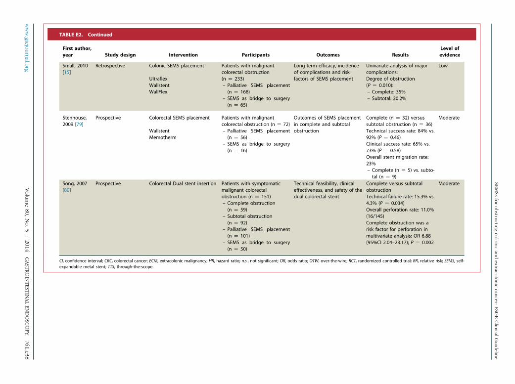
TABLE E2. Continued
First author,year Study design Intervention Participants Outcomes Results
Level ofevidence
Small, 2010[15]
Retrospective Colonic SEMS placement
UltraflexWallstentWallFlex
Patients with malignantcolorectal obstruction(n Z 233)– Palliative SEMS placement(n Z 168)
– SEMS as bridge to surgery(n Z 65)
Long-term efficacy, incidenceof complications and riskfactors of SEMS placement
Univariate analysis of majorcomplications:Degree of obstruction(P Z 0.010):– Complete: 35%– Subtotal: 20.2%
Low
Stenhouse,2009 [79]
Prospective Colorectal SEMS placement
WallstentMemotherm
Patients with malignantcolorectal obstruction (n Z 72)– Palliative SEMS placement(n Z 56)
– SEMS as bridge to surgery(n Z 16)
Outcomes of SEMS placementin complete and subtotalobstruction
Complete (n Z 32) versussubtotal obstruction (n Z 36)Technical success rate: 84% vs.92% (P Z 0.46)Clinical success rate: 65% vs.73% (P Z 0.58)Overall stent migration rate:23%– Complete (n Z 5) vs. subto-tal (n Z 9)
Moderate
Song, 2007[80]
Prospective Colorectal Dual stent insertion Patients with symptomaticmalignant colorectalobstruction (n Z 151)– Complete obstruction(n Z 59)
– Subtotal obstruction(n Z 92)
– Palliative SEMS placement(n Z 101)
– SEMS as bridge to surgery(n Z 50)
Technical feasibility, clinicaleffectiveness, and safety of thedual colorectal stent
Complete versus subtotalobstructionTechnical failure rate: 15.3% vs.4.3% (P Z 0.034)Overall perforation rate: 11.0%(16/145)Complete obstruction was arisk factor for perforation inmultivariate analysis: OR 6.88(95%CI 2.04–23.17); P Z 0.002
Moderate
CI, confidence interval; CRC, colorectal cancer; ECM, extracolonic malignancy; HR, hazard ratio; n.s., not significant; OR, odds ratio; OTW, over-the-wire; RCT, randomized controlled trial; RR, relative risk; SEMS, self-expandable metal stent; TTS, through-the-scope.
www.giejournal.org
Volume80,
No.
5:2014
GAST
ROINTEST
INALEN
DOSC
OPY
761.e38
SEMSs
forobstructing
colonicand
extracoloniccancer:
ESGEClinical
Guideline

TABLE E3. (a–c) Self-expandable metal stent (SEMS) placement as a bridge to elective surgery.
Firstauthor,year
Studydesign Intervention Participants Outcomes Results
Level ofevidence
(a) Randomized controlled trials (RCTs) comparing SEMS as bridge to surgery and emergency surgery.
Ghazal,2013 [89]
RCT Emergency stenting followed byelective resection versus totalabdominal colectomy andileorectal anastomosis
Patients with acute obstructedcarcinoma of the left colon
Preoperative stent (n Z 30),Emergency surgery (n Z 30)
Feasibility, safety, clinicaloutcomes
Outcomes of stent placementTechnical and clinical success rate: 96.7%and 100%No complications encountered during the7–10 days until surgery
Preoperative SEMS vs. emergency surgeryOperative details:– Mean time: 130 vs. 176 min(P Z 0.001)
– Mean blood loss: 250 vs. 500 ml(P Z 0.010)
– Patients requiring blood transfusion:44.8% vs. 73.3% (P Z 0.035)
– Patients requiring fresh frozen plasma:10.3% vs. 83.3% (P Z 0.010)
Overall postoperative complications: 13.8%vs. 50% (P Z 0.012)- Anastomotic leakage: 0% vs. 3.3%(P Z 1.00)
- Wound infection: 10.3% vs. 30%(P Z 0.022)
- Chest infection: 3.4% vs. 16.7%(P Z 0.098)
Median hospital stay: 13 vs. 8 days(P Z 0.102)Median bowel motions per day: 2 vs. 6(P Z 0.013)No operative mortality in both groupsOverall median follow-up: 18 months (range6–40)Recurrent disease: 17.2% vs. 13.3%(P Z 0.228)
Moderate
761.e39GAST
ROINTEST
INALEN
DOSC
OPY
Volume80,
No.
5:2014
www.giejournal.org
SEMSs
forobstructing
colonicand
extracoloniccancer:
ESGEClinical
Guideline

TABLE E3. Continued
Firstauthor,year
Studydesign Intervention Participants Outcomes Results
Level ofevidence
Tung,2013 [90]
Long-termfollow-up ofRCT
SEMS placement followed bylaparoscopic resection versusconventional open surgery
Patients with obstructing left-sidedcolorectal cancer (n Z 48)
Endo-laparoscopic (n Z 24),Open surgery (n Z 24)
Pathological staging, number oflymph nodes harvested,administration of adjuvant therapy,survival and disease recurrencefollowing curative surgery
Emergency surgery vs. preoperative SEMSMedian lymph node harvest: 11 vs. 23(P Z 0.005)Permanent stoma: 25% vs. 0% (P Z 0.03)Adjuvant chemotherapy: 54% vs. 75%(P Z 0.2)Median follow-up: 32 vs. 65 months(P Z 0.083)Curative intent: 54% (13/24) vs. 92% (22/24)(P Z 0.01)Disease recurrence rate: 23% vs. 50%(P Z 0.4)5-year overall survival rate: 27% vs. 48%(P Z 0.076)5-year disease-free survival rate: 48% vs.52% (P Z 0.63)5-year survival rate for patients with stage II/III disease: 42.8% vs. 57.1% (P Z 0.347)
Moderate
Ho, 2012[91]
RCT Colonic stenting followed byelective surgery versus immediateemergency surgeryWallFlex stent
Patients with acute left-sidedmalignant colonic obstruction withno evidence of peritonitis
SEMS as bridge to surgery (n Z 20),Emergency surgery (n Z 19)
60-days postoperative morbidityrate, stoma, hospital stay, criticalcare stay, costs
Outcomes of SEMS placementTechnical and clinical success rate: 75% and93.3%No cases of stent-related perforationPreoperative SEMS vs. emergency surgeryMedian duration of surgery: 135 vs. 135 min(P Z 0.603)Defunctioning stoma rate: 10% vs. 32%(P Z 0.127)Postoperative mortality: 0% vs. 15.8%(P Z 0.106)Overall complication rate: 35% vs. 58%(P Z 0.152)Reoperation rate: 10% vs. 11%Wound infection: 15% vs. 21%Chest infection: 10% vs. 11%Resumption of bowel function: median 4 vs.5 days (P Z 0.167)Fit for discharge: median 6 vs. 8 days(P Z 0.028)Median bowel frequency per day: 2 vs. 2(P Z 0.653)Total length of hospital stay: median 14 vs.13 days (P Z 0.430)Median length of stay in critical care: 2 vs. 3days (P Z 0.057)Median total costs: $18 132 vs. $13 301(P Z 0.194)
Moderate
www.giejournal.org
Volume80,
No.
5:2014
GAST
ROINTEST
INALEN
DOSC
OPY
761.e40
SEMSs
forobstructing
colonicand
extracoloniccancer:
ESGEClinical
Guideline

TABLE E3. Continued
Firstauthor,year
Studydesign Intervention Participants Outcomes Results
Level ofevidence
Alcantara,2011 [92]
RCT Stent placement and deferredsurgery versus emergencyintraoperative colonic lavage withprimary anastomosis
Patients with obstructive left-sidedcolonic cancer (n Z 28)
Preoperative stent (n Z 15),Emergency surgery (n Z 13)
Postoperative morbidity andmortality, staging, complicationsdue to stent placement, surgicaltime, clinical follow-up, health costs,and follow-up of survival
No complications with stent placementwere recordedTime to surgery after SEMS: 5–7 daysOverall mean follow-up: 37.6 months
Preoperative SEMS vs. emergency surgeryOverall morbidity: 13.3% vs. 53.8% (P Z0.042)Anastomotic dehiscence: 0% vs. 30.8%(P Z 0.035)Wound infection: 13.3% vs. 15.4% (P Z 1)Reoperation rate: 0% vs. 30.8% (P Z 0.035)Hospital mortality: 0% vs. 7.7% (P Z 0.464)Median postoperative hospital stay: 8 vs. 10days (P Z 0.05)Median overall hospital stay: 13 vs. 10 days(P Z 0.105)Costs: V6610 vs. V4930 (P Z 0.009)Disease-free period: 25.5 vs. 27.1 months(P Z 0.096)Tumor reappearance: 53.3% vs. 15.4%(P Z 0.055)
Moderate
761.e41GAST
ROINTEST
INALEN
DOSC
OPY
Volume80,
No.
5:2014
www.giejournal.org
SEMSs
forobstructing
colonicand
extracoloniccancer:
ESGEClinical
Guideline

TABLE E3. Continued
Firstauthor,year
Studydesign Intervention Participants Outcomes Results
Level ofevidence
Pirlet,2011 [30]
RCT Emergency surgery compared withSEMS as bridge to surgery
Bard uncovered SEMS
Patients with acute left-sidedmalignant large-bowel obstruction
SEMS as bridge to surgery (n Z 30),Emergency surgery (n Z 30)
Efficiency and reduction of thestoma placement rate
Outcomes of SEMS placementTechnical success rate: 47%Clinical success rate: 85.7%Bridge to elective colonic resection withprimary anastomosis: 40%Median time to surgery: 7 days (5–19 days)No postoperative morbidity for all 12successfully bridged patients
Preoperative SEMS vs. emergency surgeryStoma placement: 43% vs. 57% (P Z 0.30)Restoration of bowel continuity: 30% vs.13% (P Z 0.12)Median duration of stoma: 96 vs. 84 days(P Z 0.68)Successful primary anastomosis: 53% vs.43% (P Z 0.45)Mortality rate: 10% vs. 3%Overall abdominal complications: 23% vs.23% (P Z 1.000)Anastomotic leakage: 7% vs. 7%Overall extra-abdominal complications: 27%vs. 33% (P Z 0.57)Reoperation rate: 10% vs. 7%Median cumulative hospital stay: 23 vs. 17days (P Z 0.13)Colonic resection specimen showed 8clinically silent bowel perforations by thestents
Moderate
www.giejournal.org
Volume80,
No.
5:2014
GAST
ROINTEST
INALEN
DOSC
OPY
761.e42
SEMSs
forobstructing
colonicand
extracoloniccancer:
ESGEClinical
Guideline

TABLE E3. Continued
Firstauthor,year
Studydesign Intervention Participants Outcomes Results
Level ofevidence
Van Hooft,2011 [31]
RCT Colonic stenting as a bridge toelective surgery compared withemergency surgery
Wallstent,WallFlex
Patients with acute left-sidedcolorectal obstruction
SEMS as bridge to surgery (n Z 47),Emergency surgery (n Z 51)
Mean global health status, mortality,morbidity, other quality-of-lifedimensions, and stoma rate
Outcomes of SEMS placementTechnical success rate: 70.2%Clinical success rate: 70.2%Perforation rate: 12.8%Bridge to elective surgery: 93.9% (31/33)Successful primary anastomosis: 48.4%Operative specimens showed 3 silentperforations
Preoperative SEMS vs. emergency surgeryGlobal health status: 63.0 vs. 61.4 (P Z 0.36)30-day mortality rate: 10.6% vs. 9.8%; RR0.92 (95%CI 0.28–2.98); P Z 0.89Overall mortality rate: 19.1% vs. 17.6%; RR0.92 (95%CI 0.40–2.12); P Z 0.84Morbidity rate: 53.2% vs. 45.1%; RR 0.85(95%CI 0.57–1.27); P Z 0.43– Anastomotic leak: 10.6% vs. 2.0%– Abscess: 6.4% vs. 7.8%– Wound infection: 4.3% vs. 2.0%Direct stoma rate: 51.1% vs. 74.5%; RR 1.46(95%CI 1.06–2.01); P Z 0.016Stoma rate at latest follow-up: 57.4% vs.66.7%; RR 1.16 (0.85–1.59); P Z 0.35
Moderate
Cheung,2009 [93]
RCT SEMS placement followed bylaparoscopic resection versus openemergency surgery
Wallstent
Patients with an obstructing tumorbetween the splenic flexure andrectosigmoid junction (n Z 48)
SEMS as bridge to surgery (n Z 24),Emergency surgery (n Z 24)
Successful 1-stage operation,cumulative operative time, bloodloss, hospital stay, pain score, andpostoperative complications
Outcomes of SEMS placementTechnical success rate: 83%Clinical success rate: 83%Median time to laparoscopic resection: 10days (2–16 days)
Preoperative SEMS vs. emergency surgerySuccessful 1-stage operation: 67% vs. 38%(P Z 0.04)Permanent colostomy: 0% vs. 25%(P Z 0.03)Anastomotic leakage: 0% vs. 8% (P Z 0.045)Wound infection: 8% vs. 33% (P Z 0.04)Intra-abdominal abscess: 0% vs. 4%(PO0.99)Other morbidities: 0% vs. 21% (P Z 0.02)Cumulative hospital stay: 13.5 vs. 14 days(P Z 0.7)
Moderate
761.e43GAST
ROINTEST
INALEN
DOSC
OPY
Volume80,
No.
5:2014
www.giejournal.org
SEMSs
forobstructing
colonicand
extracoloniccancer:
ESGEClinical
Guideline

TABLE E3. Continued
Firstauthor,year
Studydesign Intervention Participants Outcomes Results
Level ofevidence
(b) Systematic reviews and meta-analyses on SEMS as bridge to surgery.
Huang,2014 [81]
Meta-analysisof RCTs
Preoperative colonic stents versusemergency surgery
Patients with acute left-sidedmalignant colonic obstruction
7 RCTs
SEMS as bridge to surgery(n Z 195),Emergency surgery (n Z 187)
Efficacy and safety Mean success rate of colonic stentplacement: 76.9% (46.7%–100%)
Preoperative SEMS vs. emergency surgeryPermanent stoma (P Z 0.002): OR 0.28 (95%CI 0.12–0.62); I2 Z 36%Primary anastomosis (P Z 0.007): OR 2.01(95%CI 1.21–3.31); I2 Z 0%Mortality (P Z 0.76): OR 0.88 (95%CI 0.40–1.96); I2 Z 17%Overall complications (P Z 0.03): OR 0.30(95%CI 0.11–0.86); I2 Z 77%– Anastomotic leak (P Z 0.47): OR 0.74(95%CI 0.33–1.67); I2 Z 27%
– Wound infection (P Z 0.004): OR 0.31(95%CI 0.14–0.68); I2 Z 0%
– Intra-abdominal infection (P Z 0.57):OR 0.62 (95%CI 0.12–3.19); I2 Z 0%
High
Cennamo,2013 [82]
Meta-analysisof RCTs
Colorectal stenting as palliation orbridge to surgery compared withemergency surgery
Patients with obstructing colorectalcancer (n Z 353)
8 RCTs
Palliative SEMS placement (n Z 37),SEMS as bridge to surgery(n Z 141),Emergency surgery (n Z 175)
Morbidity, mortality,stoma rate
Outcomes of SEMS placementTechnical and clinical success rate: 73.5%and 72%Stent-related complication rate: 10%– Perforation: 8.4%– Stent migration: 0.5%– Obstructions: 1.1%
SEMS versus emergency surgeryMortality: 8.4% vs. 8%; OR 0.91 (95%CI 0.29–2.79)Morbidity: 36% vs. 46.3%; OR 2.05 (95%CI0.67–6.29)Permanent stoma: 25% vs. 48.1%; OR 3.12(95%CI 1.89–5.17)
Preoperative SEMS vs. emergency surgeryPrimary anastomosis: 65.2% vs. 46.8%; OR0.42 (95%CI 0.25–0.73)Stoma creation: 36.9% vs. 55.4%; OR 2.36(95%CI 1.37–4.07)
High
www.giejournal.org
Volume80,
No.
5:2014
GAST
ROINTEST
INALEN
DOSC
OPY
761.e44
SEMSs
forobstructing
colonicand
extracoloniccancer:
ESGEClinical
Guideline

TABLE E3. Continued
Firstauthor,year
Studydesign Intervention Participants Outcomes Results
Level ofevidence
Cirocchi,2013 [83]
Meta-analysisof RCTs
Colorectal stenting as a bridge tosurgery versus emergency surgery
Patients with intestinal obstructionfrom left-sided colorectal cancer
3 RCTs
SEMS as bridge to surgery (n Z 97),Emergency surgery (n Z 100)
Clinical success, 30-day mortality,overall complications, survival,permanent stoma
Preoperative SEMS vs. emergency surgeryClinical success rate (P! 0.001): 52.5% vs.99%; OR 45.64 (95%CI 10.51–198.13)30-day postoperative mortality (P Z 0.97):8.2% vs. 9%; OR 0.99 (95%CI 0.23–4.19)Overall complication rate (P Z 0.72): 48.5%vs. 51%; OR 0.90 (95%CI 0.52–1.58)Overall survival: not analyzed in RCTsPrimary anastomosis (P Z 0.003): 64.9% vs.55%; OR 2.82 (95%CI 1.43–5.54)Overall stoma rate (P Z 0.02): 45.3% vs.62%; OR 0.48 (95%CI 0.26–0.90)Permanent stoma (P Z 0.56): 46.7% vs.51.8%; OR 0.82 (95%CI 0.42–1.59)Anastomotic leakage (P Z 0.35): 9% vs.3.7%; OR 2.33 (95%CI 0.40–13.52)Intra-abdominal abscess (P Z 0.97): 5.1% vs.4.9%; OR 1.03 (95%CI 0.25–4.18)Wound infections (P Z 0.17): 5.1% vs. 10%;OR 0.39 (95%CI 0.10–1.48)Chest infections (P Z 1.00): 6.1% vs. 6%; OR1.00 (95%CI 0.27–3.70)Urinary tract infections (P Z 0.33): 4% vs.10.2%; OR 0.45; (95%CI 0.09–2.24)
High
761.e45GAST
ROINTEST
INALEN
DOSC
OPY
Volume80,
No.
5:2014
www.giejournal.org
SEMSs
forobstructing
colonicand
extracoloniccancer:
ESGEClinical
Guideline

TABLE E3. Continued
Firstauthor,year
Studydesign Intervention Participants Outcomes Results
Level ofevidence
De Ceglie,2013 [84]
Meta-analysis Colonic stenting as bridge tosurgery versus emergency surgery
Patients with left-sided colonicobstruction
5 RCTs3 Prospective5 Retrospective1 Case-matched
SEMS as bridge to surgery(n Z 405),Emergency surgery (n Z 471)
Treatment details, short-termadverse events, mortality and lengthof hospitalization
Outcomes of SEMS placementMedian time to elective surgery: 10 daysTechnical success rate: 96.9% (95%CI94.9%–98.9%)– RCTs: 75.8% (95%CI 55.4%–96.3%)Clinical success rate: 94.2% (95%CI 91.4%–
97.0%)– RCTs: 73.4% (95%CI 51.0%–95.9%)Stent migration rate: 0% (95%CI 0.0%–0.4%)– Range: 0%–10.5%Perforation rate: 0.1% (95%CI 0.0%–0.4%)– Range: 0%–12.8%Silent perforation rate: 0.1% (95%CI 0.0%–
0.5%)– Range: 0%–26.6%
Preoperative SEMS vs. emergency surgery:Stoma creation (P Z 0.03): ES �27.1% (95%CL –51.2, –3.0); I2 Z 97.2%Protective stoma (P Z 1.0): ES 0% (95%CL–1.0%, 1.1%); I2 Z 35.5%Primary anastomosis (P! 0.001): ES 25.1%(95%CI 17.0%–33.2%); I2 Z 94.9%Successful primary anastomosis (P! 0.001):ES 23.7% (95%CI 13.6%–33.9%); I2 Z 83.9%Anastomotic leakage (P Z 0.1): ES –2.4%(95%CL –5.6%, 0.8%); I2 Z 51.2%Infection (P Z 0.006): ES –7.9% (95%CL–13.6%, –2.3%); I2 Z 59.0%Other morbidities (P! 0.001): ES –13.4%(95%CL –17.9%, –8.8%); I2 Z 0%Mortality: ES –1.9% (95%CL –4.0%, 0.3%);I2 Z 34.1%Hospital stay: ES –1.0% (95%CL –4.1%,2.0%); I2 Z 0%
High
www.giejournal.org
Volume80,
No.
5:2014
GAST
ROINTEST
INALEN
DOSC
OPY
761.e46
SEMSs
forobstructing
colonicand
extracoloniccancer:
ESGEClinical
Guideline

TABLE E3. Continued
Firstauthor,year
Studydesign Intervention Participants Outcomes Results
Level ofevidence
Tan, 2012[85]
Meta-analysisof RCTs
SEMS as bridge to surgery versusemergency surgery
Patients with acute malignantleft-sided large-bowel obstruction
4 RCTs
SEMS as bridge to surgery(n Z 116),Emergency surgery (n Z 118)
Primary anastomosis, stoma and in-hospital mortality, anastomotic leak,30-day reoperation and surgical siteinfection
Outcomes of SEMS placementTechnical success rate 70.7%Clinical success rate 69.0%Clinical perforation rate: 6.9%Silent perforation rate 14%
Preoperative SEMS vs. emergency surgeryPrimary anastomosis (I2 Z 87%):– Fixed-effect: RR 1.46 (95%CI 1.17–1.82);P! 0.001
– Random-effect: RR 1.40 (95%CI 0.84–2.35; P Z 0.20
Overall successful primary anastomosis (P!0.001): RR 1.58 (95%CI 1.22–2.04); I2 Z 0%Stoma rate (P Z 0.004): RR 0.71 (95%CI0.56–0.89); I2 Z 0%Permanent stoma (P Z 0.06): RR 0.75 (95%CI 0.55–1.01); I2 Z 47%In-hospital mortality (P Z 0.74): 6.9% vs.5.9%; RR 1.17 (95%CI 0.46–2.99); I2 Z 0%Anastomotic leak (P Z 0.71): RR 0.72 (95%CI0.13–4.00); I2 Z 51%30-day reoperation (P Z 0.82): RR 0.82 (95%CI 0.15–4.57); I2 Z 54%Surgical site infection (P Z 0.05): 12.9% vs.22.9%; RR 0.56 (95%CI 0.31–0.99); I2 Z 33%
High
Ye, 2012[86]
Meta-analysis Preoperative SEMS placementversus emergency surgery
Patients with acute left-sidedmalignant colonic obstruction
3 RCTs5 Retrospective
SEMS as bridge to surgery(n Z 219),Emergency surgery (n Z 225)
Primary anastomosis, stomaformation, short term mortality andmorbidity
Preoperative SEMS vs. emergency surgeryOne-stage stoma rate (P! 0.001): RR 0.60(95%CI 0.48–0.76); I2 Z 37%Permanent stoma rate (P Z 0.14): RR: 0.80(95%CI 0.59–1.08); I2 Z 44%Anastomosis rate (P! 0.001): RR 1.64 (95%CI 1.39–1.94); I2 Z 9%Mortality (P Z 0.77): RR 0.91 (95%CI 0.50–1.66); I2 Z 0%Overall morbidity (P! 0.001): RR 0.57 (95%CI 0.44–0.74); I2 Z 78%Anastomotic leakage (P Z 0.19): RR 0.60(95%CI 0.28–1.28); I2 Z 18%Abscess (P Z 0.68): RR 0.83 (95%CI 0.36–1.95); I2 Z 0%Extra-abdominal complications (P Z 0.13):RR 0.67 (95%CI 0.40–1.12); I2 Z 0%
High
761.e47GAST
ROINTEST
INALEN
DOSC
OPY
Volume80,
No.
5:2014
www.giejournal.org
SEMSs
forobstructing
colonicand
extracoloniccancer:
ESGEClinical
Guideline

TABLE E3. Continued
Firstauthor,year
Studydesign Intervention Participants Outcomes Results
Level ofevidence
Zhang,2012 [87]
Meta-analysis Stent as a bridge to surgery versusemergency surgery
Patients with obstructive colorectalcancer
2 RCTs6 Retrospective
SEMS as bridge to surgery(n Z 232),Emergency surgery (n Z 369)
ICU usage, success rates of stenting,primary anastomosis, stoma,perioperative mortality andcomplications, survival
Overall technical success of stenting: 87.1%Preoperative SEMS vs. emergency surgeryNeed of intensive care (P Z 0.03): RR 0.42(95%CI 0.19–0.93); I2 Z 0%Primary anastomosis (p Z 0.001): RR 1.62(95%CI 1.21–2.16); I2 Z 75%Stoma creation (P Z 0.04): RR 0.70 (95%CI0.50–0.99); I2 Z 11%Permanent stoma (P Z 0.52): RR 0.39 (95%CI 0.02–6.75); I2 Z 75%Mortality (P Z 0.47): RR 0.73 (95%CI 0.31–1.71); I2 Z 0%Overall complications (P Z 0.001): RR 0.42(95%CI 0.24–0.71); I2 Z 64%Anastomotic leakage (P Z 0.004): RR 0.31(95%CI 0.14–0.69); I2 Z 0%1-year overall survival (P Z 0.51): RR 1.07(95%CI 0.87–1.31); I2 Z 46%2-year overall survival (P Z 0.10): RR 1.14(95%CI 0.98–1.34); I2 Z 0%3-year overall survival (P Z 0.39): RR 1.08(95%CI 0.90–1.31); I2 Z 0%
High
Sagar,2011 [88]
Cochranesystematicreview
Colonic stenting (palliative andbridging) versus surgicaldecompression
Patients with obstructing colorectalcancers
5 RCTs
Colorectal stenting (n Z 102),Emergency surgery (n Z 105)
Mortality, morbidity, technical andclinical success, hospital stay
Outcome of SEMS placementTechnical success rate: 86.0%Stent-related perforation rate: 5.9%Stent migration rate: 2.1%Stent obstruction rate: 2.1%
SEMS versus emergency surgeryClinical success rate (P Z 0.001): 78.1% vs.98.8%; OR 0.06 (95%CI 0.01–0.32); I2 Z 0%30-day mortality (P Z 0.53): OR 1.41 (95%CI0.48–4.14); I2 Z 0%Complications rate (P Z 0.38): 39.2% vs.45.7%; OR 0.79 (95%CI 0.47–1.34); I2 Z 85%Wound complication rate (P Z 0.62): 5.6%vs. 12%; OR 0.54 (95%CI 0.05–6.16); I2 Z64%Mean hospital stay: 11.5 vs. 17.2 daysProcedure/operating time: 114 vs. 144 minMedian blood loss: 50 vs. 350 ml
High
www.giejournal.org
Volume80,
No.
5:2014
GAST
ROINTEST
INALEN
DOSC
OPY
761.e48
SEMSs
forobstructing
colonicand
extracoloniccancer:
ESGEClinical
Guideline

TABLE E3. Continued
Firstauthor,year
Studydesign Intervention Participants Outcomes Results
Level ofevidence
(c) Other literature on SEMS as bridge to surgery.
Gianotti,2013 [109]
Prospective Preoperative SEMS placement,palliative SEMS placement oremergency surgery
Hanarostent
Patients with colorectal obstruction
Malignant (n Z 121),Benign (n Z 11)
Preoperative SEMS placement(n Z 49),Palliative SEMS placement (n Z 32),Emergency surgery (n Z 51)
Short-term and long-term outcomesof different treatment modalities
Overall success rate of stenting:– Technical 95.3%– Clinical 98.8%Median interval to elective surgery: 6 (2–20)daysShort-term stent complications: 14.1%– Perforation rate: 1.2%– Stent migration: 4.9%– Stent occlusion: 4.9%– Tenesmus: 1.2%– Pain: 7.4%– Bleeding: 3.7%
Preoperative SEMS vs. emergency surgeryIn-hospital mortality: 2.0% vs. 2.0% (P Z 1.0)Overall morbidity: 32.7% vs. 60.8% (P Z0.006)Protective ileostomy: 14.3% vs. 21.6% (P Z0.438)Anastomotic leak: 12.2% vs. 19.6% (P Z0.416)Wound infection: 26.5% vs. 54.9% (P Z0.005)Intra-abdominal abscess: 14.3% vs. 39.2%(P Z 0.007)Respiratory tract complication: 10.2% vs.37.3% (P Z 0.002)Postoperative ICU care: 10.2% vs. 33.3%(P Z 0.007)Reoperation: 6.1% vs. 19.6% (P Z 0.052)Median overall length of hospital stay: 18(10–39) days vs. 19 (8–128) days (P Z 0.219)Definitive stoma: 6.3% vs. 26% (P Z 0.012)
Multivariate logistic regression analysis ofsurgical morbidity:Preoperative SEMS placement (P Z 0.015):RR 0.35 (95%CI 0.15–0.82)Kaplan–Meier survival curve showedsignificantly increased 36-month survival inthe SEMS group
Moderate
761.e49GAST
ROINTEST
INALEN
DOSC
OPY
Volume80,
No.
5:2014
www.giejournal.org
SEMSs
forobstructing
colonicand
extracoloniccancer:
ESGEClinical
Guideline
TABLE E3. Continued
Firstauthor,year
Studydesign Intervention Participants Outcomes Results
Level ofevidence
Lee GJ,2013 [102]
Retrospective SEMS as bridge to elective surgeryor emergency surgery
Nitinol Taewoong stents
Patients with obstructive colorectalcancer (n Z 77)
SEMS as bridge to surgery (n Z 49),Emergency surgery (n Z 28)
Short-term morbidity and mortality Preoperative SEMS vs. emergency surgeryMean number of harvested lymph nodes: 26vs. 38 (P Z 0.048)No significant difference for:– Hospital stay (P Z 0.109)– Hartmann (P Z 0.467)– Overall complications: 16.3% vs. 25%(P Z 0.355)
– Anastomotic leakage (P Z 0.297)– Mortality (P Z 0.183)3-year overall survival rate: 68.8% vs. 51.3%(P Z 0.430)
Anastomotic leakage for patients operatedwithin 10 days or after 10 days post-SEMSplacement: 20% (3/15) vs. 0% (0/28); P Z0.037
Low
Cennamo,2012 [126]
Prospective Emergency surgery or surgery afterSEMS placement as a “bridge tosurgery”WallFlex stent
Patients with acute left-sidedcolorectal cancer obstruction(n Z 86)
SEMS as bridge to surgery (n Z 47),Emergency surgery (n Z 41)
Morbidity and mortality risks with P-POSSUM and CR-POSSUM predictivescore models
Technical and clinical success of stenting:95.7% and 95.7%Stent-related complications: 6.7%– Stool impaction: 2.2%– Rectal bleeding: 2.2%– Silent stent perforation: 2.2%Mean time to surgery in SEMS group: 19days (range 6–80 days)
Preoperative SEMS vs. emergency surgeryPrimary anastomosis: 100% vs. 87.8%(P Z 0.02)30-day mortality rate: 2.4% vs. 9.8%30-day morbidity rate: 28.9% vs. 61.0Reoperation: 0% vs. 12.2% (P Z 0.02)P-POSSUM morbidity: 34.3% vs. 70.5%(P Z 0.001)P-POSSUM mortality: 2.4% vs. 13.6%(P Z 0.001)CR-POSSUM mortality: 4.9% vs. 15.1%(P Z 0.001)
Moderate
www.giejournal.org
Volume80,
No.
5:2014
GAST
ROINTEST
INALEN
DOSC
OPY
761.e50
SEMSs
forobstructing
colonicand
extracoloniccancer:
ESGEClinical
Guideline

TABLE E3. Continued
Firstauthor,year
Studydesign Intervention Participants Outcomes Results
Level ofevidence
Cui, 2011[101] *
RCT Laparoscopic resection 3 or 10 daysafter SEMS placement versusemergency open surgery
Patients with obstructing left-sidedcolon cancerSEMS followed by surgery after 3days (n Z 15),
SEMS followed by surgery after 10days (n Z 14),Open emergency surgery (n Z 20)
1-stage operation rate, length ofhospital stay, rates of permanentstoma, postoperative complications
Patients undergoing laparoscopic surgeryhad:– Less blood loss (P! 0.001)– Lower permanent stoma rate(P Z 0.024)
– Less pain (P! 0.001)– Lower incidence of postoperativecomplications
– Higher rate of 1-stage operation(P Z 0.004)
Interval to surgery 3 versus 10 daysHigher 1-stage operation rate after 10 days(P Z 0.001)Lower conversion rate after 10 days(P Z 0.046)
Moderate
Guo, 2011[100]
Retrospective SEMS insertion or primary surgery
Uncovered endoprothesis Nanjing
Patients aged R70 years diagnosedwith acute left-sided colonicobstruction
SEMS (n Z 34),Emergency surgery (n Z 58)
Mortality, avoidance of stoma, andshort-term survival in elderlypatients
SEMS versus surgeryOverall rate of successful bridging withSEMS: 79%Mean time to elective surgery: 9 days (range4–16)Successful relief of obstruction: 91% vs.100% (P Z 0.09)Primary anastomosis rate: 79% vs. 47% (P Z0.002)Temporary stoma rate: 9% vs. 53% (P!0.001)Permanent stoma rate: 6% vs. 12% (P Z0.34)Median length of hospital stay: 19 vs. 14days (P Z 0.06)Acute mortality rate: 3% vs. 19% (P Z 0.03)Acute complication rate: 24% vs. 40% (P Z0.11)
Low
761.e51GAST
ROINTEST
INALEN
DOSC
OPY
Volume80,
No.
5:2014
www.giejournal.org
SEMSs
forobstructing
colonicand
extracoloniccancer:
ESGEClinical
Guideline

TABLE E3. Continued
Firstauthor,year
Studydesign Intervention Participants Outcomes Results
Level ofevidence
Jiménez-Pérez,2011 [127]
Prospective Preoperative WallFlex stentplacement
Patients with malignant colorectalobstruction (n Z 182)
Efficacy and safety of the WallFlexcolonic stent as a bridge to surgery
Outcomes of SEMS placementProcedural success rate: 97.8%Major procedural complications: 3.3%– Perforation: 1.7%– Persistent obstruction: 1.1%– Self-limiting bleeding: 0.6%Minor procedural complications: 1.1%– Transient abdominal pain: 1.1%Major post-procedural complications: 4.2%(7/167)– Stent migration: 1.2%– Perforation: 1.2%– Fecal impaction: 1.2%– Persistent obstruction: 0.6%Minor post-procedural complications: 0.6%– Pain requiring analgesia: 0.6%Overall major stent complication rate: 7.8%(13/167)Clinical success until surgery: 94% (141/150)Bridge to elective surgery: 89.8% (150/167)– Clinical success: 97.3%– Median time to surgery: 14 days (IQR8–20 days)
– Stoma creation: 6%– Post-surgical complications: 16.7%– Post-surgical mortality: 2%– Overall stoma performance rate: 6.6%– Diverting stoma: 6%
Moderate
Kim S,2009[103] *
Nodescriptionof studydesign
SEMS placement as bridge tosurgery
Patients with obstructive colorectalcancer (n Z 62)
Operated!7 days (n Z 26),Operated O7 days (n Z 30)
Optimal time for elective radicalsurgery following colonic stentinsertion
Technical and clinical success rate: 100%and 90.3%
Surgery!7 days versus O7 daysComorbid diseases: 19.2% vs. 56.7% (P Z0.004)Postoperative morbidity: 7.7% vs. 16.7%(P Z n.s.)Postoperative mortality: 0% vs. 3.3%No difference in operation time andpostoperative recoveryAdjusted with comorbid diseases, there wasno significant difference for all the variablesbetween the two groups
Low
CI, confidence interval; CL, confidence limits; ES, effect size; ICU, intensive care unit; IQR, interquartile range; n.s., not significant; RCT, randomized controlled trial; OR, odds ratio; POSSUM, Physiological and OperativeSeverity Score for enUmeration of Mortality and Morbidity (P-, Portsmouth; CR-, colorectal); RR, relative risk; SEMS, self-expandable metal stent.*Published in abstract form only.
www.giejournal.org
Volume80,
No.
5:2014
GAST
ROINTEST
INALEN
DOSC
OPY
761.e52
SEMSs
forobstructing
colonicand
extracoloniccancer:
ESGEClinical
Guideline

TABLE E4. (a–d) Palliative placement of self-expandable metal stent (SEMS).
Firstauthor,year Study design Intervention Participants Outcomes Results
Level ofevidence
(a) RCTs comparing palliative SEMS placement with emergency surgery.
Van Hooft,2008 [123]
RCT Through-the-scope WallFlexcolorectal stent insertion versusemergency surgery
Patients with incurable stage IVleft-sided colorectal cancer
Palliative SEMS placement(n Z 11),Emergency surgery (n Z 10)
Survival in good healthout of hospital
SEMS versus surgeryMedian hospital-free survival in goodhealth: 38 vs. 56 days (P Z 0.68)Median total time in hospital: 12 vs. 11days (P Z 0.46)Median total time on ICU: 0 vs. 0 days(P Z 0.30)Median total follow-up time: 360 vs. 173days (P Z 0.67)Number of adverse events: 11 vs. 1(P Z 0.001)Patients suffering adverse event: 73% (8/11) vs. 10% (1/10); P Z 0.008; RRZ7.2
Moderate
Fiori, 2004[6] & 2012[108]
RCT Endoscopic Precision stentplacement versus divertingproximal colostomy
Patients with stage IVunresectable rectosigmoid cancerand symptoms of chronicsubacute obstruction (n Z 22)
Palliative SEMS placement(n Z 11),Colostomy (n Z 11)
Morbidity, mortality, canalizationof gastrointestinal tract,restoration of oral intake,hospital stay
Outcomes of SEMS placementTechnical and clinical success rate: 100%and 100%Hospital stay: range 2–4 daysMedian survival: 297 days (125–612 days)Late complications:– Fecal impaction: 18% (2/11)– Tumor ingrowth: 9% (1/11)Outcomes of colostomyNo postoperative mortalitySurgical revision because of partialprolapse of the colostomy: 9% (1/11)Mean hospital stay: 8 days (range 7–10days)Median survival: 280 days (135–591 days)Late complications:– Stoma prolapse: 9% (1/11)– Skin inflammation around stoma: 9%(1/11)
Moderate
Xinopoulos,2004 [128]
RCT Palliative Wallstent colonic stentplacement versus colostomy
Patients with inoperablemalignant partial obstruction inthe left colon originating fromcolorectal or ovarian cancer(n Z 30)
Palliative SEMS placement(n Z 15),Colostomy (nZ15)
Efficacy, safety, cost–effectiveness Outcomes of SEMS placementTechnical success rate: 93.3%Moderate tumor ingrowth: 43% (6/14),treated with Diomed laser, withoutreoccurrence of obstructive symptomsStent migration: 7% (1/14)SEMS versus colostomyTotal hospital stay: 28 vs. 60 daysMedian survival: 21.4 vs. 20.9 weeks(P Z n.s.)Average total cost: V2224 vs. V2092(P Z n.s.)
Moderate
761.e53GAST
ROINTEST
INALEN
DOSC
OPY
Volume80,
No.
5:2014
www.giejournal.org
SEMSs
forobstructing
colonicand
extracoloniccancer:
ESGEClinical
Guideline

TABLE E4. Continued
Firstauthor,year Study design Intervention Participants Outcomes Results
Level ofevidence
(b) Systematic reviews and meta-analyses on palliative SEMS placement.
Liang, 2014[104]
Meta-analysis SEMS versus surgery for palliativetreatment of colorectalobstruction caused by advancedcolorectal malignancy
Patients with malignant colorectalobstruction caused by advancedmalignancy
3 RCTs2 Prospective4 Retrospective
Palliative SEMS placement(n Z 195),Emergency surgery (n Z 215)
Short-term and long-termcomplications, mortality,and time of hospitalization
Major stent-related complications:– Short-term (!30 days) perforationrate: 3.7%
– Long-term (R30 days) perforationrate: 7.6%
– Overall stent migration rate: 8.9%– Re-obstruction: not analyzed.Successful relief of obstruction:– Palliative SEMS: 94%– Surgery: 100%Short-term (!30 days) complication rate(P Z 0.22):– Palliative SEMS: 26.2% (51/195)– Surgery: 34.5% (74/215)– OR 0.83 (95%CI 0.39–1.79)Long-term (R30 days) complication rate(P Z 0.03):– Palliative SEMS: 16.1% (25/155)– Surgery: 8.1% (14/173)– OR 2.34 (95%CI 1.07–5.14)Overall complication rate (P Z 0.56):– Palliative SEMS: 43.9% (68/155)– Surgery: 45.1% (78/173)– OR 1.27 (95%CI 0.58–2.77)Overall mortality rate (P Z 0.22):– Palliative SEMS: 7.1% (12/169)– Surgery: 11.6% (22/189)– OR 0.60 (95%CI 0.27–1.34)SEMS required significantly shorterhospitalization: weighted mean difference–6.07 days (95%CL –8.40, –3.74); P! 0.01
High
www.giejournal.org
Volume80,
No.
5:2014
GAST
ROINTEST
INALEN
DOSC
OPY
761.e54
SEMSs
forobstructing
colonicand
extracoloniccancer:
ESGEClinical
Guideline

TABLE E4. Continued
Firstauthor,year Study design Intervention Participants Outcomes Results
Level ofevidence
Zhao, 2013[105]
Meta-analysis Palliative stent placement vs.palliative surgical decompression
Patients with malignant colorectalobstruction that was unresectable
3 RCTs5 Prospective4 Retrospective1 Case-matched
Palliative SEMS placement(n Z 404),Palliative surgery (n Z 433)
Hospital stay, intensive care unitadmission, clinical success rate,30-day mortality, stomaformation, complications andoverall survival time
Mean length of hospital stay (P! 0.001):– Palliative SEMS: 9.6 days– Surgery: 18.8 days,ICU admission rate (P Z 0.001):– Palliative SEMS: 0.8% (1/119)– Surgery: 18.0% (22/122)– RR 0.09 (95%CI 0.02–0.38); I2 Z 0%Mean interval to chemotherapy:– Palliative SEMS: 15.5 days– Surgery: 33.4 daysClinical relief of obstruction (P! 0.001):– Palliative SEMS: 93.1% (375/403)– Surgery: 99.8% (433/434)– RR 0.96 (95%CI 0.93–0.98); I2 Z 3%In-hospital mortality rate (P Z 0.01):– Palliative SEMS: 4.2% (14/334)– Surgery: 10.5% (37/354)– RR 0.46 (95%CI 0.25–0.85); I2 Z 0%Overall complication rate (P Z 0.60):– Palliative SEMS: 34.0% (137/403)– Surgery: 38.1% (172/452)– RR 0.91 (95%CI 0.64–1.29); I2 Z 66%Early complication rate (P Z 0.03):– Palliative SEMS: 13.7% (41/300)– Surgery: 33.7% (110/326)– RR 0.45 (95%CI 0.22–0.92); I2 Z 66%Late complication rate (P! 0.001):– Palliative SEMS: 32.3% (60/186)– Surgery: 12.7% (27/213)– RR 2.33 (95%CI 1.55–3.50); I2 Z 0%Stent complications:– Perforation rate: 10.1%– Stent migration: 9.2%– Stent obstruction: 18.3%Overall survival time (P Z n.s.):– Palliative SEMS: 7.6 months– Surgery: 7.9 monthsStoma formation rate (P! 0.001):– Palliative SEMS: 12.7% (38/299)– Surgery: 54.0% (170/315)– RR 0.26 (95%CI 0.18–0.37); I2 Z 18%
High
761.e55GAST
ROINTEST
INALEN
DOSC
OPY
Volume80,
No.
5:2014
www.giejournal.org
SEMSs
forobstructing
colonicand
extracoloniccancer:
ESGEClinical
Guideline

TABLE E4. Continued
Firstauthor,year Study design Intervention Participants Outcomes Results
Level ofevidence
(c) Other literature on palliative SEMS placement.
Abbott,2014 [11]
Retrospective Palliative endoscopic SEMSinsertion
Wallstent,Taewoong,Schneider
Patients with colonic obstructiondue to colorectal cancer ormetastatic extracolonic disease(n Z 146)
Technical success andcomplication rates of SEMS, andidentifying any predictors ofstent-related complications andre-intervention
Technical success rate: 97.3%Clinical success rate: 95.8%Early complication rate: 13.0%Late complication rate: 26.7%Overall complication rate: 39.7%– Perforation rate: 4.8%– Stent migration: 13.0%– Stent re-obstruction: 18.5%– Other complications: 3.4%30-day procedural mortality rate: 2.7%Overall re-intervention rate: 30.8%– Endoscopic: 18.5%– Surgical: 14.4%Median post-procedure length of hospitalstay: 2 daysMedian survival: 9.2 months (95%CI 8.2–10.2)Stoma rate: 11.0%
Low
Gianotti,2013 [109]
Prospective Preoperative SEMS placement,palliative SEMS placement oremergency surgery
Hanarostent
Patients with colorectalobstruction
Malignant (n Z 121),Benign (n Z 11)
Preoperative SEMS placement(n Z 49),Palliative SEMS placement(n Z 32),Emergency surgery (n Z 51)
Short-term and long-termoutcomes of different treatmentmodalities
Overall technical success rate of stenting:95.3%Overall clinical success rate of stenting:98.8%Median interval to elective surgery: 6 (2–20) daysShort-term stent complications: 14.1%
Long-term outcome of SEMS (n Z 32)Clinical success rate: 81.2%Overall long-term complication rate:43.8%– Perforation: 3.1%– Stent migration: 12.5%– Occlusion: 9.4%– Tenesmus: 21.9%– Recurrent abdominal pain: 21.9%– Bleeding: 25%Hospital readmission: 34.4%Median survival (n Z 29): 10 months(95%CI 4–16)
Moderate
www.giejournal.org
Volume80,
No.
5:2014
GAST
ROINTEST
INALEN
DOSC
OPY
761.e56
SEMSs
forobstructing
colonicand
extracoloniccancer:
ESGEClinical
Guideline

TABLE E4. Continued
Firstauthor,year Study design Intervention Participants Outcomes Results
Level ofevidence
Huhtinen,2013 [111]
Retrospective Palliative SEMS insertion
Ultraflex,Hanarostent
Patients with obstructiveincurable colorectal cancer(n Z 56)
Clinical outcomes of SEMS andfactors associated with latecomplications
Technical and clinical success rate: 75%and 70% (39/56)Overall complication rate: 38% (16/42)– Perforation: 10%– Re-obstruction: 14%– Incontinence: 5%– Pain: 5%– Stent migration: 2%Stent-related mortality: 7%Late complications: 31%Re-intervention rate: 24%– Ostomy (n Z 8)– Hartmann (n Z 1)– Re-stenting (n Z 1)
Low
Yoshida,2013 [110]
Prospectivefeasibility study
Palliative Niti-S D-type uncoveredstent insertion
Patients with malignant large-bowel obstruction (n Z 33)
Efficacy and safety of the newuncovered Niti-S D-type stent
Technical success rate: 100%Clinical success rate: 97%Median follow-up: 126 days (range 20–750)Early complications:– Tenesmus: 6%Late complications:– Stent occlusion: 30%– Stent migration: 3%– Bleeding: 9%– Tenesmus: 3%Re-interventions:– Colostomy: 3% (1/33)– Endoscopic: 27% (9/33)Mean survival: 240 days
Low
761.e57GAST
ROINTEST
INALEN
DOSC
OPY
Volume80,
No.
5:2014
www.giejournal.org
SEMSs
forobstructing
colonicand
extracoloniccancer:
ESGEClinical
Guideline

TABLE E4. Continued
Firstauthor,year Study design Intervention Participants Outcomes Results
Level ofevidence
Angenete,2012 [112]
Prospective stentcohort andretrospective controlgroup
Colorectal SEMS placement oremergency surgery
Patients with colonic obstructiondue to rectal or colon cancerPalliative SEMS placement(n Z 88),SEMS as bridge to surgery(n Z 24),Control group (n Z 60)
Morbidity, mortality and hospitalstay
Overall technical success rate of SEMS:96%Overall clinical success rate of SEMS: 90%Overall stent complications:– Stent migration: 5.4%– Perforation: 5.4%– Fistula: 2.7%– Bleeding: 1.8%– Sepsis: 0.9%– Other: 4.5%– Cardiopulmonary: 1.8%Re-stenting: 10%– Clinical failure (n Z 1)– Tumor ingrowth (n Z 7)– Stent displacement (n Z 2)– Stent dysfunction (n Z 1)Outcomes in palliative SEMS groupSurgery due to complications: 18%– Poor technical success: 5%– Poor clinical success: 5%– Fistula: 2%– Perforation: 5%– Palliative resection: 2%Stoma formation: 8% (7/88) comparedwith 53% (32/60) in the surgery group
Low
www.giejournal.org
Volume80,
No.
5:2014
GAST
ROINTEST
INALEN
DOSC
OPY
761.e58
SEMSs
forobstructing
colonicand
extracoloniccancer:
ESGEClinical
Guideline

TABLE E4. Continued
Firstauthor,year Study design Intervention Participants Outcomes Results
Level ofevidence
Meisner,2012 [113]
Prospective WallFlex uncovered colonic stentplacement
Patients with colorectal stricturessecondary to malignant diseaseundergoing palliative stentplacement (n Z 255)
Procedural and clinical success,safety
Procedural success rate: 98.4%Follow-up visits and clinical success:– 30 days (n Z 206): 87.8%– 3 months (n Z 126): 89.7%– 6 months (n Z 86): 92.8%– 12 months (n Z 36): 96%Overall perforation rate: 5.1% (13/255)Overall stent migration rate: 5.5% (14/255)Cumulative complications:– Perforation rate: 13.8% (13/94)– Stent migration: 12.8% (12/94)– Tumor ingrowth/overgrowth: 17.0%(16/94)
– Fecal impaction: 8.5% (8/94)– Second colonic obstruction: 2.1% (2/94)
– Bleeding: 4.3% (4/94)– Pain: 4.3% (4/94)– Persistent obstruction: 1.1% (1/94)12-month mortality rate: 48.6%Stent-related mortality: 0.8%
Moderate
Manes, 2011[55]
Retrospective Colonic stent placement
WallFlex,Wallstent,Ultraflex,Hanarostent,Bonastent,Evolution
Patients with incurable malignantcolonic obstruction (n Z 201)
Short-term and long-term efficacyof SEMS
Technical success rate: 91.5%Immediate clinical success rate: 89.7%Mean follow-up: 115 days (1–500 days)Sustained relief of obstruction until death:77.0% (127/165)Stent patency at 6 and 12 months: 82.1%and 65.7%Overall major complications: 11.9%– Perforation: 6.0%– Stent migration: 5.5%– Stent re-obstruction: 0.5%Permanent colostomy (n Z 9)
Low
761.e59GAST
ROINTEST
INALEN
DOSC
OPY
Volume80,
No.
5:2014
www.giejournal.org
SEMSs
forobstructing
colonicand
extracoloniccancer:
ESGEClinical
Guideline

TABLE E4. Continued
Firstauthor,year Study design Intervention Participants Outcomes Results
Level ofevidence
Young, 2011[122]
Prospective Colonic SEMS insertion
Wallstent,Ultraflex,WallFlex
Patients having an attemptedSEMS insertion for large-bowelobstruction (n Z 100)
Palliative SEMS placement(n Z 89),Preoperative SEMS placement(n Z 11)
Malignant obstruction (n Z 93),Benign obstruction (n Z 7)
Stent patency, morbidity andmortality
Median follow-up: 34.5 (1–64) monthsMedian survival: 4 (95%CI 3.2–4.9) monthsTechnical success rate: 87%48-hour clinical success rate: 84%72 patients were considered to haveavoided a stoma30-day mortality: 7%– Stent-related mortality: 1%Stent-related morbidity: 20%– Perforation: 5%– Dislodgement: 4%– Migration: 1%– Obstruction: 5%– Pain: 4%– Incontinence: 1%– Impaction: 1%Patent stent at last follow-up or death:73%
Moderate
(d) Outcomes of palliative SEMS placement during chemotherapy and antiangiogenic therapy.
Abbott,2014 [11]
Retrospective Palliative endoscopic SEMSinsertion
Wallstent,Taewoong,Schneider
Patients with colonic obstructiondue to colorectal cancer ormetastatic extracolonic disease(n Z 146)
Chemotherapy (n Z 58)
Technical success andcomplication rates of SEMS, andidentifying any predictors ofstent-related complications andre-intervention
Technical success rate: 97.3%Clinical success rate: 95.8%Overall complication rate: 39.7%Overall reintervention rate: 30.8%– Endoscopic: 18.5%– Surgical: 14.4%Predictors of early complications:– Chemotherapy: OR 0.92; P Z 0.974Predictors of late complications:– Chemotherapy: OR 5.52; P Z 0.003Predictors of endoscopic reintervention:– Chemotherapy: OR 4.30; P Z 0.018Predictors of surgical treatment:– Chemotherapy: OR 2.21; P Z 0.242
Low
www.giejournal.org
Volume80,
No.
5:2014
GAST
ROINTEST
INALEN
DOSC
OPY
761.e60
SEMSs
forobstructing
colonicand
extracoloniccancer:
ESGEClinical
Guideline

TABLE E4. Continued
Firstauthor,year Study design Intervention Participants Outcomes Results
Level ofevidence
Di Mitri,2014 [117]
Retrospective Colorectal SEMS placement
WallFlex,Evolution,Ultraflex
Patients with obstructivecolorectal cancer (n Z 204)
Palliative SEMS placement(n Z 143),SEMS as bridge to surgery(n Z 61)
Chemotherapy (n Z 105)
Technical success, clinical efficacy,complications
Technical success rate: 99.0%Clinical success rate: 94.6%Median follow-up: 6 months (range 1–32)Overall complications: 15.2%– Tumor ingrowth: 8.3%– Stent migration: 4.9%– Perforation: 2.0%“None of the perforation cases were onbevacizumab”Survival at end of follow-up: 46.1%Univariable analysis of risk factorsassociated with complications:– Chemotherapy: OR 0.4; P Z 0.88Univariable analysis of risk factorsassociated with death:– Chemotherapy: OR 1.1; P Z 0.89Univariable analysis of risk factorsassociated with tumor ingrowth:– Chemotherapy: OR 0.26; P Z 0.016Multivariable analysis of risk factorsassociated with tumor ingrowth:– Chemotherapy: OR 0.44; P Z 0.009
Low
VanHalsema,2014 [51]
Meta-analysis Colorectal SEMS placement All patients who underwentcolorectal stent placement (n Z4086)
Risk factors for perforation fromcolonic stenting
Pooled perforation rate for:Patients without concomitant therapy:– 9.0% (95%CI 7.2%–11.1%)Patients treated with chemotherapy:– 7.0% (95%CI 4.8%–10.0%)Patients treated with bevacizumab:– 12.5% (95%CI 6.4%–22.8%)
Moderate
761.e61GAST
ROINTEST
INALEN
DOSC
OPY
Volume80,
No.
5:2014
www.giejournal.org
SEMSs
forobstructing
colonicand
extracoloniccancer:
ESGEClinical
Guideline

TABLE E4. Continued
Firstauthor,year Study design Intervention Participants Outcomes Results
Level ofevidence
Canena,2012 [119]
Retrospective Palliative colorectal SEMSplacement
WallFlex,Wallstent,Ultraflex
Patients with inoperablemalignant colorectal obstruction(n Z 89)
Chemotherapy (n Z 24)
Long-term clinical efficacy andfactors affecting stent patency,clinical success, and complications
Univariate analysis of factors associatedwith long-term clinical success:Chemotherapy (P Z 0.45):– Yes: 70.8% (17/24)– No: 78.5% (51/65)Multivariate logistic analysis of risk factorsfor stent migration:– Chemotherapy (P Z 0.06): OR 11.89(95%CI 0.90–156.47)
Multivariate logistic analysis of risk factorsfor obstruction:– Chemotherapy (P Z 0.35): OR 2.48(95%CI 0.50–13.08)
Multivariate Cox regression analysis offactors associated with stent patency:– Chemotherapy (P Z 0.07): HR 5.51(95%CI 0.86–35.29)
Low
Lee HJ, 2011[118]
Retrospective Colorectal SEMS placement orsurgery
WallFlex,Comvi stent,Niti-S D-type
Patients with metastaticunresectable colorectal cancerwith imminent obstruction
Palliative SEMS placement(n Z 71),Emergency surgery (n Z 73)
Long-term outcomes ofendoscopic stenting and surgery
Risk factors for late complications:Chemotherapy (P Z 0.003):– Yes: 47.8% (22/46)– No: 10% (2/20)Bevacizumab (P Z 0.645):– Yes: 20% (1/5)– No: 37.7% (23/61)Palliative chemotherapy was notsignificantly associated with perforationMultivariate analysis of risk factors for latecomplications:– Chemotherapy (P Z 0.01): OR 10.43(95%CI 1.75- 62.39)
Prognostic factors for overall survival:Chemotherapy (P! 0.001):– Yes: 15.1 months– No: 4.5 monthsTarget agent (P Z 0.020):– Yes: 18.4 months– No: 9.6 monthsMultivariate analysis of factors associatedwith survival:– Chemotherapy (P Z 0.002): HR 0.33(95%CI 0.33–0.77)
Low
www.giejournal.org
Volume80,
No.
5:2014
GAST
ROINTEST
INALEN
DOSC
OPY
761.e62
SEMSs
forobstructing
colonicand
extracoloniccancer:
ESGEClinical
Guideline

TABLE E4. Continued
Firstauthor,year Study design Intervention Participants Outcomes Results
Level ofevidence
Luigiano,2011 [76]
Prospective Endoscopic WallFlex placementfor palliation
Patients with malignant colorectalobstruction (n Z 39)
Chemotherapy (n Z 13)
Outcomes of through-the-scopelarge-diameter SEMS placementfor palliation
Technical success rate: 92.3%Clinical success rate: 89.7%Early complications:– Perforation: 5.6%– Bleeding: 2.8%Late complications:– Tumor ingrowth: 14.3%– Tumor ingrowth and bleeding: 2.8%– Stool impaction: 8.6%– Stent migration: 2.8%Overall median survival: 280 days (range32–511)No correlation between chemotherapyand late complications (P Z 0.120)Palliative chemotherapy was associatedwith longer survival (P Z 0.006)
Low
Manes, 2011[55]
Retrospective Colonic stent placement
WallFlex,Wallstent,Ultraflex,Hanarostent,Bonastent,Evolution
Patients with incurable malignantcolonic obstruction (n Z 201)
Chemotherapy (n Z 74),Bevacizumab (n Z 8)
Short-term and long-termefficacy of SEMS
Overall chemotherapy did not increasethe risk of complications
Perforation risk for bevacizumab (P!0.001):– Yes: 50% (4/8)– No: 2.5%– OR 19.6 (95%CI 5.9–64.5)
Low
Yoon, 2011[8]
Retrospective Colorectal SEMS insertion
Niti-S covered,Comvi stent,WallFlex,Niti-S D-type
Patients with malignant colorectalobstruction (n Z 412)– Palliative SEMS placement(n Z 276)
– SEMS as bridge to surgery(n Z 136)
Rates and factors predictive oftechnical and clinical failure ofSEMS
Long-term clinical failure: 36.3% (73/201)– Tumor ingrowth/overgrowth: 22.9%– Stent migration: 9.0%– Perforation: 4.0%– Bleeding: 0.5%Multivariate analysis of risk factors forlong-term clinical failure in palliationgroup:– Chemotherapy (P Z 0.015): OR 0.52(95%CI 0.31–0.88)
Low
761.e63GAST
ROINTEST
INALEN
DOSC
OPY
Volume80,
No.
5:2014
www.giejournal.org
SEMSs
forobstructing
colonicand
extracoloniccancer:
ESGEClinical
Guideline

TABLE E4. Continued
Firstauthor,year Study design Intervention Participants Outcomes Results
Level ofevidence
Fernandez-Esparrach,2010 [120]
Retrospective Colorectal SEMS insertion
Wallstent,WallFlex,Hanarostent
Patients with colorectal cancerof the left colon with obstructivesymptoms (n Z 47)– Palliative SEMS placement(n Z 38),
– SEMS as a bridge tosurgery (n Z 9)
Chemotherapy (n Z 28)
Long-term clinical successand factors predictive ofdevelopment of complications
Technical success rate: 94%Clinical success rate: 94%Overall complication rate: 51%– Stent migration: 22%– Perforation: 7%– Re-obstruction: 17%– Tenesmus: 5%Complication-related death: 12%Long-term complication rate forchemotherapy vs. no chemotherapy: 62%(16/26) vs. 33% (5/15); P Z 0.082“8/9 patients with stent migration and 2/3patients with perforation had beentreated with chemotherapy”
Low
Small, 2010[15]
Retrospective Colonic SEMS placement
Ultraflex,Wallstent,WallFlex
Patients with malignantcolorectal obstruction (n Z 233)– Palliative SEMSplacement (n Z 168),
– SEMS as bridge tosurgery (n Z 65)
Long-term efficacy, incidenceof complications, and riskfactors of SEMS placement
Univariate analysis of risk factors formajor complications:Palliative chemotherapy (P Z 0.054):– Yes: 29.8% (25/84)– No: 19.0% (16/84)Bevacizumab therapy (P Z 0.107):– Yes: 34.8% (8/23)– No: 22.8% (33/145)Univariate analysis of risk factors forperforation:Bevacizumab (P Z 0.064):– Yes: 17.4% (4/23)– No: 7.6% (11/145)
Low
Cennamo,2009 [116]
Case series WallFlex colonic stent placement Patients with occlusive coloncancer (n Z 28)
SEMS as bridge to surgery(n Z 12)Chemotherapy (n Z 9),Bevacizumab (n Z 2)
Perforation risk afterbevacizumab therapy
Median follow-up: 131 daysDelayed colonic perforation occurred inthe 2 patients treated with a combinationof capecitabine and oxaliplatin plusbevacizumab
Low
Kim JH,2009 [43]
Prospectivenonrandomized
Radiologic dual-design SEMSinsertionFlared ends (n Z 69),Bent ends (n Z 53)
Patients with malignant colorectalobstruction (n Z 122)– Palliative SEMS placement(n Z 80),
– SEMS as bridge to surgery(n Z 42)
Clinical safety and efficacy ofdual-design stents
Flared-ends versus bent-endsTechnical success rate: 94.2% vs. 96.2%Clinical success rate: 93.8% vs. 90.2%Overall complication rate: 18.5% vs. 25.5%Perforation rate: 6.2% vs. 5.9%Stent migration rate: 6.2% vs. 5.9%Stent migration was significantly relatedto chemotherapy (P Z 0.029)
Moderate
CI, confidence interval; CL, confidence limits; HR, hazard ratio; ICU, intensive care unit; n.s., not significant; OR, odds ratio; RCT, randomized controlled trial; RR, relative risk; SEMS, self-expandable metal stent.
www.giejournal.org
Volume80,
No.
5:2014
GAST
ROINTEST
INALEN
DOSC
OPY
761.e64
SEMSs
forobstructing
colonicand
extracoloniccancer:
ESGEClinical
Guideline

TABLE E5. (a, b) Adverse events related to colonic stenting
Firstauthor,year Study design Intervention Participants Outcomes Results
Level ofevidence
(a) Adverse events.
Abbott,2014 [11]
Retrospective Palliative endoscopic SEMSinsertion
Wallstent,Taewoong,Schneider
Patients with colonic obstruction dueto colorectal cancer or metastaticextracolonic disease (n Z 146)
Technical success and complicationrates of SEMS, and identifying anypredictors of stent-relatedcomplications and re-intervention
Technical success rate: 97.3%Clinical success rate: 95.8%Median post procedure length ofhospital stay: 2 daysEarly complication rate: 13.0%– Perforation: 4.8%– Stent migration: 3.4%– Re-obstruction: 2.1%– No resolution of symptoms:2.1%
– Ischemic colon: 0.7%Late complication rate: 26.7%– Perforation: 0%– Stent migration: 9.6%– Stent re-obstruction: 16.4%– Synchronous obstruction: 0.7%30-day procedural mortality rate:2.7%Overall re-intervention rate: 30.8%– Endoscopic: 18.5%– Surgical: 14.4%Median time to endoscopicreintervention: 4.6 months4/27 patients required surgicaltreatment within 30 days of insertionof a second SEMSMedian survival: 9.2 months (95%CI8.2–10.2)Stoma rate: 11.0%
Low
761.e65GAST
ROINTEST
INALEN
DOSC
OPY
Volume80,
No.
5:2014
www.giejournal.org
SEMSs
forobstructing
colonicand
extracoloniccancer:
ESGEClinical
Guideline

TABLE E5. Continued
Firstauthor,year Study design Intervention Participants Outcomes Results
Level ofevidence
Di Mitri,2014 [117]
Retrospective Colorectal SEMS placement
WallFlex,Evolution,Ultraflex
Patients with obstructive colorectalcancer (n Z 204)– Palliative SEMS placement(n Z 143),
– SEMS as bridge to surgery(n Z 61)
Technical success, clinical efficacy,complications
Technical and clinical success rate:99.0% and 94.6%Median follow-up: 6 months (range1–32)Overall complication rate: 15.2%– Tumor ingrowth: 8.3%– Stent migration: 4.9%– Perforation: 2.0%Early (%30 days) complications: 3.9%– Perforation: 1.9%– Stent migration: 1.5%– Stent ingrowths: 0.5%Late (O30 days) complications:11.2%– Stent migration 3.4%– Tumor ingrowths 7.8%SEMS migration or neoplasticingrowths were treated with asecond stentOverall clinical benefit at end offollow-up: 79.4%Survival at end of follow-up: 46.1%
Low
Geraghty,2014 [16]
Retrospective Colonic stenting for large-bowelobstruction
Patients in whom SEMS placementwas attempted for large-bowelobstruction (n Z 334)– CRC palliation (n Z 264),– CRC bridge to surgery (n Z 52),– Benign (n Z 9),– Extrinsic (n Z 9)
Outcome of colonic stenting andfactors associated with successfulintervention
Overall technical success rate: 87.4%Overall clinical success rate: 83.5%Technical failure:– Inability to deploy SEMS: 6.0%– Perforation: 2.7%– Stent migration: 2.1%– Insufficient expansion: 1.2%– Incorrect stent positioning: 0.6%– Peri-interventional cardiorespi-ratory episode: 1.2%
Additional complications:– Significant bleeds!48h (n Z 3)– Colovaginal fistula (n Z 1)– Rectal abscess (n Z 1)Re-stenting for migration or tumorovergrowth: 7.5%Surgical re-intervention: 10.8%30-day mortality: 13.2%
Low
www.giejournal.org
Volume80,
No.
5:2014
GAST
ROINTEST
INALEN
DOSC
OPY
761.e66
SEMSs
forobstructing
colonicand
extracoloniccancer:
ESGEClinical
Guideline

TABLE E5. Continued
Firstauthor,year Study design Intervention Participants Outcomes Results
Level ofevidence
Gianotti,2013 [109]
Prospective Preoperative SEMS placement,palliative SEMS placement oremergency surgery
Hanarostent
Patients with colorectal obstruction
Malignant (n Z 121),Benign (n Z 11)
Preoperative SEMS placement(n Z 49),Palliative SEMS placement (n Z 32),Emergency surgery (n Z 51)
Short-term and long-term outcomesof different treatment modalities
Overall technical success rate ofSEMS: 95.3%Overall clinical success rate of SEMS:98.8%Overall early (!30 days)complications: 14.8%– Perforation: 1.2%– Stent migration: 4.9%– Stool impaction: 4.9%– Tenesmus: 1.2%– Pain: 7.4%– Bleeding: 3.7%– Cardiac arrhythmia: 1.2%
Long-term outcome of SEMS(n Z 32)Clinically successful: 81.2%Overall long-term complications:43.8%– Perforation: 3.1%– Stent migration: 12.5%– Occlusion: 9.4%– Tenesmus: 21.9%– Recurrent abdominal pain:21.9%
– Bleeding: 25%Treatment of complications:Stent migration: SEMS wasimmediately replaced successfullyStool impaction: endoscopicallyguided colon irrigationTumor ingrowth: stent-in-stentColorectal bleeding: short-termbleeding did not require endoscopichemostasis or blood transfusion,while late bleeding requiredendoscopic hemostasis (n Z 1) andblood transfusion (n Z 3)Hospital re-admission rate: 34.4%Median survival (n Z 29): 10 months(95%CI 4–16)
Moderate
761.e67GAST
ROINTEST
INALEN
DOSC
OPY
Volume80,
No.
5:2014
www.giejournal.org
SEMSs
forobstructing
colonicand
extracoloniccancer:
ESGEClinical
Guideline

TABLE E5. Continued
Firstauthor,year Study design Intervention Participants Outcomes Results
Level ofevidence
Zhao, 2013[105]
Meta-analysis Palliative stent placement vs.palliative surgicaldecompression
Patients with malignant colorectalobstruction that was unresectable
3 RCTs5 Prospective4 Retrospective1 Case-matched
Palliative SEMS placement (n Z 404),Palliative surgery (n Z 433)
Hospital stay, intensive care unitadmission, clinical success rate, 30-day mortality, stoma formation,complications, and overall survivaltime
Stent complications:– Perforation rate: 10.1%– Stent migration rate: 9.2%– Obstruction rate: 18.3%
Outcomes of palliative SEMS forsubgroup of patients with colorectalcancer obstructions (n Z 370):– 30-day mortality rate: 3.8%– Early complication rate: 11.2%– Total complication rate: 32.1%
High
Angenete,2012 [112]
Prospective stentcohort andretrospective controlgroup
Colorectal SEMS placement oremergency surgery
Patients with colonic obstruction dueto rectal or colon cancer
Palliative SEMS placement (n Z 88),SEMS as bridge to surgery (n Z 24),Control group (n Z 60)
Morbidity, mortality, and hospitalstay
Overall technical success rate: 96%Overall clinical success rate: 90%Overall stent complications:– Stent migration: 5.4%– Perforation: 5.4%– Fistula: 2.7%– Bleeding: 1.8%– Sepsis: 0.9%– Other: 4.5%– Cardiopulmonary: 1.8%Stent-related 30-day mortality: 7%Re-stenting: 10%– Clinical failure (n Z 1)– Tumor ingrowth (n Z 7)– Stent displacement (n Z 2)– Stent dysfunction (n Z 1)All cases of re-stenting weretechnically and clinically successfulOutcome in palliative SEMS groupSurgery due to complications: 18%– Poor technical success: 5%– Poor clinical success: 5%– Fistula: 2%– Perforation: 5%– Palliative resection: 2%Stoma formation: 8% (7/88)
Low
www.giejournal.org
Volume80,
No.
5:2014
GAST
ROINTEST
INALEN
DOSC
OPY
761.e68
SEMSs
forobstructing
colonicand
extracoloniccancer:
ESGEClinical
Guideline

TABLE E5. Continued
Firstauthor,year Study design Intervention Participants Outcomes Results
Level ofevidence
Cheung,2012 [58]
RCT Colonic SEMS placement
Taewoong D-type uncoveredstent (n Z 52),Boston Scientific WallFlex stent(n Z 71)
Patients with acute malignantcolonic obstruction
Palliative SEMS placement (n Z 58),SEMS as bridge to surgery (n Z 65)
Clinical outcome and safety of the D-type stent and the WallFlex stent
WallFlex versus Taewoong D-TypeOutcomes in palliation group:Technical success rate: 100% vs.100%Clinical success rate: 100% vs. 100%Perforation rate: 3.6% vs. 0%Migration rate: 3.6% vs. 3.3%Re-stenosis rate: 3.6% vs. 0%Median stent patency:– WallFlex: 343 days (range 0–343)– D-type: no events (range 9–218days)
Moderate
Meisner,2012 [113]
Prospective WallFlex uncovered colonic stentplacement
Patients with colorectal stricturessecondary to malignant diseaseundergoing palliative stentplacement (n Z 255)
Procedural and clinical success,safety
Procedural success rate: 98.4%Follow-up visits and clinical success:– 30 days (n Z 206): 87.8%– 3 months (n Z 126): 89.7%– 6 months (n Z 86): 92.8%– 12 months (n Z 36): 96%Overall perforation rate: 5.1% (13/255)Overall stent migration rate: 5.5%(14/255)Cumulative complications:– Perforation rate: 13.8% (13/94)– Stent migration: 12.8% (12/94)– Tumor ingrowth/overgrowth:17.0% (16/94)
– Fecal impaction: 8.5% (8/94)– Second colonic obstruction:2.1% (2/94)
– Bleeding: 4.3% (4/94)– Pain: 4.3% (4/94)– Persistent obstruction: 1.1% (1/94)
12-month mortality rate: 48.6%Stent-related mortality: 0.8%
Moderate
761.e69GAST
ROINTEST
INALEN
DOSC
OPY
Volume80,
No.
5:2014
www.giejournal.org
SEMSs
forobstructing
colonicand
extracoloniccancer:
ESGEClinical
Guideline

TABLE E5. Continued
Firstauthor,year Study design Intervention Participants Outcomes Results
Level ofevidence
deGregorio,2011 [48]
Retrospective Colorectal stent placement
Wallstent,SX-ELLA intestinal stent
Patients with total or partial large-bowel obstruction secondary tomalignancy (n Z 467)
– SEMS as bridge to surgery:75.5%,
– Palliative SEMS placement:24.5%
Procedure time, radiation dose,technical success, clinical success
Technical success rate: 92.5%Clinical success rate: 88.2%Overall complication rate: 19%30-day mortality rate in bridge-to-surgery group: 4.3%Mean follow-up in palliation group:15.6 monthsPrimary stent patency withoutcomplications: 52.9%– Cumulative secondary patency:100%
Mean survival in palliation group: 234days
Low
Manes,2011 [55]
Retrospective Colonic stent placement
WallFlex,Wallstent,Ultraflex,Hanarostent,Bonastent,Evolution
Patients with incurable malignantcolonic obstruction (n Z 201)
Short-term and long-termefficacy of SEMS
Technical success rate: 91.5%Immediate clinical success rate:89.7%Technical failures:– Inability to pass guidewire: 5%– Stent malposition: 2%– Perforation: 0.5%– Failed stent deployment: 1%Early stent failure– Early migration: 3.3% (6/184)– Stent malposition: 7.1% (13/184)– Early perforation: 1.1% (2/184)Mean follow-up: 115 days (1–500days)Sustained relief of obstruction untildeath: 77.0% (127/165)Stent patency at 6 and 12 months:82.1% and 65.7%Overall major complication rate:11.9%– Perforation: 6.0%– Stent migration: 5.5%– Stent re-obstruction: 0.5%
Low
www.giejournal.org
Volume80,
No.
5:2014
GAST
ROINTEST
INALEN
DOSC
OPY
761.e70
SEMSs
forobstructing
colonicand
extracoloniccancer:
ESGEClinical
Guideline

TABLE E5. Continued
Firstauthor,year Study design Intervention Participants Outcomes Results
Level ofevidence
Meisner,2011 [12]
Prospective cohort WallFlex Colonic stentplacement
Patients with malignant colonicobstruction (n Z 463)
– Palliative SEMS placement(n Z 255),
– SEMS as bridge to surgery(n Z 182),
– Indication not specified(n Z 10)
Performance, safety, andeffectiveness of colorectal stents
Overall procedural success rate:94.8%– No stent could be placed: 3.5%– Poor stent position: 1.1%– Inability of stent to deploy: 0.2%– Perforation: 0.4%25% of patients were not eligible for30-day clinical success evaluationIntention-to-treat 30-day clinicalsuccess rate: 71.6%Per-protocol 30-days clinical successrate: 90.5%30-days mortality rate: 8.9%– 3/40 deaths related to stentperforation
30-days cumulative adverse events:– Fecal impaction: 1.6%– Mucosal/bowel impaction intostent: 0.5%
– Second colonic obstruction:0.3%
– Bleeding: 0.5%– Perforation: 3.9%– Stent migration: 1.8%– Pain: 1.8%– Persistent obstruction: 0.8%
Moderate
Park JK,2011 [59]
Retrospective Through-the-scope palliativeSEMS insertion
Uncovered:Wallstent,Niti-S,Bonastent,HanarostentCovered:Niti-S,Bonastent
Patients with incurable malignantcolorectal obstruction (n Z 103)– Uncovered SEMS (n Z 73),– Covered SEMS (n Z 30)
Success rates and complication ratesaccording to stent type
Uncovered versus covered SEMSTechnical success rate: 100% vs.100%Clinical success rate: 100% vs. 97%Overall complication rate: 26% vs.20%Stent patency up to death: 74% vs.80%Median stent patency:– Uncovered SEMS: 55 days(range 3–460)
– Covered SEMS: 62 days (range1–630)
Low
761.e71GAST
ROINTEST
INALEN
DOSC
OPY
Volume80,
No.
5:2014
www.giejournal.org
SEMSs
forobstructing
colonicand
extracoloniccancer:
ESGEClinical
Guideline

TABLE E5. Continued
Firstauthor,year Study design Intervention Participants Outcomes Results
Level ofevidence
Van Hooft,2011 [31]
RCT Colonic stenting as a bridge toelective surgery compared withemergency surgery
Wallstent,WallFlex
Patients with acute left-sidedcolorectal obstruction
SEMS as bridge to surgery (n Z 47),Emergency surgery (n Z 51)
Mean global health status, mortality,morbidity, other quality-of-lifedimensions, and stoma rate
Outcomes of SEMS placementTechnical success rate: 70.2%Clinical success rate: 70.2%Perforation rate: 12.8%Bridge to elective surgery: 93.9% (31/33)Successful primary anastomosis:48.4%Operative specimens showed 3 silentperforations
Moderate
Yoon,2011 [8]
Retrospective Colorectal SEMS insertion
Niti-S covered,Comvi stent,WallFlex,Niti-S D-type
Patients with malignant colorectalobstruction (n Z 412)– Palliative SEMS placement(n Z 276)
– SEMS as bridge to surgery(n Z 136)
Rates and factors predictive oftechnical and clinical failure of SEMS
Palliation group:– Technical success: 87.0%– Clinical success: 83.8%Bridge-to-surgery group:– Technical success: 97.8%– Clinical success: 94.7%Overall technical failure: 9.5% (39/412)– Inability to pass guidewire: 7.3%– Technical difficulty because ofcolonic immobilization and se-vere pain: 1.9%
– Nonexpansion of SEMS: 0.2%Immediate clinical failure in palliationgroup: 16.3% (39/240)– Perforation: 2.9%– Severe pain: 0.8%– Stent migration: 0.8%– No resolution of symptomsbecause of stent failure: 11.7%
Long-term clinical failure: 36.3% (73/201)– Tumor ingrowth/overgrowth:22.9%
– Stent migration: 9.0%– Perforation: 4.0%– Bleeding: 0.5%Median duration to long-term clinicalfailure: 287 days (range 4–507)
Low
www.giejournal.org
Volume80,
No.
5:2014
GAST
ROINTEST
INALEN
DOSC
OPY
761.e72
SEMSs
forobstructing
colonicand
extracoloniccancer:
ESGEClinical
Guideline

TABLE E5. Continued
Firstauthor,year Study design Intervention Participants Outcomes Results
Level ofevidence
Young,2011 [122]
Prospective Colonic SEMS insertion
Wallstent,Ultraflex,WallFlex
Patients having an attempted SEMSinsertion for large-bowel obstruction(n Z 100)
– Palliative SEMS placement(n Z 89)
– Preoperative SEMS placement(n Z 11)
– Malignant obstruction (n Z 93)– Benign obstruction (n Z 7)
Stent patency, morbidity, andmortality
Median follow-up: 34.5 months(range 1–64)Median survival: 4 months (95%CI3.2–4.9)Technical success rate: 87%– Inability to pass guidewire: 7%– Inadequate stenting: 4%– Synchronous obstruction: 1%– Perforation: 1%Patency rate:– 48-hour: 84%– 30-day: 76%30-day mortality rate: 7%– Stent-related mortality: 1%Overall stent-related morbidity rate:20%Early (!30 days) complications:– Perforation: 4%– Dislodgement: 4%– Migration: 0%– Obstruction: 2%– Pain: 4%– Incontinence: 1%– Impaction: 0%Late (O30 days) complications:– Perforation: 1%– Dislodgement: 0%– Migration: 1%– Obstruction: 3%– Pain: 1%– Incontinence: 1%– Impaction: 1%Patent stent at last follow-up ordeath: 73%
Moderate
761.e73GAST
ROINTEST
INALEN
DOSC
OPY
Volume80,
No.
5:2014
www.giejournal.org
SEMSs
forobstructing
colonicand
extracoloniccancer:
ESGEClinical
Guideline

TABLE E5. Continued
Firstauthor,year Study design Intervention Participants Outcomes Results
Level ofevidence
Van Hooft,2008 [123]
RCT Through-the-scope WallFlexcolorectal stent insertion versusemergency surgery
Patients with incurable stage IV left-sided colorectal cancer
Palliative SEMS placement (n Z 11),Emergency surgery (n Z 10)
Survival in good health out ofhospital
Outcomes of palliative SEMS:Technical success: 9/10– Inability to pass guidewire(n Z 1)
Early complications (!30 days):– Perforation (n Z 2)– Severe diarrhea (n Z 1)– Severe pain (n Z 1)30-day mortality: 2/10Late (O30 days) complications:– Perforation (n Z 4)– Fecal impaction (n Z 1)– Tumor ingrowth (n Z 1)– Stent migration (n Z 1)
Moderate
Watt, 2007[121]
Systematic review Colorectal SEMS placementcompared with surgicalprocedures
Patients with malignant colorectalobstruction
88 articles, of which 15 comparative
Palliative SEMS placement (n Z 762),SEMS as bridge to surgery (n Z 363),Clinical pathway not clear (n Z 660)
Efficacy and safety of SEMS Median rate of technical success:96.2% (range 66.6%–100%)Median rate of clinical success: 92%(range 46%–100%)Median stent patency: 106 days(range 68–288)Overall, 90.7% (118/130) of patientseither died or ended follow-up with apatent stentMedian stent migration rate: 11%(range 0%–50%)Median perforation rate: 4.5% (range0%–83%)Median re-obstruction rate: 12%(range 1%–92%)
Moderate
www.giejournal.org
Volume80,
No.
5:2014
GAST
ROINTEST
INALEN
DOSC
OPY
761.e74
SEMSs
forobstructing
colonicand
extracoloniccancer:
ESGEClinical
Guideline

TABLE E5. Continued
Firstauthor,year Study design Intervention Participants Outcomes Results
Level ofevidence
(b) Outcomes of secondary placement of self-expandable metal stent (SEMS) after initial stent failure.
Yoon,2013 [114]
Retrospective SEMS reinsertion or palliativesurgery
Niti-S covered,Comvi covered,WallFlex uncovered,Niti-S D-type uncovered
Patients who underwent palliativeSEMS insertion for the treatment ofmalignant colorectal obstruction,and had recurrence of obstructivesymptoms for various reasons andrequired secondary interventions
SEMS reinsertion (n Z 79),Palliative surgery (n Z 57)
Overall survival, progression-freesurvival, and luminal patency
Secondary SEMS outcomesTechnical success rate: 97.5%Clinical success rate: 86.1%Median follow-up: 142 daysImmediate complications: 13.9%– Migration (n Z 8)– Perforation (n Z 2)– Severe bleeding (n Z 1)Late complications: 15.2%– Migration (n Z 8)– Perforation (n Z 4)No SEMS-related mortality
SEMS (n Z 58) versus surgeryMedian overall survival: 8.2 vs. 15.5months (P Z 0.895)12-month survival: 42.1% vs. 46.3%Median progression-free survival: 4.0vs. 2.7 months (P Z 0.650)Median luminal patency: 3.4 vs. 7.9months (P Z 0.003)Immediate complications: 13.9% vs.1.8%Late complications: 15.2% vs. 1.8%Immediate mortality: 0% vs. 7%Late mortality: 0% vs. 5.3%
Low
Yoon,2011 [115]
Retrospective Secondary SEMS placement asstent-in-stent
Niti-S covered,Comvi covered,WallFlex uncovered,Niti-S D-type uncovered
Patients who underwent secondarySEMS because of the recurrence ofobstructive symptoms (n Z 36)
Immediate and long-term clinicalsuccess and complications
Median duration of primary stentpatency: 81 daysImmediate clinical success: 75%Long-term clinical failure: 51.9%– Migration (n Z 7)– Perforation (n Z 4)– Tumor ingrowth (n Z 3)Median follow-up after clinicalsuccess: 105 daysAt end of follow-up, 44.4% remainedfree of obstruction symptoms untildeathPalliative bypass surgery: 33.3%
Low
n.s., not significant; RCT, randomized controlled trial.
761.e75GAST
ROINTEST
INALEN
DOSC
OPY
Volume80,
No.
5:2014
www.giejournal.org
SEMSs
forobstructing
colonicand
extracoloniccancer:
ESGEClinical
Guideline
Related Documents