Copyright © 2007 by Lippincott Williams & Wilkins.Unauthorized reproduction of this article is prohibited. Invited Review Therapeutic Endoscopy for Nonvariceal Gastrointestinal Bleeding Marsha H. Kay and Robert Wyllie Department of Pediatric Gastroenterology and Nutrition, The Children’s Hospital, Cleveland Clinic Foundation, Cleveland, OH ABSTRACT The evaluation and management of acute gastrointestinal bleeding in infants, children, and adolescents is a reason for emergency consultation frequently cited by pediatric gastro- enterologists. After stabilization of the patient’s condition, endoscopic evaluation remains the most rapid and accurate method to identify the origin of acute bleeding in the majority of lesions in the pediatric age group. Several endoscopic techniques may be applied to bleeding lesions to achieve hemos- tasis. Familiarity with the various techniques and with the specifics of their use is essential for the pediatric endoscopist. This review focuses on the endoscopic management of acute nonvariceal bleeding in infants and children. JPGN 45:157–171, 2007. Key Words: Therapeutic endoscopy injection—Heater probe—Argon plasma coagulator—MPEC—Band ligation— Thermocoagulation. # 2007 by European Society for Pediatric Gastroenterology, Hepatology, and Nutrition and North American Society for Pediatric Gastroenterology, Hepatology, and Nutrition ETIOLOGY OF BLEEDING Acute gastrointestinal bleeding can occur from a variety of sources in pediatric patients. Upper gastroin- testinal bleeding may arise from gastric, duodenal, and jejunal ulcers; esophageal, gastric, or duodenal varices; diffuse mucosal disease; portal hypertensive gastropathy; a Dieulafoy lesion; Mallory-Weiss tears; angiectasias, including those associated with blue rubber bleb nevus syndrome; tumors, including gastrointestinal stromal tumors; and Henoch Scho ¨nlein purpura, among other causes. Acute lower gastrointestinal causes of bleeding include ulcerative lesions; angiomata from a variety of causes; polyps; bleeding polyp stalks; colonic varices; hemorrhoids; diffuse mucosal bleeding due to ulcerative colitis, Crohn disease, infectious conditions, or radiation therapy; solitary rectal ulcer syndrome; Meckel diverti- culum; intestinal duplications; and bleeding diverticula (extremely unusual in the pediatric age group), among other causes (1). Lesions can be divided into those that are amenable to endoscopic therapy and those that are not, and further characterized as those with high-risk stigmata (ie, characteristics associated with a high like- lihood of ongoing bleeding or a high risk of rebleeding after initial hemostasis). INDICATIONS Therapeutic endoscopy is indicated for patients with active bleeding at the time of endoscopy and for patients with high-risk stigmata or lesions associated with a high rebleeding rate identified at endoscopy. High-risk stig- mata are typically associated with bleeding ulcers. They include an ulcer with evidence of active bleeding, an ulcer with oozing from beneath an overlying clot (senti- nel clot), and an ulcer with a nonbleeding visible vessel at its base. A visible vessel may appear as a red, blue, or white plug or mound, sometimes referred to as a pig- mented protuberance (Fig. 1). Lesions with high-risk stigmata have a 50% risk for rebleeding after an initial bleed. Actively spurting ulcers and those larger than 2 cm in diameter with high-risk stigmata are considered to be extremely high-risk lesions (2). This contrasts to an incidence of 10% of rebleeding with other lesions, including ulcers with an overlying clot without oozing or those with flat spots (3,4). Gastroduodenal vascular malformations, although an uncommon source of upper gastrointestinal bleeding, have a high risk of bleeding. Deep ulcers located high on the lesser curvature of the stomach or along the posterior-inferior aspect of the duodenal bulb may be particularly at risk for severe bleeding because of their proximity to large blood vessels. The Dieulafoy lesion, an isolated blood vessel Received February 13, 2007; accepted April 21, 2007. Address correspondence and reprint requests to Marsha H. Kay, MD, Dept of Pediatric Gastroenterology and Nutrition, The Children’s Hospital, Cleveland Clinic Foundation, 9500 Euclid Ave, Desk A111, Cleveland, OH 44195. The mention in this article of any product or manufacturer does not imply endorsement by the authors. Journal of Pediatric Gastroenterology and Nutrition 45:157–171 # 2007 by European Society for Pediatric Gastroenterology, Hepatology, and Nutrition and North American Society for Pediatric Gastroenterology, Hepatology, and Nutrition 157

Welcome message from author
This document is posted to help you gain knowledge. Please leave a comment to let me know what you think about it! Share it to your friends and learn new things together.
Transcript

Cop
Invited Review
Therapeutic Endoscopy for Nonvariceal Gastrointestinal Bleeding
Journal of Pediatric Gastroenterology and Nutrition45:157–171 # 2007 by European Society for Pediatric Gastroenterology, Hepatology, and Nutrition andNorth American Society for Pediatric Gastroenterology, Hepatology, and Nutrition
yright © 2007 b
Marsha H. Kay and Robert Wyllie
tasis. Familiarity with
are amenable to endnot, and further chastigmata (ie, charact
Received February 13,Address correspondenc
Dept of Pediatric GastrHospital, Cleveland CliA111, Cleveland, OH 44
The mention in this artimply endorsement by th
ric Gastroenterology and Nutrition, The Children’s Hospital, Cleveland Clinic Founda
Department of Pediat tion, Cleveland, OHABSTRACT
The evaluation and management of acute gastrointestinalbleeding in infants, children, and adolescents is a reason foremergency consultation frequently cited by pediatric gastro-enterologists. After stabilization of the patient’s condition,endoscopic evaluation remains the most rapid and accuratemethod to identify the origin of acute bleeding in the
y Lippincott Williams & Wilkins.U
the various techniques and with the
oscopic therapy and those that areracterized as those with high-risk
eristics associated with a high like-
2007; accepted April 21, 2007.e and reprint requests to Marsha H. Kay, MD,oenterology and Nutrition, The Children’snic Foundation, 9500 Euclid Ave, Desk195.icle of any product or manufacturer does note authors.
157
specifics of their use is essential for the pediatric endoscopist.This review focuses on the endoscopic management of acutenonvariceal bleeding in infants and children. JPGN 45:157–171,2007. Key Words: Therapeutic endoscopy injection—Heaterprobe—Argon plasma coagulator—MPEC—Band ligation—Thermocoagulation. # 2007 by European Society for Pediatric
majority of lesions in the pediatric age group. Several endoscopic
Gastroenterology, Hepatology, and N techniques may be applied to bleeding lesions to achieve hemos-utrition and NorthAmerican Society for Pediatric Gastroenterology, Hepatology,
and NutritionETIOLOGY OF BLEEDING
Acute gastrointestinal bleeding can occur from avariety of sources in pediatric patients. Upper gastroin-testinal bleeding may arise from gastric, duodenal, andjejunal ulcers; esophageal, gastric, or duodenal varices;diffuse mucosal disease; portal hypertensive gastropathy;a Dieulafoy lesion; Mallory-Weiss tears; angiectasias,including those associated with blue rubber bleb nevussyndrome; tumors, including gastrointestinal stromaltumors; and Henoch Schonlein purpura, among othercauses. Acute lower gastrointestinal causes of bleedinginclude ulcerative lesions; angiomata from a variety ofcauses; polyps; bleeding polyp stalks; colonic varices;hemorrhoids; diffuse mucosal bleeding due to ulcerativecolitis, Crohn disease, infectious conditions, or radiationtherapy; solitary rectal ulcer syndrome; Meckel diverti-culum; intestinal duplications; and bleeding diverticula(extremely unusual in the pediatric age group), amongother causes (1). Lesions can be divided into those that
lihood of ongoing bleeding or a high risk of rebleedingafter initial hemostasis).
INDICATIONS
Therapeutic endoscopy is indicated for patients withactive bleeding at the time of endoscopy and for patientswith high-risk stigmata or lesions associated with a highrebleeding rate identified at endoscopy. High-risk stig-mata are typically associated with bleeding ulcers. Theyinclude an ulcer with evidence of active bleeding, anulcer with oozing from beneath an overlying clot (senti-nel clot), and an ulcer with a nonbleeding visible vessel atits base. A visible vessel may appear as a red, blue, orwhite plug or mound, sometimes referred to as a pig-mented protuberance (Fig. 1). Lesions with high-riskstigmata have a 50% risk for rebleeding after an initialbleed. Actively spurting ulcers and those larger than 2 cmin diameter with high-risk stigmata are considered to beextremely high-risk lesions (2). This contrasts to anincidence of �10% of rebleeding with other lesions,including ulcers with an overlying clot without oozingor those with flat spots (3,4). Gastroduodenal vascularmalformations, although an uncommon source of uppergastrointestinal bleeding, have a high risk of bleeding.Deep ulcers located high on the lesser curvature of thestomach or along the posterior-inferior aspect of the
nauthorized reproduction of this article is prohibited.
duodenal bulb may be particularly at risk for severebleeding because of their proximity to large bloodvessels. The Dieulafoy lesion, an isolated blood vessel

Copy
158 KAY AND
protruding through a small nonulcer mucosal defect, isassociated with a high complication rate if left untreated.The complication and rebleeding rates of this lesionsignificantly decrease with effective endoscopic therapy.Diffuse mucosal bleeding from duodenitis or gastritis isusually not responsive to endoscopic intervention, exceptfor portal hypertensive gastropathy. Esophageal varicesmay also have endoscopic characteristics that are associ-ated with a high rebleeding rate, but these are outside thescope of this review.
Colonic lesions amenable to endoscopic therapyinclude bleeding ulcers, angiomata, polyps and bleedingpolyp stalks, and hemorrhoids. Diffuse mucosal bleedingdue to radiation proctitis is treatable, with the recentapplication of the argon plasma coagulator (APC) for thisindication. Colonic varices, either caused by portal hy-pertension or hereditary, are less amenable to endoscopictherapy than their upper tract counterparts because oftheir diffuse nature unless a discrete bleeding point isidentified at the time of endoscopy. Some authors believethat in the colon an adherent clot in a single diverticulumor an ulcerative lesion, resistant to washing with freshblood nearby and no other visible lesion, may also be alesion at high risk for rebleeding and therefore be amen-able to endoscopic therapy (5).
PREPARATION
FIG. 1. Endoscopic view of a visible vessel in a duodenal ulcer in apediatric patient.
right © 2007 by Lippincott Williams & Wilkins.Un
Emergent gastrointestinal endoscopy carries anincreased risk of complications compared with routineendoscopy. They include risk of aspiration of gastric
J Pediatr Gastroenterol Nutr, Vol. 45, No. 2, August 2007
contents in a stomach full of blood, and a higherrisk associated with sedating an actively bleeding patientor a patient with decompensated cardiopulmonary orhepatic function. The increased risks associated withemergent gastrointestinal endoscopy necessitate closemonitoring of the airway and cautious patient sedation.In emergency situations general anesthesia offers advan-tages to the endoscopist by providing a controlled airway,close monitoring of cardiorespiratory function, and animmobile patient. If conscious or deep sedation is used,then the medication dosages should be reduced to avoidfurther impairment of the patient’s respiratory and car-diopulmonary status, and continuous monitoring of pulseoximetry and blood pressure and also intravenous accessshould be maintained. In a pediatric intensive care unit apediatric intensivist may assist with the sedation andmonitoring. Patients undergoing therapeutic endoscopyshould be volume resuscitated before the procedure ifpossible in an attempt to ensure hemodynamic stabilityduring the procedure.
Preparation for emergent endoscopy differs from thatfor routine procedures. The patient has not necessarilyfasted for several hours before the procedure and may nothave undergone an adequate colonic cleansing, in thecase of colonoscopy. In upper endoscopy, placement of anasogastric tube with irrigation and removal of the gastriccontents may be helpful. A clear nasogastric aspiratedoes not rule out a major gastrointestinal hemorrhage.Blood from the stomach can also be removed under directvision at the time of the endoscopy, an approach favoredby many endoscopists. For colonoscopic proceduresblood in the digestive tract tends to act as a cathartic,and a full preparation, although preferred, may in somecases not be required. If a patient is hemodynamicallystable, then nasogastric administration of a balancedelectrolyte solution (ie, a rapid purge) may increasethe ability of the endoscopist to visualize a single lesionthat may be acting as the bleeding source. There is also atheoretic risk of combustion in the colon when electro-cautery is performed in an inadequately prepared colon(6). In most cases of gastrointestinal bleeding, the endos-copist should be prepared to perform an upper endoscopy,even if the patient presents with hematochezia. This isdone to rule out an upper tract source with rapid gastro-intestinal transit because of the cathartic effect of bloodwithin the gastrointestinal tract.
TECHNIQUES
Five endoscopic techniques can be used to controlacute gastrointestinal bleeding: injection, coagulation/thermal therapy, laser therapy, and application of hemo-
WYLLIE
authorized reproduction of this article is prohibited.
static devices or ligation devices. The specific techniqueused depends on equipment availability and experienceof the endoscopist. The techniques seem to have roughly

Cop
R NO
equivalent efficacy, although different degrees of diffi-culty are associated with each technique (7).
Therapeutic endoscopy is most easily accomplishedwith use of a 2-channeled therapeutic scope so thattherapy (eg, injection, coagulation) may be accomplishedvia 1 channel, and simultaneous suction, irrigation, oraspiration can be performed via the second channel orwith a 1-channeled therapeutic scope. Unfortunately,therapeutic endoscopes have a larger diameter than stan-dard endoscopes and cannot be routinely used in pediatricpatients, but they may be used in some adolescents.Therapeutic endoscopy may still be performed with asingle-channel scope; however, it is technically moredifficult. Endoscope manufactures have adapted somecurrent endoscopes so that irrigation can be performedusing separate flushing pumps. In addition, equipmentmanufacturers have developed combined-use endoscopicaccessories so that, for example, sequential injection andcoagulation can be performed with use of a single probeto increase the efficiency of the endoscopic procedure.
Sclerotherapy needles for endoscopic injection consistof an outer sheath composed of plastic, Teflon, or stain-less steel and an inner hollow-core needle, whose sizemay range from 21 to 25G with a needle length from 4 to8 mm. The 23G and 25G needles are required with a2-mm endoscopic channel; 21G needles are used with a2.8-mm channel. Metal needles are particularly helpfulfor injection in the retroflexed position of the gastriccardia or elsewhere, where shearing of a plastic needlesheath may occur with repeated flexion and extension ofthe endoscope. A combined injection needle–multipolarprobe and a combined injection needle–snare are avail-able to allow for sequential injection and coagulation(Injection Gold Probe, Boston Scientific, Natick, MA,and iSnare US Endoscopy, Mentor, OH). These devicesrequire a minimum 2.8-mm endoscopic channel for thesmaller size (7F) probe and a 3.7-mm channel for the 10-Fprobe and snare. Heater probes are available in 2 sizes; thesmall 2.4-mm probe is used with a 2.8-mm channelendoscope, and the large 3.2-mm probe can be used inan endoscope with a 3.7-mm channel. Multipolar probes(MPEC) (BICAP, ACMI, MA; Gold Probe, Microvasive,MA) are also available in 2.4-mm and 3.2-mm sizes. Theuse of a laser requires at least a 2.8-mm channel. Theprobes for the APC are available in 2 sizes: 2.3 and 3.2 mm.The application of bands for ligation requires a standard-size adult upper endoscope. Other ligating devices andhemostatic clips also require a 2.8-mm channel.
Standard pediatric gastroduodenoscopes have a2.0-mm channel and a 4.9- to 6.0-mm outer diameter.These endoscopes will therefore accommodate needlesfor injection therapy but will not allow the use of a heaterprobe, multipolar probe, laser, ligating, and/or hemostatic
THERAPEUTIC ENDOSCOPY FO
yright © 2007 by Lippincott Williams & Wilkins.U
devices. Standard adult gastroduodenoscopes have a2.8-mm channel and an outer diameter in the range of8.6 to 9.8 mm. Although their channel size is sufficient
for the small MPEC and small heater probes, the outerdiameter of these endoscopes may be prohibitive insmaller pediatric patients. Adult therapeutic gastro-duodenoscopes have either 1 or 2 therapeutic channelsranging in size from 2.8 to 3.8 mm and an outer diameterin the range of 11.3 to 12.9 mm. Their larger diameterusually precludes their use in younger pediatric patients(Olympus America, Inc, Center Valley, PA; PentaxMedical Co, Montvale, NJ).
Pediatric colonoscopes have a 2.8- to 3.8-mm channeland an outer diameter ranging from 11.3 to 11.7 mm.Adult standard and therapeutic colonoscopes have chan-nel diameters ranging in size from 2.8 to 4.2 mm, withdual channel scopes available, with outer diameters ran-ging in size from 12.8 to 13.7 mm (Olympus America andPentax Medical Co).
Because of the increased channel size of dedicatedpediatric coloscopes, they can be used to perform injec-tion therapy, thermocoagulation, laser therapy, and appli-cation of hemostatic and ligating devices in mostpatients. Representative accessories for therapeutic endo-scopy are shown in Table 1.
Injection
Rationale and Mechanism of Action
Injection therapy is used for both variceal and non-variceal bleeding. Nonvariceal injection therapy isusually performed by injection of a sclerosing agent at3 or 4 sites around an exposed bleeding vessel and thendirectly at the site of the vessel (Fig. 2A). The rationalefor this technique is that a visible vessel is not an endartery and that for effective hemostasis, tamponade of thefeeding vessel is required (Fig. 2B). The precise mech-anism of the various sclerotherapeutic agents is contro-versial, although most authors believe that hemostasisresults from a combination of vasoconstriction, mechan-ical tamponade, and cytochemical mechanisms. A bleed-ing model using gastric serosal blood vessels in dogssuggests that a combination of factors may contribute toformation of the hemostatic plug (8). Whittle et al (8)tested hemostatic solutions of normal saline, 3% hyper-tonic saline, epinephrine mixed with either normal orhypertonic saline, and thrombin cocktail (thrombin,cephapirin, tetradecyl) in dogs. Significant decreases inthe blood flow rate of transected vessels were achievedwith all of the solutions, except for old thrombin cocktail,in comparison with control animals. However, the degreeof histological damage differed among the varioussolutions, ranging from mild muscular hemorrhage andsubserosal edema in the normal saline group to markededema of the gastric wall with hemorrhage into the
NVARICEAL GI BLEEDING 159
nauthorized reproduction of this article is prohibited.
muscularis and hyaline necrosis of the muscle andblood vessels in the thrombin group. The authors con-cluded that the various solutions exerted a variety of
J Pediatr Gastroenterol Nutr, Vol. 45, No. 2, August 2007

Copy
TABLE 1. Selected endoscopic accessories
Type Diameter/sizeMin endoscopic channel
required (mm) Representative products and manufacturers
Injection needle 23G, 25G 2.0 Various21G 2.8
Injection-coagulation catheter 7F (25G) 2.8 Injection gold probe/Boston Scientific10F (25G) 3.7
Injection-polypectomy snare 3.0 mm (25G) 3.7 iSnare/US EndoscopyHeater probe 2.4 mm 2.8 Olympus
3.2 mm 3.7Multipolar probe 2.4 mm 2.8 BICAP-ACMI
3.2 mm 3.7 Gold probe/Boston Scientific, Wilson-Cook, etcAPC 1.5 mm 2.0 ERBE USA Inc
2.3 mm 2.83.2 mm 3.7
Hot biopsy forceps 2.5–2.6 mm 2.8 Olympus, Wilson-Cook3.4 mm 3.7
Hemostatic clips 2.2 mm (7F) 2.8 Boston Scientific, Olympus, �Wilson-Cook, etc�Triclip (8F) 3.2
Detachable loops 2.6 mm 2.8 Olympus
als sho
160 KAY AND WYLLIE
effects. Immediately after an injection, hemostasis isfacilitated by compression and tamponade of the bleedingvessel by submucosal expansion (8–10). Epinephrine-containing solutions exert an additional vasoconstrictiveand platelet-aggregative effect to further reduce the bloodflow rate. However, this effect is transient, and thereforeepinephrine is often combined with a longer-acting hemo-static or sclerosing agent. Hypertonic solutions may pro-duce tissue edema and degeneration of the vascular lumen,thereby prolonging the effects of other injected agents(11). Although thrombin cocktail is not often used, itcombines the thrombin effect of increased conversion offibrinogen to fibrin with the sclerosant effect of alcohol.Sclerosant solutions or solutions containing ethanol mayproduce significant tissue damage and ulcer extension.This is particularly evident if follow-up endoscopy isperformed within 24 to 48 hours after injection or esopha-geal sclerotherapy. Ulceration after therapeutic injectiondoes not seem to prolong peptic ulcer healing rates. Inanimal models, after a standard volume of injection, thereis characteristically a central area of necrosis secondary toextreme tissue dehydration surrounded by a ring of edema,and inflammation with an associated vasculitis. Nearly allof the blood vessels in the central area are thrombosed.There is a clear linear relation between the volume ofalcohol injected and the extent of damage (12). Unlikeepinephrine, the lesions produced after alcohol or scler-osant injection may be more pronounced in the submucosathan in the mucosa.
Technique
Not all of the manufacturers of each accessory are included. Individumost up-to-date information.
right © 2007 by Lippincott Williams & Wilkins.Un
Table 2 lists the most commonly used solutions, theirconcentrations, appropriate volumes, and estimated
J Pediatr Gastroenterol Nutr, Vol. 45, No. 2, August 2007
maximal volumes. Several caveats should be noted.Except under unusual circumstances, injection therapyshould be confined to a single solution (single agent or acombination agent) during a given injection episode. Theuse of 2 sequential solutions may increase the risk ofcomplications with smaller volumes of sclerosant thanwould be required with a single agent alone. The injec-tion site (into vessel vs surrounding vessel vs submucosal)is specific for certain agents. Without appropriateclinical trials, changing the site of injection is probablyhazardous. Maximal volumes of sclerosant have beenestablished in adults to minimize the risk of ulcer exten-sion or perforation. Maximum volumes of sclerosants inpediatric patients have not been studied; however,maximal adult volumes should not be exceeded. Com-plications including perforation may occur with volumesof injection less than the recommended maximumvolumes.
The ability to therapeutically inject a bleeding ulcerand achieve hemostatic control may also be limited by thelocation of the ulcer (13). In some cases tangentialapplication of a heater probe or multipolar electrocoa-gulation probe may be easier to perform, especially alongthe lesser curvature of the stomach or the superior wall ofthe duodenal bulb. Successful injection of bleeding ulcerswithin other lesions, such as gastric leiomyomas, has alsobeen reported (14).
Precise volumes of injection are required, and theimportance of knowing the appropriate volumes ofspecific agents cannot be overemphasized. The volumeof absolute alcohol required to achieve hemostasis
uld consult the manufacturer’s Web site or product information for the
authorized reproduction of this article is prohibited.
may be one tenth or less of the volume required withepinephrine. Agents that require smaller volumes ofinjection may be more technically difficult to use. For
Copyright © 2007 by Lippincott Williams & Wilkins.U
FIG. 2. Endoscopic injection of a sclerosing solution around avisible vessel. Because the vessel is not an end artery, therapeuticinjection is performed circumferentially around the vessel. A, Topview. B, Side view.
THERAPEUTIC ENDOSCOPY FOR NO
example, if absolute alcohol is used as a sclerosing agent,it should be injected via a tuberculin syringe slowly at arate of 0.2 mL/2 s with maximal total volumes of 0.6 to1.2 mL.
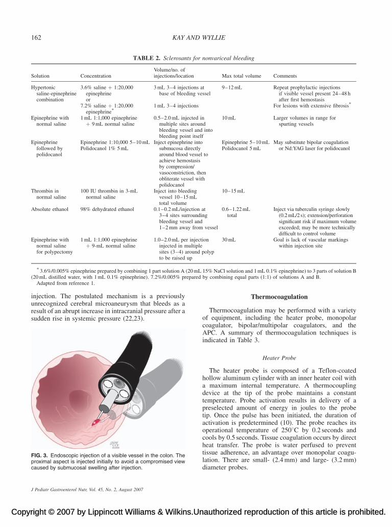
Injection therapy using epinephrine in saline 1:10,000or 1:20,000 has also been used in cases of lower gastro-intestinal bleeding. In cases of active bleeding, injectionmay be combined with thermal therapy; if a nonbleedingvisible vessel is present, thermal therapy alone may besufficient (5). Injection therapy of the colon is most easilyperformed by injection of the proximal aspect of thelesion first and distally thereafter (Fig. 3). This avoids acompromised view of the lesion caused by submucosalswelling at the injection site.
Benefits
Injection therapy has been shown in several trials to bebeneficial compared with sham therapy in patients withactive bleeding or a nonbleeding visible vessel in a pepticulcer. Several recent reports have demonstrated a benefitof combination therapy with injection and either thermaltherapy (multipolar electrocoagulation or heater probe)or mechanical therapy (hemoclip) in comparison witheither modality alone, in terms of reduction of rebleedingrate and decreased need for blood transfusions(9,10,15,16). In addition, the administration of a high-dose proton pump inhibitor after endoscopic therapy forulcers with high-risk stigmata has been shown to reducethe rate of recurrent bleeding and need for emergencysurgery (17).
Adverse Effects
The risks with injection therapy include increasedbleeding, rebleeding, bowel ischemia, and perforation(11). The injection of increased volumes of a hemostaticsclerosant solution may result in bowel wall ischemiaand subsequent perforation (18,19). The injection ofvolumes normally considered safe within the stomachmay cause perforation and/or peritonitis in the thinner-walled duodenum or colon (12). The injection of acombination of agents, such as a sclerosant with epi-nephrine, may decrease the maximum safe volumethat may be normally used with either agent alonebecause of potentiation of the ischemic effects ofthe sclerosant by the vasoconstrictive actions of theepinephrine (20).
Systemic side effects of injection therapy are a poten-tial complication of this modality. Epinephrine absorp-tion does seem to occur after submucosal injection withblood levels as much as 5 times greater than preinjectionlevels. This effect may be especially pronounced in
NVARICEAL GI BLEEDING 161
nauthorized reproduction of this article is prohibited.
patients with cirrhosis or impaired hepatic metabolism(21). Rarely, significant intracerebral hemorrhage inadult patients has been reported after epinephrine
J Pediatr Gastroenterol Nutr, Vol. 45, No. 2, August 2007
Copy
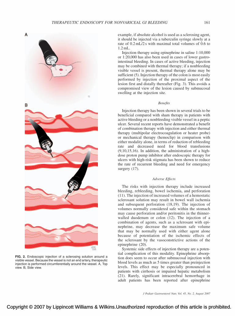
TABLE 2. Sclerosants for nonvariceal bleeding
Solution ConcentrationVolume/no. ofinjections/location Max total volume Comments
Hypertonicsaline-epinephrinecombination
3.6% saline þ 1:20,000epinephrineor
3 mL 3–4 injections atbase of bleeding vessel
9–12 mL Repeat prophylactic injectionsif visible vessel present 24–48 hafter first hemostasis
7.2% saline þ 1:20,000epinephrine
� 1 mL 3–4 injections For lesions with extensive fibrosis�
Epinephrine withnormal saline
1 mL 1:1,000 epinephrineþ 9 mL normal saline
0.5–2.0 mL injected inmultiple sites aroundbleeding vessel and intobleeding point itself
10 mL Larger volumes in range forspurting vessels
Epinephrinefollowed bypolidocanol
Epinephrine 1:10,000 5–10 mL Inject epinephrine intosubmucosa directlyaround blood vessel toachieve hemostasisby compression/vasoconstriction, thenobliterate vessel withpolidocanol
Epinephrine 5–10 mL May substitute bipolar coagulationor Nd:YAG laser for polidocanolPolidocanol 1% 5 mL Polidocanol 5 mL
Thrombin innormal saline
100 IU thrombin in 3-mLnormal saline
Inject into bleedingvessel 10–15 mLtotal volume
10–15 mL
Absolute ethanol 98% dehydrated ethanol 0.1–0.2 mL/injection at3–4 sites surroundingbleeding vessel and1–2 mm away from vessel
0.6–1.22 mLtotal
Inject via tuberculin syringe slowly(0.2 mL/2 s); extension/perforationsignificant risk if maximum volumeexceeded; may be more technicallydifficult to control volume
Epinephrine withnormal salinefor polypectomy
1 mL 1:1,000 epinephrineþ 9-mL normal saline
1.0–2.0 mL per injectioninjected in multiplesites (3–4) around polypto be raised up
30 mL Goal is lack of vascular markingswithin injection site
20 mLpared
162 KAY AND WYLLIE
injection. The postulated mechanism is a previously
�3.6%/0.005% epinephrine prepared by combining 1 part solution A (
(20 mL distilled water, with 1 mL 0.1% epinephrine). 7.2%/0.005% preAdapted from reference 1.
right © 2007 by Lippincott Williams & Wilkins.Un
unrecognized cerebral microaneurysm that bleeds as aresult of an abrupt increase in intracranial pressure after asudden rise in systemic pressure (22,23).
FIG. 3. Endoscopic injection of a visible vessel in the colon. Theproximal aspect is injected initially to avoid a compromised viewcaused by submucosal swelling after injection.
J Pediatr Gastroenterol Nutr, Vol. 45, No. 2, August 2007
Thermocoagulation
Thermocoagulation may be performed with a varietyof equipment, including the heater probe, monopolarcoagulator, bipolar/multipolar coagulators, and theAPC. A summary of thermocoagulation techniques isindicated in Table 3.
Heater Probe
The heater probe is composed of a Teflon-coatedhollow aluminum cylinder with an inner heater coil witha maximum internal temperature. A thermocouplingdevice at the tip of the probe maintains a constanttemperature. Probe activation results in delivery of apreselected amount of energy in joules to the probetip. Once the pulse has been initiated, the duration ofactivation is predetermined (10). The probe reaches itsoperational temperature of 2508C by 0.2 seconds andcools by 0.5 seconds. Tissue coagulation occurs by directheat transfer. The probe is water perfused to prevent
15% NaCl solution and 1 mL 0.1% epinephrine) to 3 parts of solution Bby combining equal parts (1:1) of solutions A and B.
authorized reproduction of this article is prohibited.
tissue adherence, an advantage over monopolar coagu-lation. There are small- (2.4 mm) and large- (3.2 mm)diameter probes.

Cop
TABLE 3. Thermocoagulation
Method Site Setting Application time No. applications Technique Notes
Heater probe Upper GI tract 30 J 3–8 s 2–4 Firm tamponade,then coagulatearound bleedingpoint, then on it
Decreased setting/time of applicationin colon or thinnergut wall
Monopolar Upper GI tract Midrange 1–2 s/pulse Directly on vessel<1-mm diameter;circumferentiallyaround vessel>1-mm diameter
Perforation likelyin colon
Bipolar/multipolar Upper GI tract 15–25 W 2 s/pulse or Multiple Firm tamponade,then coagulate
Difficult angulationlesser curve ordeformed duodenum
Up to 14-s pulse SingleColon 5 W 2 s/pulse Multiple
Argon plasmacoagulator
Upper GI tract 40–50 W 0.5–2 s Multiple Operative distance2–8 mm
Paint confluent ornear confluent areas;avoid tissue contactwith probe tip;surface should befree of liquid
0.8 L/min
Hot biopsy forceps Cecum and 10–15 W 1–2 s Tent mucosa away Use for polyps � 5 mm;
THERAPEUTIC ENDOSCOPY FOR NONVARICEAL GI BLEEDING 163
The probe is passed through the therapeutic channel ofthe endoscope. In cases of upper gastrointestinal tractbleeding the patient should be positioned so that theblood flows away from the ulcer base if possible to allowoptimal probe application. Heater probe coagulation isperformed by initially tamponading the bleeding vesselby direct firm pressure using the heater probe, and thenby coagulating the vessel (Fig. 4). This technique, whichis fundamental to the efficacy of heater probe use, isknown as coaptive coagulation. If a twin-channel instru-ment is used, the endoscopist is able to tamponade thebleeding with the probe while simultaneously suctioningin the region of the ulcer base. Coagulation is usuallyperformed in adults by 2 to 4 successive 30-J pulses (4).Coagulation should be around the bleeding point orstigmata first and then directly upon it. In several studiesin adults the greatest success seems to be with firmtamponade on the ulcer bleeding point or nonbleedingvisible vessel, and 4 pulses for a total of 120 J insuccession (4). This technique of firm tamponade andhigh-coagulation settings increases the risk of compli-cations when applied to other types of lesions, specifi-cally Mallory-Weiss tears or angiomata, that traditionallyoccur in areas with a thinner gut wall, and modification ofsettings is required (4). The heater probe may also beused in cases of colonic bleeding. The number of joulesper pulse should be reduced, especially in right-sidedcolonic lesions (5,24).
The technique of using a large probe, high setting forcoagulation, and very firm pressure seems to result in a
ascending colonLeft colon 15–20 W 2 s
Adapted from reference (1).
yright © 2007 by Lippincott Williams & Wilkins.U
lower rebleeding rate, decreased transfusion require-ment, and especially a lower emergency surgery ratein randomized control studies compared with medical
therapy alone, and in equivalent results to those achievedwith MPEC (4,24). One study reported higher permanenthemostasis rates when the heater probe was used thanwith injection therapy because of the diminished tech-nical difficulty of heater probe application to a spurtingvessel or one that requires tangential coagulation alongthe lesser curvature or superior wall of the duodenal bulb,compared with the greater technical difficulty of per-forming injection at those same sites (25). Dislodgementof the heater probe tip during therapy has been reported.In that instance the tip was retrieved by standard biopsyforceps (26). In 1% to 3% of cases perforation may occurafter heater probe application for gastrointestinal bleed-ing because of the variable depth and extent of tissueinjury after application (27). Precipitation of bleeding hasbeen reported in up to 5% of cases after heater probeapplication (10).
Electrocoagulation
There are 2 main types of electrocoagulation probes:monopolar probes and bipolar or multipolar probes(MPEC). In monopolar coagulation a continuous orintermittent current is passed via the tip or side of theprobe. The current is conducted to the patient’s groundplate. The current is converted to high-temperature heatat the tissue contact point, which coagulates the tissue,causing collagen contraction and vessel shrinkage. Forvessels <l mm in diameter, the electrode is placeddirectly on the vessel, and pressure is applied directly
contraindicatedin upper GI tract
nauthorized reproduction of this article is prohibited.
on the vessel to coapt it. With larger vessels the coagulat-ing current is placed circumferentially around the vesseluntil bleeding stops. Usually a midrange setting is used
J Pediatr Gastroenterol Nutr, Vol. 45, No. 2, August 2007

Copy
FIG. 4. Coaptive coagulation using a heater probe. A, B, Initiallythe exposed vessel is tamponaded by direct firm tangential pres-sure, using the heater probe until blanching of the vessel occurs.
164 KAY AND
for 1 to 2 seconds per pulse at a distance of 2 to 3 mmfrom the vessel. This is necessary because an artery in anulcer base may bleed from either side because it is not anend artery; therefore, a ring of tissue must be treatedaround the bleeding point to ensure adequate hemostasis.The aim is to achieve hemostasis of the underlying artery,not the overlying clot. There are 2 main problems withmonopolar coagulation. The first is that the depth of the
C, The heater probe is then used to coagulate the blood vessel.D, After coagulation, the exposed vessel appears blanched.
right © 2007 by Lippincott Williams & Wilkins.Un
burn is difficult to regulate, and perforation is thereforepossible. This is especially true in the colon. Deepnecrosis, perforation, and delayed massive bleeding have
J Pediatr Gastroenterol Nutr, Vol. 45, No. 2, August 2007
been reported with monopolar electrocoagulation. Thesecond problem is that especially in nonirrigated systems,there is a moderate amount of electrode adherence to theunderlying tissue at the treated site, and poor visibility isan additional problem. A third technical problem is theneed to clean the tip of the probe because the coagulumaccumulates during electrocoagulation.
Because of these limitations, the bipolar probe orMPEC is more commonly used. In contrast to monopolarcoagulation, a grounding plate is not required. Current istransmitted from 1 electrode on the probe to anotherelectrode. Energy is delivered when any pair of electro-des is in contact with the bleeding target. MPEC probesmay have 6 points through which current can be passed;contact between any 2 is sufficient, allowing for tangen-tial contact. The maximal temperature achieved with thismethod is significantly less than that of monopolarcoagulation or the Nd:Yag laser, resulting in less tissueinjury and also greater efficacy for vessels <2 mm indiameter (28). Two sizes of probes are available: 2.3 mmand 3.2 mm. As with the heater probe, the correct tech-nique is to compress the bleeding vessel first, then tocoagulate. Forceful application of the larger probe seemsto increase the hemostatic bond strength and the area anddepth of coagulation (28). The greatest depth of coagu-lation is usually achieved with a low- to mid-range setting(l5–25 W). Higher watt settings produce more rapidtissue desiccation. Because tissue water is necessary tocomplete the electric circuit between 2 electrodes on thebipolar probe, higher settings in effect terminate theproduction of thermal energy earlier, thereby reducinghemostatic efficacy (29). Pulses should be applied asshort, multiple pulses (2 seconds long) or a single pulse aslong as 6 to 10 seconds (28). In adults, up to 40 secondstotal of electrocoagulation may be required (28).Increased bleeding after bipolar coagulation has beenreported in cases with a visible vessel; usually thisbleeding is controllable with further bipolar coagulation,but on occasion surgery has been required.
The therapeutic endpoint for ulcer bleeding is when thebleeding stops, the visible vessel is flat, or there are flatstigmata in cases of a nonadherent clot. MPEC seems tobe equally effective to heater probe in terms of hemos-tasis, incidence of rebleeding, transfusion requirement,and need for emergency surgery. Several studies report ahemostasis rate in the range of 90% for both modalities(28,30). Difficult positioning of the MPEC probe alongthe lesser curvature or in a deformed duodenum maymake pressure application more difficult (30). Combi-nation therapy for ulcers with high-risk stigmata usinginjection and thermocoagulation, as discussed earlier,seems to be associated with higher initial hemostasis ratesand a decreased incidence of rebleeding (9,16,27,31). In
WYLLIE
authorized reproduction of this article is prohibited.
addition to sequential combination therapy, a combinationprobe (Injection Gold Probe) is available that allows forsequential injection and coagulation without the use of a

Cop
R NO
dual-channel endoscope or catheter exchange. Both 7F and10F probe sizes are available.
Angiodysplasia, involving either the stomach (gastricantral vascular ectasia, also known as watermelonstomach) of the colon, has been treated successfullyby MPEC. In cases of angiodysplasia, especially in thethin-walled colon, a maximal depth of coagulation is notdesirable. Therefore, the MPEC probe should be appliedwith decreased force, with lower watt settings, and forshorter intervals (5,10,29). Identification of colonic vas-cular ectasias may be enhanced in some cases by intra-procedural naloxone administration (32). Hemorrhagicproctocolitis with recurrent bleeding after radiationtherapy has also been successfully treated with MPEC,although the APC is becoming increasingly popular forthis indication.
Argon Plasma Coagulator
The APC is a noncontact electrocoagulation devicethat results in delivery of high-frequency monopolarcurrent through ionized gas (argon plasma) to targetedtissue (33). The system requires a high-frequency mono-polar electrosurgical generator, an argon gas source,disposable flexible probes, a grounding pad, and footcontrols that result in synchronous gas release and elec-trical current delivery (10). The probes, consisting of aTeflon tube with a tungsten monopolar electrode con-tained in a ceramic nozzle close to the distal end of theprobe, are 2.3 or 3.2 mm outer diameter and are availablein lengths of 220 or 440 cm. Probes are available to directplasma either parallel or perpendicular to the axis of thecatheter (10). Gas flow rates can be varied from 0.5 to7.0 L/min, the power settings vary from 0 to 155 W, andthe generator voltage ranges from 5000 to 6500 V. Argongas passes through the coagulation probe with an elec-trode at its tip. The electrode is activated by the footswitch, resulting in a flow of electrically activatedionized gas from the probe to the tissue (33). Ionizationof the gas and the presence of a return electrode (thegrounding pad) results in conduction of the spark to thenearest contact point. Arrival of the current at the tissueresults in coagulation. If no electrical energy is dis-charged by arcing to nearby tissue, then no ignitionoccurs, and activation of the foot switch results only ininsufflation of inert argon gas. After thermal coagulation,a thin superficial electrically insulating zone of desicca-tion occurs. After desiccation, the electrical resistance ofthe treated area increases, prompting the current to moveto another area of lower resistance (ie, an untreated area)(10). The depth of coagulation is dependent on the powersetting, the gas flow rate, the duration of application, andthe distance between the probe tip and the target tissue.
THERAPEUTIC ENDOSCOPY FO
yright © 2007 by Lippincott Williams & Wilkins.U
The noncontact nature of the technique makes it possibleto treat large areas rapidly, in comparison with the heaterprobe or MPEC (34).
Applications for the APC include hemostasis of vas-cular ectasias, including gastric antral vascular ectasia,angiectasias, and radiation-induced enteropathy/procto-pathy; treatment of bleeding ulcers; treatment of residualadenomatous tissue; and for ablative therapy (33,35–39).The primary pediatric indication is likely to be treatmentof symptomatic gastrointestinal vascular lesions. Super-ficial vascular lesions in adults are typically treated withlow power settings and gas flow rates. Multiple therapysessions are frequently required, spaced several weeksapart to allow for interval healing. In adults, settingsare in the range of 20 to 50 W with flow rates of 0.8 to1.0 L/min (34,35). In cases of vascular malformations thetreated mucosa should be rendered nonviable (whitishappearance) but not charred (black appearance). Right-sided colonic lesions may be elevated with a salinecushion before treatment to reduce the risk of perforation,and the minimum number of accurate pulses should beadministered in this location (34). Appropriate modifi-cations will be required in pediatric patients, with currentgenerators having minimum gas flow rates of 0.5 L/min.Postprocedure management for upper tract lesions mayinclude a period of fasting or dietary restriction andadministration of a proton pump inhibitor for severalweeks. Repeat sessions for vacular lesions are typicallyscheduled in 4 to 6 weeks to allow for interval healing andare then performed as needed (34). In addition to vascularlesions, the APC has been used in combination withinjection therapy for adults with peptic ulcer bleedingand high-risk stigmata. APC in combination with epi-nephrine seems to be equally efficacious as combinationtherapy consisting of heater probe and epinephrine injec-tion in terms of initial hemostasis rates (96% to 98%),risk of recurrent bleeding, and requirement for surgery ina large series of adult patients (27).
Optimal use of the APC generally requires an operativedistance of 2 to 8 mm between the probe tip and the tissue(33). The lower power settings allow for closer tissuecontact. Arcing of the current with depression of the footpedal will not occur if the correct operative distance isexceeded. The surface to be treated should optimally becleared of liquid and blood, limiting the usefulness of theAPC in cases of active bleeding. If the overlying surfaceis not clear, then a coagulated film may develop and thetissue beneath the surface may not be adequately treated(33). The correct technique is to ‘‘paint’’ the surface to betreated. This is most easily accomplished by extendingthe probe to an optimal operating distance and movingthe endoscope shaft to ‘‘paint’’ the confluent area to becoagulated. Application may be either en face or tangen-tial and is usually performed in short bursts up to2 seconds (33,35). The probe tip should not contact thetissue because this is a monopolar probe and deep tissue
NVARICEAL GI BLEEDING 165
nauthorized reproduction of this article is prohibited.
injury may occur with contact, although the safety of thetechnique is not forfeited by occasional inadvertent tissuecontact (34). Care must also be taken to continuously
J Pediatr Gastroenterol Nutr, Vol. 45, No. 2, August 2007

Copy
ND
aspirate the argon gas, which is flowing under steadypressure whenever the foot switch is activated during theprocedure, because failure to do so can result in over-distension of the stomach or bowel, especially in smallerpatients. In patients in whom the procedure is performedwithout benefit of a 2-channel endoscope, this requiresintermittent probe removal to optimize aspiration of gas.To date there has been 1 pediatric series of 13 patientsusing the APC primarily for bleeding (40). In that seriesthe APC was effective in achieving primary hemostasisafter the first session in 66% of patients, with a 25%rebleeding rate and a 17% minor complication rate.Complications have been reported in 0% to 24% ofpatients in various adult series (33–37) and includegaseous distension, pneumatosis intestinalis, pneumoper-itoneum, pneumomediastinum, subcutaneous emphy-sema, pain at the treatment site, chronic ulceration,stricture, bleeding, transmural burn, perforation, anddeath. In comparative trials the APC seems to be associ-ated with a lower complication rate than laser therapy(34). The APC is more costly than a heater probe, but thecosts are less than those associated with the Nd:YAGlaser. The APC generator can in addition be used forother applications.
Laser Photocoagulation
Laser photocoagulation is another modality occasion-ally used to achieve endoscopic hemostasis. There are 2main types of laser: argon and neodymium:yttrium-aluminum garnet (ND:Yag). The usefulness of the argonlaser is limited because of light absorption by surround-ing red blood. To use the argon laser, therefore, overlyingblood must be eliminated with a coaxial air jet. Clinically,the argon laser is used primarily for right-sided coloniclesions. In comparison with the Nd:Yag laser, ithas a lower power and depth of tissue penetration.However, mucosal abnormalities, including arteriove-nous malformations, absorb light energy well in the argonwavelength.
The ND:Yag laser is the predominant laser used ingastrointestinal endoscopy. This laser admits a continu-ous wave of infrared light of a wavelength of l064 with apower up to 100 W. This light is transmitted via a 600-mmglass fiber in a 2.5-mm Teflon catheter passed via theendoscopic channel. Carbon dioxide is passed coaxiallyalong the catheter to disperse blood away from thebleeding site and to keep the fiber tip cool and free ofdebris. A filter is attached to the eyepiece to preventreflected laser light from entering the endoscopist’s eye.The intense laser light is directed to coagulate tissuecircumferentially around the bleeding site. The recom-mendation when a noncontact laser is used in adult
166 KAY A
right © 2007 by Lippincott Williams & Wilkins.Un
patients is to deliver 0.5-second pulses, at 80 Wof energy,from a distance of l cm, and at least 2 to 3 mm away fromvisible arterial segments for upper gastrointestinal
J Pediatr Gastroenterol Nutr, Vol. 45, No. 2, August 2007
lesions. Recommendations for contact application arealso available.
Use of the laser in the colon requires modification ofboth technique and power settings. The thermal effects ofa laser beam on tissue vary according to the power density(the amount of energy converted to heat at the pointwhere the laser beam strikes tissue) and the size of thecontact area. Although the power setting and theexposure time can be preset, movement, especially inthe right colon, and varying wall thickness, especially inthe thin ascending colon, can change the time of exposurerequired to produce perforation (41). Instead of coagu-lation of tissue, vaporization of tissue can occur. Colonicperforation secondary to laser photocoagulation is aserious risk, is more frequent in the cecum and rightcolon, and occurs more frequently with the Nd:Yag laser.Laser burns may present with nausea, vomiting, and air inthe colon wall in cases of serosal burns with or withoutassociated free intraperitoneal air. Lasers have been usedfor congenital vascular lesions (hereditary hemorrhagictelangiectasia, blue rubber bleb nevi syndrome) and forsuperficial vascular lesions including angiodysplasia,telangiectasias, and arteriovenous malformation in thesmall bowel and colon (42,43). Asymptomatic, nonbleed-ing angiodysplasias are not treated. Histological diag-nosis may be difficult after laser application because oftissue destruction. As with other methods of thermocoa-gulation, laser therapy can also provoke bleeding; thisusually can be stopped with additional laser coagulation.A long learning curve is associated with use of the laser,and this modality should be used only by experiencedoperators. The laser seems to offer little advantage overthe heater probe, MPEC, and APC, and because of itsincreased cost and decreased portability, the other mod-alities are likely to predominate in the foreseeable future.
Hot Biopsy Forceps
An additional coagulation device, used primarily in thecolon, is the hot biopsy forceps. These forceps are usedprimarily for simultaneous biopsy and coagulation ofsmall sessile colonic polyps and allow for preservation ofhistological features after coagulation. In addition, theyhave been used for the treatment of vascular ectasias(44,45). The forceps require a minimum 2.8-mm endo-scopic channel. This technique combines the principlesof endoscopic biopsy and monopolar electrocoagulation.Grounding of the patient is required. The lesion toundergo biopsy is grasped in the jaw of insulated biopsyforceps, including polyps up to 5 mm. The forceps areused to tent the mucosa upward away from the colonicmuscular layer. A brief electrocoagulating current passesthrough the forceps to the mucosa and sometimes sub-
WYLLIE
authorized reproduction of this article is prohibited.
mucosa, causing coagulation at its base while preservingthe histological integrity of the specimen. The unit is seton coagulation, no cutting, at a setting of 10 to 15 W for 1
Cop
R NO
to 2 seconds in the cecum and ascending colon, or up to15 to 20 W for 2 seconds in the left colon. Smallangiomata of the colon may be coagulated by use of asimilar technique for 1 to 2 seconds (45). Higher settingsor longer application times have been associated with anincreased risk of perforation, especially in the right colon(44,46).
Perforation has also been reported after the use of hotbiopsy forceps in the upper gastrointestinal tract, both inthe stomach (secondary to increased gastric thicknesslimiting tenting of the mucosa) and in the duodenum andileum (secondary to thinness of the bowel wall andvariable depth of penetration.) (44) In the colon the riskof perforation seems to be intermediate at 0.05%, lessthan that associated with snare polypectomy but greaterthan associated with routine biopsy. Snare polypectomyis preferred for lesions larger than 5 mm because of theincreased risk of transmural injuries and complicationswith hot biopsy forceps. Significant hemorrhage has beenreported after the use of hot biopsy forceps. Failure tohold the forceps perpendicular to the mucosa increasesthe risk of this complication. Short-circuiting of thecurrent between the forceps tip and the noninsulatedportion of the forceps with resultant massive hemorrhagehas been reported if the forceps are held at an angle of 158or less to the bowel wall (46). Hemorrhage may beimmediate or delayed as long as 1 week after biopsyand may not respond to conservative therapy. Perforationmay also occur if the tip of the polyp being coagulated orthe forceps touches the opposite colonic wall, withsubsequent transmural injury. Injection, MPEC, and clip-ping, discussed below, are therapeutic options forpostpolypectomy hemorrhage.
Hemostatic Clips
Over the past several years, metallic clips have beendeveloped that can be passed through the endoscopechannel and deployed for a variety of indications, includ-ing hemostasis, attachment of catheters or tubes, mark-ing, and closure of fistulas, leaks, tears, and perforations.Originally, clips were deployed with reusable deploy-ment devices. Loading the clips with these devices wascumbersome and time-consuming, limiting their appli-cation. Subsequently, preloaded single-use clips weredeveloped. Because of their ease of use, this techniquehas found increased application, and both rotatable clipsand clips with reopening and therefore repositioningcapabilities are commercially available. In addition, pre-loaded multiclip devices are under development. Clipspecifications vary according to the manufacturer. Themajority of clips are stainless steel, with a deliverydeployment catheter consisting of a metal cable within
THERAPEUTIC ENDOSCOPY FO
yright © 2007 by Lippincott Williams & Wilkins.U
a metal coil sheath within a 2.2-mm Teflon catheter (47).Therefore, the majority of the clips require a 2.8-mmendoscope channel for deployment. Most clips are
2-pronged; a 3-pronged clip is available that requires a3.2-mm endoscopic channel (TriClip, Cook EndoscopyInc, Winston Salem, NC). The 2-pronged clips areapproximately 1.2-mm wide, available in severallengths with opening angles in the range of 908 to1358 and open from 6–12 mm depending on the specificclip used (47,48). A 2-pronged clip with reopening andrepositioning capability up to 5 times before deploymentis available (Resolution Clip, Boston Scientific). Thedifferent clips seem to be equivalent in animal models inease of application and initial hemostasis rates but differin duration of retention (49).
Clipping for hemostasis requires identification of theexact bleeding point (48). In contrast to injection orthermal techniques, the preferred technique is to clipthe bleeding point first and then to apply additional clipsaround the bleeding point if necessary. Because thisis a mechanical technique, secure clip deployment isachieved with maximal capture of tissue around thebleeding vessel (ie, not just the arterial base but alsosome of the surrounding mucosa) (48). In addition,because there is no surrounding field effect, unlikeinjection therapy, precise positioning is required (50).Optimal clip positioning is best achieved with the clipextended a relatively short distance from the endoscopetip. This allows for more precise clip application andallows for exertion of downward force on the clip duringits placement. The correct technique is to position the clipslightly away from the arterial base, allowing for an enface or tangential approach, and to push the open clipdownward while simultaneously applying suction. Theclip should be slowly closed and, if optimally positioned,deployed (48). Reopening clips can be repositionedbefore deployment if required.
The limitations of clip application relate to the locationof the lesion and to size criteria. The proximal lessercurvature and gastric cardia may be difficult to approachfor clipping directly or in the retroflexed position, and insome cases it is easier to carefully expose the clip beforeretroflexion. Duodenal ulcers may also be difficult toclip, depending on their location, especially those invol-ving the posterior wall of the duodenal bulb. Fibroticulcers or lesions may be difficult to clip. An arterialvessel larger than 2 mm in diameter may not be amenableto clipping, and there are other published criteria for sizelimitations in cases of perforations, mucosal defects, andpolyps (2,31,48). In most cases the clips dislodge spon-taneously within 2 to 4 weeks and pass in the stool,although some have been in place for >1 year (47).Although no adverse effects have been reported, mag-netic resonance imaging may be contraindicated if clipsare present.
Clipping for acute nonvariceal hemostasis is associ-
NVARICEAL GI BLEEDING 167
nauthorized reproduction of this article is prohibited.
ated with primary hemostasis rates in the range of 84%to 100%, with low rebleeding rates, comparable withthose achieved with injection, thermal, and combination
J Pediatr Gastroenterol Nutr, Vol. 45, No. 2, August 2007

Copy
ND
therapies (31,47,50,51). As with thermal therapy, hemo-static clipping has been used as part of combinationtherapy in conjunction with epinephrine injection. Forpatients with a nonbleeding visible vessel, clipping istypically performed first, followed by epinephrine injec-tion as previously described. In cases of active bleeding,some endoscopists will inject epinephrine first, followedby clip application (2). Although initial hemostatic ratesare comparable, prospective series demonstrate signifi-cantly decreased rebleeding rates and surgical require-ments in patients undergoing combination therapy (clipand injection) in comparison with injection alone (2).
Clipping and other mechanical techniques have beenshown to be more efficacious and are associated with alower rebleeding rate than nonmechanical therapies forpatients with Dieulafoy lesions, especially those withhigh-risk stigmata (52,53). These lesions, most fre-quently located in the proximal part of the stomach,may in some cases be more difficult to approach withan endoscopic clip because of flattening of the anglebetween the end of the clip and the lesion. However, theability to clip both the lesion and the surrounding normalmucosa is a benefit of this technique (53). Other hemo-static applications for hemoclips include Mallory-Weisstears and for colonic bleeding after biopsy, after poly-pectomy, from hemorrhoids, or from solitary rectal ulcersyndrome (Fig. 5). There is a single case report of
168 KAY A
right © 2007 by Lippincott Williams & Wilkins.Un
clipping as adjuvant therapy for temporary cessation ofrectal bleeding due to Klippel Trenaunay syndrome (54).Complications after clipping are extremely rare but
FIG. 5. Hemostatic clip applied in the rectum to a bleeding polypstalk. The stalk was initially injected with 1:10,000 epinephrine,and subsequently the clip was applied.
J Pediatr Gastroenterol Nutr, Vol. 45, No. 2, August 2007
include a case wherein a clip inadvertently perforateda gastric ulcer and was applied to the splenic artery, and acase of colonic perforation thought to be due to clipplacement for postpolypectomy bleeding (47,55).
Mechanical Ligation (Loops and Bands)
Detachable nylon loops (Endoloop, Olympus America)are now available and can provide mechanical ligationwithout cauterization. The sheath of the loop is Tefloncoated and has an outer diameter of 2.6 mm, requiring a2.8-mm channel for deployment and a working lengthof 230 cm. Both reloadable and single-use preloadeddevices are available. The loop is passed through theendoscope accessory channel and placed over the tar-geted lesion in a manner similar to the technique ofpolypectomy snare placement. The maximal loop open-ing width is 30 mm. The loop is tightened with advance-ment of a silicon-rubber stopper (10). The loop isthen detached after hemostasis is achieved withouttransecting the lesion. The primary indication for loopplacement is for the prevention or management ofpostpolypectomy bleeding. When the loop is appliedbefore polypectomy snare placement, care must betaken to avoid entanglement of the loop in the poly-pectomy snare. Before polypectomy, detachable loopplacement should result in change of the color of thepolyp head without transsection. If the loop is appliedtoo tightly, then amputation of the polyp may occur withresultant bleeding; if it is too loose, then bleeding mayoccur after polypectomy. Placement of a detachableloop for postpolypectomy bleeding may be difficultbecause of stalk retraction, but techniques such asthe lift and ligate technique using a 2-channel scopeare available for this condition. Hemostatic loop place-ment has also been effective in the management ofbleeding Dieulafoy lesions and has been used for bleed-ing gastric varices (10,53).
Band ligation for hemostasis is an extension of theligation technique used to treat esophageal varices.Ligation is typically performed with a multiband ligator,available from several manufacturers. After identifi-cation of the lesion to be treated, the endoscope iswithdrawn, the friction fit adaptor of the band ligatoris placed on the tip of a standard-size adult upper endo-scope, and a drawstring that extends from the cylinder isbackloaded through the endoscope working channel andconnected to the spool of the cranking handle at the inletport (51). The endoscope is repassed to the lesion to betreated, and a band is deployed and applied by suctioningthe lesion into the friction fit adaptor and simultaneouslyturning the handle; suction is subsequently released,and the efficacy of hemostasis can be determined. In
WYLLIE
authorized reproduction of this article is prohibited.
cases of nonvariceal bleeding, deployment of only asingle band may be sufficient. When multiband ligatorsare used, it may be helpful to predeploy some of the bands

Cop
R NO
outside the patient to improve the view, especially if only1 ligating band may be required.
Band ligation is most effective for bleeding fromnonfibrotic lesions and has been used to treatvarices—esophageal, gastric, and colonic, Dieulafoylesions, bleeding internal hemorrhoids, Mallory-Weisstears, angiectasia, duodenal ulcers, and polypectomysites (51,56). Ligation seems to be an extremely effica-cious method of treating Dieulafoy lesions, especiallythose located where hemostatic clip application is diffi-cult, such as the proximal gastric body (53). Ulcerationafter ligation has been reported, similar to that whichoccurs after variceal ligation. This has not been associ-ated with recurrent bleeding in the case of Dieulafoylesions (53).
Studies on resected specimens suggest that ligator usein anatomically thin segments of the bowel (ie, the smallintestine and right colon) may be associated with anincreased risk of perforation caused by entrapment ofall layers of the bowel wall (57,58). Caution should beexercised with endoscopic band ligation with avoidanceof excessive tissue aspiration by careful application ofsuction and use of banding caps that are 7 mm deep orless (58).
New and Emerging Technologies
Two endoscopic techniques have recently been devel-oped that may one day be used in pediatric patients as partof the management of acute gastrointestinal bleeding.The technique most likely to initially be used is doubleballoon enteroscopy (DBE). This technique allows forextensive antegrade or retrograde evaluation of the smallbowel by passage of a specialized commercially availableendoscope. The primary indication for this type of exam-ination is for evaluation of unexplained acute, recurrent,or obscure gastrointestinal bleeding in a patient whohas previously undergone both esophagogastroduodeno-scopy and colonoscopy. Additional indications for DBEinclude evaluation and therapy of polyposis syndromes,Crohn disease, and abdominal pain. The endoscopes havea working length of 200 cm, an outer diameter in therange of 8 to 8.5 mm, a therapeutic channel of 1.8 to2.8 mm, and a balloon at the tip; the endoscope is passedin conjunction with a flexible overtube with a balloon atits tip. The overtubes are 140 to 145 cm long and have anouter diameter of 12.0 to 13.2 mm (59–61) (FujinonCorp, Saitama, Japan). Available accessories that canbe passed through the endoscope channel include biopsyforceps, an electrocautery snare, injection needles, and athin APC catheter (ERBE, Tubingen, Germany) andhemoclips in the scopes with a larger channel (59,61).
DBE is performed after small bowel preparation and
THERAPEUTIC ENDOSCOPY FO
yright © 2007 by Lippincott Williams & Wilkins.U
consists of a series of insertion and withdrawal maneu-vers, accompanied by serial inflation and deflation of theendoscope and overtube balloons to reduce small bowel
loops, resulting in relative straightening of the smallbowel and scope advancement (60). Fluoroscopy is per-formed at the time of the procedure to evaluate scopeposition and extent of evaluation. Examination times aretypically prolonged, in the range of 60 to 360 minutes,although recent large series may have shorter proceduraltimes (60,61). Lesions identified during the procedurecan be biopsied or can be treated with availableaccessories, allowing for cauterization, resection, andhistological evaluation. DBE has been particularly usefulfor the identification of small bowel lesions associatedwith bleeding, such as gastrointestinal stromal tumors,polyps, and various types of angiodysplasia (60,61).Endoscopic biopsy may yield the diagnosis in somecases, and in others acute bleeding may resolve afterinjection or coagulation performed via the DBE channel.Bleeding as a result of diagnostic or therapeutic maneu-vers during the procedure can be treated as describedearlier for other segments of the gastrointestinal tract,using small-diameter accessories with appropriate modi-fications in settings or volumes of injection based on theanticipated small bowel wall thickness.
DBE examination in the pediatric patient is limited bythe large outer diameter of the overtube and the pro-longed procedure time and has been reported only as partof a large adult series to date (61). It is anticipated thatwith technological advancements and increasing appli-cation of the procedure, it may be performed in olderpediatric and adolescent patients.
The newest development in the field of therapeuticendoscopy is natural orifice transluminal endoscopicsurgery. This technique uses commercial endoscopes tocreate a controlled transvisceral incision, usually in thestomach, to enter the peritoneal cavity as an alternativeto conventional surgery (62,63). The technique hasprimarily been studied in animal models and is in thedevelopmental phase at present, with gastroenterolo-gists and surgeons interested in identifying applicationsof this approach in adults. As experience with thisapproach in a variety of gastrointestinal conditionsgrows, natural orifice transluminal endoscopic surgerymay in the future be performed for selected pediatricpatients.
CONCLUSIONS
Several techniques are available to the pediatricendoscopist in the management of acute gastrointestinalbleeding. Many excellent reviews of the comparativetechniques are available to the endoscopist, and tech-nology status evaluation reports are published andupdated by the American Society for GastrointestinalEndoscopy and other gastrointestinal societies that are
NVARICEAL GI BLEEDING 169
nauthorized reproduction of this article is prohibited.
invaluable resources for the treatment of patients withacute gastrointestinal bleeding (2,10,15,16,64). Thechoice of therapeutic endoscopic technique depends to
J Pediatr Gastroenterol Nutr, Vol. 45, No. 2, August 2007

Copy
ND
a significant extent on the origin of the bleeding, thepatient’s size, the availability of equipment, and thetraining of the endoscopist. If the endoscopy is properlyperformed, then the rates of primary hemostasis andrebleeding are similar between injection, thermocoagu-lation, and mechanical therapy. Improved initial hemos-tasis rates and a further reduction in recurrent bleedingrates and need for emergency surgery occur with com-bination therapy. Certain lesions may be more amenableto 1 endoscopic technique over another on the basis oftheir anatomic location or briskness of bleeding.
REFERENCES
1. Kay M, Wyllie R. Gastrointestinal hemorrhage. In: Wyllie R,Hyams JS (eds). Pediatric Gastrointestinal and Liver Disease.Philadelphia: Saunders Elsevier; 2006. pp. 203–15.
2. Lo CC, Hsu PI, Lo GH, et al. Comparison of hemostatic efficacy forepinephrine injection alone and injection combined with hemocliptherapy in treating high-risk bleeding ulcers. Gastrointest Endosc2006;63:767–73.
3. Kovacs TO, Jensen DM. Endoscopic control of gastroduodenalhemorrhage. Annu Rev Med 1987;38:267–77.
4. Jensen DM. Heat probe for hemostasis of bleeding peptic ulcers:technique and results of randomized controlled trials. GastrointestEndosc 1990;36 (Suppl):S42–9.
5. Elta GH. Urgent colonoscopy for acute lower-GI bleeding.Gastrointest Endosc 2004;59:402–8.
6. Monahan DW, Peluso FE, Goldner F. Combustible colonic gaslevels during flexible sigmoidoscopy and colonoscopy. GastrointestEndosc 1992;38:40–3.
7. Cook DJ, Guyatt GH, Salena BJ, et al. Endoscopic therapy for acutenonvariceal upper gastrointestinal hemorrhage: a meta-analysis.Gastroenterology 1992;102:139–48.
8. Whittle TJ, Sugawa C, Lucas CE, et al. Effect of hemostatic agentsin canine gastric serosal blood vessels. Gastrointest Endosc 1991;37(3):305–9.
9. Lin HJ, Tseng GY, Perng CL, et al. Comparison of adrenalineinjection and bipolar electrocoagulation for the arrest of peptic ulcerbleeding. Gut 1999;44:715–9.
10. Nelson DB, Barkun AN, Block KP, et al. Technology statusevaluation report: endoscopic hemostatic devices. GastrointestEndosc 2001;54:833–40.
11. Hirao M, Kobayashi T, Masuda K, et al. Endoscopic local injectionof hypertonic saline-epinephrine solution to arrest hemorrhage fromthe upper gastrointestinal tract. Gastrointest Endosc 1985;31:313–7.
12. Rutgeerts P, Geboes K, Vantrappen G. Experimental studies ofinjection therapy for severe nonvariceal bleeding in dogs. Gastro-enterology 1989;97:610–21.
13. Lin HJ, Perng CL, Lee FY, et al. Endoscopic injection for thearrest of peptic ulcer hemorrhage: final results of a prospec-tive, randomized comparative trial. Gastrointest Endosc 1993;39:15–9.
14. Giorcelli W, Rodi M. Injection therapy for bleeding gastric leio-myoma. Gastrointest Endosc 1992;38:730–1.
15. Barkun A, Bardou M, Marshall JK. Consensus recommendationsfor managing patients with nonvariceal upper gastrointestinalbleeding. Ann Intern Med 2003;139:843–57.
16. Calvet X, Vergara M, Brullet E, et al. Addition of a secondendoscopic treatment following epinephrine injection improves
170 KAY A
right © 2007 by Lippincott Williams & Wilkins.Un
outcome in high-risk bleeding ulcers. Gastroenterology 2004;126:441–50.
17. Sung J. Best endoscopic hemostasis for ulcer bleeding: is there sucha treatment? Gastrointest Endosc 2006;63:774–5.
J Pediatr Gastroenterol Nutr, Vol. 45, No. 2, August 2007
18. Bedford RA, van Stolk R, Sivak MV Jr, et al. Gastric perforationafter endoscopic treatment of a Dieulafoy’s lesion. Am J Gastro-enterol 1992;87:244–7.
19. Chester JF, Hurley PR. Gastric necrosis: a complication ofendoscopic sclerosis for bleeding peptic ulcer. Endoscopy1990;22:287.
20. Loperfido S, Patelli G, La Torre L. Extensive necrosis of gastricmucosa following injection therapy of bleeding peptic ulcer.Endoscopy 1990;22:285–6.
21. Sung JY, Chung SC, Low JM, et al. Systemic absorption ofepinephrine after endoscopic submucosal injection in patients withbleeding peptic ulcers. Gastrointest Endosc 1993;39:20–2.
22. Melzer E, Keter D. Intracerebral hemorrhage after therapeuticupper-GI endoscopy. Gastrointest Endosc 2006;64:468.
23. Efthymiou A, Markoglou C. Intracerebral hemorrhage aftertherapeutic upper-GI endoscopy. Gastrointest Endosc 2006;64:468.
24. Jensen DM, Machicado GA. Diagnosis and treatment of severehematochezia: the role of urgent colonoscopy after purge. Gastro-enterology 1988;95:1569–74.
25. Lin HJ, Tsai YT, Lee SD, et al. A prospectively randomized trialof heat probe thermocoagulation versus pure alcohol injectionin nonvariceal peptic ulcer hemorrhage. Am J Gastroenterol1988;83:283–6.
26. Meichner RH, Galambos J. Heater probe dislodgement during use.Am J Gastroenterol 1993;88:151–2.
27. Chau CH, Siu WT, Law BK, et al. Randomized controlled trialcomparing epinephrine injection plus heat probe coagulation versusepinephrine injection plus argon plasma coagulation for bleedingpeptic ulcers. Gastrointest Endosc 2003;57:455–61.
28. Laine L. Therapeutic endoscopy and bleeding ulcers: bipolar/multipolarelectrocoagulation. GastrointestEndosc 1990;36 (Suppl):S38–41.
29. Laine L. Determination of the optimal technique for bipolar elec-trocoagulation treatment: an experimental evaluation of the BICAPand Gold probes. Gastroenterology 1991;100:107–12.
30. Hui WM, Ng MM, Lok AS, et al. A randomized comparative studyof laser photocoagulation, heater probe, and bipolar electrocoagu-lation in the treatment of actively bleeding ulcers. GastrointestEndosc 1991;37:299–304.
31. Lin HJ, Perng CL, Sun IC, et al. Endoscopic haemoclip versusheater probe thermocoagulation plus hypertonic saline-epinephrineinjection for peptic ulcer bleeding. Dig Liver Dis 2003;35:898–902.
32. Brandt LJ, Spinnell MK. Ability of naloxone to enhance thecolonoscopic appearance of normal colon vasculature and colonvascular ectasias. Gastrointest Endosc 1999;49:79–83.
33. Ginsberg GG, Barkun AN, Bosco JJ, et al. The argon plasmacoagulator: February 2002. Gastrointest Endosc 2002;55:807–10.
34. Kwan V, Bourke MJ, Williams SJ, et al. Argon plasma coagulationin the management of symptomatic gastrointestinal vascularlesions: experience in 100 consecutive patients with long-termfollow-up. Am J Gastroenterol 2006;101:58–63.
35. Vargo JJ. Clinical applications of the argon plasma coagulator.Gastrointest Endosc 2004;59:81–8.
36. Cipolletta L, Bianco MA, Rotondano G, et al. Prospective compar-ison of argon plasma coagulator and heater probe in the endoscopictreatment of major peptic ulcer bleeding. Gastrointest Endosc1998;48:191–5.
37. Canard JM, Vedrenne B. Clinical application of argon plasmacoagulation in gastrointestinal endoscopy: has the time come toreplace the laser? Endoscopy 2001;33:353–7.
38. Venkatesh KS, Ramanujam P. Endoscopic therapy for radiationproctitis-induced hemorrhage in patients with prostatic carcinomausing argon plasma coagulator application. Surg Endosc 2002;16:
WYLLIE
authorized reproduction of this article is prohibited.
707–10.39. Smith S, Wallner K, Dominitz JA, et al. Argon plasma coagulation
for rectal bleeding after prostate brachytherapy. Int J Radiat OncolBiol Phys 2001;51:636–42.
Cop
R NO
40. Khan K, Schwarzenberg SJ, Sharp H, et al. Argon plasma coagula-tion: clinical experience in pediatric patients. Gastrointest Endosc2003;57:110–2.
41. Buchi KN. Endoscopic laser surgery in the colon and rectum. DisColon Rectum 1988;31:739–45.
42. Shahed M, Hagenmuller F, Rosch T, et al. A 19-year-old femalewith blue rubber bleb nevus syndrome: endoscopic laser photo-coagulation and surgical resection of gastrointestinal angiomata.Endoscopy 1990;22:54–6.
43. Lanthier P, d’Harveng B, Vanheuverzwyn R, et al. Colonic angio-dysplasia: follow-up of patients after endoscopic treatment forbleeding lesions. Dis Colon Rectum 1989;32:296–8.
44. Wadas DD, Sanowski RA. Complications of the hot biopsy forcepstechnique. Gastrointest Endosc 1988;34:32–7.
45. Gilbert DA, DiMarino AJ, Jensen DM, et al. Status evaluation: hotbiopsy forceps. American Society for Gastrointestinal Endoscopy.Technology Assessment Committee. Gastrointest Endosc 1992;38:753–6.
46. Quigley EM, Donovan JP, Linder J, et al. Delayed, massivehemorrhage following electrocoagulating biopsy (‘‘hot biopsy’’)of a diminutive colonic polyp. Gastrointest Endosc 1989;35:559–63.
47. Chuttani R, Barkun A, Carpenter S, et al. Endoscopic clip applica-tion devices. Gastrointest Endosc 2006;63:746–50.
48. Kaltenbach T, Friedland S, Barro J, et al. Clipping for uppergastrointestinal bleeding. Am J Gastroenterol 2006;101:915–8.
49. Jensen DM, Machicado GA, Hirabayashi K. Randomized con-trolled study of 3 different types of hemoclips for hemostasis ofbleeding canine acute gastric ulcers. Gastrointest Endosc 2006;64:768–73.
50. Saltzman JR, Strate LL, Di S, et al. Prospective trial of endoscopicclips versus combination therapy in upper GI bleeding(PROTECCT–UGI bleeding). Am J Gastroenterol 2005;100:
THERAPEUTIC ENDOSCOPY FO
yright © 2007 by Lippincott Williams & Wilkins.U
51. Binmoeller KF, Soehendra N. New haemostatic techniques: his-toacryl injection, banding/endoloop ligation and haemoclipping.Baillieres Best Pract Res Clin Gastroenterol 1999;13:85–96.
52. Yamaguchi Y, Yamato T, Katsumi N, et al. Short-term and long-term benefits of endoscopic hemoclip application for Dieulafoy’slesion in the upper GI tract. Gastrointest Endosc 2003;57:653–6.
53. Chung IK, Kim EJ, Lee MS, et al. Bleeding Dieulafoy’s lesions andthe choice of endoscopic method: comparing the hemostatic effi-cacy of mechanical and injection methods. Gastrointest Endosc2000;52:721–4.
54. Natterer J, Joseph JM, Denys A, et al. Life-threatening rectalbleeding with Klippel-Trenaunay syndrome controlled by angio-graphic embolization and rectal clips. J Pediatr Gastroenterol Nutr2006;42:581–4.
55. Tominaga K, Saigusa Y, Fujinuma S, et al. Colonic perforation afterendoclip placement for delayed post-endoscopic resection bleeding.Gastrointest Endosc 2006;64:839–41.
56. Berkelhammer C, Moosvi SB. Retroflexed endoscopic bandligation of bleeding internal hemorrhoids. Gastrointest Endosc2002;55:532–7.
57. Barker KB, Arnold HL, Fillman EP, et al. Safety of band ligator use inthe small bowel and the colon. Gastrointest Endosc 2005;62:224–7.
58. Goff JS. Endoscopic variceal ligation safety throughout the GI tract.Gastrointestinal Endoscopy 2005;62:228–9.
59. Monkemuller K, Weigt J, Treiber G, et al. Diagnostic and ther-apeutic impact of double-balloon enteroscopy. Endoscopy 2006;38:67–72.
60. Su MY, Liu NJ, Hsu CM, et al. Double balloon enteroscopy: the lastblind-point of the gastrointestinal tract. Dig Dis Sci 2005;50:1041–5.
61. Sun B, Rajan E, Cheng S, et al. Diagnostic yield and therapeuticimpact of double-balloon enteroscopy in a large cohort ofpatients with obscure gastrointestinal bleeding. Am J Gastroenterol2006;101:2011–5.
62. McGee MF, Rosen MJ, Marks J, et al. A primer on natural orificetransluminal endoscopic surgery: building a new paradigm. SurgInnov 2006;13:86–93.
NVARICEAL GI BLEEDING 171
63. Baron TH. Natural orifice transluminal endoscopic surgery. Br J
1503–8.nauthorized reproduction of this article is prohibited.
Surg 2007;94:1–2.64. Martins NB, Wassef W. Upper gastrointestinal bleeding. Curr Opin
Gastroenterol 2006;22:612–9.
J Pediatr Gastroenterol Nutr, Vol. 45, No. 2, August 2007
Related Documents