pm . mi. 51 f ~mM SU~1F~~s S iStpIM PREPORT DOCUMENTATION PAGE PO ________ I I ! b . • ham -~.'t May 1979 I -Final/ 1 Junp 1973- ft omn 17R I. IT AmD aMIUTLE IV FN-_ , -i w CARDIOVASCULAR ADAPTATION TO STRESS ~61102F (V) _________W_60_______of____________.___= 2312/A2 Vernon S. Bishop Lfl ..... .......... IftOll N:0M 1 11 1: MMM Ii 1. " UE Gy W &* r-ap" 31re . POAM M OA NAI University of Texas Health Sciences Center 4 Department of Pharmacology San Antonio TX 78284 ON*, * I S TRS ( I. S~lmnin /ONJT G GENCY NME S £50 ADOR A61OSR BAFB DC 20332-6448 AFOSR-73-2525 00 a L AUT10 IAITYSATMN V 1 ron. Biop5TIT1ON C ditibto niit The overall objective of this project was to determine the neural factors which are involved in the regulation of the cardiovascular system during various stresses. To achieve these objectives, we have investigated factors which influence the control of heart rate under normal laboratory conditions as well as during stresses such as myocardial ischemia, acute volume expansion or depletiov and during exposure to positive acceleration. Our second objective was to establi the contribution of neural reflexes originating from receptors located in the cardiopulmonary and sinoaortic regions on the beat-to-beat control of the heart and the peripheral circulation under various laboratory stresses and during the stress of positive acceleration. The detailed report is broken down into sections. Each section is a separate phase of the overall study.. - 14. SUBICT TRIU M"S lS. NUMU)AR OF PAGES 12 Ilk PRICE COOl 1. SECLAU Y MOTUIC AoI I1. 0E CraPIIT O2& REPAOM I OPLAIL ST S A GE OP ABSTRA C i 1U4 VA~m 20Wr

Welcome message from author
This document is posted to help you gain knowledge. Please leave a comment to let me know what you think about it! Share it to your friends and learn new things together.
Transcript
pm . mi. 51 f ~mM SU~1F~~s S iStpIM PREPORT DOCUMENTATION PAGE PO ________
I I ! b . • ham -~.'t
May 1979 I -Final/ 1 Junp 1973- ft omn 17R
I. IT AmD aMIUTLE IV FN-_ , -i w
CARDIOVASCULAR ADAPTATION TO STRESS
~61102F(V) _________W_60_______of____________.___= 2312/A2
Vernon S. Bishop
Lfl
..... .......... IftOll N:0M 1 11 1: MMM Ii
1. " UE Gy W &* r-ap" 31re . POAM M OA NAI
University of Texas Health Sciences Center
4 Department of PharmacologySan Antonio TX 78284 ON*, * I S TRS
( I. S~lmnin /ONJT G GENCY NME S £50 ADORA61OSR
BAFB DC 20332-6448 AFOSR-73-2525
00 a L AUT10 IAITYSATMN
V 1ron. Biop5TIT1ON C
ditibto niit
The overall objective of this project was to determine the neural factors which
are involved in the regulation of the cardiovascular system during variousstresses. To achieve these objectives, we have investigated factors whichinfluence the control of heart rate under normal laboratory conditions as well
as during stresses such as myocardial ischemia, acute volume expansion or depletiovand during exposure to positive acceleration. Our second objective was to establi
the contribution of neural reflexes originating from receptors located in thecardiopulmonary and sinoaortic regions on the beat-to-beat control of the heart andthe peripheral circulation under various laboratory stresses and during thestress of positive acceleration. The detailed report is broken down into sections.Each section is a separate phase of the overall study.. -
14. SUBICT TRIU M"S lS. NUMU)AR OF PAGES
12
Ilk PRICE COOl
1. SECLAU Y MOTUIC AoI I1. 0E CraPIIT
O2& REPAOM I OPLAIL ST S A GE OP ABSTRA
C i 1U4 VA~m 20Wr

DISCLAIMEI NOTICE
THIS DOCUMENT IS BEST
QUALITY AVAILABLE. THE COPY
FURNISHED TO DTIC CONTAINED
A SIGNIFICANT NUMBER OF
PAGES WHICH DO NOT
REPRODUCE LEGIBLY.
The University of TexasHealth Science Center at San Antonio7703 Floyd (url Drive
Sani Antonio, Texas 78284
Dcpartment of Pharmacology (512) 6I.l •
May 1, I AS7O MR A 1 b 1 18
Unannounced []Justification
Major Vincent J. Hrenak, Contracting Officer ByD1 st rlibu* I n/
Department of the Air Force . .... .Air Force Office of Scientific Research (AFSC) ,A a01Boiling Air Force Base, D.C. 20332 - '
SUBJECT: Final Report Grant AFOSR-73-2525 I
PERIOD: June 1, 1973 - May 31, 1978
OBJECTIVE: The overall objective of this project was to determine the neural
factors which are involved in the regulation of the cardiovascular
system during various stresses. To achieve these objectives, we
have investigated factors which influence the control of heart rate
under normal laboratory conditions as well as during stresses such
as myocardial ischemia, acute volume expansion or depletion and
during exposure to positive acceleration. Our second objective
was to establish the contribution of neural reflexes originating
from receptors located in the cardiopulmonary and sinoaortic regions
on the beat-to-beat control of the heart and the peripheral circu-
lation under various laboratory stresses and during the stress of
positive acceleration.
This detailed report is broken down into sections. Each section is
a separate phase of the overall study. They are as follows:
i. FACTORS INFLUENCING THE INTRINSIC CONTROL OF HEART RATE.
A. Interaction of Acetylcholine and Norepinephrine on Heart Rate
Responses of isolated rat atria to acetylcholine (Ach) indicate that
onset of bradycardia occurs when concentration in the bathing solution
-8reaches 10 molar. The degree of slowing is directly proportional to
the concentration of Ach. All hearts stopped or nearly stopped when Ach
concentration reached 10-4 M. Norepinephrine produced tachycardia. On-
set occurred at 1O-8 M and peak response occurred at 10-5 M. The dose-
response curves established the control values aqainst which other com-
parisons could be made.
When norepinephrine was added to the bathing solution prior to estab-
lishing a dose-response curve for acetylcholine, it had little influence
on the response of the atrium to acetyieholine at concentrations of 10-9
or 10-7 M. At concentrations of 10-5 M of norepinephrine, the threshold
of responsiveness to Ach was higher as was the ED50 , although the peak
response was similar to the control value. This indicates that only at
high doses of norepinephrine are low doses of Ach interfered with. High
doses of Ach were still not influenced by prior tissue exposure to high
doses of epinephrine.
When acetylcholine was added to the bathing solution prior to estab-
lishing a dose-response curve for norepinephrine, a definite influence was
observed when the -) entration reached 10-7 M. The same was true for
10- 5 M Ach. The peak responses were also reduced at these concentrations.
Thus, acetylcholine definitely inhibits both tne onset and magnitude of
responses to norepinephrine.
B. Experimental Heart Failure Effects on Atrial Response to Norepinephrine
Chronotropic and inotropic responses to norepinephrine (NE) (10- 3 -
10-5 M) were obtained for isolated rat atria. Eleven to seventeen days
postoperatively, tissue was taken from 14 sham-operated controls and 16
rats with experimental heart failure produced by aortic constriction.
Presacrificial heart rates were higher in constricted animals (F .01) but
not different from shams when isolated in the tissue bath. Chronotropic
change was observed in the heart failure group at 10-13 M NE, while sig--8
nificant change in the control group was seen at 10 M NE. The maximum
heart rate changes for NE occurred with 10-5 M for both groups. Positive
inotropic changes were observed in the heart failure group at 10- 13 M NE,
while the earliest change in the control group was at 10-9 M NE. The
maximum inotropic response for both groups occurred at 10-6 M NE. ED50
for the two groups was not significantly different. These findings show
that rat atria are more responsive to low levels of NE after heart fail-
ure than are controls. This is consistent with observations that note a
defect in uptake and storage of NE in this type of experimental conges-
tive heart Failure. If such an abnormality makes increased quantities
of NE available at the receptor site at low circulating NE levels, indi-
viduals in congestive heart failure might be hyper-responsive to normal
blood NE levels.
2. BEAT-TO-BEAT REGULATION OF HEART RATE BY AFFERENT STIMULATION OF THE AORTIC
NERVE. (See Appendix Reprint #1)
Repeated electrical stimulation of the aortic nerve, when confined
to one cardiac cycle, caused heart rate reduction in the anesthetized
rabbit. The average fall in heart rate due to supramaximal stimulation
was 8.4 + 0.3 beats/min (+ SEM). Extent of bradycardia was more closely
related to total number of impulses within the stimulus burst than to
either burst duration or Impulse frequency. The latency to onset of
the response could not be altered by changes in any of the stimulus para-
meters, nor could it be related to the position of the stimulus burst
within the cardiac cycle. These results indicate that beat-to-beat
regulation of heart rate can be accomplished when afferent aortic
activity is altered.
3. LEFT VENTRICULAR FUNCTION DURING ACUTE REGIONAL MYOCARDIAL ISCHEMIA. (See
Appendix Reprint #2)
The effects of acute ]-min. occlusion of the left circumflex coronary
artery on the inotropic state and performance of the left ventricle were
examined in adult mongrel dogs. The inotropic state, as indicated by
changes in the maximum derivative of left ventricular pressure in the
pre-ejection phase and the maximum derivative of the transverse internal
diameter, were diminished during the ischemic period. The end-systolic
diameter increased 3.8 + 0.6 mm while the end-diastolic diameter increased
only 0.4 + 0.2 mm, although the end-diastolic pressure increased 6.9 +
0.6 mmHg. Progressive decreases in the stroke volume paralleled the ap-
parent reduction in myocardial fiber shortening in the transverse plane.
Cardiac output and arterial pressure declined, concurrently, thus maintain-
ing a constant peripheral increase during the occlusion. Acute coronary
occlusion also caused an apparent increase in the myocardial wall stiff-
ness, as judged by the increase in the slope a of the equation dP/dV = aP
+ B, the increase of the slope of the pressure-diameter relationship, and
the decrease in the rate of lengthening of the diameter during diastole.
These findings suggest that acute myocardial ischemia results in an imme-
diate reduction in the effective inotropic state and an apparent increase
in the myocardial wall stiffness in the transverse plane. Both of these
changes resulted in a decrease in performance of the left ventricle.
4. SPECIFICITY OF AUTONOMIC INFLUENCES ON CARDIAC RESPONSES DURING MYOCARDIAL
ISCHEMIA. (See Appendix Reprint #3)
A possible role of the autonoenic nervous system in the left ventri-
cular response to acute regional myocardial ischemia was sought in conscious
dogs instrumented for measurement of left ventricular pressure, internal
diameter, and aortic flow. Ischemia produced by occluding the left circum-
flex coronary artery caused tachycardia and reduced contractility. Changes
during control occlusions were compared with those during occlusion after
beta-adrenergic blockade, parasympathetic blockade, and combined sympathe-
tic and parasympathetic blockade. Beta-blockade did reduce the tachycardia
and slightly reduced left ventricular diameter changes in response to coro-
nary occlusion. Results obtained in animals following surgical cardiac
sympathectomy indicated reduced tachycardia and no effects on other para-
meters. The principal effect of parasympathetic blockade was to augment
the increase in end-diastolic diameter during occlusion. Right atrial pac-
ing indicated this change was due to higher initial heart rates. Combined
parasympathetic and sympathetic blockade did not alter inotropic responses
to coronary occlusion. Results indicated that inotropic support due to
changes in activity in autonomic nerves is not increased during acute oc-
clusion of the left circumflex coronary artery.
5. REFLEX HEART RATE CONTROL VIA SPECIFIC AORTIC NERVE AFFERENTS IN THE RABBIT.
(See Appendix Reprint#4)
Reflex bradycardia was elicited in rabbits via repetitive electrical
stimulation of the central end of the sectioned :eft aortic nerve. Supra-
maximal stimulation produced 16.9 + 1.3% (SE) increase in the R-R interval
when vagal and sympathetic efferent pathways were intact. Reducing the
stimulation voltage allowed selective stimulation of the myellnated (A)
fibers, and polarizing electrodes placed central to the stimulus site per-
mitted A fiber blockade and selective stimulation of the unmyelinated (C)
fibers. When afferent A fibers were selectively stimulated, 64% of the
maximum response was obtained; selective C fiber activation elicited 63%
of the maximum observed response. Selective stimulation of A or C
fibers after either vagotomy or stellectomy indicated that A fiber affer-
ents elicit heart rate responses via both vagal and sympathetic efferents,
whereas C fiber afferent information is mediated predominantly via vagal
efferents. This afferent-efferent specificity of the aortic baroreceptor
pathways suggest baroreceptor mechanisms normally used to modulate heart
rate, Small increments in blood pressure would activate low-threshold A
fibers and result in reciprocal changes in vagal and sympatht.ic efferent
activity. More substantial increases in blood pressure would activate
afferent C fibers and produce additional heart rate effects via vagal
efferents.
6. CARDIOVASCULAR CHANGES DURING AND FOLLOWING ]-MIN EXPOSURE TO +Gz STRESS.
(See Appendix Reprint #5)
Magnitude and duration of cardiovascular responses following +GZ
forces of 1 - 5 G were studied in chronically instrumented anesthetized
dogs. During lower G forces (+1 to +3Gz), responses were variable. In
most dogs during higher G forces (+4 or +5 G Z), aortic pressure, cardiac
output, left ventricular pressure, and dP/dt were all dramatically com-
promised. These changes were observed whether the onset of the gravita-
tional inertial force was slow (0.1 G/s) or rapid (1.0 G/s). Cardio-
vascular changes after acceleration were consistent. Left atrial pres-
sure and arterial pressure rose and a transient rise in dP/dt was often
observed. Cardiac output rose briefly, then fell; hence, peripheral
resistance Increased. Magnitude and duration of these changes were directly
related to G forces during acceleration. Our results confirm that
+G stress produces major cardiovascular changes. Our experiments
also demonstrate that responses following +Gz stress may be dramatic
and prolonged. Increased peripheral resistance elevates perfusion
pressure and, concurrently, the increased preload may cause acute
cardiopulmonary congestion.
7. THE ROLE OF NEURAL FACTORS IN THE CARDIOVASCULAR RESPONSE TO ACUTE VOLUME
LOADING. (See Appendix Reprint #6)
The influence of the cardiac sympathetic nerves (CSN) and arterial
baroreceptors on the cardiovascular responses to acute volume loading
(AVL) was investigated in 20 conscious dogs. All animals were previously
instrumented with electromagnetic flow probes for the measurement of
cardiac output (CO), catheters for measuring left atrial pressure (LAP)
and arterial pressure (AP). AVL increased LAP (15 mmHg), CO (+1439 cc/
min), HR (28 b/min) and AP (13 mmHg) while decreasing peripheral resistance
(PR) (-0.87 PRU, 37%). In all 5 animals, baroreceptor denervation did not
alter the above responses to volume loading. Surgical section of the
sympathetic innervation to the heart, in 6 animals, significantly reduced
the AHR to volume loading (35 to 21 b/min) and, consequently the CO was
less (1863 to 977 cc/min). Since the AP response was unaltered, the de-
cline in PR to volume loading was significantly less (-0.52 PRU as com-
pared to -0.88 PRU). In 5 animals, selective removal of the left CSN
had no effect on the responses to AVL, while in other animals, initial
removal of the right CSN significantly reduced AHR from 35 to 17 b/min.
Vagal blockade resulted in a fall or no change in HR, during AVL.
However, a small positive AHR response to AVL was observed after combined
vagal blockade and bilateral cardiac sympathectomy. Epinephrine infusion
augmented the AHR response to AVL with or without cardiac sympathetic in-
nervation. These observations suggest the AHR is mediated via the vagus
and the magnitude is modulated by the cardiac sympathetic nerves.
8. CARDIOVASCULAR RESPONSES TO ELECTROCARDIOGRAM-COUPLED STIMULATION OF RABBIT
AORTIC NERVE. (See Appendix Reprint #7)
Electrical stimulation of the rabbit's aortic nerve during one or
more cardiac cycles resulted in a reflex fall in heart rate and mean arter-
ial pressure (MAP). The onset of bradycardia and of fall in PAP were in-
dependent of the number of beats stimulated. The initial slope of the
heart rate and MAP responses increased as the number of beats stimulated
increased, reaching a maximum at five beats of stimulation. Bradycardia
peaked 8 and 10 beats after the end of one and two cycles of stimulation,
respectively, while the peak response occurred at, or prior to, the end of
stimulation when 12 or more beats were involved. Onset and recovery of
both responses were consistent, and seldom did MAP indicate a return toward
control during stimulation, Thus, central nervous system modulation of
sympathetic activity to the peripheral vasculature was sustained as long as
the aortic nerve input was maintained. However, reflex control of heart
rate was more complex, invclving simultaneous alteration in both vagal and
sympathetic efferent activity.
9. REDUCTION IN BAROREFLEX CARDIOVASCULAR RESPONSES DUE TO VENOUS INFUSION IN
THE RABBIT. (See Appendix Reprint #8)
We studied reflex bradycardia and depression of mean arterial blood
pressure (MAP) during left aortic nerve (LAN) stimulation before and after
volume infusion in the anesthetized rabbit. Step increases in mean right
atrial pressure (MRAP) to 10 mmHg did not result in a significant change in
heart rate or MAP. After volume loading, responses to LAN stimulation
were not as great and the degree of attenuation was proportional to the
leve. of increased MRAP. A change in responsiveness was observed after
elevation of MRAP by only 1 mmfg, corresponding to less than a 10% in-
crease in average calculated blood volume. After an increase in MRAP of
10 mmHg, peak responses were attenuated by 44% (heart rate) and 52% (MAP),
and the initial slopes (rate of change) were reduced by 46% (heart rate)
and 66% (MAP). Comparison of the responses after infusion with blood and
dextran solutions indicated that hemodilution was an unlikely explanation
for the attenuation of the reflex responses. Total arterial baroreceptor
denervation (ABD) abolished the volume-related attenuation of the cardio-
vascular responses, whereas attenuation was still present following bilat-
eral aortic nerve section or vagotomy. It thus appears that the carotid
sinus responds to changes in blood volume and influences the reflex cardio-
vascular responses to afferent stimulation of the LAN. On the other hand,
cardiopulmonary receptors subserved by vagal afferents do not appear to be
involved.
10. HEMODYNAMIC RESPONSES TO CORONARY OCCLUSION IN EXERCISING DOGS (See Appendix
Reprint #9)
Exercise (EX) induces increased left ventricular function whereas
coronary occlusion depresses the heart. Their combined stress effects on
cardiac dynamics are unknown. Seven mongrel dogs were trained to run on
a level treadmill and then surgically instrumented to record left ventricu-
lar pressure, thereby permitting evaluation of systolic (LVSP) and end-
diastolic (LVEDP) pressures, the maximum derivative (dP/dt) and heart rate
(HR). Aortic flow probes were implanted to yield stroke volume (SV) and
cardiac output (CO). Cuff occluders were placed around the left circumflex
coronary artery. After full recovery, responses to coronary occlusion (0cc)
. .. . .. . ... .... .... .. . ...... . ...... . . . A & ... .. . . .. . .
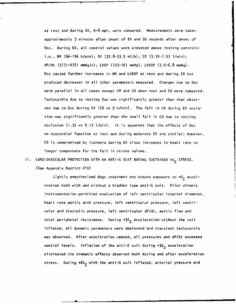
at rest and during EX, 6-8 mph, were compared. Measurements were taken
approximately 3 minutes after onset of EX and 50 seconds after onset of
Occ. During EX, all control values were elevated above resting controls:
i.e., HR (96-196 b/min); SV (32.8-33.9 ml/b); CO (3.39-7.03 1/min);
dP/dt (3171-4751 mmHg/s); LVSP (123-161 mmHg); LVEDP (3.6-6.8 mmHg).
Occ caused further increases in HR and LVEDP at rest and during EX but
produced decreases in all other parameters measured. Changes due to Occ
were parallel in all cases except HR and CO when rest and EX were compared.
Tachycardia due to resting Occ was significantly greater than that obser-
ved due to Occ during EX (29 vs 9 b/min). The fall in CO during EX occlu-
sion was significantly greater than the small fall in CO due to resting
occlusion (1.32 vs 0.13 1/min). It is apparent that the effects of Occ
on myocardial function at rest and during moderate EX are similar; however,
CO is compromised by ischemia during EX since increases in heart rate no
longer compensate for the fall in stroke volume.
II. CARDIOVASCULAR PROTECTION WITH AN ANTI G SUIT DURING SUSTAINED +GZ STRESS.
(See Appendix Reprint #10)
Lightly anesthetized dogs underwent one minute exposure to +GZ accel-
eration both with and without a bladder type anti-G suit. Prior chronic
instrumentation permitted evaluation of left ventricular internal diameter,
heart rate aortic arch pressure, left ventricular pressure, left ventri-
cular and diastolic pressure, left ventricular dP/dt, aortic flow and
total peripheral resistance. During +3GZ acceleration without the suit
inflated, all dynamic parameters were depressed and transient tachycardia
was observed. After acceleration ceased, all pressures and dP/dt exceeded
control levels. Inflation of the anti-G suit during +3GZ acceleration
eliminated the dramatic effects observed both during and after acceleration
stress. During +6Gz with the anti-G suit inflated, arterial pressure and
dP/dt were maintained whereas left ventricular end-diastolic pressure and
total peripheral resistance were much elevated and heart rate was depres-
sed. At the onset of G stress, internal diamet ' of the heart always fell
transiently. Otherwise, diameter was not significantly affected by any
of the experimental cond;;:ions. The results suggest that the anti-G suit
provides imp*Drtant maintenance of perfusion pressure at high sustained G;
however, with the anti-G suit inflated, central venous pressure is drama-
tically elevated and heart rate significant.y depressed. Thus, the bene-
ficial effects which providq tolerance to high G are accompanied by
potentially detrimental effects.
12. INFLUENCES OF SELECTIVE CARDIAC DENERVATION )N CORONARY REACTIVE HYPEREMIA IN
DOGS. (See Appendix Manuscript #11)
Mongrel dogs were chronically instrumented to me.3ure left circumflex
coronary flow, arterial pressure, left ztrial pressure, ECG, heart rate and
in some cases, left veiricular pressure or cardiac output. A cuff-type
occluder was placed distal to the coronary flow probe. Total occlusion
of the left circumflex coronary artery for one minute in unsedated, rest-
ing dogs produced reactive hyperemia with an average replacement/deficit
ratio of 2.63/1. In 11 dogs sympathetic influences were investigated by
chronic surgical cardiac sympathectomy. Surgical section of all aisae
subclaviae reduced responses from 2.61/1 to 1.67/1 (P< .001). Left sym-
pathectomy alone had no effect on the replacement/deficit ratio whereas
selective right sympathectomy reduced it from 2.25/1 to 1.38/1. Pharmacolo-
gical blockade was used to determine beta-receptor involvemeny in the re-
sponses. In 9 intact dogs, practolol (10 mg/kg) reduced the reactive hyper-
emia ratio by 12% (P<.05). Propranolol (1 rrg/kg) further reduced this ratio
by 30% (P<.001). Our results indicate that sympathetic beta influences
work primarily through the right cardiac sympathetic nerves. Also, the
magnitude of the response appears to be due, in part, to increased
metabolic activity associated with myocardial 81 receptors and heart
rate increase as well as active vasodilation through 82 receptors.
13. RABBIT CARDIOVASCULAR RESPONSES DURING VASOACTIVE DRUG INFUSION AT FIXED
CAROTID PRESSURE. (See Appendix Preprint)
In anesthetized rabbits, peak reflex bradycardia (AHR) and depres-
sion of mean arterial blood pressure (AMAP) were measured during maximal
central stimulation of the left aortic nerve (LANS). Responses were
quantified: (i) before and during steady state changes (+ 15 mmHg) in
the isolated carotid intrasinus pressure (ISP), and (ii) with ISP exclud-
ed from the circulation and maintained at a normotensive level (EP = ISP
= MAP) the MAP was changed + 20 mmHg by the infusion of either nitrogly-
cerin (NG), lysine vasopressin (AD) or phenylephrine (PE). Results indi-
cated that within + 11 mmHg of EP, the change in MAP per mmHg change in
ISP was 3, while AMAP due to LANS changed nearly double per mmHg change in
ISP. Following vagotomy a small increase in MAP was seen; however, cardio-
vascular responses to changes in ISP or LANS were unaltered. During drug
infusion with the carotid sinuses excluded from the circulation and ISP =
EP, peak AHR and AMAP to LANS were independent of the direction or magni-
tude of the drug induced change in MAP. When carotid baroreceptors were
allowed to detect the increase in MAP, the peak AHR and AMAP responses to
LANS were significantly reduced. These results suggest a high degree of
sensitivity of the carotid sinus baroreceptors around the animal's normo-
tensive region and that activity from these baroreceptors can modify re-
flex vascular tension even in the absence of significant change in heart
rate or arterial pressure.
AMmaAN Jo0m=AL 01 mfoCLMVoL 27. =,. .Sqnbw 1974. Pa i LPJ..
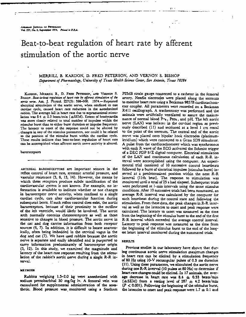
Beat-to-beat regulation of heart rate by afferent
stimulation of the aortic nerve
MERRILL B. KARDON, D. FRED PETERSON, AND VERNON S. BISHOPDepartment of Pharmacology, University of Texas Health &ience Center, San Antonio, Texas 78284
XKMnoN, Mzjmr B., D. FaR= PrrztsoN, Am Vxmo. S. P23d.B strain gauge connected to a catheter in the femoralBmxop. Beam-t&at regtdwaion of kart rau by afere stimlation of Me artery. Needle electrodes were placed along the sternumwnic ww. Am. J. Physiol. 227(3): 598--600. 1974.-Repeated to monitor heart rate using a Beckman 9857B cardiotachom-electrical stimulation of the aortic nerve, when confined to one eter coupler. All parameters were recorded on a Beckmancardiac cycle, caused heart rate reduction in the anesthetized R411 oscillograph. A tracheotomy was performed and therabbit. The average fall in heart rate due to supramaximal stimu- animal were artificially ventilated to assure the mainte-lation %as 8.4 -- 0.3 beats/min (-SEM). Extent of bradycardiawas more closely related to total number of impulses within the nance of normal blood Pn,, Pcoz, and pH. The left aorticstimulus burst than to either burst duration or impulse frequency. nerve (LAN) was isolatea in the cervical region as previ-The latency to onset of the response could not be altered by ously described (11) and sectioned at a level 1 cm rostalchanges in any of the stimulus parameters, nor could it be related to the point of the sternum. The central end of the aorticto the position of the stimulus burst within the cardiac cycle. nerve was placed onto bipolar hook electrodes (platinum-These results indicate that beat-to-beat regulation of heart rate irridium) which were connected to a Grass SD9 stimulator.can be accompished when afferent aortic nerve activity is altered. A pulse from the cardiotachometer which was synchronous
with each R wave of the ECG activated the Schmitt triggerbaroreceptors of a DEC PDP 8/E digital computer. Electrical stimulation
of the LAN and continuous calculation of each R-R in-terval were accomplished using the computer. An experi-mental tria consisted of 10 successive control heartbeats
ARTERIAL BARORECEPTORS are important s nsors in the followed by a burst of elecuical impulses (stimulus burst) in-reflex control of heart rate, systemic arterial pressure, and serted at a predetermined position within the next R-Rvascular resistance (3, 8, 13, 14). However, the means by interval (11th beat). The response to stimulation waswhich these receptors produce subtle modification of the monitored until a total of 25 s had elapsed. Subsequent trialscardiovascular system is not known. For example, no in- were performed at 1-rin intervals using the same stimulusformation is available to indicate whether or not changes conditions. After 10 successive trials had been summated, anin baroreceptor nerve activity, when confined to a single average R-R interval was calculated by the computer forcardiac cycle, can alter cardiovascular function during each heartbeat during the control state and following thesubsequent beats. If such reflex control does exist, the aortic stimulation. From these data, the peak changes in R-R inter-baroreceptors, because of their proximity to the outflow val as well as the latencies to onset and peak response wereof the left ventricle, would likely be involved. The aortic calculated. The latency to onset was measured as the timearch normally contains chemoreceptors as well as those from the beginning of the stimulus burst to the end of the firstsensitive to changes in blood pressure. The aortic nerve in R-R interval which exceeded the average control interval.the cat and dog carries information from both receptor Latency to peak response was measured as the time fromsources (6, 7). In addition, it is difficult to locate anatom- the beginning of the stimulus burst to the end of the long-ically, often being imbedded in the cervical vagus in the est heart interval monitored during the summated trials.dog and cat (7). We have used rabbits because the aorticnerve is separate and easily identified and is purported to axsuvrscarry information predominately of baroreceptor origin(5, 12). In this study, we examined the magnitude and Previous studies in our laboratory have shown that dur-latency of the heart rate response resulting from the stimu- Ing continuous aortic nerve stimulation maximaum changeslation of the rabbit's aortic nerve during a single R-R i- in heart rate can be elicited by a stimulation frequencyterval. of 80 Hz; using 10-V rectangular pulses of 0.3 ms duration
(11). Using these parameters, we stimulated the aortic nerve
tMTHoDS during one R-R interval (10 pulses at 80 Hz) to determine ifheart rate changes could be eicited. In 17 animals, the aver-
Rabbits weighing 1.5-2.0 kg were anesthetized with age decrease in heart rate was 8.4 = 0.33 beats/rainsodium pentobarbital 30 mg/kg iv. A femoral vein was (-i-SEM) from a resting level of 287 -i- 4.2 beats/mincannulated for supplemental administration of the anes- (P < 0.001). Following the beginning of the sdmulus burst,thetic. Blood pressure was. monitored using a Statham the latencies to onset and peak response were 1.7 * 0.1 and
BELT.TO-BEAT R.EGULATION OF HFART kATE 599
• ~ , .m .. s * 1 of changes in nerve discharge that we produced by in-A , creasing discharge frequency at the expense of burst
4 0. duration might, therefore, be expected to occur in response" A to increases in the slope and peak aortic pressure which.,are not accompanied by similar mean pressure changes.
" ~ In the intact animal, such an increase in slope and peakS." P . . . . of aortic pressure would stimulate a greater number of4 individual fibers within a shorter period of time leading to
t, I J. an increase in the whole-nerve discharge frequency during. " f the systolic phase, although burst duration would be shorter
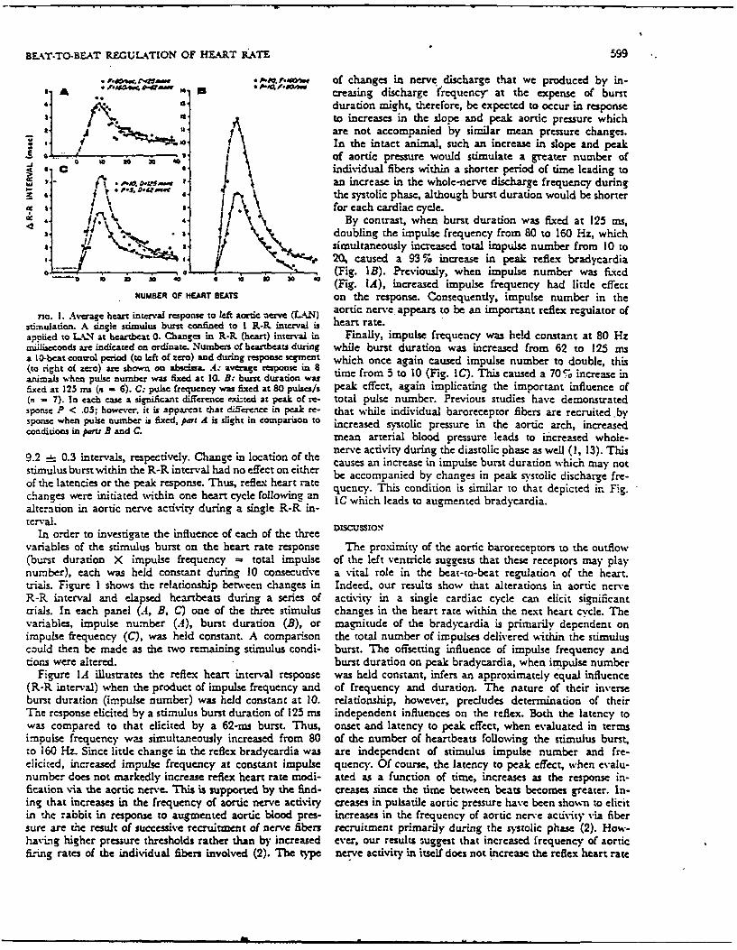
f " for each cardiac cycle., By contrast, when burst duration was fixed at 125 ms,
,....4 \,, doubling the impulse frequency from 80 to 160 Hz, which. t, simultaneously increased total impulse number from 10 to, , 20 caused a 93 % increase in peak reflex bradycardia
. .(Fig. IB). Previously, when impulse number was fixed(Fig. IA), increased impulse frequency had little effect
NUMBER OF HEART BEATS on the response. Consequently, impulse number in the
rio. I. Average heart interval response to left aortic nerve (LA4N) aortic nerve. appears to be an important reflex regulator ofstimuiation. A single stimulus burst confined to I R-R. interval is heart rate.applied to U.N at heartbeat 0. Changes in R-R (heart) interval in Finally, impulse frequency was held constant at 80 Hzmil~iseconds are indicated on ordinate. Numbes of heartbeats during while burst duration was increased from 62 to 125 msa 10-beat conwol period (to left of zero) and during response segment which once again caused impulse number to double, this(to right of zero) are shown on abscissa. A: average response in 8 time from 5 to 10 (Fig. IC). This caused a 705 increase inanimals when pulse number was fixed ac 10. B: burst duration wasfixed at 123 ms (a - 6). C: pulse frequency was fixed at 80 pulses/s peak effect, again implicating the important influence of(n - 7). In each case a significant difference exinecd at peak of re- total pulse number. Previous studies have demonstratedsponse P < .05; however, it is apparent that dfference in peak re- that while individual baroreceptor fibers are recruited .bysponse when pulse number is fixed, part A is slight in compariso to increased systolic pressure in the aortic arch, increasedconditions in paru B and C. mean arterial blood pressure leads to increased whole-
9.2 0.3 intervals, respectively. Change in location of the nerve activity during the diastolic phase as well (1, 13). Thisl bcauses an increase in impulse burst duration which may not
stimulus burst within the R-R interval had no effect on either be accompanied by changes in peak systolic discharge fre-of the latencies or the peak response. Thus, refleg heart rate nchanes ere nitateditin ne hartcycl fol i quenc. This condition is similar to that depicted in. Fig.changes were initiated within one heart cycle following an IC which leads to augmented bradycardia.alteration in aortic nerve activity during a single R-R in-terval.
In order to investigate the influence of each of the threevariables of the stimulus burst on the heart rate response The proximity of the aortic baroreceptors to the outflow(burst duration X impulse frequency - total impulse of the left ventricle suggests that these receptors may playnumber), each was held constant during 10 consecutive a vital role in the beat-to-beat regulation of the heart.trials. Figure I shows the relationship between changes in Indeed, our results show that alterations in aortic nerveR-R Lnterval and elapsed heartbeats during a series of activity in a single cardiac cycle can elicit significanttrials. In each panel (.4, B, C) one of the three stimulus changes in the heart rate within the next heart cycle. Thevariables, impulse number (A), burst duration (B), or magnitude of the bradycardia is primarily dependent onimpulse frequency (C), was held constant. A comparison the total number of impulses delivered within the stimuluscould then be made as the two remaining stimulus condi- burst. The offsetting influence of impulse frequency anddons were altered. burst duration on peak bradycardia, when impulse number
Figure IA illustrates the reflex heart interval response was held constant, infers an approximately equal influence(R-R interval) when the product of impulse frequency and of frequency and duration. The nature of their inverseburst duration (impulse number) was held constant at 10. relationship, however, precludes determination of theirThe response elicited by a stimulus burst duradon of 125 ms independent influences on the reflex. Both the latency towas compared to that elicited by a 62-ms burst. Thus, onset and latency to peak effect, when evaluated in termsimpulse frequency was simultaneously increased from 80 of the number of heartbeats following the stimulus burst,to 160 Hz. Since little change in the reflex bradycardia was are independent of stimulus impulse number and fre-elicited, increased impulse frequency at constant impulse quency. Of course, the latency to peak effect, when evalu-number does not markedly increase reflex heart rate modi- ated as a function of time, increases as the response in-fication via the aortic nerve. This is supported by the find- creases since the time between beats becomes greater. In-ing that increases in the frequency of aortic nerve activity creases in pulsatile aortic pressure have been shon to elicitin the rabbit in response to augmented aortic blood pres- increases in the frequency of aortic nerve activity via fibersure are the result of successive recruitment of nerve fibers recruitment primarily during the systolic phase (2). How-havLig higher presiure thresholds rather than by increased ever, our results suggest that increased frequency of aorticrising rates of the individual fibers involved (2). The type nerve activity in itself does not increase the reflex heart rate
600 kA.DON, PETERSON, AND BISHOP
response when the stimulus burst duration is decreased, continuous CSNS at different frequencies. Their conclusionsIndeed, augmented pulse pressure, which might lead to were that "central integrative functions" somehow reorderincreased peak systolic discharge frequency and a shorter this pulsatile information and emit the appropriate efferentburst duration, has been shown to have small reflex cardio- response without regard to afferent frequency per se. Avascular effects (1, 14). However, elevation of mean arterial similar mechanism may be operative in the aortic nerve con-pressure increases the frequency of nerve activity during trol of heart rate.both systole and diastole (4, 13), thus increasing both Analysis of the rapid reflex control of heart rate viastimulus burst duration and impulse frequency. Elevated aortic baroreceptors on the basis of peak nerve dischargemean arterial blood pressure has been shown to elicit frequencies or total impulse numbers alone may not yieldpotent reflex cardiovascular effects (1, 8). an accurate picture of how the entire reflex system re-
Recent studies in which the carotid sinus nerve was sponds to changes in arterial blood pressure. The discon-stimulated indicate results similar to our findines for aortic tinuous nature of baroreceptor nerve activity demands thatnerve stimulation. In working uneanesthetized dogs, Jonzon more than impulse frequency and impulse number beet al. (9) showed that continuous stimulation frequencies studied. The slight increase in reflex bradycardia elicitedof 60-100 Hz caused a maximal initial effect on heart by longer burst duration in the face of decreased impulserate. This supports our earlier findings in the rabbit ( 1) frequency indicates that the central integrative mechanismas well as the results of others using anesthetized dogs (7). in concert with its eflerent pathways nay be sensitive toBy contrast, steady-state blood pressure was maximally ef- the total time of afferent nerve activity as well as to changesfected at stimulus frequencies in the 30- to 50-Hz range. in impulse number.In an allied study, jonzon et al. (10) showed that the re-flex blood pressure changes elicited by carotid sinus nerve The authors thank Dr. Arthur M. Brown for his suggestions andstimulation (CSNS) were primarily dependent on impulse rJohn Gco-ackacis for his technical assistance.
This work was supported in part by AFOSR Grant 73-2525,number rather than frequency. This supports our present National Institutes of Health Grant HL 12415-06, and the Texas
findings using the aortic nerve. They showed that intermit- Heart Association (San Antonio chapter).
tent CSNS could elicit the same blood pressure effect as Received for publication 7 December 1973.
REFERENCES
I. ALWnEL. JA.mE, J. E., A"D M. Dz B. DA.LY. Comparison of the 8. Gucm, G., AND J. W. Covn-kz.L Relative importance of the carotidre.flex vasomotor responses to separate and combined stimula- and aortic baroreceptors in the reflcx control of heart ram. Am.tion of the crond sinus and aortic arch baroroceptors by pulsa- J. PFysio. 214: 955-961, 1968.tile and non-pulsatile pressures in the dog. J. Physiol., London 9. Jo. zo., A., P. A. Ota.o, G. Sxosx, AxD U. SjosTAwxo. Studies209: 237-293, 1970. of blood-pressure regulation in the unanesthetized dog. I. The
2. %crL. JAxacs, J. E. T"he effects of altering mean presure, pulse effects of constant-frequency stimulation of the carotid-sinuspresu'e and pulse frequency on the impulse activity in barorecep- nerves. Arch. Ges. Physiol. 340: 211-228, 1973.tor fibers from the aortic arch and right subclavian artery in the 10. jopzoN, A., P. A. Osro, G. SzEON, A.xN U. Sjorrmi.um. Studiesrabbit. J. Pknyial., London 214: 65--88, 1971. of blood-preure regulation in the unancsthetized dog. II. The
3. Ai-cru. JAxazs, J. E. Characteristics of single aortic and right effects of impulse train stimulation of the carotid-sinus nerves.subclavian baroreceptor fiber activity in rabbits with chronic Arch. CGe. P,4ysio!. 340: 229-249, 1973,renal byp etsion. Circulati. Rea. 32: 149-161, 1973. II. K oN, M. B., D. F. PEizasox, AxD V. S. Basxop. Reflex
4. Bx.xxr, P.. M., AxD M.I. N. Lv. Cmdiowascudar Phytiology. SL Louis: bradycardia due to aortic nerve stimulation in the rabbit. Am. J.Mosby, 1972, p. 133. Pkynel. 223: 7-11, 1973.
5. CHAuM.S, J. P., P. I. KoWe-, AND S. W. WmTr. The relative 12. Nxt., E., C. R. M. RznwooD, Axn A. ScowarrwL. Effects ofroles of the aortic and carotid sinus nerves in the rabbit in the electrical stimulation of the aortic nerve on blood presure andcontrol of respiration and circulation during arterial hypoxia and respiration in cats and rabbits under chloralose and nembuta)h.-prcapnia. . Physiol., London 188: 435-450, 1967. anesthesia. J. P ysim., London 109: 392-401, 1949.
6. DouLAs, W. W., AND W. ScHA-m.Q. A study of the depressor 13. PLLEmi , C. L., D. L. Ctau.srr, An J. T. Sram,,ea. Corn-and pressor components of the cat's carotid sinus and aortic paison of afferent activity of canine aortic and sinus erves.nerves using electrical stimuli of different intensities and fre-quencies. J. PFysieL., London 132: 173-186, 1956. Cirmd,ion Azs. 31. 557-368, 1972.
7. EDrs, A. J., AND J. T. SmarxERD. Selective denervation of aortic 14. SCHtU4MT, R. M., M. lK4uIADA, AND K. SAGAWA. Cardiovacular
arch baroreceptors and chemoreceptors in dog. J. Appl. Phy.iol. -responses to various pulsatile pressures in the carotid sinus. Am.30: 294-96, 1971. J. PhYsiol. 223: 1-7, 1972.
A.m mmmN *.--.- ~ m m -*• • m m m m m m
J~s L. Qor AP PUD PI%2IOLOGY
Voi. 37. No. 6. December 1974. P iai US.A.
Left ventricular function during acute regional
myocardial ischemia in the conscious dog
VERNON S. BISHOP, ROBERT L. KASPAR, GEORGE E. BARNES,AND MERRILL B. KARDONDepartment of Pharmacology, The University of Texas Health Scienc Centerat San Antonio, San Antonio, Texas 7828/
BISHOP, VEOaNo S., ROBERT L. KASPAR, GEORGC E. B.A'RIs, To accomplish these aims, left ventricular pressure,AND3 MERRILL B. K anoo.. Left rtentricular /unction during acute regional transverse internal diameter, and stroke volume were meas-mvocnardial ischenia in the conscious dog. J. Appl. Physiol. 37(6): 7e- ured before and during acute I -min occlusions of the left792. 1974.-The effects of acute -min occlusion of the left cir- circumflex coronary artery.cumflex coronary artery on the inotropic state and performance of
the left ventricle were examined in adult mongrel dogs. The ino- METHODStropic state, as indicated by changes in the maximum derivativeof left ventricular pressure in the preejection phase and the maxi- Surgery. In 13 adult mongrel dogs (16-20 kg) sterilemum derivative of the transverse internal diameter, were dimin- thoracotomies were performed under methoxvflurane anes-ished during the ischemic period. The end-systolic diameter in- thesia. By using the technique previously described (3, 4,creased 3.8 _ 0.6 mm while the end-diastolic diameter increased 20), two sonomicrometer transducers were implanted ononly 0.4 = 0.2 mm, although the end-diastolic pressure increased the endocardial surface of the left ventricle--one on the6.9 -. 0.6 mmHg. Progressive decreases in the stroke volume anterior and the other on the posterior left ventricular wall.paralleled the apparent reduction in myocardial fiber shortening in Through a second stab wound near the apex a calibratedthe transverse plane. Cardiac output and arterial pressure declined, solid-state pressure transducer (Konigsberg Instruments,concurrently, thus maintaining a constantd peripheral resistanceduring the occlusion. Acute coronary ocezusion also caused an P-18) was implanted in the left ventricle. A previouslyapparent increase in the myocardial wall st ffness as judged by the calibrated electromagnetic flow probe was placed around:ncrease in the slope a of the equation dP/dV - aP + B, the in- the ascending aorta, and an 18-gauge polyvinyl cathetercrease of the siope of the pressure-diameter relationship, and the was placed in the left atrial appendage. The left circumflexdecrease in the rate of lengthening of the diameter during diastole. coronary artery was exposed close to its origin and anThese findings suggest that acute myocardial ischemia results in occlusive device, similar to that reported by Chimoskey etan immediate reduction in the effective inotropic state and an al. (8), was placed proximal to the first segmnent of theapparent increase in the myocardial wall stiffness in the transverse vessel. In some animals a small electromagnetic flow probeplane. Both of these changes resulted in a decrease in performance was placed proximal to the cuff. A careful dissection wasof the left ventricle. performed in order to minimize any gross damage to the
nerve supply of the artery. Occasionally, small branches ofcoronarv artery occlusion; myocardial fiber shortening; dP/dt the left circumflex were sacrificed during the dissection,
but in no circum.stancecs did this lead to a damaged area asjudged from visual observations and pathological examina-
ALTHOUGH NUMEROUS STUDiES have demonstrated that left tion at autopsy. The electrical leads, catheters, and distalventricular function is compromised during myocardial end of the occluding device were exteriorized at the backischemia (7, 9, 11, 12) and infarction (16, 17), relatively of the neck. Approximately two weeks were allowed forlittle is known concerning the immediate changes in left recovery and, at the time coronary occlusions were per-ventricular function in conscious animals during acute formed, body temperature and ECG were normal.'myocardial ischemia. Most of our present information has Measuring of flow, pressure, and diameter. A Zepeda SW Flbeen derived either from acute animal studies (7, 12, 31) electromagnetic flowmeter was used to detect aortic .lowor from patients during stress-induced angina (11). Fur- and coronary blood flow. The flow probes were calibratedthermore, recent studies in patients have suggested that, in vitro before implantation and rechecked after the animalsduring ischemia, increases in myocardial wall stiffness may were killed. In some cases the in vivo calibration of theseverely limit and cause misinterpretation of the Frank- aor.ic flow probes was checked by using dye dilution tech-Starling reserve (9, 11). Therefore it is the purpose of this niques, and in all cases the calibrations agreed within 5 ,%.study to evaluate, in conscious dogs, the reduction in left The signal in late diastole was assumed to represent zeroventricular performance during acute regional myocardial aortic flow. Zero coronary flow was determined by occlu-ischernia. We attempted to identify, as contributing factors, sion of the coronary arter.. Stroke volume was obtainedchanges in the inotropic state, extent of myocardial fiber from aortic flow by analog integration of each ejectionshortening, and ventricular wall stiffness, period, using a Philbrick operational amplifier.
785
. l ml ml ~ml mm mmmm Mm m m l m
786 BISHOP, KASPAR, BARNES, AND K-ARDON
Left ventricular transverse internal diameter was ob- had returned to the preocclusive level. This was accom-tained by use of a sonomicrometer which measures the plished within 90 s. Occlusions were performed on at leastmean transit time for a burst of 5 mHz ultrasound between two separate days in each'animal, and hemodynamic alter-the two piezoelectric crystals at a sampling rate of 5,000 ations were reproducible at -!: 10%. As demonstrat.-d bytimes per second (30). Since the velocity of sound in blood the statistical analysis in Table 1, the group response wa.,is known, transit time is convertible to distance. also reproducible. Statistical comparisons were made using
Left ventricular pressures were measured by Konigsberg the t test for paired data. P values < 0.05 were consideredInstrument P-18 solid-state pressure transducers (3, 18-20). significant.The sensitivity of the transducers normally remains stable To evaluate the effects of increases in the initial preload,during implantation. On occasion, under local anesthetics, the left ventricular end-diastolic pressure (LVEDP) andcatheters were inserted into the left ventricle to verify the the end-diastolic diameter (EDD) were increased in Evecalibrations of the transducers. The reliability of the trans- animals by intravenous infusion of Tyrode solution (37 0C)ducers was also estimated by comparing the left ventricular (2, 3) and occlusion was performed. The LVEDP waspressure with the arterial pressure at the time of ejection. raised in each animal to a level significantly higher thanSince a routine check of zero left ventricular pressure by a the control level, but care was taken not to raise the LVEDPcatheterization technique was not possible, the zero drift to levels which produce reflex tachycardia.was normally corrected by adjusting left ventricular end- In three animals, changes in EDD during elevation indiastolic pressure to equal mean left atrial pressure at the preload by intravenous infusions of Tyrode solution werebeginning of the experiment while the animal was resting compared to the changes in EDD resulting from coronar7quietly on its right side (3, 4, 28). occlusion. To maintain a continuous rise in LVEDP, similar
Differentials of pressure and diameter were obtained by to that observed during acute coronary occlusion, Tyrodeusing an SQ10A operational amplifier.(Analog Devices) or solution was infused (300-400 ml/min) into a catheter inby digital. techniques (25). The analog derivative was the left jugular vein. The total volume infused ranged fromchecked by differentiating a sine wave, using the opera- 400 to 600 ml. This volume did not significantly alter thetional amplifier. The phase shift was 90 ° and the amplitude hematocrit (3, 4). The increments in EDD resulting fromof the differential output was linear between 0.5 and 100 these increases in LVEDP were compared to those observedHz..The differential was calibrated using a triangular wave. during coronary artery occlusion at similar LVEDP.
N.lean: arterial pressures were recorded through a catheter In four additional animals instrumented with aortic flowimplanted in the left carotid artery. Routine electrocardio- probes, infusions were performed using the above tech-grams were recorded with subcutaneous needle electrodes. nique. When the stroke volume became fixed at a maximumElectrical evaluation of the ischemia produced during level, mean left atrial pressure response was noted.coronary occlusion was obtained with a standard precordial E-stimation of ventricular wall stiffness. The passive elastice!ec:rocardiogram, with the indifferent electrode connected modulus during diastole, as defined by Diamond and For-to the ground of the coronary flow probe when the probe rester and associates (9, 10) was used to estimate the changewas not used. Peaking of the T-wave with S-T segment in myocardial wall stiffness during coronary artery occlu-elevation was observed in each animal during the period sion. With this method the equation dP/dV - aP + Bof ischemia. linearly relates the reciprocal of compliance during diastole
Recording. All signals were inscribed simultaneously on a to the pressure during diastole. The slope a of the equationType R Beckman oscillographic recorder and an Ampex is used to signify changes in myocardial wall stiffness. TheFR 1300 magnetic tape recorder. Tapes were analyzed derivative of pressure (dP) is approximated by the changewith a Philco 3000 Digital computer after analog-to- in left ventricular pressure during diastole and is equal todigital conversion. The left ventricular internal diameter the left ventricular end-diastolic pressure (LVEDP) minusand aortic flow and the integral of aortic flow were exam- the left ventricular pressure following systole, ESP (AP -ined as a function of the R-R interval of the electrocardio- EDP - ESP, Fig. I). The derivative of volume (dV) wasgram. Integrated aortic flow and left ventricular pressurewere computed as a function of left ventricular diameter(3, 4, 18, 28). TABLE 1. A.verage va!ues and mean changes during
Protocol. The coronary artery occlusive devices were coronary artery occlusion in 13 dogschecked in vitro as to the pressure and volume required toocc!ude the various size arteries. To assure the effectiveness Cadic S Volea RAne i
of the occluding device, in vivo transicnt occlusions were Condition Output (, o ea{td e
performed at the time of implantation. During the occlu- rWsion experiments identical volumes and pressures were usedto occlude the coronary artery. In those animals with Control 171=27 1.49--0.-9 114* 12 11019
cor r fCoronary occlusion 14431 1 .O tO.23 148_.L12 85 18coronary flOW probes the occlusion of the circumflex coro- .\lean difference -27-6t --O.30*0.3Ot. 35*9f -16=69nat, artery eliminated blood flow into the artery within 3 s. No. of paired ob- i13 13 13 9
Resting measurements were obtained while the animal servationswas lying quietly on its right side, unsedated and unre-strained. Following these measurements the left circumflex are means - SE. Nian difference fL SD ,ompares the changscoronary artery was occluded for I min and released. Addi- between control and coronary occlusion. * P < 0.05, t P <tional occlusions were performed only after all parameters 0.001.
REGIONAL MYOCARD\IAL ISCHE.MIA 787
CONTROL CORONARY OCCLUSION .
12400
AF
20-
LVP J C& &.0 Q 0 240 30 oa , 1.0 . , 300
o j jvej WMjf (Cc Vs VOLUMIL le)
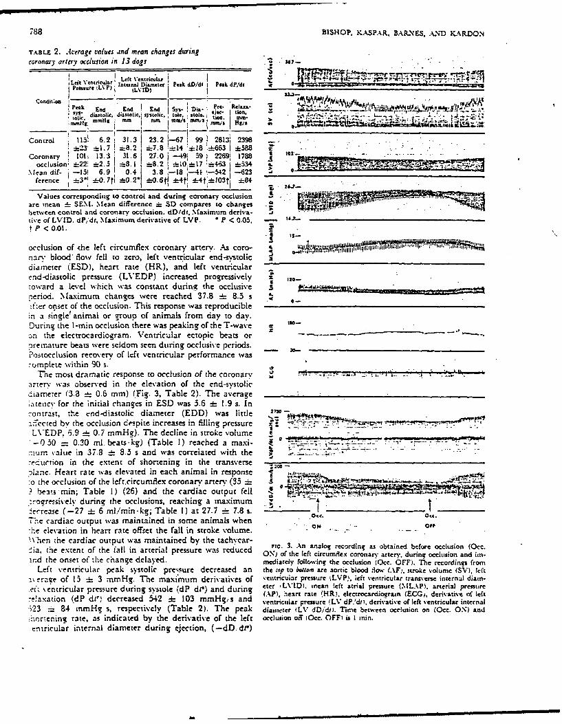
rio. 2. Relationship of left vencricular internal diameter and pres-o@sure to volume in a nonbeating heart.
autopsy, and dissecting the area of distribution (7 dogs);2) electrocardiographic changes in one acute dog, usingelectrodes placed directly on the left ventricle; 3) visual
Is- observations of discoloration during a test occlusion at theLVYOP time of surgery (all dogs); and !) histological sections fc-
"o- •]owing a permanent occlusion (I dog). In all cases thekit transducers were implanted in normal myocardial tissue.
In four additional dogs myocardial blood flow distributiona,. iCOPb- (SP
Av. sv was evaluated before and during coronary occlusion by0a.-CSO using the radioactive microsphere technique. The perfusion
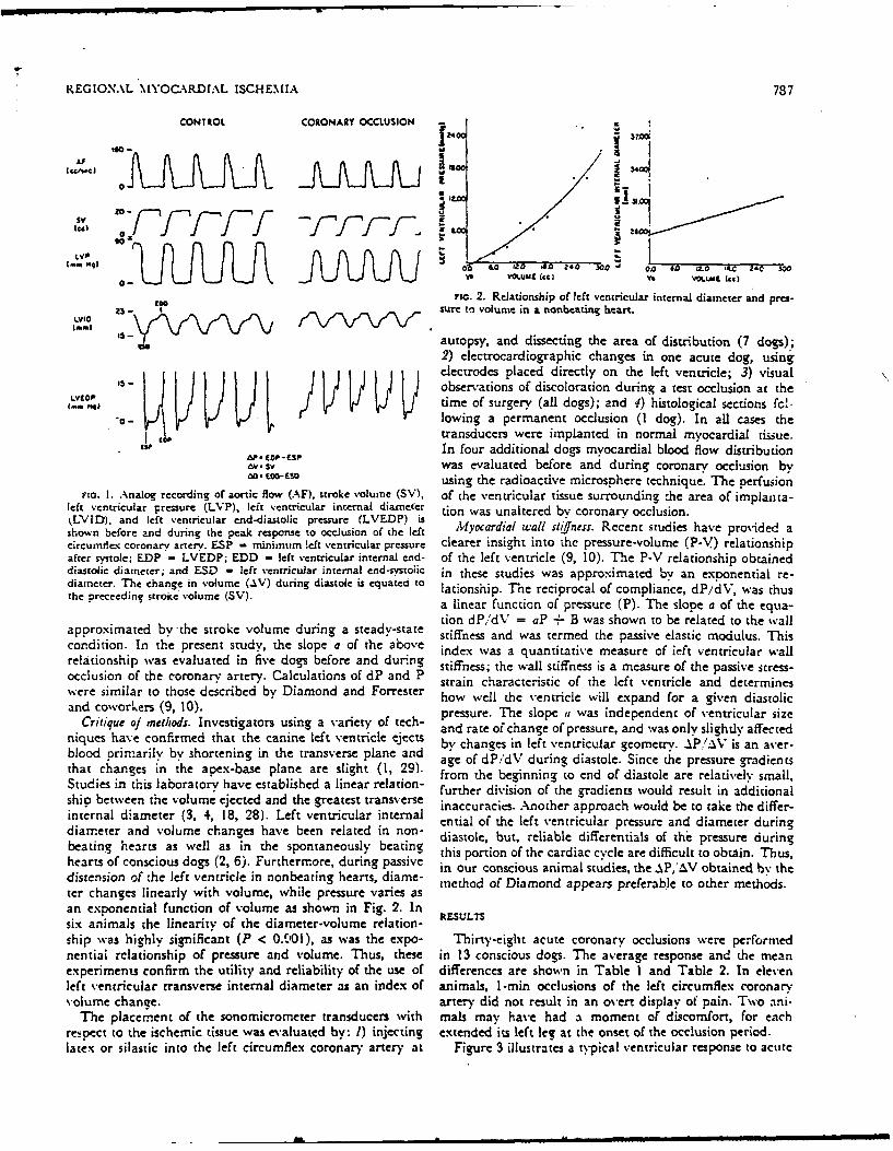
Fir. I. Analog recording of aortic flow (AF), stroke volume (SV), of the ventricular tissue surrounding :he area of implanta-left ventricular pressure (LVP), left ventricular internal diameter tion was unaltered by coronary occlusion.kLVID), and left ventricular end-diastolic pressure (LVEDP) is •oa u altd by cn ocudsiovshown before and during the peak response to occlusion of the left Myocrdial wall stifnes:. Recent studies have provided acircumie.- coronary artery. ESP = minimum left ventricular pressure clearer insight into the pressure-volume (P-V) relationshipafter systole; EDP = LVEDP; EDD - left ventricular internal end- of the left ventricle (9, 10). The P-V relationship obtaineddiastolic diameter; and ESD - left ventricuJar internal end-systoiic in these studies was approximated by an exponential re-diameter. The change in volume (AV) during diastole is equated to lationship. The reciprocal of compliance, dP/dV, was thusthe preceedine' stroke volume (SV).e sa linear function of pressure (P). The slope a of the equa-
tion dP,dV = aP + B was shown to be related to the wallapproximated by the stroke volume during a steady-state stiffness and was termed the passive elastic modulus. Thiscondition. In the present study, the slope a of the above index was a quantitative measure of left ventricular wallrelationship was evaluated in five dogs before and durng stiffness- the wall stiffness is a measure of the passive stress-occlusion of the coronary artery. Calculations of dP and P strain characteristic of the left ventricle and determineswere similar to those described by Diamond and Forrester how well the ventricle will expand for a given diastolicand coworkers (9, 10). pressure. The slope , was independent of ventricular size
Critique of methods. Investigators using a variety of tech- and rate of change of pressure, and was only slightly affectedniques have confirmed that the canine left ventricle ejects by changes in left ventricular geometry. AP.'AV is an aver-blood primarily by shortening in the transverse plane and age of dP/dV during diastole. Since the pressure gradientsthat changes in the apex-base plane are slight (1, 29). from the beginning to end of diastole are relatively small,Studies in this laboratory have established a linear relation- further division of the gradients would result in additionalshio between the volume ejected and the greatest transverse inaccuracies. Another approach would be to take the differ-internal diameter (3, 4, 18, 28). Left ventricular internal ential of the left ventricular pressure and diameter duringdiameter and volume changes have been related in non- diastole, but, reliable differentials of the pressure duringbeating hearts as well as in the spontaneously beating this portion of the cardiac cycle are difficult to obtain. Thus,hearts of conscious dogs (2, 6). Furthermore, during passive in our conscious animal studies, the .PI.V obtained b" thedistension of the left ventricle in nonbeating hearts, diame- method of Diamond appears preferable to other methods.ter changes linearly with volume, while pressure varies asan exponential function of volume as shown in Fig. 2. Insix animals the linearity of the diameter-volume relation- RESULTSship was highly significant (P < 0.001), as was the expo- Thirty-eight acute coronary occlusions were performednential relationship of pressure and volume. Thus, these in 13 conscious dogs. The average response and the meanexperiments confirm the utility and reliability of the use of differences are shown in Table I and Table 2. In elevenleft ventricular transverse internal diameter as an index of animals, 1-min occlusions of the left circumflex coronaryvolume chanze. artery did not result in an overt display of pain. Two ani-
The placement of the sonomicrometer transducers with mals may have had a moment of discomfort, for eachres pect to the ischemic tissue was evaluated by: 1) injecting extended its left leg at the onset of the occlusion period.latex or silastic into the left circumflex coronary artery at Figure 3 illustrates a typical ventricular response to acute
788 BISHOP, KASPAR, BARINES, AIND KARDON
TABLE 2. Average valucs and mean changes during.ccoronary artery occlusion in 13 dogs . in-
LetV triua eft Ventricaz X 1 "'~LPruejtLVP laterAuJ Diameter Peak dD/d i Peak dP.'di -9 ad
Prnar ~VP LVI) j _____ _______ ,AA.J
cod~uPeak Ld End IEnd Dil Pe c - 2,~ ~ f~mmiimHg mm MIT mm / o " U'
arc/si mmis. Hg/
Control 113! 6.2 131.3 23.2 1-67~ 99~ 2813; 2398 -
:L- *1. 7 8. 2 -7.8 !-14 '18_63I*8 - .- .
Coronlary 101 4.. j 6 j27.0 j -49; 59 1 269i 1788occlusion; -22! :i2. 3 * 8.1 *8. 2 *i 1 -463 -534- '~-~k -
N [an dif- j-13! 6.9 0. . 18 l:41 -:342 -623 0ference -3-' _0. 7ti -0.21 *.6t *4tl _4t:*103t1 -8
Values corresponding to control and during coronary occlusion _4are inean :L S EN . Mean difference =i SD compares to changesbctwecn control and coronary occlusion. dD/dt. Maximum deriva- Ative of LVID. dP,'dt,Maximumn derivative of LVP. *P < 0.05,
P 1< 0.01.
occlusion of -the left circumflex coronary artery. As coro- 0'-''4nary blood' flow fell to zero, left ventr icular end-systolic Xdiameter (.ESD), heart rate (HR), and left ventricularend-diastolic pressure (LVEDP) increased progressively ir 10toward a level which wvas constant during the occlusive .g________I________period. Maximum changes were reached _37.8 :L 8.3 s MA i
afier onset of the occlusion. This response was reproduciblem a single ! animal or g-roup of animals from day to day.During the 1 -min occlusion there was peaking of the T-wave C tooon the electrocardiogram. Ventricular ectopic beats or- --
?.rermature beats were seldom seen during occlusive periods.-Postocciusion recovery. of left ventricular performance was-umplete wvithin 90 s.
Tnhe most dramatic response to occlusion of the coronary 1artery was Obser-ved in the elevation of the end-systolicdiame-ter (3.8 _- 0.6 mm) (Fig. 3, Table 2). The average;.ten1cv for the initial changes in ESD was 3.6 =I 1.9 S. Incontrast, the end-diastolic diameter (EDD) was little 270:izccted by the occlusion delspitE increases in filling pressure =0 ' . ~.-LVEDP. i.9 _ - 0.7 mm-fe). The decline in stroke volume -
-0530 = 0.30 mi. beats-kg) (Table 1) reached a maxi- I! 1Lo21umn Value in 37.8 :i 8.3 s and was correiated with the-Cc Urion in the extent of shortening in the Mrnve~olane. Heart rate was elevated in each animal in response:o ;bhe occlusion of the left.circumflex coronary artery (33 .5 .. u __
b !eats min; Table 1) (26) and the cardiac output fell .
:.roressively during the occlusions, reaching a maximum - 7~errease (-27 :E 6 mi/min-kg; Table 1) at 27.7 -_ 7.8 s. Oct. Oct.
T'he cardiac output was maintained in some animals when a F:rne elevation in heart rate offset the fall in stroke volume. -..-
\\'hen the cardiac output was maintained by the tachycar- Fi.3. An analog recording as obtained before occlusion (0cc.ia, the extent Of the fall in arterial pressure was red'uced ON) of the left circumflex coronarv arterv, during occlusion and un-
ind (he onset of the change delayed. mediately following the occlusion (0cc. OFF). Thec recordings fromLeft ventricular peak systolic pressure decreased an the top to bottom arec aortic blood liow (AFi, stroke v-olume (SV), left
a1\eraze of 13 :h 3 mnmHg. The maxi'mumn derivatives of ventricular pressure %LVP), left ventricular transverse internal diain.
ve tentricular pressure during systole WdP di') and during etc- LVID1. inean left atrial pressure (MLAP1. arterial pressureAL.. AP). heirt rate H) electrocardiogramn (ECG,, derivative of left
-- !axation Wd d11,1 decreased 542 :1 103 mmHg/ s and ventrictilar pressure (L%* dP.'dit, derivative of left ventricular internal.i23 "_ 84 mmHg s, respectively (Table 2). The peak diairreter iLV dD/dmi Time between occlusion on (0cc. ON) and-:oren~ni rate, as indicated by ithe derivative of the left occlusion off (0cc. OFF) is I ini.entricular internal diameter during ejection, (-dD.. d)
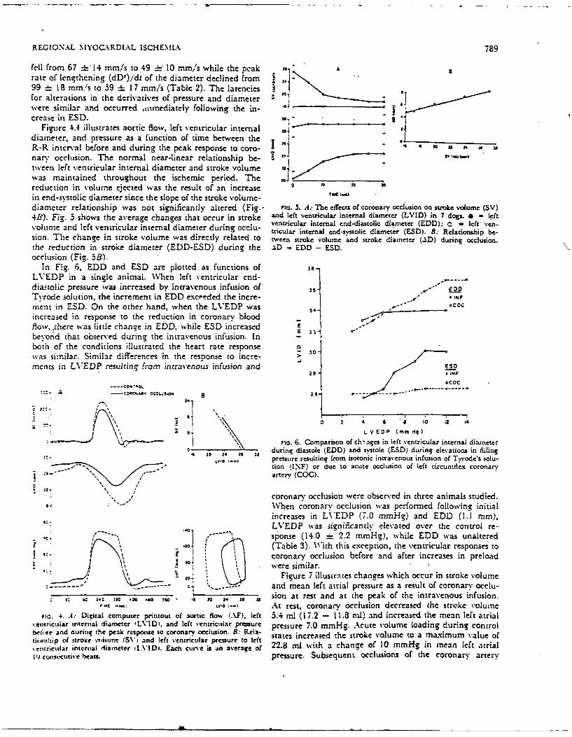
REGIONAL MYOGA\RDIAL ISCHEMIA 789
fell from 67 =h-l4 mm/s to 49 4-- 10 mm,'s while the peak "I A3
rate of lengthening (dDr)/dt of the diameter declined from 199 : _ 18 mm'/s to 59 =E 17 mm/s (Table 2). The latenciesIfor alterations in the derivatives of pressure and diameterwere similar and occurred falmediately following the in- Icrease in ESD. 30-.
Figure 4.4 illustrates aortic flow, left ventricular internal *diameter, and pressure as a function Of time between theLR-R interval before and during the pcak response to coro- -0 10 a 2nary occlusion. The normal near-linear relationship be- E IV tiwiwtWeen left ventricular internal diameter and stroke volumewas maintained throughout the ischemic period. Thereduction in volumne ejected was the result of an increase 2 TW1U W 0
in end-systolic diameter since the slope of the stroke v'olume-diameter relationship was not significantly altered (Fig.- inc. 5. A.- T1c effects of coronary occlusion on stroke volume (SV)4B3). Fig. 3 shows the average changes that occur in stroke and left ventricular internal diameter (LVID) in 7 dogs. *=left
volue ad lft vntrculr iternl dameer drin oclu- ventricular internal end-diastolic diameter (EDD); 0 - left' ven-volue ad lft vntrculr iternl dameer drin oclu-tricular internal end-systolic diameter (ESD). B: Relationship be-sion. The change in stroke volume was directly related to tween stroke volume 'and stroke diameter (AD) during occlusion.the reduction in stroke diameter (EDD-ESD) during the _%D =EDD - ESD.occlusion (Fig. 3B).
In F,(g. 6, EDD and ESD are plotted as functions of 6
LVEDP in a single animal. 'AVhen left ventricular end-diastolic pressure was increased by intravenous infusion of 35* EDOTvrodie solution, the increment in EDD exceeded the incre- 1-ment in ESD. On the other hand, when the LVEDP was 34Co
increased in response to the reduction in coronary blood1flow,,there was little change in EDD, while ESD increased 4beyond that observed during the intravenous infusion. In 3both of the conditions illustrated the heart rate response o 0was similar. Similar differences in the response to mecre->merits in LVEDP resulting tf-am intravenous infusion and UPO
29 14FN
I ,COC
*~~ a OCl.015 2.4 14. -- -
Fc6.Comparison of ch'nages in left ventricular internal diameterG ali 24 29 urn iatl (EDD) and sstole (E.SD) during elevations in Miiing
- pressure resulting from isotonic intravenous infusion of Tvrode's solu-- tion (INF) or due to acute occlusion of left circuniflex coronary
I arter (COG).
coronar-y occlusion were obser-ved in three animals studied.When coronary occlusion wvas performed following initial
LVEDP was significantly elevated over the control re-
------- nse (1, 2.2 mmn Hq), while EDD was unaltered'0- (Table 3). With this exception, the ventricular responses to
coronary occlusion before and after increases in preload- -, --- -- - \3)Figure 7 illustrates changes which occur in stroke volume
- ..--- e. *...and mean left atrial pressure as a result of coronary occlu-
5: 64 11 M~ 4 " 20 . it sion at rest and at the peak- of the intravenous infusion.,..t-'.~At rest, coronary occlusion decreased the stroke 'oIlme
Fic. 4. A: Digital computer printout of aortic Hlow (AlF). left 3.4 ml (17.2 - 11.8 ml). and increased the mean left atrial\entricular internal diameter ILVIDi, and left ventricular pressure pressure 7.0 mmHg. Acute volume loading during controlbefo're and during the peal. response to coronary occlusion. B: Rela- sae nrae h toevlm oamxmmvleoieii~oip of stroke vniimine (SV) and left ventricular pressure to left sae nrae h toevlm oamxmmvleo%entrictilar internal diameter fl[VIDi. Each curve is an average of 22.8 ml with a change of 10 mmHg in mean left atrial11) cuntccative beats. pressure. Subsequent occlusions of the coronary artery
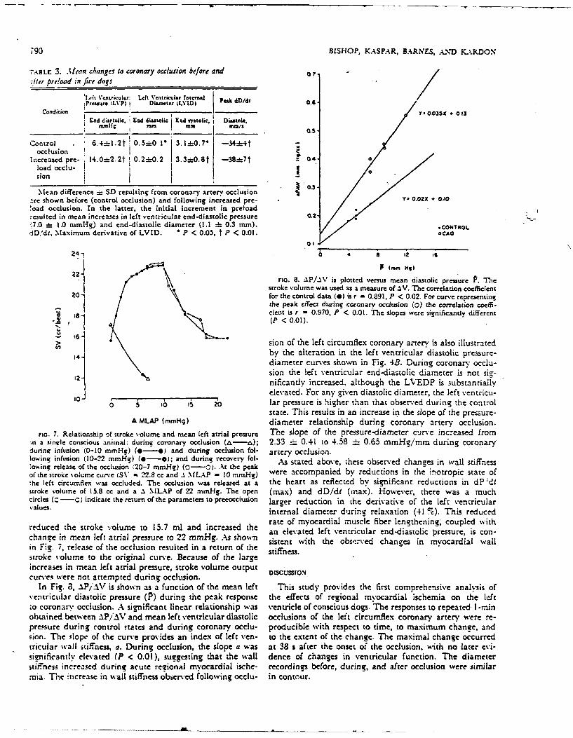
790 ~BISHOP, KASPA4R, BARNES, ANDM KARDOX
ALE3. .ltan changes to coronary occlusion before and 0
41rpredoad infive dogs______
I J'it Ventricular: Left Ventricular Internal Pa Dd .Pressure (LP) I Diarneter (YD ekMd
Condition .- Y - 0.05X + 0 13
End diactujic. End diastolic End systolic, I Diastole,mmHg EMm MM mm,./
Control . 6.4=L-.2t 0. 5:0 1 3.l1 0. 7 -34--4tZocclusion
Increased pre- 1 4.0- 2.2t 0. 2-0 .2 3.3i0. 8t -38.-!-i7f 0.4..load occlu-
4 0.3Mean difference SID resulting from coronary artery occlusion 1
,ire shown before (control occlusion) and following increased pre- VY 0.02X + OLIO
!oad occlusion. In the latter, the initial increment in preload-esulted in mean increases in left ventricular end-diastolic pressure 0.217.0 i= 1.0 nimHg) and end-diastolic diameter (1.1 : 0.3 mm). *CON4TROLdD,'dt, Maximum derivative of LVID. *P < 0.05, t P < 0.01. OCAo
21 0 2 a
22-FIG. 8. AP/AV is plotted versus mean diastolic pressure P. The
stroke volume was used as a measure of ASV. T-he correlation coefficient20- for the control data (0) is r - 0.891, P < 0.02. For curve representing
the peak effect during coronary occlusion (o) the correlation coeffi-' tient is, r 0.970, P < 0.01. The slopes were significantly different
'N (P < 0.01).
> 6 Sion of the left circumflex coronary artery is also illustrated
14 by the alteration in the left ventricular diastolic pressure-diameter curves shown in Fig. 4B. During coronary occlu-Sion the left ventricular end-diastolic diameter is not sig-nificandy increased, although the LVEDP is substantiallyelevated. For any given diastolic diameter, the left vecntricu-
0 5 b 16 5 2 lar pressure is higher than that observ-ed during the control
state. This results in an increase in the slope of the pressure-MLAP (mmHg) diameter relationship during coronary artery occlusion.
FG7.Relationship of stoevolume admean left atrial pressure T'hese of the pressure -duameter curv'e nceased fromin a single conscious animnal: during coronary occlusion (&-a); 2.33 :: 0.41 to 4.58 =i 0.65 mmHg/mm during coronarydurine infusion (0-10 mmHg) (*-*. and during occlusion fol- artery Occlusion.low.ing infusion (10-22 mmlg) (4-0); and during recovery fol- Astaeabvhseoerdcansin alsifesto%ing release of the occlusion i20-7 mmiHg) (C0.At the .peak Astaeabvhseoerdcansinwl ifCSof the stroke volume curve (SV* 22.8 cc and .1 MLAP - 10 mmg were accompanied by reductions in the inotropic state of,he left circuimflex was occluded. The occlusion wvas released at a the heart as reflected by significant reductions in dP 'dtstroke volumne of 15.8 cc and a .1 \LL-P of 22 mmHg. The open (max) and dD/di (max). However, there was a muchcircles ( -)indicate the return of the parameters to preecelcusion larger reduction in the derivative of the left ventricular'-alues. internal diameter during relaxation (41 %). This reduced
redued he troe voumeto 5.7ml ad icresedthe rate of myocardial muscle fiber lengthening", coupled withchange in mean left atria] pressure to 22 mmHg. As shown a lvtdlf etiua n-isoi rsue scnin Fig. 7, release of the occlusion resulted in a return of the stifst s.wt h bs~e hne n ycr lwlstroke volume to the original curve. Because of the large sifesincreases in mean left atrial pressure, stroke volume output DISCUSSIONcurves were not attempted during occlusion.
In Fig. 8, AP/AV is shown as a function of the mean left This study provides the first comprehensiv'e analysis ofventricular diastolic pressure (P) during the peak response the effects of regional my ocardial ischemia on the leftto coronary occlusion. A sienificant linear relationship was ventricle of conscious dogs. T he responses to repeated- I -rninobtained between AP'/A'V' and mean left ventricular diastolic occlusions of the left circumflex coronary artery were re-pressure during control states and during coronary occlu- producible with respect to time, to maximum ch~ange, andsion. The slope of the curve provides an index of left yen- to the extent of the change. The maximal change occurredtricular wall stiffness, a. During Occlusion, the slope a was at 38 s after the onset of the occlusion, with no later evi-significantlv elevated (P < 0.01), sugesting that the wall dence of changes in ventricular function. The diameterstiffness increased during acute regional myocardial ische- recordings before, during, and after occlusion were similarmia. Thre increase in wall stiffness observed tollowing occlu- in contour.
REGIONAL .MYOC.RDl.L ISCH. EMIA 791.
Acute regional myocardial ischemia resulted in a reduc- artery disease (9), but different from that reported intion in the inotropic state of the heart as assessed by the open-chested animals- during acute infarction (13). How-observed decreases in dPidi (max) and dD/dt (max). ever, it is likely that in these studies the initial state of theBoth of these variables have bcen previously shown to be myocardium was compromised by the anesthetic andsensitivc indiccs of the inotropic state (1, 14). Furthermore, experimental procedures. In our study the increment init is unlikely that these changes were significantly influenced stiffness begins to take place shortly after the occlusion,by the alterations which occurred in left ventricular end- and a stable maximum increase in myocardial wall stiffnessdiastolic pressure, heart rate, or arterial pressure, all of is reached 30-60 s into the occlusion. When the occlusionwhich have previously been shown to have minimal effects is released, the stiffness begins to return to its control value,on either dP,'di (max) or dDidi (max) (1). Reductions in and it is fully restored to its control value 15-30 s after thedP dt (max) and dD/dI (max) most likely resulted from release. The ranid increases and restorations of myocardialthe reduced contractile state of the ischemic portion of the stiffness indicated that, at least acutely, these apparentmvocardium. The extent of shortening from any given changes in myocardial stiffness are related to the directinitial length was also severely compromised during the effect of ischemia on the myocardium. Because of theischemic period and resulted in a proportional reduction rapidity and reversibility of the changes, it is likely thatin the stroke volume (Fig. 5). Since the relationship between alterations are occurring in the biochemical mechanismdiameter and stroke volume is unaltered during ischemia, responsible for relaxation (22-24).one must assume the left ventricle is still ejecting blood Myocardial ischemia reduces the performance of theprimarily by shortening in the transverse plane (9). The heart, as illustrated by the reduction in the stroke volumede -ee of systolic ballooning (7, 16, 27, 32, 33) or asvnchro- at rest and at the peak of the infusion response. Because thenous contraction could not be evaluated in the present extent of shortening from any given initial muscle length isstudy: The fact that the contour of the diameter recording severely compromised, this reduction in the Frank-Starlingwas similar before and during the occlusion suggests that reserve is obviously related in part to the decline in thethe effects of asvnchrony were small. Placement of the inotropic state. Although not so obvious, increases in mvo-transducers in normal tissue may have minimized the cardial wall stiffness may also have important functionaleffect. It also seems unlikely that s.stolic ballooning, a significance, since the Frank-Starling reserve of the heartcondition which disassociates the changes in stroke volume is dependent on the pressure-volume characteristics as wellfrornthe extent of shortening, could account for the above as the ability of the heart to develop force in response toobservations. A more likely explanation would be the loss distension. W,'hen the myocardial wall stiffness is elevated,of functional myocardial tissue. The ventricle is essentially greater increments in left ventricular filling pressures area ser.es arrangemcnt of myocardial muscle fibers, and for required to stretch the myocardium. Consequently, al-this reason, changes in the mechanics of the ischemic area though the myocardium may still be capable of developingcan affect the entire ventricle (15, 21). Therefore, the an increased force, the stroke voiume or stroke work responsereduction in dP~dt (max), dD/dt (max), and the extent of is less for any given increase in filling pressure. Thus,shortening mayl be due to a loss of functional myocardial increments in myocardial wall stiffness may cause misinter-issue (hvpokinesis) in the ischemic tissue. In isolated heart pretation of the classic Frank-Starling function curve; but,
studies, similar reductions in the extent and rate of short- more importantly, it limits the Frank-Starling reserve ofening and the rate of tension developed have been observed the heart, for extremely high filling pressure may be re-during hypoxia (34, 33). quired to stretch the myocardium.
It is apparent from our results that regional myocardial Several investigators (5, 9) have questioned, particularlyischemia alters the normal relationship between left y'en- in the diseased heart, the reliabiiity of left ventriculartricular filling pressure and the end-diastolic diameter. end-diastolic pressure as an index of changes in left yen-During ischemia, LVEDP increased without a correspond- tricular end-diastolic size. Increases in left ventricularing change in EDD. The slope of the pressure-diameter stiffness or a failing heart could both result in similarelationshio was also elevated. Assuming that changes in changes in left ventricular end-diastolic pressure. As pointed
left ventricular end-diastolic diameter reflect changes in out by Diamond and Forrester (9), a ventricular functionend-diastolic volume, it appears likely that the myocardial curve may not express the difference between change- inwall is less distensible in the transverse plane. The above ventricular stiffness and reduced inotropic state. Thus,assumption is supported by the following evidence. 1) The without an index of ventricular end-diastolic volume, theleft ventricle is part of a closed fluid-filled sys.tem and, as exact functional status of the myocardium may be impos-such, changes in diameter during systole and diastole most sible to determine. In the present study, failure to increaselikely represent the same volume. 2) Mitchell et al. (25) the stroke volume during increments in LVEDP could beha\e .hown that ventricular volume changes during systole interpreted as simply a reduction in ventricular function;and diastole are reflected by identical changes in the cross- however, the limitation of ventricular function may be insectional area of the left ventricle.* J) In the nonbeating part due to the apparent increase in stiffness of the leftheart. we have shown that left ventricular volume and ventricle.diameter change together while pressure and volume areexpnenialy elted(Fg. ).The authorn express their appreciation to .Mia Linda' Fox and .Mr.exponentially related (Fig. 1 ). Ben Wigr.ns for their technical support on this project.Uin" a more direct approach for estimating myocardial This s'tud wa supported in par by the Ntional Intitutes ofwail .tiffness, we observed dramatic chanSes in stiffness Heaith Grant 2 R01 HL12415-04, US Air Force Contract kFOS..durinz ischemia. The index of stiffness during ischemia was 71.2074, and the Texas Heart Association (San .\ntonio Chaoter).similar to that previously reported in patients with coronary Received for publication 4 January 1974.

792 BISHOP, -ASPA-R. BARNES, AND KA-RDON
REFERENCES
IBAFUNES, G.. V. S. BISHoP, L. D. HoRwiTz, AND R. L KASPAR. 18. Ho~wirz, L D., AND V. S. BisHop. Left ventricular pressure-The ma.'.imumn derivatives of left ventricular pressure and trans- dimension relationships in the conscious dog. Cardiocisscila, Res.verse internal diameter as indices of the inotropic state of the left 6: 163-1 71, 1972.ventricle in conscious dogs. J1. PhysioI.. London. 235; 571-590, 1973. 19. HoAwrrz, L D., A'JD V. S. BISHOP. The effet Of acute volumec
2. BisHOP. V. S., AND L D. HORWITZ. Left ventricular transverse loading on heart rate in the conscious dog. Cirmaioet Rts. 30:internal diameter: Value in studying left ventricular function. 316-321, 1972.Am. Her J. 80: 507-314, 1970. 20. HORwirz, L. D., V. S. Basiop, H. L. Sioxx. AND H. F. STECALI.
3. Bisitop, V. S., vo L D. Hoawrrz. Effects of altered autonomic Continuous measurement of internal left ventricular diameter.control on left ventricular function in conscious dogs. Am. J1. J1. Appl. Physiol. 24: 738-740, 1-068.Phy.noI. 221: 1278-1282, 1971. 21. j6IwiNN. M., AND E. Niz~ssoN4. A note on the influence of
4. BISHO0P. V. S.. L. D. HoRfTwrz, H. L- STONE, H. F. STECALL, AND asynchronous activation of myocardial contraction. Acta FAysiol.E. J. E'tozxLJzNo Left ventricular internal diameter and cardiac Scand. 85: 242-248, 1972.function in conscious dogs. J. Appi. Pkyriol. 27: 619-623, 1969. 22. KATz, A. M., ANDi D. I.Kuztxz. Quanti~stive aspects of dog cardiac
5. BRA.UNWAL, E., AND J. Ross, JR.. Ventricular end-diastoiic pres- microsomal calcim binding and calcium uptake. Circulatio., Res.sure: apprisa of its value in the recognition of ventricular failurt 21: 153-162, 196;.in man. Am. J. Attd. 34: 147-130, 1963. 23: KA~z, A. M., D. 1. Rznca, AND B. R_ COHEN. Control of the
6. C.AEroo, R. A., AD J. G. CL.%"-a. M easurement of left ventricu- activityr of highly purified cardiac actomyohin by Ca!', Na", andlar diameter in the dog by cardiac catheterization. Circulation Res. K*. Circulation Res. 19: 1062-1070. 1966.'22: !4i338, 1968. 2+. LUz, K. S., H. LA~NosxY, AND Jo H. Srixi~y. Decreased Ca--
7. CAsx. R. B.. E. BtRGLLND, AND S. j. SARNOP?. Ventricular func. uptake by sarcoplasmic reticulum after coronary artery occlusiontion. 11. Quan~titative relationship between coronary flow and for 60 to 90 minutes. Circulation Res. 21: 439-414. [(%7.ventricular function with observations on unilateral failure. 23. Mirca-mu., J. H., K. WILDEUNTI4AL, AND C. B. MNLtLD0N. Ceo-Circulation Res. 2: 319-323, 1954. metrical studies of the ieft ventricle utilizing biplane cinesiuorog.
8. CmsiMc.sKEV, J. E., MI. SZENTIVNYT. R. ZAKHEIM., AND A. C. raphy. Federat ion Proc. 28: 1334-1343. 1969.BARGER. Temporary coronary occlusion in conscious dogs: col- 26. .PzTSasoN, D. F., R. L. KASPAR, AND V S. Bts~op. Reflex tachv-latcral flow and electrocardiogramn. Am. J. PIysiol. 212: 1023- cardia due to temporary coronary occlusion in the conscious dog.1032, 1%67. Circ-loiion Res. 32: 632--639, 1973.
9. Dma.coro, G., AND J. S. FOaRRTR~. Effect of coronary artery dis- 27. PEINZMETAC., M., L. L.. SCHWRT.~ Z, E. CORDAY, R. SParrztjEA,ease and acute mvocardial infarction on left %entricu!ar compli- H. C. BERGMAN, AND H. E. Kituoga. Studies on the coronarvanee in man. Circ'ala.-ion. 43: 11-19, 197"-. circulation. V%. Lots of myocardial contractiiity after coronary
10. DIA~moND-. G., J. S. FoRitiSmtE, J. HARGIS, W. W. PAICULEY. R. artery occlusion. A4nn. J rnled. 31: 42u-4A49. 1-049.DANZId. AND H. J. C. SwA..N. Diastolic pressure-voluzne relation- 2 3. O'ROtCRxE, R. A., B. PEGRAI. AksD V. S. Bisno,. Variable effectiship in the canine eft ventricle. Circulwit Res. 20: 267-275, 1971. of angiotensin infusion on left %entricular function. Cardiorascia
I.Dwvxit. E. WV. Left ventricular pressure-volume alteration and Res. 6: 240-247, 1072.regiunal disorders of contractions during ischemia induced by 29. RVIMM'ER. R. F., AND D. K. CRYSTAL. Changes in confieurationatrial oacine. Circulation. 42: 1111 -11 22, 1970. of the ventricular chamnber during the cardiac. cycle. Circulation.
1-2. E.Nitic;r. L. P., H. HANNAH, A4 ND R. L. Rats. Effects of acute 4: 211-218, 1931.regional invocardial isehemia on left ventricular function in dogs. 30 SrxOALL, H. F.,.\M. B. KAROON, H. L. STONE, AND V. S. BismOP-Circujonion )Rs. 26: 307-315. &970.Apotbe inl ooiricr.JAp.Pvsl 2;8993
Ii. FORRESTER. J.C. . DIAMONDv, W. W. P~t y AND H. j. C. 19o6b7. iml ooiroee.1 4~i hsl 2:2923Sw4v Early increases in left ventricular compliance after myo- 197cardial infarction. J. C.Pin. 5w~i 1: 398-603, 1972. 31. SrONit, H. L., V. S. BisHOP, msND A. C. GtuY,-o-. Casrdiac function
14. FuCRNIVAL, C. NM., R. J. Lv.Dzx, .No H. MI. SNOW. Inotropic after emboiization of coronarie- with mnierospheres. Am. J.changes in the :eft ventricle: The effect of changes in heart rate, Physial. 204: 16-20, 1963.aortie pressure and end-diastolic pressure. J. Physiol.. Lonidon. 32. T.-LTOOLES, C. J., ANDo XV. C. RANDALL. Local ventricular bulging'211: .339-337, 1970. after acute coronary occlusion. Am. .1. P.4,siol. 201: 431-436, 1961.
15. HIERMAN. NI. V., A4N-D R. Goittw. l!n1plications of left ventricular 33. TENNANTr. R.. AND C. J. WICCEas. The enect of coronary occiusionaavnr~v Am.J. ~ardol.23: 38-47, ~9.on mvocardial contraction. .Am. J Phvuol. 112: 331-361, 1935.
6. HooD, WV. B.. JR., V. H. COVELLI, WV. H. ABE~LMN-N, AND J. C. 34 TNYstrO, Jo V.XW. WV. Pa.kttzy. AND E. H. SoNNvttsuc. In crsrNOR~mAN%. Persistance of contractile behavio'r in acutely isehemnic studies of myiocardial asynhoyadrgoa yoi.Cru4mvncardiuxn. Cardiovascular Rus. 3: 249-260, i1.-69. .nhovadrroa yoi.Crua
17. Hooo. WV. B., JR., J. JotsoN. R. KtnmAx I. KATAYAMA, R. S. fion Air. 25: 369-379, 1969.NIA. AND J. C. NORMAN. Experimental invocardiaL infarction. 33. Tysitio, J. V., L. A. YEATMAN, W. W. P.ARMLEY, C. WV. UaSCsszt.
1. P-oaUction of left ventricular failure by grda ooayocu NDB .SNL FLc.Effec-.s of hypoxia on mnechanics ofsion in :intact conscious dogs. Cardiova~scular Res. 4: 73-83, 1970. cardiac contraction. Am. J. Physiol. 21ti: 1730-1738, 1970.
I* a.. -a.,. U.S.A..a
.51(, ficity of autonomic influences on cardiac
responses during myocardial ischemnia
;0 ilil-.R' I.. KASI'AR, I E1A)R(. F-. BAI(NLS, 1), FRIA1 VI I I.R- NA\I \ L.RNON S iH'IH I'6netta Iua 'j Iharmwaauligi I 'hill'Silly aJ I xaa) II'le, ii -a It a' , C,n atn Wd Stia ll Au, 'r it Jt, aaaa, 'exits 82W?/
Na,ti .'a' it l .. a-r f. ,~ Gloaccr E. HAaaNa-S, V) FI(La, h' 1'aSON atrabla levcl &il va.-aaticuir hilc! on, as iissrd by litr .abcave
.a " -a S !la 4iaCnaai pi ildnfutli nflrtt .,I -114 91lS t..ar Ia s reat stu d illa' i) '.a11t'a1 ti - rao i i t. laeiia a)
It'l Ar 4,1 O.at llcill ficr'aaal blo .11 n lfa I. 101 pa aa atu'-. iciscirtcd aiile (lhai,''' ill the autonical-A'aa.cntC it-gaanal~ nal aaara 1, li a'.&il J a(tIt\ it fil, e iaa'raa( aaaaolllarv aacr-iswr n l
iT aaaaa, aImo ansatricavea lila inaaaa'a',aaanact ol 111daai.Aa..A: ..ar'.-aaaa, ajiernai thlrtala'la i.ad aaavlic tit)%'. 111~ ict'.iLzCd .111iaaaiais 0, 7, 20) aaaaell lbe iiflueaac-angtt-
'a r: y aaachlalngliar 111I-it klcticaaalictX (la l Chltigv~ fit 1(1 atdia (w'iara-alllaC ill forislolUb janarlal,' I
a, .alac aaciraaiaa t a i aahltcail (i111119 o, ialrit raaia al-a-i'taalaI unda( laltro ai tliatie\-, aminf.a
ii- ,-.idalttrg1C aiksalc- piaa'.vathutc~aic ialtxk- pava\ maailaia-i blota kada c aait .atapine, beta-~Acrcara-tac-I utfltiantu '. ilaldirclac and laaas aaa1)11aaliaata Wlitt kack blo-kradc \\. ith piacpiataicd, a uiaint- phat ai"(olaJvit aii
I W -Ak I,- aac aite 1,1( jj l1.1 and alihtiv ac-daaicc blockade oi I a thI div isitivas oi tie a utonotunic ner\ a us !s\ icirn,thatal~aanqv. in rr.Npua'.a ad toloaaarv alaIla- dot aa l liilal pacing, and aurac bilateral cardaaa- \-aaapa-
,:aaa-i a mmaanaI' tllinaat smaag~i( A aadiac thc. iallav.i ar t , (aaI i eaaaaa-d a IlVa aalala aaal aaa) v-iaa-c'a al
- . 1- t, a .7 Ia, ,a i aa 1.1i r al -li a( I iaatia aiait NoiatktadiI. aN1l aalav a lwa'c' ala al n ldl'alUlit tiivua i aaa (Iilc NILal* it )DS
-- t ialat pat-rag indlivated Otais ci ang lc aa clu toa 1a a raiaesI cuizaiana'i paaaympatailartic ar-ac vaapia- Iti lrti'aft oil
id& -- - ~ noI aatal altar a antropic rc'apa l.c' 10 col-ontary oc- Stv a ile hcarzarawnc 101at,-wuv 'pifriec undea anetioxv-
,,.. P Iri a. a ' a nit. corona-v a ri vrIlliltad ahala' v gi r ilcaha'l Ia .aia',Vaaa dilallce tall fiae
1;a a - a!ic'xr, anaraaaiiaa; pnopaanoloi. atropine. ridocardi( 'auifaca' of* tilt- talw(aiai amiaad posit' aar Iil a n-liiculir wal usi a t 1aaiqc-' iv(via)U.I dr-scrahrd (3, 4
Thlarouga.~h at stfl) wov unad Ila .iaa Iiva pa x a eralibraw ad Konigs.-berg iii~id-stitte pressure mi an-Aitac! aaas iariaartd inl ilicleft a'alarice. II) 10. ankill ', .a a ai- tIaraliac tlic flaow proilw
* L .'-- - . N, iCAa - :e. aif the autaaaarnac iirvaous sy514ivn was p)laid iroaund liat- *a~ac'idiaag aorta. Pol'a tiv claa] ea-u- .-'aI ' 14 ilalalocap -cradnce caftila, laca,-t duaiiang aayu- lr a. arv pLaced iii tltc- Ic-I atriai aappaidiage and into tile
-1 '.aah' aaaa !r-a ailt- a'an'trcus .awaaaaa is ajaknoii~. ctaaaaa artery aind Jugulrar a-aja tiarough a a r-iral all-aaI' ! 1li "ia'Xa" ligi raa I afrag inl or nar ait ai a r have- been a'isia n aT 1he Ia 'ft cli Ctaile oNUrc, ar aarte y was e~x~dand
.'- ( d ala raaaasaliclizad raniimals. Staoadies haarv ber(n ral ocIausiva devaice siaaaili it) thaat reported by (aaimoskra,Sas, l aa drcerani la' I he ia'mobcna' l rit cIi peri pheral a al. (a9) wvas placed a aaliia d it tiar it, alagin - Care-ful dif,-
- .a, . ca'!as ( 1o, 135, as wr-1i s c-laaiagrs inl current wrtioal m~inimizied claanaatco laata .artr's neave Nuppaia rata-aaaal .a , iavit', an crirdirar nc'rva-s (6, 7, '10) (lue tia nyo- tile timeW of surgery. lioticei , destruacion of coronary necrvaesa odl i.d1a Is 11a-lalia. q )Aiar sludae'a hav-a ala'aaontrated changes whicih: may result in ;ctteaauia i aoa c oronary' rrflecl wa%a! at aa-a,! a i ra'Sstatia'a (22), left vvtril('1ar dl'1 di(, 19), plossi ale. Intaacia'runiflex -c11aoa.alv injections of \'eatrulail acitI.-- .1 le (Mi) diurinog rnechic al stitlllationa of rafferent in aaaaial~r s' wich Ialad 1141 live-an inst51rumantaIedi and tilt1 r~anisIlla I, lit a alia- sti liatetia' nervt-s. RVc-llt fiaidirigs have lstcaeae ilh corollaary Hows probes~ and occludr,alilit raw the autoaooaic nerves ill rtrgulraioll of heart dirmlulsirated a deprrst-.Ai lesptinsa' in hmth groups. Titus,a ara cdurra'g tcultv isehemita in conscioIus doigs (23). viable reflex pathways l-itatian iaat in spite of tiae risk of
lii aaal.( :oca doags, .lrutv nayocradawi ischeira results in m-rc-n a inragrv during sio-rcrla. Thik wrat confirme'd If\.ll maa,"ia 'ci m 1 im llo ill kif- vewit rular a-uaic tiofl dIil-a 'a Inet-iral NI ialatia an fl the raita sullcl aaira iv' Itr
'a - '1 fic eqtutim-laa is chIarraa-terized by) prog)Mressive' decriecases iil I s iinflieCdia tely Al inIstrumet'a tlio n. 'Ti assui aOf abif; V0la ,11114 a'oluCaa- al itt rad rate cit imyocaadial fiber e'lc-a tivelncims of our ci-cIliaa de-vice, in vivn traaisacla aof -
aIolt14na ea, rand na N dlI /d1. I ncreases oruraU ill the left cli o'ins were perfiormulld at t ila- till (ic'a imlluran11.11 ac ala a da 1'uirifar a'aai-diiaaaniic pre'ssure and leart rrate. A new pa'a'.'.arc required fair cclasiai noted. Dillaaing~ pausa aiaal
465
I',Iii £ittothe dev ice w as aga in c bee kid Ini aima iii ili is bloc kad. A Irupi e (0.1 ilt:g kg) %% as utm( I-col lt1n inq coronary flow proiles, 4'I I icy (if he I- ocel~k p ia roclure pa rasyi li c etie b loc kade. lroiit iolol i ()It 5proceriurr was verified sioce it alwasys elimtinated bloodI 1.0 tog/kg) war, tis(d ti) produce ficta-adrenctrgic llmfk.id-flow in thle artery with in two to thiree seo(tl- iEllee tivse ll(- t hc'114 -b loc Lader Wa% immr''.d %%-tili ., 1,01115
D urinog the( 2-w k recovery picril , Ilit heal th ofl tilt- oo- ilof isoproiterenol (4 5 jug) given prio r to proi bi ll id-1
uwkI was nionitored b y bet natl-ri t i d I xxy temp ixratIurt' mn iistrait in I% will1 .Is several onot ic s altelrwatid 1 l ilterini natlbilt. All anima ls cotaild exvrrise iiormally, and noib I sl oseto it) kori re-noiiwwts clii iia ted, the lI WIa -.d 0 ill
(+-ciroca rd iosr phIc je;1bnorinal jt js were present. gir bNi'kade was i id ged t) Ix'corn le-te if noit, ..n add iii.s-iAotic flow was mleasured itsing a /veca S\Vl I I r- 0.5 ing/kg u1 propraniol was adiniktrvd, 'fhis ailma\
trtlttlal.?!ntic tiowittet., 'rhe floss probes wtere cal~ibrated resulted Iii tit(- eliitina;ton of tile response to isoproter-ili61 \ itrt before implaialio, and recltetkIed afiter the aimtals ( '~mplete --.onontl blockade was ichims-rd Ibn (olbsi mwi~i
w*rr,- sacrificed. Ili some cases, the inl vivo cailibirationi (if mitri( mdo ita~nolul in tile do,s- d(l-ndilithe antti How prohe- was chr'tk('( *lby usinq dye dilut ion Onv-miniar cc-lusions of the left ciirutinlex rw1 111.1
tchitjiie and, in all cases, the calibrations agreed within artery wrr C-airicd (.tt oin difre-rent da~ s Ii (Irtr ito -\-5',. The signal in late diastolt- was assunied to represeiit amine specrific r'lier s of parasympathetic, beta-adrenerizu.zero alortic ihow Stroke volume was obtained by analog and coiiied blockade.inteqration of the phasic aortic flow signal. Left %'cnitrietiiar Gardlac fsvmpalleram. D~uring tie( initial surgvr% , i a ilrarisvs SInternal diameter 'was obtained using a sono- oif thread was p'-red around tilt le-ft ;in'.i suilca\ i.l -
micrometer which ineasureq tile mean transit time for seven cldogs. Sterile suruery was- performerd a.imo 3t %%k Li5-nil iz ulttiasoitfld Ixiteen( the two piezoelectric crysials in ordler to loop1 a wcoind thread airound the right m,'.iit a taittpliiig rate of 5,M() times,%;. Since the velcityo of suiicl1ii. Ilth thbreads, were vxteriiirizted It tile hac ''Isoun~d in bloiod is known, transit time was convrtible- to the n-ck so that when pulled. tile aiisl %ItIis a11. ',sotiii IwI
distattee. I het Senitis ity iif thle solidI'state pressure trans- cuti, tllerelly Chliiiting the symnpathetic itloer Jtimti -fdurcr i-; qtahle Ili \itro and in vivo (161) Significant 7ero Owe heart (21). The ors ectio~n w%.I.as ,i~wa * jrf hull~
drift during impnl~iltation doles ocrieir fromn day to (lay; under general anes;thesia. To) clitill.1tl tite IX)*,Iil I
therefore, an inderpendeti 7cr') reCferentce was needed to partial cardiac denrervation resulting frotin tilt- iinlm~til itistet the pressure at tile beginning of each exp('riinenit. (if the aortic Hlow plilx, all animals used Iiilt is orttio ,I(athttr% inserted itnto the left \irnthir le under local anes- the study were not instrumented with llow% probeKs oil tilt-
ilthesia iiav, on occasion. heen used for at reference and to ascendingz aortal.co niriny the tit ivo r~aliblra i ions. The catheter which it tas- 110(rifg. -ro t-wtrit te the ci feet bf li ai it imv teilonr
urr 0 J-i ' en t ricuia r e~d -dia sti i precssure was always antimalIs underwent righ t atria I pac in \l i.i -I bil iol at pji iwn
vth i I rntnHg of thle mean I-ft ;irial pressure during catheter passed intra\ enousv\ ia tile- Itgul.ii i ithei-t mi.rorit ml conditions (4. 16). Wec have routinely ujsed the left the right atriuia. The paicing rate dluring ill 1 liisii \\.i t-ttrtal p)ressure dtaiig (ontrol conditions as5 a riferenr- for tocequal that acilievi-d by thle antnal under jiara,\m tititiithe cnd diastolic ipr('Nstirc of the %olid s tate pressure trains- blockade with atropine.dri ']'h iiis inet hod mta y a lect tilt- ii ,oltite levels of left
ittI; i~rpressic hy as it' it a;I 1tt 1 11119hut relive C.rutiques of Method.I l.i duritig the eprt~-t.n-(iOC i)
St;iithit stt-;in giltui~- (P123DW zeroed'lb'( to the( iiidlit oif ttliltlbttletur-t n i 10~ le \ viriculrtr Ilri (, it tiisndt - It i\,
ilt- ste-rn utm. Elcir onat-cli.)gtaiiol wi-i-. obi iitied I t orn be(en hl siit to Is' -Imli(ltImt (4). 'I'll( tr te, diail 1 1
Wet-I'~moo ritdiSc (irtot1N rlrdaogll- tiIIII elitcid ilh ilt Nof ln iii ltete h-rreat e liuitit iit .
All N'igitls \vrmtil)(-d ont\ p)# R Beckinrian (rscillogratpblic Iviaeh (nI-ocI~ItIttls, ivliiet %inelii- tcll'WIitilbIlsi t I
rrccrrder. The' first rienivat iv- ofi II- diatmeter and pri-ssot-e \ -ait fcniio lihirocali1ln l l
SwsrC i'toitlld colitiiitusk mi rest atid doring Ihu (ilt- load, afitilad, licatt tati, aind ace( rei'tlil tllI (1
,iil Ih ti'( itruilx ciirotldt tnt' ry. I )ifferttilts wer ischvnia~ (2 5). Asvnciltotlbl cottiractilins (lilttill!~ siri 1i.
obta ii bl'l using a I A tiperalt iotl Iainph fier (A nablogue. htaveix' bet ilI irted~ b\ no tl(rmis iS il seti il t (1I4, I15, -
IDes ics) 'The dlill trcit iat(r wwn S rSal ibrartd using ;t t ri- 211), suggi'stn thiiat [Ilit, sonon i(-ronl('t(r teeIln iqw 1,ii I
atiguiri wave gvetator. Tiet-iliasi' shift was 9WO anid tile rlating tliatt-u'eligstosittt'Ildtil timtI-
amp~litude was linear Ix'twi'eti 01.5 and 1I liz. a1las hei reliale. I losses lr. as in pteviin1,l' iliil'', (It.I1t- ..
Tile- Student M-tst fur pa ired ubsea'vation was used( Ioiin Iir ti~tstI 'rs' i mitt 'ial t IiatI leer W~l-II.I t- ll'I til I,,
statisiial aiialysis. vli ofaoicilI ltiii. IIbgirll sttlle rijo;~id (It tIll Thili. rltt -
iscfiiltlia, atauscl'' shluutiitig illt~' itali 11~llt trIIl
(a Kc/.R(st tlg t iasu ret twits Wl'ol t ai ned wileIi- tdlew wiia in it major ('11 Iiil ir to thle vi du ti ec jIt-&(1 Sti ittI'11i1t1ta1 was lying quietly on its right side, unserdaec and observations; have' lx'eti noted in clinica lu tdir, (11;unristrained. The left circumflex coronary artery wats or4- F~urther-more, the %onflhlicrometer transdurN at' tIoeltiied for I iin, ilicn released. F-olloiwing release obf tilt' placed in cardiac muscle which is rendered i'ci'nic (Ililt Ili,_,uOClttsions, all parameters were alliwed to return to tile pr-e- the occlusion. 'This has been cotnfirimed In itatkll I
ts-cltiise- Ic-vel btefore additional occlusions were perfor-mted. Silastic or latex injections into th il' itilb-qx ((in 011.llThis was usually accomplished within WQ-120 s. artery at aiutopsy as well as electroc-ardiographic C:IlitIIt
C RIII \' PERF1ORMNANCE DURING. NIYOCAMtI)AT. lSCII'-A1IA 487
350 - Thais reductionut in extenta ol slat W il ing pa1ralvls the deel itac
in xtroke v'oluinc (Fig. 1, Tale I ). Lelt \entricular cule-
4 diastolic pressure acnd heart rate were increased respe-tively (0.5 ±k 0.7 nmi It, and 33 -+ 4 litat/mia) (l'ahi
0 TI tv w a at xit Inu an t ce Ivri t-I i \-I of le-ft vveatricul ar pressuaredat; ing a he )ree-t't a itt a )jtst (dP/dt) antd thec maxit anta
A3.8 det4-ivalt. oe4 dnamlat d (urin aaa u tti ol (d'd), wich
d, 10 1 4 1tave IMet'ta pia'tviumtslv W4dant jijid 11) Ie aeialldt aIodiccs (ifI' Vmlit IIitritt)i Ntatv 0It [lht lhcmt (2, 12) were significatntly
Ii 1 ' !1 aecd (' Falc I) ' 'h I htaa te, nted at tove- agree with11i those previously ie"".attd (atirlia acute isclaeaniai aad in-
0- dicatc signilica nt I tlut-ti'ins III tne extent and rate ofamyocardial fiber shorteningt "'5, 11, 15).
~ 116- When atropine wits ad(tfinistered to conscious animals,E SIM M Sthe resultant vagal blockade caused signifirant increasesE __________________________________ ita heart rate (1 16-174 beats' tnin). Stroke volume and
a El ) were reduced as at result of the taichycardia whileESI) was not significantly changed (Table 1). This resulted
53.8 TABaLE I. IDrug effects and responses to cor onary occlusion
4r. 2~ )WI ' C"I,,nr Atroine Iteta-111orkaik
(IttI () (4) 11. 5±tl(. 7~ 5. 7 * Ii t,.±to
20- N 2.1 14 24
9L HR, brati It 1 l± 74k5** IlaS±:t
0- N 24 11 24
Al', imittig It !)71: 1 11I :tI ll ±3ISO - 0) (a) -kl2* - H =I±2 - 101:±2
___________________________N 17, 12 17
IVID R4 29l.,t .1 =. 28.6=E:. t J* M4. IiL±1 .41:DDm, ( ) (.1) llll.~ 2.0+0t.4tt a.5±tl.2t
0tt N I SI
240- LVII) R 22 .7±t1 .5 23. r±4.3 24A,.±I 4 'F.SI) 0) (A1) .1,:1±+c 1-- 1:1±1h . 0 2.7±0u..St
1111 N IS) b I -)
INI 1)al). H. t.')*0l.,- 5.2±0.6* 6.2_(l.5*t11i1t 0) (A1) -2.4±(l.4** 11. -2.1+().3
- 30- C a1iSeC I N Ii 6IS
pji I I spmm-rt ttal reconis dcpit Stheat, alterationts ini the NI .tx dl) il, R 51171: 4 .57+1 Il 49 1:--tta ,I Irk~ %vtttrcalar dat .w,,s ltcltorv mid dttriaag ctbnuiaty ai1t1t m 0 (A) - 12±1. - 21 1
ia -achtlni,. Sytbsial are the satte a% ItI 'rahc i extcept: xII.AI', N 1-1a 1)--It .,tirml loram*.t; %F, putlsatile aoirtic lhow; 1.VP, left ven-
preimirt; CO), toiget (if cutrmaary tacelusion. NMax dl' 'da, R 2741 + 151 2 5ml±i:72 2237±t21Vitarilig s(0 (a) -47b+0i- -4Q.11±Ii1it - 122:L I (
,1 .irutc dogs and histological and visual observations in N It 16 lu
anmalb with a permanently occluded circumiflex coronary SV, ttIi K 24.6A:2.2 18.1=L .7** 26i.3:12 6
artery. beait 0 (A) -t1.2+11.7'* -4 .ti*0.1t - 7.5±11. 7N in 1 Inl
RESULTSSyithso R, rst iltg s-ahit 0~( (A), maximumttlf change dtariit1
The rhangrs in left ventricuilar function are illustrated "ttltutittt N, titaluer ofi aitmimals; I.VFJP, left vetiritular eind1,~ lte hangs inahi varous ndivdua partnetrs i diaistolic pressitre; 11KR, heart rattc; APl, arterial pressore. IVI I),h) he chng i te vrius ndvidalparmeersin Ie( s'etcrjcttlar in teratl dcttataeirr; FAMl , rnd -diastolir diii,,, I a'
Fig. I anld Table 1. D~uring control tocclusions, thc mo1(st 1LSI) ettd-systolic diattis;.Al), alilfereitue (RDIF.SI) tsix(Ia.11tif6C lum t,,v w ta 4its a rapgid incareams4 in a he end -systolic 11). dt, muaxintmait d,.ris'ga is, 4t tiamtet r chatagc dtarit syuinlv;d iaaltacr ( ESD) (3.3 *1 01.3 Intl ) to it new consatant level 11"IP, aim m~~~ (14--kat* pristre at diase 4sysodr
(Fia I) Sina' he ead-iastlicdiatete (EIl))was nly SV, stroke volumtae. Staatsticail tcoumparisonts with voisotl rt-stiitg, Wi 1) Sice he ed-dantlicdiaeter(ED ) ws oly ales are ittdicaacd by *; talparisita wath cotitrol chang~es ditrii
shutltly increaseed (0.8 :k 0.2 nim), the extent of shortening Icarlsion a. or t P < tO.M. or tt P < (1.01. ,(1: I '-ESI)) was significantly reduced ( -2.4 :1 0.4 mm). ttt P < o.n
41111KASPAR, BARNIEIS, ITTLIRSON. A~ND ISH Ot'
inl .I redtittiun in the extent of sh~orteing, although (i, (1 rAittE 2. 1rt.~/uisP In (oroflary riorna-d rdl MAI were relaitivelv tinalleted. These effects oif bef orf and after surgical sympalliecomyheart rate during vagal locukade have been reported pie-viouslv (3). Coronarv occlusion during vagal blockade Control Svj.tI.-.t.
resulted in considerably gireater increases in EIDl (2.0 =1-0.4 mm) accompanied by signiificantly less fall in stroke I .VEDP. imilit 1.2* 1. t 4rI ±
volumne (Table 1), while the hecart rate response wan sub- 0 (41 o * 1.4 h±. I, k-stantiahly less (7 =I 2 beatS,'inim) when compared withN77rontrol responses. The responses of thle other variables were HiR, beats iiiii, R *I*fi, 17*7similar to the control response although a small but sig- C) (11) 26+110- 7*0tttniticant inhibition in the fall in dP,'dt during occlusion N 7 7was observe(d (Table 1), AP, ,,iJ igi it ,17+4 11:
Since El)! was significantly increased clue to coroniary 0) (A) - 111± I-- - I8±i4ttocclusion after atropine, the ellects of atropine-indlured N 7 7tachycardia were examined. When the animals were frecof drug interventions, heart rate was elevated by right LVII) EDI). min R 3t.. ± t1 32.5=17i
atrial p~acing to levels similar.to those produced by atropine. N~(~ 4)8i. .4±'Changes in ED!) due to occlusion - induced ischemnia duringpacing were found to be the saine as those after atropine LVIL) ES!), 1111 i 27-011± 1.1, 26. 2 :t 'I
administration (Fig. 2). 1() (a )2.-±(t3'* 2 IA"Tile left ventricular response to hc-ta-adrenergic blockadeN4
is characterized by increases in filling p)ressure, El)!, and sD) l.vII) D, R~ 6 1±z2.1 '
ES!). Thle increment in ES) inay exceed the increment in (A) - 2A ±1I ... - 2.4±t 4El)! (3). In these cases, thle extent of shorteninqg and N4 4stroke volume arc reduced. Aririal pressure is usually Max (1l). ill, 1111 _, i4 .11?,-=unallcted and heart rate may fall depenlding upon thle 0a - 141Z I '1~ - (o12 2initiaml rate. Small reductions occur Ii bo~th phases of d l'/dt N 4 4and dl)'dt.
After llwta-acirciwirgic blockade, thle le-ft ventricular Max di'dI imii, 11111 IQ R S112;f*±8 T1:;= I''d
mesl)1)nsc to coronat I artery-occlitisi n was q utialii a ikel () N.1 - 7 Mo-similar to control. Responlses included inlcreases iin 1: DD andESI (0.5 =J 0.2 inni and 2.7 + 0)3 ini. respctively), SV, 11il.e It' -JI 2,I±. 1 2'' f f
wileh Ow ixtcnt of shorleninv~(l) -S) was redultcedl t) (A) -7 "-1 11-1 -I 2+±(-2.1 ±* 0.3 nun). I .cft vent~ricular end diastolic prei'sure N
was i cv;\,lted 6.9 ±j 0.81 in1it1ill g and heart rate inite asid AllI syombokI air the smtiii' as iii Taeble 1.17 ±t 2 beats,' mm. Thel( only significant alterations inl tilerespoinse to roionary isehem ia during 1 et a-adclr'mrtric mimasit I after ca mdiar sv inpI thec ti in\i \a an ini .t, . iniI lc kade Nwere less dramatic rhta ge-; in thle El l , L.Sl), left Nentriictia I;icm n diastolic pressu re evuen lhow0 Owiand I ta r rate (TabIle I1). The OelIcl onl Iicait. rat hcI as I eetl a! isolli te rha nut \ Iq stimiI. ThIis \was a lm) ,lyr~e ri lpreviously replorted (23). I eta-l c kadc. A-A shown in Tl'a I 2. the mu lropic resp1 -
Thle reduction in the chanige in) El ) and ESI seemned to I -miii occlusitu oif the left circuinflex coronlary, a herstnost Ii kt l\ related to the enifarized hecart after Iwt a-adrener- after cardiar sy mpa theclotiy, wetrc sim ilar to lit),( (1gic blockade. This was investigated bw studying sev-en amn- servecd in intact trials. The total~e(xcept ion was erdocmi '
inals which had undergone surgical cardiac svnmpathecrtuiny in heart rate. As previously rcllilrt('( (23), surgicali am
after intact occlusion rI's1 lnscs had been obtained. The only patbectomiy reduced tachycardia observed durn!uiisignificant change in the resting values of thme p~aramieters occlusion (T~able 2). In adcdition. a %lightly Ev!eaiter mzmhil~
cant fall in arterial pressure was obse(rved-( (Table 2). -1 iwas pi'obabl\s related to tile dramatic reduct(ion in ,,(ch5eardia inl the( svmmp.ttltectoiized ;ininials. Thus, ill,
I ~ ED _oo- - EO(0 nificant changet inl El ) and ESL) during ocrlomi,nti,ii-E lropranolol was duet( to aI secondary effect of' the. dii- W
0 'E SO -4 ESD nototo aI rhta og in symnpathectic neu ral inflIuence (I Libl> ~I and1 2).
COAVYOL ~Since Ini basal metabolic stale of Ibe lieart iio.i\ hrtercd b\ . proplrinolol or cardiac symp~athtectoiit, lt, ivii'
24 -- - - r0- inotropie responses to coronary oclusion were oh, i~t
c 0 0 after combined parasympathetic and symp~athetic blm ki1ctii. . Cmiriin -n-daitolc nd iu-sytili: damte ihriiq in four anjinials. Resting levels of dl'j.df and dl) (it \%(-
cimmutir) eicchisiom. Hleart raite was cirvaited with atrinipitic Mid by loethndrg wo ao6rv ac babo kiright .tiinMt piaci. C, resting ceiitrol valii 0. maxiisin~ res~ponse alone. Average changes were: I'I/di, 2,73U-2,3211 111111 IQto Ucdlitim. dI)/dt, 58. 45 ,iin/s. Apparently, due to the elev.iii
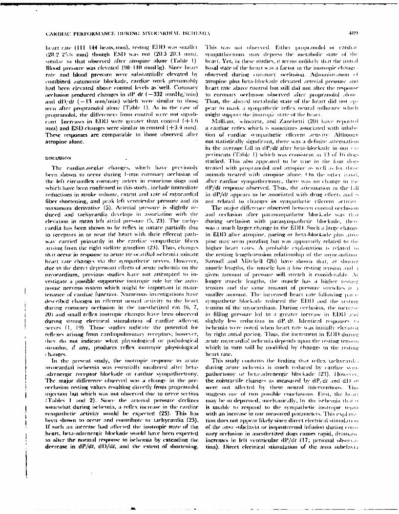
C~ARD)IAC PEAIRl: NI.\N(. DUR )1tING NIN-1111CMIA II SLtIII-At A UItt
-h~.' 2.iiit) though iFSl )%it, tIot (201.3 201.3 iiii) 111(.tt-nii ti, iies i'ttittii saen
sitili.ii it) that obst'rvcrd aifter aitroine atlone (Tale 1) heart. Yot, illiai stildjis, I! Seilis iliku ii t ilt liiiiiimii
Bloodi pressure was e'levated( (9011 ni n Il 1t0lg. Sitici lic';rt basal state of' the liv;ii "as a factor In thle illillipiC clitatiL'iratemdboc rstr were susatalIlv ted1) obtservedl luritiig utnmar\ iicclisioit. Adiititistti iioti o-orti1bi ntd a utonomi tu Wokade, carid iac work prc'stitna III) atro'p ine phlis bcltad-biii kad ecirhvai td airterial picnssult. .11i
had been i'levated above control leve'ls as wvell. Coronaryv litart rate atbove control bitt still did no01 alter then rI-tuutiseOeclusicin produced ('ha oge inl (11. di ( -332 tmuin-I g/inii) toi co rooar\ i-v rcciin iieservord ifter in upra Iolol i lon,and dl)'di ( - 13 in/ittin) whichl werc simtilaer to those Tihus, thn' altci;'d IlIetItioliC state of tht, ticairt did lititI,seecn after jiropra nok i a lone (Tat ulc I ). As inii th- cat; (if ol' war it task a s) inp!a titir tcrflex ntiral I iii e %% lit It
propraniitolol. the( difi ivncu It'e il Cron ettroul weitot sign ii' inigrltt stiuiai t t ie Iflhtiopjif stateo ic ue a hcIicant. Increasi's itt EIDl wvere greatet than control (+1 .6 Mi alliani, xhtwartz, and/.,ancietii (20) liiiv rejititdini) and ESD. chantges wvere simtilatr to cotrol (+3.4 tin). ;I Cardiac rctirx whtich is somietimtes ii-ociated with intoib;.These icsponscs arc comparable to those observed after tion of cardiac svinpatieticr eilcrint ,irtis it\- .Alttouviatropine alone, not stattisticailv sigrtiticait, there-( %is ;I defittiti' attenuetti-1ii
inl the atverage fallI inl d Pdt after beta-blockade Iit out
D I I.W; MS I ON eriiiietLsu (Table I) whtichi was nsistctti In 15 uof Il dUL!studlied. 'This also iil-telto bv' trite( ilitt Ii' itt iii *e
The cardiovasculair etiangi'swih hv tvoiiv tetd it liOtiiiil;tcatiuita' el-iii ibeen shiownt to occur dutritng I ttitt curiryr occ'lusiont of ;tttittuals treated \\.iIll atroilte ilnite. of )II the (ikt i, 1 V. I
the left cirritifex ccoonary a rtcrv itt cot use muns clogs antd a fte'cr i Cardiac Syt ipa t icc tottiv, there wasI tno u1-hi Lei I thwhich have been confirmed in) titis study. incltude utntediate d P/di responise ob-served. Titus. the at -tvinuat ion ill the 1 11redutc tions in stroke voii n, extentt and late oif invocr d ialt inl diP/dt a i tea rs t10 b e aSSOCi;ated With ii ~ it'g Ffects tind 1
fi bet' shortening, and peak left ve~lnt iar pre'ssturea id its itot related to c-iat ges in sympathiiiietic iefferet irti oima xii itni derrivative (5). Arterial p ressu re is slight ly tI-- Thc t ijor (Ii lfrenlice observed I bveit n liti A111o ftccli,1t'duced and tachycardiat dcllcps ill associatioit with tlie antd octusion after pat-tsytttpatiictrc litkldt' \%a,; tll
elevattont in mean lefIt atrtial ri'ssit c (5, 23i). 'Itte tar iiy- du ring ore iusic n wttit lpita 5y it licetic h)11c kad e, iccardia has% been shown to be reflex in inat ure partially cute was aI inucl larger change inl tite El )l. Sich 11i larer'( citaittto recreptors in or near th h~'leart xsrli tithc'irt c'ffi'ern t hi - in EI)I af'tct atriop inte, pacing or bet a -hiockad i') tln .
w.av carried prnitiarily itt the( Cardiac svmIlpitttictic fibiers pitte Iitay Sei'itt peizzliitg itt \%as ;palviltaell relattt'i t) il,-.1 rlii t fromu t ile tigiht st ella te g; InglIicti (23). '1 'hits c'i aiigvs hiigheir it';,rt ;Ites. A piob alei expl anitat ion is i Ia t d t
thatt occit in r'espItnse to actit' Iit m.ituia~l sclteiini;t initiate Ihe testinug let-il-teitsioct relatuionshtip oftil te ttvr~aiiiitri.hc'art rate changes; viat thei s tnaititetic ttilvvS. Ili)wi'Si't' Sat'niii atndc Mlitce'll (26i) iavi' shocwnt tha~t, att Shot u-i-
due to tilt-' direct dlepresant cl'ts iii iclite schctnia oil tile muscle lettgtits, th lti isl ha~s a lowV restitit, titsicilt ;1itiivocra rd iutn, p reviouts studies Ihave' t tul at telip ted to ill- gi veit at iat i t i f tsst will Siteh it cot isidiirai 11 s. A
vest igatc ;t possi ble supi )oitiv' in tric rtole fit- thei at ito- longer' Intise ii lc'tgt liii, ti'lttuscl Ichaxs a hItflIiertiil
nrutiii nervous systrit wh ich miiiightt Ill' itmtport anlt ill 111,11t- tensioni atd illte silt ic at ii ii it of i r'sun't ti't it'si .1
itenanoc' oif c'arciacr functin. Nutimeois ittestigaitiotts havce sittallci- antiouitt. T'I( iiicasid heart i-ate follow ing iai. -
clewtiitcd chiantie li i'il('nit liteiral ;Ict ivitv to iti' liti'tt svinpathi't i btickidc ed'ucedc thte ElP an) td i hit-ctidii ng corollary nc-cl usioti inl tli(' attest tizeci cat (6i. 7, ti'nlsion of t ilt- Illoca td i u . I hiri itz ccIn it in, t it icir!20) an td si natl rc'flex i not rupic cti i ms Itve i c'Ie'i iobsc'rve'd ill filling pre'ssure iM e toi a greter' intcrtease itt ED 411 i
durng a~tronig edccti-ic al stimnula tiont of car d ia;c ille'i'iiit slightly less ri tic t it inl d P, d. Idetiic a I trcsponttscs i,IIV'IrCs (1 I 1I9). Those st udic's iitd icaw th c' o t ett'tial fl. iscei ii i w'e tnt cc whe lie cart;t raIte \\ais initiall lv cv; i (i
rc'flc xes arising frot n ertriop lti Iliii a r nit- lcrs; 1 o~i'S ir, by righ t at ia l pac intg. IITus. tite in cremt it i EDD1 dil I 1
hey, do not indicate what pht~siolitgical orl paithloigical ;ttutc uimoc'ardi;l! isc'tticli'pends Itpl)ti t rn ntg et'wtil uIs, if any, prod uccs reflex iinotnco ne pihysiolto g ic-al witich in tutim \will be) int li lied by cli anqcs in theet- ii
hontges. hiatt rate.lit the piesilt tutdy, tltie inir I pic rv*;ptns to aciiv T 'h is situdy'cli ii frmis th f' intding th it t i' ex t achtive h.,i
tuv C;ttd in I i-4'hemnia was i'scrt t i ;tliv intvi c' alite'i bcta- dintitg t ic isci- nviiii;i is nt c'h ici tn-d I)\ i-aid i a s a -* irencrgic ri'c'cptoir blockade or- cardciac sy-Itipathticily . paithectoniv or bI n'~tidrene-gic bloickade (23). 1 lit\ (.\ 1* 'liii' major cliifcrenct- observecl was at change. in tilt' t~i-e- thc co'ntractile changes its incasutcied by I',d dv c dl) It
orrit~ion resti ng valutes re-sult intg dirtc'ti y front prioprt'aitolol wire tot al '-ted b y t besc nteutralI in tc'rvcnt ioni 'It lii,it 9i- t ion Ihut wvhichI wis itot obscrve Iclii' to nerve s'ctimolt suggc'sts5 li oi f two5 iossi li coiltmItsi is. Firtst , ill i' .i1
'TI*iht' I and 2). Sinte' the aicteial pre'ssurei d'clintes tilay- he So (leptessict. ilIc'cttiteally, In tin: isrii,-ttiiaii it-4noew hat dut'intg isc'lte uia, ;Ii-telcx iincteasev ii thme Cairidiac is utnatble tic respoxntd to tit- Syn epaxii i'tic i lii -it ii i 111u
ss'i pa i htt io activity womt icI lii' ex~m'c c'd (25). *1This has withi ait incrcease itt ott' t te';sltrccl ptaal etets. Tis i'X[ulItl -
Ilic'l'nflown to occutr and conitribte to tachycat'cia (23). tiolt doe's tnt aplic'ar likely sini-(- direct t'lc'cric~il ittiltiIf sic a i i ncrc'ase had ail-cti'd tilt' i not ropie statec of' t Ie of t(he ansi b- lay ia cor is' i itotctciicl infuosionit d uril ii mheart, lita~-;tdtcneric tlorkade would have Ilx'et expeced nary orchision iii ainesthuet ized dougs causeq rapid, dna itia i,
to alter the normnal respotuse to isrli'nia by c'xtcnditug tili-, increase's in ic'ft v-entric'ular ii l'/ll (17, pe'rsonatlobes--dorcreasc in dP/dl, dDi c, atnd tit, exten.t of Short(-tting. tioti). D~irect c'lectrical stimiulation of the atisa subcias ii
I ~~K.\SI' \R. It M-NIS. I'IT.IIRIS N, A\ND Ils)
.4i14 I- porao ( I 4Ing. kg.) ;Ix~lsI441( [lit, 444'. is 44444,5 44444 .]4 11544.. IThis cow i4 4454444 4'. 1v('Iqll4 (;'Il\ aIpIl'''1'''''
(I(-'(4 III 4441 4'5,4441151' (444454od ohsciva1 544\'; 4444) lit 1.44'). i III lilt(- it ',5ol 4441141 So4) Ind4441 144 c I III' lt '444i' .1( [Ill--!
Il. I IS. i e-1I IIi I I II if4''l.i4;I I (I v 'j 41'4( id 4114 (1 ifIll-s 4IIII\ 4' 1(. ~4-1' 11 44I'c4~. Is( I 541 wn an, lttle,, .L54oid IM)4 4iJ"4'
So4 s55'1141)144144c ill)i)4ll 4444 '.41411i cxper4'C that 1144 laii Ili dvo14iand(s I)v. tilt- 44c.4
luilits i 11, t h 'l(,I44 4)4 4 'j4(Iht iih lic'44*."4 i4I till(- to wson but no114444 444--cis Ill41 iti44 .4444.1 4d4
sX'14jlpa '14tr i4Inottoir suppoil4 4 (In'Nii. S4iII possibility 144 111 ,11 4 ',.it 2-IR.o' HL111211 ',444. \ir h ,' Omlimt., \Wil TI 7* ('4)41141i I4(h4d it'114.tha lo14(14'? 44141 v''(4)4'!'ill4 cond44iions, 20)7. .,4d II''I, s lsi h'rl .\ssKi.itwii,
ri ( her I I44' lI I'V(%i JolI lor4- I : I (44414 ticI I; I I q is I IilI it In h. t III; .45 .4 I pi-I- ;It 11 .'44Il Jim 1444 .41 tilt- .\4444444 m44 I'll-, 414e'w4
lilt. 4 4 4 ' c14144''"4pi cal1 il-11.c 1 l,1min(1111 inl . l. i.s 44344 A4 Ilhvsi.,1,4 '. ...I4(5111peolictic 4(~4r;z nctiva )4inputs to ) III(- herI. 1'( lo;4114: is hc4,'I 44n 4 ' 44454'' 1 .4 -.4 14'ss444 114h .1 .i4,p pN N-t , i4.
deioh4nswite14d (12. 23. 201. S t.. 1.4k.llil, N4ts I .l1s1444p 14454114444, (!f' <441thechc .
tic is Ilt Wilih ll nc I(lusion4 11,11, In tis sld", Illw44' ia~ I4'44444c'4S4Sfo44/. V_) 10122 Mij;4,. linis.
RLIA I NU.S
1. .\ NIOiW , I N.. W I'. ( Its i. NN A 1 \ 4.1414 ,1 1 . . 441' 44 In 1 . 11, .4 w \ W 4 It it. *4. H. (:*,%4 lit \ I I Nb4 %,I i4'.4 I'
* 2. l4444N4.s. G; F, ' VS. 1111011',. I. 1) AN 144444,' 4 1 .. K \N' A44. 4444444414444 1.44i/hwalulw NA' 1 21-' .44,44
14dX44444 maxin i .ttiv.,'5; (f li-ft 54444441444 'I4r .1"d4 tra.ll~- 16,. 114 lit.44 I1. I . 1) . NNII \ S. 1tI , I.t( 5444444441. llkv4'~4 i4414r44.4 ehai44ttr .45 ind4i4r, 4' f i144. 4444444qm 4IC,44t' (If IIlc lvft 4I444'4 t44 4l4444S4454 he 445cmisc44, 4I,4 (44/4444.4
v4titoc4 i cin44 tm dogs44445444 .1. P4ym.4 I.444/441 2ViS, 571 ,'4. 4:1I,.l 171. 11172.1,74S. 17. K iqm 4< L.. I N I \iku, \4.1 1. I 54.5.54I)I
.4. R1111 V. S , Nil 1 1) I 144444.544/. , 1 .I1 ( 4s (If alte c ;1,11, ti( 4444444444 44 i4; I.(Il'ct 44r444 ,44i44.4444. 4444tt -m 44 14, 14/44 4514
- 4. Visi~i, S, 1, 1). 11.ss44 1I . S44I.4sI., I I.I I I i \1 I . III,\ I I \N. A5., NI P4
ARK4S. It P1 II.Kf ;1, \N0 \ NI Iiv.
4:,N I4'444 5' I. K. F. N I t -ft I, - I.I- 1r4454.5( i.4 I N .III if - ,4 I'4 NIs I 4ii4d 5vr Ic I) I*. i.iit Fel,- '.o h4s44' 5, 5S 1,s
h1!7.1 2(4 1 . i7 ;.P I ~i5 44.A,../A444 4
6. It,,s',. V. S. let14L. ((4pr'54'k (11. 44'1I444 S',. 41I)4 \1 -I K *4 4
lIJI'N I I 11 I4 I \N4I.\ I141)4 I. PIA 'I MI. V'i. 4 1 41'. \N,4r44'1 \ 14 I1'lN
1.r44rv 44f (li c, J. IPbv4444., 14444I (""n il 1125, 836, I 'w,4 I'In4.1
'2 17 7401 74444 I'll,"4
7 At455N N5 . %NiI \ Nksi 4 itNt Spinasl ym4p.tthcn'44 ,'I,'sve 21. Mi/i mis, N . 1. mwiii44 4444 4.44445*,4 d44 4I, (.ir41144 444. r4 I,
.. 'ala,4,., by ct'44~r44444s 4i4.4'4pw4rs 1. I'1444... 1.4,44,144, 212 4,147 .7047', 141.] Ili4~ 444 14L' All4 ,&.I Itr,, 1 S2. '-",1 -27".44,.1 '7 1. U2. IN:I I Is. N, I ) F.. ...i, VN. NI 1444N. I'..44rIil, i. .d'~
It. I1iAt %%.S 5444, I. . ~4 \I I j<4
,\w St ... i,' )i 5,411,44's Lm1.45.41 the In4 slim-d4, o,i i4 ill ,4414r4'4t 4adi 344(4.44 444454' 14144 ilh4444 454 t IV s, I .4444 4444 41 h4',44''Iviiaiic f444,ctwit (.f 444, S\, 4441,.444tr 4444 \c'S 4f thr 4 (.44 . ianl 16', :t1 '4,, *4 I
irift .,4i4444 C ,r,,ipa~i,, '21 4.1 1 1,42. 144.1. 23. 111. 1 I-4454N. 1) 1'.. It.~ I.. KAS4'A, AND4 V 5. B0,11(1i All'(l
it, ( .44444445;KFN:' . I. I* , M. SF.N1 IVAN\54 1.R./,,5K44 Ill., .54 V. I :. ,,( 44114' 4415.44.IdaII, 4(444 444 lal 44444,4, kls'I'451 it, il44- I44 ~..
ItAMtull Yi,'iptiraty vo4,r4444,44'. i444'14*1444 i44 44444.4.44,44 4)441 44,1. 4, (44/,4.4 1/44 Q'2 W4,-' f.70, 44474l.ic'raI U.,w an4d 4'ltctr,cr1i4.lr444 A. J4. I'hvm,,. 212 4)425- 24. I 11A4454. 1 \ . I \ .\~ l R.4W. I'. I ;t5 11 AM) III.lo44, 1/ 1i447. 4444 4. R{,'41 wt4,,4 4.414,4.41c '14 1 iit4,, (f 4414 '-'''i'a'th~'',
to). (.s iot 44oN, 1.- L\1ra,4-.r41iac 4.4444445 -iititt4'4
4i41 4,, 4,vp,t'i,iim, 144/4444444 1/'?,. il 11411 120114, 1472.
(Iipi crerotia.4V 'socIi,4, hp .1 Cwdt1sd. It: '415 '217, 4I'4. 25. S skN4,it I S. I1 , I 44 ( ;1i\i )44RI, S K l'1s A X\,,,~ ,s' II I4,
11. I)4.v)i4. 15NV, 444 I .4ft 4.4'14444'41.4 al'4554r'4.444444 141'4,44 ANI)R, I AN5144' R,'t1 41i4444li i 44 54-44r1l,4 444411t .44ti,. \41441 ,,'.i,,iiIl o cooi5i t4Ii9' lifi (lilt4 iitgU4 414441444 444i4ial4 isc'4i54 Ow~ (.44444144 544444. ItscI'v t 444t% 4 atria 1 an \l-ttractl44 1 I4,4444
44inthk rd fly a4trial4 )4d4'141. CwtiaI4 .... 42 lilt 4422. I44744 It,. .4 112t4 It'1l,. -11,04
12. I uRi'a..i . C. NI . K .1. 1ipliaw4N, ,41) 1 L M. S44,55. 1<'(4,' Olc4 I, 2b,. SAHNI',,,I . S . A-411 MM 111- I~ii~ 1 'Ill' 4444444,41 if III,,,
4444 (h4r 144'rt J4f 5414411idalin ('ft lit ia~l ir''ptiir!. J. Phsi'/7.441 1f.4444/444 if 444ll', 4-141 44 4 1. 4<4/1'444/4- 1wr Ili ,4(I444 O55ew .4 444.4
'2111: 1147-41 II4474. 111C. AM, I 1,I S14.' .P16,c. q 2, 54,1. 4. ('(4344. 1- , 144 W4
13. IIIANI P.V, 1I. Gi. . Co~iN \:414 Nip N. S. sKINNVA, 114. Di)1'44i:41 il 2.'I'I 1"1'N ' .1, AN IX C RANDAI~s I.. IK .11lit 441( 104.14I 41 44 4
.'41wriom.'4t.ita -'-t~4iiry .srt4'ry 4"-t hisi1444. AInt. .1. (.ard44d. 27 : 51:4 H'P.o,474. "It, 14.445554. It . \NJ. I il l V1:I ' Ihr 4( 4f44~4 44
11. Iii HMAN, NI. V , ANII It. 4 4,141444 1441114.1i4,114 l4(i,'ift ve''larivi',4I at nIyixrd-i 44411441 ~ ni ten44j4.444~ .1 / Pltym. 112 4.
Awyiirogy. Aw~, .1. Cp'relI,,,. lit: 770., W,7 PH.6.1
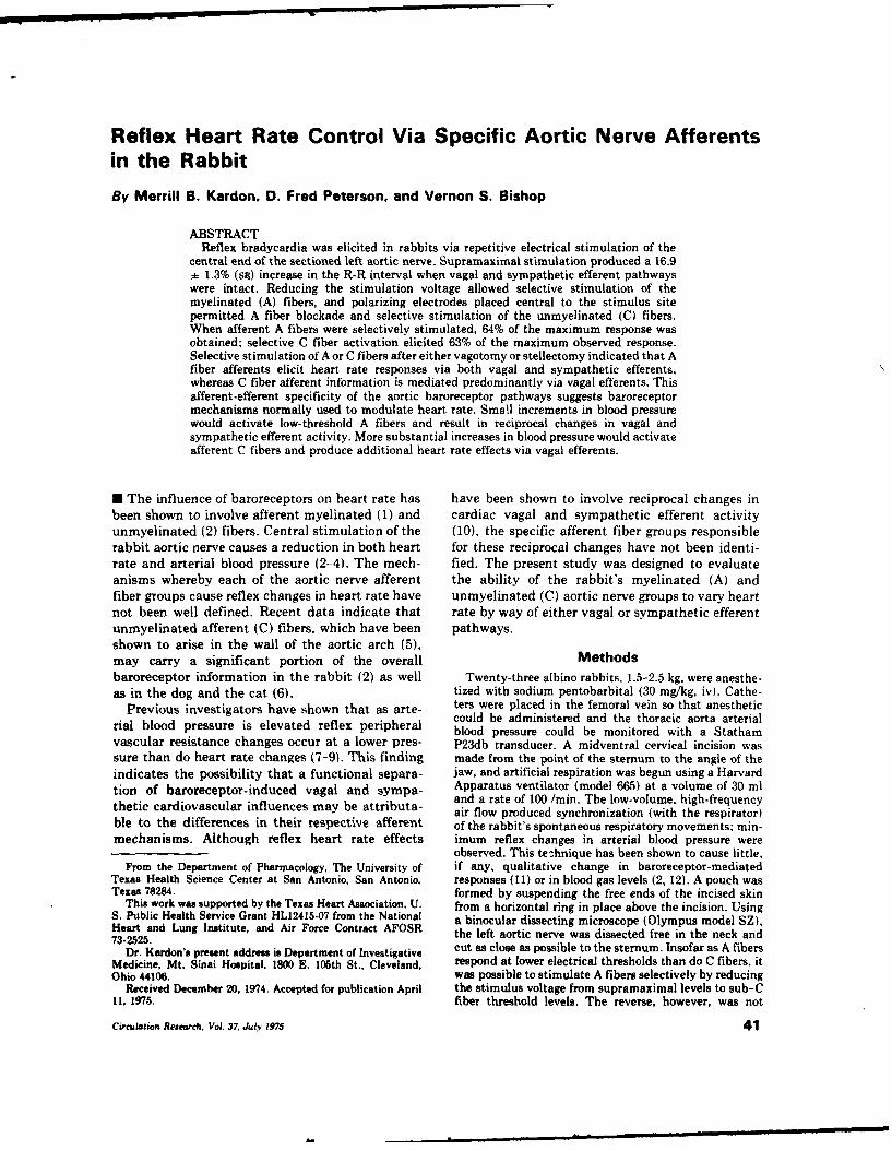
Reflex Heart Rate Control Via Specific Aortic Nerve Afferentsin the Rabbit
By Merrill B. Kardon, D. Fred Peterson. and Vernon S. Bishop
ABSTRACTReflex bradycardia was elicited in rabbits via repetitive electrical stimulation of the
central end of the sectioned left aortic nerve. Supramaximal stimulation produced a 16.9* 1.3% (SE) increase in the R-R interval when vagal and sympathetic efferent pathwayswere intact. Reducing the stimulation voltage allowed selective stimulation of themyelinated (A) fibers, and polarizing electrodes placed central to the stimulus sitepermitted A fiber blockade and selective stimulation of the unmyelinated (C) fibers.When afferent A fibers were selectively stimulated, 64% of the maximum response wasobtained; selective C fiber activation elicited 63% of the maximum observed response.Selective stimulation of A or C fibers after either vagotomy or stellectomy indicated that Afiber afferents elicit heart rate responses via both vagal and sympathetic efferents,whereas C fiber afferent information is mediated predominantly via vagal efferents. Thisafferent-efferent specificity of the aortic baroreceptor pathways suggests baroreceptormechanisms normally used to modulate heart rate. Small increments in blood pressurewould activate low-threshold A fibers and result in reciprocal changes in vagal andsympathetic efferent activity. More substantial increases in blood pressure would activateafferent C fibers and produce additional heart rate effects via vagal efferents.
U The influence of baroreceptors on heart rate has have been shown to involve reciprocal changes inbeen shown to involve afferent myelinated (1) and cardiac vagal and sympathetic efferent activityunmyelinated (2) fibers. Central stimulation of the (10). the specific afferent fiber groups responsiblerabbit aortic nerve causes a reduction in both heart for these reciprocal changes have not been identi-rate and arterial blood pressure (2-4). The mech- fled. The present study was designed to evaluateanisms whereby each of the aortic nerve afferent the ability of the rabbit's myelinated (A) andfiber groups cause reflex changes in heart rate have unmyelinated (C) aortic nerve groups to vary heartnot been well defined. Recent data indicate that rate by way of either vagal or sympathetic efferentunmyelinated afferent (C) fibers, which have been pathways.shown to arise in the wall of the aortic arch (5),may carry a significant portion of the overall Methodsbaroreceptor information in the rabbit (2) as well Twenty-three albino rabbits. 1.5-2.5 kg, were anesthe-as in the dog and the cat (6). tized with sodium pentobarbital (30 mg/kg. iv). Cathe-
Previous investigators have shown that as arte- ters were placed in the femoral vein so that anestheticcould be administered and the thoracic aorta arterial
rial blood pressure is elevated reflex peripheral blood pressure could be monitored with a Stathamvascular resistance changes occur at a lower pres- P23db transducer. A midventral cervical incision wassure than do heart rate changes (7-9). This finding made from the point of the sternum to the angle of theindicates the possibility that a functional separa- jaw, and artificial respiration was begun using a Harvardtion of baroreceptor-induced vagal and sympa- Apparatus ventilator (model 665) at a volume of 30 ml
and a rate of 100 /min. The low-volume, high-frequencythetic cardiovascular influences may be attributa- air flow produced synchronization (with the respirator)ble to the differences in their respective afferent of the rabbit's spontaneous respiratory movements; min-mechanisms. Although reflex heart rate effects imum reflex changes in arterial blood pressure were
observed. This tezhnique has been shown to cause little,From the Department of Pharmacology, The University of if any, qualitative change in baroreceptor-mediated
Texas Health Science Center at San Antonio, San Antonio, responses (11) or in blood gas levels (2, 12). A pouch wasTexas 78284. formed by suspending the free ends of the incised skin
This work was supported by the Texas Heart Association. U. from a horizontal ring in place above the incision. UsingS. Public Health Service Grant HL12415-07 from the National a binocular dissecting microscope (Olympus model SZ),Heart and Lung Institute, and Air Force Contract AFOSR the left aortic nerve was dissected free in the neck and73-2525.
Dr. Kardon's present address is Department of Investigative cut as close as possible to the sternum. Insofar as A fibersMedicine, Mt. Sinai Hospital. 1800 E. 106th St., Cleveland, respond at lower electrical thresholds than do C fibers, itOhio 44106. was possible to stimulate A fibers selectively by reducing
Received December 20, 1974. Accepted for publication April the stimulus voltage from supramaximal levels to sub-C11, 1975. fiber threshold levels. The reverse, however, was not
Circulatin Research. Vol. 37. July 1975 41
42 KARDON, PETERSON, BISHOP
possible. To elicit the preferential activation of C fibers, activation (Fig. IA), a stimulus-response run was begun.the conduction of A fibers was first blocked using a pair In a separate group of rabbits, the C fiber afferents wereof polarizing electrodes (13). Three sites were isolated activated to the exclusion of the A fibers by using thealong the length of the left aortic nerve. The free central technique of Manfredi (13). While the left aortic nerveend of the cut nerve was placed on a pair of platinum- was stimulated at supramaximal intensity (sufficient toirridium stimulating electrodes connected by way of a activate all fibers), a small blocking current (5-15 ua)Grass SIU5 stimulus isolation unit to one channel of a was applied to the nerve via the interposed blockingGrass model S-88 stimulator. At the second more rostral electrodes. By adjusting the blocking current while thesite midway along the length of the left aortic nerve in evoked potential was monitored, it was possible tothe neck, a second pair of electrodes was positioned. eliminate A fiber conduction and leave C fiber conduc-These electrodes were covered with saline-soaked cotton tion little affected (Fig. 1B). A stimulus-response runwicks and connected via a second stimulus isolation unit could then be made while only C fibers were beingto the 100,000-ohm d-c output of the second channel ofthe S-88 stimulator. As previously described, they servedas A fiber blocking electrodes (13). Bipolar recording felectrodes were placed at the third most rostral site.These electrodes were connected to a pair of cascadedGrass model P15 preamplifiers, and they served tomonitor the evoked potential. The evoked potential wasrecorded on Polaroid film from the face of a TektronixD12 storage oscilloscope. Bilateral vagotomies were per-formed in the cervical region. Stellectomies were per-formed by removing a section of the sympathetic chainbetween the first and the third thoracic interspace. Heartinterval was monitored by way of needle electrodesplaced along the sternum. These electrodes were con-nected to a Beckman type 9857B cardiotachometercoupler. Both heart rate and arterial blood pressure wererecorded continuously using a Beckman type R-M (411) boscillograph.
On-line monitoring of the R-R interval and control ofthe nerve stimulus parameters were accomplished usinga DEC PDP 8/E digital computer. Bursts of stimulusimpulses were applied to the left aortic nerve during eachR-R interval in fixed synchrony with each R wave. Theduration of each impulse was 0.3 msec as established by Bthe stimulator. The run duration and the stimulationsequence, which consisted of impulse frequency, impulsenumber, burst duration, and timing of the stimulus burstwithin each R-R interval, were established by the com-puter in accordance with the experimental protocol. Thecomputer stored each R-R interval in a given run andaveraged it with the equivalent interval in previous runsmade under identical stimulus conditions. The sequenceof average R-R interval values for each group of stimulus-response runs was then printed out on command. Threekey parameters, latency to onset, latency to peak. andthe value of the peak response were printed out alongwith their respective standard deviations. The latency toonset represents the number of heart intervals from the FIGURE 1
beginning of the stimulus to the end of the interval which A: Single stimulus compound-evoked potential recorded fromfirst exceeded the control. The latency to peak represents the left aortic nerve. Top: At a supramaximal stimulus inten-the number of heart intervals from the beginning of the sity, A fibers (fast component at left) and C fibers (slow wave atstimulus to the end of the longest interval during the center) are activated. Bottom: At a reduced stimulus voltage, Cresponse. Peak response represents the maximum per- fibers are no longer activated, but the A wave is unchanged.cent change from the control R-R interval. Interelectrode distance = 30 mm, sweep speed = 5 msec/cm. B:
Comparisons were made between the magnitude of the Overlay of three single stimulus compound-evoked potentialsreflex bradycardia elicited by either A or C fiber stimula- which were recorded in rapid succession illustrating blockage oftion before and after selective deefferentation (vagotomy A fiber conduction. Top: At a supramaximal stimulus intensityor stellectomy). To activate aortic afferent A fibers alone, all fibers are activated. Bottom: When the blocking currentthe stimulus intensity was adjusted while the evoked (5-15 Ma), is on, the A wave is blocked: the C wave is reducedpotential was monitored. When the stimulus voltage was slightly. Interelectrode distance - 35 mm, sweep speed = 5reduced enough to eliminate activation of C fiber affer- msec/cm. The horizontal bar represents I cm of the oscilloscopeents, while causing little or no change in A fiber sweep or 5 msec in time.
Circulation Research. Vol. 37. July 1975
AORTIC AFFERENT HEART RATE CONTROL 43
stimulated. Without left aortic nerve stimulation, the response. During the course of the electrical nerve block,blocking current never elicited a persistent change in the blocking current often fell somewhat during the firsteither arterial blood pressure or heart rate. Subsequent 15-30 seconds, no doubt as a result of electrode polariza-to the control runs, selective deefferentations were per- tion during the blocking phase. This fall sometimes ledformed (bilateral vagotomy or bilateral stellectomy). to a partial loss of block selectivity. However, a run wasRepeat runs were carried out to indicate the relative not made until the block was reestablished (by raisingimportance of vagal and sympathetic pathways in the the blocking current slightly) and its stability verified forcontrol of heart rate via either myelinated or un- at least 30 seconds. Subsequently, during the blockingmyelinated aortic afferents. period, no detectable blocking current creep was seen nor
Standard errors were calculated for the onset and peak was there a significant tendency for the block to degrade.latencies as well as for the peak responses. The signifi- Selective nerve block was not normally maintained forcance of the difference was determined by the t-test. P periods longer than 2 minutes. The current level neces-values < 0.05 were considered significant. sary to effect a selective aortic nerve block was essen-
tially unchanged for all trials in each rabbit. Barbitu-CRITIQUE OF METHODS rates are well known for their ability to depress the vagal
The polarizing blocking technique used in this series of centers. However, in our experiments, the rabbits wereexperiments has been shown to cause selective blockade maintained at a light surgical anesthetic level with littleof myelinated fiber conduction by producing a failure of apparent depression of the vagal component as indicatedconduction between the blocking anode and cathode by the slope of the heart rate response.(13). The A beta group is most sensitive to the d-cblockade. However, at current levels sufficient to block Resultssome of the beta group, asynchronous firing of some less In 23 rabbits, repetitive synchronous stimulussensitive nerve fibers occurs. This phenomenon has beenshown to result from cathodal excitation. With complete bursts were applied to the left aortic nerve atblockade of the beta fibers (and subsequent blockade of frequencies of 50 and 100 Hz. In 13 rabbits, a totalthe delta group as well), the asynchronous firing no of 48 trial runs was made while stimulating atlonger is seen at the recording site due to conduction supramaximal intensity (all aortic nerve fibersfailure at the anode. The conduction of unmyelinated C activated) (Fig. 2). The average R-R interval in-fibers is only slightly affected by this level of current. creased 16.9% from the control level of 209 ± 3
In the present experiments, the direct current wasadjusted to block myelinated fiber conduction while msec. The latency to onset and that to peakcausing as little asynchronous firing as possible. How- bradycardia were 4.4 intervals and 21.0 intervals,ever, insofar as d-c levels that completely suppressed respectively. Bilateral vagotomy had no significantsynchronous firing also caused significant C wave depres- effect on resting heart rate, as previously reportedsion. the level was adjusted to yield A wave blockadewith as little C wave depression as possible. The amount (2). It did result in a reduction of the peak responseof asynchronous firing passing the blocking anode was to 11.0%. The latency to onset and the latency tojudged to have little qualitative effect on the observed peak were increased to 8.4 and 32.6 intervals,
LTO LTP PR9- 3- 28-8- 30- L 24-
204 40: !K z 1S5- 15- .- 12-
ZIOZ 4- I 0- W4 8-
2Il W.0 CP(00 P C05 P(05 K001 PLo0
o Intacta VgotomyE3 Stel/ectomy
FIGURE 2
Influence of efferent neural pathways on the heart rate response during supramaximal aortic nervestimulation. Each pair of bars compares responses after either vagotomy or cardiac sympathectomyto responses obtained immediately prior to nerve section. Latency to onset (LTO) and latendy to peak(LT) are both measured in R-R intervals after initiation of electrical stimulation. Peak response(PRI is measured by the percent change in the R-R interval when the heart has slowed maximally.
Circulation Researc., Vol. 37. July 1975
44 KARDON, PETERSON, BISHOP
respectively. Thus, vagotomy reduced the peak 15.3 intervals, which was not significant. The peakresponse by 35% (P < 0.001), while the latencies to response was equally dependent on activation ofonset and to peak were significantly delayed in both A and C fiber afferents. The reduction intime. In the remaining 10 rabbits. 33 trial runs peak response was from 19.0% to 12.2% (P < 0.02)produced an average bradycardia of 14.9%. The when afferent A fibers were stimulated alone,onset and the peak bradycardia occurred at 3.8 and whereas stimulation of C fibers alone reduced the17.9 intervals, respectively, from the stimulus on- response from 13.4% to 8.0% (P < 0.01).set. Stellectomy slightly decreased the resting In 11 rabbits, a comparison was made betweenheart rate and reduced the reflex bradycardia by the reflex heart rate response to A fiber activation41% (P < 0.01). The latency to onset was not before and after either vagotomy or stellectomysignificantly affected, and the peak bradycardia (Fig. 4). Selective A fiber stimulation produced anoccurred 4.5 intervals sooner (P < 0.05). Thus, average fall (peak response) in heart rate of 10.5%.when the entire aortic nerve was activated, loss of The latencies to onset and peak were 4.1 and 11.0the sympathetic efferent pathways reduced the intervals, respectively. Vagotomy in 6 of thesemagnitude of the reflex bradycardia, and the rabbits reduced the peak response by 63%, and thelatency to peak response but had no effect on the onset of the response was delayed by 3.0 intervals,latency to onset. In contrast, loss of the parasympa- (P < 0.05). In the remaining 5 rabbits, stellectomythetic pathways extended both the onset and the reduced the peak response to 5.4% (P < 0.01) (apeak latency while it, too, reduced the magnitude 61% reduction). The latencies to onset and peakof bradycardia. were 4.8 and 10.2 intervals, respectively, but nei-
The relative influence of afferent A and C fiber ther of these changes was statistically significant.activation on heart rate response, when all efferent In ten rabbits, A fiber conduction was blocked,pathways are intact, was studied in 11 rabbits (Fig. and a total of 30 trial runs was performed (Fig. 5).3). Activation of A fibers alone caused no signifi- In trials performed in six of these rabbits, C fibercant change in the latency to onset. When C fibers stimulation reduced heart rate by 8.5%. The laten-were selectively activated, the latency to onset was cies to onset and peak were 5.8 and 16.5 intervals,increased from a control value of 4.6 intervals to 6.0 respectively. Vagotomy in this group reduced theintervals, which was a significant 31% increase (P peak response to 4.9% and increased the latencies< 0.01). The latency to peak was greatly reduced to onset and peak to 8.8 and 22.8 intervals,when C fibers were not activated. The reduction respectively. Thus. vagotomy reduced the peakobserved was 19.7 intervals to 11.4 intervals (P < response by 42% (P < 0.01). The onset of the0.01). When C fibers alone were activated, there response was delayed by 3.0 intervals (P < 0.05).was a reduction from control, 19.6 intervals to In a total of 11 trial runs performed in the remain-
LTO LTP PR7- 28- 28-
6- 24- W 24-05- 20- 020z2" 4- 16- " 6 -
MIN-3- 12 12-
WU
4 4
P (01 P(Ol P(02 P(OI
* All AffelsC9 1A" Fiber AfferentEl "C"Fiber Afferent
FIGURE 3
Afferent aortic nerve fiber influences on reflex bradycardia. Each pair of bars compares either A fiberor C fiber stimulation with supramaximal stimulation carried out immediately prior to selective stimu-lation. Comparison of A fiber stimulation with control represents a mean of 31 averaged trials in IIrabbits. C fiber comparisons with control are for 30 trials in 10 rabbits. Abbreviations are thesame as they are in Figure 2.
Circulation Research. Vol. 37, July 1975
AORTIC AFFERENT HEART RATE CONTROL 45
LTO LTP PR8- 61 14-
7 14- 12"-I~
4- 12 10
I- 0.0 ,.2...tb,,-EJ J
P(.OI P(.0I P(.OI
SStellectomy
FIGURE 4
Influence of efferent neural pathways on the heart rate response during selective aortic' A fiber stimu-lation. Each pair of bars compares responses after either vagotomnv or cardiac sympathectomy withresponses prior to nerve section. Definitions are the s ame as they are in Figure 2.
ing four rabbits. stellectomy did not cause a fibers and that its magnitude depends on thesignificant change in peak response. No significant number and the type of fibers being activated.change was observed in either latency to onset or The effect of A and C fiber afferents as well asthe latency to peak. When aortic unmyelinated that of vagal and sympathetic efferents on the peakfibers were activated, loss of parasympathetic ef- response indicates that A and C fiber afferents areferent pathways caused a reduction in peak effect approximately equipotent so long as both efferentand a delay in both onset and peak bradycardia. pathways are intact. Vagal and sympathetic effer-Loss of sympathetic efferents, however, had no ent pathways are equally important in eliciting asignificant effect on the peak response, indicating reflex bradycardia when all fibers or when only thethat aortic nerve unmyelinated f'ibers mediate A fibers are activated. However, removal of theheart rate predominantly via vagal efferents. sympathetic efferents has no effect on the heart
rate response when only C fibers are activated.Discussion Since the bradycardia to selective C fiber stimula-
This study provides the first direct evidence that tion is not abolished after vagal section, we cannotaortic nerve-mediated reflex bradycardia occurs unequivocally disregard a sympathetic efferentin response to activation of either afferent A or C involvement in the reflex response. Possibly, in the
LTO LTP PR10 281 J9 - 24 -2.
42 -AI I
P( 05 P(.OI
[3 Intc
E3 Stellectomy
FIGURE 4
Influence of efferent neural pathways on the heart rate response during selective aortic C fiber stimu-lation. Each pair of bars compares responses after either vagotomy or stellectmy with responsesprior to nerve section. Definitions are the same as they are in Fiure 2.
Circulation Research. Vat. 37. July 1975
.. .... .. ....... .fou ra bt,.......id n t c u e ies a d h t is ma nt d ep n s o h
44 KARDON. PETERSON, BISHOP
presence of intact vagal influences, sympathetic are not known at this time. However, working ininfluence is minimal, whereas in the absence of the cat, Kunze (17) has shown that activation ofvagal activity sympathetic influences are substan- the arterial baroreceptors causes demonstrabletial during selective aortic C fiber stimulation. changes in the activity of vagal efferents whichSimilar responsiveness has been demonstrated in innervate the sinoatrial node. These vagal efferentsisolated atria exposed to acetylcholine and norepi- are small-diameter myelinated fibers.nephrine (14). In any case, insofar as baroreceptor An indication of the order of activation of the Aeffects on heart rate are concerned, the cardiac and C fiber aortic baroreceptor pool may be in-sympathetic influence appears to be primarily ferred from the known progression of peripheraldependent on changes in afferent A fiber activity, vascular resistance and heart rate effects seen withIn contrast, vagal influences are modulated by both increasing arterial blood pressure. Glick and Covellafferent A and C fibers in the aortic nerve. These (8) and Allison et al. (7) have shown that withresults are supported by similar findings during increasing arterial blood pressure in the dog, reflexcarotid sinus stimulation and neural physiological reduction in peripheral vascular resistance (whichstudies of the vasomotor areas of the brainstem (15, is assumed to be largely under the influence of the16). In the brainstem of the cat and the rabbit, sympathetic nervous system) occurs at a lowerclosely situated areas have been identified which blood pressure threshold than does bradycardia.receive information from either A or C fibers (15, This finding indicates that during the course of an16). Furthermore, Kumada and Nakajima (15) increase in arterial blood pressure the afferentshave demonstrated that inputs from myelinated which are activated at lower pressures are thoseaortic nerve afferents innervate both the vagal and which tend to suppress sympathetic efferent activ-the sympathetic sensory areas of the rabbit brain- ity to the vasculature. Angell-James (21) has shownstem and that unmyelinated afferents are absent that, when they are activated physiologically, aor-from the sympathetic sensory areas. tic nerve fibers fire at a frequency which is largely
Selective activation of either A or C fiber affer- independent of the rate of change of pressure in theents demonstrates that the myelinated group is aortic arch so long as the pressure is above thresh-responsible for the earliest onset of bradycardia. old for a given receptor. This finding suggests thatThis finding, of course, is predictable, since myeli- the relative importance of recruitment of addi-nated fibers can have many times the conduction tional baroreceptor afferents as pressure increasesvelocity of unmyelinated fibers. Likewise, selective is greater than that of increasing firing rates of in-cardiac efferent nerve section demonstrates that dividual fibers during the systolic phase. Theintact vagi are essential to elicit the earliest onset results of Kardon et al. (3) demonstrate the impor-of reflex bradycardia. Again, this finding is predict- tance of impulse number to aortic nerve reflexable, since the vagus contains many myelinated heart rate effects. These two studies, in combina-efferent fibers (17, 18) and the cardiac sympathetic tion with the present work, indicate that thenerves contain few myelinated fibers of any type important features of rabbit baroreceptor control of(19, 20). heart rate are the type and the number of fibers
Our results illustrate the long time constant being activated over a given time period as well asnature of the sympathetic influence on heart rate. their individual thresholds. If aortic C fibers haveThe elimination of efferent cardiac sympathetic different pressure thresholds than do aortic Anerves causes a reduction in the number of inter- fibers, the reflex effect of C fiber recruitment onvals to peak effect with no significant effect on the heart rate should differ from the reflex effect oflatency to onset This finding infers that rapidly recruitment of additional A fibers, as shown in thisconducting A fiber afferents which modify sympa- study. Therefore, increases in the frequency ofthetic efferent activity serve a function which is not whole aortic nerve activity in response to augmen-dependent on their high conduction velocity. How- tation of arterial blood pressure must occur largelyever, rapid modification of heart rate is vagally as a result of successive recruitment of nerve fibersmediated (10). This study shows that myelinated having higher pressure thresholds rather than byaortic nerve fibers influence the vagal control of increases in the firing rates of the individual nerveheart rate. Consequently, the fastest potential fibers involved. Finally, a previous study (2) hasaortic baroreceptor influence on heart rate must shown a progressive increase in reflex effects withoccur by way of these afferent A fibers modulating increased recruitment of aortic nerve afferents:efferent vagal activity. The efferent vagal fiber blood pressure effects are seen at the lowest stimu-types which are modulated by these aortic afferents lus intensities (rapidly conducting A fiber activa-
Circulation Research. Vol. 37. July 1975
.-- J A
AORTIC AFFERENT HEART RATE CONTROL 47
tion), but heart rate effects require recruitment of 3. KARDON M. PETERSON DF. BISHOP VS: Beat-to-beat regula-more slowly conducting myelinated fibers. Thus, it tion of hart rate by afferent stimulation of the aortic
is inferred that the magnitude of the heart rate nerve. Am J Physiol 227:598-600. 19744. DOUGLAS WW. RITCHIE JM, SCHAuMANN W: Depressor re-response can be augmented until all afferent C flexes from medullated and non-medullated fibers in the
fibers are activated as well. rabbit aortic nerve. J Physiol (Lond) 132:187-198, 1956
In combination with what has been shown in the 5. NONIDEZ JF: Aortic (depressor) nerve and its associatedpresent study to be the afferent-efferent specificity epithelioid body. the glomus aorticum. Am J Anat
of the aortic baroreceptor pathways, the order of 57:259-302. 19356. COLERIDGE HM. COLERIDGE ,JCG. DANGEL A. KIDD C. LUCKelectrical activation of the afferent fibers from JC. SLEIGHT P: Impulses in slowly conducting vagal fibers
myelinated to unmyelinated fibers may illustrate frorn afferent endings in the veins, atria. and arteries ofthe baroreceptor mechanisms normally used to dogs and cats. Circ Res 33:87-97. 1973modulate heart rate. If the A fiber pool with its 7. ALLISONJL, SAGAwAK, KUMADAM: An open-loop analysis of
combined vagal-stimulatory and sympathoinhibi- the aortic arch barostatic reflex. Am J Physiol217:1576-1584, 1969tory effects is activated at lower blood pressure 8. GLICK G. COVELL JW: Relative importance of the carotid
thresholds, the normal baroreceptor control of and aortic baroreceptors in the reflex control of heart rate.heart rate would occur via their reciprocal action Am J Physiol 214:955-961, 1968on vagal and sympathetic efferents at small incre- 9. HAINSWORTH R, LEDSOME JR, CARSWELL F: Reflex responses
from aortic baroreceptors. Am J Physiol 218:423-429.ments in arterial blood pressure. More sustained 1970increases in blood pressure would activate vagal 10. WARNER HR. RUSSEL RO: Effects of combined sympatheticefferents in larger proportion via C fiber afferents and vagal stimulation on heart rate in the dog. Circ Resand would produce more profound heart rate ef- 24:567-573. 1969
fects. 11. OBERO, PA, SJOSTRAND U: Studies of blood pressure regula-tion: Ill. Dynamics of arterial blood pressure on carotidSympathetic regulation of heart rate in a pre- sinus nerve stimulation. Acta Physiol Scand 81:96-109.
dominantly high-frequency, low-threshold barore- 1971ceptor system suggests th these sympathetic 12. JoNzoN A, OBERG PA. SEDIN G, SJOSTRAND U: High fre-efferents are modulated by subtle changes in arte- quency low tidal volume positive pressure ventilation
rial blood pressure. The known frequency-response (abstr). Acta Physiol Scand 80:21A. 197013. MANFREDI M: Differential block of conduction of largercharacteristics of the sympathetic neuroeffector fibers in peripheral nerve by direct current. Arch Ital Biol
mechanism no doubt cause this subtle reflex 108:52-71. 1970bradycardia to occur over a longer time course than 14. CARRIER GO, BISHOP VS: Interaction of acetylcholine anddoes that initiated by changes in vagal activity. norepinephrine on heart rate. J Pharmacol Exp TherFluctuations in blood pressure which alter sympa- 180:31-37, 1972
15. KUMADA M. NAKAJIMA H: Field potentials evoked in thethetic influences via afferent A fibers would cer- rabbit brainstem by stimulation of the aortic nerve. Am Jtainly cause rapid beat-to-beat changes in heart Physiol 223:575-582. 1972rate via vagal efferents. When blood pressure 16. PLANCHE D. ROHNER JJ. COMTE DE VAULX G: Projectionsexceeds this range, the increased recruitment of C corticales du nerf aortique de Cyon-Ludwig. J Physiolfiber afferents would cause the potential for beat- (Paris) 66:211-218. 1973
17. KUNZE DL: Reflex discharge patterns of cardiac vagalto-beat heart rate control to be progressively efferent fibers. J Physiol ILond) 22:1-15. 1972
masked. Under these circumstances, heart rate 18. EVANS DHL, MURRAY JG: Histological and functional stud-would no doubt fall farther in response to continu- ies on the fiber composition of the vagus nerve of theous activation of both A and C fiber afferents and rabbit. J Anat 88:320-337, 1954vary less in response to beat-to-beat blood pressure 19. MALLIANI A. SCHWARTZ PJ, ZANCHETrT A: A sympatheticvane reflex elicited by experimental coronarv occlusion. Am Jchanges. Physiol 217:703-709, 1969
References 20. MALLIANI A, PARKS M. TUCKET-- RP. BROwN AM: Reflexincreases in heart rate elicited by stimulation of afferent
I. PELLETIER CL, CLEMENT DL. SHEPHERD JT: Comparison of cardiac sympathetic nerve fibers in the cat. Circ Resafferent activity of canine aortic and sinus nerves. Circ 32:9-14. 1973Res 31:557-568. 1972 21. ANGELL-JAMES JE: Effects of altering mean pressure, pulse
2. KARDON M, PETERSON DF, BISHOP VS: Reflex bradycardia pressure and pulse frequency on the impulse activity indue to aortic nerve stimulation in the rabbit. Am J fibers from the aortic arch and right subclavian artery inPhysiol 225:7-11. 1973 the rabbit. J Physiol (Lond) 214:65-88, 1971
Circulation Research. Vol. 37. July 197.5
Reprint & Copyright 0 byAerospace Medical Association, Washington, DC
Cardiovascular Changes During andFollowing 1-Min Exposure to *Gz Stress
D. FRED PETERSON, VERNON S. BISHOP, and HOWARD H.
ERICKSON
Department of Pharmacology, The University of Texas HealthScience Center at San Antonio, San Antonio, Texas 78284 andU.S.A.F. School of Aerospace Medicine, Brooks Air ForceBase, Texas 78284
PFTERSON, D. F., V. S. BisHop, and H. H. ERICKSON. Cardio- performance during acceleration in anesthetized experi-vascular changes during and following 1-min. exposure to +Gz mental animals (1,6). Left ventricular pressure fallsstress Aviat. Space Environ. Med. 46(6):775-779, 1975.
Magnitude and duration of cardiovascular resposes following progressively further during increasing +G. forces
+Gz forces of 1-5 G were studied In chronically instrumented while dp/dt responses are variable (1). Distribution ofanesthetized dog. During lower G forces (+1 to +3Gz), re- flow is not equal during acceleration, since coronaryp no 1 were varlable. In most dogs during higher G forces flow increases at low G forces but decreases at higher
(+4 or +5 Gz), aoric pressure, adliaco oItat, left ven ikular G forces (6). A redistribution of blood flow has alsopressure, and dp/dt were all dramatically co..preomued Thench gs were observed whether the oud of Th gra tonl been observed during +G. (18,19).Inertial force wa slow (0.1 G/s) or rapid (1.0 G/s). Cardlo- Further information regarding responses associatedvascular changes after acceleration were consistent. Left trial with +G. forces is becoming increasingly necessary.pres ure and arteial preure re and a transient rise in dp/dt Aircraft capable of maintaining high +G. levels havewas often observed. Cardiae output roe briefly, then fell; hence,peripheral reistance increased. M m o of thes already been developed and future aircraft will furtherchage were directly related to G forces during acceleration. expand the potential G stress on pilots. Since the cardio-Our results confirm that +Gz 'es produces maor cardlo- vascular system "-* especially vulnerable to gravitationalvascular changes. Our experiments also demnstate that re- changes, circulatory responses to such stress should beapoa following +G z shim may be dramatic and prolonged. carefully evaluated.Increased peripheral resistance elevates perfuslo. pressure ad,concurrently, the Incremed preload may cause acute cardlo- The purpose of this study is to examine in detailpulmonary congestio cardiovascular responses during and following +G.
stress in dogs. The results reinforce the necessity forcompensatory measures in order to insure adequate pilot
E ARLY experimental work confirmed that the limit- performance and safety in advanced aircraft and suggest
ing factor in +Gz stress tolerance was mainte- that factors in addition to blackout may be a source of
nance of perfusion pressure to vital organs, i.e., the central risk to the pilot.
nervous system ( 11 ). Since that time, studies of accelera- MATERIALS AND METHODStion in the +G. position have primarily been limited Fourteen mongrel dogs (10-20 kg) were chronicallyto blood pressure, heart rate, and electrocardiographic instrumented under sterile surgical conditions usingchanges during acceleration (3,4,5). In conscious man, halothane anesthesia. A left thoracotomy through theheart rate usually increases, blood pressure falls and fifth intercostal space exposed the heart and great ves-then recovers toward control (13). ECG abnormalities sels. The pericardium was opened and, through a stabmay occur but do not usually persist long after cessation incision, a solid state pressure transducer (Model P18,of acceleration (13,16,17). Similar results have been Konigsburg Instruments) was placed on the endocardialobserved in experimental animals (1,2). Recently, more surface of the left ventricle for measurement of leftsophisticated methods have been used to study cardiac ventricular pressure. An electromagnetic flow probe
(Zepeda Instruments) was placed around the ascendingaorta for measurement of cardiac output and an 18-gauge polyvinyl catheter was placed in the left atrium
The animals involved in this study were procured, maintained, for measurement of pressure. In some dogs, ECG elec-and used in accordance with the Animal Welfare Act of 1970 trodes were sutured inside the chest. Lead wires for theand the "Guide for Laboratory Animals" prepared by the In- implanted instrumentation were exteriorized at the backsitute of Laboratory Animal Resources-National Research of the neck. Two weeks or longer were allowed forCouncil. The research reported in this paper was conducted by recovery. During this time, the health of each animalpersonnel of The University of Texas Health Science Centerat San Antonio. and of the Environmental Sciences Division, was monitored daily.USAF School of Aerospace Medicine. Prior to experimentation, animals were anesthetized
Aviation, Space, and Environmental Medicine * June, 1975 775
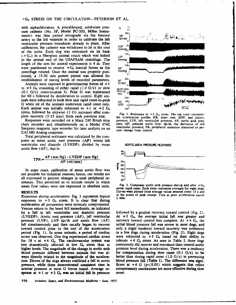
+Gz STRESS ON THE CIRCULATION-PETERSON ET AL.
with alphachloralose. A precalibrated, solid-state pres- G 03]sure catheter (No. 5F, Model PC-350, Millar Instru- 2
ments) was then passed retrograde via the femoral HR Io . ,.., .artery to the left ventricle in order to calibrate the left (nrr.] 0l.ventricular pressure transducer already in place. Aftercalibration, the catheter was withdrawn to lie in the root EDP 2
of the aorta. Each dog was restrained on its back (rrvng
(+G.) in a fiberglass animal couch which was bolted LVP 2251
to the animal end of the USAFSAM centrifuge. The (Mw*-g)length of the arm for animal experiments is 4 m. They 2
were positioned to receive +G. inertial forces as the AP 225
centrifuge rotated. Once the animal was properly posi- (rnrrit)0tioned, a 15-30 min pretest period was allowed forestablishment of resting levels of recorded parameters. zA 500
Animals were exposed to gravitoinertial forces of +1 (cced 0to +5 Gz consisting of either rapid (1.0 G/s) or slow Op/d 5o.(0.1 G/s) onset-to-peak G. Peak G was maintained ( /oHfor 60 s followed by deceleration to control. Eight ani- 201Vmals were subjected to both slow and rapid onset-to-peak PR 104G while six of the animals underwent rapid onset only. V3) oEach animal was initially subjected to + 1 or +2 G. .0o0 3ostress, followed by stepwise (I G) increases after com- Fig. 1. Responses to +3 Gz stress. The top trace representplete recovery (5-15 min) from each previous trial. the acceleration profile; HR, heart rate; EDP, end distohb.
Responses were recorded on a Mark 200 Brush strip pressure; LVP, left ventricular pressure; AP, aortic arch pres-chart recorder and simultaneously on a Model 4742 sure; AF, pulsatile aortic flow; dp/dt, the derivative of left
for later analysis on an ventricular pressure; PR, peripheral resistance measured as per-Sangamo magnetic tape recorder fo ae nlsso n cent change from control.EAI 680 Analog computer.
Total peripheral resistance was calculated by the com-puter as mean aortic root pressure (AP) minus leftventricular end diastolic (LVEDP) divided by mean AOR1IC ACH PRESSURE RESPONSEaortic flow (AF), that is:
4G3
TPR=AP (mm Hg) - LVEDP (mm Hg) 2G
AF (ml/min) 175.
150. -.............
In som e cases, calibration of m ean aortic flow w as i 1 ..............
not possible for technical reasons; hence, our results are 1 ..all expressed in percent changes in total peripheral re- 5 - \ .......sistance. This permitted us to include results in whichmean flow values were not expressed in absolute units. Fig. 2. Composite aortic arch pressure during and after +Gz
stress, rapid onset. Each trace represents averages for eight dogs.RESULTS Curves were plotted from average values selected every 15 s andResponses during acceleration: Fig. 1 represents typical at the points of peak change. Time at peak acceleration equalslmia.responses to + 3 G, stress. It is clear that during
acceleration all parameters were seriously compromised.Venous return to the heart fell immediately, as indicatedby a fall in left ventricular end diastolic pressure followed by a gradual recovery toward control (Fig. 2).(LVEDP). Aortic root pressure (AP), left ventricular At + 3 G., the average initial fall was greater andpressure (LVP), LVP dp/dt and aortic flow (AF) recovery toward control less complete. At +4 G., thefell simultaneously and then usually began to recover initial blood pressure fall was severe in most dogs, andtoward control prior to the end of the acceleration only a slight tendency toward recovery was evidencedperiod (Fig. 1). In some animals, a period of cardiac in a few dogs during acceleration (Fig. 2). Eight dogsarrest was observed. One dog experienced cardiac arrest were subjected to + 5 G. based on their ability tofor 18 s at + 4 G.. The cardiovascular system was tolerate + 4 G, stress. As seen in Table I, three dogsless dramatically affected at low G. stress than at consistently did recover and overshoot their control aortichigher levels. The magnitude of the change in aortic arch pressure level during acceleration. There was a tendencyblood pressure differed between animals but was al- for compensation during slow onset (0.1 G/s) to beways directly related to the magnitude of the accelera- better than during rapid onset (1.0 G/s) in preventingtion. Eleven of the dogs always exhibited a fall in aortic blood pressure fall (Table I). The difference was signi-pressure, while three demonstrated consistent rises in ficant at +4 G (p<0.05) which suggests that reflexarterial pressure at most G forces tested. Average re- compensatory mechanisms are more effective during slowsponse at +1 or +2 G. was an initial fall in pressure onset.
776 Aviation, Space, and Environmental Medicine * June, 1975
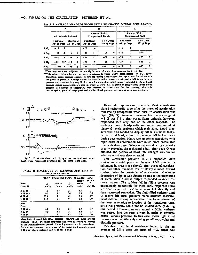
+Gz STRESS ON THE CIRCULATION-PETERSON ET AL.
TABLE 1. AVERAGE MAXIMUM BLOOD PRESSURE CHANGE DURING ACCELERATION
A B C
Animals Which Animals WhichAll Animals Included Compensated Poorly Compensated Well
Fast Onset Slow Onset Fast Onset Slow Onset Fast Onset Slow OnsetAP # Dogs AP # Dogs AP# Dogs AP# Dogs AP # Dogs AP# Dogs
I Gz -15 5 -23 4 +15 I
2 Gz -23 14 -6 8 -36 II -20 6 +22 3 +30 2
3 Gz -42 14 -33 8 -74 11 -60 6 +23 3 +35 2
4 Gz -63 12, -38 8 -97 9 -66 6 +23 3 +35 2
5 Gz -3200 4 -85 6 -94 2 -111 4 +28 2 -13 2
*Two dogs were not subjected to +4 Gs because of their slow recovery from +3 G3 ."This value is biased by the two dogs in column C which always compensated for +Gs stress.
Maximum blood pressure changes in mm HS during acceleration. Average values for all animalsare given in group A. Average values for animals which always experienced a fall in aortic archpressure are presented in group B. Averages for those dogs which usually exhibited a rise in bloodpressure during acceleration are seen in group C. Note that in group B progressively lower bloodpressure is observed to accompany each increase in acceleration. On the contrary, with onlyone exception, group C dogs produced similar blood pressure increases at each acceleration level.
4 A Heart rate responses were variable. Most animals dis-G 2 played tachycardia soon after the onset of accelerationI.50__ 60 5___J followed by bradycardia when onset to acceleration was
rapid (Fig. 3). Average maximum heart rate change at
KR 1-- ..... ....-.. - + 3 G was 8.4 s after onset. Some animals, however,responded with only one or the other response. The50-"- tendency toward bradycardia was more pronounced at
higher G levels. Animals which maintained blood pres-
48B sure well also tended to display either sustained tachy-G 2 cardia or, at least, a less than average fall in heart rate
during acceleration. Heart rate responses associated withrapid onset of acceleration were much more predictablethan with slow onset. When onset was slow, bradycardiausually preceded the tachycardia but, after peak G was
soJ attained, the pattern of heart rate changes was similarwhether onset was slow or rapid.
Fig. 3. Heart rate changes to +Gz stress, fast and slow onset. Left ventricular pressure (LVP) responses wereEach trace represents averages for the same eight dogs. similar to arterial pressure changes. LVP reached a
minimum in most trials shortly after onset of accelera-AND TIME IN tion and either remained low or slowly climbed toward
RECOVERY PHASE control during the remainder of acceleration. Maximum
depression of dp/dt was directly related to the magnitudeMLAP (4.9 mm Hg) MAP (.24mm Hg) MAP- of acceleration. Cardiac output responded in much the
Time to MLAPSlow A return A return A same manner. The sudden fall in filling pressure wasOnset N mm Hg (min) mm Hg (min) mm Hg undoubtedly responsible for these early responses since
2 G (8) 7.0 3.8 35 4.3 28 left ventricular -!nd diastolic pressure fell abruptly and3 0 (8) 9.5 4.2 36 5.3 26 then recovered somewhat. The fluid-filled catheter used
4 G (8) 12.4 5.1 41 5.6 29 to record left atrial pressure made accurate measure-5 G (6) 14.6 6.4 44 6.3 29 ment difficult during acceleration due to movement of
Fast the heart in relation to location of the transducer; thus,Onset left atrial pressure could not be studied directly during2 0 (8) 6.0 2.5 23 2.7 17 this period. However, in one animal a Millar catheter3 G (9) 8.9 3.6 34 4.4 25 was passed into the right atrium in order to estimate4 '3 (8) 13.2 5.1 43 5.9 30 central venous pressure. In this case, mean right atrial
Magnitude of mean left atrial pressure (MLAP) and mean arterial pressure was quantitatively similar to left ventricular endpressure (MAP) overshoot responses and time to return to control diastolic pressure.in the postacceleration period. Control pressures are in parentheses.Each value represents an average of the same eight animals except Calculated pe:,pheral resistance began to rise an5 G slow which included only 6 of the 8 dogs. average of 5.8 s after the onset of + G. stress and
Aviation, Space, and Environmental Medicine • June, 1975 777
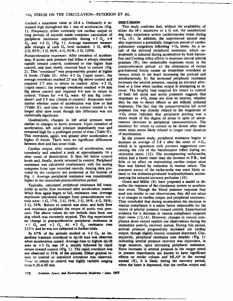
+Gz STRESS ON THE CIRCULATION-PETERSON ET AL.
reached a maximum value at 20.4 s. Ordinarily it re- DISCUSSIONmained high throughout the 1 min of acceleration (Fig. This study confirms that, without the availability of1). Frequently, either extremely low cardiac output or either the M-I maneuver or a G suit, the anesthetizedlong periods of asystole made computer calculation of dog may experience severe cardiovascular stress duringperipheral resistance impossible during +3 G., +4 +G. (6). In addition, the unprotected animal mayG., and +5 G, (Fig. 1). Average maximum measur- also experience a potentially dangerous period of cardio-able changes at each G., level included: 1 G, 40%; pulmonary congestion following +G. stress. As a re-2 G, 82%; 3 G, 96%; 4 G, 92%; 5 G, 129%. sult of the elevated peripheral resistance, which un-
Postacceleration responses: After cessation of accelera- doubtedly is initiated during acceleration by both barore-tion, if aortic arch pressure had fallen it always returned flex and Cushing reflex efforts to maintain central arterialrapidly toward control, continued to rise higher than pressure (9), two undesirable responses occur in thecontrol, and then slowly returned back to control (Fig. postacceleration period: a) the elimination of excess2). This "overshoot" was progressively greater at higher gravitational forces causes an immediate overshoot inG levels (Table II). After + 2 G. (rapid onset), the venous return to the heart increasing the preload andaverage overshoot reached 23 mm Hg above control and simultaneously, b) the increased peripheral resistancerequired 2.7 min to return to control. After +3 G. increases the arterial pressure, causing an elevated after-(rapid onset), the average overshoot reached +34 mm load at a time when cardiac output is attempting to re-Hg above control and required 4.4 min to return to cover. The lengthy time required for return to controlcontrol. Values for + 4 G, were: 43 mm Hg (over- of both left atrial and aortic pressures suggests thatshoot) and 5.9 min (return to control). Responses were responses to + G stress are slow to recover, whethersimilar whether onset of acceleration was slow or fast they be due to direct effects or are reflexly initiated(Table I), and time to return to control tended to be responses. The fact that the postacceleration left atriallonger after slow onset though this difference was not pressure rise was directly related to the magnitude ofstatistically significant. +(G. stress indicates that peripheral pooling was a
Qualitatively, changes in left atrial pressure were direct result of the degree of stress in spite of simul-
similar to changes in aortic pressure. Upon cessation of taneous increases in peripheral vasoconstriction. The
acceleration, left atrial pressure immediately rose and tendency for return to control to be slower after slowonset trials seems likely related to longer total durationremained high for a prolonged period of time (Table II). of acceleration.
This overshoot, again, was greater after acceleration at
higher G forces. There were no significant differences In the present study, peripheral resistance begins to
between slow and fast onset trials, increase an average of 5.8 s after the onset of + G,which is in agreement with previous suggestions con-
Cardiac output, after cessation of acceleration, rose cerning the role of the baroreceptor reflex during ac-transiently and reached a peak at approximately 14 s celeration stress (12). The accompanying tachycardia,after onset of deceleration. It then fell below control which had a faster onset than the increase in P.R., hadlevels and, finally, slowly returned to control. Peripheral little or no effect on maintaining cardiac output sinceresistance was calculated continuously with the analog flow was limited by reduction in venous return. Thecomputer from tape recorded records. Analog data pro- transient nature of the tachycardia was most likely re-vided by the computer are presented at the bottom of lated to the ischemia-produced bradyarrhythmia accom-Fig. 1. Average peripheral resistance was considerably panying the reduced coronary perfusion (10).higher in the immediate postacceleration period. Green and Miller (8) have proposed a model to de-
Typically, calculated peripheral resistance fell trans- scribe the response of the circulatory system to accelera-iently as aortic flow increased after acceleration ceased. tion stress. Though the blood pressure response theyWhen flow again began to fall, resistance rose. Average used was similar to our observations, they did not relatemaximum calculated changes from preacceleration con- it to changes in cardiac output and peripheral resistance.trols were: 1 G, 17%, 2 G, 19%; 3 G, 39%; 4 G, 58%; They concluded that during acceleration the decrease in5 G, 54%. Return to control was slow, and both flow venous compliance is a major factor responsible for theand resistance paralleled the return of aortic root pres- return of arterial pressure toward control. Experimentalsure. The above values do not include data from one evidence for a decrease in venous compliance supportsdog which was extremely atypical. This dog experienced their views (12,14). However, changes in venous com-no change in postacceleration peripheral resistance at pliance alone cannot explain our observations during the+ I G. and + 2 G,. At + 3 G., resistance rose immediate post-G, recovery period. During this period,233% and he was not subjected to further trials, arterial pressure progressively increased yet cardiac
In 67% of the animals studied at +3 G,, an im- output, though slightly altered, remained depressed. Con-mediate transient overshoot in dp/dt max was observed sequently, peripheral resistance rose steadily (Fig. 1)when acceleration ceased. Average time to highest dp/dt indicating arterial pressure recovery was dependent, inmax at + 3 G, was 19 s, usually followed by rapid large measure, upon increasing peripheral resistance.return toward control (Fig. 1). The rapid overshoot was Since increases in arterial pressure in the ranges notednot observed in 33% of the animals. Either gradual re- in present experiments are known to have significantturn to control or sustained overshoot was observed. effects on stroke volume and MLAP in the normalTime to return to control was highly variable ranging animal (9), it is likely during the recovery period,trom 0.20-6.50 min. when the heart is depressed, that the cardiac output and
778 Aviation, Space, and Environmental Medicine * June, 1975
+Gz STRESS ON THE CIRCULATION-PETERSON ET AL.
MLAP response are significantly influenced by the in- REFERENCEScrease in arterial pressure. 1. Burns, J.W. 1974. Influence of ethanol on cardiovascular
tolerance to +Gz acceleration. Aerospace Med. 45:19-28.The observed transient increases in aortic flow and 2. Burton, R.R. 1973. Positive ( +Gz) acceleration tolerances of
dp/dt max within 20 s post-G, which are usually ac- the miniature swine: Application as a human analog.companied by a fall in calculated total peripheral re- Aerospace Med. 44:294-298.sistance, suggest a transient increase in the capacitance of 3. Chimoskey, I.E. 1970. Cormnary blood flow and electro-
cardiogram during headward acceleration in unanesthetizedresistance vessels possibly accompanied by increased dogs. Aerospace Med. 41:1028-1030.vigor of contractility. A change in arterial capacitance 4. Cohen, G.H., and W.K. Brown. 1969. Changes in ECG con-is likely to occur since +G: stress is known to displace tour during prolonged +Gz acceleration. Aerospacevisceral organs causing distortion of vessels (18). Med. 40:874-879.
5. Cohen, G.H., and W.K. Brown. 1969. ElectrocardiographicCineradiographic studies have shown that vessels cen- changes during positive acceleration. J. Appl. Physiol. 27:
tral to the heart are elongated and reduced in diameter 858-862.as much as 50% during acceleration (15). This stretch 6. Erickson, H.H., H. Sandier, H.L. Stone, and S. Young. 1973.of the carotid arteries at the onset of acceleration com- Cardiac function during +G z acceleration. Preprints, An-
nual Scientific Meeting, Aerospace Medical Assoc., pp.plicates interpretation of heart rate responses. We pre- 192-193.sume that this initial stretch would stimulate carotid sinus 7. Folkow, B., and E. Neil. 1971. Circulation, New York, Ox-stretch receptors and produce bradycardia (7). Since a ford.transient initial bradycardia is often seen, especially dur- 8. Green, J.F., and N.C. Miller. 1973. A model describing the
response of the circulatory system to acceleration stress.ing slow onset (Fig. 3) and never extended past the Ann. Bionied. Engr., pp. 455-467.time at which peak acceleration is reached, this initial 9. Guyton, A.C. 1971. Medical Physiology, 4th Ed., Philadel-mechanical displacement is likely responsible for the phia, Saunders.bradycardia observed. As observed by others (13,16), 10. Hurst, J.W., and R.B. Logue. 1970. The Heart. New York,
during the deceleration period and shortly thereafter, ~ McGraw-Hill.11. Lambert, E.H.. and E.H. Wood. 1946. The problem of
arrhythmias ave common; neural reflex regulation of blackout and unconsciousness in aviators. Med. Clin. N.heart rate is probably minimal at this time. The most Amer. 30:833-844.common observation was a general tendency toward 12. Newberry, P.D., and A.C. Bryan. 1967. Effect on venousreturn to control rate with no consistent evidence of compliance and peripheral vascular resistance of head-
ward (+Gz) acceleration. J. Appl. Physiol. 23:150-156.baroreceptor influence. The tendency toward postac- 13. Parkhurst, M.J., S.D. Leverett, Jr., and S.J. Shubrooks, Jr.celeration bradycardia at high G levels suggests the in- 1972. Human tolerance to high, sustained +G z accelera-fluence of general cardiac depression. Nevertheless, all tion. Aerospace Med. 43:708-712.animals were conscious and alert I d following trials 14. Salzman, E.W., and S.D. Leverett, Jr. 1956. Peripheraland showed no signs of functional physiological ab- venoconstriction during acceleration and orthostasis.
Circ. Res. 4:450-545.normalities. 15. Sandier, H., J.E. Greenlead, B.D. Newsom, and S. Rositano.
Our results have demonstrated responses to +G. 1974. Lower-body negative pressure (LBNP) as a pre-stress without the benefit of either the M-1 manuever dictor for +Gz tolerance after bed rest (BR). Preprints,or a G suit. Also, it is clear that the arterial barorecep- Annual Scientific Meeting, Aerospace Medical Assoc.,
pp. 171-172.tor system contributes to the overall response, although 16. Shubrooks, S.J., Jr. 1972. Changes in cardiac rhythm duringthe absolute significance is not clear. Further studies sustained high levels of positive (+Gz) acceleration.will be necessary to define the quantitative effects of Aerospace Med. 43:1200-1206.the anti-G suit and role of the baroreceptor system. 17. Shubrooks, S.J., Jr. 1973. Positive-pressure breathing as a
protective technique during +Gz acceleration. J. Appl.Physiol. 35:294-298.
ACKNOWLEDGEMENTS 18. Stone, H.L., H.H. Erickson, and H. Sandier. 1974. ChangesThe authors are indebted to Greg Riedel, Ben Wiggins, and in mesenteric, renal, and aortic flows with +Gx accelera-
Richard Main for their technical assistance and to Linda Fox tion Aerospace Med. 45:498-504.for her surgical assistance. We also wish to express our ap- 19. Stone, H.L., H.F. Stegall, M.B. Kardon, H. Sandier, andpreciation to the USAFSAM Human Centrifuge operation staff R.M. Payne. 1971. Changes in aortic, coronary, andand to Bill Welch for his assistance in data analysis. This study carotid flows during +Gx acceleration. J. Appl. Physiol.was supported by AFOSR Grant #71-2074. 30:21-26.
Aviation, Space, and Environmental Medicine • June, 1975 779
Pathways regulating cardiovascular changesSduring volume loading in awake dogs
VERNON S. BISHOP AND D. FRED PETERSONDepartment of Pharmacology, The University of Texas Health Science Center at San Antonio,San Antonio. Texas 78284
BIsitoP. VERNON S.. AND D. FRED PETERSON. Pathways In conscious dogs. volume loading increases cardiacregulating cardiocascular changes during volume loading in output more than it increases arterial pressure, thusawake oiogs. Am. J. Physiol. 23113): 854-859. 1976.-The peripheral resistance decreases. Vatner et al. (28) re-role played hy the cardiac sympathetic nerves and arterial cently reported that the baroreflex control of heart rate.baroreceptors in the cardiovascular responses to acute volume aloading was studied in conscious dogs. In 15 normally inner- volume infuspons H ov , fo attud it wagu-vated animals, mean arterial pressure rose 10 mmHg. heart volume infusions. However. from that study it was un-rate increased 38 beats. mm and cardiac output 1.696 mlimin. clear whether the baroreceptors exerted any influencew hile peripheral resistance decreased (.99 PRU. Neither bilat- on either the heart rate or peripheral resistance re-cral baroreceptor denervation. dorsal root sections (T,-T, or sponse to volume loading. After baroreceptor denerva-surgical interruptmon of the left ansa subclavia altered the tion. the blood pressure change during volume loadingabove responses to acute volume loading. Bilateral section of was significantly reduced by both moderate and largethe ansa subciavia (total cardiac sympathectomy) signifi- volumes, whereas the heart rate response was atten-cantlv reduced the heart rate response from 35 = 5 to 20 = 5 uated at the larger volume loads. In contrast, in anes-beats min but did not alter other changes. A similar reduction thetized dogs. increases in cardiac output after barore-in heart rate response was observed following selective section ceptor denervation resulted in increased peripheral re-of the right ansa subclavia. Intravenous infusions of epineph-rine augmented the heart rate response in both normally sistance (17).innervated and cardiac sympathectomized dogs. It is sug- This study extends previously reported responses toe gested that although the primary efferent pathway for the volume infusion. Precise afferent and efferent pathwaysreflex trachycardia is via the vagus. responses are modulated of the reflex changes in heart rate associated with vol-by sympathetic neural activity. Additionally. the nervous sys- ume infusion in the conscious dog have been identifiedtem was not shown to play a measureable role in the observed and evidence for sympathetic-parasympathetic interac-peripheral resistance changes. tion presented. In addition, effects on peripheral vascu-
lar resistance have been investigated. The possible in-vagus: cardiac sympathetic nerves: peripheral resistance: bar- teraction of arterial baroreceptors on both the heart rateoreceptors: heart rate and peripheral vascular changes have been studied by
total sinoaortic baroreceptor denervation.
RECENTLY. MANY INVESTIGATIONS have centered on the METHODS
role of low-pressure mechanoreceptors in or near the Mongrel dogs were chronically instrumented underheart, which have been implicated in reflex regulation sterile surgical conditions using halothane gas. A leftof cardiovascular function. Both acute volume loading thoracotomy was performed through the fourth intercos-(1, 3, 5, 10) and localized distension of the atrial-venous tal space. An 18-gauge polyvinyl catheter was placed injunctions f4, 6. 7, 11, 14) have served as models for the left atrium through the auricular appendage. Astudying these receptors. Unfortunately, results have calibrated electromagnetic flow probe was affixedbeen inconsistent when comparing either models or re- around the ascending aorta in 15 animals. The calibra-suits of experiments by different investigators using the tion of all flow probes were checked as previously de-same model. For example, the reflex pathway for the scribed (1). A piece of surgical suture was looped aroundtachycardia produced by local stretch has been reported both left ansa subclavia for later cardiac sympathec-by some to involve vagal afferent and cardiac sympa- tomy (23). An 18-gauge polyvinyl catheter was insertedthetic efferent fibers (7, 11, 14) while other investigators into either the internal mammary artery or in the ca-have reported that both vagal afferents and efferents rotid artery through a cervical incision. At the time ofare involved (6). In the conscious dog, the tachycardia thoracotomy, or a few days later, a 10-gauge polyvinylobserved with volume loading is not eliminated by beta- catheter was inserted into the superior vena cavaadrenergic blockade, thus implicating the vagus as a through the left jugular vein. Approximately 2 wk werecontributing efferent pathway (10). Most investigators allowed for recovery, during which time the health of
- indicate that the afferent pathway is in the vagus (6, 11, the animal was'monitored daily. At the time when- 13. 18. although a recent study suggests that afferents experiments were performed, body temperature and
in the spinal cord might contribute to the response (8). electrocardiogram (ECG) were normal.-i 854
IA
HEGUI.ATION OF HEART LATE DURING INFUSION ,51-1
Experimental protocol. ECG, heart rate. systemic procedures eliminated the reflex heart rate and bloodblood pressure, and left atrial pressure were recorded on pressure responses to carotid occlusion (22. 23). Alla Beckman paper oscillograph using the required trans- branches of the vagus within 2 cm below the superiorducers. couplers, and amplifiers. Tvrode's solution laryngeal nerves were also sectioned. Presumably this
-varmed to 37.5°C was infused through the large cathe- procedure denervated the aortic arch and combined withter in the jugular vein using a Holter pump (model carotid sinus denervation resulted in an immediate riseRE161). The rate of the infusion was controlled to pro- in blood pressure. Heart rate responses to the injectionduce a steady rise in left atrial pressure. Infusions were of phenylephrine (10 Mg) were abolished or drasticallynormally performed over a 1- to 4-min period until the reduced. These animals were allowed several days toheart rate had reached a constant level, which was not recover before reentering the experimental protocol.exceeded despite further rise in left atrial pressure. Sympathetic deafferentation. In four additional ani-
The total volume of Tyrode's solution required to mals the heart rate response to acute volume loadingreach a maximum heart rate ranged between 300 and was established. Subsequently, these animals were800 ml (averaged 400 ml) (1). The hemodilution effect anesthetized with sodium pentothal (30 mg/kgi and un-ranged from -2 to -5%. In previous studies, identical der sterile conditions the dorsal roots T,-T were sec-cardiac responses were observed with intravenous infu- tioned. The animals were allowed to recover 10-14 dayssions of blood or Tyrode's solution (2). Infusions were and were reentered into the experimental protocol.performed in all animals under control conditions. Addi- Drug administration. In order to artificially simulatetional infusions were performed after each surgical de- an increase in sympathetic background activity, epi-nervation described below and in some cases during a nephrine was infused intravenously in four additionalconstant intravenous infusion of epinephrine. The animals instrumented with catheters only. The infusionabove parameters were continuously recorded during rates were controlled so that heart rate was only mini-the infusions. All experiments were performed while mally increased. Subsequently. the predetermined levelthe animals rested unrestrained in a hammock. Two of epinephrine infusion was maintained during the infu-days separated each infusion. sion of Tvrode's solution.
Total peripheral resistance, expressed in peripheralresistance units (PRU), was calculated
RESULTS
mean arterial blood pressure (mmHg, A total of 66 control volume infusions were performedPRU - central venous pressure (mmHg) in 22 conscious dogs. In each animal the infusion rate
mean aortic flow (ml; mini was controlled in order to produce a continuous rise in
x 60 s/min left atrial pressure. Increases in cardiac output andheart rate typically paralleled the increase in left atrial
Values are reported as means or mean differences z pressure during the first 1-2 min of infusion. Thesestandard errors (SE). Statistical evaluation was made responses then plateaued at levels consistent for eachby use of ti e Student t test for paired comparisons (26). individual animal at which time the infusion was termi-P < 0.05 was considered significant. nated. Mean cardiac output increased from an average
Cardiac sympathectomv. In six dogs a sterile thora- of 2,554 _t 157 ml/min to an average maximum of 4.250cotomy was performed a 2nd time in order to loop surgi- -t 280 ml/min (n = 15 dogs). Average heart rate changescal suture around the right ansa subclavia distal to the were from 109 -± 4 to 147 -- 6 beats/min. Althoughright stellate ganglion. After establishing the control cardiac output increased dramatically, the change inresponses to acute volume loading this suture. as well as arterial pressure was not great (100 ±- 3 to 110 = 3the one previously placed around the left ansa subcla- mmHg). Consequently, the peripheral vascular resist-via. was pulled to selectively cut the sympathetic nerves ance was markedly reduced (from 2.42 ± 0.19 to 1.43 ±to the heart (20). The sympathectomy was performed 0.15 PRU).when the animal was anesthetized. After recovery the In order ,o identify specific efferent pathways in-animals were reentered into the experimental protocol. volved in the heart rate response, the cardiac sympa-In three additional animals with only catheters im- thetic nerves were selectively cut. In five dogs, removalplanted. the right ansa subclavia was looped and the of the left ansa subclavia nerves only was without effectresponse to acute volume loading was observed before on the heart rate response to volume infusion. However,and after the removal of the right ansa subclavia while in three animals, section of the right ansa subclaviasympathetic innervation to the left side remained in- alone reduced the AHR response to volume loading fromtact. 35 to 17 beats/min. In six animals, a total of 24 infusion
Baroreceptor denervation. After control responses curves were obtained before and after bilateral sectionwere obtained, five animals were anesthetized with so- of the ansa subclavia nerves. Cutting both the right anddium pentothal (30 mg/kg) and a midcervical incision left ansa subclavia reduced the average resting HR fromwas made. The carotid sinuses were located and all the 110 ±t 5 to 85 :t 4 beats/min (P < 0.05). Arterial pres-vessels and other tissue above the bifurcation of the sure, cardiac output, and peripheral resistance were allcommon carotid arteries isolated, ligated, and sectioned slightly, but not significantly, reduced due to nerveexcept the external and internal carotid arteries. All section (Table 1). the peak change in heart rate due tor-maining excess tissue was stripped from the vessels infusion was also reduced from the average control",riginating from the common carotid arteries. These change of 35 ±t 5 to 20 ± 5 beatsimin (P < 0.05). These
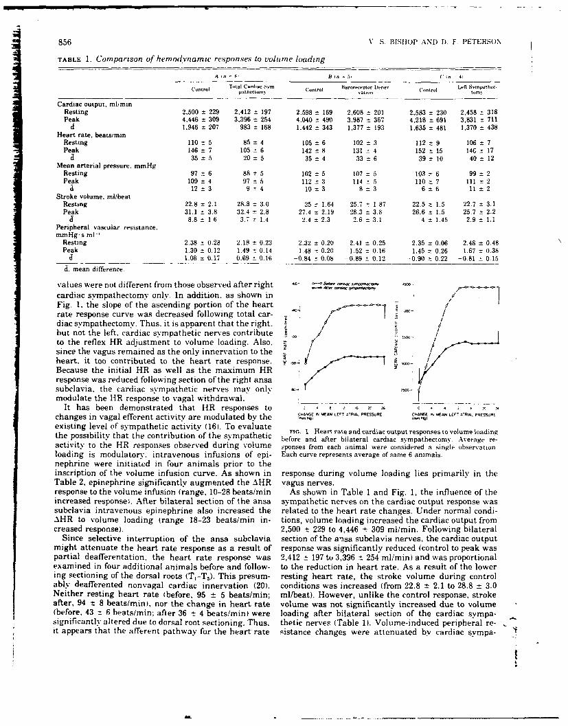
856 V S. BIIISOP AND D. F PETERSON
TABLE 1. Comparison of hemodvnamic responses to volune loading
A in 6. B In ( 5 ('in 4,
Control Total Cardiac Svm- Control Barormeptor De-ner Lefi Svrpathec-pathectour canontrol tm.
Cardiac output. ml/minResting 2.500 ± 229 2.412 -t 197 2.598 t 169 2.608 : 201 2.583 z- 230 2.458 : 318Peak 4,446 ± 309 3.396 - 254 4.040 :t 490 3.987 367 4.218 = 691 3,831 : 711
a 1,946 = 207 983 ± 168 1.442 ± 343 1,377 193 1,635 = 481 1.370 = 438Heart rate. beats/min
Resting 110 t 5 85 t 4 105 : 6 102 3 112 : 9 106 = 7Peak 146 ± 7 105 6 142 ±8 131 4 152 :t 15 146C: 17
a 35 *- 5 20 5 35 ±4 33 6 39 : 10 40 = 12Mean arterial pressure. mmHg
Resting 97 ± 6 88 ± 5 102 : 5 107 :5 103 6 99 ± 2Peak 109 ±4 97 = 5 112 ± 3 114 5 110 7 111 ± 2
i 12±3 9±4 10±t3 8:3 6-6 11 2Stroke volume, ml/beat
Resting 22.8 ± 2.1 28.8 ± 3.0 25 ± 1.64 25.7 - 1.87 22.5 ± 1.5 22.7 ± 3.1
Peak 31.1 : 3.8 32.4 ± 2.8 27.4 ± 2.19 28.3 ± 3.8 26.6 ± 1.5 25.7 ± 2.2J 8.8± 1 6 3.7 : 1.4 2.4 2.3 2.6 :3.1 4 :t 1.45 2.9 t 1.1
Peripheral vascular resistance.mmHg.s ml-'
Resting 2.38 ± 0.28 2.18 ± 0.23 2.32 = 0.20 2.41 ± 0.25 2.35 = 0.06 2.48 _ 0.48Peak 1.30 ± 0.12 1.49 ± 0.14 1.48 ± 0.20 1.52 : 0.16 1.45 ± 0.26 1.67 : 0.38
d -1.08 ±- 0.17 -0.69 ± 0.16 -0.84 ± 0.08 -0.89 i- 0.12 -0.90 z 0.22 -0.81 _ 0.15
d. mean difference.
values were not different from those observed after right =... " -- -.. ..cardiac sympathectomy only. In addition, as shown in . /Fig. 1. the slope of the ascending portion of the heart "rate response curve was decreased following total car-diac sympathectomy. Thus. it is apparent that the right. / dbut not the left. cardiac sympathetic nerves contribute ,- / .- /to the reflex HR adjustment to volume loading. Also, - /since the vagus remained as the only innervation to the - ,heart, it too contributed to the heart rate response. oBecause the initial HR as well as the maximum HRresponse was reduced following section of the right ansasubclavia. the cardiac sympathetic nerves may only omodulate the HR response to vagal withdrawal. |
It has been demonstrated that HR responses to .... - ,changes in vagal efferent activity are modulated by the c. GE IN MEAN LEr T VRAL PESSURE CH. % EAN LEF,, o S URi.
existing level of sympathetic activity (16). To evaluate FIG, 1. Hearn rate and cardiac output responses to volume loadingthe possibility that the contribution of the sympathetic before and after bilateral cardiac sympathectomy Average re-activity to the HR responses observed during volume sponses from each animal were considered a single observation.loading is modulatory, intravenous infusions of epi- Each curve represents average of same 6 animals.nephrine were initiated in four animals prior to theinscription of the volume infusion curve. As shown in response during volume loading lies primarily in theTable 2, epinephrine significantly augmented the AHR vagus nerves.response to the volume infusion (range, 10-28 beats/min As shown in Table I and Fig. 1, the influence of theincreased responsel. After bilateral section of the ansa sympathetic nerves on the cardiac output response wassubclavia intravenous epinephrine also increased the related to the heart rate changes. Under normal condi-AHR to volume loading (range 18-23 beats/min in- tions, volume loading increased the cardiac output fromcreased response). 2,500 t 229 to 4,446 t 309 ml/min. Following bilateral
Since selective interruption of the ansa subclavia section of the a'nsa subclavia nerves, the cardiac outputmight attenuate the heart rate response as a result of response was significantly reduced (control to peak waspartial deafferentation. the heart rate response was 2,412 ± 197 to 3,396 t 254 ml/min) and was proportionalexamined in four additional animals before and follow- to the reduction in heart rate. As a result of the lowering sectioning of the dorsal roots (T,-T,). This presum- resting heart rate, the stroke volume during controlably deafferented nonvagal cardiac innervation (20). conditions was increased (from 22.8 ± 2.1 to 28.8 ± 3.0Neither resting heart rate (before, 95 ± 5 beats/min; ml/beat). However, unlike the control response, strokeafter, 94 _t 8 beats/min), nor the change in heart rate volume was not significantly increased due to volume(before, 43 = 6 beats/min; after 36 ± 4 beats/min) were loading after bilateral section of the cardiac sympa-significantly altered due to dorsal root sectioning. Thus, thetic nerves (Table 1). Volume-induced peripheral re-it appears that the afferent pathway for the heart rate ;istance changes were attenuated by cardiac sympa-
A.!
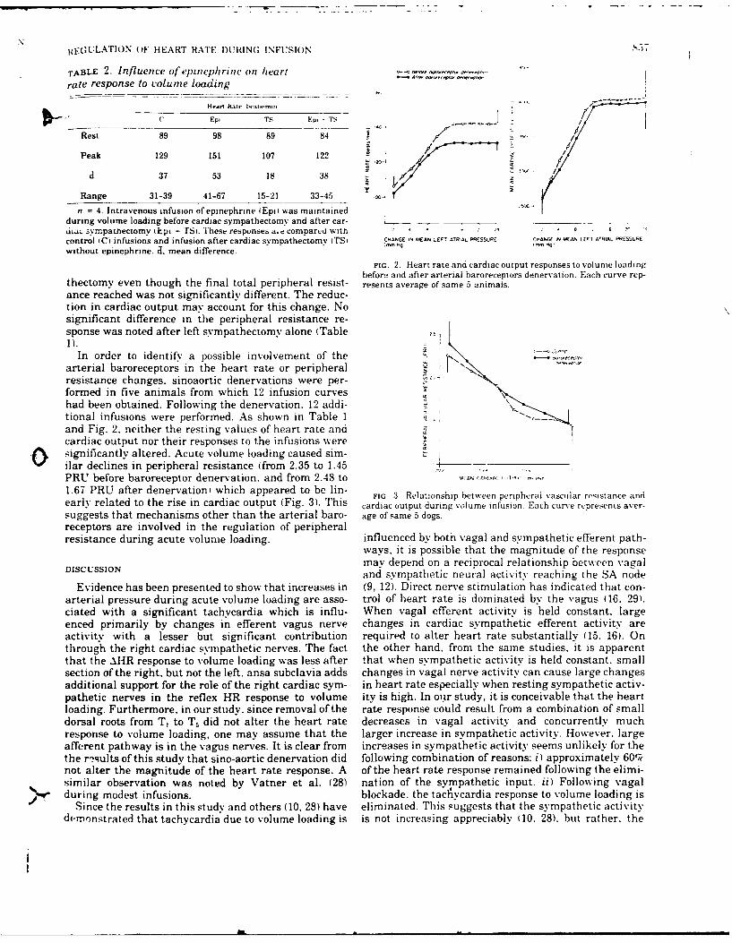
RIEGULATION OF HEART RA'E I)URING INFUSION
TABLE 2. Influence of epinephrine on heart na,. . . "
rate response to volume loading
H -i hat,. hvai, vml .
C Ep, TS Ep- TS fRest 89 98 89 84 11/
Peak 129 151 107 122 /d 37 53 18 38 A' .
Range 31-39 41-67 15-21 33-45
n = 4. Intravenous infusion of epinephrine Epi; was maintainedduring volume loading before cardiac sympathectomy and after car- - _ -
dia . sympatnectomy ukpi , r~s. These responses a,e compared with ' - ,control IC) infusions and infusion after cardiac sympathectomy (TS i CHANGE IN MEAN LEFT ATRAL PRESSURE c IAnN MEAN LuT A RIAL PRE'5URE
without epinephrine. J. mean difference. N'
FIG. 2. Heart rate and cardiac output responses to volume loadingbefore and after arterial baroreceptors denervation. Each curve rep-
thectomy even though the final total peripheral resist- resents average of same 5 animals.ance reached was not significantly different. The reduc-tion in cardiac output may account for this change. Nosignificant difference in the peripheral resistance re-sponse was noted after left sympathectomy alone (Table 11).-
In order to identify a possible involvement of thearterial baroreceptors in the heart rate or peripheral I ""..
resistance changes. sinoaortic denervations were per-formed in five animals from which 12 infusion curveshad been obtained. Following the denervation. 12 addi-tional infusions were performed. As shown in Table 1and Fig. 2. neither the resting values of heart rate andcardiac output nor their responses to the infusions were
.[\ significantly altered. Acute volume loading caused sim-ilar declines in peripheral resistance (from 2.35 to 1.45 + . .. ..PRU before baroreceptor denervation. and from 2.48 to L " c.'IA ''1 ..1.67 PRU after denervation) which appeared to be lin- FIG. 3. Relationship between peripheral vascular resistance andearly related to the rise in cardiac output (Fig. 3). This cardiac output during volume infusion. Each curve represents aver-suggests that mechanisms other than the arterial baro- age of same 5 dogs.receptors are involved in the regulation of peripheralresistance during acute volune loading, influenced by both vagal and sympathetic efferent path-
ways. it is possible that the magnitude of the responseDISC USSION may depend on a reciprocal relationship between vagal
and sympathetic neural activity reaching the SA nodeEvidence has been presented to show that increases in (9, 12). Direct nerve stimulation has indicated that con-
arterial pressure during acute volume loading are asso- trol of heart rate is dominated by the vagus (16. 29).ciated with a significant tachycardia which is influ- When vagal efferent activity is held constant, largeenced primarily by changes in efferent vagus nerve changes in cardiac sympathetic efferent activity areactivity with a lesser but significant contribution required to alter heart rate substantially (15. 16). Onthrough the right cardiac sympathetic nerves. The fact the other hand, from the same studies, it is apparentthat the AHR response to volume loading was less after that when sympathetic activity is held constant, smallsection of the right, but not the left. ansa subclavia adds changes in vagal nerve activity can cause large changesadditional support for the role of the right cardiac sym- in heart rate especially when resting sympathetic activ-pathetic nerves in the reflex HR response to volume ity is high. In our study, it is conceivable that the heartloading. Furthermore, in our study, since removal ofthe rate response could result from a combination of smalldorsal roots from T, to T, did not alter the heart rate decreases in vagal activity and concurrently muchresponse to volume loading, one may assume that the larger increase in sympathetic activity. However. largeafferent pathway is in the vagus nerves. It is clear from increases in sympathetic activity seems unlikely for thethe r-sults of this study that sino-aortic denervation did following combination of reasons: i) approximately 60%not alter the magnitude of the heart rate response. A of the heart rate response remained following the elimi-similar observation was noted by Vatner et al. (28) nation of the sympathetic input. ii) Following vagalduring modest infusions, blockade, the tacl~ycardi resptnse to volume loading is
Since the results in this study and others (10, 28) have eliminated. This suggests that the sympathetic activitydemonstrated that tachycardia due to volume loading is is not increasing appreciably (10, 28), but rather, the
IA
"1 858 V S. BISHOP AND D. F. PETERSON
vagal influences were more significant in the face of Acute volume loading in the conscious animal re-background sympathetic activity, suited in a proportionately larger increase in cardiac
The concept that the background level of sympathetic output than arterial pressure. Consequently, total pe-activity can modify vagal influences was tested in the ripheral resistance declined as cardiac output was in-
present study by controlled infusion of epinephrine creased. Liedtke et al. (17) changed cardiac output in
which had only a slight effect on the resting heart rate. anesthetized dogs by altering cardiac pump perform-This caused the peak heart rate response to acute vol- ance either through changes in coronary perfusion or
ume loading to be increased in either intact or cardiac calcium ion concentration in the perfusion of the coro-
sympathectomized animals. From this, one can con- nary beds. They also observed a similar decline in pe-
clude that the magnitude of the response to vagal with- ripheral resistance with increasing cardiac output.
drawal can be increased by increasing the existing sym- However, following bilateral baroreceptor denervationpath Lc activity (i.e.. Ljncc.'ration of catecholamine at and vagotomy, they" ohserved an increase in peripheral
the SA n, , -. This conclusion is supported by the work resistance with increasing cardiac output. They con-
of Levy a.._ Zieske (16) in which combined electrical cluded that total body autoregulation occurred when the
stimulation of the vagus and sympathetic nerves to the arterial baroreflexes from the carotid and aortic arch
heart was employed. When the basal heart rate was were eliminated. In our study, bilateral removal of the
moderately elevated by tonic sympathetic stimulation, carotid and aortic baroreceptors did not alter the rela-
increases in heart rate due to reduced vagal stimulation tionship between resistance and cardiac output.
were much more dramatic (15. 16). Similar heart rate Sagawa and Eisner (24) also failed to observe signifi-
dependency upon the resting sympathetic activity is cant whole-body autoregulation in anesthetized vagoto-
evidenced when comparing the effects of vagal blockade mized dogs either before or after the abrogation of arte-
on the control heart rate before and after elimination of rial baroreflexes. Assuming a rectilinear relationship
sympathetic influences (10. 28). between pressure and flow, they observed a near con-
Although volume loading may stimulate receptors. in stant peripheral resistance when pressure and flow
addition to those shown to be in the atrial-venous junc- were varied from 60 to 140%. Although much less than
tion. similarities do exist between the responses ob- we observed, the pressure-flow relationship in their
served during volume loading and some of those re- study was convex toward the flow axis, indicating that
ported during direct tissue stretch. Edis et al. (6) re- as cardiac output increases above normal the peripheral
ported that the magnitude of the heart rate response to resistance declines. They suggested that this may be
stretch of the pulmonary vein-atrial junction depended due to arterial baroreceptor reflexes. However, in the
upon both efferent vagal and cardiac sympathetic' conscious animal. baroreceptor denervation did not al-
nerves. In their study, when carotid sinus pressure was ter the relationship, suggesting other mechanisms formanipulated to maintain heart rate lower than 140-150 vasodilitation at flow rates above normal.beatsimin stretch of the junction produced tachycardia. A detailed study by Shepherd et al. (25) may explainWhen manipulation of the sinus pressure forced heart our observations. They noted little autoregulation inrate above 150 beatsimin (presumably withdrawing the areflexive dogs even though arterial pressure was re-vagus and activating the cardiac sympathetic nerves) duced to 50%. Oxygen delivery was found to be main-stretch of the pulmonary vein-atrial junction caused tained by increasing oxygen extraction. However. whenbradycardia. Reversal of responses is explainable if one initial 02 extraction ((a-v)0 2 difference) was increasedconsiders that the efferent activity in both groups of either by epinephrine infusions or ventilatory inducednerves could simultaneously be reduced during disten- hypoxia. flow autoregulation occurred. In these con-sion. This concept is supported by the work of Levy et al. scious animals it is unlikely that the 0., delivery is(16) in which withdrawal of vagal activity dominates limited by either extraction or flow. Consequently. atheart rate changes until the vagus is silent. Further above normal flow rates the pressure-flow relationshipevidence that cardiac sympathetic activity can with- may become convex toward the flow axis.draw during volume infusion is the bradycardia follow- Previous studies have demonstrated a role of the car-ing vagal blockade (2, 10, 28) as well as the bradycardia diopulmonary receptors in the regulation of vascularseen during coronary occlusion in sinoaortic denervated resistance 16, 19, 21). Furthermore. vagal afferents haveanimals with heart rates greater than 150 beats/min been shown to inhibit the sympathetic outflow to the(23). peripheral vascular beds (19). However, in conscious
Our study also demonstrated an important vasodila- dogs cold block of the vagus does not apparently altertor effect during volume infusion. Sinoaortic denerva- the cardiac output and arterial pressure relationshiption failed to demonstrate high pressure receptor in- during volume loading (2). However, it is possible thatvolvement in these peripheral resistance changes. Pre- volume loading decreases the restraint exerted by thevious studies have shown a small vasodilator effect by arterial baroreceptors (27), thereby minimizing the in-stimulation of atriovenous junction receptors (6) but fluence of the low-pressure receptors on the sympatheticvolume loading studies have failed to support a substan- outflow (19).tial contribution through afferent vagal involvement The authors gratefully acknowledge the technical assistance of(3). Therefore, one may presume that the observed pe- Linda Fox, Greg Riedel. Jeri McWilliams, and Jesse Rodriguez.ripheral resistance responses primarily involved direct This work was supported by Public Health Service Grant HL-vasodilator influences secondary to increased cardiac 12415-08 and Air Force Office of Scientific Research Grant 73-2525.
oUtput. Received for publication 10 Novtmber 1975.
1E(GLA lit )\ I iF HE ARTI RA.TE D)URING INF[U'IO)N
1BIs~oP. V S . ANti I. 1). IORWiTz Efle.cts of altered autonomnic heart. Circulation Res. 29. ;37 -415. 197 1-- control on itti ietricular itiction in conscious dogs. A..n 4 16. LEVY. NI.- N.. AND II. ZIESKE Autonomnic control of cardiac pau.-
*Phv~stol. 221. 127-12,S2. 1971. maker activitY anti atriovent ricular transmission. .1 Appl.2.BISHOP. V. S.. If 1, STONE. AND A. C. GLYTON. Cardiac function I-'1,.suil. 27: 4b5-47(t, 1969
cures n cnscousdogs. Am. J. Phvsil. 207: 6o7-682, 1964. 17. LIEDTKE, A. 2.. C. W. URSCHEL. AND E.S.KR.Talstei
3. BISHOP. V. S.. It. L. STONE. AND L. D. HORWITZ- Effects of' autoregulation in the :Ina and its inhibition by baroreceptorr tachycardia and ventricular filling pressure on stroke vo~lume in reflexes. Circulation Res. 32: 673 977. 1973.
the conscious dog. Am,. J. Ph 'vsiol. 220: 436-439. 1971. 18. LINDEN. R. J. Function of cardiac receptc.- Circulaton~ 4S: 46:3-4. CARSWELL. F. R. IHAINSWORTH. AND J. R. LEDSOME_ The effects 480. 1973.
of distention of the pulmonarv vein-atria] junctions upon periph- 19. MANCIA. G.. D. E. D)ONALD. AND J_ T. SHEPHER.D. Inhibition oferal vascuiar resistance. 4. Ph/ivsiol.. London 207: 1-14. 1970. adrenergic outflow% to peripheral blood vessels by vagal aflerents
5. COLERIDGE. J. C. G.. AND R. J. LINDEN. The effect of intravenous from the card iopulimonary region in the dog. Circulation Res. 33:infusions upon thlt.ewart rate of the anesthetized dog. .1. PhYs. 713-721. 1973.iot.. Lonidon 12S. 310-319. 1955. 211. NiIZERES. N. J3. Origin and course of Card ioaccleraiioii .rS in
6. EDM A. J.. D. E. DOINALD. AND J T. SHEPHERD. Cardiovascular the dog. Anal. Rt-cord 132: 261-279. 1958.reflexes from strvtch of pulmonary vein-atria] junctions in the 21. PELLETIER. C. L.. A. J. EDIS. AND J. T. SHEPHERD. Circulatorydog. Circifiation l,.27: 10191-1100O. 1970f. reflex from vagal aflerents in response to hemorrhage in the dlog.
7. FuRNIVAL. C. NI.. R, J. LINDEN. AND H. M. SNOW. Reflex effects Circulation Res. 29: 626-634. 1971.on the heart of stimulating left atrial receptors. J. Phviuoi.. 22. PETERSON. D. F.. AND V. S. BisHop. Reflex blood pressure controlLondjon 21S: 447-463. 19711. during acute myocardial ischemnia in the conscious dog. Circila-
S. GUPTA. P. D. Spinai autonomic afferents in elicitation of tachy- lion Res. 34: 226-232. 1974.cardia in vniume infusion in the dog. Am. J. PhYsiol. 229: 303- 23. PETERSON. D. F.. R. L. KASPAR. AND V. S. BISHOP. Reflex tach.308. 1975. cardia due to temporary coronary occlusion in the conscious dog.
9. HAKUIAKZ. Nl. () K. Vaga) and sYmpathetic efferent discharge Circulation Res. 32: 652-659. 1973in the Batnbridi.! Itfiex of dogs Acta Plivsiol. Scand. 85: 414- 24. SAGAWA. K.. AND A. EISNER. Static pressure-flow relation in the417. 1972 total systemic vascular bed of the dog and its modification by the
10. HORITwcZ. . [11 . .AND V S. Bissioe. Effect of acute volume loading baroreceptor reflex. Circulation Res. 36: 466-413. 1975.on heart rate tn t n, conscious dog. Circulation Res. 30: 316-321. 25. SHEPHERD. A. P.. H. J. GRANGER. E. E. SMITH. AND A. C.1972. GUYTON. Local control of tissue oxygen delivery and its contribu-
11. KAPPAGODA. C. T_. R. . LINDEN. AND D. A. SAUNDERS. The effect tion to the regulation ofcardiac output. Am. J. Phsiol. 225: 747-on heart rote ot distending the atria] appendages in the dog. J. 755. 1973.Ph vsiol.. Luriw _'25: 705-719. 1972. 26. SINEDECOR. G. W_. AND W. G. COCHRAN. Statistical Methods. (6th
12. KARDON. NI. B.. 1) F PETERSON. AND V. S. BISHOP. Reflex ed., Ames. Iowa: Iowa State Univ. Press. 1967. p. 91-98.brad% card~a ad.uet u: ;rtic nerve stimulation in the rabbit. An. J. 27. STINNETr. H. 0.. D. F. PETERSON. AND V. S. BISHOP. Cardiovas.Phivsaiol. 225. -,-1:. 1973, cular responses tco e lectrocardiogram -coupled stimulation of rab-
13. KARIM. F . C. KID[). C. NI. NIALPUS. AND P. E. PENNER. The bit aortic nerve. A4m. J. Ph vNsjol. 230: 1374-1378. 1976.__effects of'stimuiation of the left atrial receptors on sympathetic 28. VATNER. S. F.. D_ H. BOETTCHER, G. R. HEYNDRICKX, AND R. J.
efferent nerve acttviia. J_ Phvsiol.. London 227: 243-260. 1972. McRITCHIE. Reduced baroreflex sensitivity with volume loading14. LEOSOME. J .. AND H. J. LINDEN. A reflex increase in heart rate in conscious dogs. Circulation Res. 37: 236-242. 1975.
from distention of the pulmonar ' -vein-atrial junctions. J. Ph~vs. 29. WARNER. H. R.. AND R. 0. RU.SSELL. Effect of combined SN mpa-iol.. Lonaon 170. 4.7,6-473. 1964. thetic and vagal stimulation on heart rate in the dog. Circula-
15. LEVY. N1. N. Sv nipatthetic-parasvm T.pathetic interactions in the lion Re's. 24: 567-573. 1969.
ANnWC" ,,um" or Pn"mmy~oVd. M. N. 5. hy amn Ptuea in USA.
Cardiovascular responses to electrocardiogram-coupledstimulation of rabbit aortic nerve
H. 0. STINNETT, D. F. PETERSON, AND V. S. BISHOPDepartment of Pharmacology, University of Texa Healh Science Center, San Antonio, Texas 78284
S'rm r, H. 0., D. F. PlTERSON, AND V. S. BISHOP. Car- their relationship to each other were dependent on thediovascular responses to electrocardiogram-coupled stimula- number of cardiac cycles activated.gion of rabbit aortic nerve. Am. J. Physiol. 230(5): 1374-1378. The rabbit was used in this study because its aortic1976.-Electrical stimulation of the rabbit's aortic nerve nerve is easily identifiable, anatomically separate, andduring one or more cardiac cycles resulted in a reflex fall in composed almost entirely of afferent fibers originatingheart rate and mean arterial blood pressure (MAP). The onset com ao s en of atert f r oriinainof bradycardia and of fall in MAP were independent of the from baroreceptors in or near the aortic arch (1, 10).number of beats stimulated. The initial slope of the heart rateand MAP responses increased as the number of beats stimu- METHODSlated increased, reaching a maximum at five beats of stimula-tion. Bradycardia peaked 8 and 10 beats after the end of one Fourteen rabbits weighing 1.7-2.5 kg were anesthe-and two cycles of stimulation, respectively, while the peak tized with pentobarbital sodium via an ear vein (Diabu-response occurred at, or prior to, the end of stimulation when tal, Diamond Laboratories, Inc., 30 mg/kg iv). Supple-12 or more beats were involved. Onset and recovery of both mental anesthetic was administered through a cannu-responses were consistent, and seldom did MAP indicate a lated femoral vein. The femoral artery was also cannu-return toward control during stimulation. Thus, central nerv- lated and connected to a Statham P23db strain gauge forous system modulation of sympathetic activity to the periph- blood pressure recordings. Heart rate was monitored viaeral vasculature was sustained as long as the aortic nerve sternal needle electrodes connected to a Beckman 9857Binput was maintained. However, reflex control of heart rate cardiotachometer coupler. Blood pressure and heartwas more complex, involving simultaneous alteration in both rate were initially recorded on a Beckman R411 oscillo-vagal and sympathetic efferent activity, graph with parallel output signals to a Digital Equip-
ment Corporation PDP-8/E digital computer. A trache-baroreceptor reflexes; heart rate; blood pressure otomy was performed and the animals were artificially
ventilated by the technique published previously (6) toassure maintenance of normal blood Po2 , Pco, and pH.
CONTINUOUS, SUPRAMAXIMAL STIMULATION of the aortic Through a midventral incision, the left aortic nervenerve results in sustained depression in heart rate (9) (LAN) was located in the cervical region, carefully iso-and blood pressure (3, 10). The chronotropic response lated from surrounding tissue for about 1 cm, sectionedhas been shown to involve activation of both myelinated near the sternum, and bathed in mineral oil, as previ-and unmyelinated afferent fibers that reflexly modify ously described (6). The central end of the LAN wasboth vagal and sympathetic influences on the heart (6, placed onto bipolar (plantinum iridium) electrodes that7). Blood pressure changes appear to be influenced by were connected to a Grass SD9 stimulator. Nerve stimu-activation of myelinated and unmyelinated afferents (3, lation was accomplished by synchronization of the10), and the efferent limb associated with this response Schmidt trigger of the computer with the R wave of theis in the sympathetic nerves that alter peripheral vascu- ECG. The Schmidt trigger then activated the stimula-lar tone (6, 7). Recent work has indicated that electrical tor. Thus, regulation of stimulus timing and stimulusstimulation of the afferent aortic nerve, confined to one parameters. as well as continuous calculation of thecardiac cycle, can produce transient bradycardia (5). length of each R-R interval and beat-to-beat mean bloodSimilar direct evidence for beat-to-beat blood pressure pressure, ws accomplished with the computer and spe-regulation has not been demonstrated. In addition, lit- cial computer program systems. An experimental trialtie is known about the time course of reflex blood pres- consisted of: 1) 10 successive control cardiac cycles, 2)sure adUjustments or of their temporal relationship with bursts of electrical stimuli coupled to each R wave of theheart rate changes during aortic nerve activation. ECG beginning with the l1th interval and continuing
The purpose of this study was to examine qur atita- through a predetermined number of beats, and 3) con-tively the extent and time course of the reflex change in tinuous data collection through recovery to control.heart rate and blood pressure resulting from electrical Each burst of electrical stimulation was made up to 10stimulation of the left aortic nerve in the rabbit. Stimuli square-wave impulses (10 V) inserted 10 ms after thewere synchronized with the R wave of the electrocardi- recorded R wave of the ECG. The impulse duration wasogram (ECG) and the total duration of stimulation was 0.3 ms, stimulus frequency was 80 Hz, and burst dura-varied in order to determine whether the responses or tion was 113 ms. Each combination of stimulus condi-
1374
CARDIOVASCULAR RESPONSES TO AORTIC NERVE STIMULATION 1375
tions was repeated 10 times in uccession, and the aver- #AA .0age R-R interval and blood pressure were calculated bythe computer for each cardiac cycle during control as ....well as during and after stimulation periods. The num-ber of beats stimulated included 1, 2, 5, 12, 20, 40, and -"120, and the order of stimulation was randomized be- '20tween animals. From the averaged trial data, the peak 3.
change in R-R interval duration and blood pressure, as 30 -well as the latencies to onset and peak responses, were Wcalculated. Digital analysis was made by the computerand computer-linked oscilloscope displays of the re- •-. -
sponses were obtained and photogrqphed.. -The latency to onset (LTO) was measured as the time . X
from the beginning of stimulation to the first beat indi- _-_
cating a fall in heart rate or blood pressure below the 200so 1o0 ISOaverage control values. The initial mean slope values aG. 1. Oscillograph display of reflex bradycardia (change in R-Rfr the various curve components, representing the ini- interval time of ECG) and blood pressure fall Ichange in meantial rate of change in the respective response, were arterial pressure (MAP)] produced in I rabbit during electrical stim-calculated based on the average unit change (ms or ulation of left aortic nerve (LAN) for 120 heartbeats. Both responsemmHg, respectively) per unit time during the first 10 curves are average of 10 trails. Stimulation initiation is at zero. Eachbeats after onset. Latency to peak (LTP) bradycardia point represents average R-R or MAP value recorded per heartbeat
and displayed on a Digital Equipment Corporation PDP-8 computerwas measured as the time from the beginning of the oscilloscope. Ordinate: R-R (5 ms/division left scale; bottom to top)stimulus to the end of the beat with the longest interval and decrease in MAP (5 mmHg/division right scale; top to bottom;.time. The LTP hypotension was measured as the time Abscissa: heartbeat number from beginning of stimulus (zero). Ar-
from the beginning of stimulation to the end of the first ro*w indicate beginning and end of stimulation.
beat having the minimum mean blood pressure. Timeconstants for the recovery after stimulation were deter- lar onsets were also observed in all animals, but varia-mined by measuring the time from initiation of the tions in the latency to peak and the peak response wererecovery response until heart rate or blood pressure had found (Fig. 2B).recovered toward control by 67% of the original displace- When stimulation of the LAN was terminated, thement. onset of recovery for heart rate began in two cardiac
Values are reported as the mean or mean difference ± cycles, whereas mean arterial pressure changes, on theSE. Statistical evaluation was made by use of the Stu- average, were delayed eight cycles.dent t test for paired comparisons; P < 0.05 was consid- Bradycardia occurs after supramaximal stimulationered significant. of the aortic nerve during one R-R interval. Our results
confirm this and further demonstrate that a blood pres-lRBStL7S sure depression also occurs (Fig. 2P). As shown in Fig.
2, the time course and magnitude o, the heart rate andFigure I presents average heart rate (measured in R- blood pressure responses were influenced by the number
R interval time) and mean blood pressure response of beats in which the LAN was stimulated. Each curvecurves recorded simultaneously during 120 beats of represents the average of response for all animals sub-stimulation for one animal. During LAN stimulation of jected to that number of beats of stimulation. For clar-this number of heartbeats, both maxima] reflex brady- ity, results for 2 and 12 beats of stimulation were omit-cardia and hypotension were attained in all animals ted from Fig. 2. The average control R-R interval wasstudied. Each curve and point in the curve represents 203.5 ± 3.8 ms (SE); the range was 195.4-208.7 ms. Thethe average value for 10 successive trials. After aortic onset of bradycardia was essentially the same for allnerve stimulation began (zero time) onset of heart rate levels of stimulation, occurring between the first andchanges occurred quickly (2nd beat). Onset of mean iecond beat, mean 1.4 ± 0.3 beats (LTO, 235 t 77 ms).blood pressure change, however, did not occur until the However, the initial mean slope increased progressively9th or 10th beat. as the number of beats stimulated increased from one to
The heart rate response during 120 beats of stimula- five (Figs. 2A, 3A). Beyond five beats, no further in-tion was characterized by an initial rapid fall inter- crease was observed. The slope value for the curverupted by an abrupt decrease in slope ("shoulder") (Fig. associated with the response at one beat of stimulation1). Usually, heart rate continued to fall slightly but the was significantly different from that for five (P < 0.05),difference between the shoulder and the peak response -but not that for two. The respective mean peak brady-did not prove to be significant. In all cases there was a ceardia values are illustrated in Fig. 2A and 3C forslight tendency for heart rate to return toward control comparison at each increased number of cardiac cyclesbefore stimulation ceased, probably a result of intact stimulated. As the number of beats stimulated in-carotid sinus baroreceptors responding to the falling creased, the peak bradycardia increased until the num-blood pressure. ber stimulated exceeded 40. Thus, the maximum brady-'A reflex arterial blood pressure response similar La cardia elicited from a supramaximal electrical stimula-
that shown in Fig. 1 was obtained in all animals. Simi- tion of the LAN occurred on the average at 47 beats and
1376 .STINNETT, PETERSON. AND BISHOP
so TABLE 1. Latency to peak bradycardia and decrease inblood pressure from left aortic nerve stimulation
40Stmu* Lsttmnesr .40 "________"__.__- l__s___- Du
20 t_2_laer _____________Blodpessre.au __l
I " 1 5 1836 ± 129 9 3733 t 215 1820 2 8 2234 = 158 10 4924 300 25: k.. 5 8 2307 ± 307 10 5106 ±350 25
.12 7 2924 ± 231 12 5445 ±323 26'." 20 6 4350 ±117 20 6619 252 32
- 40 6 9356 ±264 41 11234 ±302 48" * ,V "4'.. ' . "'"120 7 11288 1253 47 18110 1815 91
0 SO oo . O 20 ,"'Values are means = SE.so 10 IS 2, , I
number of beats stimulated and occurred between theIQ Ao eighth and ninth beat after stimulus initiation (LTO
* 1,880 -t 230 ms). Hence, the onset of MAP change is20 V' / / delayed for at least six beats or more than 1,600 ms
I' / / compared with bradycardia onset. Initial mean slopeaverages were calculated for each duration of stimula-
B tion (Figs. 2B and 3B). The slopes for one and two beatswere significantly less than the initial slope for fivestimulated beats or more. No further significant in-
no. 2. Oscillograph display of computer-controlled overlays of crease in slope was observed when more than five beats
magnitude and time course of average HR and MAP response when were stimulated (Fig. 3B). On the average, maximalLAN was stimulated for 1, 5, 20. 40, and 120 cardiac cycles in the MAP depression occurred after 90 successive beats ofrabbit (abbreviations same as in Fig. 1). A: average bradycardia stimulation of the LAN and averaged 28.9 ± 5.1 mmHg(AR-R) for I (n = 5). 5 (n - 8). 20 (n - 6), and 120 (n = 7) beats of or a 35% decrease from control values (Fig. 3D). TheLAN stimulation (n - number of animals). Ordinate: change in R-R LTP hypotension averages for the various number ofinterval time. Abscissa: heartbeat number from beginning of stimu-hs (zero). Stimulation starts at zero for each curve. B: average blood cardiac cycles stimulated are listed in Table 1. Thepressure fall (L MAP) for same animals and number of beats of LAN average LTP for 2 beats was significantly different (P <stimulation as in A. Ordinate: change in MAP. Abscissa: same as 0.05) from I but not from five or 12 stimulated beats. TheFig. 1. Stimulation starts at zero for each curve. LTP averages for 20, 40, and 120 stimulated beats were
progressively and significantly larger (P < 0.05) thanresulted in a 23% reduction in heart rate from control each prior value respectively. When the LAN had beenlevels. In Fig. 2A it is also apparent that the average stimulated for 120 beats, MAP continued to remainbradycardia response curves for 40 and 120 beats of maximally depressed for an average of eight beats afterstimulation display an abrupt change in slope or shoul- the stimulation was stopped. The MAP did not peakder. The magnitude of the responses at this shoulder until after the end of stimulation when stimulation waswas 35.6 ± 8 and 32.8 ± 10.2 ms increase in R-R inter- confined to 40 beats or less and in all cases recovery wasval, respectively. These shoulder values are not signifi- delayed at least 8 beats after stimulation ceased. Bloodcantly different from the peak values of their respective pressure did not recover as rapidly as heart rate. Timecurves or the peak value after 20 intervals of stimula- constants were calculated for the recovery part of thetion. However, these shoulders were always easy to blood pressure response curve. No significant differenceidentify and occurred significantly earlier (17 and 20 was found between these time constants and the meanintervals) on the respective response curves. The LTP of the averages was 8.5 ± 0.7 s or 45 beats.bradycardia values for the various beats of stimulation Finally, to determine whether or not blood pressureare listed in Table 1. The LTP values for one, two, and depression during aortic nerve stimulation might befive beats of stimulation were not significantly different influenced by a fall in cardiac output due to bradycar-from each other. Values for 12, 20, 40, and 120 beats of dia, the peripheral end of the sectioned right vagus wasstimulation were significantly different (P < 0.05) from stimulated. Electrical stimulation parameters were thethe prior values and from each other (Table 1). same as LAN stim' ulation except that voltage was ad-
After stimulation, individual time constants were de- justed (ranging between 3 and 3.5 V) to obtain a heartternmned for the respective bradycardia recovery curves rate change similar to that observed during LAN stimu-and not found to be significantly different; the mean of lation. As a result of vagal stimulation in two animals,the averages was 2.5 s or 11.5 beats. average R-R interval increased 67 ms or 31% over con-
The respective curves representing mean blood pres- trol while MAP fell only 5.7 mmHg or 5.9% below con-sure responses are shown in Fig. 2B for the same ani- trol. Thus, the significant hypotension observed duringmals as in Fig. 24. The average of the control MAP left aortic nerve stimulation cannot be explained solelyvalues was 84.6 ± 2.8 inmHg (range 81.6-90.9). Onset of as a result of a depressed cardiac output secondary tohypotension is essentially the same regardless of the the bradycardia.
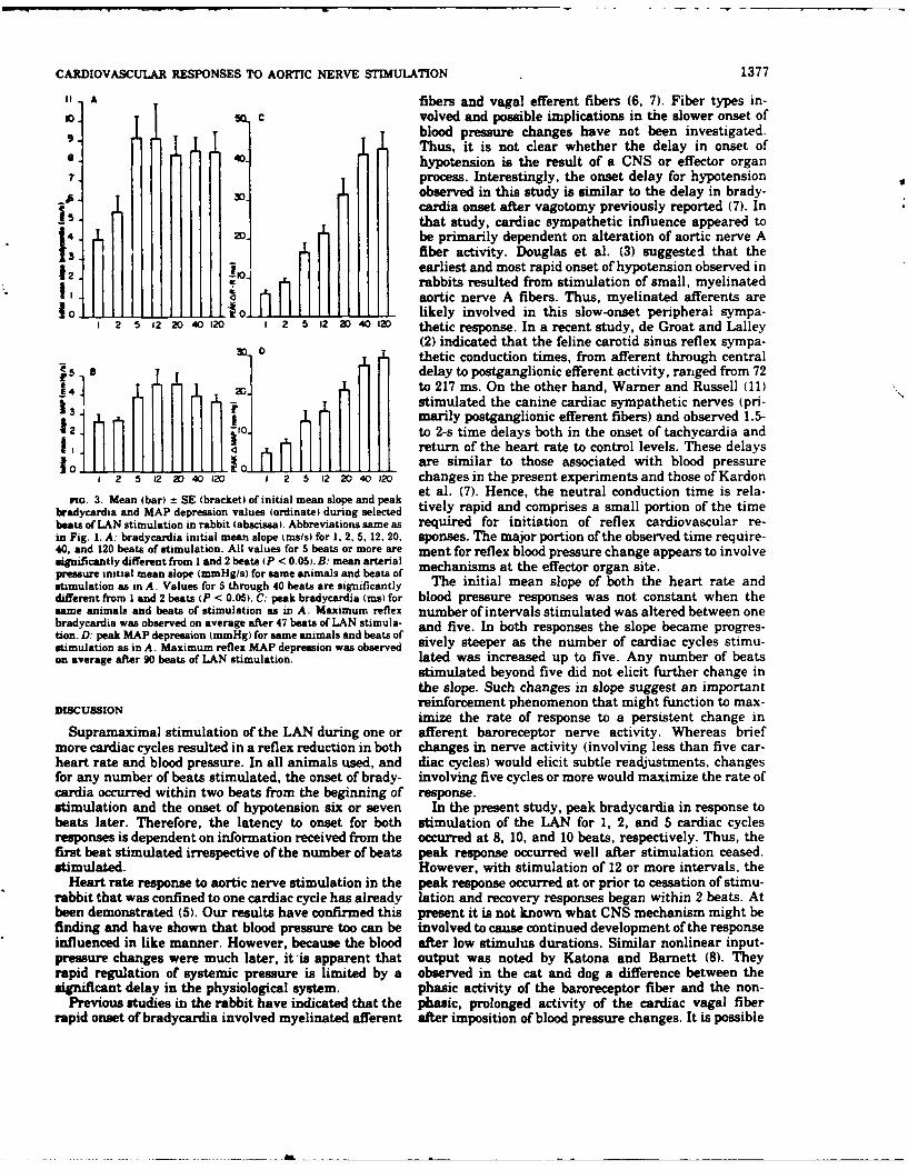
CARDIOVASCULAR RESPONSES TO AORTIC NERVE STIMULATION 1377
I. A fibers and vagal efferent fibers (6, 7). Fiber types in-10. sQc volved and possible implications in the slower onset of9. ,t blood pressure changes have not been investigated.
Thus, it is not clear whether the delay in onset ofhypotension is the result of a CNS or effector organ
. Iprocess. Interestingly, the onset delay for hypotension6 0.observed in this study is similar to the delay in brady-
15. cardia onset after vagotomy previously reported (7). In4 20 that study, cardiac sympathetic influence appeared to
I~1 be primarily dependent on alteration of aortic nerveA3. fiber activity. Douglas et al. (3) suggested that the
12. '' earliest and most rapid onset of hypotension observed inIt ;rabbits resulted from stimulation of small, myelinated
I I LI 11 - - aortic nerve A fibers. Thus, myelinated afferents are101 o . . 10. . likely involved in this slow-onset peripheral sympa-
I 2 5 12 20 40 120 1 2 5 12 2D 40 120 thetic response. In a recent study, de Groat and Lalley(2) indicated that the feline carotid sinus reflex sympa-
Sthetic conduction times, from afferent through central
64 to 217 ms. On the other hand, Warner and Russell (11)marily postganglionic efferent fibers) and observed 1.5-
12 . to 2-s time delays both in the onset of tachycardia andIIIIreturn of the heart rate to control levels. These delays
11 ...... are similar to those associated with blood pressure1 2 5 2 20 40 120 1 2 5 12 20 40 120 changes in the present experiments and those of Kardon
rio. 3. Mean (bar) = SE (bracket) of initial mean slope and et al. (7). Hence, the neutral conduction time is rela-bradycardia and MAP depression values (ordinate) during selected tively rapid and comprises a small portion of the timebeats of LAN stimulation in rabbit (abscissa). Abbreviations same as required for initiation of reflex cardiovascular re-in Fig. 1. A: bradycardia initial mean slope tins/s) for 1, 2, 5, 12, 20. sponses. The major portion of the observed time require-40, and 120 beats of stimulation. All values for 5 beats or more are ment for reflex blood pressure change appears to involvesignificantly different from I and 2 beats (P < 0.05,. B: mean arterial mechanisms at the effector organ site.pressure initial mean slope (mmHg/s) for same animals and beats ofstimulation as in A. Values for 5 through 40 beats are significantly The initial mean slope of both the heart rate anddifferent from 1 and 2 beats (P < 0.05). C: peak bradycardia (ms) for blood pressure responses was not constant when thesame animals and beats of stimulation as in A. Maximum reflex number of intervals stimulated was altered between onebradycardia was observed on average after 47 beats of LAN stimula- and five. In both responses the slope became progres-tion. D: peak MAP depression (mmHg) for same animals and beats ofstimulation as in A. Maximum reflex MAP depression was observed sively steeper as the number of cardiac cycles stimu-on average after 90 beats of LAN stimulation. lated was increased up to five. Any number of beats
stimulated beyond five did not elicit further change inthe slope. Such changes in slope suggest an important
DISCUSSION reinforcement phenomenon that might function to max-imize the rate of response to a persistent change in
Supramaximal stimulation of the LAN during one or afferent baroreceptor nerve activity. Whereas briefmore cardiac cycles resulted in a reflex reduction in both changes in nerve activity (involving less than five car-heart rate and blood pressure. In all animals used, and diac cycles) would elicit subtle readjustments, changesfor any number of beats stimulated, the onset of brady- involving five cycles or more would maximize the rate ofcardia occurred within two beats from the beginning of response.stimulation and the onset of hypotension six or seven In the present study, peak bradycardia in response tobeats later. Therefore, the latency to onset for both stimulation of the LAN for 1, 2, and 5 cardiac cyclesresponses is dependent on information received from the occurred at 8, 10, and 10 beats, respectively. Thus, thefirst beat stimulated irrespective of the number of beats peak response occurred well after stimulation ceased.stimulated. However, with stimulation of 12 or more intervals, the
Heart rate response to aortic nerve stimulation in the peak response occurred at or prior to cessation of stimu-rabbit that was confined to one cardiac cycle has already lation and recovery responses began within 2 beats. Atbeen demonstrated (5). Our results have confirmed this present it is not known what CNS mechanism might befinding and have shown that blood pressure too can be involved to cause continued development of the responseinfluenced in like manner. However, because the blood after low stimulus durations. Similar nonlinear input-pressure changes were much later, it is apparent that output was noted by Katona and Barnett (8). Theyrapid regulation of systemic pressure is limited by a observed in the cat and dog a difference between thesignificant delay in the physiological system. phasic activity of the baroreceptor fiber and the non-
Previous studies in the rabbit have indicated that the phasic, prolonged activity of the cardiac vagal fiberrapid onset of bradycardia involved myelinated afferent after imposition of blood pressure changes. It is possible
1378 STINNETT, PETERSON, AND BISHOP
that baroreceptor afferents initiate a "reverberating ac- reflex bradycardia after stellectomy in the rabbit has,tivity" in the CNS similar in nature to that hypothe- been shown to occur near the 20th interval; after vagot-sized by Hockman and Talesnik (4), which modifies omy with sympathetic nerves intact the LTP was almostafferent input resulting in prolonged efferent output doubled (7). That study also demonstrated that the la-after short-duration stimuli. However, to explain the tency to onset of heart rate changes after vagotomyrapid "off" and recovery response observed at longer occurred much later, at the eighth or ninth beat, whichstimulus durations, an additional mechanism must be corresponds to onset of blood pressure responses ob-considered. One possibility could be that when stimulus served in this study. Thus, in the rabbit, changes induration is extended into the period when blood pres- end-organ responses mediated by sympathetic tonesure is depressed, and is detectable by the intact aortic seem to have similar temporal characteristics for theand carotid sinus baroreceptors, attenuation or inhibi- heart and vascular areas during aortic afferent stimula-tion of this reverberating modulatory activity results tion.from altered afferent activity from these receptors., Finally, since the abrupt changes in the slope of the
Significant hypotension was not associated with bra- heart rate change occurs as blood pressure falls, perhapsdycardia produced by efferent vagal stimulation in this the decrease in neural activity from intact barn-eceptorsstudy. Hence, the fall in blood pressure during LAN influences vagal outflow selectively. This is supportedstimulation was primarily due to a reflex decrease in by evidence that the rabbit aortic afferent C fibers dovascular sympathetic tone. If blood pressure was influ- not elicit changes in heart rate through efferent sympa-enced by bradycardia, the relationship was too subtle to thetic pathways, whereas they are important in vagalbe identified by our methods. . control of heart rate (7). Furthermore, unmyelinated
Onset and recovery of both heart rate and blood pres- aortic afferents reportedly are absent from sympatheticsure responses were consistent and there was seldom sensory areas of the brainstem (9). Thus, changes inany indication that blood pressure returned toward con- afferent C fiber activity from intact baroreceptors maytrol during stimulation. This suggests that CNS modu- be occurring that influence vagal but not sympatheticlation of sympathetic activity to the peripheral vascula- outflow.ture is sustained as long as the aortic nerve input issustained in spite of the falling blood pressure. On the We thank Mark Priesand for his valuable assistance in designingother hand, the reflex control of heart rate is more com- the computer programs.plex, involving simultaneous alterations in both vagaland sympathetic efferent activity. This work was supported in prt by National Institutes of Health
The onset interval and initial mean slope for reflex Grant HL12415-07, AFOSR 73-2525, and the Texas Heart Associa-
bradycardia during the first 10 intervals must be me-
diated principally through vagal efferents. The LTP for Received for publication 27 August 1975.
REFERENCES
1. CHALuxas. J. P., P. I. Ko&Nxa, ANm S. W. Wmr. The relative Physiol. 225: 7-11, 1973.role of the aortic and carotid sinus nerves in the rabbit in the 7. KARDON, M. B., D. F. P315RSON, AND V. S. BIsHOP. Reflex heartcontrol of respiration and circulation during arterial hypoxia rate control via specific aortic nerve afferents in the rabbit.and hypercapma. J. Physiol., London 188: 435-450, 1967. Circulation Res. 37: 4147, 1975.
2. DR GROAT, W. C., AND P. M. LALLzY. Reflex sympathetic firing in 5. KATONA, P. G., An G. 0. BAmmNTr. Central origin of asymmetryresponse to electrical stimulation of the carotid sinus nerve in in the carotid sinus reflex. Ann. N. Y. Acad. Sci. 156: 779-786,the cat. Brain Res. 80: 17-40, 1974. 1969.
3. DouGLAs, W. W., J. M. RrrcHIE, AND W. SCiAUtANN. Depressor 9. KUMADA. M., AND H. NAKAJIMA. Field potentials evoked inreflexes from medullated and nonmedullated fibres in the rab- rabbit brain stem by stimulation of the aortic nerve. Am. J.bits aortic nerve. J. Physwil., London 132: 187-198, 1956. Physiol. 223: 575-582, 1972.
4. HOCKMAN, C. H., AD J. TAL mz. Central nervous system 10. NSuL, E., C. R. M. RXDWOOD, Axn A. Scwzrrzza. Effects ofmodulation of baroreceptor input. Am. J. Physiol. 221: 515-519, electrical stimulation of the aortic nerve on blood pressure and1971. respiration in cats and rabbits under chloralose and Nembutal
5. KARDON, M. B., D. F. PMsON, AND V. S. BISoP. Beat-to-beat anesthesia. J. Physiol., London 109: 392-401, 1949.regulation of heart rate by afferent stimulation of the aortic 11. WASmHe, H. R., AND R. 0. Russzu,, JR. Effect of combinednerve. Am. J. Physiol. 227: 598-00, 1974. sympathetic and vagal stimulation on heart rate in the dog.
S. KAKDON. M. B., D. F. PmaoRN, ArrD V. S. BSHOP. Reflex Circulation Res. 24: 567-573, 1969.bradycardia due to aortic nerve stimulation in the rabbit. Am. J.
Rcrrmlcd tron ( jrhul,it," R r h
Reduction in Baroreflex Cardiovascular ResponsesDue to Venous Infusion in the Rabbit
HENRY 0. STINNETT, PH.D., VERNON S. BISHOP, PH.D., AND D. FRED PETERSON, PH.D.
SUMMARY We studied reflex brudycardia and depression of 52% (MAP), and the initial slopes (rate of change) were reducedmean arterial blood pressure (MAP) during left aortic nerve by 46% (heart rate) and 66% (MAP). Compariaon of the re-(LAN) stimulation before and after volume infusion in the anes- sponses after infusion with blood and dextran solutions indicatedthetized rabbit. Step increases in mean right atrial pressure that hemodilution was an unlikely explanation for the attenuation(MRAP) to 10 mm Hg did not result in a significant change in of the reflex responses. Total arterial baroreceptor denervationheart rate or MAP. After volume loading, responses to LAN (ABD) abolished the volume-related attenuation of the cardio-stimulation were not as great and the degree of attenuation was vascular responses, whereas attenuation was still present follow.proportional to the level of increased MRAP. A change in re- ing bilateral aortic nerve section or vugotomy. It thus appears thatsponsiveness was observed after elevation of MRAP by only I the carotid sinus responds to changes in blood volume and influ-mm Hg, corresponding to less than a 10% increase in average ences the reflex cardiovascular responses to afferent stimulationcalculated blood volume. After an increase in MRAP of 10 mm of the LAN. On the other hand, cardiopulmnonary receptorsHg, peak responses were attenuated by 44% (heart rate) and subserved by vagal afferents do not appear to be involved.
VOLUME LOADING. sufficient to raise the venous unlikely that specific modification of systemic arterial bar-pressure and dilate the heart. has been observed to pro- oreceptors is involved. In the dog, Gupta et al.2" foundduce striking cardiovascular responses.- 3 Since Bain- dramatic increases in atrial type B receptor activity withbridge3 first reported an increase in heart rate during volume expansion, while observing only small changes inintravenous infusion many studies have attempted to de- activity of individual aortic fibers. However, later work byscribe the efferent pathways" of reflexes originating from Edis2' demonstrated that the aortic baroreceptor in thereceptors located in the cardiopulmonary region.' 0 - Still. dog shows little tonic activity, consequently, in the dog.the interaction of the low pressure cardiopulmonary re- one would not expect to see large changes in aortic nerveceptors %ith the arterial baroreflex system is not clear.' 5 activity with modest alterations in vascular volume. On theUnder conditions of arterial baroreceptor isolation or de- other hand, in the rabbit, Kumada and Sagawa 2 found thenervation. tne receptors in the cardiopulmonary region aortic baroreceptor nerve activity recorded from multifi-that are subserved by afferent vagal fibers have been ber preparations to be proportional to modest blood pres-shown to exert a restraint on the sympathetic adrenergic sure changes during 20% volume loading and 10% bloodoutflow to the peripheral vasculature in the dog" and loss.rabbit." Recently. Vatner et al.2 found that arterial barn- This study was designed to investigate the influence ofreflex sensitivity in dogs is reduced as atrial pressure is volume expansion on responses to aortic nerve stimulationincreased by volume loaJing and suggested that the set in the intact and selectively denervated rabbit. Heart ratepoint or gain of the arterial baroreflex system is altered and blood pressure responses were measured before andduring infusion. These alterations might occur at either the after steady state alterations in mean right atrial pressurereceptor site or in the central nervous system. Although (MRAP). The relative influence of low and high pressurerecent evidence"' '
" does suggest that reflex responses receptors was examined and the threshold for atrial pres-from one input are modified by other afferent inputs sure necessary to effect altered responses was determined.through integration in the central nervous system, it is The rabbit was chosen as the model for study because it
has an easily identified aortic nerve which subserves onlyFrom the Department of Pharmacology. The University ofTexas Health baroreceptors, and the vascular responses are well de-
Science Center at San Antonio. San Antonio. Texas. fined."- z"Supported by National Institutes of Health Grant HL 12415, AFOSR
73-2525, and the San Antonio Chapter of the Americ.an Heart Associa- Methodstion.
Address for reprints: Vernon S. Bishop. Ph.D.. Department of Pharma- Twenty-four rabbits weighing between 1.48 and 2.23 kgcology. rhe University of Texas Health Science Center at Antonio. 7703Floyd Curl Drive. San Antonio. Texas 78284. were anesthetized with sodium pentobarbital (Diabutal.
Received January 22. 1976; accepted for publication July 9. 1976. Diamorl Laboratories), 30 mg/kg, iv, via an ear vein for
BAROREFLEX ATTENUATION BY INFUSION/Stinnett et al. 767
this study. Supplemental anesthetic was administered infusion was continued at a rate necessary to maintainthrough a cannulated femoral vein to maintain a light level MRAP at that level. Trial stimulations of the LAN wereof surgical anesthesia. The descending aorta (via the femo- initiated after heart rate and MAP reached stabilizedral artery) and the right atrium (via the jugular vein) also steady state values.were cannulated and connected to Statham P23Db and Reflex responses to LAN stimulation were also studiedP23Bb strain gauges to record arterial and right atrial in four rabbits which were bled after prior infusion ofpressures, respectively. Heart rate was monitored through dextran solution. Each rabbit was bled until the volume ofsternal needle electrodes connected to a Beckman 9847B blood removed equaled the volume of dextran solutioncardiotachometer coupler.24 Blood pressures and heart previously infused. In some instances, slightly more bloodrate initially were recorded on a Beckman R411 oscillo- was withdrawn to reduce MRAP to control level (range,graph with parallel output signals from the arterial pres- 0-1.5 mm Hg). In no case did the volume withdrawn plussure and heart rate channels connected to a DEC PDP 8/E urine volume collected during this period exceed the pre-digital computer. A tracheostomy was performed and the viously infused volume by more than 10 ml. Followingrabbits were artificially ventilated by the technique pub- each bleeding, a period of 1-2 minutes was allowed forlished previously24 to ensure maintenance of normal blood stabilization of the rabbit's heart rate and blood pressurePo. Pco2 , and pH. Through a midventral incision, the left before further experimental trials were undertaken.and right aortic nerves were located in the cervical region To monitor the red blood cell concentration, hemato-and carefully isolated from surrounding tissue for about I crits were measured before and after each infusion orcm.2 4. 2 In addition, in four rabbits the vagi, and in five bleeding. In addition, urine flow and volume also wererabbits the carotid sinus nerves, also were carefully iso- routinely monitored throughout each experiment.lated and looped with a loose thread for identification Values are reported as the mean or mean difference -prior to bilateral sectioning. Loss of the reflex responses the standard error of the mean (sEM). Statistical evalua-due to bilateral carotid occlusion verified carotid sinus tion was made by use of the appropriate Student's t-test fordenervation.3s After carotid sinus denervation total arte- paired or unpaired comparisons. P values (<0.05) wererial baroreceptor denervation (ABD) was considered considered significant.complete when both aortic nerves also were sectioned. Inall rabbits the left aortic nerve (LAN) was sectioned near Remiltsthe sternum and bathed in mineral oil, as previously de-scribed 4. - The central end of the LAN was placed on In 14 rabbits MRAP was elevated to 5 mm Hg and inbipolar (platinum-iridium) electrodes which were con- seven of these, to 10 mm Hg, using an average of 41 andnected to a Grass SD9 stimulator. The stimulator was 71 ml of dextran solution. This acute volume expansionactivated by the Schmitt trigger of the computer which was did not significantly affect either heart rate or blood pres-synchronized with the R wave of the electrocardiogram sure (Table 1). Representative changes in response to(ECG). Regulation of the stimulus timing and stimulus LAN stimulation are shown in Figure 1 before and afterparameters, as well as continuous calculation of the length step increases in right atrial pressure. The duration ofof each R-R interval and beat-to-beat mean arterial pres- stimulation used (120 cardiac cycles) previously has beensure (MAP), were accompfished using the computer and shown to produce maximum heart rate and blood pressurespecial computer program systems. An experimental trial responses.' Note that following the increases in MRAP.consisted of: (1) 10 successive control cardiac cycles, (2) the amplitude of each response was further decreasedbursts of electrical stimuli coupled to each R wave of the (Fig. 1). The average maximum increase in R-R interval ofECG beginning with the 11 th interval and continuing -45.4 ± 3.0 msec (n = 14) induced by aortic nerve stimula-through 120 intervals, and (3) continuous data collection tion was significantly reduced (P < 0.01) to 31.6 ± 4.2 (nthrough recovery to control. Each burst of electrical stim- = 14) and 25.4 ± 3.2 (n = 7) following increases inulation was made up of 10 rectangular pulses (10 V) MRAP of 5 and 10 mm Hg, respectively (Fig. 2A). Simi-delivered 10 msec after the recorded R wave of the ECG. larly, for the same rabbits, the average preinfusion MAPThe impulse duration was 0.3 msec, the stimulus fre- response of -30.0 ± 2.3 mm Hg was reduced to -17.6 -tquency was 80 Hz, and burst duration was 113 msec. For 2.4 (P < 0.005) and -14.3 ± 1.0 (P < 0.005), respec-each experimental condition data from five or more trials tively (Fig. 2B). Neither the latency to onset (LTO) norwere averaged for each rabbit. The peak change in R-R the latency to peak (LTP) was significantly altered forinterval and blood pressure, the latencies to onset and either reflex response and both were similar to thosepeak responses, as well as the initial slope for each re- previously described." However, the initial slopes forsponse. were calculated as previously described.2 both responses (Fig. 2C and D) were diminished with each
The heart rate and blood pressure responses to LAN increase in atrial pressure and this change, in conjunctionstimulation were studied in 14 rabbits infused with dextran with the lower maximum responses, would account for thein physiologic solution, 5 g/1.000 ml (dextran 40, mol wt essentially unchanged LTP's. The initial slope of the heart40,000, Pharmacia), heated at 38"C, and one rabbit was rate response decreased from 9.29 ± 0.88 msecisec toinfused with whole blood from a heparinized donor. The 5.66 ± 0.84 (P < 0.01) and 5.03 ± 0.85 (P < 0.05).rate of infusion through the femoral vein was adjusted to Similarly, the initial slope of the control MAP response,ensure a steady rise in right atrial pressure. Infusions were 3.68 ± 0.34 mm Hg/sec, was decreased to 2.14 ± 0.41 (Pgiven overa period of 6-14 minutes. Once the MRAPhad < 0.005) and 1.25 ± 0.13 (P < 0.005) by increases inreached a predetermined level (1, 2.5, 5, or 10 mm Hg) MRAP of 5 and 10 mm Hg, respectively.
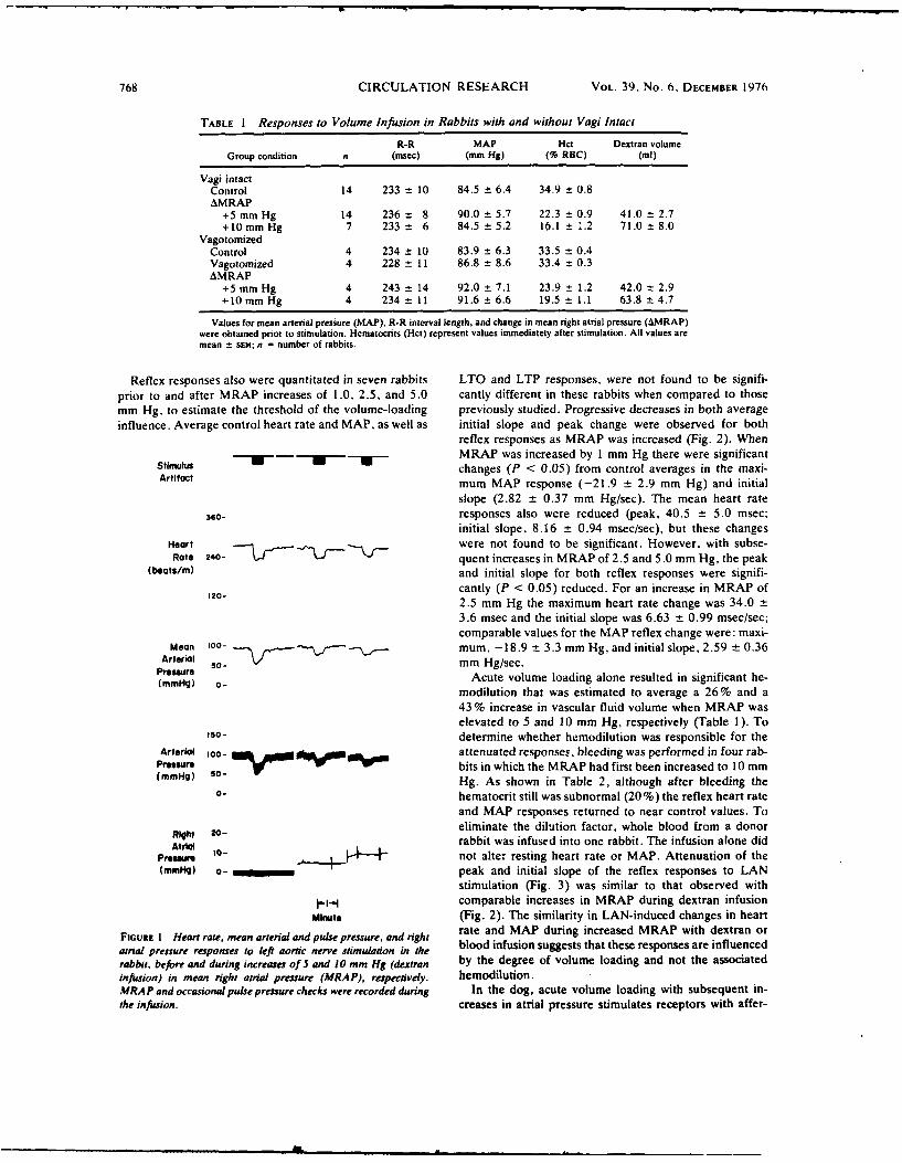
768 CIRCULATION RESEARCH VOL. 39. No. 6, DECEMBER 1976
TABLE I Responses to Volume Infusion in Rabbits with and without Vagi Intact
R-R MAP Hct Dextran volumeGroup condition n (msec) (mm Hg) (% RBC) (ml)
Vagi intactControl 14 233 ± 10 84.5 ± 6.4 34.9 ± 0.8AMRAP
+5mm Hg 14 236 ± 8 90.0 ± 5.7 22.3 ± 0.9 41.0 t 2.7+10mm Hg 7 233 ± 6 84.5 ± 5.2 16.1 ± .2 71.0 ± 8.0
VagotomizedControl 4 234 t 10 83.9 ± 6.3 33.5 ± 0.4Vagotomized 4 228 ± 11 86.8 ± 8.6 33.4 ± 0.3AMRAP
+5 mm Hg 4 243 ± 14 92.0 ± 7.1 23.9 ± 1.2 42.0 ± 2.9+10mm Hg 4 234 ±11 91.6 ± 6.6 19.5 ±1.1 63.8 ± 4.7
Values for mean arterial pressure (MAP). R-R interval length, and change in mean right atrial pressure (AMRAP)were obtained prior to stimulation. Hematocrits (Hct) represent values immediately after stimulation. All values aremean - sEM; n = number of rabbits.
Reflex responses also were quantitated in seven rabbits LTO and LTP responses, were not found to be signifi-prior to and after MRAP increases of 1.0, 2.5, and 5.0 cantly different in these rabbits when compared to thosemm Hg, to estimate the threshold of the volume-loading previously studied. Progressive decreases in both averageinfluence. Average control heart rate and MAP, as well as initial slope and peak change were observed for both
reflex responses as MRAP was increased (Fig. 2). WhenMRAP was increased by 1 mm Hg there were significant
Stimulus changes (P < 0.05) from control averages in the maxi-Artifact mum MAP response (-21.9 ± 2.9 mm Hg) and initial
slope (2.82 ± 0.37 mm Hg/sec). The mean heart rate
360- responses also were reduced (peak, 40.5 ± 5.0 msec;initial slope, 8.16 ± 0.94 msec/sec), but these changes
Heart were not found to be significant. However, with subse-Rate 240- ~quent increases in MRAP of 2.5 and 5.0 mm Hg, the peak
(beats/m) and initial slope for both reflex responses were signifi-120- cantly (P < 0.05) reduced. For an increase in MRAP of
2.5 mm Hg the maximum heart rate change was 34.0 ±
3.6 msec and the initial slope was 6.63 ± 0.99 msec/sec;comparable values for the MAP reflex change were: maxi-
Mean 100- mum, -18.9 ± 3.3 mm Hg, and initial slope, 2.59 ± 0.36Arterial 50- mm Hg/sec.Premsre Acute volume loading alone resulted in significant he-(mmHg) o- modilution that was estimated to average a 26% and a
43% increase in vascular fluid volume when MRAP waselevated to 5 and 10 mm Hg, respectively (Table 1). To
1so- determine whether hemodilution was responsible for theArterial 1O ___0-__ attenuated responses, bleeding was performed in four rab-
Pressure " - bits in which the MRAP had first been increased to 10 mm(mmHg) 5 Hg. As shown in Table 2, although after bleeding the
o- hematocrit still was subnormal (20 %) the reflex heart rate
and MAP responses returned to near control values. ToRiOh 20- eliminate the dilution factor, whole blood from a donorAtrto- rabbit was infused into one rabbit. The infusion alone did
Pressure ,... -" not alter resting heart rate or MAP. Attenuation of the(mmHg) a- peak and initial slope of the reflex responses to LAN
stimulation (Fig. 3) was similar to that observed withI--.i comparable increases in MRAP during dextran infusion
Minute (Fig. 2). The similarity in LAN-induced changes in heart
FIGURE 1 Heart rate, mean arterial and pulse pressure, and right rate and MAP during increased MRAP with dextran or
atrial pressure responses to left aortic nerve stimulation in the blood infusion suggests that these responses are influenced
rabbit, before and during increases of 5 and 10 mm Hg (dextran by the degree of volume loading and not the associatedinfusion) in mea right atrial pressure (MRAP), respectively. hemodilution.MRAP and occasional pulse pressure checks were recorded during In the dog, acute volume loading with subsequent in-the infusion, creases in atrial pressure stimulates receptors with affer-
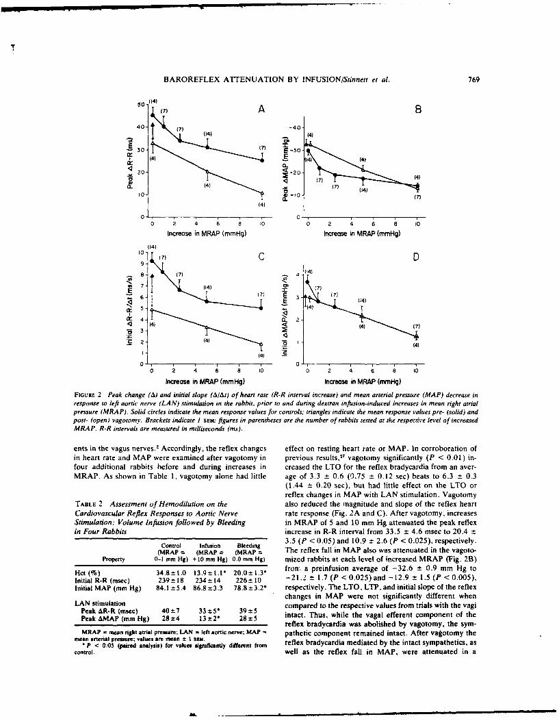
BAROREFLEX ATTENUATION BY INFUSIONIStinnet et al. 769
50 (1)47)A)
(7)-O 74(44
(1) ( )(
0- (7)E 30-
(4) . 14) (4)4 4_
2- 20! (4) (7) 4
(44)
0- 0 -
E
(4 (4
0 2 4 6 a 10 0 2 4 6 8 (0
Increase in MRAP (mmHg) Increase in MRAP (mmHg)
FIGURlE 2 Peak change (A) and initial slope (Al/At) of heart rate (R-R interval increase) and mean arterial pressure (MAP) decrease inresponse to left aortic nerve (LAN) stimulation in the rabbit, prior to and during dextran infusion-induced increases in mean right atrialpressure (MRAP). Solid circles indicate the mean response values for controls; triangles indicate the mean response values pre. (solid) andpost- (open) vagotomy. Brackets indicate 1 Srss" figures in parentheses are the number of rabbits tested at the respective level of increasedMRAP. R-R intervals are measured in milliseconds (ins).
ents in the vagus nerves.2 Accordingly, the reflex changes effect on resting heart rate or MAP. In corroboration ofin heart rate and MAP were examined after vagotomy in previous results,27 vagotomy significantly (P < 0.01) in-four additional rabbits before and during increases in creased the LTO for the reflex bradycardia from an aver-MRAP. As shown in Table 1, vagotomy alone had little age of 3.3 -± 0.6 (0.75 ±- 0.12 sec) beats to 6.3 ±_ 0.3
(1 .44 ± 0.20 sec), but had little effect on the LTO orreflex changes in MAP with LAN stimulation. Vagotomy
T~ALE 2 Assessment of Hemodilution on the also reduced the magnitude and slope of the reflex heartCardiovascular Reflex Responses to Aortic Nerve rate response (Fig. 2A and C). After vagotomy. increasesStimulation: Volume Infusion followed by Bleeding in MRAP of 5 and 10 mm Hg attenuated the peak reflexin Four Rabbits increase in R-R interval from 33.5 ± 4.6 msec to 20.4 -,
t ( i d 3.5 (P <0.05) and 10.9 ± 2.6 (P <0.025), respectively.
(MRAP :s (MRAP :" (MRAP s The reflex fall in MAP also was attenuated in the vagoto-Pr'operty 0-1 mm Hg) + 10 mm Hg) 0.0 mm Hg) mized rabbits at each level of increased MRAP (Fig. 2B)
Hct (%) 34.8±t.0 13.9±-t.t * 20.0±t.35 frorr a preinfusion average of -32.6 ± 0.9 mm Hg toInitial R-R (msec) 239±18 234± 14 226± 10 -21.2 ± 1.7 (P < 0.025) and -12.9 _± 1.5 (P < 0.005).Initial MAP (mam Hg) 84.1 ± 5.4 86.8 ±3.3 78.8 ± 3.2" respectively. The LTO, LTIP, and initial slope of the reflex
changes in MAP were not significantly different when
Pak R stmuela4±7tion 9± compared to the respective vaiues from trials with the vagiPeak &R-RP (mmg) 24- 13±2" 28±5 intact. Thus, while the vagal efferent component of the
Pek MP mmu) 7- ± 14)" 8
reflex bradycardia was abolished by vagotomy, the sym-MRAP - mean right atrial pressure; LAN - left aorsic nerve; MAP = pathetic component remained intact. After vagotomy the
mean arterial pressure; values are mean ± t SeMI.elxbayadamdae b h natsmahtca•P < 0.05 (pie analysis) for values siginificantly different from rfe rdcri eitdb h natsmahtc.a
control. well as the reflex fall in MAP, were attenuated in a
E (7) / I I i I i u l I m l
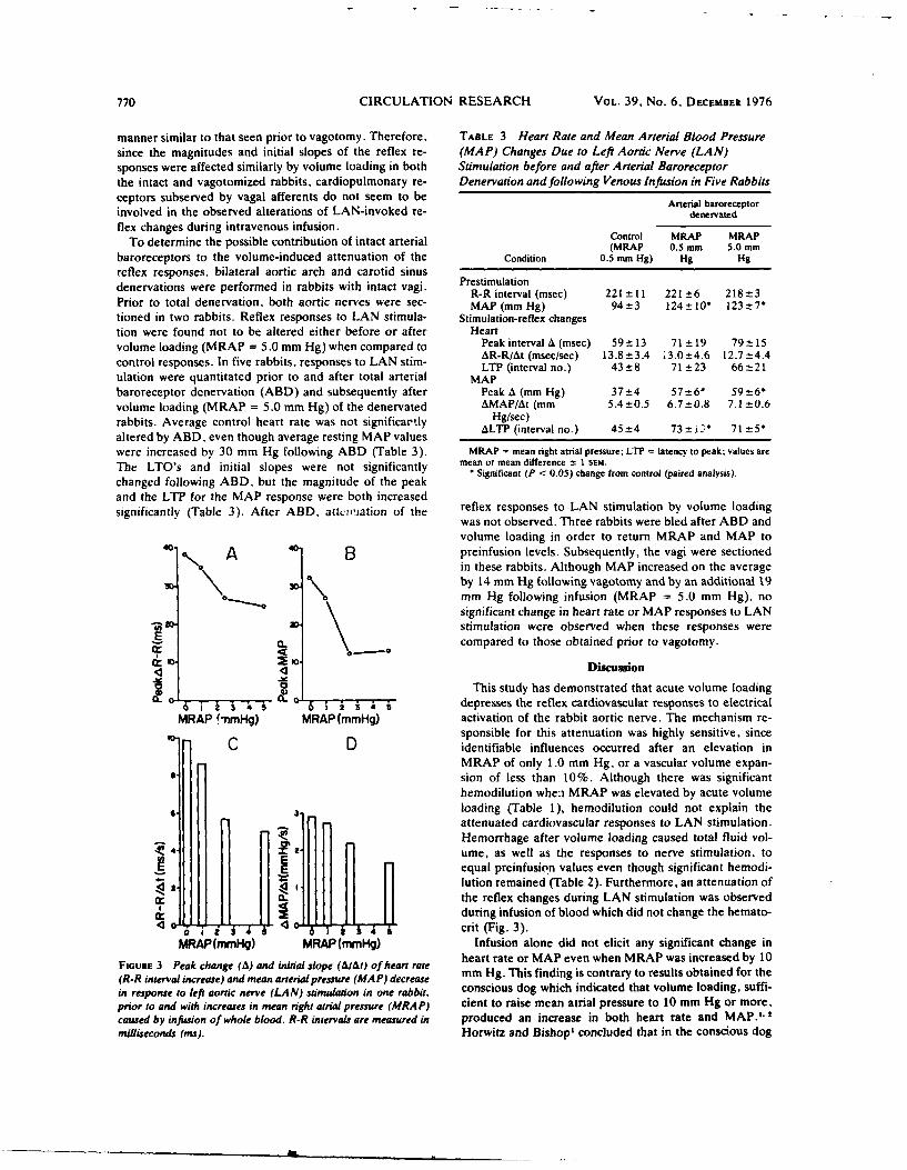
770 CIRCULATION RESEARCH VOL. 39, No. 6, DECEMBER 1976
manner similar to that seen prior to vagotomy. Therefore, TABLE 3 Heart Rate and Mean Arterial Blood Pressuresince the magnitudes and initial slopes of the reflex re- (MAP) Changes Due to Left Aortic Nerve (LAN)sponses were affected similarly by volume loading in both Stimulation before and after Arterial Baroreceptorthe intact and vagotomized rabbits, cardiopulmonary re- Denervation and following Venous Infusion in Five Rabbitsceptors subserved by vagal afferents do not seem to be Arterial baroreceptorinvolved in the observed alterations of LAN-invoked re- denervatedflex changes during intravenous infusion. ofitatateilControl MRAP MRAP
To determine the possible contribution of intact arterial (MRAP 0.5 mm 5.0 mm
baroreceptors to the volume-induced attenuation of the Condition 0.5 mm Hg) Hg Hgreflex responses, bilateral aortic arch and carotid sinusdenervations were performed in rabbits with intact vagi. Prestimulation
R-R interval (msec) 221 - 11 221 ±6 218-3Prior to total denervation, both aortic nerves were sec- MAP (mm Hg) 94±3 124-± 10 123±t7"tioned in two rabbits. Reflex responses to LAN stimula- Stimulation-reflex changestion were found not to be altered either before or after Heartvolume loading (MRAP = 5.0 mm Hg) when compared to Peak interval A (msec) 59 ± 13 71 ± 19 79 ± 15
AR-R/At (msec/sec) 13.8 ±3.4 .3.0±4.6 12.7:±4.4control responses. In five rabbits, responses to LAN stim- LTP (interval no.) 43- 8 71 ±23 66-±-21ulation were quantitated prior to and after total arterial MAPbaroreceptor denervation (ABD) and subsequently after Peak A (mm Hg) 37±4 57±t6* 59±61volume loading (MRAP = 5.0 mm Hg) of the denervated AMAP/At (mm 5.4 ±0.5 6.7 ±-0.8 7.1 ±0.6
rabbits. Average control heart rate was not significartly Hg/sec)
altered by ABD, even though average resting MAP values ALTP (interval no.) 45±4 73±i 71±5*
were increased by 30 mm Hg following ABD (Table 3). MRAP = mean right atrial pressure; LTP = latency to peak; values are
The LTO's and initial slopes were not significantly mean or mean difference ±-1 SIum.
changed following ABD, but the magnitude of the peak Significant (P < 0.05) change from control (paired analysis).
and the LTP for the MAP response were both increasedsignificantly (Table 3). After ABD, attciviation of the reflex responses to LAN stimulation by volume loading
was not observed. Three rabbits were bled after ABD andvolume loading in order to return MRAP and MAP to
40. A 4 B preinfusion levels. Subsequently, the vagi were sectionedin these rabbits. Although MAP increased on the average
30. .by 14 mm Hg following vagotomy and by an additional 19
mm Hg following infusion (MRAP = 5.0 mm Hg), nosignificant change in heart rate or MAP responses to LANstimulation were observed when these responses werecompared to those obtained prior to vagotomy.
Discussion
0 oThis study has demonstrated that acute volume loading
6 3 i i 4 0 1 2 3 4 depresses the reflex cardiovascular responses to electricalMRAP t -nmHg) MRAP(mmHg) activation of the rabbit aortic nerve. The mechanism re-
sponsible for this attenuation was highly sensitive, sinceC D identifiable influences occurred after an elevation in
MRAP of only 1.0 mm Hg, or a vascular volume expan-G sion of less than 10%. Although there was significant
hemodilution when MRAP was elevated by acute volume6 loading (Table 1), hemodilution could not explain the
attenuated cardiovascular responses to LAN stimulation.Hemorrhage after volume loading caused total fluid vol-
4- ume, as well as the responses to nerve stimulation, toequal preinfusion values even though significant hemodi-
4 -. lution remained (Table 2). Furthermore, an attenuation ofl- the reflex changes during LAN stimulation was observedk during infusion of blood which did not change the hemato-4 -- 0. crit (Fig. 3).
MRAP(mmHg) MRAP(mmHg) Infusion alone did not elicit any significant change in
FIoUtE 3 Peak change (A) and mnitial slope (AI) of heart rate heart rate or MAP even when MRAP was increased by 10
(R-R interval increase) and mean arterial pressure (MAP) decrease mm Hg. This finding is contrary to results obtained for the
in response to left aortic nerve (LAN) stimulaion in one rabbit, conscious dog which indicated that volume loading, suffi-
prior to and with increases in mean right atrial pressure (MRAP) cient to raise mean atrial pressure to 10 mm Hg or more,caused by infusion of whole blood. R-R intervals are measured in produced an increase in both heart rate and MAP.",milliseconds (ms). Horwitz and Bishop' concluded that in the conscious dog
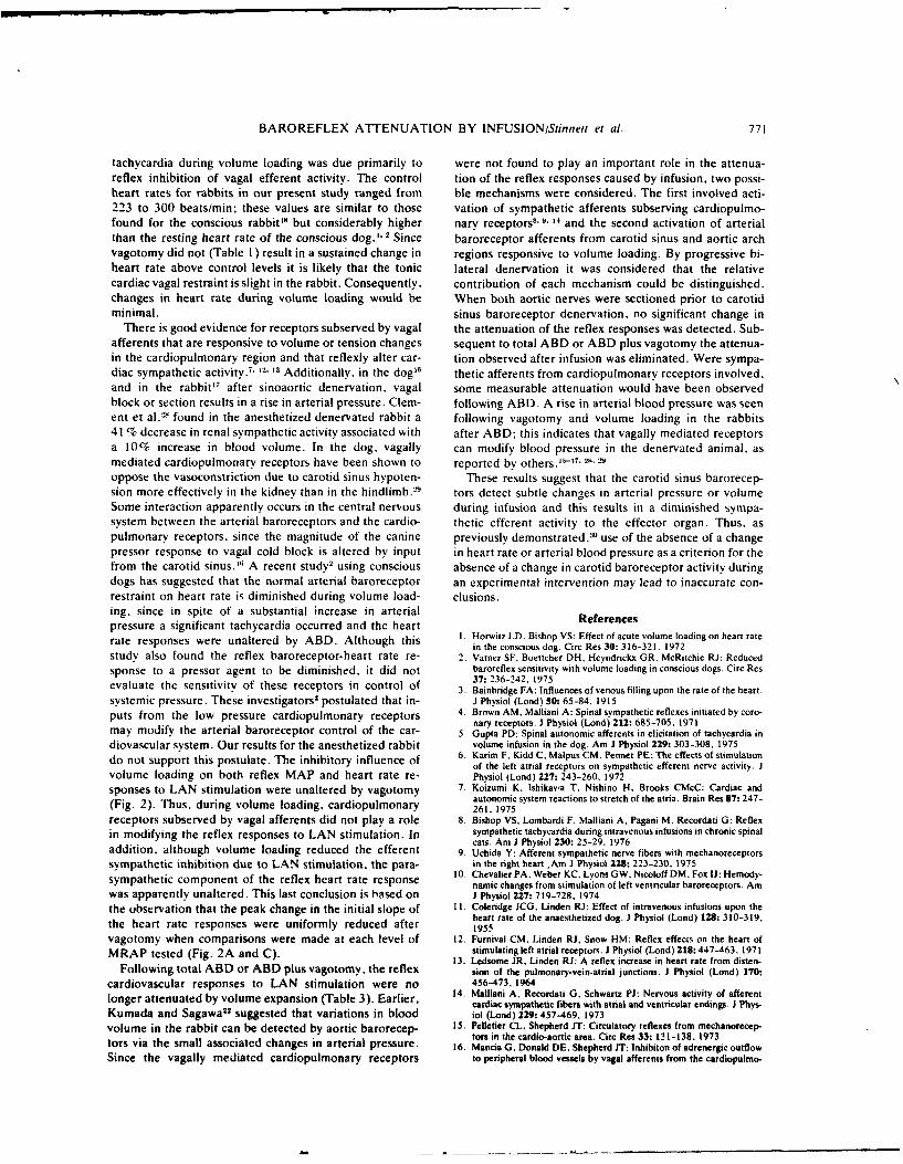
BAROREFLEX ATTENUATION BY INFUSION/Stinnett et al. 771
tachycardia during volume loading was due primarily to were not found to play an important role in the attenua-reflex inhibition of vagal efferent activity. The control tion of the reflex responses caused by infusion, two possi-heart rates for rabbits in our present study ranged from ble mechanisms were considered. The first involved acti-223 to 300 beats/min; these values are similar to those vation of sympathetic afferents subserving cardiopulmo-found for the conscious rabbit'" but considerably higher nary receptors" 9- '4 and the second activation of arterialthan the resting heart rate of the conscious dog." I Since baroreceptor afferents from carotid sinus and aortic archvagotomy did not (Table 1) result in a sustained change in regions responsive to volume loading. By progressive bi-heart rate above control levels it is likely that the tonic lateral denervation it was considered that the relativecardiac vagal restraint is slight in the rabbit. Consequently, contribution of each mechanism could be distinguished.changes in heart rate during volume loading would be When both aortic nerves were sectioned prior to carotidminimal, sinus baroreceptor denervation, no significant change in
There is good evidence for receptors subserved by vagal the attenuation of the reflex responses was detected. Sub-afferents that are responsive to volume or tension changes sequent to total ABD or ABD plus vagotomy the attenua-in the cardiopulmonary region and that reflexly alter car- tion observed after infusion was eliminated. Were sympa-diac sympathetic activity.- 1' " Additionally. in the dog" thetic afferents from cardiopulmonary receptors involved.and in the rabbit 7 after sinoaortic denervation, vagal some measurable attenuation would have been observedblock or section results in a rise in arterial pressure. Clem- following ABD. A rise in arterial blood pressure was seenent et al. 6 found in the anesthetized denervated rabbit a following vagotomy and volume loading in the rabbits41% decrease in renal sympathetic activity associated with after ABD; this indicates that vagally mediated receptorsa 10% increase in blood volume. In the dog. vagally can modify blood pressure in the denervated animal, asmediated cardiopulmonary receptors have been shown to reported by others.5 -
17
. 28, 29
oppose the vasoconstriction due to carotid sinus hypoten- These results suggest that the carotid sinus barorecep-sion more effectively in the kidney than in the hindlimb.2' tors detect subtle changes in arterial pressure or volumeSome interaction apparently occurs in the central nervous during infusion and this results in a diminished sympa-system between the arterial baroreceptors and the cardio- thetic efferent activity to the effector organ. Thus. aspulmonary receptors, since the magnitude of the canine previously demonstrated,25 use of the absence of a changepressor response to vagal cold block is altered by input in heart rate or arterial blood pressure as a criterion for thefrom the carotid sinus." A recent study2 using conscious absence of a change in carotid baroreceptor activity duringdogs has suggested that the normal arterial baroreceptor an experimental intervention may lead to inaccurate con-restraint on heart rate is diminished during volume load- clusions.ing. since in spite of a substantial increase in arterialpressure a significant tachycardia occurred and the heartrate responses were unaltered by ABD. Although this t. Horwitz LD. Bishop VS: Effect of acute volume loading on heart rate
in the conscious dog. Circ Res 30: 316-321. 1972study also found the reflex baroreceptor-heart rate re- 2. Vatner SF, Boettcher DH, Heyndrickx GR. McRitchie RJ: Reducedsponse to a pressor agent to be diminished, it did not baroreflex sensitivity with volume loading in conscious dogs. Circ Res
37: 236-242. 1975evaluate the sensitivity of these receptors in control of 3. Bainbridge FA: Influences of venous filling upon the rate of the heart.systemic pressure. These investigators' postulated that in- J Physiol (Lond) 50: 65-84, 1915
puts from the low pressure cardiopulmonary receptors 4. Brown AM, Malliani A: Spinal sympathetic reflexes initiated by coro-c onary receptors. I Physiol (Lond) 212: 685-705, 1971may modify the arterial baroreceptor control of the car- 5. Gupta PD: Spinal autonomic afferents in elicitation of tachycardia in
diovascular system. Our results for the anesthetized rabbit volume infusion in the dog. Am J Physiol 229: 303-308, 1975
do not support this postulate. The inhibitory influence of 6. Karim F. Kidd C, Malpus CM, Penner PE: The effects of stimulationof the left atria] receptors on sympathetic efferent nerve activity. Jvolume loading on both reflex MAP and heart rate re- Physiol (Lond) 227: 243-260, 1972
sponses to LAN stimulation were unaltered by vagotomy 7. Koizumi K, Ishikavia T, Nishino H. Brooks CMcC: Cardiac andautonomic system reactions to stretch of the atria. Brain Res 87: 247-(Fig. 2). Thus, during volume loading, cardiopulmonary 261. 1975
receptors subserved by vagal afferents did not play a role 8. Bishop VS. Lombardi F, Malliani A, Pagani M, Recordati G: Reflexin modifying the reflex responses to LAN stimulation. In sympathetic tachycardia during intravenous infusions in chronic spinal
cats. Am J Physiol 230: 25-29. 1976addition, although volume loading reduced the efferent 9. Uchida Y: Afferent sympathetic nerve fibers with mechanoreceptorssympathetic inhibition due to LAN stimulation, the para- in the right heart ,Am J Physio 228: 223-230, 1975
tof the reflex heart rate response 10. Chevalier PA. Weber KC. Lyons GW. Nicoloff DM. Fox IU: Hemody-namic changes from stimulation of left ventricular baroreceptors. Am
was apparently unaltered. This last conclusion is hosed on J Physiol 227: 719-728, 1974the observation that the peak change in the initial slope of 11. Coleridge JCG. Linden RJ: Effect of intravenous infusions upon the
heart rate of the anaesthetized dog. J Physiol (Lond) 128: 310-319.the heart rate responses were uniformly reduced after 1955
vagotomy when comparisons were made at each level of 12. Furnival CM. Linden RJ. Snow HM: Reflex effects on the heart ofMRAP tested (Fig. 2A and C). stimulating left atrial receptors. J Physiol (Lond) 218: 447-463, 1971
13. Ledsome JR. Linden RJ: A reflex increase in heart rate from disten-Following total ABD or ABD plus vagotomy, the reflex sion of the pulmonary-vein-atrial junctions. I Physiol (Lond) 170:
cardiovascular responses to LAN stimulation were no 456-473, 196414. Malliani A, Recordati G. Schwartz PJ: Nervous activity of afferentlonger attenuated by volume expansion (Table 3). Earlier, cardiac sympathetic fibers with atrial and ventricular endings. J Phys-
Kumada and Sagawa22 suggested that variations in blood iol (Lond) 229: 457-469, 1973volume in the rabbit can be detected by aortic barorecep- 15. Pelletier CL. Shepherd JT: Circulatory reflexes from mechanorecep-
tors in the cardio-aotic area. Circ Res 33: 13 1-138, 1973tors via the small associated changes in arterial pressure. 16. Mancia G. Donald DE. Shepherd JT: Inhibiton of adrenergic outflow
Since the vagally mediated cardiopulmonary receptors to peripheral blood vessels by vagal afferents from the cardiopulmo-
772 CIRCULATION RESEARCH VOL. 39, No. 6, DECEMBER 1976
nary region in the dog. Circ Res 33: 713-721, 1973 24. Kardon MB, Peterson DF. Bishop VS: Reflex bradycardia due to17. Pillsbury HRC III, Guazzi M, Preis ED: Vagal afferent depressor aortic nerve stimulation in the rabbit. Am J Physiol 225: 7-11. 1973
nerves in the rabbit. Am J Physiol 217: 768-770. 1969 25. Chalmers JP, Korner PI. White SW: The relative roles of the aortic18. Korner Pl, Shaw J. West M. Oliver JR, Hilder RG: Integrative reflex and carotid sinus nerves in the rabbit in the control of respiration and
control of heart rate in the rabbit during hypoxia and hyperventila- circulation during arterial hypoxia and hypercapnia. J Physiol (Lond)tion. Circ Res 33: 63-73, 1973 18: 435-450. 1967
19. Kumada M. Nogami K, Sagawa K: Modulation of carotid sinus baro- 26. Stinnett HO, Peterson DF. Bishop VS: Heart rate and blood pressurereceptor reflex by sciatic nerve stimulation. Am J Physiol 228: 1535- responses to synchronized stimulation of the aortic nerve. Am J1541. 1975 Physiol 230: 1374-1378. 1976
20. Gupta PD. Henry JP, Sinclair R. Von Baumgarten R: Responses to 27. Kardon MB. Peterson DF, Bishop VS: Reflex heart rate control viaatrial and aortic baroreceptors to nonhypotensive hemorrhage and to specific aortic nerve afferents in the rabbit. Circ Res 37: 41-47. 1975transfusion. Am J Physiol 211: 1429-1437, 1966 28. Clement DL. Pelletier CL, Shepherd .T: Role of vagal afferents in the
21. Edis AJ: Aortic baroreflex function in the dog. Am J Physiol 221: control of renal sympathetic nerve activity in the rabbit. Circ Res 31:1352, 1971 824-830, 1972
22. Kumada M, Sagawa K: Aortic nerve activity during blood volume 29. Mancia G, Shepherd JT, Donald DE: Role of cardiac pulmonary andchanges. Am J Physiol 218: 961-965, 1970 carotid mechanoreceptors in the control of hindlimb and renal circula-
23. Neil E. Redwood CRM. Schweitzer A: Effects of electrical stimulation tion in dogs. Circ Res 37: 200-208, 1975of the aortic nerve on blood pressure and respiration in cats and 30. Peterson DF, Kaspar RL, Bishop VS: Reflex tachycardia due torabbits under chloralose and Nembutal anesthesia. J Physiol (Lond) temporary coronary occlusion in the conscious dog. Circ Res 32: 652-109: 392-401. 1949 659, 1973
Effect of regional myocardial ischemia oncardiac pump performance -during exercise
LAWRENCE D. HORWITZ, D. FRED PETERSON, AND VERNON S. BISHOPDepartments of Medicine and Pharmacology, University of Texas Health Science Center,San Antonio, Texas 78284
Hoawrrz, LAWUZNCz D., D. FEZD PTRSON, AND VEKNON shortening of left ventricular diameter (14). However,S. BsHop. Effect of regional myocardial ischemia on cardiac when only a portion of the ventricle is ischemic, thepump performance during exercise. Am. J. Physiol. 234(2):H157-H162, 1978 or Am. J. Physiol.: Heart Circ. Physiol. responses could differ both qualitatively and quantita-3(2): H157-H162, 1978. -The effect of brief periods of regional tively because of the Presence of some normally perfusedlchemia upon left ventricular pump performance was studied left ventricular myocardium and, possibly, because ofin nine dogs standing quietly at rest and during running more effective collateral flow channels to the region inexercise on a treadmill. Transient occlusions of the left circum- which the normal vascular supply is damaged (11). Aflex coronary artery resulted in increase in heart rate at rest related question is whether a drug that reduces myocar-(+30 beats/ain) but not during exercise. Other changes due dial oxygen consumption, such as a beta-adrenergicto occlusion were similar at rest and during exercise and blocking agent, might be expected to exert a salutaryincluded decreases in stroke volume (-25% standing, -23% effect upon cardiac pump function during regional is-running); in dP/dt max, the maximum first derivative of the chemia either with rest or exercise.left ventricular pressure (-20% standing or running); and in However, in the face of myocardial regional perfusionleft ventricular peak systolic pressure (-13% standing, -21% abnormalities, beta-adrenergic blocking agents couldrunning); and rises in left ventricular end-diastolic pressure be harmful since the normal sympathetic-mediated aug-(+4.5 mmHg standing, +6.3 mmHg running). Cardiac outputwas unchanged by occlusions at rest but fell (-18%) during mentation in contractile force generation and pumpocclusions while the dogs were running. Propranolol reduced performance which accompanies exercise (7, 8) wouldabsolute levels of cardiac performance during exercise occlu. be blocked. On the other hand, it is not known whethersions but had no effect at rest. Inotropic agents with ischemia an independent increase in contractile function due tohad some effects at rest but did not alter exercise hemodynam- pharmacological agents would be beneficial in counter-ics. It is concluded that integrated left ventricular function acting ischemic impairment of stroke volume either atduring ischemia is not impaired by exercise, probably because rest or during exercise.of beta-adrenergic stimulation of nonischemic myocardium. The purpose of this study was to evaluate the disturb-
ance in integrated pump performance of the left ventri-oronary occlusion; cardiac output; propranolol; ouabain; man- cle due to transient occlusion of the left circumflex
nitol coronary artery in conscious dogs at rest and whilerunning on a treadmill. In addition, the extent to whichresponses to ischemia were modified by beta-adrenergic
RZGIONAL MYOCARIAL iscUMIA reduces the ability of blockade with propranolol and two agents that augmentthe left ventricle to pump blood (2, 4, 6). Because myocardial contractile force-ouabain and hypertoniccontractile function of the ischemic portion is impaired, mannitol (1)-was assessed under both resting andthe rate of pressure development and the ejection frac- exercise conditions.tion of the ventricle are reduced (2, 10). Consequently,stroke volume falls and maintenance of cardiac output MITHODS
at normal levels is dependent upon an increase in heartrate (13). Nine mongrel dogs, weighing 14.5-27.3 kg, were
It is a reasonable assumption that local myocardial trained to run on a level treadmill. After the trainingoxygen demand is a major factor influencing the extent period, each dog underwent a sterile thoracotomy underto which regional ischemia disturbs cardiac pump per- sodium pentobarbital anesthesia. A solid-state pressureformance. Accordingly, the substantial increase in my- transducer (Konigsberg P18) was implanted within theocardial oxygen consumption with strenuous physical left ventricle, an electromagnetic flow probe was placedactivity might be expected to exacerbate the deleterious around the proximal portion of the ascending aorta,effects of ischemia. Of interest in this regard is a report and 18-gauge polyvinyl catheters were inserted into thethat in global ischemia of the entire left ventricle due left atrial appendage, the left jugular vein, and via theto stenosis of the left main coronary artery, exercise left internal mammary artery, into the aorta (7). Thecaused severe deterioration in the rate of left ventricu- left circumflex coronary artery was isolated near itslar pressur development and the rate and extent of origin, and a polyvinyl occlusive device (2) was placed
H157
H158 HORWITZ, PETERSON, AND BISHOP
around the vesel. Care was taken to avoid damage to later, five dogs received mannitol, as a 25% solution, atthe nerve supply of the artery. The electrical leads, a dosage of 1.25 mg/kg iv, administered over approxi-catheters, and distal end of the occlusive device were mately 12 min and were studied immediately thereaf-exteriorized at the back of the neck. Three weeks were ter. Arterial blood samples were drawn for determina-allowed for recovery from surgery and retraining. At tion of serum osmolality prior to the rest occlusion.the time of study, each dog could exercise at levels Osmolalities were measured by freezing-point determi-attained prior to surgery. nation with an Advanced Instruments osmometer.
Aortic flow was measured with a Zepeda square-wave Preliminary data established that a steady state waselectromagnetic flowmeter. The flow probes were cali- present between 2 and 4 min of running 6-8 mphbrated in vitro before implantation and rechecked after without occlusion and between 40 and 70 s after occlu-the animals were sacrificed; the two calibrations always sion at rest or during exercise; during these periods,agreed within 5%. Flow in late diastole was assumed to heart rate, stroke volume, peak systolic pressure, andbe zero. Stroke volume was obtained by integration of dP/dt max varied by less than 5% at end-expirationthe flow signal, with an active circuit, during each and showed no discernible trend toward change. Theejection period. The frequency response of the flowmeter dosage of propranolol used abolished the hemodynamicwas limited only by the response of the recorder which effects of isoproterenol, 5 lzg/min iv, between 15 and 45was flat to 80 Hz. min after the propranolol was given. Data were oh-
The solid-state pressure transducers were precali- tained for analysis immediately prior to the onset ofbrated. Sensitivity was stable during the period of occlusions and during the last few seconds of each'implantation. Small amounts of day-to-day zero drift occlusion. The results of eight consecutive beats werewere corrected by setting the left ventricular end-dia- averaged to reduce the effects of respiratory variationstolic pressure equal to the mean left atrial pressure at or atypical beats (7, 8). Statistical analyses were per-the beginning of each study while the dogs were at rest formed by the paired t-test, using each animal as its(9). The natural frequency of the solid-state transducers own control, and by an analysis of variance for compar-has been reported to be in excess of 3,000 H-z (9). The ison of results with drugs versus control studies.first differential of the left ventricular pressure (dP/dt)was obtained with an active circuit which was linear to ERSULTS70 Hz. Mean aortic pressure was measured via the Effect of Occlusion at Rest and During Exerciseimplanted catheter with a Statham P23 Db manometer.All signals were inscribed on a Beckman RM oscillo- The results of acute occlusion of the left circumflexgraph and an Ampex FR 1300 magnetic tape recorder. coronary artery in nine dogs are shown in Table 1. A
The occlusive device on the left circumflex coronary typical record is shown in Fig. 1. At rest, occlusionartery was inflated by injection of saline. Collapse of resulted in statistically significant increases in heartthe vessel and discoloration of tissue distal to the rate (+30 beats/ain) and left ventricular end-diastolicocclusion were observed when the device was tested at pressure (+4.5 mmHg). Decreases occurred in strokesurgery. After experiments were completed, all hearts volume (-8.2 ml), peak systolic left ventricular pres-were carefully inspected when the animals were sacr- sure (-18 mmHg), and the maximum first derivativeficed, and no evidence of tissue damage was noted in of the left ventricular pressure, dP/dt max, (-664the region distal to the occluder. mmHgls). Mean aortic pressure fell in four of the six
Resting measurements were obtained with the dog dogs in which it was measured (-11 mmHg), but thisstanding quietly on the treadmill. After control data change was not statistically significant. Cardiac outputwere measured, the left circumflex coronary artery was and systemic vascular resistance did not change signif-occluded for 50 s and then released. Five minutes later, icantly, nor were there any consistent trends in theseby which time hemodynamics had returned to the parameters.initial resting levels, the treadmill was started with Without occlusion, as described previously (7), exer-the dog running at a speed between 6 and 8 mph. The cise was associated with substantial increases in heartspeed was preselected as one which the particular dog rate, left ventricular systolic pressure, dP/dt max, andcould accomplish consistently, yet appeared to require left ventricular end-diastolic pressure (Table 1 and Fig.strenuous effort. Three minutes after running began, 1). Coronary occlusion during exercise caused a signifi-the left circumflex coronary artery was again occluded cant rise in left ventricular end-diastolic pressure (+6.3for 50 s. In each case the dog continued to run at the mmHg) and decreases in stroke volume (-7.7 ml), peaksame speed during occlusion. The occluder was then systolic left ventricular pressure (-29 mmHg), and dP/released, and the treadmill was gradually slowed and dt max (-920 mm g/s). In contrast with resting occlu-then stopped. After one or two "control" studies had son results, there was no significant change in heartbeen obtained without drug intervention in all nine rate but a significant fall in cardiac output (-1,220 ml/dogs, on later experimental days seven dogs were stud- min). Mean aortic pressure fell (-18 mmHg), but sys-ied 15 min after administration of propranolol (1.0 mg/ temic vascular resistance was not altered. Thus, thekg iv). The exercise level and duration were the same major difference between the effects of occlusion at restas in the control study. On separate days, five dogs and during exercise was that in the presence of similarreceived ouabain (0.025-0.030 mg/kg iv) and were re- falls in stroke volume, cardiac output was maintainedstudied 15 min later. As a third intervention, 3-4 days at rest through an increase in heart rate; but during
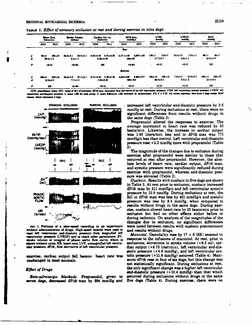
REGIONAL MYOCARDIAL ISCHEMIA H159
Tzz 1. Effect of coronar occlusion at rest and during wxrise in nine dogs
Emt aes". awoke vdin. C66Ma Ont'Ut. a/* am. LYOP, -. LVKDP, KAP.bmealmi .lkbet ut.,.!min -=maw# mmiig mmi. -aft
CON 0CC CON 0 CC CON 0Ce CON 0CC CON 0CC CON 0CC CON 0CC
Rot-
A 21±5 123±10 163.1 31.2±2.1 8.3±080 3.10.311 3X171210 27142 127 .111±t7 3.1S1.0 91±1 10 26± 07
41 3.6±7.2 &.21.1 0.2±0.9 6612 - 17.7±23.7 4.621.1 10.825.9
P <1.01 0.001 N9 <.001 .01 C0.01 NS
A 126± 210±16 $4.8z2.3 27.1=2.1 6.71s±0.40 6.020.36 4.615±3* 3,34±317 165±10 126±10 7.6±2.0 12.6±6.3 130*11 102%1341 12.6211.0 1.7±1.2 1.12±0.33 =20234 31.11±54.2 6.3±1.3 11.2±6.2
P N8 <A.111 <00 0.01 <00 <O0 C.0 O1 NO
CON. vabs. vemCC. veims .10 a stuiuen; d om wma Am darrneave oftbe Mef ,iUWWla pine; LVSP. left ventrwal" Syitalieo u ; LVEDF. leftu~imem~~e~g pue.4. n ± i gek eepJ. m. &34.in 31 inle. e. midNa S, P a 0.05. All veim ispemt da ez . dege sepL MAP
STANING OCCLUSION RUNING OCCLUSION increased left ventricular end-diastolic pressure by 3.6No mmHg at rest. During occlusions at rest, there were no
LVP owsignificant differences from results without drugs in(mm~gthe same dogs (Table 2).
Propranolol altered the responses to exercise. Theaverage increment in heart rate was reduced by 33beats/min. Likewise, the increase in cardiac output
dp/dtwas 1.68 liters/min less and in dP/dt max was 772(mm~/sac B~f ' F9MR-1 mmHg/s less than control. Left ventricular end-diastolic
. . .. . .pressure rose +3.2 mm g more with propranolol (Table
The magnitude of the changes due to occlusion duringexercise after propranolol were Similar to those that
Iocc O I ctc occurred at rest after propranolol. However, the abso-Off ~ . lute levels of heart rate, cardiac output, dP/dt max,
and systolic pressure were significantly reduced duringexercise with propranolol, whereas end-diastolic pres-
SV sure was elevated (Table 2).Oasabain. Results with ouabain in five dogs are shown
in Table 3. At rest prior to occlusion, ounbain increaseddP/dt max by 511 mnHg/s and left ventricular systolic
PHASICpressure by 10.8 mmHg. During occlusions at rest, theAOT 1 IInc fall in dP/dt max was less by 451 minHg/s and systolicFLOW pressure was less by 9.4 mmHg, when compared to
,j,-----'~~~ results without drugs in the same dogs. During exer-HR ~' cise, ouabain slowed heart rate by 32 beats/min prior to
(b/ian) -. - occlusion but had no other effects either before orduring ischemia. On analysis of the magnitudes of thechanges due to occlusion, no significant differences
no. 1. Portions of a alow-speed recording in an experiment were noted between results with ouabain pretreatmentwithout1 administrationoOf drugs. High-peed records wereO used to and results without drugs.med left ventricular end-diastolic pressure Iom magnifed left Mannitol. Osmolality rose by 17 ± 6 (SE) mosmol in
vantricular pressure (LVEDP) and to check other parameters. SV,stroke volume or integral of phasic aortic Raow curv. whc is response to the infusions Of mannitol. At rest, prior toAiw witheut unita; HR. heart rate; LW,. unmagnified left ventmc- occlusions, eievations in stroke volume (+6.2 ml), car-alar presuez dt, firat derivative of left ventricular pressure. diac output (+0.73 liter/min), left ventricular end-dia-
stolic pressure (+4.8 mmHg), and left ventricular sys-exercise, cardiac output fell becaUP heart rate was tolic pressure (+11.8 mnmHg) occurred (Table 4). Maxi-amchanged in nxs nin mum dP/dt rose in four of six dogs, but this change was
not statistically significant. During occlusions at rest,Effect of Drugs the only significant change was a higher left ventricular
and-diastolic pressure (+10.4 mmHg) than that whichet-ad renergic blockade. Propranolol, given to occurred during occlusions without drugs in the same
seven dogs, decreased dP/dt maxr by 594 mmHg and five dogs (Table 4). During exercise, there were no
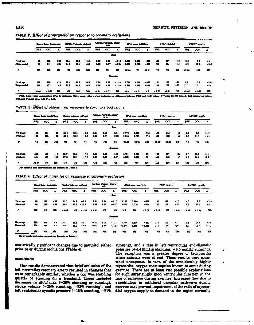
H160 HORWITZ. PETERSON, AND BISHOP
TAMIz 2. Effect of propranolol on response to coronary occlusions
Smm smft. 6=nWul 0110010 VduM, MiAmt 0I*.eo"I U" &a mu im LYW. umlig LYID? neaf
pox 0C A P31 ccC A POZ 0CC A 331 0CC A P33 0cc A 331 0CC A
Noamp 16 in +325. VI 3.6 -6.2 &JO 11.11 -2.14 9,171 2,8" a? in 16 -16 11-4 7.5 +4.1PPmwmm 6f 111 *.3 84.6 3.4 4.2 LUS 2.67 -. 26 U157 2.18 -410 lid 18 -14 5.8 I0s +..
P NO NS #43 NS me No NS No NI <11.06 NS <41.0 KS KS KS CO.O NS NS
1N6 drp n *10 U2.4 27.2 -11.11 7.2 5.70 -1.83 4.781 8,7k4 -22 161 in -83 6.2 13.5 +6.6pftpumbw 1in 171 +8 32 2.8 -5. _0 8.36 4.U -1.10 2.221 2,22 -U4 1in 106 -2a 11.7 17.1 +5.4
P <0.61 <6.03 #5 N48 NO NS <0.01 <0.01 KS <0.01 <41.01 No <41.06 40.01 NS <4.06 4806 NS
335. mw u lummtaeI prw is nc mm. C. alue during ~iama A. diurmm hat O. OW O. CC Valens. P Values an he pound s-IMa insprng v.1mwfth and wi~tiu *An. N42,P a 0.22.
TABLz 3. Effect of ouabain on response to coronary occlusions
mart Raoa. b am fuiea Volum, a. l/bet Cada ,,.liee dPI& Mae. MMMWO/ LYS?. mmnaf LVEDP. mHg
Paz 0CC A Pits 0CC a PRE 0CC A PRE 0C A PRE ccC A PRE 0cc a
He dnw so 114 +22 U.4 5.6 -9.6 8.14 8.22 -0.14 8.'072 2,38 -718 12n 1132 11 4.2 8.3 +4.0Ouaa 22 122 +18 113.11 5.6 -6.2 2.22 2.27 -0.02 3,864 2,20 -775 136 1.2 -12 6. 7 11.1 .+4.4
p No NS No NO No NS KS No #48 <0.22 <0.22 NS <0.06 <0.05 NS NS No NS
me drain 1M 2i5 .3 3.0 3.2 -7.2 6.79 6.61 -1.28 4,761 4M -V71 163 137 -56 6.7 11.1 +4.400@bm 161 178 +17 87.0 3.1 -7.2 8 2.15 -0.77 4,621 SAW2 -721 166 14 -17 L.5 14.7 +11.9
p <0.22 NS NO NS NS NS NO NS NS NS NO us NS NS No NO KS KS
For smalomis and abqu,,ateam m aoa to Ta" 2.
TABLE 4. Effect of mannitol on response to coronary occlusion
San Row. busttaUI Shah. Volun... *jjt Cardia Otuput. BM 431t Ewa. nuiaft LVII'. umli LVEDP -- Hig
FU1 occ A Paz 0CC A M3 0CC A PRE 0CC A ME1 0CC A PRE 0CC a
No druge 2 116 +U 3.2 24.2 -5.0 2.1 2.74 -0.17 2,103 2.22 -525 140 in2 -17 4.2 6.7 +2.5Uausual 96 in2 +21 .4 271 -113 8.24 5.16 -0.47 8,471 2.712 -7116 182 13 -24 2.0 17.1 +11.1
P No NS #48 <0221 NO <022 <0.01 NS No NS NS <0.22 <0.06 NO <0 01 <0.05 <0.06 KS
Medrop 22n O04 + U.2 U.4 -4.7 6.32 15.111 -1.17 5.132 4,32 -227 17581 IN 27 7.1 10.5 +3.8mom," 176 184 +7 84.6 271 -7.5 6.01 4LOS -1.18 111 &,M2 -1,82 173 A -91 6.7 15.6 +12.1
P me8 NS KS No m4e KO KS KS KS No NS Ko N5 NS NI NS NO KS
Fur ayi ud abbuqmitam ma bha to Yaie 2.
statistically significant changes due to mannitol either running), and a rise in left ventricular end-diastolicprior to or during occlusions (Table 4). pressure (+4.4 mmnHg standing, +6.3 mmHg running).
The exception was a greater degree of tachycardiaDISCUSON when animals were at rest. These results were some-
what unexpected in view of the considerably higherOur results demonstrated that brief occlusion of the myocardial oxygen consumption known to occur during
left circumflex coronary artery resulted in changes that exercise. There are at lest two poesible explanationswere remarkably similar, whether a dog was standing for such surprisingly good ventricular function in thequietly or running on a treadmill. These included face of iachemnia during exercise. Increased flow due todecreases in dP/dt max (-20% standing or running), vasodilation in collateral vascular pathways duringstroke volume (-25% standing, -23% running), and exercise may prevent impairment of the ratio of myocar-left ventricular systolic pressure (- 1% standing, -21% dial oxygen supply to demand in the region normally
REGIONAL MYOCARDIAL IJCHEMIA H161
served by the occluded artery (11). Another"sibility beta-adrenergic receptor sites (8). As has been reportedis that the higher levels of generalized sympathetic previously (10), propranolol had little effect on theactivity associated with exercise sufficiently enhance inotropic state during resting conditions either beforefunction in normally perfused regions to offset impaired or during occlusions. On the basis of this finding, itfunction in the ischemic region. would appear that increases in sympathetic tone may
There is some disparity between our results in the be small during occlusions at rest. During exercise,studies with occlusion of the left circumflex coronary when sympathetic tone is markedly augmented, pre-artery and the results of Vatner et a1. (14), in which treatment with propranolol significantly limited in-stenosis without occlusion of the main left coronary creases in heart rate, cardiac output, and rate of forceproduced only slight decrements in left ventricular development (dP/dt max). However, exercise occlusionsperformance at rest but profound deterioration during resulted in changes similar in magnitude to those thatexercise. Distal vasodilation can often maintain flow occurred without propranolol. Thus, since beta-adrener-through a stenotic coronary artery at nearly normal gic blockade depressed cardiac performance during ex-levels under resting conditions (5), but when myocardial ercise, and changes due to ischemia were the same asoxygen demands increase with exercise, flow through those seen without propranolol, it is likely that propran-the stenosis may not increase further. In contrast, total olol primarily influenced function in normal ratherocclusion of any coronary artery is likely to result in a than ischemic regions. This is compatible with theregion of distal ischemia at rest (2-4, 6, 10), as well as concept that, when autonomic control is intact, theduring exercise. Since obstruction of the main left ameliorative effect of catecholamines on the nonis-coronary may cause global ischemia of the left ventricle, chemic myocardiun overcomes the deleterious effectswhereas distal occlusions permit normal perfusion in of increased myocardial oxygen demand in the ischemicsome portions of the ventricle, the extent to which region.ischemia-induced decrements in integrated left ventric- Ouabain increased the rate of force development atular performance can be exacerbated by exercise stress rest prior to occlusion. The rate of force developmentmay depend upon the percentage of the left ventricle and peak systolic pressure were higher during restingthat is not ischemic. In addition, the possibility that -occlusion than those that occurred without drugs, al-collateral flow can increase during exercise in regional though the changes due to occlusion did not differ.ischemia cannot be excluded. Ouabain did not alter dP/dt max during exercise either
The major difference in hemodynamics between the before or during ischemia. The lack of effect of ouabaineffects of regional myocardial ischemia at rest and during exercise may mean that stimulation by catechol-during running was in heart rate. Although stroke amines increased myocardial contractile force in nor-volume fell in both settings, cardiac output was main- mally perfused regions to such high levels that inotropictained at preocclusion levels at rest as a result of the agents can add little additional benefit.heart rate increase. During exercise occlusions, cardiac Mannitol also increased dP/dt max prior to occlusionoutput fell because heart rate did not increase signifi- as has been reported previously (1). However, mannitolcantly. A previous study has concluded that tachycardia has no effect on dP/dt may during rest occlusions ordue to temporary left circumflex coronary artery occlu- during exercise states. Mannitol increased left ventric-sion in the resting, conscious dog is reflex in origin ular end-diastolic pressure during ischemia at rest.(13). This rMflex is initiated by receptors near the heart, Whether this reflects increased stiffness of the leftinvolves both vagal and sympathetic efferent pathways, ventricle or enhanced venous return due to expansionand is modulated by arterial baroreceptor activity (13). of blood volume or other factors cannot be determinedDuring exercise, sympathetic tone is high and vagal from our data.tone is substantially reduced (7, 8). These changes maybe so marked that the capability of the reflex mecha- The authors gratefully acknowledge the technical assistance ofnism to induce further sympathetic stimulation or vagal Donald Watkins, Pamela Reichenbach, Stanley A. Dunbar, Charleswithdrawal via the efferent pathways is limited. Alter- D. Montgomery, and Linda Fox.natively, as appears to be the case with baroreceptor This work was supported by Public Health Service Grants HL-reflex control of heart rate (12), the sensitivity of the 16656 and HL-12415 from the National Heart and Lung Institute.
Send requests for reprints to L. D. Horwitz, Dept. of Medicine,reflex decreases, either through diminished receptor University of Te s Health Science Center at San Antonio, 7703sensitivity or central nervous system changes. Floyd Curl Drive, San Antonio, Tex. 78284.
In the dosage used in this study, propranolol effec-tively competes with endogenous catecholamines for Received for pubiciation 13 June 1977.
REFERENCES
I. Arm., J. U., K. WnmuriuA., sun L. D. Hoawmr. Cardiovas- 4. Rumour, L. P., H. HjmuA, A" R. L. Ems. Effects of acuteeular reponses to hyperosmotic mannitol in anesthetized and regional myocardial isehemia in left ventricular function incncious dop. Am. J. Physio. 22: 132-137, 1973. dogs. Circulation Re. 26: 307-315, 1970.
2. Bum , V. S., R. L. K.Asa, G. E. B AaS, AND M. B. KAnON. A. OoiUs., K. L, K. Lnsoxa, ANv G. W. HAMILTON. Physio-Left ventricular function during cute regional myocardial is- logic basis for assesing critical coronary stenosis. Am. J.chsaia in the coscious dog. J. App. Physiol. 37: 785-792, 1974. Cardil. 33: 87-94, 1974.
$. Dwrua, E. W. Left ventricular pressure-volume alteration and 6. Hoon, W. B., Ja., V. H. Covuw, W. H. AanmAxm, Au J. C.regional disorders of centractions during isobemia induced by Noams . Persistence of contractile behavior in acutely ischemicarbal pean. Cin-ulsmm 42: 1111-1122, 1970. ayocardium. Cardiavsccdar Re. 3: 249-260,1969.
|1
H162 HORWrm, PETERSON, AND BISHOP
7. Hoawrrz, L. D.. J. M M. um, Am S. J. Lwmn. Role of the en pe1fuaim o collateral-dependent myocardium in dogs withFrank-Starling mechanism in exercise. Circulation Re. U1 868- chnic coronary artery occlusion. J. Chn. Invest. 9: 1-7,1977.
74, 1972. 12. McRvrcm, Ft. J., S. F. VATmia, D. BAm=cmm, G. R. HYN-8. Hoawmrz, L. D., J. M. ATKINS, Am S. J:LummN. Effects of DRcz, T. A. PATRcC, AND E. BmAuN Lwun. Role of arterial
beta-adrenrgic blockade on left ventricular function in exercise. "barreceptore in mediating cardiovascular response to exercise.Am. J. Physiol. 227: 839-842, 1974. Am. J. Physiol. 230: 86-89, 1976.
9. Hoawrrz, L. D., Am V. S. Bsop. Left ventricular presure- 1. Pwurnouo, D. F., R. L. K.&PA, A." V. S. Bunop. Reflexdimension relationships in the conscious dog. Cargiovascular .. tachycardia due to temporary coronary occlusion in the con-ke. 6: 163-171, 1972. acious dog. Circulatdon Re. 32: 652-8, 1973.
10. KAAa, R. L., G. E. BDAw , D. F. PzmutsoN, Amn V. S. 14. VATNUE, S. F., R. J. McRrcxms, P. R. MAXOKo, T. A. PATRc,Bumo. Specificity ofauto omic influences on cardiac responses A" E. BaAn.rwA.n. Effects of catecholamines, exercise andduring myocardial iscuimia. J. App. Physiol. 38: 485-490, 1975. iutroglycerm on the normal and isechemic myocardium in con-
11. LAsuar. P. R., D. S. Ham, Am R. J. Bacnz. Effect of exercise scious dogs. J. Chn. Invest. 54: 563-575, 1974.
C -
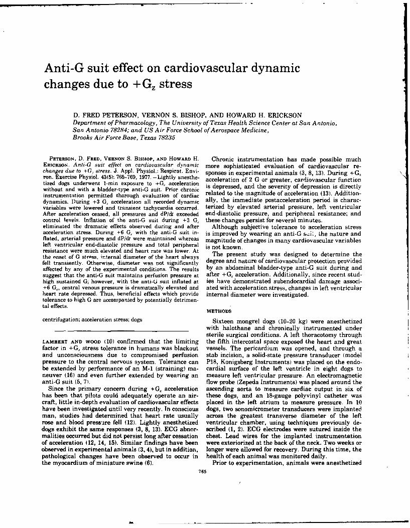
Anti-G suit effect on cardiovascular dynamicchanges due to +G, stress
D. FRED PETERSON, VERNON S. BISHOP, AND HOWARD H. ERICKSONDepartment of Pharmacology, The University of Texas Health Science Center at San Antonio,San Antonio 78284; and US Air Force School of Aerospace Medicine,Brooks Air Force Base, Texas 78235
PETERSON, D. FRED, VERNON S. BISHOP, AND HOWARD H. Chronic instrumentation has made possible muchERICKSON. Anti-G suit effect on cardiovascular dynamic more sophisticated evaluation of cardiovascular re-changes due to +G, stress. J. Appl. Physiol.: Respirat. Envi- sponses in experimental animals (3, 8, 13). During +G,ron. Exercise Physiol. 43(5): 765-769, 1977. -Lightly anesthe- acceleration of 2 G or greater, cardiovascular functiontized dogs underwent 1-min exposure to +G, acceleration is depressed, and the severity of depression is directlywithout and with a bladder-type anti-G suit. Prior chronicinstrumentation permitted thorough evaluation of cardiac related to the magnitude of acceleration (13). Addition-dynamics. During +3 G, acceleration all recorded dynamic ally, the immediate postacceleration period is charac-variables were lowered and transient tachycardia occurred. terized by elevated arterial pressure, left ventricularAfter acceleration ceased, all pressures and dP/dt exceeded end-diastolic pressure, and peripheral resistance; andcontrol levels. Inflation of the anti-G suit during +3 G, these changes persist for several minutes.eliminated the dramatic effects observed during and after Although subjective tolerance to acceleration stressacceleration stress. During +6 G, with the anti-G suit in- is improved by wearing an anti-G suiL, Lhe nature andflated, arterial pressure and dP/dt were maintained whereas magnitude of changes in many cardiovascular variablesleft ventricular end-diastolic pressure and total peripheral is not known.resistance were much elevated and heart rate was lower. At The present study was designed to determine thethe onset of G stress, internal diameter of the heart alwaysfell transiently. Otherwise, diameter was not significantly degree and nature of cardiovascular protection providedaffected by any of the experimental conditions. The results by an abdominal bladder-type anti-G suit during andsuggest that the anti-G suit maintains perfusion pressure at after +G, acceleration. Additionally, since recent stud-high sustained G; however, with the anti-G suit inflated at ies have demonstrated subendocardial damage associ-+6 G,, central venous pressure is dramatically elevated and ated with acceleration stress, changes in left ventricularheart rate depressed. Thus, beneficial effects which provide internal diameter were investigated.tolerance to high G are accompanied by potentially detrimen-tal effects.
METHODS
centrifugation; acceleration stress; dogs Sixteen mongrel dogs (10-20 kg) were anesthetizedwith halothane and chronically instrumented understerile surgical conditions. A left thoracotomy through
LAMBERT AND WOOD (10) confirmed that the limiting the fifth intercostal space exposed the heart and greatfactor in +G, stress tolerance in humans was blackout vessels. The pericardium was opened, and through aand unconsciousness due to compromised perfusion stab incision, a solid-state pressure transducer (modelpressure to the central nervous system. Tolerance can P18, Konigsberg Instruments) was placed on the endo-be extended by performance of an M-1 (straining) ma- cardial surface of the left ventricle in eight dogs toneuver (16) and even further extended by wearing an measure left ventricular pressure. An electromagneticanti-G suit (5, 7). flow probe (Zepeda Instruments) was placed around the
Since the primary concern during +G, acceleration ascending aorta to measure cardiac output in six ofhas been that pilots could adequately operate an air- these dogs, and an 18-gauge polyvinyl catheter wascraft, little in-depth evaluation of cardiovascular effects placed in the left atrium to measure pressure. In 10have been investigated until very recently. In conscious dogs, two sonomicrometer transducers were implantedman, studies had determined that heart rate usually across the greatest transverse diameter of the leftrose and blood press-are fell (12). Lightly anesthetized ventricular chamber, using techniques previously de-dogs exhibit the same responses (3, 8, 13). ECG abnor- scribed (1, 2). ECG electrodes were sutured inside themalities occurred but did not persist long after cessation chest. Lead wires for the implanted instrumentationof acceleration (12, 14, 15). Similar findings have been were exteriorized at the back of the neck. Two weeks orobserved in experimental animals (3, 4), but in addition, longer were allowed for recovery. During this time, thepathological changes have been observed to occur in health of each animal was monitored daily.the myocardium of miniature swine (6). Prior to experimentation, animals were anesthetized
765
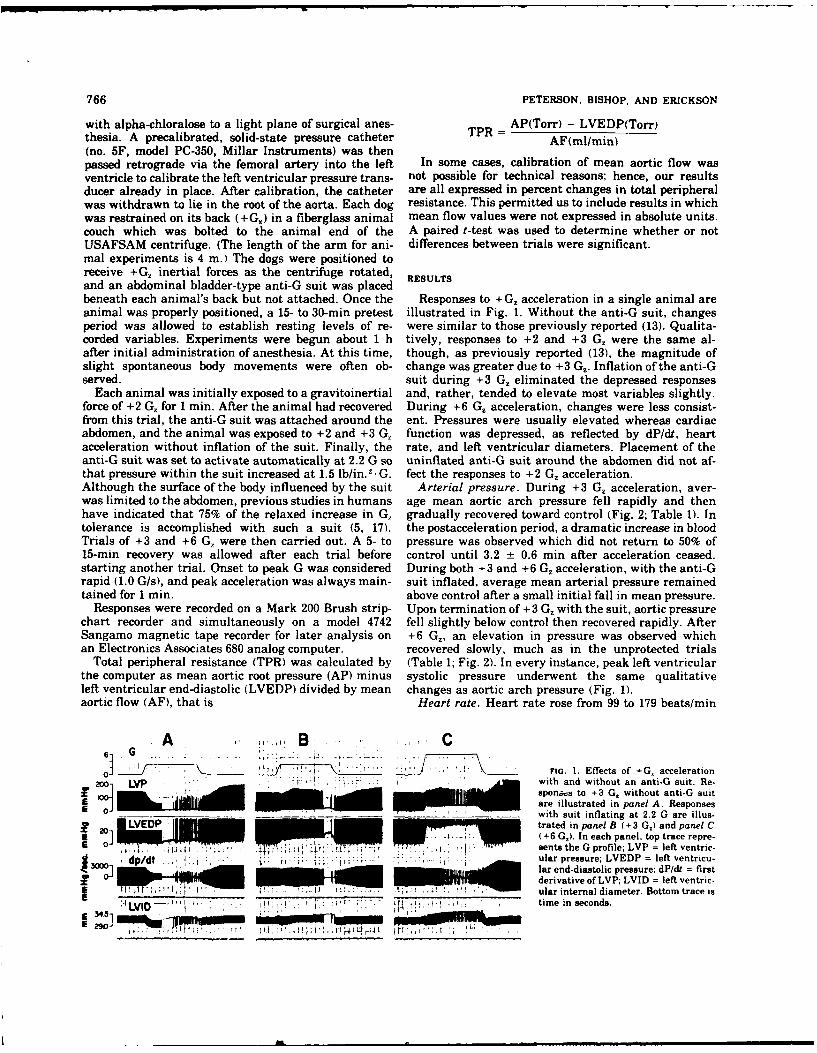
766 PETERSON, BISHOP, AND ERICKSON
with alpha-chloralose to a light plane of surgical anes- AP(Torr) - LVEDP(Torr)thesia. A precalibrated, solid-state pressure catheter TPR AF(ml/min)(no. 5F, model PC-350, Millar Instruments) was thenpassed retrograde via the femoral artery into the left In some cases, calibration of mean aortic flow wasventricle to calibrate the left ventricular pressure trans- not possible for technical reasons; hence, our resultsducer already in place. After calibration, the catheter are all expressed in percent changes in total peripheralwas withdrawn to lie in the root of the aorta. Each dog resistance. This permitted us to include results in whichwas restrained on its back (+G) in a fiberglass animal mean flow values were not expressed in absolute units.couch which was bolted to the animal end of the A paired t-test was used to determine whether or notUSAFSAM centrifuge. (The length of the arm for ani- differences between trials were significant.mal experiments is 4 m.) The dogs were positioned toreceive +G., inertial forces as the centrifuge rotated, RESULTSand an abdominal bladder-type anti-G suit was placedbeneath each animal's back but not attached. Once the Responses to +G, acceleration in a single animal areanimal was properly positioned, a 15- to 30-min pretest illustrated in Fig. 1. Without the anti-G suit, changesperiod was allowed to establish resting levels of re- were similar to those previously reported (13). Qualita-corded variables. Experiments were begun about 1 h tively, responses to +2 and +3 G, were the same al-after initial administration of anesthesia. At this time, though, as previously reported (13), the magnitude ofslight spontaneous body movements were often ob- change was greater due to +3 Gz. Inflation of the anti-Gserved. suit during +3 G. eliminated the depressed responses
Each animal was initially exposed to a gravitoinertial and, rather, tended to elevate most variables slightly.force of +2 G, for 1 min. After the animal had recovered During +6 G, acceleration, changes were less consist-from this trial, the anti-G suit was attached around the ent. Pressures were usually elevated whereas cardiacabdomen, and the animal was exposed to +2 and +3 G, function was depressed, as reflected by dP/dt, heartacceleration without inflation of the suit. Finally, the rate, and left ventricular diameters. Placement of theanti-G suit was set to activate automatically at 2.2 G so uninflated anti-G suit around the abdomen did not af-that pressure within the suit increased at 1.5 lb/in .
. G. fect the responses to +2 G. acceleration.Although the surface of the body influenced by the suit Arterial pressure. During +3 G, acceleration, aver-was limited to the abdomen, previous studies in humans age mean aortic arch pressure fell rapidly and thenhave indicated that 75% of the relaxed increase in G, gradually recovered toward control (Fig. 2; Table 1). Intolerance is accomplished with such a suit (5, 17). the postacceleration period, a dramatic increase in bloodTrials of +3 and +6 G, were then carried out. A 5- to pressure was observed which did not return to 50% of15-min recovery was allowed after each trial before control until 3.2 t 0.6 min after acceleration ceased.starting another trial. Onset to peak G was considered During both +3 and +6 G, acceleration, with the anti-Grapid (1.0 GIs), and peak acceleration was always main- suit inflated, average mean arterial pressure remainedtained for 1 min. above control after a small initial fall in mean pressure.
Responses were recorded on a Mark 200 Brush strip- Upon termination of +3 G. with the suit, aortic pressurechart recorder and simultaneously on a model 4742 fell slightly below control then recovered rapidly. AfterSangamo magnetic tape recorder for later analysis on +6 G,, an elevation in pressure was observed whichan Electronics Associates 680 analog computer. recovered slowly, much as in the unprotected trials
Total peripheral resistance (TPR) was calculated by (Table 1; Fig. 2). In every instance, peak left ventricularthe computer as mean aortic root pressure (AP) minus systolic pressure underwent the same qualitativeleft ventricular end-diastolic (LVEDP) divided by mean changes as aortic arch pressure (Fig. 1).aortic flow (AF), that is Heart rate. Heart rate rose from 99 to 179 beats/min
, A ,' ,',, B ,, .. Co! /
7,-. , . G. 1. Effects of +G, accelerationLVP "" ' n with and without an anti-G suit. Re-
F 10 sponzwe to +3 G, without anti-G suit0_ are illustrated in panel A. Responses
__ _'__ ___ _ with suit inflating at 2.2 G are illus-LVEDP l trated in panel B (+3 G.) and panel C2D]J" (+6 G,). In each panel, top trace repre-0sents the G profile; LVP = left ventric
~dp/.I _.. . .ular pressure; LVEDP = left ventricu-i nWs lar end-diastolic pressure; dP/dt = firstderivative of LVP; LVID = left ventric-
__________.____________ _____"____... .__________ ular internal diameter. Bottom trace isI 4 ALV I ... L. • .T7;7TT-T . : : fI ,iL I , ; "time in seconds.34bi
wn
E 29 1 1 L-: llm m m m m m
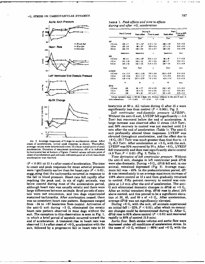
+G, STRESS ON CARDIOVASCULAR DYNAMICS 767
Aortic Arch Pressure TABLE 1. Peak effects and time to effects,..,.-o.s. during and after +G: acceleration
X ...... During G Poet CPre G Control Peak effect Time to peak Peak effect Time to 50q
rcvery
Tor" Torr s Tort 'nin
APo 3G6 wb _4#1 3 G w/o 128 ± 7 67 t 13- 11.5 t 1.5 168 ± 12t 3.2 t 0.6
Heart Rote * J6 Sot 3Gw 122±9 96 ±9. 5.9-±0.4 117 t 11 0.6 0.2o2660Sa 6Gw 125 ±9 100 ± 9t 6.1 t0.7 193 ± 65 2.5 ± 0.6
1 bewlinrn beotsn,, S beowst,,u MlI.
03 G wo 99 ± 8 179 ± 15" 13.4 ± 1.2 94 ± 9 1.7 ± 0.53Gw 112 ± 13 140 - 14 9.0 ± 0.8 108 t 7 0.5 = 0 1
0 6Gw 116 t 12 143 ± 15 11.5 -1.5 126 ± 14 2.1 ± 0.6
Tor or To- MI.n50 LVEDP
3 G w/o 1.4 t 0.7 -9.0 ± 1.80 6.9± 0.8 16.0 ± 3.3t 2.4 t 0.4Left Ventricular End Diastolic Pressure 3Gw 1.0 ±0.8 .3 ± 1.1" 6 0.14 1'3 = 0.8 0.5 ± 0.1
6 G w 1.9 ± 0.8 20.7 ± 2.5 76 ± 0.3 4.9 ± 1Ot 1.4 - 0 3
20 - 0..aGo9 0oa 0i
'0 -o dP ldt
E 3 G wo 100 58 ± 7" 8.0 ±0.9 216 ± 47t 13 ± 0.50 ,- -- o.-- 3Gw 100 82 ± 70 6.3 ±0.6 112 ± 11 0.5 ±0.1
.......... . . 6Gw 100 68 = 9t 6.2 ±0.7 191 ± 24t 0.9 ± 0.2
Values represent mean ± SE for 8 dogs: w/o, without inflation of the anti-G suit; w.
dp/dt Change with inflated suit. * P < 0.001. t P < 0.01. t P < 0.05.
2o /1beats/min at 60 s. All values during G after 15 s weresignificantly less than control (P < 0.001), Fig. 2.
1.o0_.Left ventricular end-diastolic pressure (LVEDP).0 ° Without the anti-G suit, LVEDP fell significantly (-5.0Torr) but recovered before the end of acceleration. A
0 large increase was observed after G stress (15.0 Torr),
Yand 50% recovery to control was not reached until 2.4min after the end of acceleration (Table 1). The anti-G
6/ suit profoundly altered these responses. LVEDP was
elevated throughout acceleration, and the effect due toFoG. 2. Average responses of 8 dogs to acceleration stress. After +6 G, (20.7 Torr) was much greater than that due to +3
onset of acceleration, initial peak response is shown. Thereafter, G, (6.3 Torr). After acceleration at +3 G. with the suit,average values were determined every 15 s from initial point of peak LVEDP was 50% recovered by 30 s. After +6 G,, LVEDPacceleration. Duration of maximum acceleration (60 a) is indicated fell transiently and then rose significantly above controlby horizontal bar at bottom of figure. Control values indicate point of (4.9 Torr; P < 0.01) (Fig. 2; Table 1).onset of acceleration and ordinate indicates point at which maximumacceleration was reached. Time derivative of left ventricular pressure. Without
the anti-G suit, changes in left ventricular peak dP/dt(P < 0.001) at 13.4 s after onset of acceleration. The time were also dramatic. During +3 G,, dP/dt fell and, in oneto onset and peak responses for mean arterial pressure animal, remained depressed (Fig. 2). Average maxi-were !-ignificantly earlier than for heart rate (P < 0.01), mum fall was -45%. In the postacceleration period, dP/suggi.sting that the tachycardia occurred in response to dt rose immediately to an average maximum increase ofthe fall in blood pressure. Heart rate fell rapidly after 116% above control at 15 s and then gradually returnedreaching the peak and, in six of eight animals, was to control. Fifty percent recovery to control was com-below control during most of the acceleration period, plete at 1.3 min after the end of acceleration. The anti-although heart rate was usually erratic and there were G suit eliminated dramatic changes in dP/dt at +3 G,.large differences between animals. Brief periods of asys- After an initial transient drop, dP/dt rose to about 15%tole were not uncommon, and two dogs experienced above control; and this proved to be a significant eleva-sustained tachycardia. After acceleration ceased, there tion at 30, 45, and 60 s of +3 G,. After acceleration,was no consistent heart rate pattern. Responses ranged average dP/dt was not significantly elevated.from -34 to +67 beats/min from control. Activation of During +6 G,, with the suit, all animals experiencedthe anti-G suit during +3 G. eliminated the erratic an initial fall (-32%; P < 0.01), after which no consist-heart rate pattern observed in most dogs without the ent changes could be demonstrated during G. After G,suit. The exception to this observation is seen in Fig. 1, dP/dt rose to 91% above control (P < 0.01) and recoveredin which a brief period of asystole occurred toward the rapidly to 50% of control (0.9 min).end of acceleration. A transient rise in heart rate was Aortic flow. Both stroke volume and aortic flow wereobserved 11.5 s after onset of +6 Gz acceleration with the depressed under all conditions of acceleration stress. Insuit, followed by a progressive fall in heart rate to 54 the cases of +3 Gz without (-66%) and +6 G. with the
768 PETERSON, BISHOP. AND ERICKSON
suit (-55%), magnitude of mean flow depression was DISCUSSIONgreater than during +3 G, with the suit inflated(-27%). Flow remained depressed throughout accelera- Results of this study demonstrated that activation oftion and, immediately upon stopping the centrifuge, an abdominal bladder-type anti-G suit reversed the de-rose back toward control under all three conditions. leterious cardiovascular effects due to +3 G. stress.After +3 G. without the suit, however, this recovery Elevation of aortic arch pressure above control suggestswas transient. Flow was depressed again by 23% (P < an especially beneficial effect in preventing blackout0.01). On the other hand, after +3 and +6 G. with the and unconsciousness which occurs in man at low centralsuit, no significant deviation from control was demon- perfusion pressures. Most other untoward effects of ac-strated. celeration were not observed to occur at +3 G. with the
Peripheral resistance. Calculation of peripheral re- suit, except at the onset of acceleration. A transient fallsistance at regular intervals was complicated by the in arterial pressure, left ventricular pressure, and dP/dtperiods of extremely erratic cardiac output at low heart were observed. Since the suit was not activated until 2.2rates as well as the occasional periods of observed G was reached, these changes were undoubtedly due toasystole. For this reason peripheral resistance was suddenly reduced afterload associated with onset of in-sampled during periods of regular cardiac output. In all creased gravitoinertial forces. The slightly delayedcases, calculated TPR was elevated during acceleration. tachycardia indicates a baroreflex response to the pres-During +3 G with the suit, the elevation was less sure fall. After activation of the anti-G suit, both eleva-than in the other cases. The maximum value was 60% tion of arterial pressure and left ventricular end-dia-above control. During +3 G, without the suit, average stolic pressure might be accounted for by a shift inmaximum TPR was 124% above control. During +6 G; circulating body fluids. Reduction of venous pooling inwith the suit inflated, average maximum TPR was the abdomen would increase venous return and increase217% above control. After 3 G. without the suit, TPR pulmonary blood volume (9), whereas the anti-G-suit-fell transiently toward control, then climbed signifi- induced increase in total peripheral resistance wouldcantly above control (58 ± 21; P < 0.05) at 1 min after tend to impede cardiac output. The likelihood that leftacceleration ceased. After +3 G. with the suit inflated, ventricular end-diastolic pressures do mirror the fillingTPR returned rapidly to control. After +6 G. with the pressure of the left heart is supported by the results insuit inflated, TPR returned gradually to control but did Fig. 1 indicating that end-diastolic heart size was di-remain significantly elevated at 1 min post-G (+37 + rectly related to LVEDP in this animal. On the other15; P < 0.05). hand, as previously reported (9), pressure on the abdo-
Left ventricular internal diameter. Immediately after men may displace the diaphragm and increase intra-the onset of acceleration, the left ventricular internal thoracic pressure, thus directly influencing thoracicdiameter (LVID) began to get smaller. During +3 G. vascular pressures. Near-normal heart rate suggestedwithout the suit, at the time when arterial pressure that neither direct stress nor indirect reflex influenceswas lowest (64 Torr for this group), systolic size of the were dramatically altering heart function. In the post-Gheart was significantly reduced by 2.2 mm (P < 0.01) period all variables approached control rapidly, indicat-to 28.3 mm. In no case did the left ventricle approach ing lack of sustained adverse cardiovascular effects atcollapse. The average diameter in diastole fell 0.8 mm, +3 G, with the anti-G suit inflated. Thus, the prolongedbut the change was not significant. As arterial pressure elevation of arterial pressure and LVEDP observed hererecovered, LVID's got larger and no other significant and previously (13) in the post-G period were completelychanges in left ventricular size were observed during or eliminated.following +G, acceleration. It was suspected that the At +6 G,, partial protection from adverse cardiovas-magnitude of change in diameter size might be related cular effects was obtained by suit inflation. Mostto either arterial pressure or heart rate. Analysis of changes were intermediate between those observed atvariance showed no significant relationship in either +3 G, with and without the suit. Average aortic archcase. Even in animals whose arterial pressure fell pressure was maintained above control, and although itdramatical':, diameter size was not significantly al- was not a significant elevation, this is in marked con-tered. In five dogs whose left ventricular systolic pres- trast to trials without the anti-G suit. In a previoussure fell below 60 Torr (avg, 49 Torr), end-systolic study, due to poor recovery from +3 or +4 G., only 4 ofdiameter fell by only 2.4 mm. In three dogs whose 14 dogs could be tested at +5 G, without the anti-G suitheart rates rose 50 beats/min or more during accelera- (13). In a similar study hypotension, bradycardia, and ation (avg, 101 beats/min), end-systolic diameter fell by profound decrease in coronary blood flow were reported4.7 mm to an average of 25.3 mm. during exposure to +5 or +6 G. stress (8). Thus, mainte-
Both increases and decreases in internal diameter nance of perfusion pressure to the coronary and CNSwere observed with the suit activated. Responses in vasculature was dramatically improved by the suit dur-four dogs were similar to those illustrated in Fig. 1. In ing high acceleration stress.these experiments LVID got small during unprotected Although arterial pressure was maintained, signifi-acceleration and larger if the anti-G suit was inflated, cant decrements in other cardiovascular indices wereIn the other six dogs no general pattern could be observed. Heart rate was down, and this could not beidentified. Thus, there were no consistent changes in attributed to reflex bradycardia. Aortic flow was dra-internal diameter during activation of the anti-G suit matically down, which could perhaps be related to bothwhich could be demonstrated statistically. depressed he-rt rate and limited circulating blood vol-
+G, STRESS ON CARDIOVASCULAR DYNAMICS 769
ume. The most consistent observation was the dramatic Although peak left ventricular pressure fell as low as 30elevation in LVEDP. This suggests that during high Torr in one dog, and heart rate rose to 280 beats/min insustained + G. with an anti-G suit, elevated intrathora- another, in no animal was the left ventricle observed tocic pressures are probably a necessary compromise in get smaller than 15.5 mm across its greatest diameterorder to maintain aortic arch perfusion pressure. On the during end-systole. In an earlier study, severity of theother hand, in the post-6-G period, the overshoot pathological findings was related to a) level and dura-changes in aortic pressure, dP/dt, and LVEDP were not tion of G exposure, b) heart rate, and c) catecholamineas great nor did they persist as long as in the unpro- activity (6). It does not appear that these changes cantected trials at +3 G2 . induce striking of the internal walls of the left ventricle
Our results indicated that although lightly anesthe- during stresses similar to those in this study. Changestized dogs did not tolerate +3 G. acceleration well with- during inflation of the anti-G suit were even less re-out the protection of an anti-G suit, changes in left markable. In those cases no significar.. changes in leftventricular internal diameter were not as dramatic as ventricular internal diameter could be demonstrated.were changes in other variables. Numerous studieshave indicated that pathological changes in cardiac The authors acknowledge the fine technical assistance of Lindamuscle are associated with high sustained acceleration Fox, Ben Wiggins, Jesse Rodriguez, R. V. Fairchild, Hubert Tomer-stress (6, 11). In the present study, we considered the lin, and personnel of the Crew Technology Division, USAF School ofpossibility that the subendocardial hemorrhage re- Aerospace Medicine.ported by MacKenzie et al. (11) might be due to striking The work was supported by AFOSR Contract 73-2525.of the internal walls of the left ventricle during acceler-ation. No evidence for this possibility was obtained. Received for publication 4 April 1977.
REFERENCES
1. BISHOP. V. S., AND L. D. HORWITZ. Effects of altered autonomic York: Pergamon, 1965, chapt. 23, p. 615-618.control of left ventricular function in conscious dogs. Am. J. 10. LAMBERT, E. H., AND E. H. WOOD. The problem of blackout andPhysiol. 221: 1278-1282, 1971. unconsciousness in aviators. Med. Clin. N. Am. 30: 833-844,
2. BISHOP. V. S.. L. D. HORWITZ, H. L. STONE, H. F. STEGALL, AND 1946.E. J. ENGELKEN. Left ventricular internal diameter and cardiac 11. MACKENZIE, W. F., R. R. BURTON, AND W. I. BUTcHER. Cardiacfunction in conscious dogs. J. Appl. Physiol. 37: 785-792, 1969. pathology associated with high sustained +G,. II. Stress cardio-
3. BURNS, J. W. Influence of ethanol on cardiovascular toler- myopathy. Aviation Space Environ. Med. 47: 718-725, 1976.ance to +G, acceleration. Aerospace Med. 45: 19-28, 1974. 12. PARKHURST, M. J., S. D. LtVERETr, JR., AND S. J. SHUsROOKS,
4. BURTON, R. R. Positive (+G,) acceleration tolerances of the JR. Human tolerance to high, sustained +G, acceleration. Aero-miniature swine: application as a human analog. Aerospace space Med. 43: 708-712, 1972.Med. 44: 294-298, 1973. 13. PETERSON, D. F., V. S. BISHOP, AND H. H. ERICKSON. Cardiovas-
5. BURTON, R. R., AND R. W. KRUTZ, JR. G-tolerance and protection cular changes during and following 1-min exposure to +G,with anti-G suit concepts. Aviation Space Environ. Med. 46: stress. Aviation Space Environ. Med. 46: 775-779, 1975.119-124, 1975. 14. SHUBROOKS, S. J., JR. Changes in cardiac rhythm during sus-
6. BURTON, R. R., AND W. F. MACKRNZIE. Cardiac pathology tained high levels of positive (+G,) acceleration. Aerospaceassociated with high sustained +G,. I. Subendocardial hemor- Med. 43: 1200-1206, 1972.rhage. Aviation Space Environ. Med. 47: 711-717, 1976. 15. SHUBROOKS, S. J., JR. Positive pressure breathing as a protective
7. BURTON, R. R., M. J. PARKHURST, AND S. D. LzVzRaTr, JR. +G., technique during +G, acceleration. J. Appl. Physiol. 35: 294-protection afforded by standard and preacceleration inflations 298, 1973.of the bladder and capstan-type G-suits. Aerospace Med. 44: 16. SHUBROOKS, S. J., JR., AND S. D. LEVERETT, JR. Effect of the488-494, 1973. valsalva maneuver on tolerance to +G, acceleration. J. Appl.
8. ERICKSON, H. H., H. SANDLER, AND H. L. STONE. Cardiovascular Physiol. 34: 460-466, 1973.function during sustained +G, stress, Aviation Space Environ. 17. WOOD, E. H., AND E. H. LAMaZRT. Some factors which influenceMed. 47: 750-758, 1976. the protection afforded by pneumatic anti-G suits. Aerospace
9. HOWARD. P. The physiology of positive acceleration. In: A Med. 23: 218-228, 1952.Textbook of Aviation Physiology, edited by J. A. Gillies. New
T
Influences of Selective Cardiac Denervation on
Coronary Reactive Hyperemia In Conscious Dogs
By
D. FRED PETERSON, PH.D. AND VERNON S. BISHOP, PH.D.
Department of Pharmacology, The University of Texas Health
Science Center at San Antonio, San Antonio, Texas 78284
Grant Support: NIH Grant HL12415 and AFOSR #73-2525
Abbreviated Title: Denervation and reactive hyperemia
Send Correspondence and Proofs to:
D. Fred Peterson, Ph.D.
Department of Pharmacology
The University of Texas Health Science Center atSan Antonio
7703 Floyd Curl Drive
San Antonio, Texas 78284
Abstract
Mongrel dogs were chronically instrumented to measure left
circumflex coronary flow, arterial pressure, left atrial pres-
sure, ECG, heart rate and in some cases left ventricular pres-
sure or cardiac output. A cuff type occluder was placed distal
to the coronary flow probe. Total occlusion of the left circum-
flex coronary artery for one minute in unsedated, resting dogs
produced reactive hyperemia with an average replacement/deficit
ratio of 2.68/1. In 10 dogs sympathetic influences were investi-
gated by total, chronic cardiac sympathectomy. Surgical section
of all ansae subclavia reduced responses from 2.76/1 to 1.71/1
(P <.OOl). Left sympathectomy alone :iad no effect on the re-
placement/deficit ratio whereas selective right sympathectomy
reduced it from 2.61/1 to 1.67/1. Pharmacological blockade was
used to deterr'ne beta-receptor involvement in the responses.
In 9 intact d ; practolol (10 mg/kg) reduced the reactive hyper-
emia ratio by 11% (P<.05). Propranolol (1 mg/kg) further re-
duced this ratio by 30% (P<.OOl). Our results indicate that
sympathetic beta influences work primarily through the right
cardiac sympathetic nerves. Also, the magnitude of the response
appears to be due, in part, to increased metabolic activity
associated with myocardial /fl receptors as well as vasodilation
through /2 receptors.
KEY WORDS: reflex circumflex coronary artery
propranolol practolol cardiac sympathectomy
Numerous studies have indicated that coronary blood flow
can be influenced by both the parasympathetic and sympathetic
nervous systems (1-6). Direct efferent stimulation of the
vagus nerve produces coronary vasodilation independent of
chronotropic or inotropic effects suggesting the potential
for a parasympathetic component in the regulation of coronary
blood flow (4). More recently, reflex vasodilation via vagal
efferents has been demonstrated to accompany injection of
veratrum alkaloids into the coronary circulation (7). Sym-
pathetic involvement in coronary blood flow regulation is
more complex. Evidence indicates that direct neural control
of coronary resistance is partially mediated through sympa-
thetic alpha receptors located in the vessel walls (3,5).
However, vasodilation is observed during cardiac sympathetic
nerve stimulation. In general this dilation has been assumed
to be secondary to the increased metabolic requirements
associated with the inotropic and chronotropic responses to
myocardial ,8-receptor stimulation (6). Thus, in the intact
heart, the regulation of oxygen delivery through changes in
coronary resistance appears to be dominated by changes in
local oxygen requirements. However, there is some evidence
for an intrinsic adrenergic vasodilator mechanism (2). Further-
more, in the cat, stimulation of coronary sC fibers has been
shown to decrease coronary resistance independent of changes
in the chronotropic or inotropic state of the myocardium (8).
2
Brief occlusion of a coronary artery results in a re-
active hyperemia response in which the flow far exceeds the
apparent deficit resulting during the period of occlusion.
This response has been linked to both myogenic relaxation of
the vessels (9) and to the myocardial metabolic requirements
(1,10,11). Whether or not the nervous system is involved in
coronary reactive hyperemia has not been determined.
In the present study, we have investigated the involve-
ment of the cardiac sympathetic neural reflexes in modulating
the magnitude of the coronary reactive hyperemia in the
conscious dog. The magnitude of the hyperemia was determined
following one minute occlusion of the left circumflex coronary
artery. Control responses were compared to those after both
partial and total cardiac sympathectomy as well as after
selective beta adrenergic blockade and after atropine.
Methods
Chronic Surgery.
Twenty-six mongrel dogs weighing between 10-25 kg were
chronically instrumented using sterile surgical techniques
under halothane (Fluothane, Ayerst) anesthesia. A left thora-
cotomy was performed through the fifth intercostal space ex-
posing the heart and great vessels. The pericardium was opened
and a longitudinal incision made in the connective tissue over
the left circumflex artery just beyond its origin. A 6-10 mm
length was lifted away from surrounding tissue and a balloon
3
type polyethylene catheter cuff, similar to that previously
reported (12) was placed around the vessel. The catheter
was then sutured to the wall of the ventricle, thus, immobil-
izing it. A saline filled syringe was used to verify occlusion.
Both the volume and pressure required for occlusion were
determined. An electromagnetic flow probe (Zepeda) was placed
around the left circumflex coronary artery proximal to the
occluder in all dogs for measurement of coronary flow. During
implantation of the occluder and flow probe, special care was
taken to avoid damage to cardiac nerves. This was accomplished
by instrumenting only those dogs in which the circumflex artery
was located superficially and could be lifted away from under-
lyinq tissue with minimal tissue trauma. Undoubtedly, some
damage was done to innervation of this segment of the artery,
however, the main nerve trunk was carefully avoided.
An 18-gauge polyvinyl catheter was placed in the left
atrial appendage for measurement of pressure or infusion of
drugs. A second catheter was placed directly into the aortic
arch at the time of thoracotomy or into a carotid artery through
a cervical incision several days later. During the initial
surgery a loop of surgical suture (Tevdek) was placed around
the left ansae subclavia as they emerged from the stellate
ganglion, thus encircling the left cardiac sympathetic nerves (13).
In 8 dogs an electromagnetic flow probe was placed around the
ascending aorta and in 16 a solid state pressure transducer
placed in the left ventricle via a stab incision in the
4
myocardium. All catheters, leads, thread, and the occlusive
device were exteriorized at the back of the neck. Approximately
two weeks were allowed for recovery and, at the time coronary
occlusions were performed, body temperature and ECG were
normal.
Experimental Protocol.
ECG, blood pressure, heart rate, left ventricular pressure,
aortic flow and left circumflex coronary flow were recorded
simultaneously on a Beckman Type R Dynograph and Ampex tape
recorder using appropriate transducers, couplers, and amplifiers.
Coronary occlusions were performed by inflating the coronary
cuff with saline and occlusions were maintained for one minute.
In our experiments, one minute occlusions of the left circum-
flex coronary artery only rarely produced extrasystolic beats.
Unless otherwise indicated, experiments were performed
while each dog was conscious, lying on its right side unre-
strained. After control values had been obtained, responses to
selective cardiac sympathectomy were studied. Sterile surgery
was performed a second time in order to denervate the right
cardiac sympathetic innervation. In 3 dogs, only the left ansae
subclavia were sectioned. In 11 dogs, both right and left were
sectioned. However, in 5 of these dogs fhe left side remained
intact until responses could be studied after selective right-
side section. In' all cases the dogs were anesthetized prior.
to pulling the thread which encircled the left ansae subclavia.
Post morteum examination verified that all branches emerging
from the stellate ganglia were severed on both the right and
5
left sides. On each experimental dog, three responses were ob-
tained at 5-10 minute intervals. The average of each three
responses was considered the mean response for that series
and represented a single response for statistical purposes.
Control responses as well .as responses after practolol
(10 mg/kg), propranolol (1 mg/kg) and atropine (0.1 mg/kg) were
obtained in dogs which had not undergone any surgical denerva-
tion. Responses after pharmacological blockade were also
observed in dogs which had undergone selective nerve section.
In every case 20-30 minutes elapsed between the last pre-drug
occlusion and the first post-drug occlusion. Three responses
were always averaged under each experimental condition.
Flow debt and reactive hyperemia flow were obtained by
measuring the area under the mean flow curve with a polar
planimeter. Percentage repayment of flow debt was obtained
by the method of Coffman and Gregg (14). Systolic and diastolic
stroke coronary flow was obtained by replaying taped responses
at a high recording speed. Systolic flow was obtained by
measuring the area under the coronary flow curve during aortic flow
and diastolic flow was measured durina the remainder of the cardiac
cycle.
Venous blood samples were analyzed for dopamine ,4-hydroxy-
lase (DBH) in 3 dogs by the method of Nagatsu and Undenfriend
(15). Samples were taken prior to onset of coronary occlusion
and again immediately prior to release. In 2 dogs, the same
sampling procedure was used after admin-istration of propranolol.
6
The student t-test for paired observations was employed
to determine significant differences in responses obtained on
the same day. Values obtained on different days were compared
by analysis of variance.
Res ul ts
Cardiovascular changes during one minute occlusion of the
left circumflex coronary artery were not significantly differ-
ent from those already reported (16,17,18,19). Average peak
responses of all 26 animals studied include: heart rate
(+26 + 3 b/min), arterial pressure (-6 + 1 mmHg), left atrial
pressure (+4.5 + 0.6 mmHg), left ventricular dp/dt (-350 + 54
mmHg/sec), cardiac output (-320 + 55 ml/min), and stroke volume
(-7.3 + 0.7 ml/b).
Typical left circumflex coronary flow changes associated
with one minute occlusion of the artery are demonstrated in
Figure 1A. Average replacement/deficit ratios for the 26 dogs
studied was 2.63/1. A comparison of systolic and diastolic
left circumflex coronary flow was made between control conditions
and the peak of the reactive hyperemia. At rest, systolic flow
was 21.5% of total flow. At the peak of reactive hyperemia,
systolic flow was still 21.5' of total flow. Thus, there was
no change in the relatio,:ship between sy ;tolic and diastolic
peak flow following one minute of coronary occlusion (Fig. 2).
Surgical Cardiac Sympathectomy.
Total cardiac sympathectomy was performed in 11 dogs in
order to determine a possible role of the cardiac sympathetic
7
nerves in mediating coronary reactive hyperemia. Sympathectomy
did not significantly change resting circumflex coronary flow.
However, these dogs did demonstrate much reduced reactive
hyperemia when compared with control trials. The averaged
control responses for this group was 2.61/1, replacement to
deficit, whereas after sympathectomy the response was reduced
to 1.67/1 (Figs. 1B, 3). The arterial pressure response was
not significantly different after sympathectomy but the heart
rate change during occlusion was significantly less after
denervation (+30.2 b/min, before; +9.7 b/min, after: P<.00l).
Three dogs underwent relective left sympathectomy. This
procedure did not consistently alter reactive hyperemia and
changes which did occur were small. The average replacement
to deficit ratios were: 2.38/1 before; 2.30/1 after section
(Fig. 3).
The right cardiac sympathetic nerves were selectively
sectioned in 5 dogs. In this group, the right ansae subclavia
were looped and the incision closed. Prior to recovery from
anesthesia both pre- and post right sympathectomy occlusions
were performed in the closed-chest dog. Responses were sub-
stantially less after nerve section in 4 of the 5 dogs (average
of all 5: 1.93/1 before; 1.48/1 after). The change approached
but did not meet the criterion for significance at P< .05
which was not surprising since the anesthetic apparently had
depressed the control response. After recovery from surgery,
reactive hyperemia in this group of dogs was significantly
8
less than the presurgery control (2.25/1 before; 1.38/1 after)
(Fig. 3). As in the totally sympathectomized group, the heart
rate change during occlusion was significantly less after
right sympathectomy (23.0 vs 13.4 b/min, P< .01). Subsequent
left sympathectomy in this group had no significant effect
on the response (1.59/1). Thus, it appears that the riqht
cardiac sympathetic nerves have a direct role in regulation
of coronary reactive hyperemia.
Pharmacological Blockade.
The role played by specific cardiac receptors was investi-
gated pharmacologically. First, beta adrenergic blockade was
accomplished with propranolol in 22 dogs. Reactive hyperemia
was reduced to 1.81/1 (replacement/deficit) which was sig-
nificantly less than control (2.71, P< .001) (Fig. 4, Table 1).
As previously reported (17) there were no significant changes
in inotropic indices during coronary occlusion which could
account for the difference between pre- and post-propranolol
responses. Additionally, since the fall in arterial pressure
during occlusion was similar in both cases (control -7 mmHg,
post-propranolol -9 mmHg), the difference in afterload was not
a factor.
Propranolol and cardiac sympathectomy have both been shown
to significantly reduce reflex heart rate changes due to coro-
nary occlusion (TB). Nevertheless, in this study, trials
from 8 dogs were found in which pre- and post-propranolol
heart rate responses were similar (less than 6 b/min difference
in peak response). These trials were compared to determine
if differences in heart rate responses could account for the
differencj in reactive hyperemia after propranolol. A sig-
nificant reduction in reactive hyperemia was observed after
propranolol (2.27/1 before, 1.62/1 after, P< .01) in these
selected occlusions even though there was no significant
difference in the average degree of tachycardia. Thus, heart
rate differences were not a major factor in the pre- and post-
propranolol reactive hyperemia differences.
The relative percent of coronary flow during systole and
diastole was determined after propranolol. At rest, systolic
flow was 21.2% of total flow. During peak reactive hyperemia,
systolic flow contributed 24.8% of total flow. Thus, beta
.eceptor blockade did not alter the phase distribution of
circumflex coronary arterial flow.
Vasodilation may potentially be affected by inotropic
cnanges secondary to increased sympathetic nerve activity. Prac-
tolol was administered to 9 dogs to selecti'ely block myocardial
,9 receptors and, consequently, abolish any positive inotropic
effect. After practolol, the averaqe reactive hyperemia was
attenuated from 2.68/1 to 2.36/1 (P <.05). Subsequently, pro-
pranolol was infused and the reactive hypiremia was further
attenuated to 1.66/1 (P< .001). On a second experimental day,
propranolol was administered first in 6 dogs causing a reduction
in reactive hyperemia from 3.07/1 to 2.05/1. Practolol was then
given and a further, slight reduction was observed (1.81/1)
(Fig. 4). Thus, although a metabol4 c vasodilatory effect was
revealed, the major influence appeared to be via the coronary
10
192 receptors.
Possible involvement of the parasympathetic nervous system
was also investigated. Atropir.2 infusion alone in 8 dogs in-
creased heart rate by 82 b/min and mean coronary blood flow
by 46% (P<.O01). The magnitude of reactive hyperemia was
reduced by atropine from 2.36/1 to 1.90/1 (P<.Ol). The subse-
quent addition of practolol did not affect the response, however,
propranolol did significantly reduce the response further
(Fig. 5). Thus, either directly or indirectly, parasympathetic
nerves also appear to influence the magnitude of reactive
hyperemia.
After administrat, , of propranolol to those dogs which
had previously undergone selective left sympathectomy, the
reactive hyperemia was further reduced (Fig. 3). On the other
hand, after right sympathectomy, neither practolol nor propranolol
plus practolol further reduced the degree of the response (Fig. 3).
Circulating Catecholamines.
The possibility that circulating catecholamines changed
during reactive hyperemia was tested by taking blood samples
from the left atria of 3 dogs both at rest and during peak
reactive hyperemia. Results indicated that circulating dopamine
A-hydroxylase were not different between resting and hyperemia
samples. It has been demonstrated that dopamine /,!-hydroxylase
is released with catecholamines and it therefore indicates blood
levels of catecholamines (15,21). These results indicate that
circulating catecholamines are not involved in the hyperemic
response following coronary occlusion.
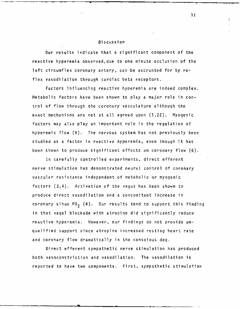
11
Discussion
Our results indicate that a significant component of the
reactive hyperemia observed,due to one minute occlusion of the
left circumflex coronary artery, can be accounted for by re-
flex vasodilation through cardiac beta receptors.
Factors influencing reactive hyperemia are indeed complex.
Metabolic factors have been shown to play a major role in con-
trol of flow through the coronary vasculature although the
exact mechanisms are not at all agreed upon (1,22). Myogenic
factors may also play an important role in the regulation of
hyperemic flow (9). The nervous system has not previously been
studied as a factor in reactive hyperemia, even though it has
been shown to produce significant effects on coronary flow (6).
In carefully controlled experiments, direct efferent
nerve stimulation has demonstrated neural control of coronary
vascular resistance independent of metabolic or myogenic
factors (3,4). Activation of the vagus has been shown to
produce direct vasodilation and a concomitant increase in
coronary sinus PO2 (4). Our results tend to support this finding
in that vagal blockade with atropine did significantly reduce
reactive hyperemia. However, our findings do not provide un-
qualified support since atropine increased resting heart rate
and coronary flow dramatically in the conscious dog.
Direct efferent sympathetic nerve stimulation has produced
both vasoconstriction and vasodilation. The vasodilation is
reported to have two components. First, sympathetic stimulation
IC
has been shown to increase the metabolic needs of the myocardium,
thus producing an autoregulatory increase in flow (1). This
can be blocked by the selective d 1l blocking agent practolol,
which prevents the increased metabolic requirements (23). In
our experiments, reactive hyperemia was less after practolol
suggesting that metabolic activity of the myocardium was reduced
due to the drug. Secondly, sympathetic nerve stimulation has
been shown to produce coronary vasodilation independent of a
change in metabolic requirements (8,23,24). These data support
our findings in that they demonstrate the potential for neurally-
mediated coronary vasodilation independent of metabolic changes.
Feigl (3), has demonstrated that during direct left sympathetic
nerve stimulation, .alpha vasoconstriction is the dominant re-
sponse after total beta blockade. Unfortunately, such a phenomenon
could not be investigated in our study due to complicating alter-
ations in arterial pressure after alpha blockade in the conscious
dog.
One minute coronary occlusion in the conscious dog has been
demonstrated to have dramatic effects on cardiac performance
(16,17). This was true both before and after beta receptor
blockade or cardiac sympa comy. In each case, inotropic re-
sponses to coronary occlusion were not significantly different
from each other (17). Similar results were observed in the
present experiments. This suggests that co-onary occlusion did
not produce reflex changes in the inotrop;. state of the heart,
thus, altered metabolic demands could not account for the large
component of the reactive hyperemia blocked by propranolol or
13
cardiac sympathectomy in our experiments. Direct right stellate
ganglion stimulation after practolol has been shown to increase
coronary blood flow without changing myocardial oxygen consumption
(24). Thus, an increase in selective sympathetic neural
activity associated with the reactive hyperemia could be a true
overpayment leaving a reserve potential for oxygen extraction by
the myocardium.
Our results indicate that the efferent pathway for the re-
flex beta vasodi.lator effect lies primarily in the right sympa-
thetic nerves. Left cardiac sympathectomy, either before or
after right side section, has no apparent affect on reactive
hyperemia. Right cardiac sympathectomy or total cardiac sympa-
thectomy produced a large, significant reduction in reactive
hyperemia. Such specificity of the pathway is not surprising
since the efferent limb of sympathetic mediated heart rate
changes in response to coronary occlusion and volume expansion
have been found primarily in the right nerves (18,25). This
may also explain why direct sympathetic stimulation seldom has
provided good support for a reflex sympathetic vasodilator
effect. In most previous studies, the left cardiac sympathetic
nerves have been stimulated (1,3,8,26). The majority of these
studies emphasized an alpha vasoconstricto r influence due to
left stellate stimulation. Ross and Mulder (27) showed an
interesting separation of effects between stimulation of the
right and left cardiac sympathetic nerves before and after pro-
pranolol. Before propranolol, end-diastolic vascular re-
sistance in the circumflex coronary artery was decreased by
14
stimulation of either left (-26%) or right (-19%) cardiac
sympathetic nerves. After propranolol, left sympathetic
stimulation increased vascular resistance 75%; whereas, right
sympathetic stimulation caused only a 9% increase. Such re-
sults suggest that alpha effects would be emphasized and beta
effects minimized during left cardiac sympathetic nerve stimu-
lation. Further support is found in the study by Nayler and
Carson (24) in which right stellate ganglion stimulation after
practolol caused coronary vasodilation without a change in
myocardial 02 consumption. In this same study several
different /62 blocking drugs were used and in every case sub-
sequent right stellate stimulation caused a reduction in
coronary blood flow.
In our study, curculating catecholamines did not influence
the response to coronary occlusion since dopamine beta hydroxide
levels did not change. Lack of alterations in circulating
catecholamine levels during acute coronary occlusions has been
previously observed (28).
In our study, beta blockade did reduce mean coronary flow
significantly. It can be presumed that this reduced the
metabolic requirements of the heart either through a direct
effect on the myocardium or by blocking circulatina catecholamine
effects. Cardiac sympathectomy, on the other hand, produced
no significant fall in coronary flow. In this case both circu-
lating catecholamines and spontaneous release of norepinephrine
from postganglionic nerves in the heart may have been signifi-
cant factors. Previous work has indicated that in conscious
A-m m m m ~ m•• mi
15
dogs which had undergone both beta blockade and cardiac sym-
pathectomy on separate occasions, beta blockade caused
measureable reduction in inotropic indices whereas sympathectomy
did not (17). Thus, similar reductions in reactive hyperemia
due to coronary occlusion subsequent to either beta blockade
or sympathctomy in our study indicates an active vaso-
dilator component under neural control. This phenomenon
appears to contribute to the substantial over repayment of
coronary blood flow during reactive hyperemia.
Acknowl edgements
The authors wish to express their appreciation to Linda
Fox for technical assistance and to Eugene Sprague for the
dopamine-beta-hydroxylase assays.

References
I. Berne, RM, De Geest, H, Levy, MN: Influence of the cardiac
nerves on coronary resistance. Am J Physiol 208: 763-
769, 1965
2. Klocke, FJ, Kaiser, GA, Ross, J, Jr, Braunwald, E: An
intrinsic adrenergic vasodilator mechanism in the coronary
vascular bed of the dog. Circ Res 16: 376-382, 1965
3. Feigl, EO. Sympathetic contro of coronary circulation.
Circ Res 20: 262-271, 1967
4. Feigl, EO: Parasympathetic control of coronary blood flow
in dogs. Circ Res 25: 509-519, 1969
5. Vatner, SF, Franklin, D, Van Citters, RL, Braunwald, E:
Effects of carotid sinus nerve stimulation on the coronary
circulation of the conscious dog. Circ Res 27: 11-21, 1970
6. Rubio, R, Berne, RM: Regulation of coronary blood flow.
Prog Cardiovasc Dis 18: 105-122, 1975
7. Feigl, FO: Reflex parasympathetic coronary vasodilation
elicited from cardiac receptors in the dog. Circ Res 37:
175-182, 1975
8. Brown, AM: Motor innervation of the coronary arteries of the
cat. J Physiol 198: 311-328, 1968
9. Eikens, E, Wilcken, DEL: Myocardial reactive hyperemia and
coronary vascular reactivity in the dog. Circ Res 33: 267-
274, 1973
10. Bache, RJ, Cobb, FR, Greenfield, JC, Jr: Effects of increased
myocardial oxygen consumption on coron.ary reactive hyperemia
in the awake dog. Circ Res 33: 588-596, 1973
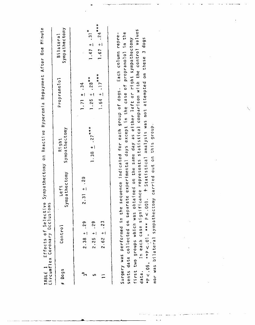
CU'
M- 42 ce' 4' '
$- Q~ I W
Cci4- o) m. .
C)j 0. w '0
01) tn* M- 0 a)CU
4- u ea EU -C
5.. >.% 4-3
4-j icU 0 C C
Cc to4 9 S- 4*-J 0
0 Cl) (\j 4- 4J CC * 0 4-)
cu $.- +1 + + a) s- 2
C. 0 '0 (1 = C)
S- - cli '0U 4.) 0 -
0l. 4- . 4- *- O
E 0 aJ ) L
S-~~ CL CJ C
E C
C. 0 C C 0
>1 4-) - 3 0
C) 2N =' a. w) '0
>0 u' U C.U U ' 0
4--) ca r U CA - *-
4-' C'j cj C) X rO~ 'A 0
U) _r_ C '
to crC S-s - '
c) -J +1 0 ') (Z4- +
reo 4- >,-0 to C
D. co +J r O 4-J
0 >1 C) - C
ca V) 4-)) UO ' t
2 U4-) C 4--l
4--) >1 (D C) . 'A ' 0U E 4J ( C--
C) 0 ,... 4) -0
413 ci L. CCL ea C)
4-J u U ) C) C 4-J --
to 4~C)U C0 L 1) 5.-
0. C +1 CX "aS
204-) o) ) Ucu C4- ro
V) CL m )*w - C
a)'A > C\J' tn 4j) E-~
>A cI .- C, 0
o ej o .004-0C 4-
4 C C2. .- U
u'tA 4-C 'j A C v
'A r )-CO
4- - *' . -
(A 4-* +' +' +'. 0 W C 0'
0A( 0 *o *O -~ U 0)'
4-3 -L 4-.- t C)0. '4 M
'Au 0C n .0 U- '- -
c)LS- C\J C' C4 Q) V (
4-0 0. 0( -
'AJ 0 C a 9 -k
(U 34-) 0.r. to
4-V 4-I LO 'A
E Ln ) 0(a
miJ 0) _ ) A 4-) . 3:.. U 0 (c n LOtm4-) 'A to V
m -- c s- 4-j L= CU .- 100-L 0
(A 'A 4- '0
11. Bach, RJ, Cobb, RF, Greenfield, JC. Limitation of the
coronary vasoconstriction in the dog. Circ Res 37: 88-
95, 1974
12. Debley, VG: Miniature hydraulic occluder for zero blood
flow determination. J Appl Physiol 31: 138-139, 1971
13. Mizeres, NJ: Origin and course of the cardioaccelerator
fibers in the dog. Anat Rec 132: 261-279, 1958
14. Coffman, JD, Gregg, DE: Reactive hyperemia characteristics
of the myocardium. Am J Physiol 199: 1143-1149, 1960
15. Nagatsu, T, Udenfriend, S: Photometric assay of dopamine-
/A-hydroxylase activity in human blood. Clin Chem 18: 980-
983, 1972
16. Bishop, VS, Kaspar, RL, Barnes, GE, Kardon, MB: Lef ven-
tricular function durinq acute regional myocardial ischemia
in the conscious dog. J Appl Physiol 37: 785-792, 1974
17. Kaspar, RL, Barnes, GE, Peterson, DF, Bishop, VS: Specificity
of autonomic influences on cardiac responses during myo-
cardial ischemia. J Appl Physiol 38: 485-490, 1975
18. Peterson, OF, Kaspar, RL, Bishop, VS: Reflex tachycardia
due to temporary coronary occlusion in the conscious dog.
Circ Res 32: 652-659, 1973
19. Peterson, DF, Bishop, VS: Reflex blood oressure control dur-
ing acute myocardial ischemia in the conscious dog. Circ
Res 34: 226-232, 1974
20. Reigl, EO: Control of myocardial oxygen tension by sympathetic
coronary vasoconstriction in the dog. Circ Res 37: 88-95,
1975
21. Echanberg, SM, Stone, RA, Kirshner, N, Gunnells, JC,
Robinson, RR: Plasma dopamine-/-hydroxylase: A possible
aid in the study and evaluation of hypertension. Science
183: 523-525, 1974
22. Olsson, RA: Myocardial reactive hyperemia. Circ Res 37:
263-269, 1975
23. Hamilton, FN, Feigl, EO: Coronary vascular sympathetic beta-
receptor innervation. Am J Physiol 230: 1569-1576, 1976
24. Nayler, WG, Carson, V: Effect of stellate qanglion stimu-
lation on myocardial blood flow, oxygen consumption, and
cardiac effeciency during beta-adrenoreceptor blockade.
Cardiovas Res 7: 22-29, 1973
25. Bishop, VS, Peterson, DF: Pathways regulating cardiovascular
changes during volume loading in awake dogs. Am J Physiol
231: 854-859, 1976
26. Granata. L, Olsson, RA, Huvos, A, Gregg, DE: Coronary in-
flow and oxygen usage following cardiac sympathetic nerve
stimulation in unanesthetized dog. Circ Res 16: 114-120, 1965
27. Ross, G, Mulder, DG: Effects of right and left cardio-
sympathetic nerve stimulation on blood flow in the major
coronary arteries of the anesthetized dog. Cardiovasc Res
3: 22-29, 1969
28. Toltzis, RJ, Satinsky, VP, Reed, V, Angelakos, ET, Scott, JC:
Effects of coronary artery occlusion with a swan-ganz catheter
on plksma norepinephrine. Fed Proc 33: 413, 1974
Legends
FIGURE 1 Left circumflex coronary flow associated with one
minute occlusion. The top trace is pulsatile flow, the bottom
trace is mean flow. Resting mean flow is indicated by the
dashed line. Part A is the response in the control state;
part B is after recovery from total cardiac sympathectomy.
FIGURE 2 Comparison of systolic and diastolic components of
coronary flow (CF) at rest and during peak reactive hyperemia.
The top trace is pulsat le aortic flow.
FIGURE 3 Effects of cardiac sympathectomy on over repayment
during coronary reactive hyperemia. Values represent percentage
repayment above unity. C, control; T.S., total sympathectomy;
L.S., left sympathectomy; R.S., right sympathectomy; Pp, pro-
pranolol blocked; Pt, practolol blocked; .001 p value compared
to preceeding observation.
FIGURE 4 Effects of blocking agents on reactive hyperemia.
Statistical comparisons are made with the preceeding value. C,
control; Pp, after propranolol; Pt, after practolol.
FIGURE 5 Effects of blocking agents on reactive hyperemia.
C, control; At, atropine; Pt, practolol; Pp, propranolol; **P< .01
compared with preceeding value.
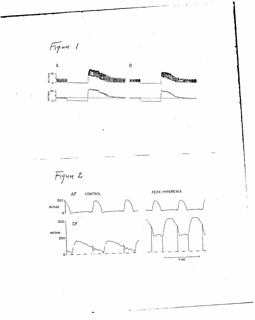
/i
A B
,50
AF CONTROL PEAK HYPEREMIA
mil/sec
0500' CF k
0I
Isec
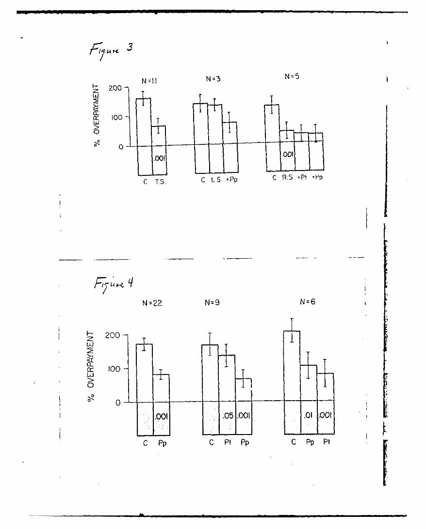
N=fl N=3 N=5F- 200-
Li
tx 100-
U
0uJ TS 0 1
.00, 7f 9
C T.S C LS. Pp C RS P1 ,p
N=22 N=9 N=6
200-ZT -7-c 100 TLUL
00
.0 .05 .001 .01 .001
C Pp C Pt Pp C Pp Pt
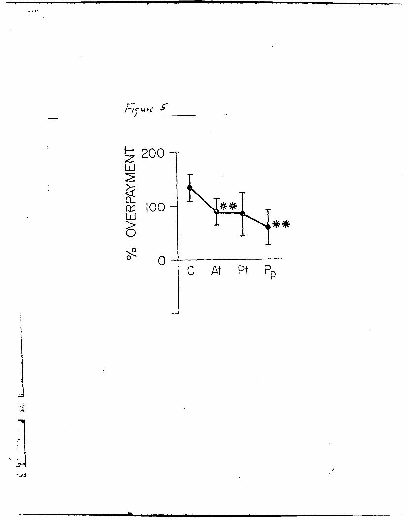
-200-
a--e 100
C At Pt Pp
.1]
Rabtit Cardiovascular Responses During
Vasoactive Druc Infusion at FLxed Carotid Pressure
By
H-enry 0. Stinnett, Ph.D., D. Fred Peterson, Ph.D. , and Vernon S.
Bishop, Ph.D., Department of Physiology and Pharmacology
University of North Dakota, Grand Forks, North Dakota 58202;
Department of Pharmacology, Unvversity of Texas Health Science
Center of San Antonio, 7703 Flcyd CLrl Dr.
San Antonio, Texas 78284
rr;: Sur;cr:: NI Grant HiLi2415, AFOSR =71-2.375
and :he San Antonio Chapter of :-e
American Hea.- Asscc1at2cn.
: :a-ed T : ........ Baroreflex alteraticn to ..aso ctive crugs
Sen d FS tc: Her!.y 0. Stinnett, Ph.D.
Depar:ment of Phys0iog. and Pharmacology
The University cf North DaKz:a
Grand Fcrks, N:o.--n Daksta 58'02
ABSTRACT
In anesthelized rabbits, peak reflex brodyca7r:.a 4~ HR) and
depression of mean arterial blood pressure ( A*.eemeasured
during maximal central stimulation of the left ac-rtL: narve (LANS).
Responses .were quantified; (i) before and duing t ay state changes
(+ 15 mm Hg) in the isolated carotid intrasinus presSI..re 'IS?), and (ii)
with ISP excluded fromn the circulation and maintain~ed at a normotenstve
level (EP = ISP = MAP) the MAP was chanced 20 m~m Hg by the infusion
of either nitroglycerin (NG) , lysine vasopress ion (.?)or phenylephrine
(PE) . Results indicated that within + 15 mnm Hg of ?.the change in MAP
per mm Hg change in ISP was 3, while & MA? d-.e : LANS changed nearly
double per mrn Hg change in ISP. Fzollowing a;zt: sm-all increase
in MAP was seen, hovwever, cardiovascular eoesto ch-anges in ISF
ar LA,' S were -unaltered. Dur~no druc rfs:.. a~rotuz s,,nuses
e x - Iuded f-rom the circu.latlon and TS C, . 3c. ~Ka-d AvP to
TLANS were indeperident of the direction or ten ne *zrug induced
change in MA.When carotid baroreceptorr, *.er ::)et detect -.h-e
increase in MAthe peac A HR and n, '.:A7 -e p: s :z 0A were
significa.-t'ly reduced. These results suc;;es: a :i;- cree of sensi-
tivity of the carotid sinus baroreceptors aro_:" -n -a .ma Zs nzormotensive
recion and that activ!ity from these barore:e;-_-- c~7 mo-ify reflex vassullar
tens ion even in the atsence of significar.: hr~ -..:ar rate or arterial
pressure .
Index term.s: L-aroreflex chan-a, vasoactr.'o *:r..zs :rt z nerve stimuj-ation
INTROD'CTICN
Many studies have characterized the hear:- an: vascular responses to
a wide range of controlled pressures in the isoiea--: car:.c or aortic sinus,
and defined the response relationship as the sest:v:" of the respective
baroreflex system (1, 5, 6, 8, 12, 13, 21, 22). Rc-:ent stucies have charac-
terized low pressure cardiopulmonary mechanc-rece;:ors (2, 11, 14, 15, 17,
18, 19, 20, 25, 26) which, when activated, -,BV a.er a-erial baroreceptor
sensitivity. Vatner,et al. (27) found that :he re.e:,: heart ratc response to
a pressure challenge was reduced during venous v:i-me loading and suggested
that the set point or gain of the arterial barorefle:es waS altered as atrial
pressure was raised. Indeed more recent studies 4'n, ave indicated that
volume infusion may actuially nave a direct ff.t.--... a.terlal berCreflex
through subtle cardiovascular alterations. -hs w-s a;a-re!t w'hen infusion
.~enuazed relex responses to left aortic ne-,e -- :. _,-..,', were
abolished foilowing carotid sinus barorece-.- z:- -t2:n '-t nct vacctom..' .
Additionally, since heart rate (HR) an: 7-ea... - . :-ss-es .. ) were
no: altered b rf:us:on, the usual asZ;.-: . "- ..... a chaz oe
in HR or : is an indication of a . ... ta 3 ...... ... ay
no: be valid. For this reason, it was os.u a i. th e :ar:t~ sinus
barorecetors are able to detect sute c.a:e :- ---- r cr volu.me whicri
may ,ea i to aitered reflex sympathetic efferen ca.a.: 1...::t sicnificantly
altered arterial pressure. This may lead z. -h : .. zs:xo sinus
bufferI:n c a.azi-tv at appare nt' .nc ~nz- y .-. _:. :-.anzed
zlocd Oress'-res.
A-m i mlelll I II i
2
The purpose of this study was to exa.-rine th._ -ostulate by determinng
whether or not reflex HR and MAP responses :: :::S are altered as a re-
sult of a change in the carotid sinus buffer-.-. ac-.:. Responses were
quantified firstduring controlled carotid sinrus =ressu.-e disturbances within
a physiological range of norrrotensive amer~ai e,.:e5. Then, vaso-
active drugs were used to change arterial pressure ---ir.g conditions in
which the carotid sinuses pressure was held co..stz-: and then subse-
quently allowed to compensate for the drug induce o-a.-.rge in systemic
blood pressure.
METHODS
Twenty-two rabbits of either sex (2.7 0.2 -were anesthetized
with sodium pentobarbital via an ear vein (Diazuta Diamond Laboratories,
Inc. , 30 m /gkg IV). A light level of suria e.-es:--: ".'.as --aint~ ed
by supplemental administration through a c -- .i vein. The
descending aorta (via femoral artery) and -.c "-;n . .. . (vi- the ;uga.
vein) were also cannuiared and connected t z:a:ha- e2 b and P23ES
strain ca"7,-7?Es for recording of arteriai and r -.: r .c pressures,
respective ly. Hear' rate was recorde:"' vic:-r. ' -' iec roces con-
nec:ed to a Beckman 9^4B car -c.4chcnmeter z_-;e.. >' I4 cre.ssre and
heart rate "w';ere initially reccrded on a Bec r-. £.41 :<o~rsh with
parallel output signdls to a DEC PD? 8/E D:Tin>.i or-:_:-r. ?ne tracne:
was cannulatd via a -racneostomy an: eat-. Sp-.:-a .......... soontane-
ously throughcuat the experiment. Frequer.: b...'......... blood samples
na,ze. r 2, p'-2 and p on a Eo :- C., ..' blood cas
-3
analyzer to assure maintenance of physiological eels for blood gases and
pH. Through a midventral incision, the left a.:-4 Bortic and vagus
nerves were located in the cervical region, caref-Il!. :soiated f_: sJr-
rounding tissue for about I cm, and looped ".:t a 1:cse thread for identifi-
cation pricr :z sectioning.
Carotid Sinus Isolation:
A modified Moissejeff technique was ,sed '3 permi" reversible isola-
tion of a carotid sinuses from the systemic circul=_ticr. (21, 22). Bot, internal
carctids were tied at approximately 0.5 cm from -. -. ,- :o'.'s of origin. The
external carotid artery was carefully isolated -ut re-. aned Daent. All other
small vessels were ligated. Following hezarin::z.. ,-= %,,g L' 50
units/mg, Hynson, Westcott, Dunning, Inc., 2a:--..re, :aryiand 21201)
the carotid arteries were cannuiated bei&c :e.... .ha. ex:.razorporeel
loop. Tiis permitted seif periuson of -.nc -r.s zy z-.z o:-s r rever-
sible pressure regulaion of the sinuses -i: a: a-> :S - _re .. Cott1ie
.a::er cclusion of the external carz:. ar-er:es. .. : zzl .:-zr.s
intrasinus pressure ,11SP) was m .nit..z. =--a -C . .-- in rl
ex:racorqoreal syste. near tne sinus.
In most animals, the sensitc':y * :-e z r:: e . ir "he
control of NIAP was evaluated at !- 5 mm '.- 3: tr pressure.
This was done bv observing the refle.: c-.ar:=s in a':- d s:-=:e '.A? as
a result of small stez cha..ces in the isc!:e7d Car: . S C.- .. S S r
Thus, when holdirg IS? cznsar.:, It ..as -- : ..e " - 2:s - of carotid
sinus was not altered during that portion of the expe% 7e-.al rctocol.
Both the right and left aortic nerves were c,;- an-- -.!-e cen.al end of the
left placed on hipolar hook electrodes, p~r~~ .electrical stir-,-
lation (9, 16). When not being stimulated, thke ner'Ve *.;=s Ziaced4 on saline
soaked cotton to prevent drying. Responses tc; left :irner-.e stimulation
(L.A'X'S) were observed both with the carotid s-~.~e n : ci.rc-_at~on and with
therr isolated and their pressure maintained czrns-ar.-: E: t -e :eeemre
equilibrium pressure. Responses to LINS wvere c*btE-. :de: z4 te above con-
di'tions bcth before and after vagotomy and a.-er :7.: It atrations in
bcdcressure (sce belowv).
~lectrical stimulation was generated. -:ia a -:asz5 E :_.'Cc activate
by; the Scnr:;-,idt t-i:,ger of the computer v;-.i:: : . :777
wav-e o' the EC k'10 ,23) . Regulation of t -_e s:.u.3 - &b
ne t er s, a s -w;elIl a s c _-nt iu s c a 1cuiair I: E7.. Dr.. ~.
hzz:e cozmzuter and spec_-,a conm 'u.Cr r~-~------ r-:zton o
e~cc~ica simu~tonparametLers used in a:- -::2ru :. :-.is SzuL';
haz bDcen prev.icously pubDi~B'..ed 23.r'ev, OLut over7
12" 0ardiac cycle~s, each burz ,:,A' elec-r, ci S::I;.:a p c te n
so-,-are wav'e impulses (ten v1s n-ed:D -cc: i-: : :07z: ed P-wave
of ',-,a G The i-. pulse 3ua~n .~ .n.%:t :~::eun V *as
:-ar.: cr:irto w,3s M3 msec.
5
Dr-c infusions:
In ten aortic denervated armals, after obtnairr; control 'ata, reflex
cardzovascu;ar responses were studied follo-.ving sysitemc Lrfu-sio., cf a
pressor or de:ressor drug to obtain a small (approx. 2'9 7= Hql %se cr fall
in steady state MAP. Drug infusion was initiated (HaPLardsyr.nce pump:
Har-vard Apparatus Co., Inc., Millis, Mass) with the carc:id sinuses
isolated frc.. the system and held at a pressure equa,?) t -e control
mean arterial pressure of that animal. Following the .:eadv state alteration
in systemic arerial pressure induced by the vascac:i.'e .;-, rhe :L.N was
stimulated. Following recovery and while dr-g infOs:sn c:.-e, a
steady rate, the animal- wes allowed to perfuse nis ov. :artd snuses
(carotid sinuses were included in the circulaticn ) and LX _timuaton re-
peaed. .Systemic arterial pressure was increased 'y Z. z.. e...l-
phrine nydroonioride (W.\inrbroo Labor~tzr~es, ;.2 ... v -s"- .... [z~nc.Ne','vZ;o " . . .;',
New Yor k 1 or Lysin 'asc-,es in (Sandoz 7"-- - r ""
re a s e! in svstemic a,:a p ressure were c:- .c.. c. s ..
nitro lycerin Eli -.l' & Co., 1:d1- Dp s , a). .: !es "..ere .c-
, c : .- 'es'red e t (vo Z , es tiMe -. .... n .-= - - "- "7-.n e
1). All values are recored as a mean cr mean z"r=.:e :-e ::r: error
of the mean (SE,) Statistical evalua,-. -as maze s e :o a: ..roria te
studerz t-.e-- for PaLred or unr.a reo c=t mPari:cr.. .: -.. ] .ere
ccns idered s:znificant.
nSRT Sin S7 n'
, a% ,-'
6
:: eft ac.-*Li: nerve stimrulation (LAINS) are surrmzarized in Table 2.
.7.-~z_-, e:.::>.'ing zhe carotid sinus f7rn . :-.e circulat~on at equili-
P-_=-_- ir c reasec the .rnacnitude of the ref'ex '-:.adycardia due to LANS
-4i:~ 50 e~t/m~.Removal of the vagi _ eCre6sed tne magnitude
c: tne refle:.: :radvcard ia when the carotid sinusesi vere included in the
c~rcu'Jticn. but had no significant effect when the s inuses were excluded .
nus, rec~zr:iess of vwhether or not the vagi are ir.:aC:. :e refllex HR response
LAS . en-.anced v:ner, the caroticd sinuses are :eventec rz sensing
-.-e re*.,--:: jnd:2ced changes in MAP. Excludi the carctid sinu.ses from the
c.rcula:=~ -Ia: a small, significant effect on. :ne re_-ex fa',i in MAP due to
.~N, e~en.scthe fall I-rom -48.2-2.6 rrnm.- tc -:-4.74;-2.2 MmHg (p< 0.05).
.~~L c-_-, zI.the small extenS ion of ',.e reie-Z -... , .:as not sicnifican.
r~ae-.aiu~e :wcnances in car::.: SIr.:r- surema altc: "he
S C -n-.- S-- . a.; 0,:... 2 -_ a- 'eC 3r
c -,.~.. -;ere , z -Tr-_ 4 -*Z -- - - - _z Z S C),rle
wsa:r::.smaM! c:eps ~e:e2 n 7, FmH :5 ... S .,.
C:.-..: I-, 78.-.a:e there a~~ & near :.a eairsipbetw.een
2 7- S -371Cr a "d e 7e: ai~ :?: .X .11 Thselationsllp
C., tl-e sa::e an,:imals -az :y hs
- ~ ~--!~ 7... . .. . . Dt!7 ... ... reS sur resulted in
*...-~ :z:::~cner-"e
stimulation. The extent of bradycardia durtnc LA'S, hzwe'er, was unaltered
during 1 15 m-.mHg changes in ISP around the ec_ .b.-., zressure. Furthermcre,
while vagotomy reduced the peak bradycardia :n LYS, no relattonship of peak
brad.'cardia to iSP level was observed fcllz;:..- regai -e.ation.
The effec:s : cressor and deoresscr acer .s :rn - e:c res=_-nse toLANS stimulation:
Lvs,.ne Vasocressin (ADH): With the carc:i sin_-es exluded (Ex) from
the circulation, the intravenous infsion of " nzn-specific pressor agent)
was adjusted to increase the MAP by about 2: .--- , 'v. 95.7-3.0 to 112.1 -
3.6 mmHc) (Fig. 3A). Subsequent reflex f-c :t 7? -t .-. atior. cf the
aortic nerve (LA-S) was not signif .cant,, z---. ,.. r: o--3.4
and during in.,zsion -51. --4.1 mrn?.z, resmea.- . ,.-: t:e infusion r-
tained, the carotid sinuses were include/ (1 ia-,: :r.:c Ce syste. rc crc.-iation.
.ean art3ria! oressur ',_an decree- " --7. 7 "a uC nt z i--
........ .,.zne :s-s ". i i . -. .-.-E- .s e r
nif-:cently ifrn from "ne eiuSz*:e ..- 2 m2-c.?Ewvr
Jurin c ruc .... s on in the (i) co,,c,.,o.. . . .., ....- 7.., rsts :. ..
wa s 3 less (-3 .9- 3 .. . .... :c.... ..2. .cr
c -c 31 2 7 14'tHtcocruc :r,,'S Z2 ',-51.3 - 2.7 m
:..e rrn e: A cons nz ~n 7 a,.-. niZ .7. 1r:rrA e 07Z
wit h .tne crc. : .td sinuses e:,:cl ed .. . := .. . . e.... -:re (? ,
fr 2m 9 6 - 1. 9 o I IS - 2 6 , 'T, F: z=:. 3 C' .. -: i: . :.. ttn 3 z he
MAP res:cn e (-33.4 6.4 m..Hc) to LA'S .. .....- :." (p < 3.5)
re uce .- hat cr bsC.',ed 7". .:hc :.-::i .:._-- -:::.:: - -.,:ne
8
car:i si.-.:- s :cluded n the ci-ua'o -a .a of PE maintained
constant, .-: as :'u~:o 108. 3 3.- .I , >v0 s.nf:n~. 0z .5~
gr e er -"a t 6a. wbe--~.ith :he inrti s*~e n.e n e c~rcuiation
pric. t3 :-.e cn-,'H 9 .(G7 l.51. e- ::e e.scn-e tc AY
wds-3 o.4 -?i -. wh -ich was also Si;.-.Zfzan-.>' less :n-an th8: cbDser-Ved
during A:S "'srior :Co PE inf"Usi or v~t", Ca rc::d stse inc:-,ded in -he circulation.
N it.-c l .c e r.n: Nitroglycerin (\TG"- i:nfusic. F:z. %v ith -. e carotid
sinu-.ses cxiued-on- th e circulation, 9ed6. 3.A2c 9 - i3 . 5 to 62. 9
-. i-c P< '305) . Including the car:n i .se :'Do t;e c:rczulation
resuted - in a s~gnnificant (P< 0.05) increaze -.n F :c 0 . i nrr
M:;eer:>.s ;S i -.waS Still einic~l 1 m~~c' sf~ : si on
:' o~n z. av er ae of' 9. ..... Y: -'o~fs
zu.~~~~~c:::r.~~~~Se -2-.: Cris~l.dfecn, -2---
*, r- C-.-.., i' 'o.2 v~---; -Wt- ~
~n re n u s ns :)n s c: .- .- nzcsr c.: .e.r lPressur Lt
~ ' 'c-'re- level, eeh n ::: 2rs :-szrt Zg 3D)
~~'.I~~~ i n.. . . . . . . . .
9
:~e caSz. nuses Lr tne c ircula ion-prod ucec e a.-f:~ .. reflex
S272 "- 3 to 249 +36. Wlt6. Ca7::.: :' from
:-. c.:A:.:. -.e reilex r. :~ri to LA2:S wv _ rei Iric-.ever, on~ce
carcz,:.: su-.uses -.:e-e a!lcwe-d to senisE tr: ::c:~~.:-e r~f1c..
bra;c~:.a:o L.ANIS s~i7,ulation was red-uce: :: -: -3 4 (p .CS
=;t~.. re".ced t-.e reflex bradvcarb- i to LAS.Howz:er, ;ual.t :ively the
-9anzes -,.serve-- af:-.r *;agotomy wer.sm~ o~ 3~ v a.. v :,,'a--t.
-~. e:ects of P7' on the restin~g HR (Fig. 3?' .-:=-e Aer to- =:- ..- nh:Se ob-
se rvIe _ A~n D F i n f sior. With carotidJ sinu~ses a::>:e _-.7jSjon
re~z~ r~ ~i~frm272 - 6 to 260 - E X~-. :~:i s,:..-ses
were ealic.::ed to sen~se 'he pressur;,e c7 anes a t: ~ :te?~WS
L a 7er.y ~P< 05 re dc ed n c o. r.e ed s i e .
T S 7--...r- t oP .Eifus o'n frm 2,,'74 - c : 2 4 y z a.c
s i- n, - . n n .... c.
not alter :-e rei:::cre;-~~~. e. . .: se.
~c~~: : rql~'ce~.ha' !.itt? sffic: z.-. -z-e n~.; - r the re;flex
S.Ss~~it: 7csr .. e> 1 e ex
:~~rc:~~~z-.7 (:7 St) Cs e."e ar;:E; s. ca.. ..... z C.W 5e
.<.. -.. -. ~-- ."Z S. C:-e.-------------- S- - - - - - - - - -
.,-----
10
DISCUSSIC,
A Results of this study demonstrate :h: "ne reflex responses to left
1 aortic ner'e stimulation (ANS) are de'edent u- -he stead-.' state carotid
sinus cressure. At intrasinus cressures ( .S? o 5 mm- H =-"g-ove the
equilibrium pressure (EP), were ISP and ,-? are eq ual, the reflex fall
in mean arterial pressure (MAP) during L"S ":a_ dlmnished. At IS? levels
I 1 to 15 mm Eg below EP the responses to A'S ".ere enhanced -%hen compared
to responses during control conditions. Re-ef*...... C ,ef.ex radycardia
to TANS, as seen during-pressor drug iofs-., .cq . . .o'ure of the
carotid sinus baroreceptor to pressures in. excess C_ ",m Hg ng-er than
EP. These results suggest that the !S trreshoit ?.ressure for .ascular
change in the rabbit is in the normotes'.-e g.. '..e t-a: :r -ea
rate chance is higher. Such findings ere .-....- :hose zreviously
reoorted for the dog (1, 4, 6, ZI) arnd t-. - --. :, ubisnedU
ob serv.at ions).4
',-.h n .iat:et raI carotid sinuS c -re:s r " .- ...-. : .
:rom "hc- z:rculation, the zrdi........ - e -- s L... ".'e:r.ecen'
ent of eh= . ? increase rocuza: c' cg..s:~r: .: .. ..-- .'a-cr~ssz-.
(ADH). W',hen the carotid s,.nuses ".'erc eci'.ve sa.;- :no AZE- :r-ouced
pressure chage, systemic arter:al re---------. .. 0st :Z "e zconrol
level. "Zow retlex fall in M.IAP due to L."S .v.a -... :i*, :euced when
comparei to the preinfuslcn c . l:esl-c-.s s. These results _-Jggest
that the refflex adjustm.ents . ne carct: s,.._ .. - . s-- -. - Er- a, I
cres zure -i.-ur L.; ADH -on re.. Su.d V..........
activi-t..; over. though the blood press..re wa- z e~.1. : same as the
preinfusicn M?
D)Ur, i ne.~ri (pr) in w~n.it- -,-e :tzsi-.usez isolated
at eiiri cressure, the average MA? rose s; c:vr
reflex '3.1 in Ato LANS was significantly. _nTese resuits are
in sharp contrast to those d':rin ADH.infuslon. 7Fe-exzo'-.g tne carotid
sinuses to the systemic pressure, during PE in.s: sle nan ad-
diticnal attienuation of the MAP response to _-.. e_-_ resultS succgest
that , unlike ADH , a nonspecific -:scnsrzz,=-~~p:n ;rOzJ1ce~s
an increase in. arterial pressure by cc.nipet i r. g v: . :: .e :7,r re f-_r
alpha receptz rs. Consequently, when sv-ti ~:v~ S reflexiy;
wt~drnby, 7~ Z3,' the available 7= ~i:ec::e ze :z~
sites and- 'he refle x lall in arterial pressr _7s tr~.i Z -: tton
is also 3:bsar%;Cd ...!en the carot:id a.:uss
sfle> reded ,."A?-r~inutE
stta z a1 c f z ym -re :-, i z;,-, . Z-- - . . . -
we:re al,:o czucr-ied %wnr. tne caro:tu S .:s ... ~ .
no ite~~ir .vso~e.e n the i-7 rsp.s .. : :e :-crx
Sinu-.ses *.,ere f::cc rom tiie :e:-:; ..... - - --.. ~
t P-fl A C
aru:c~e : :-e r~iex~i? cs~cze . - - 7
ecr-2e of a::enr,;tion of *he h-R resconsn-.- :.e -- E5-agent r.%,hwen the
carz-ti s,_-uses were included in the ci.-,:Z'~ :c. -: the at~enuatior,
cf -le HR response to !; NS occurred-4, az e.1' E zecondzary result
: caox c! snu t 7nte nt s t6o thIe P, ,e s :cr..Z e,-;e
syrrpat etic eff-erent activity (7). The :s~ ~-s:ccm-.cnent fr
reflex heart rate change was apparently Ttrz .-S agrees with a
previous sztudy in which the attenuation -::).-,se to LANS during
volum~e loadinc was also lound to be de-z:tn: uzzc:. ::-.e szp~ei
e'.:erernt activity (24).
Intravenous infusion of nitrogllyce:.-....e in a fall .'n MA?
wrn t-he carotid sinuses were excludiced- B...... .u;n bcn efore
and_ following vagctonr.y. The reflex fall .. > .:as nzt signifi-
cerIi ai'ereC4 d-uring infusion. vhen th------ :::r :T:aze-
1:%r.zs on *,alUes . EXZosure o): - -e Cart ;- -Cc::cny:
te~ls jcn CaluseJ a - nic:-s:*ncre>:r:. v:3r
e----------e---------------------------
t , w ~ r e o u e a rn e n z ~ > ~ . . . . . . ~ . ~ a . ~
e.. t hn~ en& croi 0 ~ ie L lZ- e:,.~- 27.. I.~Snc'
~t~nifz-ri d~~~r theC..L.~.......z nfusin
wrnt!h carot id s inuse s vwore Inc 1-_i- z z- n.... S. i r ie s uIt S
*:ldnst ta: een zrediceffi from :ca~~E : -~S~it:
daa F;.I, )That is, cart> -"t vn. rE~.e az:.e, s
7n Cn 2 rr ed pr-:s su r-2s : .Z S 3.. . .. .- : 71r~- 10o
carot_ stru :rssr arc *7-2....
. . . . . . . .. . . . .7s-rs
13
This relationship was linear within the near normc:ensi'e range of
pressures tested. From this relationship, one voud expect that when
press-re was lowered by infusion of a nonspecific .asdilator, such as
nitroglycerine, there w-ould be an enhancement of t:e vascular response
to LANS. This was not observed. This discrepenzy ,s further surprising in
view of previous results from our laboratory in whizh the reflex HR and MA?
responses to LANS were significantly increased after total arterial barore-
ceptor denervation (24). In this earlier work, tote arterial baroreceptor
denervation significantly increased the resting M.? indicating a higher
initial sympathetic efferent activity. Important, .... ,er, is that under
conditions of total arterial baroreceptor dener-ati:.-., the carotid sinuses
cannot reflexly adjust to the falling "AP c -rina ... n this study, with
the carotid sinuses isolated, with IS? held a: EP, , c-: NTG resulted
in a rela:ively smal decrease in MAP. :t :s : i: tha" . this drcc
in %-A? her detected by the intact carotid Lr:.e-ztsres-i 4:"inan
in;--t to :!e CNS ecuivalent to total zarcti S.-s-_s -...-. n."Further-
more, frL. .- e first cart of this stuc>: it ca'. a-I_ : r"s: te caroti
:arorefic-: .mecnanisms are oera:n; , t .c: .:c:sn--::v levels
during 7T G irfusicn. Therefore, a -ossi - _:..:-.. :r. e apparent
contradiction of the resonses to LANS d~rin: WT ris~on to :r.ose pre-dicted may b,- d frnces in co-:en.... .ec: wcri when
pressure is lowered h: vascdilaticn verss :: raise-. vasocnstric-
tion. T.hat iS, in the case of car-tZd stnus ct:.:. to a r-_- ucei
rSe in crcssure, th -.st effect is a rec .... ... .. s-m-. t.,tic
tone. Sub 'enr :, f'-rther v.u:hdrs.'a. t: -'. :. - -. 3... -. '?t tOno
1-4
. 5cg~sr-: nerve stimnulation would be less e:e:=: s Lr.ica-2e: b-y our
.es:-,',ts. Thi;s res:nse would result from a cczr~~ f '.he iitial lower
.eSting sym:':hetiz lone and the buffering e::ec- c: : cecarotid- sinus as
zressure falls. How.ev.er, when pressure ijs .:wre *ascdila-.0r and
zartiall': ccrrected b:the carotid sinuses zer.:r-era.svzhecanvt
-s elevated. LANS causes withdrawal of' sy3~e. u-zut b-ut as cressure
Ils, t~ie decrease --n carotid sinus activity. --i~ :e syvm:.ehetic with-
rawal and- tends to prevent an enhanoemer. :n -. e e::ec- predicted; at a
.e-;ucec: isolated carotid sinus pressure.
Heart rate responses to LANS were nc: ali-ere: -- -e -.c N:G wh.en re-
sponses during infusion are compared to = :::- i a c :r.rl -=es o s es
Z7:07 -0 infusion, either before or after vz:- rs res - _ _
essentially expected .-hen one consize4=rs-.e 3:~: 7-.e 7=-.
.r ~ac"!oardia resoconses to LJCNS d-,un::sg::3~e
~a~cto ~~rasnuspressure as dizcusseo er'er
.l>,thne '-e SaOf v a SOa -t; .-e na -7,.s: 1-c ::n-s .7e-- olve
zlrec-- .- '.- E-'e t Wh--'n1 ~ ~ Z
GZ ~sic-. 'nCL;':.ide a d irec: e::e:: fi :-,ec...-~.;.sole
.'.:nin th-e carotid susregion resu iting :-:.Z Cr'
z ~r~f e ea r by ta ror e ce -? z t. ~~c ree
m7eorim,'enoeptor i:unein the cr o.: ::
- ~ .. : v-~seis -ese -. 72-25
Refere nces
1. AT:2 ,TT, SAGAA, M. KUMAIDA: An open-1co; analysis of the
acr~ic arch~ bar-ostanic reflex. Am P hysicl 27(6): 1570'-JlI4, 1969.
2. -2:,3?.HC?, V:S, F. LCMEAB-'.D1, A. IMALLIA'.1 ,. GA T 0. RECORDATI:
Aefllex sv=-athetic tachycardia du.-,no iritra';eric's inrU~Sicz ll nronlic
spinal cats. Am j Physiol 230 (1): .25-29, 1976.
3. CCHEN, XVV, ES. KIRK: Differential response cf largc and small
co:zr. a:-eries to nitroglycerin and angiotensin. Oi,-c Res 33:
4. COW.LEY, AWv ;r, E. NION'S, AC. GUYTCN: Tn~eraction of vasopressin
an-- the barorecepior reflex systen in tne 07.a~o o: a:er .I_- blood
p~~s -ein :nhe dog. Circ Res 34: SUDS-314 , 1974 .
3. _=S , A,-: Aotrtic baror--.fex funo:tzr. irt thle _:og Ar ?~ic 2
5 7 71.
a:c t ar C,-Cez cr: S, Pnys.51 __S 2': 2-:,1.
7. ~:1:7 -c:" C-P D-H 7: : -:: 5?. C..P S~ zf: an.:te n n
:aSZ~e~n. nd Tet"oxr nC on ,r a
2. p2l~ H, e 'N: ~:rai nerv'ous S.,~ :u.:.no
Z n re e 77 n D r. Am Psii2: 1-L~ 1.
10. KARDCN, .'B, DF. PETERSON, VS. BISHOP: Re/:ex neart rate control
via specific aortic nerve afferents in the ratbit. Circ Res 37: 41-47, 1975.
11. KOIKE, H, AL. MARK, DD. HEISTA'D, PG. SC:.,.,D:: influence of
card ionu lmcnory va;al afferent activity on carztzl chemcreceptor and
barorecector reflexes in the dog. Circ Res 37: 22-429, !975.
12. KUMADA, M,, K. SAGAWA: Aortic nr-ve acttvity during blood volume
changes. Am I Physiol 218 (4): 961-965, 1970.
1.3.- LEVY, MN, ML. NG, H. ZIESKE: Cardiac and reseir-etory eects of
aortic arch baroreceotor stimulation. Cir Fes 1i: 292 , 1966.
14. MANCIA, G, DE. DONALD, JT. SHEPHERD: :nhb:ti'n of acrenergic
outflow to peripheral blood vessels by a;:a affe:ents Erc. te
cardiopumona / region in the dog. Cir Res : 3-72'1, 1973.
15. MA'CIA, G, IT. SHEPHERD, DE. ON.LD: .ntr.' amcnc carot.,d
s ar--, C a o pim,.r-arv., , a nd- carc L.c "::::-:".; • c
' ,"?, (1): 19-2-., 1976.
,. NTL, E, ITR' .D2,XVO D, A. SC.','- .::-: .7ee:r:cal
stu--..uati:r. z: the azrt~c n"er-ve on h:.c- cr-3:--r: >.i .'s...
in nc rabbit uncer c-or,3--- -b-------
, Physic: i , 7 1,D 2!-. 194 .
17. OTT, :T, ;T. SHE!HERD: ,'snferezsor re:1e:.. .l. 'K ation in
the c-a-..', A- sio, 2 2- 3 :
18. PELLETiE7, %C." 7D:S, -: va
,..e....n .s ~resc*.se......ae- in, i :. C . 4es_.: ~ 24, 1071
19. Z E' T7 -- 1 Cl , IT. SHEPHERD: Circula'zry :-efexes :rmmechano-
recez-tors in the cardc-aor-tic area. Circ Pes -3 (2): i' 1-136, 1973.
20. 7?TLL;.U', i-iRC I'll, M. GUAZZI, EID. Fr S: .;a ffcrent deores-
:zcr n e -s in the rez.An, I ?hysiol 21-2:p6-7,9~
21. SCh.',IDT, RM, N1. KUZAADA, K. SAGAW.'-.: Cardiac -utout and4 total
periphneral resistance in carotid sinus reflex.:. Am:7 7 Fnysicl 221 (2):
460-487, 1971.
22. SCHii"iDT, R M.K ADK. SAO-AWA: Crv ol responses to
various p.-Isatile pressures in the caronud s. Ph% 'iol 2 2 (1):
1-7, 1972.
23. STINNETT:, HO, DF. PETERSON, VS. ::-C :d:as'lrresponses
to :lcrcrioa-ouldsiuain~- - er-ic. Ann
PhY-s ;ol 2 3 (5): 13 74-1378, 1976.
1 r .- 2scuTT HO ,ecno VSc .o.o~. FH FF P.....- Z -e 2
Res 39 '6): 7 6-7 7Z., 1S 7 6
2.- ,G. ::CL, 7 H 7
Am Ths~c1229 (5): 1410-1-113, 7.
26. JC- 11A , Y: Affe re n- S'."Pa tne- c ner-ve -. :s :.S~ 7CC nrcc crs
27. AT',SF, D-. 5~HP . .
n' 1~. '7 -
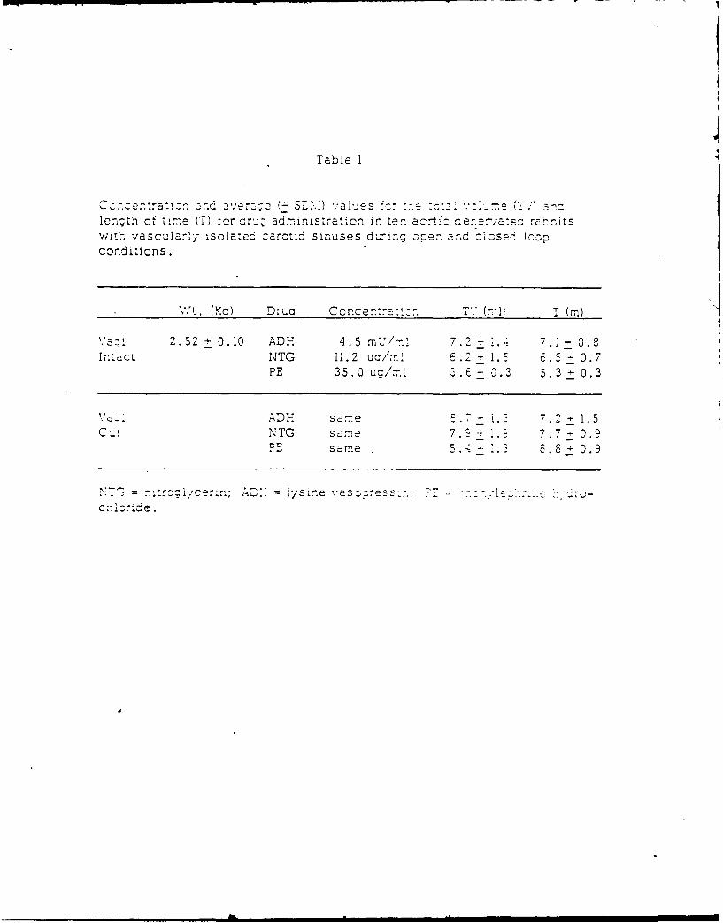
Table I
Cc--enrati. a ve ( - 3F:.:) valu.es -o 3Y- c .: -'e...-
lenzth of time (T) for dr's: administration in ten acrt : dene,,aed rabbitswith vascl.zry isolated carotid sinuses during oper. and cizsed lcopconditions.
'vt. (Kc) Drua Concen:-::-. TV (m1 T (m)
2.52 + 0.10 ADH 4 5 mU/mi 7.2 - 7. 1- 0.8Ir.tact NTG 11.2 uc/ 1.5 . 5 - 0.7
PE 35.0 ua/z. 3.E + 0.3 5.3 + 0.3
a same 7 + 1.5NTG same 7. . .7 0.
same 5.- 1.3 8 0.9
XCq = nitrglyce..; AD-, = 1ysine v'ss:ress..; T z = .:-:'l.h..-. -.c c!e
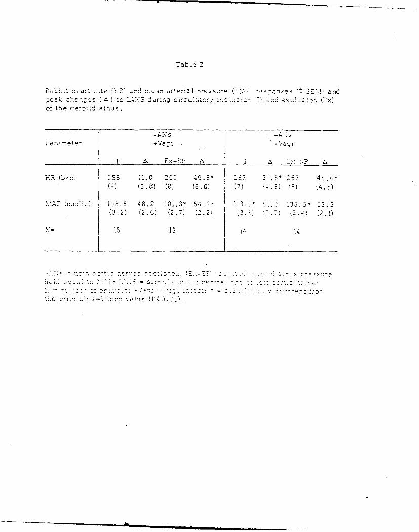
Table 2
Rab~:t heart aate (i-, an-d . .i arterBl pressure (',.. 7E-- ,) and.eaz" 1- ')'s during circulatorv 1"-(Ex)of the c-otid sinus.
-ANs -AsParameter +Vagi -Vag I
______Ex-EP i Ex-EP
HR b.) 258 411.0 260 49.* .* 2 7 45.*(9) (5.8) (8) (6.0) (7) -. 6) (c) (4.5)
,,P ,. n' iz) 108.S 48.2 101.3* 54.7, 3 15.* 55.5(3.2) (2.6) (2.7) (2.2, (3. " 7) 2.4) (2.1)
- £. 1
"c . "- (. .....i - c c ,, ::. . 5, : -.. .. = . :7 v -.t73 : " = Z ~ ..- C . :..- e"- " c
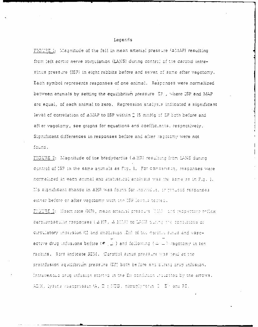
Leg endAs
~T 1: a~r'iudeof t~.e fall in~ mean arterial cress...e ~2.P eut
from left acr-:.o nerve stimulation. (LNS) during ccntr : Z- carct~d intra-
s-' - : eic-t re bt ero-re andc seven c- =7 after vagotomy .
Eazh symbol rezresents responses of one animal. Respcznszs were normalized
be'-w.een amnmals by setting the equilbrum press-, re 7-:, ,TeeIP n A
are equal, of each animal to zero. .Regression ia i-s:;Cated a significant
level of correlation of AMto ISP wit'j-.in IS~ g. z.-.ef'cre anoi
aft er vagotomy, see graphis for equations and coeffic.en :s, eetvey
Sizn'.iant differences in responses before anad af-'-er :- .-- z ere not
F 7 G E 2: Ma g-i t -,de of te br-a yar d'a I~ .- ? res.~ S-~- J.Sor
czn.r:n of :5 n te s am:e a n i 7nIs a s c~ 1. So z: sn, e S oSe s . e
- ~ ~ i -- 3-7-- tef'Uo, - .
e~rv3 f r c.-:e -? c: - 30 ; t- e r m 7
ac:i.e dr..: Cr:s~n be:re C )ar..: fo~z c : 111 -; ten~m'
-. r .fs ion .j e iiebre m - jZ~ .. . .-. ; -r
.enLZ~2;:.:c.........
i-2-----------------------
.) * .'cates sic.-izant n(. . j z:: :e-ece ..nen th s
- t:e re pec:'.'e (I) : (£x-E') oor..r 1 . e. Fcr fu '-
descrp~z~~s - e ::.:j
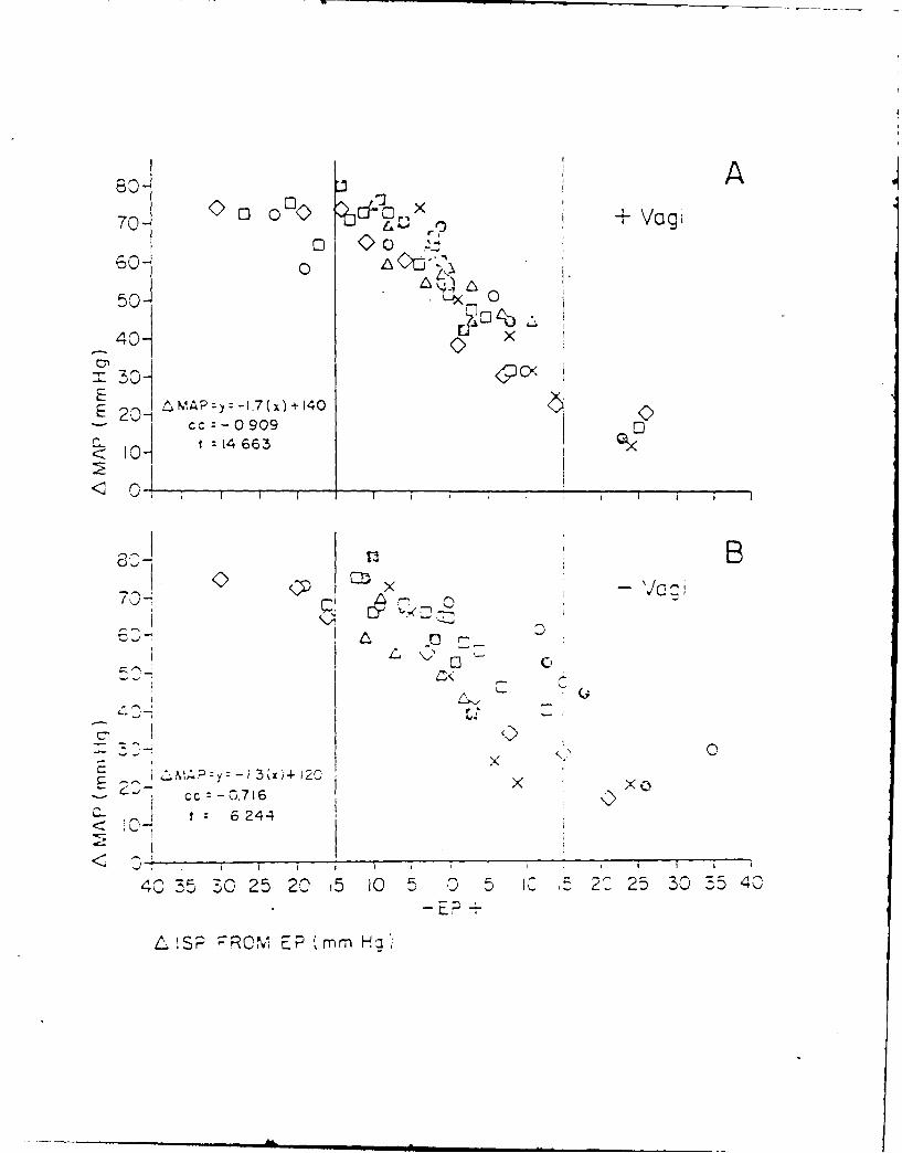
7 o) ,9 + Vagi
o0 0 05o-c)50- L '< 0
40- x
.30E I MAP-y: -.7(x) + 140E 2--
cc:- 0909C.. I t 14 663< l
r- -7c
,, x '7ogi
C C
, p..
x0
-- 2- cc :-0.716 ,
<" f: 6 24-4< 0-
, I I I *i I
40 3530 25 20 15 05 05 5 2 25 30 3540
LS2 PR0" EP m m. H q
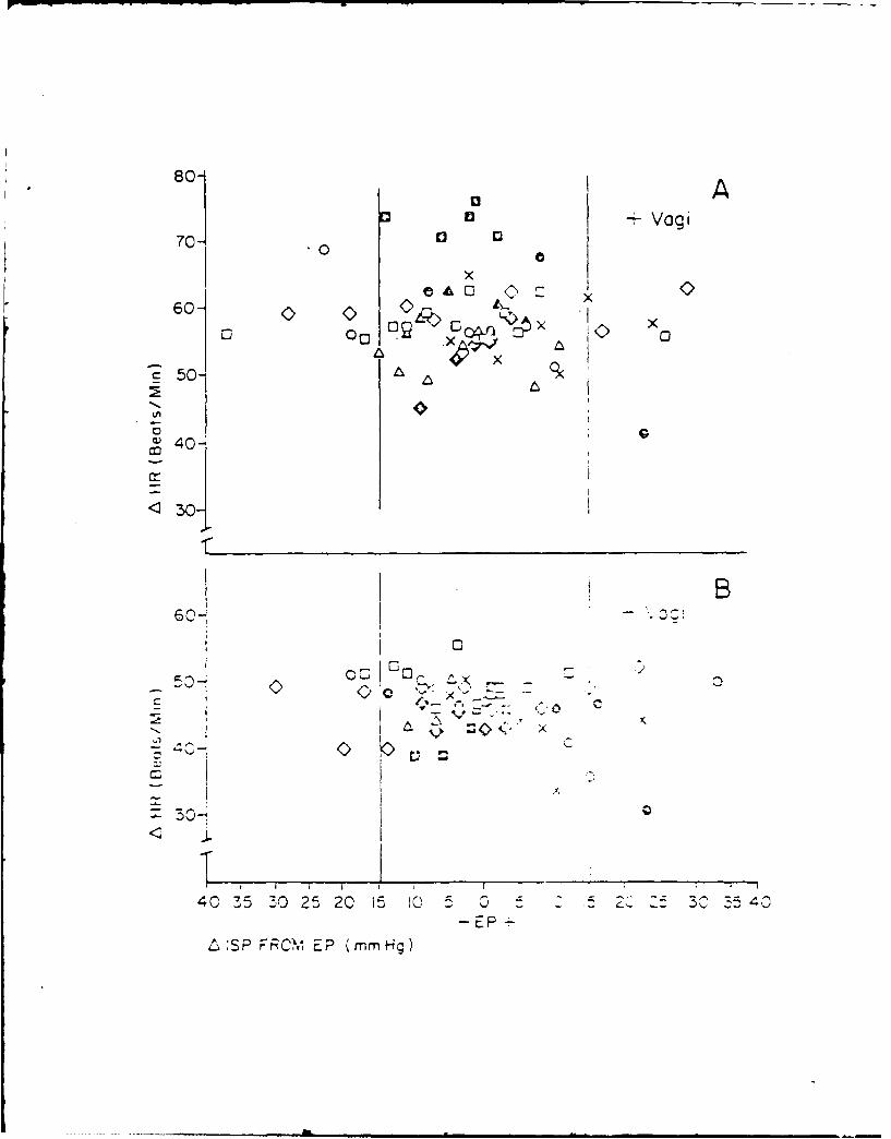
801-Vog~
70-4 0 a
x
£50- O A
~40-
O-~
< 301
00'
- EP -
^:SP FRC\1I EP (mnmHg)
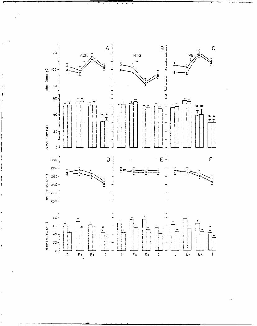
2> Al Bi C23 ADH NTG PEe
T 7-
4- i
20J
1 ,F- .-1 -- - .*
] i i W'
1 l-
A E -Ex E E xLI
-- 2gS, ---
- _ - - -
" i I ~ ~~~~I -, . , . .- '-_
. - ,! :! , ;j f , ' i
<: C, i
... ... ..... .il~, ,m~m~mm~lm l
I
Related Documents