See discussions, stats, and author profiles for this publication at: https://www.researchgate.net/publication/233939699 β-Catenin-Driven Cancers Require a YAP1 Transcriptional Complex for Survival and Tumorigenesis Article in Cell · December 2012 DOI: 10.1016/j.cell.2012.11.026 · Source: PubMed CITATIONS 159 READS 104 21 authors, including: Joseph Rosenbluh Monash University (Australia) 50 PUBLICATIONS 1,153 CITATIONS SEE PROFILE James T Neal Stanford Medicine 9 PUBLICATIONS 299 CITATIONS SEE PROFILE Aviad Tsherniak Broad Institute of MIT and Harvard 48 PUBLICATIONS 913 CITATIONS SEE PROFILE All content following this page was uploaded by Aviad Tsherniak on 29 December 2013. The user has requested enhancement of the downloaded file. All in-text references underlined in blue are linked to publications on ResearchGate, letting you access and read them immediately.

Welcome message from author
This document is posted to help you gain knowledge. Please leave a comment to let me know what you think about it! Share it to your friends and learn new things together.
Transcript
Seediscussions,stats,andauthorprofilesforthispublicationat:https://www.researchgate.net/publication/233939699
β-Catenin-DrivenCancersRequireaYAP1TranscriptionalComplexforSurvivalandTumorigenesis
ArticleinCell·December2012
DOI:10.1016/j.cell.2012.11.026·Source:PubMed
CITATIONS
159
READS
104
21authors,including:
JosephRosenbluh
MonashUniversity(Australia)
50PUBLICATIONS1,153CITATIONS
SEEPROFILE
JamesTNeal
StanfordMedicine
9PUBLICATIONS299CITATIONS
SEEPROFILE
AviadTsherniak
BroadInstituteofMITandHarvard
48PUBLICATIONS913CITATIONS
SEEPROFILE
AllcontentfollowingthispagewasuploadedbyAviadTsherniakon29December2013.
Theuserhasrequestedenhancementofthedownloadedfile.Allin-textreferencesunderlinedinbluearelinkedtopublicationsonResearchGate,lettingyouaccessandreadthemimmediately.
b-Catenin-Driven Cancers Requirea YAP1 Transcriptional Complexfor Survival and TumorigenesisJoseph Rosenbluh,1,3,5 Deepak Nijhawan,1,3,5 Andrew G. Cox,3,4,6 Xingnan Li,7 James T. Neal,7 Eric J. Schafer,1,3,5
Travis I. Zack,2,5,8 Xiaoxing Wang,1,3,5 Aviad Tsherniak,5 Anna C. Schinzel,1,3,5 Diane D. Shao,1,3,5
Steven E. Schumacher,2,5 Barbara A. Weir,1,5 Francisca Vazquez,1,5 Glenn S. Cowley,5 David E. Root,5 Jill P. Mesirov,5
Rameen Beroukhim,2,3,5 Calvin J. Kuo,7 Wolfram Goessling,1,3,4,6 and William C. Hahn1,2,3,5,*1Department of Medical Oncology2Department of Cancer Biology
Dana-Farber Cancer Institute, 450 Brookline Avenue, Boston, MA 02215, USA3Department of Medicine4Division of Genetics
Brigham and Women’s Hospital and Harvard Medical School, 75 Francis Street, Boston, MA 02115, USA5Broad Institute of Harvard and MIT, 7 Cambridge Center, Cambridge, MA 02142, USA6Harvard Stem Cell Institute, Cambridge, MA 02138, USA7Department of Medicine, Hematology Division, Stanford University School of Medicine, Stanford, CA 94305, USA8Program in Biophysics, Harvard University, Boston, MA 02115, USA
*Correspondence: [email protected]
http://dx.doi.org/10.1016/j.cell.2012.11.026
SUMMARY
Wnt/b-catenin signaling plays a key role in the patho-genesis of colon and other cancers; emergingevidence indicates that oncogenic b-catenin regu-lates several biological processes essential forcancer initiation and progression. To decipher therole of b-catenin in transformation, we classifiedb-catenin activity in 85 cancer cell lines in which weperformed genome-scale loss-of-function screensand found that b-catenin active cancers are depen-dent on a signaling pathway involving the transcrip-tional regulator YAP1. Specifically, we found thatYAP1 and the transcription factor TBX5 form acomplex with b-catenin. Phosphorylation of YAP1by the tyrosine kinase YES1 leads to localization ofthis complex to the promoters of antiapoptoticgenes, including BCL2L1 and BIRC5. A small-molecule inhibitor of YES1 impeded the proliferationof b-catenin-dependent cancers in both cell linesand animal models. These observations definea b-catenin-YAP1-TBX5 complex essential to thetransformation and survival of b-catenin-drivencancers.
INTRODUCTION
b-catenin signaling plays a key role in colon development and
cancer (Clevers, 2006). The destruction complex composed
of AXIN1, GSK3b, and adenomatous polyposis coli (APC)
phosphorylates serine residues in b-catenin, which leads to its
C
proteasomal degradation (Clevers, 2006). Binding of Wnts to
the LPR6-Frizzled receptor inactivates this complex, leading to
accumulation and nuclear translocation of b-catenin. In the
nucleus, b-catenin forms a complex with TCF4 that drives the
transcription of genes that contribute to cell proliferation (Klaus
and Birchmeier, 2008). Individuals carrying APC germline
mutations (familial adenomatous polyposis) develop colonic
polyps that progress to colon cancer (Kinzler and Vogelstein,
1996), and mutations in the tumor suppressor APC or the
oncogene b-catenin have been found in the majority of sponta-
neously arising colon cancers (Cancer Genome Atlas Network,
2012).
b-catenin is a component of the adherent junctions (Baum and
Georgiou, 2011) and, in the nucleus, binds to TCF4 and several
transcriptional regulators. For example, when cancer cell lines
are cultured under hypoxic conditions, b-catenin forms a
complex with HIF-1, leading to hypoxia adaptation (Kaidi
et al., 2007), and in prostate cancer cells, a b-catenin-androgen
receptor (AR) complex increases the transcription of AR
(Mulholland et al., 2002). b-catenin and YAP1 also coregulate
genes that are essential for cardiac development (Heallen
et al., 2011). These observations suggest that, through interac-
tions with different partners, b-catenin regulates many biological
processes.
Yes-associated protein 1 (YAP1) is a transcriptional modulator
that has been implicated in stem cell differentiation and the
control of organ size (Pan, 2010). YAP1 regulates several
context-specific transcriptional programs (Badouel et al., 2009)
and promotes proliferation and tumor growth (Overholtzer
et al., 2006; Zhao et al., 2008). Indeed, YAP1 is recurrently ampli-
fied in hepatocellular cancer, in which YAP1 is essential for
survival of tumors that harbor YAP1 amplifications (Zender
et al., 2006). Furthermore, inducible transgenic expression of
ell 151, 1457–1473, December 21, 2012 ª2012 Elsevier Inc. 1457
a stabilized YAP1 mutant (S127A) in mice induced liver hyper-
plasia and colonic adenomas (Camargo et al., 2007).
YAP1 transcriptional activity is regulated by several mecha-
nisms. In quiescent cells, Hippo pathway-mediated serine phos-
phorylation of YAP1 inhibits nuclear import and promotes its
degradation (Zhao et al., 2012). In contrast, YES1-mediated
phosphorylation of YAP1 activates YAP1 in embryonic stem
cell self-renewal (Tamm et al., 2011), and ABL-mediated phos-
phorylation of YAP1 in response to DNA damage results in tran-
scription of proapoptotic genes (Levy et al., 2008). Recent work
suggests that YAP1 also plays a role in mechanotransduction in
a Hippo-independent manner (Dupont et al., 2011).
Although stabilization and localization of b-catenin contribute
to adenoma formation, our understanding of b-catenin regulation
and function in cancer remains incomplete. For example, Rac1-
mediated phosphorylation of b-catenin has been shown to affect
b-catenin activation and localization (Wu et al., 2008). Moreover,
in zebrafish and some human cell lines, APC loss alone resulted
in impaired differentiation but failed to induce nuclear localization
of b-catenin and transformation (Phelps et al., 2009). To gain
insights into b-catenin activity in malignant transformation, we
classified b-catenin activity in a panel of human cancer cell lines
in which we have systematically characterized genetic alter-
ations, gene expression, and gene essentiality. Here, we report
the identification of an alternative transcriptional regulatory
complex required for the b-catenin-driven transformation and
tumor maintenance.
RESULTS
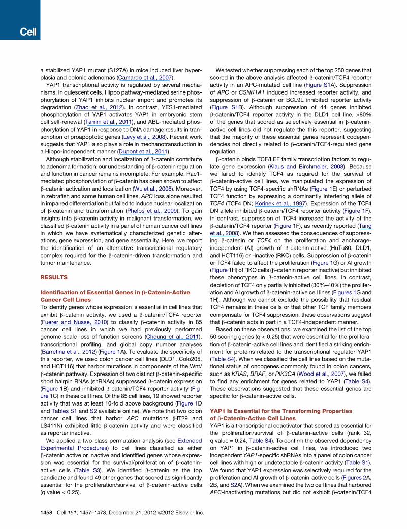
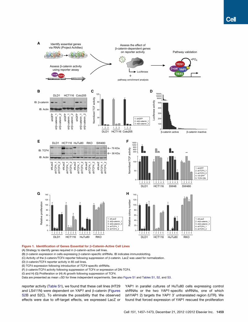
Identification of Essential Genes in b-Catenin-ActiveCancer Cell LinesTo identify genes whose expression is essential in cell lines that
exhibit b-catenin activity, we used a b-catenin/TCF4 reporter
(Fuerer and Nusse, 2010) to classify b-catenin activity in 85
cancer cell lines in which we had previously performed
genome-scale loss-of-function screens (Cheung et al., 2011),
transcriptional profiling, and global copy number analyses
(Barretina et al., 2012) (Figure 1A). To evaluate the specificity of
this reporter, we used colon cancer cell lines (DLD1, Colo205,
and HCT116) that harbor mutations in components of the Wnt/
b-catenin pathway. Expression of two distinct b-catenin-specific
short hairpin RNAs (shRNAs) suppressed b-catenin expression
(Figure 1B) and inhibited b-catenin/TCF4 reporter activity (Fig-
ure 1C) in these cell lines. Of the 85 cell lines, 19 showed reporter
activity that was at least 10-fold above background (Figure 1D
and Tables S1 and S2 available online). We note that two colon
cancer cell lines that harbor APC mutations (HT29 and
LS411N) exhibited little b-catenin activity and were classified
as reporter inactive.
We applied a two-class permutation analysis (see Extended
Experimental Procedures) to cell lines classified as either
b-catenin active or inactive and identified genes whose expres-
sion was essential for the survival/proliferation of b-catenin-
active cells (Table S3). We identified b-catenin as the top
candidate and found 49 other genes that scored as significantly
essential for the proliferation/survival of b-catenin-active cells
(q value < 0.25).
1458 Cell 151, 1457–1473, December 21, 2012 ª2012 Elsevier Inc.
We testedwhether suppressing each of the top 250 genes that
scored in the above analysis affected b-catenin/TCF4 reporter
activity in an APC-mutated cell line (Figure S1A). Suppression
of APC or CSNK1A1 induced increased reporter activity, and
suppression of b-catenin or BCL9L inhibited reporter activity
(Figure S1B). Although suppression of 44 genes inhibited
b-catenin/TCF4 reporter activity in the DLD1 cell line, >80%
of the genes that scored as selectively essential in b-catenin-
active cell lines did not regulate the this reporter, suggesting
that the majority of these essential genes represent codepen-
dencies not directly related to b-catenin/TCF4-regulated gene
regulation.
b-catenin binds TCF/LEF family transcription factors to regu-
late gene expression (Klaus and Birchmeier, 2008). Because
we failed to identify TCF4 as required for the survival of
b-catenin-active cell lines, we manipulated the expression of
TCF4 by using TCF4-specific shRNAs (Figure 1E) or perturbed
TCF4 function by expressing a dominantly interfering allele of
TCF4 (TCF4 DN; Korinek et al., 1997). Expression of the TCF4
DN allele inhibited b-catenin/TCF4 reporter activity (Figure 1F).
In contrast, suppression of TCF4 increased the activity of the
b-catenin/TCF4 reporter (Figure 1F), as recently reported (Tang
et al., 2008). We then assessed the consequences of suppress-
ing b-catenin or TCF4 on the proliferation and anchorage-
independent (AI) growth of b-catenin-active (HuTu80, DLD1,
and HCT116) or -inactive (RKO) cells. Suppression of b-catenin
or TCF4 failed to affect the proliferation (Figure 1G) or AI growth
(Figure 1H) of RKO cells (b-catenin reporter inactive) but inhibited
these phenotypes in b-catenin-active cell lines. In contrast,
depletion of TCF4 only partially inhibited (30%–40%) the prolifer-
ation and AI growth of b-catenin-active cell lines (Figures 1G and
1H). Although we cannot exclude the possibility that residual
TCF4 remains in these cells or that other TCF family members
compensate for TCF4 suppression, these observations suggest
that b-catenin acts in part in a TCF4-independent manner.
Based on these observations, we examined the list of the top
50 scoring genes (q < 0.25) that were essential for the prolifera-
tion of b-catenin-active cell lines and identified a striking enrich-
ment for proteins related to the transcriptional regulator YAP1
(Table S4). When we classified the cell lines based on the muta-
tional status of oncogenes commonly found in colon cancers,
such as KRAS, BRAF, or PIK3CA (Wood et al., 2007), we failed
to find any enrichment for genes related to YAP1 (Table S4).
These observations suggested that these essential genes are
specific for b-catenin-active cells.
YAP1 Is Essential for the Transforming Propertiesof b-Catenin-Active Cell LinesYAP1 is a transcriptional coactivator that scored as essential for
the proliferation/survival of b-catenin-active cells (rank 32,
q value = 0.24, Table S4). To confirm the observed dependency
on YAP1 in b-catenin-active cell lines, we introduced two
independent YAP1-specific shRNAs into a panel of colon cancer
cell lines with high or undetectable b-catenin activity (Table S1).
We found that YAP1 expression was selectively required for the
proliferation and AI growth of b-catenin-active cells (Figures 2A,
2B, and S2A).Whenwe examined the two cell lines that harbored
APC-inactivating mutations but did not exhibit b-catenin/TCF4
1 2 3 4 5
DLD11 2 3 4 5
HuTu801 2 3 4 5
HCT1161 2 3 4 5
RKO1 2 3 4 5
DLD11 2 3 4 5
HuTu801 2 3 4 5
HCT1161 2 3 4 5
RKO
0
30
60
90
120N
orm
ilize
d TC
F ac
tivity
IB: Actin
shG
FP
shβ-
cate
nin_
2
IB: β-catenin
shβ-
cate
nin_
1
shG
FP
shβ-
cate
nin_
2sh
β-ca
teni
n_1
shG
FP
shβ-
cate
nin_
2sh
β-ca
teni
n_1
DLD1 HCT116 HuTu80 RKO SW480
shLa
cZsh
TCF4
_1sh
TCF4
_2
IB: TCF4
IB: Actin
shLa
cZsh
TCF4
_1sh
TCF4
_2sh
LacZ
shTC
F4_1
shTC
F4_2
shLa
cZsh
TCF4
_1sh
TCF4
_2
shLa
cZsh
TCF4
_1sh
TCF4
_2
58 KDa
79 KDa
Luci
fera
se a
ctiv
ity
β-catenin active β-catenin inactive0
100
200
300
400
500
50001000015000
1 2 3 4 5 1 2 3 4 5 1 2 3 4 5 1 2 3 4 50
50
100
300600900
12001500
Nor
mili
zed
TCF
activ
ity
DLD1 HCT116 SW48 SW480
1: shGFP2: shTCF4_13: shTCF4_2 4: V5-GFP5: TCF4 DN
0
20
40
60
80
100
120
140
Rel
ativ
e pr
olife
ratio
n
0
20
40
60
80
100
120
140
Rel
ativ
e co
lony
num
ber
Identify essential genesvia RNAi (Project Achilles)
Assess β-catenin activityusing reporter assay
TCF binding site LuciferaseTCF4β-cat
Assess the effect ofβ-catenin-dependent genes
on reporter activity Pathway validation
Luciferaseβ-cat
TBX5YAP1
YES1
A
B C D
E F
G H
1: shLacZ2: shβ-catenin_13: shβ-catenin_24: shTCF4_15: shTCF4_2
1: shLacZ2: shβ-catenin_13: shβ-catenin_24: shTCF4_15: shTCF4_2
+PO4
pathway enrichment analysis
1 2 3
DLD1
1: shGFP2: shβ-catenin_13: shβ-catenin_2
1 2 3
HCT1161 2 3
Colo205
Colo205HCT116DLD1
+
Figure 1. Identification of Genes Essential for b-Catenin-Active Cell Lines
(A) Strategy to identify genes required in b-catenin-active cell lines.
(B) b-catenin expression in cells expressing b-catenin-specific shRNAs. IB indicates immunoblotting.
(C) Activity of the b-catenin/TCF4 reporter following suppression of b-catenin. LacZ was used for normalization.
(D) b-catenin/TCF4 reporter activity in 85 cell lines.
(E) TCF4 expression following introduction of TCF4-specific shRNAs.
(F) b-catenin/TCF4 activity following suppression of TCF4 or expression of DN-TCF4.
(G and H) (G) Proliferation or (H) AI growth following suppression of TCF4.
Data are presented as mean ±SD for three independent experiments. See also Figure S1 and Tables S1, S2, and S3.
reporter activity (Table S1), we found that these cell lines (HT29
and LS411N) were dependent on YAP1 and b-catenin (Figures
S2B and S2C). To eliminate the possibility that the observed
effects were due to off-target effects, we expressed LacZ or
C
YAP1 in parallel cultures of HuTu80 cells expressing control
shRNAs or the two YAP1-specific shRNAs, one of which
(shYAP1 2) targets the YAP1 30 untranslated region (UTR). We
found that forced expression of YAP1 rescued the proliferation
ell 151, 1457–1473, December 21, 2012 ª2012 Elsevier Inc. 1459

A B
C D
E F G
H
0
50
100
150
Rel
ativ
e pr
olife
ratio
n
+ – + –0
40
80
120
160
Rel
ativ
e pr
olife
ratio
n
HT55Colo205SW480HuTu80DLD1HCT116RKOKM12
β-catenin activity:
shYAP1_1 shYAP1_2
+LacZ
shLa
cZ
shY
AP
1_1
shY
AP
1_2
+YAP1
shLa
cZ
shY
AP
1_1
shY
AP
1_2
IB: YAP1
IB: ActinN
o D
ox
+Dox
1
+Dox
2
+Dox
3
+Dox
4
IB: YAP1
IB: Actin
HA1EM+β-catenin
HA1EM+YAP1
IB: Actin
IB: β-catenin
IB: Actin
IB: YAP1
shLa
cZsh
LacZ
shY
AP
1_1
shY
AP
1_2
shβ-
cate
nin_
2
shβ-
cate
nin_
1
0
40
80
120
160
Rel
ativ
e co
lony
num
ber
0
50
100
150
Rel
ativ
e pr
olife
ratio
n
LacZYAP1 0
50
100
150
200
Rel
ativ
e co
lony
num
ber
0
50
100
150
Rel
ativ
e co
lony
num
ber
HT55Colo205SW480HuTu80DLD1HCT116RKOKM12
+ – + –β-catenin activity:
shYAP1_1 shYAP1_2
LacZYAP1
shLa
cZ
shY
AP
1_1
shY
AP
1_2
shLa
cZ
shY
AP
1_1
shY
AP
1_2
shLa
cZ
shY
AP
1_1
shY
AP
1_2
shLa
cZ
shY
AP
1_1
shY
AP
1_2
I J
No Dox +Dox 1 +Dox 2
+Dox 3 +Dox 4
shLa
cZ
shY
AP
1_1
shY
AP
1_2
shLa
cZ
shβ-
cate
nin_
1
shβ-
cate
nin_
2
+β-catenin+YAP1
shLa
cZ
shY
AP
1_1
shY
AP
1_2
shLa
cZ
shβ-
cate
nin_
1
shβ-
cate
nin_
2
+β-catenin+YAP1
Figure 2. YAP1 Is Essential for Tumorigenicity of b-Catenin-Dependent Cancer Cell Lines
(A and B) (A) Proliferation and (B) AI growth of the indicated cancer cell lines following suppression of YAP1. Classification of b-catenin activity in each cell line
is noted.
(legend continued on next page)
1460 Cell 151, 1457–1473, December 21, 2012 ª2012 Elsevier Inc.
and AI growth of HuTu80 cells in which YAP1 was suppressed
(Figures 2C–2E).
The YAP1-related protein TAZ has been reported to bind to
YAP1 and also to regulate Wnt signaling by inhibiting DVL1
(Varelas et al., 2010). TAZ did not score as essential for
b-catenin-active cell lines, and when we suppressed the expres-
sion of TAZ in b-catenin-active cells (Figure S2D), we failed to
observe an effect on proliferation (Figure S2E). These observa-
tions suggest that TAZ is not required in cells that exhibit
b-catenin activity.
We found that suppression of YAP1 failed to affect the activity
of the b-catenin/TCF4 reporter (Figure S1A). Because YAP1 was
reported to affect reporter activity in SW480 cells (Zhou et al.,
2011), we suppressed YAP1 in four additional colon cancer lines
that harbor mutations that activate the Wnt/b-catenin pathway
and failed to detect decreased reporter activity (Figure S2F) or
alterations in the transcription of known b-catenin/TCF4 target
genes such as c-Myc, AXIN2, and SOX4 (Figure S2G) (He
et al., 1998; Yan et al., 2001). Moreover, suppression of YAP1
failed to affect the stability of b-catenin (Figure S2H).
To determine whether YAP1 is required for tumorigenicity, we
developed an orthotopic colon cancer model in which subcuta-
neous tumor xenografts derived from an established colon
cancer cell line are implanted into the cecum of a second host.
Orthotopic implantation of these tumors resulted in infiltration
of the colon and liver metastases (Figure S3A). We used this
model to determine whether b-catenin or YAP1 were required
for tumor growth. Specifically, we developed vectors that
harbored doxycycline-inducible shRNAs targeting either
b-catenin or YAP1 and introduced these vectors into HCT116
cells. shRNA expression was induced after cecal implantation
with doxycycline. We found that tumors expressing the inducible
b-catenin-specific shRNAs showed diminished expression of
b-catenin and were substantially smaller (Figures S3B and
S3C). When we analyzed tumors expressing an inducible
YAP1-specific shRNA, we found that suppression of YAP1 also
inhibited tumor growth by 80%–90% (Figures 2F and 2G), indi-
cating that YAP1 was essential for tumorigenic growth.
These observations confirmed that YAP1 expression is
required for the tumorigenicity of b-catenin-active cells. To
determine whether YAP1 contributes to cell transformation, we
expressed a stabilized form of b-catenin (S33A, S37A, T41A,
and S45A) that cannot be phosphorylated (Morin et al., 1997)
or YAP1 (Zhao et al., 2010) in HA1EM cells, a nontumorigenic
immortalized kidney epithelial cell line that is rendered tumori-
genic by the forced expression of myristoylated AKT1 (Boehm
et al., 2007). Expression of stabilized b-catenin or YAP1 sufficed
to promote AI growth (Figure 2H), indicating that expression of
either YAP1 or activated b-catenin transforms these cells. These
immortalized cells were not dependent on stabilized b-catenin or
(C–E) (C) Proliferation, (D) AI growth, and (E) expression of YAP1 in HuTu80 cell line
shRNAs.
(F) Effects of suppressing YAP1 on orthotopic colon tumors.
(G) YAP1 expression in tumors shown in (F).
(H–J) HA1EM cells expressing b-catenin or YAP1 and YAP1- or b-catenin-specific
representative images from AI growth assay.
Data are presented as mean ±SD for three independent experiments. See also F
C
YAP1 for proliferation (Figure 2I). However, suppression of
b-catenin inhibited the AI growth of cells expressing stabilized
YAP1, and suppression of YAP1 reduced the AI growth of cells
expressing stabilized b-catenin (Figures 2H and 2J). Together,
these observations implicate YAP1 as an essential gene in
b-catenin-mediated transformation and suggest that YAP1 and
b-catenin cooperate to induce transformation.
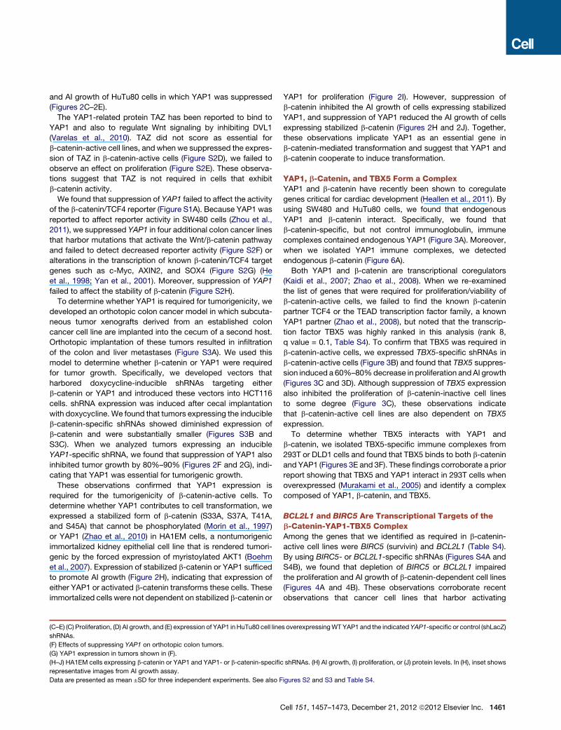
YAP1, b-Catenin, and TBX5 Form a ComplexYAP1 and b-catenin have recently been shown to coregulate
genes critical for cardiac development (Heallen et al., 2011). By
using SW480 and HuTu80 cells, we found that endogenous
YAP1 and b-catenin interact. Specifically, we found that
b-catenin-specific, but not control immunoglobulin, immune
complexes contained endogenous YAP1 (Figure 3A). Moreover,
when we isolated YAP1 immune complexes, we detected
endogenous b-catenin (Figure 6A).
Both YAP1 and b-catenin are transcriptional coregulators
(Kaidi et al., 2007; Zhao et al., 2008). When we re-examined
the list of genes that were required for proliferation/viability of
b-catenin-active cells, we failed to find the known b-catenin
partner TCF4 or the TEAD transcription factor family, a known
YAP1 partner (Zhao et al., 2008), but noted that the transcrip-
tion factor TBX5 was highly ranked in this analysis (rank 8,
q value = 0.1, Table S4). To confirm that TBX5 was required in
b-catenin-active cells, we expressed TBX5-specific shRNAs in
b-catenin-active cells (Figure 3B) and found that TBX5 suppres-
sion induced a 60%–80%decrease in proliferation and AI growth
(Figures 3C and 3D). Although suppression of TBX5 expression
also inhibited the proliferation of b-catenin-inactive cell lines
to some degree (Figure 3C), these observations indicate
that b-catenin-active cell lines are also dependent on TBX5
expression.
To determine whether TBX5 interacts with YAP1 and
b-catenin, we isolated TBX5-specific immune complexes from
293T or DLD1 cells and found that TBX5 binds to both b-catenin
and YAP1 (Figures 3E and 3F). These findings corroborate a prior
report showing that TBX5 and YAP1 interact in 293T cells when
overexpressed (Murakami et al., 2005) and identify a complex
composed of YAP1, b-catenin, and TBX5.
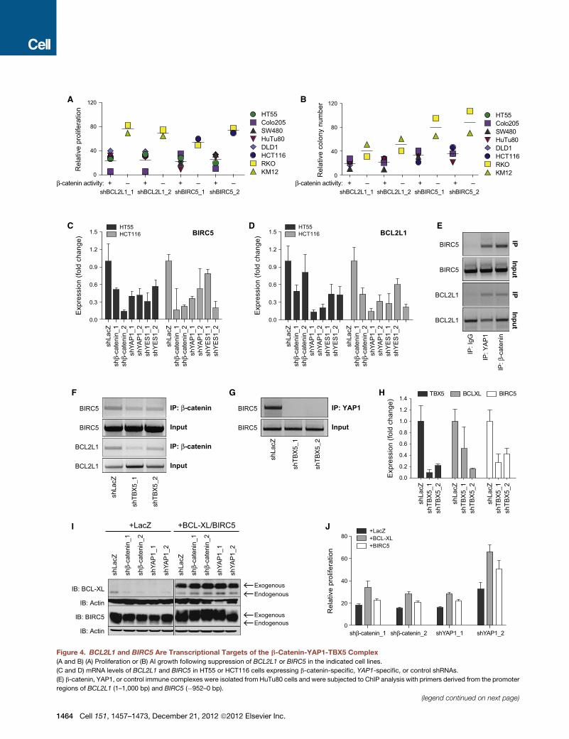
BCL2L1 and BIRC5 Are Transcriptional Targets of theb-Catenin-YAP1-TBX5 ComplexAmong the genes that we identified as required in b-catenin-
active cell lines were BIRC5 (survivin) and BCL2L1 (Table S4).
By using BIRC5- or BCL2L1-specific shRNAs (Figures S4A and
S4B), we found that depletion of BIRC5 or BCL2L1 impaired
the proliferation and AI growth of b-catenin-dependent cell lines
(Figures 4A and 4B). These observations corroborate recent
observations that cancer cell lines that harbor activating
s overexpressingWTYAP1 and the indicated YAP1-specific or control (shLacZ)
shRNAs. (H) AI growth, (I) proliferation, or (J) protein levels. In (H), inset shows
igures S2 and S3 and Table S4.
ell 151, 1457–1473, December 21, 2012 ª2012 Elsevier Inc. 1461
A B
E F
IB: β-catenin
IB: YAP1
IP: F
LAG
IP: I
gG
Inpu
t
DLD1
FLA
G-T
BX
5 +
HA
-β-c
aten
in
FLA
G-T
BX
5
HA
-β-c
aten
in
FLA
G-T
BX
5 +
HA
-YA
P1
FLA
G-T
BX
5
HA
-YA
P1
IP: FLAG
Input
IB: HA
IB: HA
293T
C D
+ – + –0
40
60
80
100
Rel
ativ
e pr
olife
ratio
n
HT55Colo205SW480HuTu80DLD1HCT116RKOKM12
β-catenin activity:
shTBX5_1 shTBX5_2
0
40
80
100
120
Rel
ativ
e co
lony
num
ber
HT55Colo205SW480HuTu80DLD1HCT116RKOKM12
+ – + –β-catenin activity:
shTBX5_1 shTBX5_2
20
60
20
1 2 3 1 2 3 1 2 3 1 2 3 1 2 3 1 2 3 1 2 3 1 2 30.0
0.3
0.6
0.9
1.2
1.5
Exp
ress
ion
(fold
cha
nge)
DLD1HCT116KM12RKOColo205HT55HuTu80SW480
1: shLacZ2: shTBX5_13: shTBX5_2
SW480
IB: YAP1
IB: β-catenin
β
IP: I
gG
Inpu
t 4%
IP:
-cat
enin
Figure 3. YAP1, b-Catenin, and TBX5 Interact
(A) b-catenin or control IgG immune complexes were isolated from SW480 lysates, and the indicated proteins were analyzed by immunoblotting.
(B) TBX5mRNA levels measured by quantitative PCR in cells expressing TBX5-specific or control (shLacZ) shRNAs. Data are presented as mean ±SD for three
independent experiments.
(C and D) (C) Proliferation or (D) AI growth of the indicated cells following suppression of TBX5.
(E) The indicated expression vectors were introduced into 293T cells, and FLAG immune complexes were isolated and analyzed by immunoblotting with an
anti-HA antibody.
(F) FLAG-immune complexes were isolated from DLD1 cells stably expressing a FLAG-epitope-tagged TBX5 protein and analyzed by immunoblotting with the
indicated antibodies.
mutations in b-catenin are particularly sensitive to navitoclax,
an inhibitor of BCL2 family members, including BCL-XL
(S. Schreiber, personal communication).
To test whether BIRC5 and BCL2L1 are transcriptional
targets of the b-catenin-YAP1-TBX5 complex, we examined
1462 Cell 151, 1457–1473, December 21, 2012 ª2012 Elsevier Inc.
the messenger RNA (mRNA) levels of BIRC5 and BCL2L1 in
cell lines (HT55 and HCT116) in which we had suppressed
either YAP1 or b-catenin. We found that the expression of
both BIRC5 and BCL2L1 was dependent on the presence of
b-catenin and YAP1 (Figures 4C and 4D), suggesting that the
b-catenin-YAP1-TBX5 complex is involved in the transcriptional
regulation of these genes.
To determine whether b-catenin and YAP1 directly regulate
BCL2L1 and BIRC5 expression, we performed a chromatin
immunoprecipitation (ChIP) assay focused on sites in the
BCL2L1 and BIRC5 promoters identified by b-catenin-specific
ChIP sequencing (ChIP-seq) and found that both b-catenin and
YAP1 were bound to these promoters (Figure 4E). Furthermore,
suppression of TBX5 expression in HuTu80 cells abrogated the
binding of b-catenin (Figure 4F) or YAP1 (Figure 4G) to these
promoters. Similar to what we found when we suppressed
YAP1, YES1, or b-catenin (Figures 4C and 4D), suppression of
TBX5 expression resulted in decreased expression of BIRC5
and BCL2L1 (Figure 4H).
To investigate whether BCL2L1 and BIRC5 contribute to the
proliferation arrest that is observed following suppression of
either b-catenin or YAP1 in b-catenin-active cancer cell lines,
we stably expressed the antiapoptotic isoform of BCL2L1
(BCL-XL) or BIRC5 in HuTu80 (b-catenin active). Following over-
expression of these genes, we expressed YAP1- or b-catenin-
specific shRNAs. Ectopic expression of BCL-XL or BIRC5
rendered the levels of these proteins independent of b-catenin
or YAP1 (Figure 4I) and partially restored the proliferation of
cell lines in which we suppressed either b-catenin or YAP1 (Fig-
ure 4J), suggesting that these genes are targets of the b-catenin-
YAP-TBX5 complex.
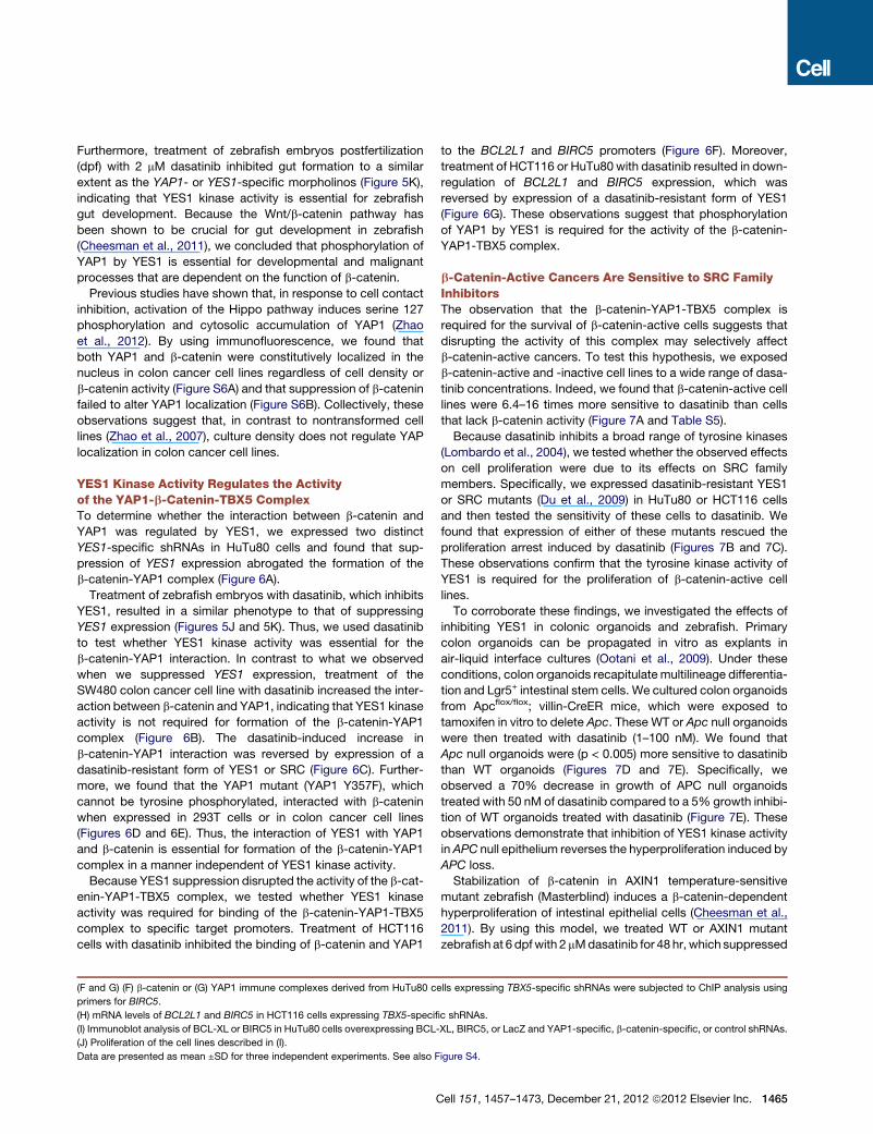
YES1 Kinase Activity Is Essential for the TransformingProperties of b-Catenin-Dependent CancersYAP1 was originally identified as a YES1-associated protein
(Sudol et al., 1995). We found that the SRC family tyrosine kinase
YES1 was essential for the growth of b-catenin-active cell lines
(rank 30, q value = 0.24, Table S4). As we observed for YAP1,
suppression of YES1 inhibited the proliferation, AI growth, and
tumor formation of b-catenin-active cell lines (Figures 5A–5D
and S5A). Furthermore, when we suppressed YES1 expression,
we also found reduced levels of BIRC5 and BCL2L1 (Figures 4C
and 4D). We confirmed that YES1-specific shRNAs did not alter
the expression of the closely related kinase SRC (Figures S5A
and S5B). These observations confirmed that YES1 expression
was required in b-catenin-active cell lines.
YAP1 binds to YES1 and is phosphorylated by SRC family
kinases in embryonic stem cells (Tamm et al., 2011). We
confirmed that YES1 and YAP1 interact in the b-catenin-active
colon cancer cell line SW480 (Figure 5E). Previous studies
have shown that YAP1 is able to bind to other SRC family
members such as SRC in HeLa cells (Zaidi et al., 2004). However,
in colon cancer cell lines, we failed to detect an interaction
between YAP1 and SRC or FYN (Figure 5E).
To determine whether YES1 or SRC phosphorylates YAP1, we
expressed YAP1 in 293T cells and assessed YAP1 tyrosine
phosphorylation when coexpressed with YES1 or SRC (Fig-
ure 5F). We detected phosphorylated YAP1 only when YAP1
was coexpressed with SRC or with activated mutant version of
YES1 (Y537F). We failed to detect phosphorylation of YAP1
when coexpressed with wild-type (WT) YES1, indicating that
YAP1 phosphorylation requires the active form of YES1 (Fig-
ure 5F). Although both YES1 and SRC phosphorylated YAP1,
C
suppression of SRC failed to inhibit the proliferation and AI
growth of b-catenin-active cell lines (Figures S5C–S5E). Thus,
we concluded that both YES1 and SRC are able to phosphory-
late YAP1, but only YES1 is essential for the survival of
b-catenin-active cell lines.
In 293T cells, we did not detect phosphorylated YAP1 when
expressed alone (Figure 5F). In contrast, we readily detected
YAP1 tyrosine phosphorylation in HuTu80 or SW480 cells
expressing WT YAP1 (Figure 5H). Furthermore, treatment of
colon cancer cells expressing WT YAP1 with the tyrosine kinase
inhibitor dasatinib (Lombardo et al., 2004) inhibited the tyrosine
phosphorylation of YAP1 (Figure 5I). These results confirm
reported observations that demonstrated that YES1 is activated
in colon cancer cell lines and tumors (Pena et al., 1995).
Prior work has demonstrated that SRC family members phos-
phorylate tyrosine residues contained with the sequence motif
YXXP (Levy et al., 2008). YAP1 harbors one tyrosine residue
with this motif (tyrosine 357). Under conditions in which active
YES1 phosphorylated WT YAP1 in 293T cells, we failed to detect
tyrosine phosphorylation of the YAP1 Y357F mutant in either
293T or colon cancer cell lines (Figures 5G and 5H). These
observations confirm that YES1 phosphorylates YAP1 at
tyrosine 357.
We then tested whether phosphorylation of YAP tyrosine 357
was essential for YAP1 function. Specifically, when we ex-
pressed WT or mutant Y357F YAP1 in HuTu80 cells expressing
YAP1-specific shRNAs, we found that WT, but not Y357F,
YAP1 was able to rescue the antiproliferative and AI growth
effects of the YAP1-specific shRNA (compare Figures 2C and
2D to S5F and S5G) when expressed at equivalent levels (Figures
2E and S5H). Together, these observations confirm that YES1 is
essential for the tumorigenicity of b-catenin-dependent cell lines
and suggest that YES1-mediated phosphorylation of tyrosine
357 regulates YAP1 activity.
To assess the relationship between YES1 andYAP1 in vivo, we
examined the effect of suppressing these genes on zebrafish
development. Microinjection of zebrafish embryos with a high
concentration (200 mM) of YAP1- or YES1-specific morpholinos
resulted in severe developmental phenotypes (Figure S5I).
Specifically, the YAP1 morphants developed craniofacial
abnormalities and cardiac edema, whereas the YES1morphants
exhibited craniofacial abnormalities associated with pharyngeal
defects (Figure S5I). These phenotypes resemble defects
observed when high concentrations of b-catenin-specific mor-
pholinos were injected (Zhang et al., 2012) and confirm previous
reports showing that YAP1 and YES1 are essential for early
embryonic development in zebrafish (Jiang et al., 2009; Tsai
et al., 2005).
Microinjection of YAP1 or YES1 morpholinos at lower doses
(50 mM) avoided global toxicity but impaired gut development
(Figure S5I). Intestinal fatty-acid-binding protein (IFABP and
FAPB2) is expressed in intestinal epithelial cells, where it plays
a key role in gutmetabolism and is used as amarker of gut devel-
opment (Goessling et al., 2008). Morpholino-mediated suppres-
sion of YAP1 or YES1 expression dramatically inhibited gut
formation as determined by both fluorescence microscopy of
Tg(fabp2:RFP)as200 gut reporter embryos and by examination
of IFABP expression by in situ hybridization (Figure 5J).
ell 151, 1457–1473, December 21, 2012 ª2012 Elsevier Inc. 1463
E
F
C D
A B
+ –0
40
80
120
Rel
ativ
e pr
olife
ratio
n HT55
Colo205SW480HuTu80DLD1HCT116RKOKM12
β-catenin activity:shBCL2L1_1 shBCL2L1_2
0
40
80
120
Rel
ativ
e co
lony
num
ber
HT55Colo205SW480HuTu80DLD1HCT116RKOKM12
IP: β
-cat
enin
IP: Y
AP
1
IP: I
gG
BIRC5
BIRC5
BCL2L1
BCL2L1
InputInput
IPIP
0.0
0.3
0.6
0.9
1.2
1.5
Exp
ress
ion
(fold
cha
nge)
BIRC5
0.0
0.3
0.6
0.9
1.2
1.5
Exp
ress
ion
(fold
cha
nge)
HT55HCT116
0.0
0.2
0.4
0.6
0.8
1.0
1.2
1.4E
xpre
ssio
n (fo
ld c
hang
e)TBX5 BCLXL BIRC5
shLa
cZ
shT
BX
5_1
shT
BX
5_2
BCL2L1
BIRC5
BIRC5
BCL2L1
Input
Input shT
BX
5_1
shT
BX
5_2
BIRC5
BIRC5
shLa
cZ
IP: YAP1
Input
IB: Actin
IB: BCL-XL
+LacZ +BCL-XL/BIRC5
IB: BIRC5
IB: Actin0
20
40
60
80
Rel
ativ
e pr
olife
ratio
n
+ – + – + –shBIRC5_1 shBIRC5_2
+ –β-catenin activity:shBCL2L1_1 shBCL2L1_2
+ – + – + –shBIRC5_1 shBIRC5_2
HT55HCT116
shβ-
cate
nin_
2sh
YAP
1_1
shLa
cZsh
β-ca
teni
n_1
shYA
P1_
2sh
YE
S1_
1sh
YE
S1_
2
shβ-
cate
nin_
2sh
YAP
1_1
shLa
cZsh
β-ca
teni
n_1
shYA
P1_
2sh
YE
S1_
1sh
YE
S1_
2
shβ-
cate
nin_
2sh
YAP
1_1
shLa
cZsh
β-ca
teni
n_1
shYA
P1_
2sh
YE
S1_
1sh
YE
S1_
2
shβ-
cate
nin_
2sh
YAP
1_1
shLa
cZsh
β-ca
teni
n_1
shYA
P1_
2sh
YE
S1_
1sh
YE
S1_
2HG
IP: β-catenin
IP: β-catenin
shLa
cZsh
TBX
5_1
shTB
X5_
2
shLa
cZsh
TBX
5_1
shTB
X5_
2
shLa
cZsh
TBX
5_1
shTB
X5_
2
I J
shβ-
cate
nin_
2
shYA
P1_
1
shLa
cZ
shβ-
cate
nin_
1
shYA
P1_
2
shβ-
cate
nin_
2
shYA
P1_
1
shLa
cZ
shβ-
cate
nin_
1
shYA
P1_
2
EndogenousExogenous
EndogenousExogenous
shβ-catenin_2 shYAP1_1shβ-catenin_1 shYAP1_2
+LacZ+BCL-XL+BIRC5
BCL2L1
Figure 4. BCL2L1 and BIRC5 Are Transcriptional Targets of the b-Catenin-YAP1-TBX5 Complex
(A and B) (A) Proliferation or (B) AI growth following suppression of BCL2L1 or BIRC5 in the indicated cell lines.
(C and D) mRNA levels of BCL2L1 and BIRC5 in HT55 or HCT116 cells expressing b-catenin-specific, YAP1-specific, or control shRNAs.
(E) b-catenin, YAP1, or control immune complexes were isolated from HuTu80 cells and were subjected to ChIP analysis with primers derived from the promoter
regions of BCL2L1 (1–1,000 bp) and BIRC5 (�952–0 bp).
(legend continued on next page)
1464 Cell 151, 1457–1473, December 21, 2012 ª2012 Elsevier Inc.
Furthermore, treatment of zebrafish embryos postfertilization
(dpf) with 2 mM dasatinib inhibited gut formation to a similar
extent as the YAP1- or YES1-specific morpholinos (Figure 5K),
indicating that YES1 kinase activity is essential for zebrafish
gut development. Because the Wnt/b-catenin pathway has
been shown to be crucial for gut development in zebrafish
(Cheesman et al., 2011), we concluded that phosphorylation of
YAP1 by YES1 is essential for developmental and malignant
processes that are dependent on the function of b-catenin.
Previous studies have shown that, in response to cell contact
inhibition, activation of the Hippo pathway induces serine 127
phosphorylation and cytosolic accumulation of YAP1 (Zhao
et al., 2012). By using immunofluorescence, we found that
both YAP1 and b-catenin were constitutively localized in the
nucleus in colon cancer cell lines regardless of cell density or
b-catenin activity (Figure S6A) and that suppression of b-catenin
failed to alter YAP1 localization (Figure S6B). Collectively, these
observations suggest that, in contrast to nontransformed cell
lines (Zhao et al., 2007), culture density does not regulate YAP
localization in colon cancer cell lines.
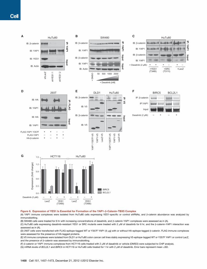
YES1 Kinase Activity Regulates the Activityof the YAP1-b-Catenin-TBX5 ComplexTo determine whether the interaction between b-catenin and
YAP1 was regulated by YES1, we expressed two distinct
YES1-specific shRNAs in HuTu80 cells and found that sup-
pression of YES1 expression abrogated the formation of the
b-catenin-YAP1 complex (Figure 6A).
Treatment of zebrafish embryos with dasatinib, which inhibits
YES1, resulted in a similar phenotype to that of suppressing
YES1 expression (Figures 5J and 5K). Thus, we used dasatinib
to test whether YES1 kinase activity was essential for the
b-catenin-YAP1 interaction. In contrast to what we observed
when we suppressed YES1 expression, treatment of the
SW480 colon cancer cell line with dasatinib increased the inter-
action between b-catenin and YAP1, indicating that YES1 kinase
activity is not required for formation of the b-catenin-YAP1
complex (Figure 6B). The dasatinib-induced increase in
b-catenin-YAP1 interaction was reversed by expression of a
dasatinib-resistant form of YES1 or SRC (Figure 6C). Further-
more, we found that the YAP1 mutant (YAP1 Y357F), which
cannot be tyrosine phosphorylated, interacted with b-catenin
when expressed in 293T cells or in colon cancer cell lines
(Figures 6D and 6E). Thus, the interaction of YES1 with YAP1
and b-catenin is essential for formation of the b-catenin-YAP1
complex in a manner independent of YES1 kinase activity.
Because YES1 suppression disrupted the activity of the b-cat-
enin-YAP1-TBX5 complex, we tested whether YES1 kinase
activity was required for binding of the b-catenin-YAP1-TBX5
complex to specific target promoters. Treatment of HCT116
cells with dasatinib inhibited the binding of b-catenin and YAP1
(F and G) (F) b-catenin or (G) YAP1 immune complexes derived from HuTu80 c
primers for BIRC5.
(H) mRNA levels of BCL2L1 and BIRC5 in HCT116 cells expressing TBX5-specifi
(I) Immunoblot analysis of BCL-XL or BIRC5 in HuTu80 cells overexpressing BCL-
(J) Proliferation of the cell lines described in (I).
Data are presented as mean ±SD for three independent experiments. See also F
C
to the BCL2L1 and BIRC5 promoters (Figure 6F). Moreover,
treatment of HCT116 or HuTu80 with dasatinib resulted in down-
regulation of BCL2L1 and BIRC5 expression, which was
reversed by expression of a dasatinib-resistant form of YES1
(Figure 6G). These observations suggest that phosphorylation
of YAP1 by YES1 is required for the activity of the b-catenin-
YAP1-TBX5 complex.
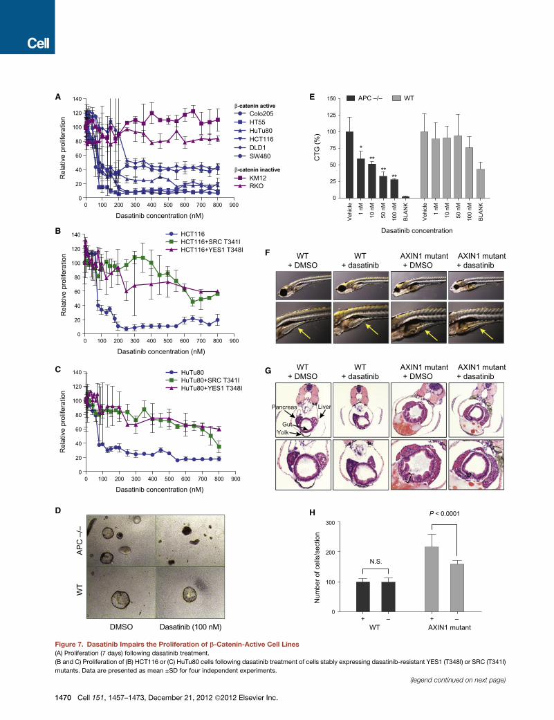
b-Catenin-Active Cancers Are Sensitive to SRC FamilyInhibitorsThe observation that the b-catenin-YAP1-TBX5 complex is
required for the survival of b-catenin-active cells suggests that
disrupting the activity of this complex may selectively affect
b-catenin-active cancers. To test this hypothesis, we exposed
b-catenin-active and -inactive cell lines to a wide range of dasa-
tinib concentrations. Indeed, we found that b-catenin-active cell
lines were 6.4–16 times more sensitive to dasatinib than cells
that lack b-catenin activity (Figure 7A and Table S5).
Because dasatinib inhibits a broad range of tyrosine kinases
(Lombardo et al., 2004), we tested whether the observed effects
on cell proliferation were due to its effects on SRC family
members. Specifically, we expressed dasatinib-resistant YES1
or SRC mutants (Du et al., 2009) in HuTu80 or HCT116 cells
and then tested the sensitivity of these cells to dasatinib. We
found that expression of either of these mutants rescued the
proliferation arrest induced by dasatinib (Figures 7B and 7C).
These observations confirm that the tyrosine kinase activity of
YES1 is required for the proliferation of b-catenin-active cell
lines.
To corroborate these findings, we investigated the effects of
inhibiting YES1 in colonic organoids and zebrafish. Primary
colon organoids can be propagated in vitro as explants in
air-liquid interface cultures (Ootani et al., 2009). Under these
conditions, colon organoids recapitulate multilineage differentia-
tion and Lgr5+ intestinal stem cells. We cultured colon organoids
from Apcflox/flox; villin-CreER mice, which were exposed to
tamoxifen in vitro to delete Apc. These WT or Apc null organoids
were then treated with dasatinib (1–100 nM). We found that
Apc null organoids were (p < 0.005) more sensitive to dasatinib
than WT organoids (Figures 7D and 7E). Specifically, we
observed a 70% decrease in growth of APC null organoids
treated with 50 nM of dasatinib compared to a 5% growth inhibi-
tion of WT organoids treated with dasatinib (Figure 7E). These
observations demonstrate that inhibition of YES1 kinase activity
in APC null epithelium reverses the hyperproliferation induced by
APC loss.
Stabilization of b-catenin in AXIN1 temperature-sensitive
mutant zebrafish (Masterblind) induces a b-catenin-dependent
hyperproliferation of intestinal epithelial cells (Cheesman et al.,
2011). By using this model, we treated WT or AXIN1 mutant
zebrafish at 6 dpfwith 2 mMdasatinib for 48 hr, which suppressed
ells expressing TBX5-specific shRNAs were subjected to ChIP analysis using
c shRNAs.
XL, BIRC5, or LacZ and YAP1-specific, b-catenin-specific, or control shRNAs.
igure S4.
ell 151, 1457–1473, December 21, 2012 ª2012 Elsevier Inc. 1465
E FC D
A B
+ –0
60
90
120
Rel
ativ
e pr
olife
ratio
n
HT55Colo205SW480HuTu80DLD1HCT116RKOKM12
β-catenin activity:shYES1_1 shYES1_2
Rel
ativ
e co
lony
num
ber
HT55Colo205SW480HuTu80DLD1HCT116RKOKM12
+ –
HG I
J
IB: YES1
IB: SRC
IP: I
gG
IP: Y
AP
1
Inpu
t
SW480
IB: FYN
No
Dox
+Dox
1
+Dox
2
+Dox
3
+Dox
4
YES1
Actin
+ V
5-La
cZ+
V5-
YA
P1
+ V
5-Y
AP
1 Y
357F
SW480
IP: V5IP: V5
InputInput
+ D
MS
O (0
.5 µ
M)
+ D
asat
iinib
(2 µ
M)
+ D
asat
iinib
IB: pY
IB: pY
IB: V5
IB: V5
SW480
HuTu80
IP: V5
293T
30
+ –0
60
90
120
β-catenin activity:shYES1_1 shYES1_2
+ –
30
No Dox +Dox 1 +Dox 2
+Dox 3 +Dox 4
V5-YAP1YES1 (WT)
Active YES1 (Y537F)SRC
IB: pY
IB: V5
IP: V5
+–
+ + +––
–+– –+
– – – +
IB: pY
IB: pY
IB: YAP1
IB: YAP1
IP: FLAG
InputInput
293T
YAP1 (WT)YAP1 (Y357F)
YES1 (WT)Active YES1 (Y537F)
+–
+ – +–+
––+ ––
– – + +
K
IB: pY
IB: pY
IB: YAP1
IB: YAP1
+ V
5-La
cZ+
V5-
YA
P1
+ V
5-Y
AP
1 Y
357F
HuTu80
Con
trol
YA
P1
YE
S1
Tg IFABP:dsRed WISH:IFABP
Mor
phol
ino
inje
cted
(50
µM)
Das
atin
ib(2
µM
)D
MS
O
WISH:IFABP
Figure 5. YES1 Is Essential for Tumorigenicity of b-Catenin-Active Cells
(A and B) (A) AI growth or (B) proliferation following suppression of YES1 expression.
(C) Effects of suppressing YES1 on orthotopic HCT116 colon tumors.
(D) YES1 expression in tumors shown in (C).
(E) YAP1 immune complexes were isolated from SW480 cells, and the indicated proteins were analyzed by immunoblotting.
(F) V5-epitope-tagged YAP1 and WT or activated (Y537F) YES1 or SRC expression constructs were introduced into 293T cells. V5 immune complexes were
analyzed by immunoblotting with the PY99 phosphotyrosine-specific antibody.
(legend continued on next page)
1466 Cell 151, 1457–1473, December 21, 2012 ª2012 Elsevier Inc.
intestinal hyperplasia in the AXIN1 mutants as assessed by
morphology (Figure 7F) or hematoxylin and eosin staining (H&E)
(Figure 7G). Furthermore, the number of epithelial cells was
significantly (p < 0.0001) decreased in AXIN1 mutant zebrafish
treated with dasatinib (Figure 7H). In contrast, we failed to
observe changes in the intestinal structure or cell number in
WT zebrafish treated with dasatinib, indicating that the effect of
dasatinib was specific to AXIN1 mutant animals (Figures 7G
and 7H). We concluded that inhibition of YES1 kinase activity
inhibits the b-catenin-dependent proliferation in cultured human
cancer cells, in colon epithelial organoids, and in a zebrafish
model of intestinal hyperplasia.
DISCUSSION
Identification of Codependent Genes in Cells thatExhibit Active b-Catenin SignalingBy using loss-of-function data derived from Project Achilles
(Cheung et al., 2011), we identified 50 genes whose expression
was preferentially required for the proliferation of b-catenin-
active cell lines. The use of a large number of cell lines allowed
us to perform permutation analyses to ensure that the genes
identified by this approach robustly distinguished the two groups
(FDR q < 0.25). Indeed, when we arbitrarily assigned cell lines to
two classes, we failed to identify genes that distinguished these
groups.
This approach allowed us to identify a set of proteins related to
the transcriptional regulator YAP1 that is essential for the prolif-
eration/survival of b-catenin-active cell lines. b-catenin forms
a complex with YAP1 and TBX5, which promotes colon cancer
cell survival and contributes to malignant transformation. These
observations reveal hitherto unidentified components of the
b-catenin pathway that play key roles in survival of b-catenin-
active cells.
Cancer Cell Lines that Exhibit b-Catenin ActivityRequire YAP1YAP1 is an effector of the Hippo pathway (Zhao et al., 2011), an
oncogene in hepatocellular cancers (Zender et al., 2006), and
a protein involved in mechanotransduction (Dupont et al.,
2011). In hepatocellular carcinomas that harbor 11q22 amplifica-
tions, YAP1 cooperates with a coamplified gene CIAP1 to
accelerate tumor formation (Zender et al., 2006). Genetically
engineered mice that express a stabilized YAP1 mutant
(S127A) develop colonic adenomas and liver hyperplasia
(Camargo et al., 2007). YAP1 has also been reported to be over-
expressed in many epithelial cancers (Steinhardt et al., 2008).
However, when we analyzed YAP1 expression in 807 cancer
(G) FLAG-epitope-tagged WT or mutated (Y357F) YAP1 were transfected int
complexes were isolated and analyzed by immunoblotting with a phosphotyrosi
(H) V5 immune complexes were isolated from HuTu80 or SW480 cells stably ex
complexes were analyzed by immunoblotting with a phosphotyrosine antibody.
(I) HuTu80 or SW480 cells stably expressing V5-epitope-tagged WT YAP1 wer
analyzed by immunoblotting with a phosphotyrosine antibody.
(J) Transgenic IFABP:RFP zebrafish were injected with 50 mM of YAP1- or YE
expression was assessed 3 dpf by using whole-mount in situ hybridization with
(K) Embryos were exposed to 2 mM of dasatinib at 2 dpf, and IFABP expression
See also Figures S5, S6, and S7.
C
cell lines (Barretina et al., 2012), we found that YAP1 was ex-
pressed in most epithelial cell lines (Figure S7), and we failed
to identify a correlation between YAP1 dependence and YAP1
copy number or expression. Based on the strong correlation
between YAP1 and b-catenin dependency, we conclude that
YAP1 induces transformation in b-catenin-active cancers
primarily through its interactions with b-catenin.
The Hippo pathway controls organ size by regulation of
YAP1. Specifically, upon cell contact inhibition, the Hippo
pathway is triggered, leading to a cascade of phosphorylation
events resulting in activation of the MST1/2 kinases. Activated
Mst1/2 phosphorylate LATS1/2, which, in turn, phosphorylate
YAP1 on serine 127, leading to inactivation of YAP1 by protea-
somal degradation and cytosolic retention of YAP1 (Zhao et al.,
2011). Inactivation of the Hippo pathway by tissue-specific
germline deletion of Mst1/2 induced hepatocellular cancers
(Zhou et al., 2009) or colonic hyperplasia and adenomas
(Zhou et al., 2011). However, recurrent deletions or loss of
heterozygosity involving these genes have not been identified
in human cancers. Here, we failed to find a correlation between
YAP1 nuclear localization or expression and YAP1 depen-
dency, corroborating prior work (Zhou et al., 2011). Because
YAP1 is regulated by Hippo-dependent and -independent
mechanisms (Dupont et al., 2011), further studies are neces-
sary to determine the role of Hippo signaling in colon cancer
pathogenesis.
b-catenin regulates YAP1 expression by binding to the YAP1
promoter (Konsavage et al., 2012). Specifically, both b-catenin
and TCF4 bind to sequences upstream of YAP1, and suppres-
sion of b-catenin expression resulted in decreased YAP1
mRNA levels. However, we found comparable levels of YAP1
across a large number of cell lines, including b-catenin-inactive
colon cancer cell lines (Figures S2A and S7), suggesting that
b-catenin is not the primary driver of YAP1 sensitivity in these
cell lines. However, it remains possible that the b-catenin-
TCF4 complex participates in a feedback loop that enhances
YAP1 expression.
The b-Catenin-YAP1-TBX5 ComplexTBX5, a member of the T-box family of transcription factors,
plays key roles in cardiac muscle development and limb identity
(Rodriguez-Esteban et al., 1999). Germline mutations in TBX5
occur in the Holt-Oram syndrome (Mori and Bruneau, 2004).
TBX5 has also been shown to form a complex with TAZ and
YAP1 that induces transcription of atrial natriuretic factor (Mura-
kami et al., 2005). Here, we found that TBX5 forms a complex
with b-catenin and YAP1 that is found at the BCL2L1 and
BIRC5 promoters. These observations extend prior work that
o 293T cells together with activated (Y537F) YES1. FLAG-epitope immune
ne antibody.
pressing WT or Y357F V5-epitope-tagged YAP1 or control V5-LacZ. Immune
e treated for 6 hr with 0.5 or 2 mM of dasatinib. V5 immune complexes were
S1-specific morpholinos. Red fluorescence was assessed 4 dpf, or IFABP
an IFABP-specific probe.
was assessed after 24 hr by using whole-mount in situ hybridization.
ell 151, 1457–1473, December 21, 2012 ª2012 Elsevier Inc. 1467
E F
C
D
A B
0.0
0.3
0.6
0.9
1.2
1.5
Exp
ress
ion
(fold
cha
nge)
HCT116 HuTu80
LacZ LacZYES1(T348I)
shLa
cZ
shY
ES
1 1
shY
ES
1 2
HuTu80
IP: YAP1
Input
IB: YAP1
IB: β-catenin
IB: YES1
IB: Actin
LacZ
-V5
LacZ
-V5
YA
P1-
V5
YA
P1
Y35
7F-V
5
YA
P1
Y35
7F-V
5
YA
P1-
V5
IB: V5
IB: V5
IP: V5Input
HuTu80DLD1
IB: YAP1
IB: YAP1
IB: Actin
IP: YAP1
Input
+ D
MS
O
+ Dasatinib [nM]
SW480
50 500 1000 2000
Input
IB: YAP1
IB: YAP1
IP: YAP1
Input
HuTu80
IP:YAP1
Input
BIRC5 BCL2L1
IB: β-catenin
IB: β-catenin
IB: β-catenin
IB: β-catenin
IP: β-cateninIB: β-catenin
IB: β-catenin
IB: HA
IB: HA
IB: YAP1
IB: YAP1
FLAG-YAP1 Y357FFLAG-YAP1
HA-β-cateninIP: FLA
GInput
293T
+–
+ – –++
+–– –+
Dasatinib (2 µM): – +–+
BIRC5BCL2L1
Dasatinib (2 µM): – +–+ – +–+
YES1(T348I)
G
+SRC(T311I)
+LacZ+YES1(T348I)
+ Dasatinib (2 µM): + –++ + –
Figure 6. Expression of YES1 Is Essential for Formation of the YAP1-b-Catenin-TBX5 Complex
(A) YAP1 immune complexes were isolated from HuTu80 cells expressing YES1-specific or control shRNAs, and b-catenin abundance was analyzed by
immunoblotting.
(B) SW480 cells were treated for 6 hr with increasing concentrations of dasatinib, and b-catenin-YAP1 complexes were assessed as in (A).
(C) HuTu80 cells expressing dasatinib-resistant YES1 or SRC mutants were treated with 2 mM of dasatinib for 6 hr, and the b-catenin-YAP1 interaction was
assessed as in (A).
(D) 293T cells were transfected with FLAG-epitope-tagged WT or Y357F YAP1 (5 mg) with or without HA-epitope-tagged b-catenin. FLAG immune complexes
were assessed for the presence of HA-tagged proteins.
(E) V5 immune complexes were isolated from DLD1 or HuTu80 colon cancer cell lines stably expressing V5-epitope-tagged WT or Y357F YAP1 or control LacZ,
and the presence of b-catenin was assessed by immunoblotting.
(F) b-catenin or YAP1 immune complexes from HCT116 cells treated with 2 mM of dasatinib or vehicle (DMSO) were subjected to ChIP analysis.
(G) mRNA levels of BCL2L1 and BIRC5 in HCT116 or HuTu80 cells treated for 1 hr with 2 mM of dasatinib. Error bars represent mean ±SD.
1468 Cell 151, 1457–1473, December 21, 2012 ª2012 Elsevier Inc.
showed that TBX5 binds the BIRC5 and BCL2L1 promoters
when overexpressed in 293T cells (He et al., 2011).
YAP1 interacts with the TEAD family of transcriptional factors
to regulate both developmental and cancer-associated pheno-
types (Zhao et al., 2008). We did not find that TEAD family
members were differentially required for proliferation of
b-catenin-active cells. Although the YAP1-TEAD complex
regulates other cancer phenotypes (Lamar et al., 2012), our
observations implicate TBX5 as a key transcription factor target
of the b-catenin-YAP1 complex. Moreover, because b-catenin
interacts with different transcription factors such as the AR
(Mulholland et al., 2002) and HIF-1 (Kaidi et al., 2007), these
observations suggest that both YAP1 and b-catenin regulate
several transcriptional programs through interactions with
distinct transcription factors.
TCF4 Dependency in b-Catenin-Dependent CancersAlthough the b-catenin-TCF4 complex plays an important role in
colon adenoma initiation, several lines of evidence suggest that
b-catenin may also contribute to cancer in a TCF4-independent
manner. Specifically, although expression of a dominantly inter-
fering allele of TCF4 inhibits the b-catenin/TCF4 reporter activity
in b-catenin-dependent colon cancer cell lines (Korinek et al.,
1997; Figure 1H), suppression of TCF4 induces a substantial
increase in b-catenin/TCF4 reporter activity and only partially
affects the proliferation and AI growth of b-catenin-dependent
cell lines (Figures 1I and 1J) (Tang et al., 2008). Moreover, the
HT29 and LS411N colon cancer cell lines harbor APCmutations
and depend on b-catenin expression for survival yet failed to
exhibit detectable b-catenin/TCF4 reporter activity and were
dependent on YAP1 for survival.
Furthermore, compound genetically engineered mice that
express the APCmin allele and lack one TCF4 allele develop
aggressive, metastatic colon cancers (Angus-Hill et al., 2011).
Whole-genome sequencing of colon cancer genomes has re-
vealed recurrent TCF4-VTI1A translocations that create a
dominantly interfering allele of TCF4 (Bass et al., 2011) as well
as inactivating mutations and copy number loss involving TCF4
(Cancer Genome Atlas Network, 2012). In aggregate, these
observations suggest that TCF4 may contribute initially to
adenoma formation but then is mutated or lost to foster malig-
nant transformation. We cannot exclude the possibility that
a residual amount of TCF4 remains in the experiments presented
herein, and these observations do not exclude the possibility that
other TCF or LEF proteins may be essential for b-catenin-medi-
ated transformation.
YES1 Regulates the Formation and Activity of theb-Catenin-YAP1-TBX5 ComplexWe found that the SRC family member YES1 is also specifically
essential for the proliferation and transformed phenotype of
b-catenin-active cells both because YES1 is necessary for the
formation of the b-catenin-YAP1-TBX5 complex and because
phosphorylation of YAP1 on Y357 by YES1 is required for the
localization and activity of this complex.
Several tyrosine kinases, including YES1 (Tamm et al., 2011),
SRC (Zaidi et al., 2004), and ABL (Levy et al., 2008), phosphory-
late YAP1. We found that suppression of SRC or ABL failed to
C
affect the proliferation/survival of b-catenin-active cells, demon-
strating that, in this context, YES1 plays a dominant role in regu-
lating the b-catenin-YAP1-TBX5 complex. Moreover, treatment
of b-catenin-active cells with dasatinib inhibited the activity of
the b-catenin-YAP1-TBX5 complex and the survival of
b-catenin-active cancer cell lines in a manner that is rescued
by expression of dasatinib-resistant YES1 or SRC mutants.
Extending these findings, we found that dasatinib induces an
antiproliferative effect in both murine and fish experimental
models of APC loss/WNT pathway activation. These findings
corroborate a recent report that showed that treatment of
APCmin mice with dasatinib decreases intestinal adenomas
(Nautiyal et al., 2011). Together, these observations support
a role for YES1 phosphorylation in b-catenin-driven cancers.
ConclusionsWehave identified a b-catenin-YAP1-TBX5 complex required for
the survival and transformation of b-catenin-active cancer cell
lines. YES1 regulates the formation of this complex and localiza-
tion to specific promoters, which is dispensable for b-catenin/
TCF4 activity, but which regulates transcription of prosurvival
genes. These observations demonstrate that deregulated
b-catenin stability and function drive malignant transformation
through interactions with at least two distinct transcrip-
tional complexes (b-catenin-YAP1-TBX5 and b-catenin-TCF4).
Although further work is necessary to decipher the specific roles
of each of these complexes in tumorigenesis, these observations
provide a framework to explain recent observations that loss of
TCF4 activity is associated with tumor progression (Angus-Hill
et al., 2011).
Although no specific inhibitors of YES1 exist, the sensitivity of
b-catenin-active cancer cell lines and animal models to dasatinib
suggests that YES1 is an attractive target in b-catenin-driven
cancers. Moreover, we found that suppression of BCL2L1 and
BIRC5 also inhibited the proliferation/survival of b-catenin-active
cell lines. Because small-molecule inhibitors of BCL2L1 and
BIRC5 are currently under investigation, these molecules may
also prove useful for targeting of b-catenin-active cancers.
EXPERIMENTAL PROCEDURES
In Vivo Orthotopic Tumor Model
4 3 106 HCT116 cells were injected into the flanks of immunodeficient mice
(NCr Nude, Taconic). Tumors were extracted, cut into 1 mm3 cubes, and
implanted into a pouch created in the cecum of a second mouse. For experi-
ments with inducible shRNAs, the mice were fed a doxycycline diet 2 days
after cecal implantation. Tumors were examined 3 weeks postimplantation.
Three-Dimensional Primary Intestinal Organoid Culture
Colons from Apcflox/flox ; villin-CreER mice were dissected lengthwise and
washed in cold PBS. 0.5–1 cm segment per dish was minced extensively
on ice and embedded in a 3D collagen gel by using a double-dish culture
system (Ootani et al., 2009). Tamoxifen (Sigma, 2 mM in ethanol) or vehicle
(ethanol) was applied on the day of initial plating for 7 days to generate
APC null or WT organoids. Organoids were recovered from collagen gel by
collagenase IV incubation followed by 0.05% trypsin/EDTA incubation to
dissociate organoids into single cells. 5,000 cells per well were seeded
into 96 well transwell plates (Fisher Scientific). Organoids were treated
with the indicated concentration of dasatinib (in DMSO) for 7 days and
were quantified by using CellTiter 96 AQueous One Solution Cell Proliferation
Assay (Promega).
ell 151, 1457–1473, December 21, 2012 ª2012 Elsevier Inc. 1469
E
F
C
D
A
B
H
G
Rel
ativ
e pr
olife
ratio
n
0 100 200 300 400 500 600 700 800 9000
20
40
60
80
100
120
140 HCT116HCT116+SRC T341IHCT116+YES1 T348I
Rel
ativ
e pr
olife
ratio
n
0 100 200 300 400 500 600 700 800 9000
20
40
60
80
100
120
140 HuTu80HuTu80+SRC T341IHuTu80+YES1 T348I
Rel
ativ
e pr
olife
ratio
n
0 100 200 300 400 500 600 700 800 9000
20
40
60
80
100
120
140
Colo205HT55HuTu80HCT116DLD1SW480
KM12RKO
WT+ DMSO
AXIN1 mutant+ DMSO
AXIN1 mutant+ dasatinib
WT+ dasatinib
+ – + –0
100
200
300
Num
ber o
f cel
ls/s
ectio
n
WT AXIN1 mutant
N.S.
P < 0.0001
0
25
50
75
100
125
150
Dasatinib concentration
CTG
(%)
***
****
APC –/– WT
Vehi
cle
1 nM
10 n
M
50 n
M
100
nM
BLAN
K
Vehi
cle
1 nM
10 n
M
50 n
M
100
nM
BLAN
K
AP
C –
/–W
T
DMSO Dasatinib (100 nM)
Pancreas Liver
GutYolk
WT+ DMSO
AXIN1 mutant+ DMSO
AXIN1 mutant+ dasatinib
WT+ dasatinib
Dasatinib concentration (nM)
Dasatinib concentration (nM)
β-catenin active
β-catenin inactive
Dasatinib concentration (nM)
Figure 7. Dasatinib Impairs the Proliferation of b-Catenin-Active Cell Lines(A) Proliferation (7 days) following dasatinib treatment.
(B and C) Proliferation of (B) HCT116 or (C) HuTu80 cells following dasatinib treatment of cells stably expressing dasatinib-resistant YES1 (T348I) or SRC (T341I)
mutants. Data are presented as mean ±SD for four independent experiments.
(legend continued on next page)
1470 Cell 151, 1457–1473, December 21, 2012 ª2012 Elsevier Inc.
Zebrafish Experiments
Zebrafish were maintained according to institutional animal care and use
committee (IACUC-BIDMC) protocols. Validated morpholinos (MO) (Gene-
Tools, PhiloMath, OR) designed against the ATG site of YES1 (50-CCTCTTTACTCTTGACACAGCCCAT-30 ) (Jopling and Hertog, 2007) or YAP1
(50-AGCAACATTAACAACTCACTTTAGG-30 ) (Skouloudaki et al., 2009) were
injected into WT or gut reporter (Tg(fabp2:RFP)as200) zebrafish at the one-
cell stage. At 4 dpf, the gut morphology of intestinal reporter embryos was
imaged by fluorescent microscopy (Discovery, Carl Zeiss). Whole-mount
in situ hybridization experiments were conducted by using standard zebrafish
protocols (http://zfin.org), and the gut tissue was visualized by using the estab-
lished marker IFABP (Mudumana et al., 2004). The axin1tm213 mutant line
Masterblind was reared at 30�C (temperature required for homozygote pheno-
type to be fully penetrant). Larvae were fixed overnight in 4% PFA, processed
and embedded in JB-4 resin, cut into 7 mM sections, and stained with Hema-
toxylin and Eosin (Sullivan-Brown et al., 2011). The total number of intestinal
epithelial cells and intestinal wall thickness was quantified in sections at a loca-
tion of the intestinal bulb that had a comparable amount of pancreatic and liver
tissue (20 sections quantified represent four sections of five animals).
Sequences of shRNAs and Primers
The target sequences of the shRNAs used are listed in Table S6. The
sequences of the primers used for RT-PCR and CHIP are listed in Table S7.
For further details, see Extended Experimental Procedures.
SUPPLEMENTAL INFORMATION
Supplemental Information includes Extended Experimental Procedures, seven
figures, and seven tables and can be found with this article online at http://dx.
doi.org/10.1016/j.cell.2012.11.026.
ACKNOWLEDGMENTS
We thank the members of the Hahn lab for helpful discussions and L. Gafney
and L. Solomon for assistance with graphic arts. This work was supported in
part by NIH/NCI grants R01 CA140545 (W.C.H.), RC2 CA148268 (W.C.H.),
U54 CA143798 (R.B.), U01 DK085527 (C.J.K.), U01 CA151920 (C.J.K.), R01
DK085720 (C.J.K.), U54 CA112962 (A.T., J.P.M., and W.C.H.), R01
DK090311 (W.G.), the Pew Charitable Trust (W.G.), a Fidelity Foundation grant
(C.J.K.), a Stanford University Dean’s Fellowship (J.T.N.), a DoD breast cancer
postdoctoral fellowship W81XWH-10-1-0062 (X.W.), Conquer Cancer Foun-
dation Young Investigator Award and Sass Foundation Fellowship (D.N.),
a SPORE career development award P50 CA127003 (J.R.), and an NIH F32
postdoctoral fellowship F32 GM090437 (J.R.). J.R. is a John Svenson post-
doctoral fellow.W.C.H. andR.B. are consultants for Novartis Pharmaceuticals.
Received: April 28, 2012
Revised: September 23, 2012
Accepted: November 13, 2012
Published online: December 13, 2012
REFERENCES
Angus-Hill, M.L., Elbert, K.M., Hidalgo, J., and Capecchi, M.R. (2011). T-cell
factor 4 functions as a tumor suppressor whose disruption modulates colon
cell proliferation and tumorigenesis. Proc. Natl. Acad. Sci. USA 108,
4914–4919.
(D) Representative images of colon organoids derived from WT or APC null mice
(E) Quantification of results in (D). Error bars represent SD from four replicates. B
(F) WT or AXIN1 mutant (Masterblind) zebrafish were treated with 2 mM of dasati
(G) H&E staining of zebrafish in (F). Width of epithelium is noted by bars.
(H) The number of epithelial cells/section was measured in WT or AXIN1 mutant z
from 20 different sections from five treated fish. p value was calculated by using
See also Table S5.
C
Badouel, C., Garg, A., and McNeill, H. (2009). Herding Hippos: regulating
growth in flies and man. Curr. Opin. Cell Biol. 21, 837–843.
Barretina, J., Caponigro, G., Stransky, N., Venkatesan, K., Margolin, A.A., Kim,
S., Wilson, C.J., Lehar, J., Kryukov, G.V., Sonkin, D., et al. (2012). The Cancer
Cell Line Encyclopedia enables predictive modelling of anticancer drug sensi-
tivity. Nature 483, 603–607.
Bass, A.J., Lawrence, M.S., Brace, L.E., Ramos, A.H., Drier, Y., Cibulskis, K.,
Sougnez, C., Voet, D., Saksena, G., Sivachenko, A., et al. (2011). Genomic
sequencing of colorectal adenocarcinomas identifies a recurrent VTI1A-
TCF7L2 fusion. Nat. Genet. 43, 964–968.
Baum, B., and Georgiou, M. (2011). Dynamics of adherens junctions in epithe-
lial establishment, maintenance, and remodeling. J. Cell Biol. 192, 907–917.
Boehm, J.S., Zhao, J.J., Yao, J., Kim, S.Y., Firestein, R., Dunn, I.F., Sjostrom,
S.K., Garraway, L.A., Weremowicz, S., Richardson, A.L., et al. (2007). Integra-
tive genomic approaches identify IKBKE as a breast cancer oncogene. Cell
129, 1065–1079.
Camargo, F.D., Gokhale, S., Johnnidis, J.B., Fu, D., Bell, G.W., Jaenisch, R.,
and Brummelkamp, T.R. (2007). YAP1 increases organ size and expands
undifferentiated progenitor cells. Curr. Biol. 17, 2054–2060.
Cancer Genome Atlas Network (2012). Comprehensive molecular character-
ization of human colon and rectal cancer. Nature 487, 330–337.
Cheesman, S.E., Neal, J.T., Mittge, E., Seredick, B.M., and Guillemin, K.
(2011). Epithelial cell proliferation in the developing zebrafish intestine is regu-
lated by theWnt pathway and microbial signaling via Myd88. Proc. Natl. Acad.
Sci. USA 108(Suppl 1), 4570–4577.
Cheung, H.W., Cowley, G.S., Weir, B.A., Boehm, J.S., Rusin, S., Scott, J.A.,
East, A., Ali, L.D., Lizotte, P.H., Wong, T.C., et al. (2011). Systematic investiga-
tion of genetic vulnerabilities across cancer cell lines reveals lineage-specific
dependencies in ovarian cancer. Proc. Natl. Acad. Sci. USA 108, 12372–
12377.
Clevers, H. (2006). Wnt/beta-catenin signaling in development and disease.
Cell 127, 469–480.
Du, J., Bernasconi, P., Clauser, K.R., Mani, D.R., Finn, S.P., Beroukhim, R.,
Burns, M., Julian, B., Peng, X.P., Hieronymus, H., et al. (2009). Bead-based
profiling of tyrosine kinase phosphorylation identifies SRC as a potential target
for glioblastoma therapy. Nat. Biotechnol. 27, 77–83.
Dupont, S., Morsut, L., Aragona, M., Enzo, E., Giulitti, S., Cordenonsi, M., Zan-
conato, F., Le Digabel, J., Forcato, M., Bicciato, S., et al. (2011). Role of YAP/
TAZ in mechanotransduction. Nature 474, 179–183.
Fuerer, C., andNusse, R. (2010). Lentiviral vectors to probe andmanipulate the
Wnt signaling pathway. PLoS ONE 5, e9370.
Goessling, W., North, T.E., Lord, A.M., Ceol, C., Lee, S., Weidinger, G., Bour-
que, C., Strijbosch, R., Haramis, A.P., Puder, M., et al. (2008). APC mutant
zebrafish uncover a changing temporal requirement for wnt signaling in liver
development. Dev. Biol. 320, 161–174.
He, T.C., Sparks, A.B., Rago, C., Hermeking, H., Zawel, L., da Costa, L.T.,
Morin, P.J., Vogelstein, B., and Kinzler, K.W. (1998). Identification of c-MYC
as a target of the APC pathway. Science 281, 1509–1512.
He, A., Kong, S.W., Ma, Q., and Pu, W.T. (2011). Co-occupancy by multiple
cardiac transcription factors identifies transcriptional enhancers active in
heart. Proc. Natl. Acad. Sci. USA 108, 5632–5637.
Heallen, T., Zhang, M., Wang, J., Bonilla-Claudio, M., Klysik, E., Johnson, R.L.,
and Martin, J.F. (2011). Hippo pathway inhibits Wnt signaling to restrain
cardiomyocyte proliferation and heart size. Science 332, 458–461.
treated for 6 days with 100 nM of dasatinib.
lank denotes wells where no organoids were added.
nib from 6–8 dpf. Arrow indicates developing gut.
ebrafish treated with 2 mM of dasatinib or DMSO. Error bars represent the SD
Student’s t test.
ell 151, 1457–1473, December 21, 2012 ª2012 Elsevier Inc. 1471
Jiang, Q., Liu, D., Gong, Y., Wang, Y., Sun, S., Gui, Y., and Song, H. (2009). yap
is required for the development of brain, eyes, and neural crest in zebrafish.
Biochem. Biophys. Res. Commun. 384, 114–119.
Jopling, C., and Hertog, J. (2007). Essential role for Csk upstream of Fyn and
Yes in zebrafish gastrulation. Mech. Dev. 124, 129–136.
Kaidi, A., Williams, A.C., and Paraskeva, C. (2007). Interaction between
beta-catenin and HIF-1 promotes cellular adaptation to hypoxia. Nat. Cell
Biol. 9, 210–217.
Kinzler, K.W., and Vogelstein, B. (1996). Lessons from hereditary colorectal
cancer. Cell 87, 159–170.
Klaus, A., and Birchmeier, W. (2008). Wnt signalling and its impact on develop-
ment and cancer. Nat. Rev. Cancer 8, 387–398.
Konsavage, W.M., Jr., Kyler, S.L., Rennoll, S.A., Jin, G., and Yochum, G.S.
(2012). Wnt/b-catenin signaling regulates Yes-associated protein (YAP) gene
expression in colorectal carcinoma cells. J. Biol. Chem. 287, 11730–11739.
Korinek, V., Barker, N., Morin, P.J., van Wichen, D., de Weger, R., Kinzler,
K.W., Vogelstein, B., and Clevers, H. (1997). Constitutive transcriptional
activation by a beta-catenin-Tcf complex in APC-/- colon carcinoma. Science
275, 1784–1787.
Lamar, J.M., Stern, P., Liu, H., Schindler, J.W., Jiang, Z.G., and Hynes, R.O.
(2012). The Hippo pathway target, YAP, promotes metastasis through its
TEAD-interaction domain. Proc. Natl. Acad. Sci. USA 109, E2441–E2450.
Levy, D., Adamovich, Y., Reuven, N., and Shaul, Y. (2008). Yap1 phosphoryla-
tion by c-Abl is a critical step in selective activation of proapoptotic genes in
response to DNA damage. Mol. Cell 29, 350–361.
Lombardo, L.J., Lee, F.Y., Chen, P., Norris, D., Barrish, J.C., Behnia, K.,
Castaneda, S., Cornelius, L.A., Das, J., Doweyko, A.M., et al. (2004). Discovery
of N-(2-chloro-6-methyl- phenyl)-2-(6-(4-(2-hydroxyethyl)- piperazin-1-yl)-2-
methylpyrimidin-4- ylamino)thiazole-5-carboxamide (BMS-354825), a dual
Src/Abl kinase inhibitor with potent antitumor activity in preclinical assays.
J. Med. Chem. 47, 6658–6661.
Mori, A.D., and Bruneau, B.G. (2004). TBX5 mutations and congenital heart
disease: Holt-Oram syndrome revealed. Curr. Opin. Cardiol. 19, 211–215.
Morin, P.J., Sparks, A.B., Korinek, V., Barker, N., Clevers, H., Vogelstein, B.,
and Kinzler, K.W. (1997). Activation of beta-catenin-Tcf signaling in colon
cancer by mutations in beta-catenin or APC. Science 275, 1787–1790.
Mudumana, S.P., Wan, H., Singh, M., Korzh, V., and Gong, Z. (2004). Expres-
sion analyses of zebrafish transferrin, ifabp, and elastaseBmRNAs as differen-
tiation markers for the three major endodermal organs: liver, intestine, and
exocrine pancreas. Dev. Dyn. 230, 165–173.
Mulholland, D.J., Cheng, H., Reid, K., Rennie, P.S., and Nelson, C.C. (2002).
The androgen receptor can promote beta-catenin nuclear translocation inde-
pendently of adenomatous polyposis coli. J. Biol. Chem. 277, 17933–17943.
Murakami, M., Nakagawa, M., Olson, E.N., and Nakagawa, O. (2005). A WW
domain protein TAZ is a critical coactivator for TBX5, a transcription factor
implicated in Holt-Oram syndrome. Proc. Natl. Acad. Sci. USA 102, 18034–
18039.
Nautiyal, J., Banerjee, S., Kanwar, S.S., Yu, Y., Patel, B.B., Sarkar, F.H., and
Majumdar, A.P. (2011). Curcumin enhances dasatinib-induced inhibition of
growth and transformation of colon cancer cells. Int. J. Cancer 128, 951–961.
Ootani, A., Li, X., Sangiorgi, E., Ho, Q.T., Ueno, H., Toda, S., Sugihara, H.,
Fujimoto, K., Weissman, I.L., Capecchi, M.R., and Kuo, C.J. (2009). Sustained
in vitro intestinal epithelial culture within aWnt-dependent stem cell niche. Nat.
Med. 15, 701–706.
Overholtzer, M., Zhang, J., Smolen, G.A., Muir, B., Li, W., Sgroi, D.C., Deng,
C.X., Brugge, J.S., and Haber, D.A. (2006). Transforming properties of YAP,
a candidate oncogene on the chromosome 11q22 amplicon. Proc. Natl.
Acad. Sci. USA 103, 12405–12410.
Pan, D. (2010). The hippo signaling pathway in development and cancer. Dev.
Cell 19, 491–505.
Pena, S.V., Melhem, M.F., Meisler, A.I., and Cartwright, C.A. (1995). Elevated
c-yes tyrosine kinase activity in premalignant lesions of the colon. Gastroenter-
ology 108, 117–124.
1472 Cell 151, 1457–1473, December 21, 2012 ª2012 Elsevier Inc.
Phelps, R.A., Chidester, S., Dehghanizadeh, S., Phelps, J., Sandoval, I.T., Rai,
K., Broadbent, T., Sarkar, S., Burt, R.W., and Jones, D.A. (2009). A two-step
model for colon adenoma initiation and progression caused by APC loss.
Cell 137, 623–634.
Rodriguez-Esteban, C., Tsukui, T., Yonei, S., Magallon, J., Tamura, K., and
Izpisua Belmonte, J.C. (1999). The T-box genes Tbx4 and Tbx5 regulate
limb outgrowth and identity. Nature 398, 814–818.
Skouloudaki, K., Puetz, M., Simons, M., Courbard, J.R., Boehlke, C., Hartle-
ben, B., Engel, C., Moeller, M.J., Englert, C., Bollig, F., et al. (2009). Scribble
participates in Hippo signaling and is required for normal zebrafish pronephros
development. Proc. Natl. Acad. Sci. USA 106, 8579–8584.
Steinhardt, A.A., Gayyed, M.F., Klein, A.P., Dong, J., Maitra, A., Pan, D.,
Montgomery, E.A., and Anders, R.A. (2008). Expression of Yes-associated
protein in common solid tumors. Hum. Pathol. 39, 1582–1589.
Sudol, M., Chen, H.I., Bougeret, C., Einbond, A., and Bork, P. (1995). Charac-
terization of a novel protein-bindingmodule—theWWdomain. FEBS Lett. 369,
67–71.
Sullivan-Brown, J., Bisher, M.E., and Burdine, R.D. (2011). Embedding, serial
sectioning and staining of zebrafish embryos using JB-4 resin. Nat. Protoc. 6,
46–55.
Tamm, C., Bower, N., and Anneren, C. (2011). Regulation of mouse embryonic
stem cell self-renewal by a Yes-YAP-TEAD2 signaling pathway downstream of
LIF. J. Cell Sci. 124, 1136–1144.
Tang, W., Dodge, M., Gundapaneni, D., Michnoff, C., Roth, M., and Lum, L.
(2008). A genome-wide RNAi screen for Wnt/beta-catenin pathway compo-
nents identifies unexpected roles for TCF transcription factors in cancer.
Proc. Natl. Acad. Sci. USA 105, 9697–9702.
Tsai, W.B., Zhang, X., Sharma, D., Wu, W., and Kinsey, W.H. (2005). Role of
Yes kinase during early zebrafish development. Dev. Biol. 277, 129–141.
Varelas, X., Miller, B.W., Sopko, R., Song, S., Gregorieff, A., Fellouse, F.A.,
Sakuma, R., Pawson, T., Hunziker, W., McNeill, H., et al. (2010). The Hippo
pathway regulates Wnt/beta-catenin signaling. Dev. Cell 18, 579–591.
Wood, L.D., Parsons, D.W., Jones, S., Lin, J., Sjoblom, T., Leary, R.J., Shen,
D., Boca, S.M., Barber, T., Ptak, J., et al. (2007). The genomic landscapes of
human breast and colorectal cancers. Science 318, 1108–1113.
Wu, X., Tu, X., Joeng, K.S., Hilton, M.J., Williams, D.A., and Long, F. (2008).
Rac1 activation controls nuclear localization of beta-catenin during canonical
Wnt signaling. Cell 133, 340–353.
Yan, D., Wiesmann, M., Rohan, M., Chan, V., Jefferson, A.B., Guo, L., Saka-
moto, D., Caothien, R.H., Fuller, J.H., Reinhard, C., et al. (2001). Elevated
expression of axin2 and hnkdmRNA provides evidence thatWnt/beta -catenin
signaling is activated in human colon tumors. Proc. Natl. Acad. Sci. USA 98,
14973–14978.
Zaidi, S.K., Sullivan, A.J., Medina, R., Ito, Y., van Wijnen, A.J., Stein, J.L., Lian,
J.B., and Stein, G.S. (2004). Tyrosine phosphorylation controls Runx2-
mediated subnuclear targeting of YAP to repress transcription. EMBO J. 23,
790–799.
Zender, L., Spector, M.S., Xue, W., Flemming, P., Cordon-Cardo, C., Silke, J.,
Fan, S.T., Luk, J.M., Wigler, M., Hannon, G.J., et al. (2006). Identification and
validation of oncogenes in liver cancer using an integrative oncogenomic
approach. Cell 125, 1253–1267.
Zhang,M., Zhang, J., Lin, S.C., andMeng, A. (2012). b-Catenin 1 and b-catenin
2 play similar and distinct roles in left-right asymmetric development of zebra-
fish embryos. Development 139, 2009–2019.
Zhao, B., Wei, X., Li, W., Udan, R.S., Yang, Q., Kim, J., Xie, J., Ikenoue, T., Yu,
J., Li, L., et al. (2007). Inactivation of YAP oncoprotein by the Hippo pathway is
involved in cell contact inhibition and tissue growth control. Genes Dev. 21,
2747–2761.
Zhao, B., Ye, X., Yu, J., Li, L., Li, W., Li, S., Yu, J., Lin, J.D., Wang, C.Y.,
Chinnaiyan, A.M., et al. (2008). TEADmediates YAP-dependent gene induction
and growth control. Genes Dev. 22, 1962–1971.

Zhao, B., Li, L., Tumaneng, K., Wang, C.Y., and Guan, K.L. (2010). A coordi-
nated phosphorylation by Lats and CK1 regulates YAP stability through
SCF(beta-TRCP). Genes Dev. 24, 72–85.
Zhao, B., Tumaneng, K., and Guan, K.L. (2011). The Hippo pathway in organ
size control, tissue regeneration and stem cell self-renewal. Nat. Cell Biol.
13, 877–883.
Zhao, B., Li, L., Wang, L., Wang, C.Y., Yu, J., and Guan, K.L. (2012). Cell
detachment activates the Hippo pathway via cytoskeleton reorganization to
induce anoikis. Genes Dev. 26, 54–68.
C
Zhou, D., Conrad, C., Xia, F., Park, J.S., Payer, B., Yin, Y., Lauwers, G.Y., Thas-
ler, W., Lee, J.T., Avruch, J., and Bardeesy, N. (2009). Mst1 and Mst2 maintain
hepatocyte quiescence and suppress hepatocellular carcinoma development
through inactivation of the Yap1 oncogene. Cancer Cell 16, 425–438.
Zhou, D., Zhang, Y., Wu, H., Barry, E., Yin, Y., Lawrence, E., Dawson, D., Willis,
J.E., Markowitz, S.D., Camargo, F.D., and Avruch, J. (2011). Mst1 and Mst2
protein kinases restrain intestinal stem cell proliferation and colonic tumori-
genesis by inhibition of Yes-associated protein (Yap) overabundance. Proc.
Natl. Acad. Sci. USA 108, E1312–E1320.
ell 151, 1457–1473, December 21, 2012 ª2012 Elsevier Inc. 1473
Related Documents