UVA Generates Pyrimidine Dimers in DNA Directly Yong Jiang, †‡ Mahir Rabbi, †‡ Minkyu Kim, †‡ Changhong Ke, †‡{ Whasil Lee, †‡ Robert L. Clark, †‡ Piotr A. Mieczkowski, § and Piotr E. Marszalek †‡ * † Center for Biologically Inspired Materials and Material Systems, ‡ Department of Mechanical Engineering and Materials Science, Duke University, Durham, North Carolina; § Department of Molecular Genetics and Microbiology, Duke University Medical Center, Durham, North Carolina; and { Department of Mechanical Engineering, State University of New York at Binghamton, Binghamton, New York ABSTRACT There is increasing evidence that UVA radiation, which makes up ~95% of the solar UV light reaching the Earth’s surface and is also commonly used for cosmetic purposes, is genotoxic. However, in contrast to UVC and UVB, the mechanisms by which UVA produces various DNA lesions are still unclear. In addition, the relative amounts of various types of UVA lesions and their mutagenic significance are also a subject of debate. Here, we exploit atomic force microscopy (AFM) imaging of indi- vidual DNA molecules, alone and in complexes with a suite of DNA repair enzymes and antibodies, to directly quantify UVA damage and reexamine its basic mechanisms at a single-molecule level. By combining the activity of endonuclease IV and T4 endonuclease V on highly purified and UVA-irradiated pUC18 plasmids, we show by direct AFM imaging that UVA produces a significant amount of abasic sites and cyclobutane pyrimidine dimers (CPDs). However, we find that only ~60% of the T4 endo- nuclease V-sensitive sites, which are commonly counted as CPDs, are true CPDs; the other 40% are abasic sites. Most impor- tantly, our results obtained by AFM imaging of highly purified native and synthetic DNA using T4 endonuclease V, photolyase, and anti-CPD antibodies strongly suggest that CPDs are produced by UVA directly. Thus, our observations contradict the predominant view that as-yet-unidentified photosensitizers are required to transfer the energy of UVA to DNA to produce CPDs. Our results may help to resolve the long-standing controversy about the origin of UVA-produced CPDs in DNA. INTRODUCTION Ultraviolet (UV) radiation spans the range of wavelengths between 200 and 400 nm and is divided into three groups: UVC (200–290 nm), UVB (290–320 nm), and UVA (320– 400 nm). The biological effects of UVC and UVB have been studied extensively, and it has been generally concluded that both types of UV light may directly and indi- rectly damage DNA, contributing to various types of skin cancer (1–8). The main DNA lesions generated by UVC and UVB include direct products of photochemical reactions within DNA, such as cyclobutane pyrimidine dimers (CPDs) and 6-4 lesions (5,8–11). Other types of damage include single- and double-strand breaks (SSBs and DSBs, respec- tively), and numerous modified bases, such as 8-oxo- guanine, thymine glycol, 5,6-dihydrothymine, and cytosine photohydrate (5,12–15). All of these DNA alterations are well characterized chemically and have been precisely quan- tified for various absorbed doses of UV (2,3,16). On the other hand, the biological effects of UVA have been studied only fairly recently (6–8,10,13,16–20), even though it is the predominant UV radiation to which humans are exposed. The initial results suggest strong mutagenic properties of this ever-present radiation (5–8,10,13,16,21– 24). However, the distribution and accurate fractions of various DNA lesions attributed to UVA radiation are still unknown, and the various results obtained in different studies are a subject of a debate (6–8,20,24,25). Recently, Mouret et al. (7) and Douki et al. (10) postulated that UVA-induced CPDs are the main promutagenic DNA lesions. However, the mechanism by which they are gener- ated remains unclear (7,24,26). In earlier works by Kielbassa et al. (13), Kuluncsics et al. (20), and Perdiz et al. (25), UVA was proposed to generate CPDs directly. However, based on the most recent analysis of the types of CPDs induced by UVA, and the apparent lack of 6-4 photoproducts among UVA-produced lesions, other investigators (5,7,10,16) support the notion that these CPDs are produced indirectly. The putative mechanism would involve a triplet energy transfer of UVA that must be absorbed by an as-yet-uniden- tified photosensitizer (7,16,27). However, this view seems to downplay the important fact that the absorbance of UVA light by DNA, although believed to be small, is not insignif- icant (26), and experiments with UVA light typically involve very large radiation doses. In our opinion, the lack of an understanding of the fundamental mechanism by which UVA generates CPDs warrants new studies in this area. Because of the extraordinary complexity of the intracel- lular environment, it is difficult to examine the mechanism of UVA damage to DNA in vivo. Therefore, to clarify the origin of UVA-induced CPDs, we simplified the system to be studied and examined isolated and dialyzed DNA that was purified of any possible photosensitizers. There are many methods that can be used to detect and quantify DNA damage (2,7–11,28–32). These methods typically involve fairly harsh DNA degradation, processing, staining, and labeling procedures, which by themselves may obscure or even alter the DNA damage. It should be also noted that in some of the earlier works on UVA damage to DNA, T4 Submitted September 8, 2008, and accepted for publication October 31, 2008. *Correspondence: [email protected] Editor: Laura Finzi. Ó 2009 by the Biophysical Society 0006-3495/09/02/1151/8 $2.00 doi: 10.1016/j.bpj.2008.10.030 Biophysical Journal Volume 96 February 2009 1151–1158 1151

Welcome message from author
This document is posted to help you gain knowledge. Please leave a comment to let me know what you think about it! Share it to your friends and learn new things together.
Transcript

UVA Generates Pyrimidine Dimers in DNA Directly
Yong Jiang,†‡ Mahir Rabbi,†‡ Minkyu Kim,†‡ Changhong Ke,†‡{Whasil Lee,†‡ Robert L. Clark,†‡
Piotr A. Mieczkowski,§ and Piotr E. Marszalek†‡*†Center for Biologically Inspired Materials and Material Systems, ‡Department of Mechanical Engineering and Materials Science,Duke University, Durham, North Carolina; §Department of Molecular Genetics and Microbiology, Duke University Medical Center, Durham,North Carolina; and {Department of Mechanical Engineering, State University of New York at Binghamton, Binghamton, New York
ABSTRACT There is increasing evidence that UVA radiation, which makes up ~95% of the solar UV light reaching the Earth’ssurface and is also commonly used for cosmetic purposes, is genotoxic. However, in contrast to UVC and UVB, the mechanismsby which UVA produces various DNA lesions are still unclear. In addition, the relative amounts of various types of UVA lesionsand their mutagenic significance are also a subject of debate. Here, we exploit atomic force microscopy (AFM) imaging of indi-vidual DNA molecules, alone and in complexes with a suite of DNA repair enzymes and antibodies, to directly quantify UVAdamage and reexamine its basic mechanisms at a single-molecule level. By combining the activity of endonuclease IV andT4 endonuclease V on highly purified and UVA-irradiated pUC18 plasmids, we show by direct AFM imaging that UVA producesa significant amount of abasic sites and cyclobutane pyrimidine dimers (CPDs). However, we find that only ~60% of the T4 endo-nuclease V-sensitive sites, which are commonly counted as CPDs, are true CPDs; the other 40% are abasic sites. Most impor-tantly, our results obtained by AFM imaging of highly purified native and synthetic DNA using T4 endonuclease V, photolyase,and anti-CPD antibodies strongly suggest that CPDs are produced by UVA directly. Thus, our observations contradict thepredominant view that as-yet-unidentified photosensitizers are required to transfer the energy of UVA to DNA to produceCPDs. Our results may help to resolve the long-standing controversy about the origin of UVA-produced CPDs in DNA.
Biophysical Journal Volume 96 February 2009 1151–1158 1151
INTRODUCTION
Ultraviolet (UV) radiation spans the range of wavelengths
between 200 and 400 nm and is divided into three groups:
UVC (200–290 nm), UVB (290–320 nm), and UVA (320–
400 nm). The biological effects of UVC and UVB have
been studied extensively, and it has been generally
concluded that both types of UV light may directly and indi-
rectly damage DNA, contributing to various types of skin
cancer (1–8). The main DNA lesions generated by UVC
and UVB include direct products of photochemical reactions
within DNA, such as cyclobutane pyrimidine dimers (CPDs)
and 6-4 lesions (5,8–11). Other types of damage include
single- and double-strand breaks (SSBs and DSBs, respec-
tively), and numerous modified bases, such as 8-oxo-
guanine, thymine glycol, 5,6-dihydrothymine, and cytosine
photohydrate (5,12–15). All of these DNA alterations are
well characterized chemically and have been precisely quan-
tified for various absorbed doses of UV (2,3,16).
On the other hand, the biological effects of UVA have
been studied only fairly recently (6–8,10,13,16–20), even
though it is the predominant UV radiation to which humans
are exposed. The initial results suggest strong mutagenic
properties of this ever-present radiation (5–8,10,13,16,21–
24). However, the distribution and accurate fractions of
various DNA lesions attributed to UVA radiation are still
unknown, and the various results obtained in different
studies are a subject of a debate (6–8,20,24,25).
Submitted September 8, 2008, and accepted for publication October 31,
2008.
*Correspondence: [email protected]
Editor: Laura Finzi.
� 2009 by the Biophysical Society
0006-3495/09/02/1151/8 $2.00
Recently, Mouret et al. (7) and Douki et al. (10) postulated
that UVA-induced CPDs are the main promutagenic DNA
lesions. However, the mechanism by which they are gener-
ated remains unclear (7,24,26). In earlier works by Kielbassa
et al. (13), Kuluncsics et al. (20), and Perdiz et al. (25), UVA
was proposed to generate CPDs directly. However, based on
the most recent analysis of the types of CPDs induced by
UVA, and the apparent lack of 6-4 photoproducts among
UVA-produced lesions, other investigators (5,7,10,16)
support the notion that these CPDs are produced indirectly.
The putative mechanism would involve a triplet energy
transfer of UVA that must be absorbed by an as-yet-uniden-
tified photosensitizer (7,16,27). However, this view seems to
downplay the important fact that the absorbance of UVA
light by DNA, although believed to be small, is not insignif-
icant (26), and experiments with UVA light typically involve
very large radiation doses. In our opinion, the lack of an
understanding of the fundamental mechanism by which
UVA generates CPDs warrants new studies in this area.
Because of the extraordinary complexity of the intracel-
lular environment, it is difficult to examine the mechanism
of UVA damage to DNA in vivo. Therefore, to clarify the
origin of UVA-induced CPDs, we simplified the system to
be studied and examined isolated and dialyzed DNA that
was purified of any possible photosensitizers. There are
many methods that can be used to detect and quantify
DNA damage (2,7–11,28–32). These methods typically
involve fairly harsh DNA degradation, processing, staining,
and labeling procedures, which by themselves may obscure
or even alter the DNA damage. It should be also noted that
in some of the earlier works on UVA damage to DNA, T4
doi: 10.1016/j.bpj.2008.10.030

1152 Jiang et al.
endonuclease V was used as a gold standard for detection
and/or calibration of these lesions (10,13,20,25,32).
However, this enzyme detects not only CPDs but also abasic
sites. Thus, it is possible that the number of CPDs reported in
studies that exploited T4 endonuclease V may have been
significantly overestimated. In the work presented here, we
addressed this issue by directly determining the fraction of
UVA-induced T4 endonuclease V-sensitive sites that corre-
spond to abasic sites. To identify and accurately quantify
UVA damage in this work, we used a novel direct single-
molecule approach (33,34). This method involves atomic
force microscopy (AFM) imaging of individual DNA mole-
cules (35–41) and DNA complexes with photodamage-
specific enzymes and antibodies (see Materials and
Methods). AFM detection of DNA damage has several
advantages over other methods in that harsh DNA treatment
and degradation are avoided, extremely small amounts of
DNA and protein material are needed for observation, and
DNA and proteins do not need to be labeled or stained. Of
importance, AFM imaging of photolyase and antibodies
allows for direct detection of the location of each lesion.
To the best of our knowledge, this work shows the first direct
images of individual anti-CPD antibodies on DNA captured
by AFM.
MATERIALS AND METHODS
Materials
The enzymes and antibody used for UVA damage detection are summarized
in Table 1 (42). pUC18 (2686 basepairs) was isolated from Escherichia coli
and purified using the Qiafilter plasmid maxi kit (Qiagen, Valencia, CA).
Poly(dA)-poly(dT) was purchased from Sigma-Aldrich (St. Louis, MO).
DNA was dialyzed in ultrapure Millipore water or buffer using a Slide-
A-Lyzer mini dialysis unit from Pierce Biotechnology (Rockford, IL)
following its normal dialysis protocol. The molecular weight cutoff of the
membrane of the chosen dialysis tube was 10K to remove all possible
small-molecule chemicals, such as salts and possible chromophores. Tris-
Biophysical Journal 96(3) 1151–1158
EDTA buffer 100� concentrate, sodium chloride solution (~5 M), and
magnesium chloride (1 M) were purchased from Sigma-Aldrich.
UVA irradiation
For broadband UVA, irradiations were performed at the peak wavelength of
365 nm using a high-intensity UVA lamp (model B-100A) from UVP
(Upland, CA). The intensity of UV light was measured by a UVX radiometer
from UVP and read a value of ~450 W/m2 at the distance of 10 cm, where
the DNA was irradiated. For narrowband UVA, a bandpass filter was
inserted between the UVA source and sample to cut off both sides of the
UVA spectrum (43). The high-transmission UV bandpass filter (model
XHQA365; Asahi Spectra, Torrance, CA) had a central wavelength of
365 nm, a full width at half-maximum of 10 nm, and a minimum transmis-
sion of 70%. Then 50 mL of a 40 mg/mL supercoiled pUC18 DNA solution
in different buffers were put into an open NMR tube exposed to UVA light at
room temperature for different durations. NMR tubes were cut to 1 cm
length and cleaned by 12 M hot HCl for 30 min before use. NMR tubes
were used instead of plastic tubes to reduce a possible reaction of the tube
material to the radiation, causing the release of possible DNA damaging
products (20).
Endonuclease enzyme treatment of DNA
Irradiated or control supercoiled pUC18 plasmids (5.64 nM or 10 mg/mL,
respectively) were incubated with different kinds of enzymes to a total
volume of 40 mL at 37�C for 30–60 min in their preferred reaction buffer,
as suggested by the enzyme provider. Control experiments showed that
when the concentration of the enzymes was twice that suggested by the
manufacturer, the number of lesions detected was only slightly higher as
compared to the number of lesions detected with the manufacturer-suggested
amount of enzyme. Thus, to make sure that all of the lesions generated were
detected, we used concentrations of the enzymes that were five times higher
than suggested by the manufacturers. After enzyme incubation, all of the
solutions were diluted by DNA buffer (10 mM Tris HCl, 1 mM EDTA,
and 100 mM NaCl) to the final DNA concentration of 0.5–1 mg/mL and
then deposited on 1-(3-aminopropyl)silatrane-functionalized mica (APS-
mica) (44) for imaging.
Photolyase treatment of DNA
DNA (10 mg/mL) was incubated with photolyase (2 mg/mL) in 1� REC
buffer 14 (20 mM Tris-HCl, pH ¼ 7.8; 1 mM EDTA; 1 mM DTT; and
TABLE 1 Enzymes and antibody used for UVA damage detection
Enzyme or antibody for damage detection Supplier Specific lesions these enzymes can detect Enzyme’s activity (42)
E. coli endonuclease IV New England Biolabs AP site base paired with adenine 100%
5,6-dihydrothymine <10%
T4 endonuclease V New England Biolabs
and Epicentre
CPDs 100%
AP site base paired with adenine 100%
E. coli endonuclease III Trevigen thymine glycol 100%
AP site base paired with adenine 100%
5,6-dihydrothymine <10%
E. coli Fpg formamidopyrimidine
DNA glycosylase
Trevigen 8 oxoguanine base paired with a
cytosine or guanine
100%
AP site base paired with adenine <10%
5,6-dihydrothymine <10%
2,6-diamino-4-hydroxy-5-N-
methylformamidopyrimidine
E. coli photolyase Trevigen cis-syn CPDs
Antithymine-dimer antibody, clone KTM53 Kamiya Biomedical Thymine dimers
UVA Generates CPD Directly 1153
50 mM NaCl) at a total volume of 20 mL. The incubation was performed for
30 min at room temperature in darkness to prevent the photolyase from re-
pairing damage and disassociating from the damage sites. The solution was
then diluted with the addition of 10 mM Tris HCl, 1 mM EDTA, and 5–10
mM MgCl2 buffer to a final DNA concentration of 0.5–1 mg/mL. The sample
was then deposited onto a freshly cleaved mica surface immediately for
AFM imaging.
Antithymine-dimer antibody treatment of DNA
DNA (10 mg/mL) was incubated with an antithymine dimer (0.5–1.0 mg/mL)
in PBS buffer at a total volume of 20 mL. The incubation was performed for
30 min at 37�C. Then the solution was diluted by PBS buffer again to the
final DNA concentration of 0.5–1 mg/mL. The sample was then deposited
onto an APS-mica surface immediately for AFM imaging.
Immobilization of DNA molecules for AFM imaging
For experiments in which the imaging buffer did not have MgCl2, APS-mica
was used for the binding of DNA molecules. APS-mica was prepared as
described by Shlyakhtenko et al. (44). A drop of 10–50 mL of DNA solution
(DNA concentration of 0.5–1 mg/mL) was deposited on the APS-mica
surface at room temperature for 3 min. The sample was rinsed and air-dried
before imaging.
AFM imaging
Images were taken using a Nanoscope IIIa MultiMode scanning probe
microscope (Veeco Instruments, Santa Barbara, CA) using Tapping mode
with an E scanner. RTESP probes (Veeco) were used for imaging in air.
The spring constant of the AFM cantilevers was 20–80 N/m and their reso-
nance frequency was 275–316 kHz. All images were collected at a scan rate
of 2.0–3.0 Hz, a scan resolution of 512� 512 pixels, and scan sizes of 1000–
5000 nm. In each experiment, 24–36 AFM images were captured and more
than 500 DNA molecules were analyzed to determine the fractions of super-
coiled, circular, and linear molecules. The results are expressed as the
mean 5 SD for each fraction.
Quantification and verification of DNA damagefrom AFM images
Supercoiled DNA is a good model for studying DNA damage because its
configuration is highly sensitive to environmental conditions and structural
modifications, such as SSB and DSB. It responds to different buffer or salt
conditions by undergoing large topological changes (33,45,46), such as
switching freely between supercoiled and circular forms. One SSB will relax
its superhelical structure to a circular form permanently. Similarly, one DSB
will open the circular plasmid to a linear form. Plasmid relaxation will occur
spontaneously if SSBs are generated by the UV light. In addition, typical
UV-produced damage sites will be converted to SSBs by means of
damage-specific endonucleases. All of these topological or length variations
of DNA are easy to resolve using AFM imaging (44,47–53). Our criteria for
differentiating between intact and damaged DNA are described in detail
elsewhere (33). Briefly, pUC18 molecules with more than five supercoiled
nodes were considered intact, whereas molecules with five or fewer nodes
indicated relaxed DNA. The number of various lesions was quantified by
a Poisson distribution (54,55) based on percentages of supercoiled and
relaxed DNA. The average number of lesions per molecule, P, can be
obtained from P ¼ �ln[f(P,0)], where f(P,0) is the fraction of supercoiled
(intact) plasmids (34). Using this method, we can directly detect and quan-
tify UVA damage to supercoiled DNA by visualizing and counting indi-
vidual intact and damaged plasmids captured by AFM imaging. To remove
bias and verify the accuracy of damage quantification, we performed
numerous blind tests in which the person who analyzed the AFM images
did not know which sample they originated from. We also verified our
AFM assay by performing gel electrophoresis (33,34) (see Fig. S3 and
Table S1 in the Supporting Material).
RESULTS AND DISCUSSION
UVA produces a variety of lesions in highlypurified DNA
As our model system for investigating UVA damage, we
chose supercoiled DNA because its configuration is highly
sensitive to structural modifications such as SSB and DSB,
to which it responds with large topological changes that
are easily identified with AFM imaging (33,45,46,50) (see
Materials and Methods). Since UVA is expected to produce
SSB and DSB, these lesions can be detected directly. In addi-
tion, CPDs (7,10,17,23,24) and other oxidative lesions can
be easily converted into SSBs with the use of appropriate
endonucleases (e.g., T4 endonuclease V for CPDs; see
Table 1 for the enzymes used for damage detection). The
supercoiled DNA plasmid pUC18, which we used in this
study, has been extracted from E. coli cells. Thus, it might
have certain impurities that could potentially mediate energy
transfer to DNA. The purpose of dialysis is thus to remove
these impurities before the DNA is treated with UVA (see
Materials and Methods).
The AFM image in Fig. 1 A shows the UVA damage
detected directly without using any enzyme, and Fig. 1 Cshows the distribution of the different plasmid configura-
tions. After ~1.3 MJ/m2 of UVA irradiation at 365 5 5 nm
(see Materials and Methods), 88.2 5 1.9% of DNA re-
mained supercoiled, and 11.1 5 1.0% of plasmid relaxed
to a circular form. According to a Poisson distribution (see
Materials and Methods), the number of SSBs developed is
0.125 SSB/pUC18, i.e., 46.7 5 1.5 lesions/Mbp. By sub-
tracting the number of background damage of 14.7 5 0.4
lesions/Mbp (see Fig. S1), we calculated the number of new
SSB lesions to be 32.0 5 1.9 SSB/Mbp per 1.3 MJ/m2
of UVA radiation, i.e., 21.2 5 1.5 SSB/Mbp/MJ/m2. We
similarly quantified other lesions by converting them into
SSBs using specific enzymes (Table 1). Typically, these
DNA enzymes target multiple lesions. Here, we list only
their primary targets that we aimed to detect. Thus, we
used E. coli endonuclease IV to detect apurinic/apyrimidinic
(AP) sites, T4 endonuclease V for CPDs and AP sites, E. coliendonuclease III for oxidized pyrimidines and AP sites, and
E. coli Fpg for oxidized purines. The results obtained by
incubating UVA-irradiated pUC18 with these enzymes are
summarized in Fig. 1 E (see also Fig. S2).
T4 endonuclease V detects CPDs produceddirectly by UVA
T4 endonuclease V is commonly used to detect CPDs in UV-
irradiated DNA (13,20,25,32). However, we note that T4
endonuclease V has both DNA glycosylase and AP-lyase
activity, which means that it has the same endonucleolytic
Biophysical Journal 96(3) 1151–1158

1154 Jiang et al.
activity as E. coli endonuclease IV to cleave the phospho-
diester bond at AP sites (56). Thus, it is possible that some
or even all of the T4 endonuclease V-sensitive sites may
actually be AP sites, not CPDs. Therefore, to determine the
actual number of CPDs generated by UVA, in a separate
experiment we first treated the dialyzed and irradiated
DNA by E. coli endonuclease IV to relax all the AP sites
A
C
E
D
B
FIGURE 1 AFM images on APS-mica (48) of different pUC18 DNAs
that were subjected to 1.3 MJ/m2 UVA radiation and different enzyme treat-
ments before imaging. DNA was dialyzed in 10 mM Tris-HCl, 1 mM
EDTA, and 100 mM NaCl buffer, and irradiated in the same solution by
UVA. After that, the sample was diluted back to a suitable buffer for
different enzyme incubations: (A) no enzyme treatment as control, (B) T4
endonuclease V. The scan size in all the images is 1 � 1 mm2. (C and D)
Histograms of the occurrence of various configurations of pUC18 plasmids
determined from the AFM images, such as these shown in A and B. Color
code: red, supercoiled DNA (S); green, relaxed circular plasmids (R);
blue, linear DNA (L). The error bars in the figures represent the SD. Each
histogram is based on 600–1000 DNA molecules from 30–36 AFM images.
(E) Histogram summarizes the number of different lesions/Mbp/MJ/m2 after
UVA irradiation and specific enzyme treatments. The values shown in the
histogram represent averages from two to five separate experiments.
Biophysical Journal 96(3) 1151–1158
(28.7 5 4.8 lesions/Mbp/MJ/m2), and then incubated the
same sample with T4 endonuclease V, which resulted in
the total number of lesions being 70.9 5 7.3 lesions/Mbp/
MJ/m2. The results are shown in Fig. 2. After subtracting
all the lesions detected by E. coli endonuclease IV treatment,
we determined 70.9–28.7 ¼ 42.2 lesions/Mbp/MJ/m2. Thus,
the real number of CPD lesions generated by UVA is ~67%
of the sites detected by T4 endonuclease V alone. Of impor-
tance, in previous studies of UVA damage, the AP-lyase
activity of T4 endonuclease V was ignored, which could
have resulted in a significant overestimation of the number
of CPDs generated by UVA adding more confusion to the
interpretation of UVA damage. Yet, from our results it is
clear that in the absence of photosensitizers, UVA directly
generated a significant number of CPDs as detected by a
combined action of E. coli endonuclease IV and T4 endo-
nuclease V. The detailed mechanism underlying the forma-
tion of these CPDs by UVA may be quite complex and
involve some intra-DNA energy transfer, and warrants
further studies (2,3). The above results were independently
reproduced by gel electrophoresis measurements as shown
in Fig. S3 and Table S1.
CPDs are not generated by the UVB tail of the UVlamp spectrum
To verify that CPDs were indeed generated by light at UVA
wavelengths and not the UVB tail of the UV lamp spectrum,
we repeated the same experiments without the narrowband
UVA filter, which attenuates the intensity of transmitted
UVB by a factor of 105 (see above). The number of T4 endo-
nuclease V-sensitive sites increased after the filter was
removed, as shown in Fig. 3. At 365 5 5 nm, filter transmis-
sion was ~70%. We calculated the slope of both lines and
FIGURE 2 Histogram summarizes the actual CPD number/Mbp/MJ/m2
of UVA-irradiated DNA by combining E. coli endonuclease IV and T4
endonuclease V enzyme treatments. The values shown in the histogram
represent averages from two to five separate experiments.
UVA Generates CPD Directly 1155
found that generation rate of T4 endonuclease-sensitive sites
without and with the filter were 90 5 3 sites/Mbp and 61 5
2 sites/Mbp, respectively, i.e., a 68% difference, consistent
with filter transmission. We determined that in the absence
of the filter, the number of T4 endonuclease-sensitive sites
generated within the same exposure time increased only by
47% and not by a factor of 105. Along the same line, in
another experiment (data not shown) without the filter, we
combined E. coli endonuclease IV with T4 endonuclease
V treatment and determined that ~60% of the T4 endonu-
clease V-sensitive sites were true CPDs, confirming the
results from our experiment with the filter and ruling out
the possibility that those CPDs were generated by UVB light.
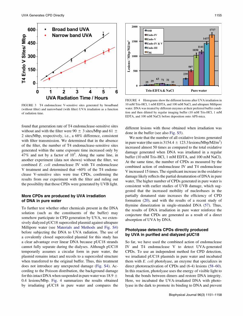
More CPDs are produced by UVA irradiationof DNA in pure water
To further test whether other chemicals present in the DNA
solution (such as the constituents of the buffer) may
somehow participate in CPD generation by UVA, we exten-
sively dialyzed pUC18 supercoiled plasmid against ultrapure
Millipore water (see Materials and Methods and Fig. S4)
before subjecting the DNA to UVA radiation. The use of
a covalently closed supercoiled plasmid for this study has
a clear advantage over linear DNA because pUC18 strands
cannot fully separate during the dialyses. Although pUC18
temporarily assumes a circular form in pure water, the
plasmid remains intact and recoils to a supercoiled structure
when transferred to the original buffer. Thus, this treatment
does not introduce any unexpected damage (Fig. S4). Ac-
cording to the Poisson distribution, the background damage
for this intact DNA when suspended in pure water was 18.9 5
0.4 lesions/Mbp. Fig. 4 summarizes the results obtained
by irradiating pUC18 in pure water and compares the
FIGURE 3 T4 endonuclease V-sensitive sites generated by broadband
(without filter) and narrowband (with filter) UVA irradiation as a function
of radiation time.
different lesions with those obtained when irradiation was
done in the buffer (see also Fig. S5).
We note that the number of all oxidative lesions generated
in pure water (the sum is 3154.4 5 123.3 lesions/Mbp/MJ/m2)
increased almost 50 times as compared to the total oxidative
damage generated when DNA was irradiated in a regular
buffer (10 mM Tris-HCl, 1 mM EDTA, and 100 mM NaCl).
At the same time, the number of CPDs as measured by the
combined action of endonuclease IV and T4 endonuclease
V increased 15 times. The significant increase in the oxidative
damage likely reflects the partial denaturation of DNA in pure
water, The higher number of CPDs generated in pure water is
consistent with earlier studies of UVB damage, which sug-
gested that the increased mobility of nucleobases in the
partially denatured state increases the efficiency of CPD
formation (20), and with the results of a recent study of
thymine dimerization in single-stranded DNA (57). Thus,
the results of DNA irradiation in pure water reinforce the
conjecture that CPDs are generated as a result of a direct
absorption of UVA by DNA.
Photolyase detects CPDs directly producedby UVA in purified and dialyzed pUC18
So far, we have used the combined action of endonuclease
IV and T4 endonuclease V to detect UVA-generated
CPDs. To use an independent method for CPD detection,
we irradiated pUC18 plasmids in pure water and incubated
them with E. coli photolyase, an enzyme that specializes in
direct photoreactivation of CPDs and (6-4) lesions (58–60).
In this reaction, photolyase uses the energy of visible light to
break the bonds between dimers and restore DNA integrity.
Here, we incubated the UVA-irradiated DNA with photo-
lyase in the dark to promote its binding to DNA and prevent
FIGURE 4 Histograms show the different lesions after UVA irradiation in
10 mM Tris-HCl, 1 mM EDTA, and 100 mM NaCl, and ultrapure Millipore
water. DNA was treated by different enzymes at their preferred buffer condi-
tion and then diluted by regular imaging buffer (10 mM Tris-HCl, 1 mM
EDTA, and 100 mM NaCl) before deposition onto APS-mica.
Biophysical Journal 96(3) 1151–1158

1156 Jiang et al.
photoreactivation. Photolyase (MW ¼ 54 KDa) is large
enough to be directly visualized by AFM imaging (61).
Before AFM imaging, the DNA was suspended in a buffer
that contained 5 mM Mg and no Na (33). Under these condi-
tions, supercoiled plasmids are forced to assume a circular
form with no supercoiled strand crossings, although they
remain covalently closed. This DNA structure allowed us
to image and count photolyase particles on DNA more accu-
rately. The AFM image in Fig. 5 A shows photolyase bound
onto 1 MJ/m2 UVA-irradiated pUC18 plasmids, and Fig. 5 Bshows the image of untreated DNA after incubation with the
enzyme. We counted the number of photolyase particles on
pUC18 molecules directly from AFM images and plotted
their frequency histograms together with the Poisson distri-
bution fits (green curves in Fig. 5, C and D). After subtract-
ing the number of enzyme molecules bound nonspecifically
to the DNA, we estimate 0.98 photolyase-sensitive sites per
plasmid at 1 MJ/m2, i.e., 365 CPD/Mbp/MJ/m2.
Thus, the number of photolyase-sensitive sites is close to
but somewhat lower than the number of CPDs determined
from the combined results of T4 endonuclease V and
E. coli endonuclease IV treatments (634 lesions/Mbp/MJ/m2).
We conclude that, consistent with the results obtained by
means of specific endonucleases, the photolyase assay detects
a significant amount of CPDs produced by UVA in the
absence of any photosensitizers. This result additionally
A
C
B
D
FIGURE 5 AFM images show photolyases binding to the CPD sites of
pUC18 (some of them marked by blue arrows) with (A) 1 MJ/m2 UVA radi-
ation and (B) no UVA radiation. Irradiation was performed on dialyzed plas-
mids suspended in pure water. The scan size in all the images is 1 � 1 mm2.
(C and D) Histograms show the distribution of photolyase on pUC18 mole-
cules as shown in A and B. The curves show the Poisson distribution fits,
which give the average damage l ¼ 1.52/plasmid for the UVA-radiated
DNA and 0.54 for control DNA, respectively.
Biophysical Journal 96(3) 1151–1158
supports the hypothesis that UVA can produce CPDs by direct
action on DNA.
Photolyase and antibodies detect CPDs produceddirectly by UVA in synthetic homopolynucleotide
To further test whether UVA directly generates CPDs in DNA
without possible exposure to cellular photosensitizers, we
irradiated, in a Tris-EDTA buffer, a synthetic double-stranded
homopolynucleotide, poly(dA)-poly(dT), with 6 MJ/m2
UVA at 365 5 5 nm, and incubated it with photolyase and
with anti-CPD antibodies (see Materials and Methods).
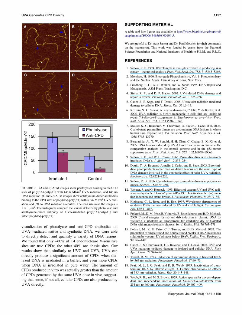
Fig. 6 shows the AFM images, which clearly reveal numerous
protein particles on the irradiated DNA proving that CPDs
have indeed been formed. To the best of our knowledge,
Fig. 6 C shows for the first time direct images of individual
anti-CPD antibodies on DNA captured by AFM. The numbers
of CPD sites determined from these and similar images were
(after subtracting nonspecifically bound proteins): for poly
(dA)-poly(dT) 78 5 12 CPD/Mbp/MJ/m2 as detected by pho-
tolyase, and 68 5 5 CPD/Mbp/MJ/m2 as detected by antith-
ymine-dimer antibodies. These numbers of CPDs are higher
than the number of CPDs detected by the combined action
of T4 endonuclease V and endonuclease IV on pUC18 mole-
cules in the same buffer (42 CPD/Mbp/MJ/m2). This result is
understandable because poly(dA)-poly(dT) has more TT
neighbors per unit length that can potentially form TT dimers
than does pUC18.
The number of CPDs produced by UVA in vitroexceeds the number of CPDs produced in vivo
Finally, we compare the number of CPDs generated by UVA
in isolated DNA that we determined using our AFM method-
ology with the number of CPDs determined in previous
studies, in which cells and skin tissues were subjected to pro-
longed UVA radiation (6–8). The latter number varies
among different systems and studies, and ranges from 1.5
to 18 CPD/Mbp/MJ/m2 (5–8,10,22,23). Thus, the number
of CPDs generated in cellular DNA by UVA is lower than
the number of CPDs generated in isolated DNA (42.2
CPDMbp/MJ/m2). Since our results strongly suggest that
UVA generates CPDs in isolated DNA directly, it is possible
that some, if not all, cellular CPDs are also generated by
UVA directly. However, it is also possible that the protection
of cellular DNA against direct UVA damage is so high that
some photosensitizers are still required to account for the
numbers of CPDs generated in vivo. Further studies to clarify
these difficult issues are warranted.
CONCLUSIONS
By executing endonuclease-driven supercoiled plasmid
relaxation assays on the AFM imaging platform, and AFM
UVA Generates CPD Directly 1157
visualization of photolyase and anti-CPD antibodies on
UVA-irradiated native and synthetic DNA, we were able
to directly detect and quantify a variety of DNA lesions.
We found that only ~60% of T4 endonuclease V-sensitive
sites are true CPDs; the other 40% are abasic sites. Our
results show that, similarly to UVC and UVB, UVA can
directly produce a significant amount of CPDs when dia-
lyzed DNA is irradiated in a buffer, and even more CPDs
when DNA is irradiated in pure water. The amount of
CPDs produced in vitro was actually greater than the amount
of CPDs generated by the same UVA dose in vivo, suggest-
ing that some, if not all, cellular CPDs are also produced by
UVA directly.
A B
DC
FIGURE 6 (A and B) AFM images show photolyases binding to the CPD
sites of poly(dA)-poly(dT) with (A) 6 MJ/m2 UVA radiation, and (B) no
UVA radiation. (C and D) AFM images show antithymine-dimer antibodies
binding to the CPD sites of poly(dA)-poly(dT) with (C) 6 MJ/m2 UVA radi-
ation, and (D) no UVA radiation as control. The scan size in all the images is
1 � 1 mm2. The histograms compare the lesions detected by photolyase and
antithymine-dimer antibody on UVA-irradiated poly(dA)-poly(dT) and
intact poly(dA)-poly(dT).
SUPPORTING MATERIAL
A table and five figures are available at http://www.biophysj.org/biophysj/
supplemental/S0006-3495(08)00104-5.
We are grateful to Dr. Aziz Sancar and Dr. Paul Modrich for their comments
on the manuscript. This work was funded by grants from the National
Science Foundation and National Institutes of Health to P.E.M. and R.L.C.
REFERENCES
1. Setlow, R. B. 1974. Wavelengths in sunlight effective in producing skincancer—theoretical analysis. Proc. Natl. Acad. Sci. USA. 71:3363–3366.
2. Morrison, H. 1990. Bioorganic Photochemistry, Vol. 1, Photochemistryand the Nucleic Acids. John Wiley & Sons, New York.
3. Friedberg, E. C., G. C. Walker, and W. Siede. 1995. DNA Repair andMutagenesis. ASM Press, Washington, D.C.
4. Sinha, R. P., and D. P. Hader. 2002. UV-induced DNA damage andrepair: a review. Photochem. Photobiol. Sci. 1:225–236.
5. Cadet, J., E. Sage, and T. Douki. 2005. Ultraviolet radiation-mediateddamage to cellular DNA. Mutat. Res. 571:3–17.
6. Kozmin, S., G. Slezak, A. Reynaud-Angelin, C. Elie, Y. de Rycke, et al.2005. UVA radiation is highly mutagenic in cells that are unable torepair 7,8-dihydro-8-oxoguanine in Saccharomyces cerevisiae. Proc.Natl. Acad. Sci. USA. 102:13538–13543.
7. Mouret, S., C. Baudouin, M. Charveron, A. Favier, J. Cadet, et al. 2006.Cyclobutane pyrimidine dimers are predominant DNA lesions in wholehuman skin exposed to UVA radiation. Proc. Natl. Acad. Sci. USA.103:13765–13770.
8. Besaratinia, A., T. W. Synold, H. H. Chen, C. Chang, B. X. Xi, et al.2005. DNA lesions induced by UV A1 and B radiation in human cells:comparative analyses in the overall genome and in the p53 tumorsuppressor gene. Proc. Natl. Acad. Sci. USA. 102:10058–10063.
9. Setlow, R. B., and W. L. Carrier. 1966. Pyrimidine dimers in ultraviolet-irradiated DNA’s. J. Mol. Biol. 17:237–254.
10. Douki, T., A. Reynaud-Angelin, J. Cadet, and E. Sage. 2003. Bipyrimi-dine photoproducts rather than oxidative lesions are the main type ofDNA damage involved in the genotoxic effect of solar UVA radiation.Biochemistry. 42:9221–9226.
11. Setlow, R. B. 1966. Cyclobutane-type pyrimidine dimers in polynucle-otides. Science. 153:379–386.
12. Wehner, J., and G. Horneck. 1995. Effects of vacuum UV and UVC radi-ation on dry Escherichia-coli plasmid Puc19. 1. Inactivation, lacz(�) muta-tion-induction and strand breaks. J. Photochem. Photobiol. B. 28:77–85.
13. Kielbassa, C., L. Roza, and B. Epe. 1997. Wavelength dependence ofoxidative DNA damage induced by UV and visible light. Carcinogen-esis. 18:811–816.
14. Folkard, M., K. M. Prise, B. Vojnovic, B. Brocklehurst, and B. D. Michael.2000. Critical energies for ssb and dsb induction in plasmid DNA byvacuum-UV photons: an arrangement for irradiating dry or hydratedDNA with monochromatic photons. Int. J. Radiat. Biol. 76:763–771.
15. Folkard, M., K. M. Prise, C. J. Turner, and B. D. Michael. 2002. Theproduction of single strand and double strand breaks in DNA in aqueoussolution by vacuum UV photons below 10 eV. Radiat. Prot. Dosimetry.99:147–149.
16. Cadet, J., S. Courdavault, J. L. Ravanat, and T. Douki. 2005. UVB andUVA radiation-mediated damage to isolated and cellular DNA. PureAppl. Chem. 77:947–961.
17. Tyrrell, R. M. 1973. Induction of pyrimidine dimers in bacterial DNAby 365 nm radiation. Photochem. Photobiol. 17:69–73.
18. Peak, M. J., J. G. Peak, and R. B. Webb. 1973. Inactivation of trans-forming DNA by ultraviolet-light. 3. Further observations on effectsof 365 nm radiation. Mutat. Res. 20:143–148.
19. Webb, R. B., and M. S. Brown. 1979. Action spectra for oxygen-depen-dent and independent inactivation of Escherichia-coli-WP2S from254-nm to 460-nm. Photochem. Photobiol. 29:407–409.
Biophysical Journal 96(3) 1151–1158
1158 Jiang et al.
20. Kuluncsics, Z., D. Perdiz, E. Brulay, B. Muel, and E. Sage. 1999. Wave-length dependence of ultraviolet-induced DNA damage distribution:involvement of direct or indirect mechanisms and possible artefacts.J. Photochem. Photobiol. B. 49:71–80.
21. Peak, M. J., and J. G. Peak. 1989. Solar-ultraviolet-induced damage toDNA. Photodermatol. 6:1–15.
22. Douki, T., M. Court, S. Sauvaigo, F. Odin, and J. Cadet. 2000. Forma-tion of the main UV-induced thymine dimeric lesions within isolatedand cellular DNA as measured by high performance liquid chromato-graphy-tandem mass spectrometry. J. Biol. Chem. 275:11678–11685.
23. Courdavault, S., C. Baudouin, M. Charveron, A. Favier, J. Cadet, et al.2004. Larger yield of cyclobutane dimers than 8-oxo-7,8-dihydrogua-nine in the DNA of UVA-irradiated human skin cells. Mutat. Res.556:135–142.
24. Mitchell, D. 2006. Revisiting the photochemistry of solar UVA inhuman skin. Proc. Natl. Acad. Sci. USA. 103:13567–13568.
25. Perdiz, D., P. Grof, M. Mezzina, O. Nikaido, E. Moustacchi, et al. 2000.Distribution and repair of bipyrimidine photoproducts in solar UV-irra-diated mammalian cells—possible role of Dewar photoproducts in solarmutagenesis. J. Biol. Chem. 275:26732–26742.
26. Sutherland, J. C., and K. P. Griffin. 1981. Absorption-spectrum of DNAfor wavelengths greater than 300-nm. Radiat. Res. 86:399–410.
27. Hiraku, Y., K. Ito, K. Hirakawa, and S. Kawanishi. 2007. Photosensi-tized DNA damage and its protection via a novel mechanism. Photo-chem. Photobiol. 83:205–212.
28. Sutherland, B. M., P. V. Bennett, O. Sidorkina, and J. Laval. 2000. Clus-tered DNA damages induced in isolated DNA and in human cells by lowdoses of ionizing radiation. Proc. Natl. Acad. Sci. USA. 97:103–108.
29. Sutherland, B. M., A. G. Georgakilas, P. V. Bennett, J. Laval, and J. C.Sutherland. 2003. Quantifying clustered DNA damage induction andrepair by gel electrophoresis, electronic imaging and number averagelength analysis. Mutat. Res. 531:93–107.
30. Collins, A. R. 2004. The comet assay for DNA damage and repair—principles, applications, and limitations. Mol. Biotechnol. 26:249–261.
31. Brendler-Schwaab, S., A. Hartmann, S. Pfuhler, and G. Speit. 2005. Thein vivo comet assay: use and status in genotoxicity testing. Mutagenesis.20:245–254.
32. Sutherland, B. M., P. V. Bennett, K. Conlon, G. A. Epling, and J. C.Sutherland. 1992. Quantitation of Supercoiled DNA cleavage in nonra-dioactive DNA—application to ionizing-radiation and synthetic endo-nuclease cleavage. Anal. Biochem. 201:80–86.
33. Jiang, Y., C. H. Ke, P. A. Mieczkowski, and P. E. Marszalek. 2007.Detecting ultraviolet damage in single DNA molecules by atomicforce microscopy. Biophys. J. 93:1758–1767.
34. Ke, C., Y. Jiang, P. A. Mieczkowski, G. G. Muramoto, J. P. Chute, et al.2008. Nanoscale detection of ionizing radiation damage to DNA byatomic force microscopy. Small. 4:288–294.
35. Hansma, H. G., D. E. Laney, M. Bezanilla, R. L. Sinsheimer, and P. K.Hansma. 1995. Applications for atomic-force microscopy of DNA.Biophys. J. 68:1672–1677.
36. Lyubchenko, Y. L., and L. S. Shlyakhtenko. 1997. Visualization ofsupercoiled DNA with atomic force microscopy in situ. Proc. Natl.Acad. Sci. USA. 94:496–501.
37. Guthold, M., M. Bezanilla, D. A. Erie, B. Jenkins, H. G. Hansma, et al.1994. Following the assembly of RNA-polymerase DNA complexes inaqueous-solutions with the scanning force microscope. Proc. Natl.Acad. Sci. USA. 91:12927–12931.
38. Shao, Z. F., J. Mou, D. M. Czajkowsky, J. Yang, and J. Y. Yuan. 1996.Biological atomic force microscopy: what is achieved and what isneeded. Adv. Phys. 45:1–86.
39. Hansma, H. G. 2001. Surface biology of DNA by atomic force micros-copy. Annu. Rev. Phys. Chem. 52:71–92.
40. Hansma, H. G., J. Vesenka, C. Siegerist, G. Kelderman, H. Morrett,et al. 1992. Reproducible imaging and dissection of plasmid DNA underliquid with the atomic force microscope. Science. 256:1180–1184.
Biophysical Journal 96(3) 1151–1158
41. Lindsay, S. M., T. Thundat, L. Nagahara, U. Knipping, and R. L. Rill.1989. Images of the DNA double helix in water. Science. 244:1063–1064.
42. New England Biolab. DNA Repair Glycosylases on Various DamagedBases. Available at: http://www.neb.com/nebecomm/tech_reference/modifying_enzymes/dna_repair_damaged_bases.asp.
43. Woollons, A., C. Kipp, A. R. Young, C. Petit-Frere, C. F. Arlett, et al.1999. The 0.8% ultraviolet B content of an ultraviolet A sunlampinduces 75% of cyclobutane pyrimidine dimers in human keratinocytesin vitro. Br. J. Dermatol. 140:1023–1030.
44. Shlyakhtenko, L. S., A. A. Gall, A. Filonov, Z. Cerovac, A. Lushnikov,et al. 2003. Silatrane-based surface chemistry for immobilization ofDNA, protein-DNA complexes and other biological materials. Ultrami-croscopy. 97:279–287.
45. Cherny, D. I., and T. M. Jovin. 2001. Electron and scanning forcemicroscopy studies of alterations in supercoiled DNA tertiary structure.J. Mol. Biol. 313:295–307.
46. Vologodskii, A. V., and N. R. Cozzarelli. 1994. Conformational andthermodynamic properties of supercoiled DNA. Annu. Rev. Biophys.Biomol. Struct. 23:609–643.
47. Bussiek, M., N. Mucke, and J. Langowski. 2003. Polylysine-coatedmica can be used to observe systematic changes in the supercoiledDNA conformation by scanning force microscopy in solution. NucleicAcids Res. 31:e137.
48. Shlyakhtenko, L. S., L. Miloseska, V. N. Potaman, R. R. Sinden, andY. L. Lyubchenko. 2003. Intersegmental interactions in supercoiledDNA: atomic force microscope study. Ultramicroscopy. 97:263–270.
49. Murakami, M., H. Hirokawa, and I. Hayata. 2000. Analysis of radiationdamage of DNA by atomic force microscopy in comparison with agarosegel electrophoresis studies. J. Biochem. Biophys. Methods. 44:31–40.
50. Pang, D., B. L. Berman, S. Chasovskikh, J. E. Rodgers, and A. Drit-schilo. 1998. Investigation of neutron-induced damage in DNA byatomic force microscopy: experimental evidence of clustered DNAlesions. Radiat. Res. 150:612–618.
51. Pang, D., J. E. Rodgers, B. L. Berman, S. Chasovskikh, and A. Drit-schilo. 2005. Spatial distribution of radiation-induced double-strandbreaks in plasmid DNA as resolved by atomic force microscopy. Radiat.Res. 164:755–765.
52. Boichot, S., M. Fromm, S. Cunniffe, P. O’Neill, J. C. Labrune, et al.2002. Investigation of radiation damage in DNA by using atomic forcemicroscopy. Radiat. Prot. Dosimetry. 99:143–145.
53. Psonka, K., S. Brons, M. Heiss, E. Gudowska-Nowak, and G. Taucher-Scholz. 2005. Induction of DNA damage by heavy ions measured byatomic force microscopy. J. Phys. Condens. Matter. 17:S1443–S1446.
54. Lobachevsky, P. N., T. C. Karagiannis, and R. F. Martin. 2004. PlasmidDNA breakage by decay of DNA-associated auger electron emitters:approaches to analysis of experimental data. Radiat. Res. 162:84–95.
55. Sachs, R. K., A. L. Ponomarev, P. Hahnfeldt, and L. R. Hlatky. 1999. Loca-tions of radiation-produced DNA double strand breaks along chromo-somes: a stochastic cluster process formalism. Math. Biosci. 159:165–187.
56. Gruskin, E. A., and R. S. Lloyd. 1986. The DNA scanning mechanismof T4 endonuclease-V—effect of NaCl concentration on processivenicking activity. J. Biol. Chem. 261:9607–9613.
57. Schreier, W. J., T. E. Schrader, F. O. Koller, P. Gilch, C. E. Crespo-Hernandez, et al. 2007. Thymine dimerization in DNA is an ultrafastphotoreaction. Science. 315:625–629.
58. Sancar, A. 2003. Structure and function of DNA photolyase and cryp-tochrome blue-light photoreceptors. Chem. Rev. 103:2203–2237.
59. Kao, Y. -T., C. Saxena, L. Wang, A. Sancar, and D. Zhong. 2005.Inaugural article: direct observation of thymine dimer repair in DNAby photolyase. Proc. Natl. Acad. Sci. USA. 102:16128–16132.
60. Sancar, G. B., and A. Sancar. 2006. Purification and characterization ofDNA photolyases. Methods Enzymol. 408:121–156.
61. van Noort, J., F. Orsini, A. Eker, C. Wyman, B. de Grooth, et al. 1999.DNA bending by photolyase in specific and non-specific complexesstudied by atomic force microscopy. Nucleic Acids Res. 27:3875–3880.
Related Documents