Transcriptome analysis reveals cyclobutane pyrimidine dimers as a major source of UV-induced DNA breaks George A Garinis 1 , James R Mitchell 1 , Michael J Moorhouse 2 , Katsuhiro Hanada 1 , Harm de Waard 1 , Dimitri Vandeputte 1 , Judith Jans 1,5 , Karl Brand 1 , Marcel Smid 3 , Peter J van der Spek 2 , Jan HJ Hoeijmakers 1 , Roland Kanaar 1,4 and Gijsbertus TJ van der Horst 1, * 1 Department of Cell Biology and Genetics, Erasmus University Medical Center, Rotterdam, The Netherlands, 2 Department of Bioinformatics, Erasmus University Medical Center, Rotterdam, The Netherlands, 3 Department of Medical Oncology, Josephine Nefkens Institute, Erasmus University Medical Center, Rotterdam, The Netherlands and 4 Department of Radiation Oncology, Erasmus University Medical Center, Rotterdam, The Netherlands Photolyase transgenic mice have opened new avenues to improve our understanding of the cytotoxic effects of ultraviolet (UV) light on skin by providing a means to selectively remove either cyclobutane pyrimidine dimers (CPDs) or pyrimidine (6-4) pyrimidone photoproducts. Here, we have taken a genomics approach to delineate pathways through which CPDs might contribute to the harmful effects of UV exposure. We show that CPDs, rather than other DNA lesions or damaged macromolecules, comprise the principal mediator of the cellular transcrip- tional response to UV. The most prominent pathway in- duced by CPDs is that associated with DNA double-strand break (DSB) signalling and repair. Moreover, we show that CPDs provoke accumulation of c-H2AX, P53bp1 and Rad51 foci as well as an increase in the amount of DSBs, which coincides with accumulation of cells in S phase. Thus, conversion of unrepaired CPD lesions into DNA breaks during DNA replication may comprise one of the principal instigators of UV-mediated cytotoxicity. The EMBO Journal (2005) 24, 3952–3962. doi:10.1038/ sj.emboj.7600849; Published online 27 October 2005 Subject Categories: genome stability & dynamics; genomic & computational biology Keywords: DNA damage; functional genomics; photolyase; UV irradiation Introduction Ultraviolet (UV) radiation comprises one of the major exogen- ous toxic agents through which cellular macromolecules can be damaged, thereby inducing deleterious effects such as sunburn, immune suppression, skin cancer and photoaging (Friedberg et al, 1995). As nucleic acids, lipids and proteins are simultaneously damaged during UV exposure, it has been difficult to pinpoint the principal cellular target for the UV response. With respect to DNA, cyclobutane pyrimidine dimers (CPDs) and pyrimidine (6-4) pyrimidone photoproducts (6- 4PPs) are the predominant lesions caused by short-wavelength UV radiation (Mitchell, 1988). Both types of lesions impinge on vital cellular functions, including transcription, DNA replica- tion and cell cycle progression. Their persistence in the genetic material also increases the chance of fixation into mutations, a hallmark event of cancer initiation (Friedberg et al, 1995). A number of mechanisms have evolved to recognize and remove DNA damage. For bulky helix-distorting damage, such as the main UV-induced lesions, the principal repair mechanism is the evolutionarily conserved nucleotide excision repair (NER) pathway (Hoeijmakers, 2001). Two different modes of lesion recognition delineate distinct NER subpathways. Transcription- coupled NER is restricted to repair of damage on the transcribed strand of active genes and is efficient at removing CPDs and 6- 4PPs, in addition to other types of transcription-blocking lesions. Global genome NER (GG-NER) removes DNA damage from any position in the genome (Hoeijmakers, 2001). However, whereas 6-4PPs and other types of helix-distorting lesions are efficiently repaired, CPDs form a poor substrate and are removed at significantly reduced rates in human cells (Tung et al, 1996), and virtually not at all in rodent cells (Bohr et al, 1985). During DNA replication, cells can bypass persisting CPDs by employing relatively error-free or error-prone bypass polymerases, or by utilizing template switching, involving proteins of the homo- logous recombination repair pathway (Hoeijmakers, 2001). Failure of these pathways may lead to arrested replication forks, requiring subsequent processing for recombinational re- pair and thereby increasing the chance of chromosomal aberra- tions. Thus, based on their high relative abundance, slow repair kinetics and known mutagenicity, CPDs are thought to contri- bute significantly to the effects of UV radiation (Mitchell, 1988; Sage, 1993; Tung et al, 1996; Yoon et al, 2000). In humans, the essential role of NER in the repair of UV-induced cytotoxic photolesions is illustrated by the occurrence of photosensitive disorders (e.g. xeroderma pigmentosum) that originate from inborn errors in NER genes (Bootsma et al, 2002). A distinct strategy to repair UV-induced photolesions is photoreactivation (PR), an enzymatic reaction in which 6-4PPs or CPD lesion-specific photolyases directly revert photolesions into undamaged bases using visible light energy (Carell et al, 2001). However, although photolyases are pre- sent across species boundaries, they are absent in placental mammals. We have previously established mice that express a Received: 9 June 2005; accepted: 30 September 2005; published online: 27 October 2005 *Corresponding author. Department of Cell Biology and Genetics, Center for Biomedical Genetics, Erasmus University Medical Center, PO Box 1738, 3000 DR Rotterdam, The Netherlands. Tel.: þ 3110 408 7455; Fax: þ 3110 408 9468; E-mail: [email protected] 5 Present address: Medical Genetic Center, Department of Molecular and Cell Biology, University of California at Berkeley, 125 Koshland Hall, Berkeley, CA, USA The EMBO Journal (2005) 24, 3952–3962 | & 2005 European Molecular Biology Organization | All Rights Reserved 0261-4189/05 www.embojournal.org The EMBO Journal VOL 24 | NO 22 | 2005 & 2005 European Molecular Biology Organization EMBO THE EMBO JOURNAL THE EMBO JOURNAL 3952

Welcome message from author
This document is posted to help you gain knowledge. Please leave a comment to let me know what you think about it! Share it to your friends and learn new things together.
Transcript

Transcriptome analysis reveals cyclobutanepyrimidine dimers as a major source ofUV-induced DNA breaks
George A Garinis1, James R Mitchell1,Michael J Moorhouse2, Katsuhiro Hanada1,Harm de Waard1, Dimitri Vandeputte1,Judith Jans1,5, Karl Brand1, Marcel Smid3,Peter J van der Spek2, Jan HJHoeijmakers1, Roland Kanaar1,4
and Gijsbertus TJ van der Horst1,*1Department of Cell Biology and Genetics, Erasmus University MedicalCenter, Rotterdam, The Netherlands, 2Department of Bioinformatics,Erasmus University Medical Center, Rotterdam, The Netherlands,3Department of Medical Oncology, Josephine Nefkens Institute, ErasmusUniversity Medical Center, Rotterdam, The Netherlands and4Department of Radiation Oncology, Erasmus University Medical Center,Rotterdam, The Netherlands
Photolyase transgenic mice have opened new avenues to
improve our understanding of the cytotoxic effects of
ultraviolet (UV) light on skin by providing a means to
selectively remove either cyclobutane pyrimidine dimers
(CPDs) or pyrimidine (6-4) pyrimidone photoproducts.
Here, we have taken a genomics approach to delineate
pathways through which CPDs might contribute to the
harmful effects of UV exposure. We show that CPDs, rather
than other DNA lesions or damaged macromolecules,
comprise the principal mediator of the cellular transcrip-
tional response to UV. The most prominent pathway in-
duced by CPDs is that associated with DNA double-strand
break (DSB) signalling and repair. Moreover, we show that
CPDs provoke accumulation of c-H2AX, P53bp1 and Rad51
foci as well as an increase in the amount of DSBs, which
coincides with accumulation of cells in S phase. Thus,
conversion of unrepaired CPD lesions into DNA breaks
during DNA replication may comprise one of the principal
instigators of UV-mediated cytotoxicity.
The EMBO Journal (2005) 24, 3952–3962. doi:10.1038/
sj.emboj.7600849; Published online 27 October 2005
Subject Categories: genome stability & dynamics; genomic
& computational biology
Keywords: DNA damage; functional genomics; photolyase;
UV irradiation
Introduction
Ultraviolet (UV) radiation comprises one of the major exogen-
ous toxic agents through which cellular macromolecules can
be damaged, thereby inducing deleterious effects such as
sunburn, immune suppression, skin cancer and photoaging
(Friedberg et al, 1995). As nucleic acids, lipids and proteins are
simultaneously damaged during UV exposure, it has been
difficult to pinpoint the principal cellular target for the UV
response. With respect to DNA, cyclobutane pyrimidine dimers
(CPDs) and pyrimidine (6-4) pyrimidone photoproducts (6-
4PPs) are the predominant lesions caused by short-wavelength
UV radiation (Mitchell, 1988). Both types of lesions impinge on
vital cellular functions, including transcription, DNA replica-
tion and cell cycle progression. Their persistence in the genetic
material also increases the chance of fixation into mutations,
a hallmark event of cancer initiation (Friedberg et al, 1995).
A number of mechanisms have evolved to recognize and
remove DNA damage. For bulky helix-distorting damage, such
as the main UV-induced lesions, the principal repair mechanism
is the evolutionarily conserved nucleotide excision repair (NER)
pathway (Hoeijmakers, 2001). Two different modes of lesion
recognition delineate distinct NER subpathways. Transcription-
coupled NER is restricted to repair of damage on the transcribed
strand of active genes and is efficient at removing CPDs and 6-
4PPs, in addition to other types of transcription-blocking lesions.
Global genome NER (GG-NER) removes DNA damage from any
position in the genome (Hoeijmakers, 2001). However, whereas
6-4PPs and other types of helix-distorting lesions are efficiently
repaired, CPDs form a poor substrate and are removed at
significantly reduced rates in human cells (Tung et al, 1996),
and virtually not at all in rodent cells (Bohr et al, 1985). During
DNA replication, cells can bypass persisting CPDs by employing
relatively error-free or error-prone bypass polymerases, or by
utilizing template switching, involving proteins of the homo-
logous recombination repair pathway (Hoeijmakers, 2001).
Failure of these pathways may lead to arrested replication
forks, requiring subsequent processing for recombinational re-
pair and thereby increasing the chance of chromosomal aberra-
tions. Thus, based on their high relative abundance, slow repair
kinetics and known mutagenicity, CPDs are thought to contri-
bute significantly to the effects of UV radiation (Mitchell, 1988;
Sage, 1993; Tung et al, 1996; Yoon et al, 2000). In humans, the
essential role of NER in the repair of UV-induced cytotoxic
photolesions is illustrated by the occurrence of photosensitive
disorders (e.g. xeroderma pigmentosum) that originate from
inborn errors in NER genes (Bootsma et al, 2002).
A distinct strategy to repair UV-induced photolesions is
photoreactivation (PR), an enzymatic reaction in which
6-4PPs or CPD lesion-specific photolyases directly revert
photolesions into undamaged bases using visible light energy
(Carell et al, 2001). However, although photolyases are pre-
sent across species boundaries, they are absent in placental
mammals. We have previously established mice that express aReceived: 9 June 2005; accepted: 30 September 2005; publishedonline: 27 October 2005
*Corresponding author. Department of Cell Biology and Genetics,Center for Biomedical Genetics, Erasmus University Medical Center, POBox 1738, 3000 DR Rotterdam, The Netherlands. Tel.: þ 31 10 408 7455;Fax: þ 31 10 408 9468; E-mail: [email protected] address: Medical Genetic Center, Department of Molecular andCell Biology, University of California at Berkeley, 125 Koshland Hall,Berkeley, CA, USA
The EMBO Journal (2005) 24, 3952–3962 | & 2005 European Molecular Biology Organization | All Rights Reserved 0261-4189/05
www.embojournal.org
The EMBO Journal VOL 24 | NO 22 | 2005 &2005 European Molecular Biology Organization
EMBO
THE
EMBOJOURNAL
THE
EMBOJOURNAL
3952

marsupial (Potorous tridactylis) CPD-specific photolyase trans-
gene either ubiquitously or specifically in the basal keratino-
cytes of the epidermis. These mice were used to dissect the
specific contributions of CPDs versus 6-4PPs to the biological
consequences of UV irradiation (Chigancas et al, 2000; Schul
et al, 2002). Importantly, light-dependent removal of CPDs but
not 6-4PPs was found to promote UV survival (Chigancas
et al, 2000; Schul et al, 2002; Nakajima et al, 2004). Moreover,
CPDs appeared to be the primary cause of the vast majority of
(semi) acute responses in the UV-exposed skin (i.e. sunburn,
apoptosis, hyperplasia and mutation induction) and have
been unequivocally identified as the principal instigator of
non-melanoma skin cancer (Jans et al, 2005).
In the current study, we aimed at identifying those path-
ways through which unrepaired CPDs might induce their
harmful effects. By implementing a functional genomics
approach, we demonstrate that during DNA replication,
unrepaired CPD lesions can be converted into a substantial
source of toxic, replication-dependent single-strand breaks
(SSBs) and double-strand breaks (DSBs), highlighting their
role in UV-mediated cytotoxicity.
Results
Impact of CPDs on the transcriptional response
to UV irradiation
To ascertain the global transcriptional response to UV damage
in general and to CPDs specifically, CPD photolyase trans-
genic mouse dermal fibroblasts (MDFs) were irradiated with
a series of UV doses (0, 2, 4 and 8 J/m2), treated with PR light
or kept in the dark for 30 or 60 min and harvested at intervals
from �30 min (relative to the 0 h time point that marked one
full hour of PR light exposure) to þ 24 h (Figure 1A). Under
the conditions used, we observed neither cell loss nor signs of
apoptosis (i.e. TUNEL staining, ligation-mediated PCR ampli-
fication of fragmented DNA; data not shown). The effective-
ness of CPD repair by photolyase was confirmed by
immunocytochemical visualization of CPDs. As shown in
Figure 1B, CPDs were no longer detectable after 1 h exposure
to PR light. In contrast, when cells were kept in the dark (thus
withholding the energy required for photolyase activity),
CPDs remained visible, which is consistent with their poor
removal by GG-NER in rodent cells (Bohr et al, 1985). Next,
we determined the gene expression profiles by microarray
analysis, using 15K cDNA arrays (see Materials and meth-
ods). To assess whether PR of UV-induced CPDs has a
substantial impact on gene expression, we examined, by
unsupervised hierarchical clustering, the similarity of tran-
scription profiles of all responsive genes (defined as those
genes for which the expression changed significantly in time
and with UV dose; ANOVA P-value p0.05, 71.5-fold
change). As shown in Figure 2A, the profound effect of PR
(and therefore CPD removal) on the transcriptional response
to UV was readily seen at each of the time points examined
after PR. For each time point, at a given UV dose, all matrix
points clustered into two main groups correlating solely with
A
UV-C
D
L
0 h 2 h 4 h 8 h 24 h2 J/m2
4 J/m28 J/m2
UV-C
No-UV D
−1/2 h
0 h 2 h 4 h 8 h 24 h−1/2 h
2 J/m24 J/m2
8 J/m2
Experimental design
B
LightDark Dark
UV (8 J/m2)No UV
Anti-CPD
DAPI
0 h 24 hTime (post-PR)
Figure 1 Experimental approach and removal of CPDs upon PR. (A) Graphical representation of the experimental design. For each dose andtime point, labelled cDNAs derived from irradiated, PR and non-PR samples (upper and lower horizontal bars) were hybridized to unirradiated,non-PR material from the same time point (middle bar). D: dark; L: light. (B) CPDs were detected by indirect immunofluorescence; nuclei werevisualized by DAPI staining. UV dose (No UVor 8 J/m2) and PR status (light or dark) are indicated on the bottom; time after the 1 h PR period isindicated on top.
CPD-dependent transcriptional responsesGA Garinis et al
&2005 European Molecular Biology Organization The EMBO Journal VOL 24 | NO 22 | 2005 3953

their PR status, suggesting that light-dependent removal of
CPDs was a major determinant of significant gene expression
changes. This effect was further confirmed by an additional
clustering over all 15 241 probes (Supplementary Figure S1A–
F), thus avoiding any gene preselection or potential introduc-
tion of bias. These findings demonstrate the overriding
influence of unrepaired CPDs on the genome-wide transcrip-
tional shifts observed after UV exposure, validating the
biological effect of PR and our ability to measure it on a
transcriptional level using this experimental approach.
Relative contribution of CPD lesions to the UV-induced
transcriptional response
To delineate the relative contribution of CPDs to the overall
transcriptional response to UV exposure in normal cells,
we first created a series of ‘biosets’ composed of all genes
Figure 2 PR of CPDs is a major determinant of significant gene expression profiles. (A) Tree graph representation of the similarity betweensignificant expression profiles (ANOVA Pp0.05, X71.5-fold change) at a given time point. Note the clustering of all matrix points (defined as aunique combination of dose and time point) into two main groups correlating to dark or light exposure during PR (to the left and right of thedotted orange line, respectively). (B) Venn diagrams of 2, 4 and 8 J/m2 biosets. Each bioset represents all significant genes in at least one timepoint (0–24 h) at the corresponding dose (2, 4 and 8 J/m2) in the absence of PR. (C) CPD-dependent and -independent gene expression. Eachbioset from (B) was divided into blue, depicting the percentage of genes shared between both PR and non-PR MDFs (CPD-independent), andred, depicting the percentage of genes present only in the absence of PR (CPD-dependent). (D) Time dependence of the shared transcriptionalresponse. The 8 J/m2 bioset representing all significant genes at each individual time point (�30 min to 24 h) was divided proportionately intoblue and red representing CPD-independent and -dependent gene expression, respectively.
CPD-dependent transcriptional responsesGA Garinis et al
The EMBO Journal VOL 24 | NO 22 | 2005 &2005 European Molecular Biology Organization3954
responding significantly to a given UV dose (2, 4 or 8 J/m2 of
UV-C) in any of the time points within 24 h after irradiation
(Figure 2B). Then, we examined which of the genes in each of
the three UV dose biosets obtained from non-PR cells (when
CPDs were present) also varied significantly in the 24 h
period following exposure of cells to PR light (when CPDs
were below the level of detection). This allowed us to identify
those genes whose expression levels were affected by UV
exposure in a CPD-dependent manner (Figure 2C). At all
doses tested, only B20% of the UV-responsive genes were
also regulated in UV-treated cells that were exposed to PR
light (Figure 2C). Similarly, when we compared the distinct
and shared significant transcriptional responses of PR and
non-PR cells (exposed to 8 J/m2), we identified the highest
percentage of shared genes (38%) to occur at the earliest time
point (�30 min; Figure 2D). This percentage gradually de-
clined to only 3% at the latest time point (24 h), suggesting
that most non-CPD lesions (including 6-4PPs) are repaired
within the 24 h period following UV, with the transcriptional
response at the latest time point (24 h) reflecting the presence
of persisting CPDs in the genome. Taken together, our data
provide strong evidence that DNA lesions, rather than
damaged proteins and/or lipids, are the principal mediators
of the transcriptional response to UV exposure with CPDs
representing the primary determinant.
Impact of CPD-dependent transcriptional responses
on UV-induced biological processes
To identify the biological processes provoked by CPDs in
UV-exposed cells, all responsive genes from each bioset
were subjected to gene ontology (GO) classification and
subsequent network analysis (Supplementary Figure S2;
data available at http://www.eur.nl/fgg/ch1/gene_network).
Those biological processes containing a significantly dispro-
portionate number of responsive genes relative to those
printed on the microarrays were red-flagged as over-repre-
sented. Upon 8 J/m2 of UV-C and in the absence of PR, a
broad range of biological processes were significantly over-
represented including that of nucleic acid (GO:0006139), lipid
(GO:0006629) and protein metabolism (GO:0009058), corro-
borating our previous findings that the majority of UV-
responsive genes required the presence of CPDs. A smaller
number of processes were identified at lower doses, indicat-
ing that, over the 24 h period examined, the biological effect
exerted by UV was proportional to the dose range.
Contribution of CPD lesions to the transcriptional
regulation of genes associated with SSB/DSB
signalling and repair
How and why do CPDs—which contrast 6-4PPs in that they
are poorly recognized and inefficiently repaired by GG-NER
and yet are tolerated in cells—exert influence on such a broad
range of physiological processes? To answer this, we exam-
ined in an unbiased way the most predominant molecular
networks underlying the response to DNA damage it self by
implementing the Ingenuity Molecular Network analysis
approach (www.ingenuity.com). First, all genes encoding
products functioning in common networks (pathways) were
annotated, and subsequently the statistical significance of
each network was listed (see ‘network analysis’ online). In
combination with the previous analysis for the over-repre-
sentation of biological processes, this method avoided any
arbitrary data preselection, or bias in interpretation of the
initially identified significant expression profiles. Strikingly,
for the 8 J/m2 bioset, the most significant network identified
within the GO-tree categorization ‘response to endogenous
stimulus’ (identified as an over-represented biological pro-
cess itself and including all modes of DNA repair and the
response to DNA damage itself; see online visualizations)
was associated with strand break repair pathways. Impor-
tantly, all genes encoding products that share a common role
in the repair of DSBs via homologous recombination (Rad51,
Rad54, Xrcc3 and Blm) and non-homologous end joining
(Ku80) were differentially expressed only upon the continu-
ing presence of CPDs (Figure 3A). Similarly, a network of
genes with an overlapping role in the response to DNA break
(including Mre11a, Rad50, Rad51 and Parp-2) was identified
at lower doses as well (Figure 3A and online visualization).
With the exception of Rad50, all other genes were signifi-
cantly differentially transcribed in UV-irradiated, non-PR
MDFs only (and thus as a consequence of the presence
of CPDs).
The initial finding that CPDs could contribute substantially
to the transcriptional regulation of genes associated with SSB
and DSB repair prompted us to further examine those path-
ways involved in signalling of such DNA breaks. Noticeably,
within the GO-tree categorization ‘physiological processes’,
we identified the most significantly over-represented network
in the 8 J/m2 bioset to contain genes directly involved in the
ATM signalling pathway that is centrally involved in the
detection and signalling of SSBs and DSBs in mammalian
cells (Atm, Chk2, Hus1, c-Abl-1, Mdm2, Blm and Cdc25a;
Figure 3A and online visualization). With the exception of
Mdm2, which has a prominent role outside the signalling
of DNA breaks as well, changes in expression levels of each
of these genes correlated significantly with the presence of
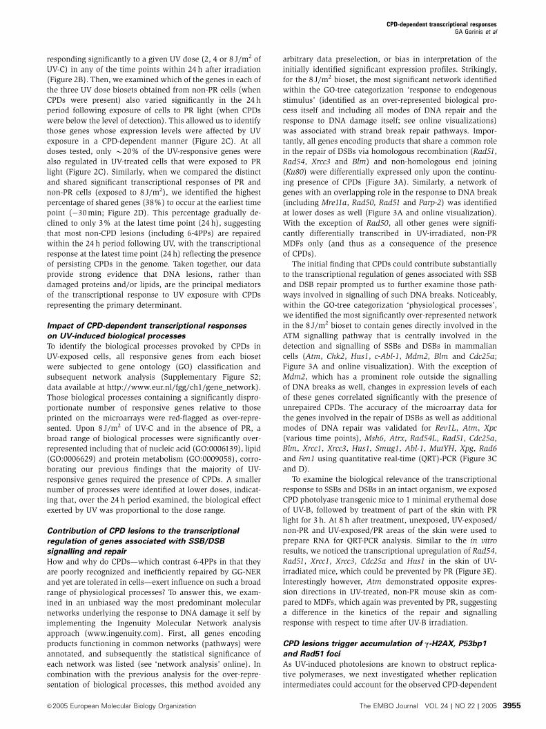
unrepaired CPDs. The accuracy of the microarray data for
the genes involved in the repair of DSBs as well as additional
modes of DNA repair was validated for Rev1L, Atm, Xpc
(various time points), Msh6, Atrx, Rad54L, Rad51, Cdc25a,
Blm, Xrcc1, Xrcc3, Hus1, Smug1, Abl-1, MutYH, Xpg, Rad6
and Fen1 using quantitative real-time (QRT)-PCR (Figure 3C
and D).
To examine the biological relevance of the transcriptional
response to SSBs and DSBs in an intact organism, we exposed
CPD photolyase transgenic mice to 1 minimal erythemal dose
of UV-B, followed by treatment of part of the skin with PR
light for 3 h. At 8 h after treatment, unexposed, UV-exposed/
non-PR and UV-exposed/PR areas of the skin were used to
prepare RNA for QRT-PCR analysis. Similar to the in vitro
results, we noticed the transcriptional upregulation of Rad54,
Rad51, Xrcc1, Xrcc3, Cdc25a and Hus1 in the skin of UV-
irradiated mice, which could be prevented by PR (Figure 3E).
Interestingly however, Atm demonstrated opposite expres-
sion directions in UV-treated, non-PR mouse skin as com-
pared to MDFs, which again was prevented by PR, suggesting
a difference in the kinetics of the repair and signalling
response with respect to time after UV-B irradiation.
CPD lesions trigger accumulation of c-H2AX, P53bp1
and Rad51 foci
As UV-induced photolesions are known to obstruct replica-
tive polymerases, we next investigated whether replication
intermediates could account for the observed CPD-dependent
CPD-dependent transcriptional responsesGA Garinis et al
&2005 European Molecular Biology Organization The EMBO Journal VOL 24 | NO 22 | 2005 3955
transcriptional response. Stalling of replication forks (as well
as SSBs and DSBs produced by a variety of agents) can be
visualized by the appearance of phosphorylated histone
H2AX (g-H2AX)-containing foci that accumulate at sites of
DNA breaks (Fernandez-Capetillo et al, 2004; Squires et al,
2004; Ward et al, 2004; Halicka et al, 2005). In B10% of the
untreated MDFs, we observed 1–2 g-H2AX foci per nucleus
(Figure 4A), which likely reflects unrepaired, spontaneous
DNA damage. In marked contrast, g-H2AX staining of UV-
irradiated, non-PR MDFs revealed a punctuate pattern of foci
that gradually accumulated from 1–2 foci/nucleus 2 h after
UV irradiation (B20% positive cells) to approximately 17
0 h 2 h 4 h 8 h 24 hA
B POLL 8 J/m2
ATRX 8 J/m2
RAD23B 8 J/m2
REV1L 8 J/m 2
MUS81 2 J/m2
FEN1 8 J/m2
REV1L 2 J/m2
HNRPD 8 J/m2
POLB 8 J/m2
XPC 8 J/m2
SMUG1 8 J/m2
MSH6 8 J/m2
MUTYH 2 J/m2
GTF2H2 2 J/m2
POLR2G 8 J/m2
XPG 8 J/m2
GTF2H1 8 J/m2
RAD6 8 J/m2
XRCC3 8 J/m2
RAD51 8 J/m2
HUS1 8 J/m2
ABL -1 8 J/m2
RPA-1 8 J/m2
ATM 8 J/m2
PARP-2 2 J/m2
RAD54 8 J/m2
BLM 8 J/m2
CDC25A 8 J/m2
MRE11A 2 J/m2
XRCC1 2 J/m2
KU80 8 J/m2
RAD50 2 J/m2
RAD51 2 J/m2
�1.5-fold change
�−1.5-fold change
% o
f rel
ativ
e m
RN
A e
xpre
ssio
n le
vels
−3
−1
1
3
0 h 2 h 4 h 8 h 24 hTime
XPC mRNA levels
Fol
d ch
ange
% o
f rel
ativ
e m
RN
Aex
pres
sion
leve
ls
0
100
200
300
400
Rad
54
Rad
51
Xrc
c1
Xrc
c3
Cdc
25a
Hus
1
Atm
Rad
54
Rad
51
Xrc
c1
Xrc
c3
Cdc
25a
Hus
1
Atm
Dark Light
MicroarraysQRT-PCR
050
100150200250300350
Xrcc3
(8 h
)
Rad51
(24
h)
Hus1
(8 h
)
Abl-1
(2 h
)
Rpa-1
(2 h
)
Atm (2
h)
Rad54
(8 h
)
Blm (2
4 h)
Cdc25
a (4
h)
Xrcc1
(4 h
)
050
100150200250300350
Xpc (0
h)
Rev1L
(0 h
)
Msh
6 (2
h)
Atrx (0
h)
Smug
1 (2
h)
Rad6
(4 h
)
Xpg (2
h)
Mut
YH (2 h
)
Fen1
(4 h
)
C
D
E
Figure 3 CPDs provoke the transcriptional response of genes associated with replication-dependent and -independent modes of DNA damagerepair. (A) Heat map representation of time-dependent expression profiles of genes associated with SSB/DSB repair and signalling.(B) Additional modes of DNA repair in UV-irradiated, non-PR-treated MDFs as compared to non-UV, non-PR-treated MDFs. Changes in foldexpression are represented by red (upregulated Xþ 1.5-fold change) and green (downregulated X�1.5-fold change) as indicated. All othercolors represent intermediate fold changes. Gene entries in orange represent those genes that displayed significant expression profiles uponremoval of CPDs by PR. The 0 h time point includes the 1 h exposure to PR. (C) QRT-PCR verification of microarray data. Fold changes areexpressed as percentage of relative mRNA expression for each gene in UV-irradiated (8 J/m2 of UV-C), non-PR-treated MDFs as compared tonon-irradiated, non-PR-treated MDFs at the indicated time points. (D) Time-dependent mRNA expression levels of XPC obtained frommicroarrays and real-time PCR as indicated in UV-irradiated, non-PR-treated MDFs as compared to non-irradiated, non-PR-treated MDFs. Foldchanges are expressed in log2 ratios. (E) QRT-PCR evaluation of genes associated with DSB repair and signalling in UV-irradiated, non-PR-treated mouse skin. Fold changes are expressed as percentage of the relative mRNA expression levels for each gene in the UV-irradiated,PR-treated mouse skin over the non-UV, non-PR-treated mouse skin 5 h after UV irradiation and subsequent PR treatment (3 h).
CPD-dependent transcriptional responsesGA Garinis et al
The EMBO Journal VOL 24 | NO 22 | 2005 &2005 European Molecular Biology Organization3956

A
B C
2 h 4 h 8 h 24 h 48 h
8 J
/m+
da
rk2
8 J
/m+
ligh
t2
1.3±0.4 4.6±0.7 6.2±1.5 14.2±2.4 17.2±2.3
0.6±0.1 1.7±0.5 3.3±1.4 5.4±1.6 9.2±2.2
81% 73% 64% 50% 35%
92% 80% 78% 70% 66%
89%
0.5±0.3
92%
0.5±0.2
91%
0.6±0.2
89%
0.3±0.1
90%
0.4±0.1
No
UV
UV+darkUV+lightNo UV
48 h
4 h
8 h
24 h
1.5±0.4
1.4±0.1
1.3±0.5
82%
85%
81%
83%
1.4±0.1
72%
67%
62%
54%
65%
57%
50%
31%
3.2±1.4 4.3±2.4
4.3±2.3
5.4±2.5
6.3±3.2
6.3±3.4
8.3±4.3
13±5.1
83%
85%
89%
84%
81%
72%
82%
70%
51%
0.8±0.5
1.5±0.3
1.1±0.2
1.2±0.6
2.3±0.7
3.3±2.4
2.4±1.1
5.1±2.2
16±5.7
UV+darkUV+lightNo UV
48 h
4 h
8 h
24 h
D
58%
84%
7.3±3.4
10±4.2
68%
65%
9.2±2.4
0
5
10
15
20
2 h 4 h 8 h 24 h 48 h 2 h 4 h 8 h 24 h 48 h 2 h 4 h 8 h 24 h 48 h
UV+light No UVUV+dark
% o
f foc
i (+
) ce
lls
∗P<0.05
H2ax
p53bp1
Rad51
∗ ∗
Figure 4 CPDs induce accumulation of g-H2AX, P53bp1 and Rad51 foci. (A) MDFs stained with anti-g-H2AX at 2, 4, 8, 24 and 48 h after UVexposure and subsequent PR (or not). Upper, middle and lower panels: UV-exposed, non-PR-treated (UVþdark), PR-treated (UVþ light) andunirradiated (No UV) MDF cultures. (B) MDFs stained with anti-P53bp1 at 4, 8, 24 and 48 h after UVexposure and subsequent PR (or not). Left,middle and right panels: Unirradiated (No UV), UV-irradiated, PR-treated (UVþ light) and non-PR-treated (UVþdark) MDF cultures. (C) MDFsstained with anti-Rad51 at 4, 8, 24 and 48 h after UV irradiation and subsequent PR (or not). Left, middle and right panels: Unirradiated (NoUV), UV-irradiated, PR-treated (UVþ light) and non-PR-treated (UVþdark) MDF cultures. Each image represents a projection of all opticalsections through a typical cell. The number of foci per cell (average7standard deviation) representing only those fluorescent spots with an arealarger than 0.24 mm2 (see Materials and methods) and the percentage of foci-free cells are shown in the lower and upper right corners,respectively. (D) Number of g-H2AX, P53bp1 and Rad51 foci per cell in UV-irradiated, non-PR-treated (UVþdark), PR-treated (UVþ light) andunirradiated (No UV) MDFs at the indicated time points.
CPD-dependent transcriptional responsesGA Garinis et al
&2005 European Molecular Biology Organization The EMBO Journal VOL 24 | NO 22 | 2005 3957
foci/nucleus at 48 h after UV exposure (B65% positive cells;
Figure 4A, 8 J/m2þ dark). Removal of CPD lesions by PR
substantially decreased the incidence of g-H2AX foci to
approximately 9 foci/nucleus (34% positive cells) at 48 h
after treatment. Therefore, UV-induced g-H2AX foci forma-
tion depends on the presence of persisting CPDs (Figure 4A
and D). To further substantiate this finding, we examined the
ability of P53bp1 and Rad51 to form foci upon UV irradiation.
P53bp1 responds to ionizing radiation-induced DSBs by
relocalizing to discrete nuclear foci, along with Mre11–Nbs–
Rad50 complex and phosphorylated g-H2AX (Schultz et al,
2000; Anderson et al, 2001; Rappold et al, 2001). Rad51
promotes DNA strand exchange on the processed single-
stranded ends of the broken DNA during repair of DSBs by
homologous recombination and can also be visualized as foci
(Elliott and Jasin, 2002). In line with the accumulation of
g-H2AX foci, P53bp1 and Rad51 foci accumulated from 3–4
foci/nucleus to approximately 13 and 16 foci/nucleus,
respectively, at 48 h after UV exposure (69 and 49% positive
cells, respectively; Figure 4B and C). Importantly, PR-
mediated removal of CPDs reduced significantly the inci-
dence of P53bp1 and Rad51 foci to approximately 6 and 10
foci/nucleus, respectively (Figure 4B–D), indicating that the
UV-induced formation of both P53bp1 and Rad51 foci
requires the continuing presence of CPDs in the genome.
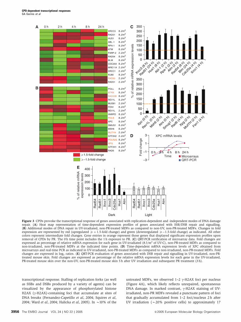
Detection of DSBs in the presence of CPDs
In addition to marking DSBs or potentially collapsed replica-
tion forks, g-H2AX foci also form upon NER- and ATR-
dependent processing of UV-induced photolesions
(O’Driscoll et al, 2003), a process involving breaks in only
one DNA strand. In order to distinguish CPD-induced DSBs
from DNA excision repair intermediates, we analyzed geno-
mic DNA from UV-treated PR and non-PR cells by pulsed-field
gel electrophoresis (Figure 5A). In the absence of PR, we
detected increased amounts of low-molecular-weight DNA,
indicative of the presence of DSBs beginning at 16 h after UV
irradiation (Figure 5A, irradiated cells, lanes marked �).
Importantly, in line with the immunocytochemical results,
DSB levels remained at background level upon complete
removal of CPDs (Figure 5A, irradiated cells, lanes marked
þ ). Under these experimental conditions, MDFs were refrac-
tory to apoptosis or necrosis. It is therefore unlikely that the
CPD-dependent DSBs result from DNA fragmentation; rather
they represent replication-dependent DNA breaks.
CPD-induced DSBs interfere with cell cycle progression
We next sought evidence for the DNA replication dependence
of the observed CPD-induced DSBs by examining the impact
of CPDs on cell cycle progression of UV-irradiated MDFs
(8 J/m2) exposed to PR light or kept in the dark, using
BrdU labelling and subsequent fluorescent activated cell
sorting (FACS) sorting. As shown in Figure 5B, UV-exposed
cells continued to incorporate BrdU for up to 8 h regardless of
PR, but at a substantially lower level than unirradiated cells,
an observation that is consistent with a slowing down of DNA
replication. After 16 h however, PR cells incorporated BrdU as
efficiently as unirradiated cells, thus indicating a return to
normal cell cycling after an initial replication delay. In con-
trast, non-PR cells largely stopped incorporating BrdU alto-
gether, suggesting that persisting CPD lesions cause a DNA
replication catastrophe. Importantly, the moment of S-phase
cell cycle arrest coincided with the time point at which DSBs
were clearly detected (16 h post-UV), which strongly impli-
cates an S-phase DNA replication dependency of DSB forma-
tion. From these data, we conclude that the initial slowing
UV+dark UV+lightNo UV
Brd
U c
onte
nt
− + +− +− +− +−
M 2 h 4 h 8 h 16 h 24 h
B
ANo UV
UV (8 J/m2)
2 h
4 h
8 h
24 h
48 h
16 h
100
102
104100
102
104100
102
104100
102
104100
102
104100
102
104
59% 13% 60% 59%15%
58%
11%
56% 14%
59%58% 15%
52%
58%
15%
61% 11% 53%
16%
13%
57%
11% 53%50%
59%
62% 10% 38% 22% 64%
16%
16%
14%
11%
13%
9%
G1
S
G2
PI0 10000 1000 0 1000
− + +− +− +− +−
Figure 5 CPDs induce the accumulation of DSBs and an S-phasecell cycle arrest. (A) Detection of DSBs by pulsed-field electrophor-esis in genomic DNA isolated from unirradiated (No UV) and UV-irradiated MDFs (UV) exposed to PR light (þ ) or not (�). (B) FACSanalysis of the cell cycle of irradiated MDFs in the presence orabsence of CPDs. The DNA (stained with propidium iodide, PI) andBrdU content of the cells is shown on the x- and y-axis, respectively.The left, middle and right panels show the effect in non-irradiated(No UV), irradiated/non-PR-treated (8 J/m2þdark) and irradiated/PR-treated (8 J/m2þ light) cells, respectively, from 2 to 48 h sub-sequent to 8 J/m2 of UV exposure and PR. The dotted line indicatesthe BrdU content of non-irradiated (left panel) versus that ofirradiated cells in the absence (middle panel) or presence (rightpanel) of PR. Percentage of cells in G1 and G2 phases of the cellcycle is indicated.
CPD-dependent transcriptional responsesGA Garinis et al
The EMBO Journal VOL 24 | NO 22 | 2005 &2005 European Molecular Biology Organization3958
down of cell cycle progression (up to 8 h post-UV) was
independent of persisting CPDs (likely due to combined
cis and trans effects from the initial presence of CPDs,
6-4PPs and non-DNA-based lesions), whereas S-phase arrest
was CPD-dependent and coincided with DSBs, which further
points to DSBs as the consequence of collapsed replication
forks or intermediates in their repair.
CPD-mediated transcriptional response of genes
associated with additional modes of DNA repair
Consistent with the replication-dependent secondary effects
of CPDs, we observed a significant upregulation of the
expression of genes involved in error-free post-replication
repair (PPR) (Rad6A, 4 h/8 J/m2) and error-prone PPR
(Rev1L, 0 h at both low and high UV doses) (Figure 3B).
Furthermore, our analysis revealed a CPD-dependent and
-independent upregulation of two mismatch repair genes
(Msh6 at 2 h/8 J/m2 and MutY at 2 h/2 J/m2).
Replication-independent modes of DNA repair, including
NER and base excision repair (BER) pathways, also appeared
transcriptionally regulated in cells exposed to 8 J/m2 UV-C.
After an initial decrease in the steady-state transcript levels at
early time points, expression of the Xpc (Figure 3B–D) and
Rad23B (Figure 3B) genes, encoding two initiators of GG-
NER, is upregulated. Unlike Xpc, Rad23B expression was
independent of persisting CPDs. In addition, the endonu-
clease Xpg (2 h/8 J/m2), two components of the DNA re-
pair/transcription initiation complex TFIIH (p44, 2 h/2 J/m2
and p62 2 h at both 2 and 8 J/m2) as well as Polb (8 h/8 J/m2)
and Smug1 (2 h/8 J/m2), typically associated with BER and/
or repair of SSBs, were transcriptionally regulated, with Polbdisplaying a CPD-independent regulation (Figure 3B). Finally,
in cells exposed to 8 J/m2 UV-C, we observed the early
transcriptional upregulation of ATR-X, a type II helicase
with homology to Rad54 that has been previously implicated
in NER and transcription (Stayton et al, 1994).
Stochastic cis-acting effects of UV on global gene
transcription
It has long been hypothesized that expression of genes with
relatively longer primary transcript lengths may be at greater
risk to transcription-blocking lesions than shorter ones.
Within the first 4 h following PR, we observed a significant
correlation between transcript length, increased number of
genes with a negative fold change and decreased number of
genes with a positive fold change at both 2 and 8 J/m2 data
sets (Supplementary Figure S3, solid bars, shown at 0 h after
PR). Importantly, this effect was lost upon removal of CPD
lesions following PR, with the difference between up- and
downregulated genes being equally pronounced regardless of
their length (Supplementary Figure S3, open bars). Thus, a
stochastic cis-acting steric hindrance induced by CPDs may
comprise a substantial threat to the timely, coordinated
transcriptional response to immediate threats (i.e. exogenous
DNA-damaging agents).
Discussion
We have previously shown that complete removal of CPDs by
PR in vivo prevents the onset of acute effects (apoptosis,
epidermal hyperplasia and erythema) and long-term res-
ponses (non-melanoma skin cancer) in the UV-exposed skin
of CPD photolyase transgenic mice (Jans et al, 2005). To gain
insight into the transcriptional response elicited by CPDs,
we employed a functional genomic approach on UV-exposed
isogenic murine cells expressing a CPD photolyase transgene
that allows rapid removal of CPD lesions in a light-dependent
manner.
CPDs have a profound effect on the transcriptional
response to UV irradiation
As could be predicted on the basis of the pronounced
attenuation of (semi)-acute effects in UV-exposed cells and
skin through PR of CPD lesions, we have found that photo-
lyase-mediated removal of CPDs has a considerable impact
on gene expression profiles in our model cellular system. The
fact that all matrix points clustered into two main groups
correlating with the PR status of the UV-exposed cell
(Figure 2A) confirmed the effect of PR on the transcriptional
response to UV and validates our experimental approach to
detect transcriptional changes.
Besides DNA, other UV wavelength-absorbing cellular
macromolecules such as RNA (Iordanov et al, 1997, 1998)
and proteins (Coffer et al, 1995) have been put forward as
primary instigators of the response to UV exposure.
Furthermore, radiation-induced effects can also be observed
in unirradiated cells via the bystander effect (Goldberg and
Lehnert, 2002). As the light-dependent removal of CPDs
negated B80% of the observed transcriptional response
(Figure 2C) and the affinity of CPD photolyase for CPD
dimers in rRNA is considerably less than in DNA (Ka¼ 102
in rRNA versus 108 in DNA) (Yasui and Eker, 1998), our data
provide evidence that damaged DNA (rather than RNA) is the
principal mediator of the cellular transcriptional response to
UV with CPDs as the primary (if indirect) stimulus. As the
initial CPD damage load is rapidly declining during the 1 h PR
period, our data indicate that the remaining B20% of UV-
responsive genes shared by PR and non-PR UV-exposed cells
represent the transcriptional response to non-CPD DNA
lesions such as 6-4PPs, thymine glycols and protein–DNA
crosslinks, as well as a variety of other damaged cellular
macromolecules, including proteins and lipids.
CPDs induce the transcriptional regulation of genes
associated with repair and signalling of SSBs and DSBs
It remained elusive, however, why the removal of CPDs
(which are poorly recognized by the GG-NER system) nega-
ted most of the (semi) acute responses in vivo (Schul et al,
2002; Jans et al, 2005) as well as the observed effects on the
transcriptional response (both in terms of number of genes
and categories of different responses) to UV in vitro (Figure
2C and D; a detailed overview is available online). We have
tackled this question by employing an unbiased approach
(instead of an arbitrary gene preselection) that combined
network analysis, GO categorization and analysis of signifi-
cantly over-represented biological processes. This led us to
identify (i) the nature of the DNA damage itself, (ii) the
signalling mechanisms involved and (iii) the implicated
biological processes.
This approach revealed several genes implicated in the
repair of DNA breaks (Rad51, Rad54, XRCC3, Blm, KU80,
Mre11a and Parp-2) to respond to the continuous presence of
CPDs in the genome (Figure 3A). Thus, DNA replication forks
CPD-dependent transcriptional responsesGA Garinis et al
&2005 European Molecular Biology Organization The EMBO Journal VOL 24 | NO 22 | 2005 3959
blocked by CPDs may eventually collapse, giving rise to
single-strand intermediates and eventually DSBs, or else
may require processing that involves formation of transient
DSBs. In either case, homologous recombination will be
employed to repair the collapsed fork (Cox et al, 2000).
In mammalian cells, however, a successful response to
DNA damage is heavily dependent on their ability to activate
a series of signalling events that will delay cell cycle progres-
sion until optimal repair can be achieved. Having identified
persistent CPDs in the genome as the likely trigger for the
induction of a combination of repair intermediates including
stalled replication forks, SSBs and/or DSBs, we examined
whether pathways relevant to the detection and signalling of
these highly toxic lesions were regulated as well. Strikingly,
among the most significantly over-represented networks
detected in this microarray study, was the ATM signalling
pathway (Figure 3A and online visualization), centrally in-
volved in the detection and signalling of DSBs in mammalian
cells (Kurz and Lees-Miller, 2004). In MDFs, this pathway
displayed a noticeable specificity to CPDs (Figure 3A and
online visualization), which was confirmed by QRT-PCR at
the corresponding time points. The in vivo relevance of this
finding was confirmed in UV-irradiated, PR and non-PR
mouse skin. Taken together, these data suggest that in rapidly
dividing cells, such as the basal keratinocytes, UV-induced
photolesions (i.e. CPDs) with the potential to block repli-
cation cause subsequent damage when encountered by a
replication fork, including SSBs and DSBs. These toxic inter-
mediates, rather than CPDs themselves, are therefore most
likely to be the signals responsible for the triggering of the
above gene networks.
Unrepaired CPD lesions induce DSBs and S-phase
cell cycle arrest
g-H2AX, P53bp1 and Rad51 foci form rapidly following
ionizing irradiation (Schultz et al, 2000; Elliott and Jasin,
2002; Fernandez-Capetillo et al, 2004; Squires et al, 2004;
Ward et al, 2004; Halicka et al, 2005) and are thought to mark
the presence of genomic DSBs. Although it is known that UV
irradiation can lead to replication-dependent g-H2AX foci
formation and DSBs (Squires et al, 2004; Ward et al, 2004;
Halicka et al, 2005), this toxic intermediate is still not
commonly associated with the normal spectrum of UV-
induced DNA lesions. The present study unequivocally
shows that g-H2AX, P53bp1 and Rad51 foci accumulated
gradually in the presence of persisting CPDs. The number
of g-H2AX foci did not substantially increase until 8 h after
exposure to UV irradiation in non-PR cells and required an
additional 16 h to accumulate in 50% of the cells (Figure 4A).
Similarly, the number of P53bp1 and Rad51 foci did not
accumulate significantly until 24 h, whereas in the case of
Rad51, it required an additional 24 h to accumulate in 50% of
cells (Figure 4B and C). Thus, CPD-mediated g-H2AX, P53bp1
and Rad51 foci formation clearly differs from g-irradiation-
mediated foci formation. For example, foci of g-H2AX are
known to appear in all cells within minutes after irradiation
(Rogakou et al, 1999). Importantly, the detection of DSBs
indicates that CPD-dependent formation of g-H2AX, P53bp1
and Rad51 foci proceeds through formation of DSBs
(Figure 5A).
The kinetics of CPD-induced g-H2AX, P53bp1 and Rad51
foci formation upon UV treatment, and the coincidence with
DSB formation suggest cell cycle dependency. Primary
human fibroblasts do not respond to DSBs by arresting cell
cycle progression until they reach S phase (Kaufmann and
Kies, 1998). UV-irradiated non-PR MDFs accumulated gradu-
ally in S phase (Figure 5B). The onset of S-phase arrest (16 h)
coincided with the time frame in which DSBs were detected
by pulsed-field gel electrophoresis, suggesting that the S
phase of the cell cycle is critical for DSB formation, probably
in response to stalled replication forks. However, in the
presence of CPDs, the fraction of cells arrested in S phase
48 h after UVexposure was substantially less than the fraction
of g-H2AX, P53bp1 and Rad51 foci positive cells within the
same time period, which suggests that foci may not only
signal the formation of DSBs but also the appearance of
stalled replication forks and SSBs.
CPDs exert pleiotropic effects via both replication-
dependent and replication-independent repair
intermediates
Whatever its origin, replicative blockage needs to be repaired
or bypassed to resume the process of replication. In mam-
mals, mutagenic bypass of DNA damage is equivalent to
error-prone translesion replication (Pages and Fuchs, 2002),
a process that requires the RAD6A protein (Barbour and Xiao,
2003). Here, we observed a CPD-dependent upregulation of
Rad6A gene expression 4 h after cells have been exposed to
8 J/m2 UV-C (Figure 3B). Error-free and error-prone modes of
PPR differ, based on the polymerase employed to bypass the
lesion. We also observed that UV exposure (2 and 8 J/m2)
elicits an immediate (0 h time point) upregulation of Rev1L
expression. Inhibition of error-prone bypass by disruption of
the polymerase zeta-associated Rev1L gene product greatly
reduces the UV-induced mutation frequency without affecting
cell survival (Gibbs et al, 2000). Thus, upregulation of Rad6A
and Rev1L, although important for prevention of replication
blocks, is likely to contribute to mutation induction after UV
exposure. Lesion bypass, especially by error-prone polyme-
rases, can lead to mismatches in nascent DNA opposite the
photolesion and mismatch repair proteins have been impli-
cated in the repair of such damage (Wang et al, 1999). The
significant upregulation of both Msh6 and MutY genes sug-
gests that UV-exposed DNA may directly signal the presence
of CPD lesions in the genome, a strategy that is anticipated to
avoid mutation fixation by replication or excision repair.
Although most of the NER genes are thought to be ubiqui-
tously expressed in mammalian cells, the Xpc and p48 genes
(encoding GG-NER-specific proteins) have been shown to be
transcriptionally induced upon UV in a p53-dependent, repli-
cation-independent manner (Hwang et al, 1999; Adimoolam
and Ford, 2002). Here, upon exposure to 8 J/m2 UV-C, we
observed coordinate transcriptional regulation of Xpc and
Rad23b encoding the binding partner of the XPC protein
(Ng et al, 2003; Figure 3B and D). Unlike Xpc, Rad23B
expression was independent of persisting CPDs, suggesting
its involvement in a wider range of stress-induced activities
than NER alone. Interestingly, the initial decrease in Xpc
mRNA levels (Figure 3D) was previously documented in
normal human fibroblasts and adenocarcinoma cells
(Adimoolam and Ford, 2002). Although the Xpc gene spans
a relatively large region (B30 kb), a UV-induced cis-mediated
transcription-blocking effect cannot fully explain the under-
lying cause of the early decrease in Xpc mRNA levels, as a
CPD-dependent transcriptional responsesGA Garinis et al
The EMBO Journal VOL 24 | NO 22 | 2005 &2005 European Molecular Biology Organization3960
variety of genes with similar or larger primary transcript
lengths failed to demonstrate comparable expression profiles.
Other transcriptionally regulated NER components include
the endonuclease Xpg and two components of the DNA
repair/transcription initiation complex TFIIH (p44 and p62)
involved in unwinding DNA surrounding a lesion. Compo-
nents involved in BER and/or repair of SSBs such as Polb and
Smug1 were regulated at the level of gene transcription,
although Polb did not require the presence of CPDs in the
genome.
CPDs induce cis-acting effects on global gene
expression
Finally, our results show that gene-length-dependent tran-
scriptional interference (Supplementary Figure S3), long pre-
dicted but never visualized on a global level in mammalian
cells, is likely to be one source of selective pressure against
long intron size in genes required for immediate response to
genotoxic insult, as has been shown previously for genes
required at high constitutive levels (Castillo-Davis et al, 2002)
and for genes that are transcriptionally activated by the
stress-induced tumor suppressor p53 (McKay et al, 2004).
Concluding remarks
Using photolyase-transgenic mouse cells that specifically
remove UV-induced CPDs upon visible light exposure, we
found that the presence of unrepaired CPD lesions (i) repre-
sents the principal mediator of the transcriptional response to
UV (ii) induces the transcriptional regulation of genes asso-
ciated with SSB and DSB signalling and repair, (iii) provokes
the time-dependent accumulation of g-H2AX, P53bp1 and
Rad51 foci and (iv) increases the amount of DSBs coincident
with an accumulation of cells in S phase. The relative
abundance of CPDs over 6-4PPs (3:1 ratio) and the ability
of NER-proficient cells to remove 6-4PPs faster than CPDs
raises the question of whether a comparable amount of
unrepaired 6-4PPs can elicit a response similar to the one
described in this study. The answer to this question should
come from studies with cells from totally NER-deficient
(Xpa�/�) CPD-PL and 6-4PP-PL (double) transgenic mice,
in which the UV dose can be adapted to generate cells with
equal amounts of unrepaired CPDs or 6-4PPs.
Taken together, our findings provide evidence that among
UV-absorbing cellular macromolecules, DNA plays the most
prominent role in downstream signalling of the damage
response, implicating CPD-dependent replication products,
rather than CPDs themselves, as the primary mediators of
the bulk transcriptional response to UV light. The fact that the
vast majority of (semi) acute and long-term responses in the
UV-exposed skin (i.e. sunburn, apoptosis, hyperplasia, cancer
initiation) have been previously ascribed to the continuing
presence of CPDs in the genome raises the possibility for a
direct role of replication-dependent SSBs and DSBs in UV-
mediated cytotoxicity. Importantly, by identifying the nature
of UV-induced SSBs and DSBs, our findings may pave the way
for specific, downstream intervention strategies.
Materials and methods
Cell cultures, mouse skin samples, UV irradiation and PRCulturing of MDFs, UV irradiation and PR were performed asdescribed previously (Schul et al, 2002). Cells were UV-treated and
PR-treated (or not) for 30 or 60 min and harvested at the indicatedtime point. Mouse skin samples were obtained from the unexposedor irradiated skin area exposed to PR light (or not), as describedpreviously (Jans et al, 2005).
Labelling and hybridization protocolsLabelling and hybridization protocols were adapted from theNational Institute of Aging (NIA, Bethesda, MD). Samples werehybridized to unirradiated, non-PR MDFs of the pertinent timepoint. 15K cDNA microarrays were obtained from the NetherlandsCancer Institute. Detailed information on experimental design, totalRNA isolation, cDNA labelling, hybridization and data extractioncan be found in Supplementary data and at http://microarrays.nki.nl/download/index.html.
Data analysisHierarchical and K clustering, self-organizing maps and analysisof variance were performed by the Spotfire Decision Sitesoftware package 7.2 version 10.0 (Spotfire Inc., MA, USA). Detailedinformation on data processing, transcript retrieval and dataanalysis, GO classification and network analysis can be found athttp://www.eur.nl/fgg/ch1/gene_network.
ImmunofluorescenceCPDs, g-H2AX and Rad51 were visualized by indirect immuno-fluorescence as previously described in irradiated (or not) cells thatwere subjected to PR (or not) and harvested for the indicated timepoints (Schul et al, 2002; Niedernhofer et al, 2004; van Veelen et al,2005). P53bp1 foci were visualized with a rabbit polyclonalantibody to P53bp1 (Novus Biologicals, CO, USA). To assess, on asemiquantitative basis, the extent of foci formation, we consideredonly those foci with an area larger than 0.24 mm2. This conservativecutoff eliminates the contribution of nonspecific fluorescent spots inthe background and actually results in an underestimate of thenumber of legitimate foci.
Pulsed-field gel electrophoresisSubconfluent MDF cultures were exposed to 8 J/m2 of UV-C, treatedwith PR (or not) and harvested for the pertinent time points. DSBswere detected by pulsed-field gel electrophoresis as described(Niedernhofer et al, 2004).
Flow cytometry and BrdU incorporationSubconfluent MDF cultures were exposed to 8 J/m2 of UV-C, treatedwith PR (or not), washed and grown for 2–48 h. At 1 h before eachof the indicated time points, MDFs were incubated with BrdU(15 mg/ml), then harvested by trypsinization, fixed in 70% ethanoland stained with propidium iodide and a-BrdU antibody (1:1000;DAKO). The DNA content of the cells was determined by FACSsorting (Facscan, Becton Dickinson). The percentage of cells in G1,S and G2 phases was calculated with CellQuest.
QRT-PCRQRT-PCR was performed with the DNA engine Opticon (MJResearch, USA). For primer sequences and data analysis, seeSupplementary data.
Data retrievalMicroarray data complied with the Minimum Information forMicroarray Experiments (MIAME), submitted to European Bioinfor-matics Institute (EBI, Hinxton) and can be retrieved at http://www.ebi.ac.uk/miamexpress (Array: A-MEXP-76, Experiment:E-MEXP-117).
Supplementary dataSupplementary data are available at The EMBO Journal Online.
Acknowledgements
This work was supported by the Dutch Cancer Foundation (EUR 98-1774, EUR 2001-2437, EMCR 2002-2701), the Erasmus MCRevolving Fund (01-432), the Association for International CancerResearch (AICR 98-259, AICR 03-128) and the EuropeanCommission. JRM was a fellow of the Damon Runyon CancerResearch Fund (DRG 1677).
CPD-dependent transcriptional responsesGA Garinis et al
&2005 European Molecular Biology Organization The EMBO Journal VOL 24 | NO 22 | 2005 3961
References
Adimoolam S, Ford JM (2002) p53 and DNA damage-inducibleexpression of the xeroderma pigmentosum group C gene. ProcNatl Acad Sci USA 99: 12985–12990
Anderson L, Henderson C, Adachi Y (2001) Phosphorylation andrapid relocalization of 53BP1 to nuclear foci upon DNA damage.Mol Cell Biol 21: 1719–1729
Barbour L, Xiao W (2003) Regulation of alternative replicationbypass pathways at stalled replication forks and its effects ongenome stability: a yeast model. Mutat Res 532: 137–155
Bohr VA, Smith CA, Okumoto DS, Hanawalt PC (1985) DNA repairin an active gene: removal of pyrimidine dimers from the DHFRgene of CHO cells is much more efficient than in the genomeoverall. Cell 40: 359–369
Bootsma D, Kraemer KH, Cleaver JE, Hoeijmakers JH (2002)Nucleotide excision repair syndromes: xeroderma pigmentosum,Cockayne syndrome, and trichothiodystrophy. In The GeneticBasis of Human Cancer, Vogelstein B, Kinzler KW (eds) pp 211–237. New York: McGraw-Hill Medical Publishing Division
Carell T, Burgdorf LT, Kundu LM, Cichon M (2001) The mechanismof action of DNA photolyases. Curr Opin Chem Biol 5: 491–498
Castillo-Davis CI, Mekhedov SL, Hartl DL, Koonin EV, KondrashovFA (2002) Selection for short introns in highly expressed genes.Nat Genet 31: 415–418
Chigancas V, Miyaji EN, Muotri AR, de Fatima Jacysyn J, Amarante-Mendes GP, Yasui A, Menck CF (2000) Photorepair preventsultraviolet-induced apoptosis in human cells expressing themarsupial photolyase gene. Cancer Res 60: 2458–2463
Coffer PJ, Burgering BM, Peppelenbosch MP, Bos JL, Kruijer W(1995) UV activation of receptor tyrosine kinase activity.Oncogene 11: 561–569
Cox MM, Goodman MF, Kreuzer KN, Sherratt DJ, Sandler SJ,Marians KJ (2000) The importance of repairing stalled replicationforks. Nature 404: 37–41
Elliott B, Jasin M (2002) Double-strand breaks and translocations incancer. Cell Mol Life Sci 59: 373–385
Fernandez-Capetillo O, Lee A, Nussenzweig M, Nussenzweig A(2004) H2AX: the histone guardian of the genome. DNA Repair(Amst) 3: 959–967
Friedberg EC, Walker GC, Siede W (1995) DNA Repair andMutagenesis. San Francisco: WH Freeman and Company
Gibbs PE, Wang XD, Li Z, McManus TP, McGregor WG, LawrenceCW, Maher VM (2000) The function of the human homolog ofSaccharomyces cerevisiae REV1 is required for mutagenesisinduced by UV light. Proc Natl Acad Sci USA 97: 4186–4191
Goldberg Z, Lehnert BE (2002) Radiation-induced effects in unir-radiated cells: a review and implications in cancer. Int J Oncol 21:337–349
Halicka HD, Huang X, Traganos F, King MA, Dai W, DarzynkiewiczZ (2005) Histone H2AX phosphorylation after cell irradiation withUV-B: relationship to cell cycle phase and induction of apoptosis.Cell Cycle 4: 339–345
Hoeijmakers JH (2001) Genome maintenance mechanisms for pre-venting cancer. Nature 411: 366–374
Hwang BJ, Ford JM, Hanawalt PC, Chu G (1999) Expression of thep48 xeroderma pigmentosum gene is p53-dependent and isinvolved in global genomic repair. Proc Natl Acad Sci USA 96:424–428
Iordanov MS, Pribnow D, Magun JL, Dinh TH, Pearson JA, Chen SL,Magun BE (1997) Ribotoxic stress response: activation of thestress-activated protein kinase JNK1 by inhibitors of the peptidyltransferase reaction and by sequence-specific RNA damage tothe alpha-sarcin/ricin loop in the 28S rRNA. Mol Cell Biol 17:3373–3381
Iordanov MS, Pribnow D, Magun JL, Dinh TH, Pearson JA, MagunBE (1998) Ultraviolet radiation triggers the ribotoxic stress re-sponse in mammalian cells. J Biol Chem 273: 15794–15803
Jans J, Schul W, Sert YG, Rijksen Y, Rebel H, Eker AP, Nakajima S,van Steeg H, de Gruijl FR, Yasui A, Hoeijmakers JH, van der HorstGT (2005) Powerful skin cancer protection by a CPD-photolyasetransgene. Curr Biol 15: 105–115
Kaufmann WK, Kies PE (1998) DNA signals for G2 checkpointresponse in diploid human fibroblasts. Mutat Res 400: 153–167
Kurz EU, Lees-Miller SP (2004) DNA damage-induced activation ofATM and ATM-dependent signaling pathways. DNA Repair(Amst) 3: 889–900
McKay BC, Stubbert LJ, Fowler CC, Smith JM, Cardamore RA,Spronck JC (2004) Regulation of ultraviolet light-inducedgene expression by gene size. Proc Natl Acad Sci USA 101:6582–6586
Mitchell DL (1988) The relative cytotoxicity of (6-4) photoproductsand cyclobutane dimers in mammalian cells. PhotochemPhotobiol 48: 51–57
Nakajima S, Lan L, Kanno S, Takao M, Yamamoto K, Eker AP, YasuiA (2004) UV light-induced DNA damage and tolerance for thesurvival of nucleotide excision repair-deficient human cells. J BiolChem 279: 46674–46677
Ng JM, Vermeulen W, van der Horst GT, Bergink S, Sugasawa K,Vrieling H, Hoeijmakers JH (2003) A novel regulation mechanismof DNA repair by damage-induced and RAD23-dependent stabi-lization of xeroderma pigmentosum group C protein. Genes Dev17: 1630–1645
Niedernhofer LJ, Odijk H, Budzowska M, van Drunen E, Maas A,Theil AF, de Wit J, Jaspers NG, Beverloo HB, Hoeijmakers JH,Kanaar R (2004) The structure-specific endonuclease Ercc1-Xpf isrequired to resolve DNA interstrand cross-link-induced double-strand breaks. Mol Cell Biol 24: 5776–5787
O’Driscoll M, Ruiz-Perez VL, Woods CG, Jeggo PA, Goodship JA(2003) A splicing mutation affecting expression of ataxia-telan-giectasia and Rad3-related protein (ATR) results in Seckel syn-drome. Nat Genet 33: 497–501
Pages V, Fuchs RP (2002) How DNA lesions are turned intomutations within cells? Oncogene 21: 8957–8966
Rappold I, Iwabuchi K, Date T, Chen J (2001) Tumor suppressor p53binding protein 1 (53BP1) is involved in DNA damage-signalingpathways. J Cell Biol 153: 613–620
Rogakou EP, Boon C, Redon C, Bonner WM (1999) Megabasechromatin domains involved in DNA double-strand breaksin vivo. J Cell Biol 146: 905–916
Sage E (1993) Distribution and repair of photolesions in DNA:genetic consequences and the role of sequence context.Photochem Photobiol 57: 163–174
Schul W, Jans J, Rijksen YM, Klemann KH, Eker AP, de Wit J,Nikaido O, Nakajima S, Yasui A, Hoeijmakers JH, van der HorstGT (2002) Enhanced repair of cyclobutane pyrimidine dimers andimproved UV resistance in photolyase transgenic mice. EMBO J21: 4719–4729
Schultz LB, Chehab NH, Malikzay A, Halazonetis TD (2000)p53 binding protein 1 (53BP1) is an early participant in thecellular response to DNA double-strand breaks. J Cell Biol 151:1381–1390
Squires S, Coates JA, Goldberg M, Toji LH, Jackson SP, Clarke DJ,Johnson RT (2004) p53 prevents the accumulation of double-strand DNA breaks at stalled-replication forks induced by UV inhuman cells. Cell Cycle 3: 1543–1557
Stayton CL, Dabovic B, Gulisano M, Gecz J, Broccoli V, GiovanazziS, Bossolasco M, Monaco L, Rastan S, Boncinelli E (1994) Cloningand characterization of a new human Xq13 gene, encoding aputative helicase. Hum Mol Genet 3: 1957–1964
Tung BS, McGregor WG, Wang YC, Maher VM, McCormick JJ(1996) Comparison of the rate of excision of major UV photo-products in the strands of the human HPRT gene of normaland xeroderma pigmentosum variant cells. Mutat Res 362:65–74
van Veelen LR, Cervelli T, van de Rakt MW, Theil AF, Essers J,Kanaar R (2005) Analysis of ionizing radiation-induced foci ofDNA damage repair proteins. Mutat Res 574: 22–33
Wang H, Lawrence CW, Li GM, Hays JB (1999) Specific bindingof human MSH2.MSH6 mismatch-repair protein heterodimersto DNA incorporating thymine- or uracil-containing UV lightphotoproducts opposite mismatched bases. J Biol Chem 274:16894–16900
Ward IM, Minn K, Chen J (2004) UV-induced ataxia-telangiectasia-mutated and Rad3-related (ATR) activation requires replicationstress. J Biol Chem 279: 9677–9680
Yasui A, Eker APM (1998) DNA photolyases. In DNA Damageand Repair, Vol. 2: DNA Repair in Higher Eukaryotes, NickoloffJA, Hoekstra MF (eds) Vol.2, pp 9–32. Totowa, NJ: Humana Press
Yoon JH, Lee CS, O’Connor TR, Yasui A, Pfeifer GP (2000) The DNAdamage spectrum produced by simulated sunlight. J Mol Biol 299:681–693
CPD-dependent transcriptional responsesGA Garinis et al
The EMBO Journal VOL 24 | NO 22 | 2005 &2005 European Molecular Biology Organization3962
Related Documents