J Physiol 587.17 (2009) pp 4213–4233 4213 Upregulation of inward rectifier K + (Kir2) channels in dentate gyrus granule cells in temporal lobe epilepsy Christina C. Young 1,2 , Michael Stegen 1,3 , Ren´ e Bernard 4 , Martin M ¨ uller 3,5 , Josef Bischofberger 6 , R¨ udiger W. Veh 4 , Carola A. Haas 5 and Jakob Wolfart 1 1 Cellular Neurophysiology, Dept. of Neurosurgery, University Medical Center Freiburg, Breisacher Str. 64, 79106 Freiburg, Germany 2 Faculty of Biology, University of Freiburg, Schaenzlestrasse 1, 79104 Freiburg, Germany 3 Faculty of Pharmaceutical Sciences, University of Freiburg, Hebelstraße 27, 79085 Freiburg, Germany 4 Institut f¨ ur Integrative Neuroanatomie, Charit´ e – Universit¨ atsmedizin Berlin, Philippstr. 12, 10115 Berlin, Germany 5 Experimental Epilepsy Research Group, Dept. of Neurosurgery, University Medical Center Freiburg, Breisacher Str. 64, 79106 Freiburg, Germany 6 Physiological Institute, University of Freiburg, Hermann-Herder-Straße 7, 79104 Freiburg, Germany In humans, temporal lobe epilepsy (TLE) is often associated with Ammon’s horn sclerosis (AHS) characterized by hippocampal cell death, gliosis and granule cell dispersion (GCD) in the dentate gyrus. Granule cells surviving TLE have been proposed to be hyperexcitable and to play an important role in seizure generation. However, it is unclear whether this applies to conditions of AHS. We studied granule cells using the intrahippocampal kainate injection mouse model of TLE, brain slice patch-clamp recordings, morphological reconstructions and immunocytochemistry. With progressing AHS and GCD, ‘epileptic’ granule cells of the injected hippocampus displayed a decreased input resistance, a decreased membrane time constant and an increased rheobase. The resting leak conductance was doubled in epileptic granule cells and roughly 70–80% of this difference were sensitive to K + replacement. Of the increased K + leak, about 50% were sensitive to 1 mm Ba 2+ . Approximately 20–30% of the pathological leak was mediated by a bicuculline-sensitive GABA A conductance. Epileptic granule cells had strongly enlarged inwardly rectifying currents with a low micromolar Ba 2+ IC 50 , reminiscent of classic inward rectifier K + channels (Irk/Kir2). Indeed, protein expression of Kir2 subunits (Kir2.1, Kir2.2, Kir2.3, Kir2.4) was upregulated in epileptic granule cells. Immunolabelling for two-pore weak inward rectifier K + channels (Twik1/K2P1.1, Twik2/K2P6.1) was also increased. We conclude that the excitability of granule cells in the sclerotic focus of TLE is reduced due to an increased resting conductance mainly due to upregulated K + channel expression. These results point to a local adaptive mechanism that could counterbalance hyperexcitability in epilepsy. (Received 18 February 2009; accepted after revision 24 June 2009; first published online 29 June 2009) Corresponding author J. Wolfart: Cellular Neurophysiology, Dept of Neurosurgery, University Medical Center Freiburg, Breisacher Str. 64, 79106 Freiburg, Germany. Email: [email protected] Abbreviations AHS, Ammon’s horn sclerosis; GCD, granule cell dispersion; g rest , resting leak conductance; KA, kainic acid; K2P/Twik, two-pore weak inward rectifier K + ; Kir2/Irk, classic inward rectifier K + ; Kir3/Girk, G-protein-coupled inward rectifier K + ; R in , input resistance; R m , specific membrane resistance; τ m , membrane time constant; TLE, mesial temporal lobe epilepsy; V rest , resting membrane potential Mesial temporal lobe epilepsy (TLE), one of the most prevalent forms of focal epilepsies, is often intractable but surgical resection of the hippocampus and adjacent medial temporal structures leads to seizure cessation in most cases. Resected hippocampi of TLE patients often show Ammon’s horn sclerosis (AHS), characterized by marked hippocampal cell death, gliosis and network Christina C. Young and Michael Stegen contributed equally to this work. disorganization (Blumcke et al. 2002; Thom et al. 2002). Closely linked to hippocampal damage is granule cell dispersion (GCD) in the dentate gyrus (Houser, 1990; Thom et al. 2002). The role of granule cells in TLE is not clear. Granule cells survive hippocampal damage better than neighbouring cell populations and have been implicated in seizure generation (Sloviter, 1991; Heinemann et al. 1992; Okazaki et al. 1999; Selke et al. 2006). On the other hand, doubts have been raised whether granule cells actively contribute C 2009 The Authors. Journal compilation C 2009 The Physiological Society DOI: 10.1113/jphysiol.2009.170746 ) at Physiologisches Institut on March 12, 2010 jp.physoc.org Downloaded from J Physiol (

Welcome message from author
This document is posted to help you gain knowledge. Please leave a comment to let me know what you think about it! Share it to your friends and learn new things together.
Transcript

J Physiol 587.17 (2009) pp 4213–4233 4213
Upregulation of inward rectifier K+ (Kir2) channels indentate gyrus granule cells in temporal lobe epilepsy
Christina C. Young1,2, Michael Stegen1,3, Rene Bernard4, Martin Muller3,5, Josef Bischofberger6,Rudiger W. Veh4, Carola A. Haas5 and Jakob Wolfart1
1Cellular Neurophysiology, Dept. of Neurosurgery, University Medical Center Freiburg, Breisacher Str. 64, 79106 Freiburg, Germany2Faculty of Biology, University of Freiburg, Schaenzlestrasse 1, 79104 Freiburg, Germany3Faculty of Pharmaceutical Sciences, University of Freiburg, Hebelstraße 27, 79085 Freiburg, Germany4Institut fur Integrative Neuroanatomie, Charite – Universitatsmedizin Berlin, Philippstr. 12, 10115 Berlin, Germany5Experimental Epilepsy Research Group, Dept. of Neurosurgery, University Medical Center Freiburg, Breisacher Str. 64, 79106 Freiburg, Germany6 Physiological Institute, University of Freiburg, Hermann-Herder-Straße 7, 79104 Freiburg, Germany
In humans, temporal lobe epilepsy (TLE) is often associated with Ammon’s horn sclerosis(AHS) characterized by hippocampal cell death, gliosis and granule cell dispersion (GCD) inthe dentate gyrus. Granule cells surviving TLE have been proposed to be hyperexcitable andto play an important role in seizure generation. However, it is unclear whether this appliesto conditions of AHS. We studied granule cells using the intrahippocampal kainate injectionmouse model of TLE, brain slice patch-clamp recordings, morphological reconstructions andimmunocytochemistry. With progressing AHS and GCD, ‘epileptic’ granule cells of the injectedhippocampus displayed a decreased input resistance, a decreased membrane time constant andan increased rheobase. The resting leak conductance was doubled in epileptic granule cellsand roughly 70–80% of this difference were sensitive to K+ replacement. Of the increasedK+ leak, about 50% were sensitive to 1 mm Ba2+. Approximately 20–30% of the pathologicalleak was mediated by a bicuculline-sensitive GABAA conductance. Epileptic granule cells hadstrongly enlarged inwardly rectifying currents with a low micromolar Ba2+ IC50, reminiscentof classic inward rectifier K+ channels (Irk/Kir2). Indeed, protein expression of Kir2 subunits(Kir2.1, Kir2.2, Kir2.3, Kir2.4) was upregulated in epileptic granule cells. Immunolabelling fortwo-pore weak inward rectifier K+ channels (Twik1/K2P1.1, Twik2/K2P6.1) was also increased.We conclude that the excitability of granule cells in the sclerotic focus of TLE is reduced due to anincreased resting conductance mainly due to upregulated K+ channel expression. These resultspoint to a local adaptive mechanism that could counterbalance hyperexcitability in epilepsy.
(Received 18 February 2009; accepted after revision 24 June 2009; first published online 29 June 2009)Corresponding author J. Wolfart: Cellular Neurophysiology, Dept of Neurosurgery, University Medical Center Freiburg,Breisacher Str. 64, 79106 Freiburg, Germany. Email: [email protected]
Abbreviations AHS, Ammon’s horn sclerosis; GCD, granule cell dispersion; g rest, resting leak conductance; KA, kainicacid; K2P/Twik, two-pore weak inward rectifier K+; Kir2/Irk, classic inward rectifier K+; Kir3/Girk, G-protein-coupledinward rectifier K+; R in, input resistance; Rm, specific membrane resistance; τm, membrane time constant; TLE, mesialtemporal lobe epilepsy; V rest, resting membrane potential
Mesial temporal lobe epilepsy (TLE), one of the mostprevalent forms of focal epilepsies, is often intractablebut surgical resection of the hippocampus and adjacentmedial temporal structures leads to seizure cessation inmost cases. Resected hippocampi of TLE patients oftenshow Ammon’s horn sclerosis (AHS), characterized bymarked hippocampal cell death, gliosis and network
Christina C. Young and Michael Stegen contributed equally to this work.
disorganization (Blumcke et al. 2002; Thom et al. 2002).Closely linked to hippocampal damage is granule celldispersion (GCD) in the dentate gyrus (Houser, 1990;Thom et al. 2002).
The role of granule cells in TLE is not clear. Granule cellssurvive hippocampal damage better than neighbouringcell populations and have been implicated in seizuregeneration (Sloviter, 1991; Heinemann et al. 1992; Okazakiet al. 1999; Selke et al. 2006). On the other hand, doubtshave been raised whether granule cells actively contribute
C© 2009 The Authors. Journal compilation C© 2009 The Physiological Society DOI: 10.1113/jphysiol.2009.170746
) at Physiologisches Institut on March 12, 2010jp.physoc.orgDownloaded from J Physiol (
4214 C. C. Young and others J Physiol 587.17
to seizures (Sloviter, 1994; Liu et al. 2000; Harvey &Sloviter, 2005). Furthermore, although the structuralchanges in AHS are often considered a ‘pathologicalsubstrate’ for TLE (Blumcke et al. 2002; Thom et al. 2002),it is unclear whether the sclerotic tissue really is the originof hyperexcitability in TLE (King et al. 1997; Mueller et al.2007; Le Duigou et al. 2008). In particular, only little isknown about the electrophysiology of granule cells inrelation to AHS and GCD.
Recently, we found a reduced input resistance (R in) andan increased inwardly rectifying conductance in granulecells of TLE patients with AHS (Stegen et al. 2009), aresult which contrasted with a large number of previousresults on intrinsic properties of granule cells in TLEpatients and TLE animal models (e.g. Mody et al. 1988,1992; Williamson et al. 1995; Isokawa, 1996b; Molnar &Nadler, 1999; Okazaki et al. 1999; Scharfman et al. 2000;Dietrich et al. 2005; Selke et al. 2006). To further study theunderlying mechanisms under controlled conditions, weutilized the focal KA injection mouse model of TLE wherenot only recurrent, focal, pharmaco-resistant seizuresbut also AHS and GCD have been well characterized(Suzuki et al. 1995; Bouilleret et al. 1999; Riban et al.2002; Kralic et al. 2005; Le Duigou et al. 2005; Heinrichet al. 2006). Using patch-clamp recordings of granulecells in brain slices, we found an increased restingconductance with progressing AHS. In addition, we usedquantitative pharmacology, quantitative morphology,computer simulations and immunocytochemistry toidentify mechanisms responsible for the increased leakconductance. Our results suggest that the increased leakwas due to an upregulation of inward rectifier K+ (Kir)channels, in particular of the Kir2 type, in combinationwith an increased GABAA conductance. These resultspoint to homeostatic mechanisms in TLE that could resultin shunting of epileptic input and thereby protect granulecells in the sclerotic focus.
Methods
Animals
All animal procedures were performed in accordancewith the guidelines of the European Community CouncilDirective of November 24, 1986 (86/609/EEC) and wereapproved by the regional council and local animalwelfare officer according to the German animal protectionact (Tierschutzgesetz). Experiments were conducted onmale C57Bl/6 mice (64 controls, 119 KA injections, 3saline injections and 3 mice for immunocytochemistry).Animals were held in a 12 h light–dark cycle (roomtemperature, 21.5–22.5◦C) with food and water adlibitum. Before decapitation and brain removal mice wereanaesthetized with isofluorane.
KA injections
Kainic acid (KA, in 0.9% NaCl) or saline injectionswere performed as previously described (Heinrich et al.2006). Briefly, 5- to 6-week-old mice were anaesthetizedI.P. with a mixture of (in μg (g body weight)−1) 100ketamine, 5 xylazin and 0.1 atropin and placed into astereotaxic frame. Fifty nanolitres of a 20 mM KA solutionin 0.9% NaCl were injected into the right dorsal CA1region of the hippocampus during 1 min using a 0.5 μlmicrosyringe (Hamilton, Bonaduz, Switzerland) and amicropump (CMA/100, Carnegie Medicine, Stockholm,Sweden). The coordinates for injection (with bregmaas reference) were: anteroposterior 1.9 mm, mediolateral1.5 mm and dorsoventral 1.9 mm. The cannula was lefttwo additional minutes after injection to avoid refluxalong the cannula track. After recovery from anaesthesia astatus epilepticus of several hours was observed. Since,in this model morphological, electrophysiological andbehavioural symptoms of chronic TLE develop within2 weeks post-KA injection (Suzuki et al. 1995; Bouilleretet al. 1999; Heinrich et al. 2006), experiments wereperformed 2–8 weeks after KA injection, if not statedotherwise.
Brain slice preparation
Mice (6 to 15 weeks old; control 6–11 weeks, KA-injected6–15 weeks) were deeply anaesthetized with isoflurane andkilled by decapitation. The brain was immersed in ice-coldartificial cerebrospinal fluid (ACSF) containing (in mM):87 NaCl, 25 NaHCO3, 2.5 KCl, 1.25 NaH2PO4, 0.5 CaCl2,7 MgCl2, 75 sucrose and 10 glucose (equilibrated with95% O2–5% CO2). Coronal slices 400 μm thick werecollected with a vibratome VT1200S (Leica, Bensheim,Germany) from the dorsal hippocampus (in KA-injectedmice approximately ±0.8 mm from the injection site).Slices were incubated for 30 min at 35–36◦C andsubsequently kept at room temperature in oxygenatedsucrose ACSF for more than 1 h until they weretransferred individually for electrophysiologicalexperiments.
Electrophysiology
Patch-clamp methods were adopted from(Schmidt-Hieber et al. 2004, 2007). For patch-clamprecordings, brain slices were transferred to a recordingchamber and continuously superfused at roomtemperature with ACSF containing (in mM): 125 NaCl,25 NaHCO3, 2.5 KCl, 1.25 NaH2PO4, 2 CaCl2, 1 MgCl2
and 25 glucose (equilibrated with 95% O2–5% CO2).Recordings were obtained from neurons in the upperblade of the dentate gyrus granule cell layer visualized bydifferential interference contrast video microscopy using
C© 2009 The Authors. Journal compilation C© 2009 The Physiological Society
) at Physiologisches Institut on March 12, 2010jp.physoc.orgDownloaded from J Physiol (

J Physiol 587.17 Kir2 upregulation in the epileptic hippocampus 4215
a 63×/1.0 objective in an upright microscope (Axioskop2FS, Zeiss, Oberkochen, Germany). Cells of KA-injectedanimals were from the injected side (‘epileptic’) or theun-injected side (contralateral) or from naive animals(‘control’). To exclude new-born neurons from theanalysis we avoided recording at the border betweengranule cell layer and hilus (Schmidt-Hieber et al. 2004).Patch pipettes were pulled from borosilicate glass usinga DMZ-universal puller (Zeitz, Martinsried, Germany).They were filled with a solution containing (in mM): 135potassium gluconate, 20 KCl, 10 Hepes, 0.1 EGTA, 2MgCl2, 2 Na2ATP and 0.2% biocytin (pH = 7.28) and hadtip resistances of 5.2 ± 0.04 M� (range 3.3–7.7). To obtainoptimal biocytin labelling, the whole-cell configurationwas maintained for at least 25 min and the pipette wasretrieved via the outside-out configuration. Records werefiltered at 8–10 kHz using a SEC05LX amplifier (NPI,Tamm, Germany) and digitized at 10–20 kHz (voltage andcurrent clamp, respectively) using a ITC18 D/A converter(Instrutech, Port Washington, NY, USA) and PatchMastersoftware (Heka, Lambrecht, Germany). Series resistances(7–16 M�) were compensated via bridge balance andpipette capacitance was compensated via fast capacitancecontrols of the SEC amplifier. Seal resistances (Rseal)were >1 G� (control, 3.3 ± 0.2 G�, n = 125; epileptic,2.5 ± 0.1 G�, n = 197, P < 0.001). Accepted ratios ofR in/Rseal were less than 0.3 (control, 0.15 ± 0.01, n = 125;epileptic, 0.11 ± 0.004, n = 197, P < 0.001), except whenstated otherwise (<0.1). The liquid junction potential wasdetermined to be 10 mV and voltages were appropriatelycorrected (Staley & Mody, 1992; Okazaki et al. 1999).Only cells with resting membrane potentials (V rest)negative to −65 mV and overshooting action potentialswere included in the analysis.
Pharmacology
In K+ replacement experiments cells were first recordedwith normal intracellular solution and (via theoutside out configuration) subsequently repatchedwith K+-free intracellular solution. In these cases, K+
was replaced equimolar with TEA in the ACSF andthe pipette solution contained (in mM): 135 TEACl,20 CsCl, 0.1 EGTA, 2 MgCl2, 2 Na2ATP and 0.2%biocytin (pH 7.28 adjusted with TEA hydroxide).Except in current-clamp conditions, experimentswere conducted in the presence of the Na+ channelblocker tetrodotoxin (TTX, 0.5 μM) and inhibitors ofAMPA/KA-type and NMDA-type glutamate receptorsusing 50 μM D(-)-2-amino-5-phosphonopentanoic acid(D-AP5) and 20 μM 1,2,3,4-tetrahydro-7-nitro-2,3-dioxoquinoxaline-6-carbonitrile disodium (CNQX) asmentioned in the text. The following drugs were keptin H2O stocks at −20◦C: 1(S),9(R)-(−)-bicuculline
methiodide (BMI), bupivacaine-HCl, CsCl, CNQX,D-AP5, TEACL, TTX, BaCl2, 4-aminopyridine (4-AP),1,3-dihydro-1-phenyl-3,3-bis(4-pyridinylmethyl)-2H-indol-2-one (linopirdine), apamin, r-tertiapinQ, 10,10-bis(4-pyridinylmethyl)-9(10H)-anthracenone dihydro-chloride (XE991), 4-ethylphenylamino-1,2-dimethyl-6-methylaminopyrimidinium chloride (ZD7288). PTX andtolbutamide were kept in DMSO stocks at −20◦C anddiluted (1 : 1000) freshly in oxygenated glucose ACSF.(RS)-3-amino-2-(4-chlorophenyl)propylphosphonic acid(phaclofen) was solved in 100 mM NaOH. Drugs werekept in glass syringes of an application system (Auto-Mate Scientific, Berkeley, CA, USA) under carbogenpressurized at 1300–1600 hPa before bath application.We obtained D-AP5, CNQX, XE991 and ZD7288 fromAscent Scientific (Weston-Super-Mare, UK), apaminand linopirdine from Tocris (Bristol, UK), r-tertiapin-Qfrom Alomone (Jerusalem, Israel), tolbutamide from ICNBiomedicals (Aurora, OH, USA) and all other substancesfrom Sigma-Aldrich (Taufkirchen, Germany).
Morphology and immunocytochemistry
For cell type identification, slices were fixed overnightwith 4% paraformaldehyde in 0.1 M PB pH 7.4. After3 washing steps with PB, slices were treated for 30 minwith a blocking solution containing 0.3% Triton X-100and 10% normal goat serum (NGS) and incubatedeither for more than 3 h at room temperature orovernight at 4◦C with a rabbit polyclonal anti-Prox1antibody, (1 : 1000, Chemicon, Temecula, CA, USA) in0.1% Triton and 1% NGS. After 3 washes, slices wereincubated with Alexa Fluor-546-streptavidin (1 : 500,Invitrogen, Karlsruhe, Germany) for biotin detectionand a secondary anti-rabbit antibody conjugated withAlexa Fluor-488 (1 : 200, Invitrogen) either for more than3 h at room temperature or overnight at 4◦C. After 5washes, slices were mounted in fluorescence mountingmedium (DAKO, Glastrup, Denmark) or ProLong goldantifade reagent (Invitrogen). For overviewreconstructions, GCD measurements and cellidentification immunofluorescence was analysedwith an Axioplan 2 microscope equipped with Apotometechnology (Zeiss) using the 20×/0.75 objective andextended focal images. GCD was quantified as widthof granule cell layer measured at the position andlevel of reconstructed neurons. If a recorded cell couldnot unambiguously be identified by biocytin staining,three GCD measurements per slice were averaged. Theexperimenters performing the GCD measurements wereunaware of the respective electrophysiological results.
For detailed reconstructions using confocalmicroscopy, slices were washed 4 times after fixationand were incubated with FITC–AvidinD (1 : 500, Vector
C© 2009 The Authors. Journal compilation C© 2009 The Physiological Society
) at Physiologisches Institut on March 12, 2010jp.physoc.orgDownloaded from J Physiol (

4216 C. C. Young and others J Physiol 587.17
Laboratories, Burlingame, CA, USA) in 0.3% Triton (in0.1 M PB) overnight at 4◦C. After 3 washes, slices weremounted in ProLong gold antifade reagent (Invitrogen)and stored 7 days prior to confocal acquisition. Fordetailed morphological reconstructions of cellularcompartments, we used a Zeiss LSM 510 equipped withan argon laser (488 nm), a long-pass emission filter(LP505) and a 40×/1.3 NA oil-immersion objective.Soma, axon and dendrites were traced using image stacks(0.25 × 0.25 μm pixel−1 to 0.32 × 0.32 μm pixel−1 inx–y plane, 0.5 μm in z-axis) with the filament tracingsoftware Neurolucida (mbf Bioscience, Williston, VT,USA). Spines were manually counted with variably sizedmarkers and spine surface was estimated by assumingsphere-like shapes neglecting spine necks. We did notcorrect for hidden spines and for tissue shrinkage.
For Kir channel and Twik channel immunoperoxidasecytochemistry, animals were deeply anaesthetized byintraperitoneal injections of a cocktail consisting of 45%ketamine (100 mg ml–1), 35% xylazine (20 mg ml–1)and 20% saline, at a dose of 0.16 ml (100 g of bodyweight)–1. After 200 IU heparin I.P. they were fixed viatranscardial perfusion with 4% paraformaldehyde, 0.05%glutaraldehyde and 0.2% picric acid in 0.1 M PB, pH 7.4(PGPic). Brains were removed from the skull, cut intopreselected blocks, cryoprotected in 0.4 M sucrose forabout 4 h and in 0.8 M sucrose overnight, shock-frozenin hexane at −70◦C, and stored at −80◦C until use.Coronal 20-μm-thick cryostat sections were stored at−20◦C in a cryoprotectant solution (30% sucrose and30% ethylene glycol in 0.1 M PB, pH 7.4) until use.Freely floating sections were rinsed in 0.01 M PBS at pH7.4, treated for 15 min with 1% sodium borohydridein PBS and thoroughly washed in PBS. Thereafter,sections were pretreated for 30 min in a blocking andpermeabilizing solution, consisting of 10% NGS(Interchem, Bad Kreuznach, Germany), 0.3%Triton X-100 and 0.05% phenylhydrazine (Merck,Darmstadt, Germany) in PBS at RT. Primary antibodiesagainst Kir2 and Kir3 channels (Pruss et al. 2003; Eulitzet al. 2007) or against Twik channels were diluted (Kir2.1,1 : 500; Kir2.2; 1 : 100; Kir2.3, 1 : 100; Kir2.4, 1 : 5000;Kir3.1, 1 : 5000; Kir3.2, 1 : 1000; Kir3.3, 1 : 5000; Kir3.4,1 : 100; Twik1, 1 : 200, sc-11483, Santa Cruz, Santa Cruz,CA, USA; Twik, 1 : 500, APC-040, Alomone Labs) inPBS containing 10% NGS, 0.3% Triton X-100, 0.1%sodium azide and 0.01% thiomersal, and applied for36 h at 2◦C. Thereafter, sections were thoroughly rinsedin PBS, pretreated for 1 h with 0.2% bovine serumalbumin in PBS (PBS-A), and exposed for another 24 hto the secondary antibodies (biotinylated goat anti-rabbitIgG, 1 : 2000; biotinylated horse anti-goat IgG, 1 : 2000;both from Vector Labs). After repeated washings in PBSand preincubation for 1 h in PBS-A, the biotinylatedsecondary antibodies in the sections were complexed for
another 12 h with a preformed ABC-complex (VectorLabs, 1 : 200 in PBS-A). After further thorough rinses inPBS, preincubation for 15 min in a solution of 0.05%diaminobenzidine and 10 mM imidazole in 50 mM Trisbuffer, pH 7.6, the visualization of the antigen–antibodycomplexes was started by the addition of hydrogenperoxide (0.0015%; 25 μl of 0.03% hydrogen peroxideto 500 μl solution) and stopped after 15 min at RT byrepeated washings with PBS. Sections were mountedonto gelatin-coated slides, air-dried, dehydrated througha graded series of ethanol, transferred to xylene, andcoverslipped with Entellan (Merck). For cresyl violetstaining slide-mounted sections were left in 70% ethanolovernight, rinsed in bidistilled water, and stained with0.2% cresyl violet acetate in 20 mM acetate buffer, pH4.0, for 30 min at RT. After rinsing in bidistilled water,sections were dehydrated and coverslipped.
Cable modelling
Passive cable models were obtained using NEURON 6.2or 7.0 for Windows XP (Carnevale & Hines, 2006) withmethods adopted in part from (Schmidt-Hieber et al.2007). Digitized morphological data were imported inNEURON from Neurolucida format (see morphology andimmunocytochemistry). Spines were modelled explicitlyand consisted of a neck with variable length but fixeddiameter of 0.18 μm (Hama et al. 1989) and a headwith measured size. We did not correct for hiddenspines and tissue shrinkage. Segment length was adjustedaccording to the ‘d_lambda rule’ by calculation of thealternating current length constant λ at 1 kHz for eachsection (Carnevale & Hines, 2006; Schmidt-Hieber et al.2007). The integration time step was 50 μs. The specificmembrane resistance (Rm), membrane capacitance (C m)and intracellular resistivity (Ra) were obtained by directleast-squares fitting of the response of the model tothe experimental data in NEURON using the built-in‘Brent’s principal axis’ algorithm that minimizes the sumof squared errors (χ2). To constrain model parameters,voltage responses to 300 ms current pulses were used andχ2 was calculated in this period.
Data analysis
Electrophysiological records were analysed using IgorPro(WaveMetrcis, Portland, OR, USA) and FitMaster (Heka).The input resistance (R in) was calculated from the slopeof the steady-state current–voltage (I–V ) relation fromvoltage responses of less than ±10 mV from V rest. Themembrane time constant (τm) was derived by the averageof four to six single exponentials fitted to voltage responsesto 450 ms current steps. The resting conductance (g rest)was determined in voltage-clamp experiments at holding
C© 2009 The Authors. Journal compilation C© 2009 The Physiological Society
) at Physiologisches Institut on March 12, 2010jp.physoc.orgDownloaded from J Physiol (

J Physiol 587.17 Kir2 upregulation in the epileptic hippocampus 4217
potentials of −80 mV and calculated as 1/slope of fittedcurrent–voltage relationships (4 steps of 3 mV). For thecalculation of IC50 values, fractional block of g rest or ofcurrents at −80 to −140 mV was determined by fitting thevalues to the Hill curve y = B + (1 - B)/(1 + (IC50/C)∧S),where y is the g rest or current during or before Ba2+,B is the Ba2+-resistant component and S is the slope.Statistical significance of group differences was measuredusing Prism 4.0 software (GraphPad, San Diego, CA, USA)applying the following tests: ANOVA plus post hoc Tuckey’stest for more than two groups with normal distribution,Kruskal–Wallis test plus Dunn’s test for more than twogroups not normally distributed, Mann–Whitney’s testfor two groups not normally distributed, Student’s t testsfor two groups normally distributed and Wilcoxon signedrank test for paired tests. Significance of correlation wasdetermined according to a table of Pearson’s r values.Levels of significance are indicated in figures as ∗ (< 0.05),∗∗ (< 0.01) and ∗∗∗ (< 0.001). Mean values are ± S.E.M.and numbers represent cells if not mentioned otherwise.Figures were produced using Prism, Illustrator and Photo-shop (Adobe, Munchen, Germany).
Results
The intrahippocampal kainate injection mousemodel of TLE
Mice with unilateral intrahippocampal KA injectionssuffered an initial status epilepticus and developedTLE-like AHS with marked GCD in the injectedhippocampus, while the contralateral side resembled thatof naive mice (Fig. 1A and B). Morphological analysisrevealed a severe loss of neurons especially in the CA1and CA3c region of the KA-injected hippocampus whilethe CA3a and CA3b regions still contained viable neurons(Fig. 1A, arrow, and B). The dentate gyrus showedGCD and was 3- to 4-fold broader as compared to thecontralateral side (Fig. 1A and B). Described data are ingood agreement with previous descriptions of the intra-hippocampal KA mouse model of TLE (for details seeSuzuki et al. 1995; Bouilleret et al. 1999; Riban et al. 2002;Kralic et al. 2005; Le Duigou et al. 2005, 2008; Heinrichet al. 2006). These previous studies also showed that theanatomical changes develop over the first 2 weeks after KAinjection. Therefore, the following results were obtainedfrom animals more than 14 days after injection (exceptwhen stated otherwise).
At the cellular level, granule cells on the KA-injectedside displayed untypical large proximal excrescences,altered spine morphology, hypertrophic cell bodies andoften mossy fibre backsprouting (Figs 1C, and 3A andB) confirming previous observations from KA-injectedmice (Suzuki et al. 1997) (see below for morphologicalquantifications). We found at least one basal dendrite per
cell in 47 of 62 (75.8%) epileptic cells while none wasfound in examined control granule cells (n = 43).
The input resistance is reduced in epileptic granulecells of kainate-injected mice
Passive membrane properties such as R in and τm areimportant factors for postsynaptic signal integration. TheR in is also commonly recorded as a starting routineto test the viability of recorded cells which impliesthat changes in the passive properties may have beenmissed in many studies in the selection process. Wecompared the R in and τm values of granule cells fromnaive mice (control) and KA-injected mice (contralateraland ipsilateral/‘epileptic’). To ensure that recorded cellswere granule cells they were labelled with biocytin duringrecordings for post hoc anatomical reconstructions andimmunocytochemical identification. All recovered cellswere positioned in the upper blade of the granule celllayer and had spiny apical dendrites extending into themolecular layer (Fig. 1C, Fig. 3A and B). As the positionand morphology of granule cells is altered in KA-injectedmice, cells were co-labelled with Prox1, a granule cellmarker (Fig. 1C) (Liu et al. 2000). Prox1-negative cells(4 of 196 recovered cells) were excluded fromfurther analysis. Electrophysiological and morphologicalproperties of control granule cells (Fig. 1C–F) matchedthose previously described for mature granule cellsrecorded in adult rodents using similar techniques (Staley& Mody, 1992; Schmidt-Hieber et al. 2004).
The R in was strongly reduced in epileptic granulecells (Fig. 1D and E) (control, 421 ± 12 M�, n = 125;epileptic, 218 ± 5 M�, n = 197, P < 0.001). The R in ofgranule cells from saline-injected hippocampi was notdifferent from control granule cells (n = 9, P > 0.05,data not shown). Although V rest values of granulecells were largely overlapping, the mean of 197epileptic granule cells was 3.4 mV depolarized comparedto control cells (control, −81.1 ± 0.5 mV, n = 125;epileptic, −77.7 ± 0.4 mV; n = 197, P < 0.001, see belowfor discussion on V rest). Since overall contralateralgranule cells displayed a morphological and electro-physiological phenotype similar to control granule cells(Fig. 1C–E) (V rest: −82.5 ± 1.3 mV, R in: 513 ± 33 M�,n = 21, P > 0.05 vs. control, respectively) we comparedonly control vs. epileptic granule cells for the followinganalyses.
To rule out that differences in recording Rseal resultedin erroneous underestimation of epileptic R in values, weverified that the ratio of R in/Rseal was sufficiently low toavoid a significant influence on the measured R in (seeMethods). To provide extra security against this error, weadditionally selected only those cells with a R in/Rseal ratioof< 0.1 and these ratios were not different between control
C© 2009 The Authors. Journal compilation C© 2009 The Physiological Society
) at Physiologisches Institut on March 12, 2010jp.physoc.orgDownloaded from J Physiol (

4218 C. C. Young and others J Physiol 587.17
and epileptic granule cells (control, 0.076 ± 0.004, n = 33;epileptic, 0.066 ± 0.002, n = 103, P > 0.05). Even withthese stringent criteria the R in difference remained robust(control, 371 ± 22 M�, n = 33; epileptic, 187 ± 6 M�,n = 103, P < 0.001), suggesting that control and epilepticgranule cells did indeed possess a different R in.
Figure 1. The input resistance is reducedin epileptic granule cells ofkainate-injected miceThe input resistance (Rin) is reduced in‘epileptic’ granule cells of kainate(KA)-injected mice. A and B, cresyl violetstaining of KA-injected (ipsilateral) hippocampishow a complete loss of neurons in the CA1region (B, arrowheads) and a severe loss in theCA3c region of the injected hippocampus. TheCA3a and CA3b regions still contained viableneurons (A arrow, B). The dentate gyrusgranule cell layer (DG) of the injectedhippocampus is 3- to 4-fold broader,displaying largely dispersed cells in this area(GCD). Note that the molecular layer of theDG can still be separated from the formerouter stratum lacunosum-moleculare viaresidual capillaries (B, asterisks; see alsoFig. 7A1 and C2). The upper border of theCA1 region against the corpus callosum is alsovisible (B, arrows). C, biocytin-labelled granulecells of control and KA-injected mice (KA,contralateral and ipsilateral, respectively)co-labelled with the granule cell markerProx-1. Scale bars, 100 μm. D, current–voltagerelation of control (C, open circles),contralateral and epileptic granule cells of theKA-injected mice (KAc, open squares, and KAi,grey triangles, respectively) as it was used forthe calculation of Rin in E. V rel, voltage relativeto the resting membrane potential (V rest).Insets, current-clamp recordings of granulecells shown in C. Upper and lower traces,voltage and current traces, respectively. Scalebars, 0.2 s, 10 mV, 10 pA. E and F, R in valuesand membrane time constants (F, τ m) wereboth decreased in KAi granule cells comparedto KAc and control granule cells. G and H, τ m
values of KAi granule cells decreased incorrelation with the degree of GCD (G, widthof granule cell layer averaged per respectiveslice) in particular during the first 2 weeksafter KA injection (H). Several days post KA,τ m values were control-like, but decreasedsubsequently and stabilized at low levels30–40 days post-KA. All values aremean ± S.E.M. Values in bars are number ofcells.
The R in change may reflect a decrease in Rm and/or anincrease in membrane surface. The τm is the product ofRm and C m. The latter is relatively constant in biologicalmembranes with values around 1 μF cm−2 including thoseof granule cells (Schmidt-Hieber et al. 2007). If this valueis assumed to be identical in epileptic granule cells, τm is
C© 2009 The Authors. Journal compilation C© 2009 The Physiological Society
) at Physiologisches Institut on March 12, 2010jp.physoc.orgDownloaded from J Physiol (

J Physiol 587.17 Kir2 upregulation in the epileptic hippocampus 4219
independent of membrane surface and can consequentlybe used as a direct estimate of Rm. In epileptic granulecells, τm values were strongly reduced compared to controlcells (Fig. 1F) (control, 32.2 ± 0.9 ms, n = 125; epileptic,19.9 ± 0.5 ms, n = 197, P < 0.001). Contralateral cellspossessed slightly larger τm values than control granulecells (Fig. 1F), an interesting difference we have notfurther investigated here. Importantly, the low τm valuesof epileptic granule cells indicate that the reduced R in
was not due to an increased membrane surface (see alsomorphological analysis below). Instead, the data suggest areduction of Rm by approximately 10 k� cm2 at V rest.
In humans and KA-injected mice, GCD can be used asa measure of TLE-related hippocampal damage (Houser,1990; Suzuki et al. 1995; Bouilleret et al. 1999; Thom et al.2002; Heinrich et al. 2006). To test whether the changesin passive properties correlate with the extent of GCD werecorded granule cells 1–14 days after KA injection, as inthis period AHS and GCD progressively develop (Suzukiet al. 1995; Bouilleret et al. 1999; Heinrich et al. 2006).Indeed, R in and τm values negatively correlated with thedegree of GCD (Fig. 1G) (τm vs. GCD, r = 0.56, n = 26slices, P < 0.01), comparable to our results from TLEpatients (Stegen et al. 2009). Following the first 2 weekspost-KA injection, during which τm gradually decreased(Fig. 1H) (r = 0.75, n = 13 days, P < 0.01), the R in andτm values remained relatively constant for the rest of thetesting period (Fig. 1H) (post-KA days 15–40, r = 0.36,n = 22, P = 0.10). In control mice, R in did not correlatewith the age of the animal (r = 0.15, n = 125, P = 0.10,data not shown).
In summary, these data show that the Rm of granulecells in the sclerotic focus becomes substantially reducedwith increasing AHS and GCD. In other words, our resultssuggest that the membrane leak conductivity increases inparallel to epileptogenesis during which morphological,electroencephalographic and behavioural symptoms ofthe chronic epileptic condition have been shown togradually develop (Suzuki et al. 1995; Bouilleret et al. 1999;Riban et al. 2002; Kralic et al. 2005; Le Duigou et al. 2005,2008; Heinrich et al. 2006).
The excitability is reduced in ‘epileptic’ granule cellsof kainate-injected mice
Ohm’s law (R = U /I) predicts that the reduced R in
of epileptic granule cells should lead to a decreasedexcitability, i.e. more current should be necessary toevoke action potential firing. We tested this hypothesisby measuring the minimal current injection needed totrigger at least one action potential (rheobase) (Fig. 2).The rheobase was approximately 3-fold increased inepileptic compared to control granule cells, confirming thepredicted decrease in excitability (Fig. 2A–C) (rheobase:
control, 70 ± 6 pA, n = 32; epileptic, 228 ± 9 pA, n = 87,P < 0.001). Epileptic granule cells also showed aramp-shaped delay before the first action potential(Fig. 2B). The mechanism underlying this difference wasnot further analysed here, but important for the presentstudy is that current injections triggering strong activityin control cells were well below the potential needed toeven start the ramp-shaped delay in epileptic cells. Infact, control granule cells were occasionally killed withcurrent amplitudes used to evoke spiking in epilepticgranule cells. Consistent with the hypothesis that thelow R in critically diminished the propensity of epilepticgranule cells to generate action potentials, there was aclear relation between the R in and the rheobase (Fig. 2D)
Figure 2. The excitability is reduced in epileptic granule cells ofkainate-injected miceEpileptic granule cells of KA-injected (ipsilateral) hippocampi needmore depolarizing current to generate action potentials (APs). A, asmall current injection was sufficient to evoke an AP in control granulecells (left panel), but not in epileptic granule cells (right panel). B, thecurrent needed to evoke minimal AP firing in epileptic granule cells(right panel) either killed control granule cells (not shown) or evokedstrong AP firing in control cells (left panel). C, summary of experimentsas in A and B. The current needed to generate at least one AP(rheobase) was strongly increased in epileptic granule cells (KAi). D,the rheobase was related to the R in, suggesting that the latter wasresponsible for the reduced excitability of epileptic granule cells. Upperpanel (circles), control granule cells; lower panel (grey triangles),epileptic granule cells). Note different y and x axis scales in thesepanels.
C© 2009 The Authors. Journal compilation C© 2009 The Physiological Society
) at Physiologisches Institut on March 12, 2010jp.physoc.orgDownloaded from J Physiol (

4220 C. C. Young and others J Physiol 587.17
Figure 3. Changes in membrane morphology are notresponsible for the decreased input resistance of epilepticgranule cellsQuantitative morphology and cable modelling of control and epilepticgranule cells. A and B, Neurolucida reconstructions of a control (A)and an epileptic (B) granule cell showing morphological changes aspreviously described (see text). Scale bars, 50 μm. C, the total dendriticlength was not significantly different in control and epileptic (KAi)granule cells (n = 5, respectively). D, the number of dendritic branches(1–3 branch order) was slightly reduced in epileptic granule cells. E,membrane surface quantification of sub-compartments showing thatthe total membrane surface (Cell) and the surface of apical dendrites(DA) and spines (SPA) was similar, while somata (SO) were enlarged inepileptic granule cells. Basal dendrites (BA) include spines. F, computersimulation of the passive cable properties of reconstructed granulecells to test the influence of changes in geometry on the Rin. Passivecable parameters Rm, Ra and Cm of the control granule cell shown inA were fitted to its charging curve evoked by 10 pA (black line). Fittedcontrol parameters were then implemented in the cable model fromreconstructed epileptic granule cell shown in B. The resulting ‘hybrid’cell with epileptic geometry but control cable properties showed noobvious changes in the charging curve (lower dashed line). Correctingfor the slight difference in total membrane surfaces resulted inidentical charging curves (upper dashed line), suggesting that themeasured Rin reduction was not due to the changed geometry.
(control, r = 0.46, n = 32, P < 0.05; epileptic, r = 0.52,n = 87, P < 0.001). These results suggest that theexcitability of epileptic granule cells from the sclerotichippocampus is decreased rather than increased.
Changes in membrane morphology are notresponsible for the decreased input resistanceof epileptic granule cells
Although the reduced τm values of epileptic granulecells indicated that the reduced R in was not dueto an increased membrane surface, this interpretationdepends on the assumption of a constant C m (seeabove). To independently verify the interpretation wedirectly quantified the membrane surface of recorded andidentified granule cells using 3-D reconstruction fromconfocal image stacks of biocytin-labelled cells via thesoftware Neurolucida (Fig. 3A and B). The accumulatedlength of all dendritic branches, the ‘total dendritic length’(TDL), was not significantly different in epileptic andcontrol cells (Fig. 3C) (control, 2012 ± 186 μm, n = 5;epileptic, 1724 ± 156 μm, n = 5, P = 0.31). Epilepticgranule cells had a slightly reduced number of dendriticbranches especially in the first three branch orders(Fig. 3D) (control, 11.2 ± 1.6, n = 5; epileptic, 7.0 ± 0.6,n = 5, P = 0.056), but an increase in surface frombasal dendrites, dendritic excrescences and enlargedsomata (Fig. 3E, ‘SO’) (control, 492 ± 20 μm2, n = 5;epileptic, 926 ± 58 μm2, n = 5, P < 0.01). The previouslynoted proximal surface increase (Suzuki et al. 1997)apparently was compensated by the reduced number ofapical branches such that the total membrane surfaceof reconstructed granule cells was not significantlydifferent (Fig. 3E, ‘Cell’) (control, 8799 ± 978 μm2;epileptic, 9173 ± 649 μm2, P = 0.55). Also, the totalsurface of spines was similar (Fig. 3E, ‘SPA’) (control,2477 ± 257 μm2, n = 5; epileptic, 2249 ± 207 μm2, n = 5,P = 0.69). However, their distribution was changed:proximally, the density of spines was increased in epilepticgranule cells in particular when basal dendrites whereincluded (1st branch order: control, 0.055 ± 0.04 1 μm−1,n = 5; epileptic, 0.398 ± 0.03 1 μm−1, n = 5; P < 0.01),while on distal dendrites the opposite effect was observable(4th branch order: control, 0.770 ± 0.043 1 μm−1, n = 5;epileptic, 0.566 ± 0.054 1 μm−1, n = 5; P < 0.05). Hiddenspines were not considered in this analysis. Importantly,the recorded R in of the same reconstructed granule cellswith similar total membrane surfaces was significantlylower in epileptic vs. control neurons (control,402 ± 57 M�; epileptic, 249 ± 29 M�, n = 5, P < 0.05),suggesting that the decrease in R in was not due to anincrease in membrane surface.
Although granule cells are generally considered electro-tonically very compact (such that the determination
C© 2009 The Authors. Journal compilation C© 2009 The Physiological Society
) at Physiologisches Institut on March 12, 2010jp.physoc.orgDownloaded from J Physiol (

J Physiol 587.17 Kir2 upregulation in the epileptic hippocampus 4221
of R in at steady state should be possible without acontribution of so-called space-clamp artefacts and sub-cellular morphology changes), it is theoretically possiblethat the disproportional change of proximal vs. distalsurface affected the R in of granule cells independently ofchanges in total membrane surface. To supply an estimateof such a potential impact of cell geometry on R in wecomputed passive cable models using the morphology ofreconstructed granule cells obtained from the above 3-Dreconstructions (see Methods). If changes in geometryindependent of total surface were responsible for thereduced R in of epileptic granule cells, then transferring thepassive cable properties (Rm, Ra and C m) from a controlcell to the morphology of an epileptic cell with similartotal surface should reduce the R in considerably. UsingNEURON we fitted the parameters Rm, Ra and C m to thevoltage responses of the control cell shown in Fig. 3A (R in,375 M�, τm, 31 ms, surface, 8771 μm2). The resultingfit is shown in Fig. 3F (black upper line). The passiveparameters obtained from the fit (Rm, 30.4 k�cm2, Ra,287 �cm, C m, 1.00 μF cm−2) were in good agreement withprevious cable models of granule cells (Schmidt-Hieberet al. 2007). For epileptic granule cells no such referenceexists, therefore we refrained from fitting their parameters.The control parameters were then implemented in themodel of the reconstructed epileptic cell shown in Fig. 3Bwhich in the patch-clamp recording had the typical lowR in of epileptic granule cells (R in, 228 M�, τm, 22 ms,surface, 9237 μm2). The resulting ‘hybrid’ model cell withcontrol-like cable properties but epileptic morphologypossessed a charging curve similar to the control cell(Fig. 3F , lower dashed line) and accordingly very similarR in and τm values (model cells: control, R in, 378 M�,epileptic, R in, 355 M�, control, τm, 30 ms, epileptic, τm,30 ms). As the epileptic cell had a slightly larger membranesurface (factor 1.053), we scaled the charging curve by thisfactor to correct for the small surface effect. The resultingcharging curve was now virtually identical to that of thecontrol cell model (Fig. 3F , upper dashed line).
In summary, the combined results of this sectionstrongly support our conclusion (from τm measurements)that neither the changes in membrane surface nor changesin the geometry were responsible for the reduced R in ofepileptic granule cells. By contrast, the main factor was anincrease in membrane conductivity.
A potassium conductance is increased in epilepticgranule cells
To obtain more information about the conductanceunderlying the decreased R in of epileptic granule cellswe characterized the pharmacology of the restingconductance measured by fitting the slope of the I–Vrelation recorded in voltage clamp at a holding potentialof −80 mV (‘g rest’) (Fig. 4A). These experiments were
conducted in ACSF containing antagonists of Na+
channels (TTX, 0.5 μM), AMPA receptors (CNQX, 20 μM)and NMDA receptors (AP-5, 50 μM). These drugs didnot affect the g rest significantly (sensitive g rest: control,−0.083 ± 0.088 nS, n = 8; epileptic, −0.078 ± 0.165 nS,n = 17, P = 0.98) suggesting that glutamatergic events,for example, by tonic NMDA receptor activation(Isokawa & Mello, 1991), did not contribute to the g rest.Overall, the g rest of epileptic granule cells was increasedby a factor of ∼2.2 (by 2.79 nS) compared to control cells(see Fig. 5C, left panel) (control, 2.41 ± 0.16 nS, n = 28;epileptic 5.20 ± 0.34 nS, n = 25, P < 0.001).
When the intracellular and extracellular K+ wasreplaced with TEA+ (via re-patching, see Methods),the g rest of control granule cells was reducedby 1.32 ± 0.22 nS (Fig. 4B and C) (control, from2.34 ± 0.30 nS to 1.03 ± 0.14 nS, n = 4, P < 0.05). Inepileptic granule cells, K+ replacement decreased theg rest by 3.70 ± 0.65 nS (Fig. 4B and C) (epileptic,from 5.28 ± 0.75 nS to 1.58 ± 0.24 nS, n = 6, P < 0.01).Accordingly, the K+-sensitive g rest was 2.8 times higher inepileptic compared to control granule cells (difference,2.38 nS, P < 0.01). The g rest values of epileptic granulecells were still enlarged after K+ replacement althoughthe difference was not significant anymore (Fig. 4B)(control, 1.03 ± 0.14 nS, n = 4; epileptic, 1.63 ± 0.26 nS,n = 5, P = 0.19). Numbers of epileptic cells are unequal,because in one case picrotoxin was present throughoutthe experiment which could affect the K+-insensitive g rest
(but not the K+-sensitive g rest). When using the initialdifference in g rest averaged for these experiments (2.94 nS)as 100%, the difference in K+-sensitive g rest (2.38 nS)amounted to 80.9%. This suggests that K+ conductancesaccounted for most of the R in difference (see below forfurther assessment of the relative contributions to theincreased g rest). Indeed, the amount of K+-sensitive g rest
correlated with the R in of these cells and had a hyper-bolic relation (Fig. 4D) (r = 0.97, n = 10, P < 0.01). Bysubtraction of the currents following K+ replacementfrom those before K+ replacement, the I–V relationship of‘K+ currents’ was obtained (Fig. 4E). Consistent with fluxthrough K+ channels, these currents reversed close to theNernst equilibrium potential of K+ ions (EK: −104 mV;control, −104.3 mV; epileptic −100.0 mV). Figure 4E alsoshows that there was a disproportionate increase in inwardK+ currents compared to outward K+ currents in epilepticgranule cells.
Barium- and bicuculline-sensitive componentsunderlie the increased resting conductanceof epileptic granule cells
Many mechanisms may contribute to the resting leakconductance of neurons, but K+ and Cl− channels are
C© 2009 The Authors. Journal compilation C© 2009 The Physiological Society
) at Physiologisches Institut on March 12, 2010jp.physoc.orgDownloaded from J Physiol (

4222 C. C. Young and others J Physiol 587.17
the most likely ones. The shape of the K+ current I–V(Fig. 4E) suggested Ba2+-sensitive inward rectifier K+
(Kir) channels as good candidates (Goldstein et al. 2001;Stanfield et al. 2002). In addition, tonic GABAA activationmay be increased in granule cells during TLE (Nusseret al. 1998; Bouilleret et al. 2000; Loup et al. 2000; Farrant& Nusser, 2005). Therefore we tested the effect of Ba2+
(200 μM) and GABAA inhibitors bicuculline (BMI, 20 μM)or picrotoxin (PTX, 100 μM) on the g rest of epilepticand control granule cells (Fig. 5). These experiments wereconducted in ACSF containing TTX, CNQX and AP-5.
Application of BMI or PTX did not significantlyreduce the g rest in control granule cells (Fig. 5A–C) (BMI,from 2.94 ± 0.29 nS to 2.70 ± 0.30 nS; BMI-sensitiveg rest, 0.23 ± 0.21 nS, n = 4, P = 0.125; PTX-sensitive g rest,0.31 ± 0.12 nS, n = 5, P = 0.22) and the holding currentnecessary to keep the cells hyperpolarized at −90 mVwas not changed (from −26 ± 9 pA to −29 ± 12 pA,n = 4, P = 0.63, not shown). This is consistent withprevious results suggesting that in hippocampal slicesof adult animals tonic GABAA current has little effecton the R in and is small unless artificially increasedby pharmacological manipulations such as elevatingthe extracellular GABA concentration (Stell et al.2003; Farrant & Nusser, 2005; Glykys et al. 2008).
Figure 4. A potassium conductance isincreased in epileptic granule cellsK+ replacement experiments showing thecontribution of K+ ions to the resting leakconductance (grest) of granule cells. A, thegrest was determined in voltage-clamprecordings as the fitted slope of I–V relationsat −80 mV holding potential (control andepileptic, white circles and grey triangles,respectively). Sample current traces of controland epileptic granule cells (upper blacktraces) in response to ±6 mV voltagecommands shown below. These experimentswere conducted in the presence of TTX,CNQX and AP-5. Scale bars, 0.2 s, 20 pA. B,overlaid current responses of control andepileptic granule cell during the replacementof the entire intracellular and extracellular K+([K+
i/o] = 0). x-axis break indicatesre-patching of cell with new intracellularsolution. Sample current responses to −5 mVvoltage pulses for each condition are shownin the upper panels (black, control; grey,epileptic). Scale bars, 20 pA, 20 ms. C,summary of experiments as in B (C, control,KAi, epileptic). D, the amount ofK+-dependent grest correlated with the initialinput resistance (Rin) of granule cells inhyperbolic manner. E, K+ current familyobtained by subtraction of currents after K+replacement from those before thereplacement. Currents were evoked byvoltage commands decreasing with 10 mVsteps. Values in bars are numbers of cells.
However, in epileptic granule cells the g rest was clearlyreduced by bicuculline or picrotoxin (Fig. 5A–C) (BMI,from 5.23 ± 0.45 nS to 4.30 ± 0.41 nS; BMI-sensitiveg rest, 0.93 ± 0.06 nS, n = 6, P < 0.05; PTX-sensitive g rest,1.18 ± 0.22 nS, n = 7, P < 0.05) and the holding currentnecessary to keep the cells hyperpolarized at −90 mVwas slightly though not significantly decreased (from−30 ± 14 pA to −19 ± 15 pA, n = 5, P = 0.063, notshown). These results suggest that in contrast tocontrol granule cells, in epileptic granule cells, aGABAA conductance contributed to the g rest (difference:BMI, 0.70 nS, P < 0.01; PTX, 0.87 nS, P < 0.01). Notethat this difference corresponds roughly to the g rest
difference remaining after K+ removal (0.6 nS, see above).When using the initial difference in g rest averaged forthese experiments (2.29 nS) as 100%, the differencein BMI-sensitive g rest (0.70 nS) amounted to 30.6%.The apparent discrepancy to the ∼20% non-K+ g rest
mentioned above is probably due to the variabilityacross experiments (see also below). The employed ionconcentrations determine that Cl− currents should have adepolarizing influence and also under undisturbed intra-cellular conditions GABAA is depolarizing in granule cells(Staley & Mody, 1992; Soltesz & Mody, 1994). Consistentwith this, the application of PTX hyperpolarized epileptic
C© 2009 The Authors. Journal compilation C© 2009 The Physiological Society
) at Physiologisches Institut on March 12, 2010jp.physoc.orgDownloaded from J Physiol (

J Physiol 587.17 Kir2 upregulation in the epileptic hippocampus 4223
granule cells (from −76.9 ± 1.7 to −82.0 ± 2.1 mV, n = 8,P < 0.05) and PTX- or BMI-sensitive currents had adepolarizing reversal potential in epileptic granule cells(EPTX, −62 mV; EBMI, −67 mV, not shown).
Subsequent to BMI we applied 200 μM Ba2+ whichinhibited a major component of g rest. Notably, the effectwas 1.7-fold in epileptic compared to control granulecells (Fig. 5A–C) (g rest sensitive to 200 μM Ba2+: control,0.96 ± 0.07 nS, n = 11; epileptic, 1.66 ± 0.17 nS, n = 9,P < 0.01). The subsequent application of 5 mM Cs+
had minor additional effects (Fig. 5A–C) (Cs+ in thepresence of BMI and 200 μM Ba2+, sensitive g rest: control,0.16 ± 0.01 nS, n = 2; epileptic, 0.35 ± 0.17 nS, n = 4).
We further quantified the Ba2+ sensitivity of g rest inthe presence of TTX, CNQX, AP-5 and PTX. At 40 μM,the Ba2+-sensitive g rest was still more than 2-fold inepileptic granule cells (Fig. 5C) (control 0.35 ± 0.07 nS,n = 6; epileptic 0.98 ± 0.22 nS, n = 6, P < 0.01). TheIC50 of the sensitive g rest was in the micromolar range(Fig. 5D) (IC50 of g rest ± S.D.: control, 79.8 ± 15.2 μM,n = 39; epileptic, 122.3 ± 43.2 μM, n = 34), but at thesepotentials approximately half of g rest was not sensitiveto 1 mM Ba2+ (Fig. 5C) (sensitive g rest 1 mM: control1.10 ± 0.19 nS, n = 7; epileptic 2.41 ± 0.44 nS, n = 5,P < 0.05). Importantly, similar to our K+ replacementexperiments, it was the Ba2+-sensitive g rest that correlatedwith the initial R in and had a hyperbolic relation (Fig. 5E)(1 mM, r = 0.97, n = 17, P < 0.01) consistent with thehypothesis that the Ba2+-sensitive conductance controlledthe R in.
In summary, several leak conductances appearupregulated in epileptic granule cells. Most prominent isthe increase in a K+ resting conductance. In addition,a GABAA leak appears in epileptic granule cells. It isdifficult to give an estimate of the relative contributions tothe increased leak, because comparisons are only possibleacross cells and animals (and the variability between cellsis high). Stressing this concern, we nevertheless offer arough estimate, the result of which depends on the g rest
difference between epileptic and control granule cells usedas 100%. According to the K+ removal experiments, therelative contribution of K+ conductances (2.38 nS) is 80%,leaving 20% non-K+ conductance. According to the BMIexperiments, a GABAA conductance (0.70 nS) accountedfor 30% leaving 70% for the K+ conductance. Thus, incombination, a reasonable estimate for the pathologicalleak conductance appears to be: K+, 70–80% and GABAA,20–30%.
The upregulated K+ leak consists of a componentwith high and a component with low Ba2+ sensitivity.Approximately 55.0% were sensitive to 1 mM Ba2+ at V rest
(1.31 nS, ∼47.0% of total). Thus, there was an additionalK+ leak which was not sensitive to Ba2+ concentrationsbelow 1 mM (∼45%) at V rest.
Additional pharmacology of the resting conductanceof epileptic granule cells
To assess whether other ion channels contribute to theincreased g rest of epileptic granule cells, in particular thecomponent with low Ba2+ sensitivity, we tested variouschannel inhibitors. However, none of these tests resulted inevidence for an additional leak mechanism. The followinginhibitors of K+ channels (Coetzee et al. 1999; Patel &Honore, 2001; Kubo et al. 2005) did not significantlyreduce the g rest of epileptic granule cells or the effectwas not significantly different between epileptic andcontrol cells (listed is the drug-sensitive g rest): tertiapin,an inhibitor of Kir1.1, Kir3.1, Kir3.2 and Kir3.4 channels(0.05–1 μM, −0.01 ± 0.07 nS, n = 5); bupivacaine, aninhibitor of Kir3 and some K2P channels (1 mM,control, 0.92 ± 0.36 nS, n = 3; epileptic, 0.65 ± 0.16 nS,n = 5); 4-AP, an inhibitor of fast inactivatingvoltage-gated K+ (Kv) channels (4 mM, 0.24 ± 0.10 nS,n = 4); TEA, an inhibitor of slowly inactivatingKv channels and big conductance Ca2+-sensitive K+
channels (10–20 mM, 0.26 ± 0.32 nS, n = 5); XE991and linopirdine, inhibitors of M-current-mediating Kv7(KCNQ) K+ channels (10–250 μM, 0.40 ± 0.35 nS, n = 4;100 μM, 0.20 ± 0.14 nS, n = 4, respectively); apamin,an inhibitor of small conductance Ca2+-activatedK+ channels (500 nM, 0.15 ± 0.18 nS, n = 2) andtolbutamide, an inhibitor of ATP-sensitive Kir (Kir6)channels (500 μM, −0.17 ± 0.33 nS, n = 3). Loweringthe pH to 6.5, which should affect acid-sensitiveK2P (Task) channels (Patel & Honore, 2001), alsodid not differentially affect the g rest of control andepileptic cells (control, 0.68 ± 0.26 nS, n = 5; epileptic,0.53 ± 0.15 nS, n = 11). Hyperpolarization-activated,cyclic nucleotide-gated cation (HCN) channels whichmediate the ‘I h’ current may also contribute to amembrane leak although only small I h currents havebeen measured in granule cells (Brauer et al. 2001).Application of ZD7288 did affect the g rest of epilepticgranule cells; however, this effect was similar in controland epileptic granule cells (50 μM, control, 1.14 ± 0.13 nS,n = 3; epileptic, 1.19 ± 0.25 nS, n = 5), indicating thatI h does not mediate the increase in g rest. We haveno evidence that GABAB receptor-mediated currents(other than Kir3-mediated, see above) contribute to theincreased g rest of epileptic granule cells as the antagonistphaclofen had little effect (500 μM, −0.22 ± 0.38 nS,n = 2). Overall, these data suggest that the mentionedmechanisms are unlikely to be main players in theincreased g rest of epileptic granule cells. Therefore, wedid not further investigate these channels in control cellsand with respect to the K+ conductance increased inepileptic granule cells, Kir channels were still the bestcandidates.
C© 2009 The Authors. Journal compilation C© 2009 The Physiological Society
) at Physiologisches Institut on March 12, 2010jp.physoc.orgDownloaded from J Physiol (

4224 C. C. Young and others J Physiol 587.17
Figure 5. Barium- andbicuculline-sensitive componentsunderlie the increased restingconductance of epileptic granule cellsSuccessive bicuculline (BMI) and Ba2+application show the relative componentsresponsible for the increased restingconductance (grest) of epileptic granulecells. A, upper panels show superimposedaveraged sample currents (evoked by−5 mV pulses) of control (C) and epilepticgranule cells for each of the successivelyapplied pharmacological condition shownin the lower panel (black traces and opencircles, control; grey traces ‘KAi’ andtriangles, epileptic). Middle panels showfitted I–V plots for each condition as usedfor the calculation of grest values. The initialapplication of Na+ channel and glutamatereceptor blockers (ACSF1): 0.5 μM TTX,20 μM CNQX, 50 μM AP-5) did not changethe grest of control and epileptic granulecells (not shown). Additional inhibition ofGABAA receptors (BMI, 20 μM) reduced thegrest in epileptic granule cells but not incontrol granule cells. Additional applicationof 200 μM Ba2+ inhibited a majorcomponent of the grest in epileptic but alsoreduced the leak in control granule cells.Additional application of 5 mM Cs+ hadonly minimal further effects. Dashed lines inupper panel indicate current levels in theACSF1) condition. Scale bars: upper panel,10 pA, 20 ms; middle panel, 10 pA, 5 mV.B, summary of experiments as in A showingthat 200 μM Ba2+ had a larger effect ongrest than BMI. C, grest differences in controland epileptic granule cells (white and greybars, respectively) and their relative drugsensitivities. Cs+2), as Cs+ was applied inaddition to 200 μM Ba2+, only a minoradditional effect appeared. D, inhibition ofgrest by Ba2+ was described by a Hillfunction. IC50 ± S.D. values, Hill coefficientsand relative base levels were79.8 ± 15.2 μM, −1.03 and 0.38 forcontrol granule cells (n = 39) and122.3 ± 43.2 μM, −0.77 and 0.32 forepileptic granule cells (n = 34), respectively,suggesting that ∼32% of the epileptic grest
were Ba2+ insensitive even to millimolarconcentrations. These experiments wereconducted in the presence of TTX, CNQX,AP-5 and PTX. E, the Ba2+-sensitive grest
correlated with the respective initial Rin ofcontrol and epileptic granule cells (opencircles and grey triangles, respectively)consistent with the hypothesis that thisconductance controls the Rin. Values in barsare numbers of cells.
C© 2009 The Authors. Journal compilation C© 2009 The Physiological Society
) at Physiologisches Institut on March 12, 2010jp.physoc.orgDownloaded from J Physiol (

J Physiol 587.17 Kir2 upregulation in the epileptic hippocampus 4225
Inwardly rectifying currents with properties of Kir2channels are increased in epileptic granule cells
The above results suggested that in particular in epilepticgranule cells, but also in control granule cells, oneconductance controlling the R in of granule cells is highlyBa2+ sensitive. In particular, ‘classic’ or ‘resting’ Kir(Irk1–4/Kir2.1–2.4) channels are known to possess highopen probabilities around V rest and have Ba2+ sensitivitiesin the micromolar range (Stanfield et al. 2002; Kuboet al. 2005). However, around V rest, where the aboveg rest data were collected, Kir2 conductances are likely tobe mixed with other conductances and pharmacologicalcharacteristics are usually determined at unphysiologicallyhyperpolarized potentials. To better isolate and comparethe properties of Kir conductances of granule cells wetherefore analysed Ba2+ sensitivity of Kir currents at hyper-polarized potentials in the presence of TTX, CNQX, AP-5and PTX.
Subtracting voltage-evoked currents after applicationof 40 μM Ba2+ from those before application resultedin currents with marked inward rectification (Fig. 6Aand B). Importantly, these Kir currents were of clearlyincreased amplitudes in epileptic granule cells (Fig. 6Aand B) (e.g. at −120 mV, control, 58 ± 19 pA, n = 6;epileptic, 134 ± 20 pA, n = 6, P < 0.05). In addition, alinear component insensitive to 40 μM Ba2+ was alsoincreased (Fig. 6B inset). In both control and epilepticgranule cells, the Ba2+-insensitive currents decreased withhyperpolarization. This is evident when comparing thedose–response curve of g rest (Fig. 5D) to the dose–responsecurve of evoked currents at −120 mV (Fig. 6C). At
Figure 6. Inwardly rectifying currents withproperties of Kir2 channels are increased inepileptic granule cellsInward currents were highly Ba2+ sensitive andstrongly increased in epileptic granule cells. A,subtraction traces of currents sensitive to 40 μM
Ba2+ in a control and epileptic granule cell evokedby 10 mV step commands from −60 to −140 mV.Scale bars, 150 pA, 200 ms. B, I–V relationship ofcurrents sensitive to 40 μM Ba2+ as in A. Notestrong increase of inwardly rectifying component inepileptic granule cells. Inset, the Ba2+-insensitivecurrents lacked inward rectification and were alsoincreased in epileptic granule cells. Scale bars,50 pA, 10 mV. C, inhibition of inward currents(evoked at −120 mV) by Ba2+ revealed that a majorcomponent of these currents displays a high Ba2+sensitivity. IC50 ± S.D. values, Hill coefficients andrelative base levels were 14.3 ± 2.5 μM, −1.22 and0.26 for control (n = 37) and 19.2 ± 2.3 μM,−1.37 and 0.19 for epileptic cells (n = 34),respectively. D, the Ba2+ sensitivity of inwardcurrents was decreased towards more physiologicalpotentials as has been described for Kir2 channels(see text). Values in bars are numbers of cells.
−120 mV approximately 75% of currents were blockedby Ba2+ concentrations below 0.5 mM (IC50 ± S.D.,control, 14.3 ± 1.5 μM, n = 37; epileptic, 19.2 ± 2.3 μM,n = 34), which is similar to sensitivities of Kir2 channels(Preisig-Muller et al. 2002; Schram et al. 2002). It is knownthat the sensitivity of Kir currents to Ba2+ decreases withdepolarization (Preisig-Muller et al. 2002; Schram et al.2002) and we also measured this effect (Fig. 6D). Thiscould mean that the contribution of Kir channels to theleak conductance may be underestimated when judged bythe Ba2+ sensitivity of g rest.
In summary, inwardly rectifying currents weresignificantly increased in epileptic granule cells and thebiophysical and pharmacological profile indicated Kir2channels as the most likely candidates for these currents.We tested this hypothesis by immunocytochemicallocalization of Kir2 channel subunits.
Kir2 channel proteins are elevated in epilepticgranule cells
All proteins of the Kir2 (Kir2.1–2.4) channel family arepresent in moderate to high levels in the granule celllayer (Karschin et al. 1996; Liao et al. 1996; Pruss et al.2005), but no morphological data on their expressionin relation to AHS were available. Using antibodies withwell characterized specificity (Pruss et al. 2003), we foundall four Kir2 subunit proteins at elevated levels in theepileptic compared to the contralateral granule cell layer(Fig. 7A–D), supporting our electrophysiological data.Although precise quantification of protein levels via an
C© 2009 The Authors. Journal compilation C© 2009 The Physiological Society
) at Physiologisches Institut on March 12, 2010jp.physoc.orgDownloaded from J Physiol (

4226 C. C. Young and others J Physiol 587.17
Figure 7. Kir2 channel proteins are elevated in epileptic granule cellsStaining of Kir2 (Kir2.1–Kir2.4) family proteins was strongly increased in the epileptic as compared to the contra-lateral dentate gyrus (A, B, C, D, left vs. right hippocampus). At higher magnification (see boxed areas in A)elevated Kir2.1 expression was detected primarily in somata of epileptic (Aa) vs. contralateral (Ab) granule cells.Expression of Kir2.2 (B, Ba) and Kir2.3 (C, Ca) proteins was also elevated in the injected hippocampus. Kir2.4subunit protein was more strongly expressed in the injected hippocampus (D, Da) as compared to the contralateralside (Db) and still more to that of the Kir2.2 and Kir2.3 proteins. Note the elevated level of the Kir2.4 protein insubareas of the CA3 field (D; compare to Fig. 1A and B).
C© 2009 The Authors. Journal compilation C© 2009 The Physiological Society
) at Physiologisches Institut on March 12, 2010jp.physoc.orgDownloaded from J Physiol (
J Physiol 587.17 Kir2 upregulation in the epileptic hippocampus 4227
immunoperoxidase reaction is not feasible, the increase inthe case of Kir2.1 subunits appeared particularly strong(Fig. 7A). As the granule cell density is lower on theinjected side, this is a conservative estimate. At highermagnification the increased Kir2 labelling on the epilepticside could be attributed to an intense cytoplasmaticlabelling of granule cells (Fig. 7, compare Aa with Ab,Ba with Bb, Ca with Cb and Da with Db, respectively).Interestingly, in the CA3a and CA3b subareas, wherepyramidal cells survived epilepsy (Fig. 1A arrow, B),Kir2.2, Kir2.3, and especially Kir2.4 channel proteins alsoappeared upregulated (Fig. 7B–D).
In summary, consistent with our electrophysiologicalresults, the expression of Kir2 channels was substantiallyenhanced in epileptic granule cells suggesting that it wasresponsible for the increased Kir currents of epilepticgranule cells in TLE.
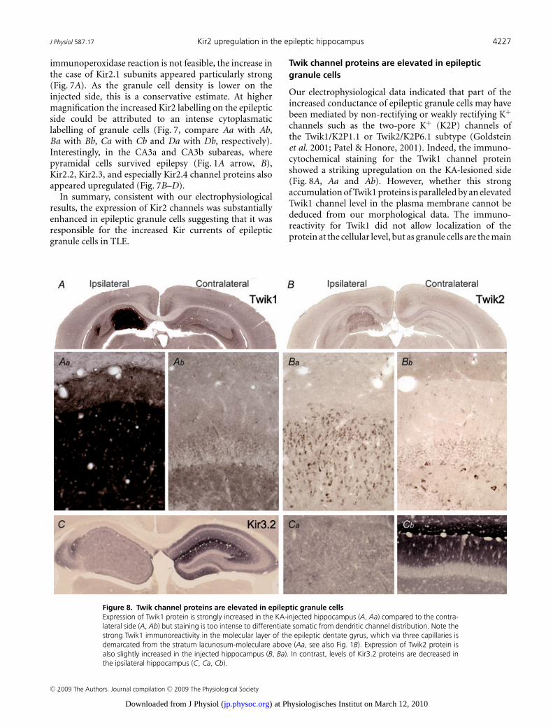
Figure 8. Twik channel proteins are elevated in epileptic granule cellsExpression of Twik1 protein is strongly increased in the KA-injected hippocampus (A, Aa) compared to the contra-lateral side (A, Ab) but staining is too intense to differentiate somatic from dendritic channel distribution. Note thestrong Twik1 immunoreactivity in the molecular layer of the epileptic dentate gyrus, which via three capillaries isdemarcated from the stratum lacunosum-moleculare above (Aa, see also Fig. 1B). Expression of Twik2 protein isalso slightly increased in the injected hippocampus (B, Ba). In contrast, levels of Kir3.2 proteins are decreased inthe ipsilateral hippocampus (C, Ca, Cb).
Twik channel proteins are elevated in epilepticgranule cells
Our electrophysiological data indicated that part of theincreased conductance of epileptic granule cells may havebeen mediated by non-rectifying or weakly rectifying K+
channels such as the two-pore K+ (K2P) channels ofthe Twik1/K2P1.1 or Twik2/K2P6.1 subtype (Goldsteinet al. 2001; Patel & Honore, 2001). Indeed, the immuno-cytochemical staining for the Twik1 channel proteinshowed a striking upregulation on the KA-lesioned side(Fig. 8A, Aa and Ab). However, whether this strongaccumulation of Twik1 proteins is paralleled by an elevatedTwik1 channel level in the plasma membrane cannot bededuced from our morphological data. The immuno-reactivity for Twik1 did not allow localization of theprotein at the cellular level, but as granule cells are the main
C© 2009 The Authors. Journal compilation C© 2009 The Physiological Society
) at Physiologisches Institut on March 12, 2010jp.physoc.orgDownloaded from J Physiol (

4228 C. C. Young and others J Physiol 587.17
surviving cells in this area it is likely that Twik1 expressionwas enhanced in granule cells. Possibly it was elevated inother cell types (e.g. glial cells) in addition. Twik2 channelswere clearly expressed in granule cells although their mainexpression sites have been reported to be outside the CNS(Medhurst et al. 2001). Also, the Twik2 immunoreactivitywas increased in epileptic granule cells albeit to a smallerextent (Fig. 8B, Ba and Bb).
Although our pharmacological evidence argued againsta contribution of Kir3.2 channels to the increased g rest wewanted to verify whether Kir3.2 channels, presumably thepredominant conducting subunit of G-protein-coupledKir (Girk/Kir3) channels in granule cells (Karschin et al.1996; Liao et al. 1996), showed elevated immunolabellingin the KA-injected hippocampus. However, neither theKir3.2 protein (Fig. 8C), nor any of the other Kir3channel proteins (data not shown) were elevated on theinjected side. As expected (Liao et al. 1996), the Kir3.2subunit displayed a highly laminar distribution in thecontralateral hippocampus with highest densities in thestratum lacunosum-moleculare of the CA1 region and themolecular layer of the dentate gyrus (Fig. 8C and Cb).In contrast, Kir3.2 expression on the epileptic side washomogeneously low (Fig. 8C and Ca) and even at highermagnification rather down- than upregulated.
In summary, while the elevated Twik channelexpression probably contributed to the increased g rest
and may contribute to leak components with less inwardrectification and lower Ba2+ sensitivity, the enhancedexpression of Kir2 channels explains the epilepsy-inducedincrease in Kir currents with high Ba2+ sensitivity inepileptic granule cells.
Discussion
The main finding reported here is that the passivemembrane properties of dentate gyrus granule cells arefundamentally altered in a TLE model with AHS and GCD.Previous work has suggested that granule cells becomeintrinsically hyperexcitable in TLE via changes in ionchannels (Isokawa & Mello, 1991; Beck et al. 1998; Selkeet al. 2006). The data presented here suggest that changesin the passive cellular properties related to the degree ofAHS have to be taken into account in addition, to fullycharacterize the excitability of granule cells in TLE.
Epileptic (dispersed) granule cells of mice withintrahippocampal kainate injection display increasedmembrane conductivity and a decreased excitability
In the sclerotic focus of KA-injected mice, granule cellspossessed a strongly reduced R in and τm, both importantfactors in postsynaptic signal integration (Fig. 1). Indeed,much more current was needed to drive epileptic granule
cells to action potential firing (Fig. 2), suggesting theirexcitability was decreased rather than increased (our label‘epileptic’ for ipsilateral cells should not be confoundedwith ‘more excitable’). As previously observed, granulecells of KA-injected mice displayed an altered morphology(Suzuki et al. 1995, 1997) which we considered here onlywith respect to its impact on R in. Our morphologicalquantifications of reconstructed cells and computersimulations show that the R in difference between controland epileptic cells was not due to the increased somasurface or a changed cellular geometry (Fig. 3). Thus, thesimultaneous decrease of R in and τm is only compatiblewith the interpretation that the membrane conductivity(1/Rm) of epileptic granule cells was increased. No suchdecrease in R in has been mentioned in any of the manyearlier patch-clamp studies of granule cells using otherTLE animal models (e.g. Mody et al. 1992; Isokawa, 1996b;Molnar & Nadler, 1999; Okazaki et al. 1999; Scharfmanet al. 2000; Dietrich et al. 2005; but see Isokawa & Mello,1991). This discrepancy could be related to the seizureextent and the degree of hippocampal damage. In manyof the earlier TLE studies, models were used where CApyramidal and granule cell layers appear to be less affectedcompared to the TLE model used here. In particular,very little GCD is observed in these rat TLE models(Okazaki et al. 1999; Scharfman et al. 2000; Dietrich et al.2005). Interestingly, in one study a decrease in τm wasreported which was associated with increased frequencyof seizures, whereas with lower seizure frequency τm
remained unchanged (Isokawa, 1996a).We found a correlation of granule cell membrane
conductivity and the degree of GCD in the presentwork (Fig. 1) and in TLE patients (Stegen et al. 2009).How could GCD and R in be related? It has been shownthat GCD is not a direct effect of the KA itself butrather a consequence of disturbed reelin signalling whichalso occurs in TLE (Haas et al. 2002; Suzuki et al.2005; Heinrich et al. 2006). We cannot fully excludethat conductivity changes are triggered by the KA, butas they first occur several days after KA injection, thisseems unlikely. In addition, similar effects occur in TLEpatients (Stegen et al. 2009). Reeler mice share GCDmechanisms with TLE but lack hippocampal epilepsy. IfGCD itself would directly cause the changed conductivity,one would expect to find a decreased R in in granule cellsof reeler mice. However, this is not the case (J. Kowalski,personal communication). The combined data favourthe hypothesis that GCD and the leakiness of granulecells are two independent consequences of seizure activitywhich may vary in different forms of TLE (Mueller et al.2007). This hypothesis implies that the morphologicaland physiological changes reported here are either delayedresponses to the status epilepticus or a response to seizuresoccurring in the first 2 weeks after KA injection which hassometimes been called ‘latent period’ (but see Sloviter,
C© 2009 The Authors. Journal compilation C© 2009 The Physiological Society
) at Physiologisches Institut on March 12, 2010jp.physoc.orgDownloaded from J Physiol (

J Physiol 587.17 Kir2 upregulation in the epileptic hippocampus 4229
2008). Indeed, EEG recordings revealed that subclinicalseizures are frequent in the first 2 weeks after intra-hippocampal KA injection (Bouilleret et al. 1999; Ribanet al. 2002; U. Haussler, personal communication).
Epileptic granule cells possessed increased potassiumand GABAA leak conductances
Our K+ replacement experiments suggest that theincreased leak conductance of epileptic granule cells wasmainly (∼70–80%) due to an increased K+ conductance(Fig. 4). At V rest roughly 50% of this increased K+
conductance was sensitive to 1 mM Ba2+ while theremaining part was not. In addition, a (∼20–30%)GABAA conductance contributed to the leak increased inepileptic granule cells (Fig. 5). These estimates of relativecontributions should be treated with caution becausethey had to be calculated across cells and animals amongwhich the variance was high. In individual experiments,the contributions to the leak ranged between almostequal BMI-sensitive and (200 μM) Ba2+-sensitive g rest,respectively, in one cell and only ∼5% GABAA and ∼95%highly Ba2+-sensitive leak in other cells. The increasedGABAA conductance of epileptic granule cells couldexplain why these cells were not hyperpolarized despitethe increase in K+ conductance as the reversal potentialof GABAA currents was depolarizing in our conditions(see also Staley & Mody, 1992; Soltesz & Mody, 1994).In the context of excitability, it should be stressed thatthe slight depolarization (∼3 mV) of epileptic granulecells was largely overruled by the reduction in R in. Thisis evident from the rheobase which was tripled in epilepticcells.
So-called tonic GABAA currents, which generally canonly be revealed in granule cells by pharmacologicalmanipulations, constitute a good candidate for theGABAA-mediated leak conductance we found (Stell et al.2003; Farrant & Nusser, 2005; Glykys et al. 2008). Ingranule cells, tonic currents are thought to be mediatedby extrasynaptic GABAA receptors likely to contain δ
subunits associated to α5 and/or α4 subunits (Peng et al.2002; Stell et al. 2003; Farrant & Nusser, 2005; Glykys et al.2008). The expression of α5 and α4 subunits has indeedbeen found upregulated in TLE although not in all cases(Schwarzer et al. 1997; Brooks-Kayal et al. 1998; Loupet al. 2000; Peng et al. 2004; Sun et al. 2007; Zhang et al.2007). In most of these studies, the expression of δ sub-units appeared downregulated but other subunits usuallynot mediating tonic currents may compensate for this loss(Nusser et al. 1998; Peng et al. 2002; Sun et al. 2007; Zhanget al. 2007). Importantly, in the TLE model we used, α5expression is increased in the dentate gyrus (Bouilleretet al. 2000). Thus, it is likely that we measured thefunctional correlate of the GABAA receptor upregulation
in TLE. As the competitive GABAA antagonist BMI hadalmost the same effect on g rest as the non-competitivechloride channel blocker PTX, our results suggest thatmost of the GABAergic leak was mediated by receptoractivation rather than constitutive opening of GABAA
receptors independent of agonist binding.Our results suggest that inwardly rectifying K+ (Kir)
channels mediated the Ba2+-sensitive component ofthe increased g rest (see below). However, another K+
conductance with lower Ba2+ sensitivity (> 2 mM) andless rectification (Fig. 6B) appeared to contribute to theleak of epileptic granule cells (see 32% residual g rest fittedto the dose–response data in Fig. 5D). These properties arecompatible with two-pore leak K+ (K2P) channels such asweak inward rectifier K+ (Twik) channels (Goldstein et al.2001; Patel & Honore, 2001). Indeed, we found a markedupregulation of Twik1/K2P1.1 channels and a weakincrease of Twik2/K2P6.1 channels on the KA-injectedside, suggesting Twik channels also contributed to theincreased leak. However, because the Ba2+ sensitivityof Twik channels under physiological conditions is notentirely clear (Goldstein et al. 2001; Patel & Honore, 2001)we cannot conclude with certainty about the contributionof Twik channels. Less Ba2+-sensitive K2P channels suchas Twik-related acid-sensitive K2P (Task) channels havenow also been reported as increased in the pilocarpinemodel of TLE (Kim et al. 2009) although it is unclear howthis relates to the lack of R in changes reported repeatedlyfrom the same model (see above). In the TLE modelused here, we could not find an increased sensitivity ofthe pathological g rest to lowered pH. Other ion channelsaffected in TLE include HCN channels (Bender et al. 2003)and the effectors of GABAB receptors (Kir3/Girk channels)(Straessle et al. 2003). However, our pharmacologicalexperiments gave no evidence that any of these channels(and Kv channels or calcium-activated K+ channels)contributed to the g rest of epileptic granule cells. Also,increased NMDA receptor activation could contribute toa decreased R in in TLE (Isokawa & Mello, 1991), but inour TLE model inhibition of glutamate receptors did notaffect the g rest of epileptic granule cells.
Kir2 channels are responsible for the increasedinwardly rectifying K+ currents of epilepticgranule cells
In epileptic granule cells, highly Ba2+-sensitive currents(IC50: ∼19 μM at −120 mV and physiological K+) withmarked inward rectification were strongly increased(Fig. 6). These properties match the profile ofclassical inward rectifier K+ (Irk1–4/Kir2.1–2.4) channels(Goldstein et al. 2001; Preisig-Muller et al. 2002; Schramet al. 2002; Kubo et al. 2005), but are incompatible withthe properties of weak inward or even outward rectifier
C© 2009 The Authors. Journal compilation C© 2009 The Physiological Society
) at Physiologisches Institut on March 12, 2010jp.physoc.orgDownloaded from J Physiol (
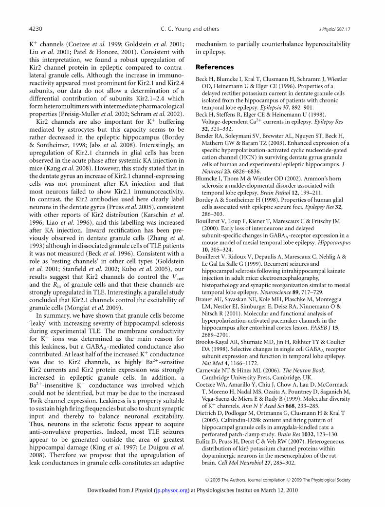
4230 C. C. Young and others J Physiol 587.17
K+ channels (Coetzee et al. 1999; Goldstein et al. 2001;Liu et al. 2001; Patel & Honore, 2001). Consistent withthis interpretation, we found a robust upregulation ofKir2 channel protein in epileptic compared to contra-lateral granule cells. Although the increase in immuno-reactivity appeared most prominent for Kir2.1 and Kir2.4subunits, our data do not allow a determination of adifferential contribution of subunits Kir2.1–2.4 whichform heteromultimers with intermediate pharmacologicalproperties (Preisig-Muller et al. 2002; Schram et al. 2002).
Kir2 channels are also important for K+ bufferingmediated by astrocytes but this capacity seems to berather decreased in the epileptic hippocampus (Bordey& Sontheimer, 1998; Jabs et al. 2008). Interestingly, anupregulation of Kir2.1 channels in glial cells has beenobserved in the acute phase after systemic KA injection inmice (Kang et al. 2008). However, this study stated that inthe dentate gyrus an increase of Kir2.1 channel-expressingcells was not prominent after KA injection and thatmost neurons failed to show Kir2.1 immunoreactivity.In contrast, the Kir2 antibodies used here clearly labelneurons in the dentate gyrus (Pruss et al. 2005), consistentwith other reports of Kir2 distribution (Karschin et al.1996; Liao et al. 1996), and this labelling was increasedafter KA injection. Inward rectification has been pre-viously observed in dentate granule cells (Zhang et al.1993) although in dissociated granule cells of TLE patientsit was not measured (Beck et al. 1996). Consistent with arole as ‘resting channels’ in other cell types (Goldsteinet al. 2001; Stanfield et al. 2002; Kubo et al. 2005), ourresults suggest that Kir2 channels do control the V rest
and the R in of granule cells and that these channels arestrongly upregulated in TLE. Interestingly, a parallel studyconcluded that Kir2.1 channels control the excitability ofgranule cells (Mongiat et al. 2009).
In summary, we have shown that granule cells become‘leaky’ with increasing severity of hippocampal sclerosisduring experimental TLE. The membrane conductivityfor K+ ions was determined as the main reason forthis leakiness, but a GABAA-mediated conductance alsocontributed. At least half of the increased K+ conductancewas due to Kir2 channels, as highly Ba2+-sensitiveKir2 currents and Kir2 protein expression was stronglyincreased in epileptic granule cells. In addition, aBa2+-insensitive K+ conductance was involved whichcould not be identified, but may be due to the increasedTwik channel expression. Leakiness is a property suitableto sustain high firing frequencies but also to shunt synapticinput and thereby to balance neuronal excitability.Thus, neurons in the sclerotic focus appear to acquireanti-convulsive properties. Indeed, most TLE seizuresappear to be generated outside the area of greatesthippocampal damage (King et al. 1997; Le Duigou et al.2008). Therefore we propose that the upregulation ofleak conductances in granule cells constitutes an adaptive
mechanism to partially counterbalance hyperexcitabilityin epilepsy.
References
Beck H, Blumcke I, Kral T, Clusmann H, Schramm J, WiestlerOD, Heinemann U & Elger CE (1996). Properties of adelayed rectifier potassium current in dentate granule cellsisolated from the hippocampus of patients with chronictemporal lobe epilepsy. Epilepsia 37, 892–901.
Beck H, Steffens R, Elger CE & Heinemann U (1998).Voltage-dependent Ca2+ currents in epilepsy. Epilepsy Res32, 321–332.
Bender RA, Soleymani SV, Brewster AL, Nguyen ST, Beck H,Mathern GW & Baram TZ (2003). Enhanced expression of aspecific hyperpolarization-activated cyclic nucleotide-gatedcation channel (HCN) in surviving dentate gyrus granulecells of human and experimental epileptic hippocampus. JNeurosci 23, 6826–6836.
Blumcke I, Thom M & Wiestler OD (2002). Ammon’s hornsclerosis: a maldevelopmental disorder associated withtemporal lobe epilepsy. Brain Pathol 12, 199–211.
Bordey A & Sontheimer H (1998). Properties of human glialcells associated with epileptic seizure foci. Epilepsy Res 32,286–303.
Bouilleret V, Loup F, Kiener T, Marescaux C & Fritschy JM(2000). Early loss of interneurons and delayedsubunit-specific changes in GABAA-receptor expression in amouse model of mesial temporal lobe epilepsy. Hippocampus10, 305–324.
Bouilleret V, Ridoux V, Depaulis A, Marescaux C, Nehlig A &Le Gal La Salle G (1999). Recurrent seizures andhippocampal sclerosis following intrahippocampal kainateinjection in adult mice: electroencephalography,histopathology and synaptic reorganization similar to mesialtemporal lobe epilepsy. Neuroscience 89, 717–729.
Brauer AU, Savaskan NE, Kole MH, Plaschke M, MonteggiaLM, Nestler EJ, Simburger E, Deisz RA, Ninnemann O &Nitsch R (2001). Molecular and functional analysis ofhyperpolarization-activated pacemaker channels in thehippocampus after entorhinal cortex lesion. FASEB J 15,2689–2701.
Brooks-Kayal AR, Shumate MD, Jin H, Rikhter TY & CoulterDA (1998). Selective changes in single cell GABAA receptorsubunit expression and function in temporal lobe epilepsy.Nat Med 4, 1166–1172.
Carnevale NT & Hines ML (2006). The Neuron Book.Cambridge University Press, Cambridge, UK.
Coetzee WA, Amarillo Y, Chiu J, Chow A, Lau D, McCormackT, Moreno H, Nadal MS, Ozaita A, Pountney D, Saganich M,Vega-Saenz de Miera E & Rudy B (1999). Molecular diversityof K+ channels. Ann N Y Acad Sci 868, 233–285.
Dietrich D, Podlogar M, Ortmanns G, Clusmann H & Kral T(2005). Calbindin-D28k content and firing pattern ofhippocampal granule cells in amygdala-kindled rats: aperforated patch-clamp study. Brain Res 1032, 123–130.
Eulitz D, Pruss H, Derst C & Veh RW (2007). Heterogeneousdistribution of kir3 potassium channel proteins withindopaminergic neurons in the mesencephalon of the ratbrain. Cell Mol Neurobiol 27, 285–302.
C© 2009 The Authors. Journal compilation C© 2009 The Physiological Society
) at Physiologisches Institut on March 12, 2010jp.physoc.orgDownloaded from J Physiol (

J Physiol 587.17 Kir2 upregulation in the epileptic hippocampus 4231
Farrant M & Nusser Z (2005). Variations on an inhibitorytheme: phasic and tonic activation of GABAA receptors. NatRev Neurosci 6, 215–229.
Glykys J, Mann EO & Mody I (2008). Which GABAA receptorsubunits are necessary for tonic inhibition in thehippocampus? J Neurosci 28, 1421–1426.
Goldstein SA, Bockenhauer D, O’Kelly I & Zilberberg N (2001).Potassium leak channels and the KCNK family oftwo-P-domain subunits. Nat Rev Neurosci 2, 175–184.
Haas CA, Dudeck O, Kirsch M, Huszka C, Kann G, Pollak S,Zentner J & Frotscher M (2002). Role for reelin in thedevelopment of granule cell dispersion in temporal lobeepilepsy. J Neurosci 22, 5797–5802.
Hama K, Arii T & Kosaka T (1989). Three-dimensionalmorphometrical study of dendritic spines of the granule cellin the rat dentate gyrus with HVEM stereo images. J ElectronMicrosc Tech 12, 80–87.
Harvey BD & Sloviter RS (2005). Hippocampal granule cellactivity and c-Fos expression during spontaneous seizures inawake, chronically epileptic, pilocarpine-treated rats:implications for hippocampal epileptogenesis. J CompNeurol 488, 442–463.
Heinemann U, Beck H, Dreier JP, Ficker E, Stabel J & ZhangCL (1992). The dentate gyrus as a regulated gate for thepropagation of epileptiform activity. Epilepsy Res Suppl 7,273–280.
Heinrich C, Nitta N, Flubacher A, Muller M, Fahrner A, KirschM, Freiman T, Suzuki F, Depaulis A, Frotscher M & Haas CA(2006). Reelin deficiency and displacement of matureneurons, but not neurogenesis, underlie the formation ofgranule cell dispersion in the epileptic hippocampus. JNeurosci 26, 4701–4713.
Houser CR (1990). Granule cell dispersion in the dentate gyrusof humans with temporal lobe epilepsy. Brain Res 535,195–204.
Isokawa M (1996a). Decreased time constant in hippocampaldentate granule cells in pilocarpine-treated rats withprogressive seizure frequencies. Brain Res 718,169–175.
Isokawa M (1996b). Decrement of GABAA receptor-mediatedinhibitory postsynaptic currents in dentate granule cells inepileptic hippocampus. J Neurophysiol 75, 1901–1908.
Isokawa M & Mello LE (1991). NMDA receptor-mediatedexcitability in dendritically deformed dentate granule cells inpilocarpine-treated rats. Neurosci Lett 129, 69–73.
Jabs R, Seifert G & Steinhauser C (2008). Astrocytic functionand its alteration in the epileptic brain. Epilepsia 49,3–12.
Kang SJ, Cho SH, Park K, Yi J, Yoo SJ & Shin KS (2008).Expression of kir2.1 channels in astrocytes underpathophysiological conditions. Mol Cells 25, 124–130.
Karschin C, Dissmann E, Stuhmer W & Karschin A (1996).IRK(1–3) and GIRK(1–4) inwardly rectifying K+ channelmRNAs are differentially expressed in the adult rat brain. JNeurosci 16, 3559–3570.
Kim JE, Kwak SE & Kang TC (2009). UpregulatedTWIK-related acid-sensitive K+ channel-2 in neurons andperivascular astrocytes in the hippocampus of experimentaltemporal lobe epilepsy. Epilepsia 50, 654–663.
King D, Bronen RA, Spencer DD & Spencer SS (1997).Topographic distribution of seizure onset and hippocampalatrophy: relationship between MRI and depth EEG.Electroencephalogr Clin Neurophysiol 103, 692–697.
Kralic JE, Ledergerber DA & Fritschy JM (2005). Disruption ofthe neurogenic potential of the dentate gyrus in a mousemodel of temporal lobe epilepsy with focal seizures. Eur JNeurosci 22, 1916–1927.
Kubo Y, Adelman JP, Clapham DE, Jan LY, Karschin A, KurachiY, Lazdunski M, Nichols CG, Seino S & Vandenberg CA(2005). International Union of Pharmacology. LIV.Nomenclature and molecular relationships of inwardlyrectifying potassium channels. Pharmacol Rev 57, 509–526.
Le Duigou C, Bouilleret V & Miles R (2008). Epileptiformactivities in slices of hippocampus from mice afterintra-hippocampal injection of kainic acid. J Physiol 586,4891–4904.
Le Duigou C, Wittner L, Danglot L & Miles R (2005). Effects offocal injection of kainic acid into the mouse hippocampus invitro and ex vivo. J Physiol 569, 833–847.
Liao YJ, Jan YN & Jan LY (1996). Heteromultimerization ofG-protein-gated inwardly rectifying K+ channel proteinsGIRK1 and GIRK2 and their altered expression in weaverbrain. J Neurosci 16, 7137–7150.
Liu GX, Derst C, Schlichthorl G, Heinen S, Seebohm G,Bruggemann A, Kummer W, Veh RW, Daut J &Preisig-Muller R (2001). Comparison of cloned Kir2channels with native inward rectifier K+ channels fromguinea-pig cardiomyocytes. J Physiol 532, 115–126.
Liu M, Pleasure SJ, Collins AE, Noebels JL, Naya FJ, Tsai MJ &Lowenstein DH (2000). Loss of BETA2/NeuroD leads tomalformation of the dentate gyrus and epilepsy. Proc NatlAcad Sci U S A 97, 865–870.
Loup F, Wieser HG, Yonekawa Y, Aguzzi A & Fritschy JM(2000). Selective alterations in GABAA receptor subtypes inhuman temporal lobe epilepsy. J Neurosci 20, 5401–5419.
Medhurst AD, Rennie G, Chapman CG, Meadows H,Duckworth MD, Kelsell RE, Gloger II & Pangalos MN(2001). Distribution analysis of human two pore domainpotassium channels in tissues of the central nervous systemand periphery. Brain Res Mol Brain Res 86, 101–114.
Mody I, Kohr G, Otis TS & Staley KJ (1992). Theelectrophysiology of dentate gyrus granule cells in whole-cellrecordings. Epilepsy Res Suppl 7, 159–168.
Mody I, Stanton PK & Heinemann U (1988). Activation ofN-methyl-D-aspartate receptors parallels changes in cellularand synaptic properties of dentate gyrus granule cells afterkindling. J Neurophysiol 59, 1033–1054.
Molnar P & Nadler JV (1999). Mossy fiber-granule cellsynapses in the normal and epileptic rat dentate gyrusstudied with minimal laser photostimulation. J Neurophysiol82, 1883–1894.
Mongiat LA, Esposito MS, Lombardi G & Schinder AF (2009).Reliable activation of immature neurons in the adulthippocampus. PLoS ONE 4, e5320.
Mueller SG, Laxer KD, Schuff N & Weiner MW (2007).Voxel-based T2 relaxation rate measurements in temporallobe epilepsy (TLE) with and without mesial temporalsclerosis. Epilepsia 48, 220–228.
C© 2009 The Authors. Journal compilation C© 2009 The Physiological Society
) at Physiologisches Institut on March 12, 2010jp.physoc.orgDownloaded from J Physiol (
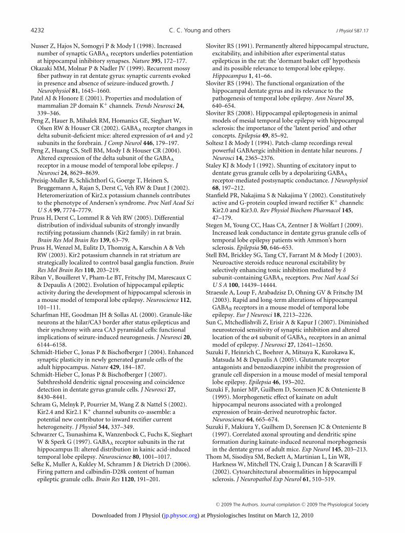
4232 C. C. Young and others J Physiol 587.17
Nusser Z, Hajos N, Somogyi P & Mody I (1998). Increasednumber of synaptic GABAA receptors underlies potentiationat hippocampal inhibitory synapses. Nature 395, 172–177.
Okazaki MM, Molnar P & Nadler JV (1999). Recurrent mossyfiber pathway in rat dentate gyrus: synaptic currents evokedin presence and absence of seizure-induced growth. JNeurophysiol 81, 1645–1660.
Patel AJ & Honore E (2001). Properties and modulation ofmammalian 2P domain K+ channels. Trends Neurosci 24,339–346.
Peng Z, Hauer B, Mihalek RM, Homanics GE, Sieghart W,Olsen RW & Houser CR (2002). GABAA receptor changes indelta subunit-deficient mice: altered expression of α4 and γ2subunits in the forebrain. J Comp Neurol 446, 179–197.
Peng Z, Huang CS, Stell BM, Mody I & Houser CR (2004).Altered expression of the delta subunit of the GABAA
receptor in a mouse model of temporal lobe epilepsy. JNeurosci 24, 8629–8639.
Preisig-Muller R, Schlichthorl G, Goerge T, Heinen S,Bruggemann A, Rajan S, Derst C, Veh RW & Daut J (2002).Heteromerization of Kir2.x potassium channels contributesto the phenotype of Andersen’s syndrome. Proc Natl Acad SciU S A 99, 7774–7779.
Pruss H, Derst C, Lommel R & Veh RW (2005). Differentialdistribution of individual subunits of strongly inwardlyrectifying potassium channels (Kir2 family) in rat brain.Brain Res Mol Brain Res 139, 63–79.
Pruss H, Wenzel M, Eulitz D, Thomzig A, Karschin A & VehRW (2003). Kir2 potassium channels in rat striatum arestrategically localized to control basal ganglia function. BrainRes Mol Brain Res 110, 203–219.
Riban V, Bouilleret V, Pham-Le BT, Fritschy JM, Marescaux C& Depaulis A (2002). Evolution of hippocampal epilepticactivity during the development of hippocampal sclerosis ina mouse model of temporal lobe epilepsy. Neuroscience 112,101–111.
Scharfman HE, Goodman JH & Sollas AL (2000). Granule-likeneurons at the hilar/CA3 border after status epilepticus andtheir synchrony with area CA3 pyramidal cells: functionalimplications of seizure-induced neurogenesis. J Neurosci 20,6144–6158.
Schmidt-Hieber C, Jonas P & Bischofberger J (2004). Enhancedsynaptic plasticity in newly generated granule cells of theadult hippocampus. Nature 429, 184–187.
Schmidt-Hieber C, Jonas P & Bischofberger J (2007).Subthreshold dendritic signal processing and coincidencedetection in dentate gyrus granule cells. J Neurosci 27,8430–8441.
Schram G, Melnyk P, Pourrier M, Wang Z & Nattel S (2002).Kir2.4 and Kir2.1 K+ channel subunits co-assemble: apotential new contributor to inward rectifier currentheterogeneity. J Physiol 544, 337–349.
Schwarzer C, Tsunashima K, Wanzenbock C, Fuchs K, SieghartW & Sperk G (1997). GABAA receptor subunits in the rathippocampus II: altered distribution in kainic acid-inducedtemporal lobe epilepsy. Neuroscience 80, 1001–1017.
Selke K, Muller A, Kukley M, Schramm J & Dietrich D (2006).Firing pattern and calbindin-D28k content of humanepileptic granule cells. Brain Res 1120, 191–201.
Sloviter RS (1991). Permanently altered hippocampal structure,excitability, and inhibition after experimental statusepilepticus in the rat: the ‘dormant basket cell’ hypothesisand its possible relevance to temporal lobe epilepsy.Hippocampus 1, 41–66.
Sloviter RS (1994). The functional organization of thehippocampal dentate gyrus and its relevance to thepathogenesis of temporal lobe epilepsy. Ann Neurol 35,640–654.
Sloviter RS (2008). Hippocampal epileptogenesis in animalmodels of mesial temporal lobe epilepsy with hippocampalsclerosis: the importance of the ‘latent period’ and otherconcepts. Epilepsia 49, 85–92.
Soltesz I & Mody I (1994). Patch-clamp recordings revealpowerful GABAergic inhibition in dentate hilar neurons. JNeurosci 14, 2365–2376.
Staley KJ & Mody I (1992). Shunting of excitatory input todentate gyrus granule cells by a depolarizing GABAA
receptor-mediated postsynaptic conductance. J Neurophysiol68, 197–212.
Stanfield PR, Nakajima S & Nakajima Y (2002). Constitutivelyactive and G-protein coupled inward rectifier K+ channels:Kir2.0 and Kir3.0. Rev Physiol Biochem Pharmacol 145,47–179.
Stegen M, Young CC, Haas CA, Zentner J & Wolfart J (2009).Increased leak conductance in dentate gyrus granule cells oftemporal lobe epilepsy patients with Ammon’s hornsclerosis. Epilepsia 50, 646–653.
Stell BM, Brickley SG, Tang CY, Farrant M & Mody I (2003).Neuroactive steroids reduce neuronal excitability byselectively enhancing tonic inhibition mediated by δ
subunit-containing GABAA receptors. Proc Natl Acad SciU S A 100, 14439–14444.
Straessle A, Loup F, Arabadzisz D, Ohning GV & Fritschy JM(2003). Rapid and long-term alterations of hippocampalGABAB receptors in a mouse model of temporal lobeepilepsy. Eur J Neurosci 18, 2213–2226.
Sun C, Mtchedlishvili Z, Erisir A & Kapur J (2007). Diminishedneurosteroid sensitivity of synaptic inhibition and alteredlocation of the α4 subunit of GABAA receptors in an animalmodel of epilepsy. J Neurosci 27, 12641–12650.
Suzuki F, Heinrich C, Boehrer A, Mitsuya K, Kurokawa K,Matsuda M & Depaulis A (2005). Glutamate receptorantagonists and benzodiazepine inhibit the progression ofgranule cell dispersion in a mouse model of mesial temporallobe epilepsy. Epilepsia 46, 193–202.
Suzuki F, Junier MP, Guilhem D, Sorensen JC & Onteniente B(1995). Morphogenetic effect of kainate on adulthippocampal neurons associated with a prolongedexpression of brain-derived neurotrophic factor.Neuroscience 64, 665–674.
Suzuki F, Makiura Y, Guilhem D, Sorensen JC & Onteniente B(1997). Correlated axonal sprouting and dendritic spineformation during kainate-induced neuronal morphogenesisin the dentate gyrus of adult mice. Exp Neurol 145, 203–213.
Thom M, Sisodiya SM, Beckett A, Martinian L, Lin WR,Harkness W, Mitchell TN, Craig J, Duncan J & Scaravilli F(2002). Cytoarchitectural abnormalities in hippocampalsclerosis. J Neuropathol Exp Neurol 61, 510–519.
C© 2009 The Authors. Journal compilation C© 2009 The Physiological Society
) at Physiologisches Institut on March 12, 2010jp.physoc.orgDownloaded from J Physiol (

J Physiol 587.17 Kir2 upregulation in the epileptic hippocampus 4233
Williamson A, Telfeian AE & Spencer DD (1995). ProlongedGABA responses in dentate granule cells in slices isolatedfrom patients with temporal lobe sclerosis. J Neurophysiol 74,378–387.
Zhang L, Valiante TA & Carlen PL (1993). Contribution of thelow-threshold T-type calcium current in generating thepost-spike depolarizing afterpotential in dentate granuleneurons of immature rats. J Neurophysiol 70,223–231.
Zhang N, Wei W, Mody I & Houser CR (2007). Alteredlocalization of GABAA receptor subunits on dentate granulecell dendrites influences tonic and phasic inhibition in amouse model of epilepsy. J Neurosci 27, 7520–7531.
Author contributions
C.C. Young: Completion and analysis of electrophysiologicalexperiments. Completion and analysis of morphologicalquantifications and Neurolucida analysis. Analysis of immuno-cytochemical experiments (Fig. 1C). Critical revision ofmanuscript. M. Stegen: Surgical procedures of KA injections.Completion and analysis of electrophysiological experiments.Computation of cable models. Critical revision of manuscript.R. Bernard: Completion of immunocytochemical experiments(Figs 1A, B, 7 and 8). Critical revision of manuscript.
M. Muller: Surgical procedures of KA injections. Criticalrevision of manuscript. J. Bischofberger: Supervision andcompletion of confocal reconstructions. Critical revision ofmanuscript. R.W. Veh: Supervision and analysis of immuno-cytochemical experiments (Figs 1A, B, 7 and 8). Criticalrevision of manuscript. C.A. Haas: Conception of study.Critical revision of manuscript. J. Wolfart: Conception,design and supervision of study. Design and supervisionof electrophysiological experiments. Conception of immuno-histochemical and morphological experiments. Drafting themanuscript.
Acknowledgements
We thank F. Moos, H. Heilmann, R. Lommel and I. Wolterfor help with immunocytochemical procedures; Dr M. Lofflerfor laboratory support and Dr F. Aiple for help withcomputation; Dr J. Staiger and C. David for help withmorphological reconstructions; Drs C. Schmidt-Hieber and P.Jonas for valuable comments and help with methods; Dr H.Cuntz/ACCN 2008 course and Dr C. Schmidt-Hieber for helpwith cable modelling. This work was supported by the Mini-stry of Science, Research and the Arts of Baden-Wurttemberg[Juniorprofessorenprogramm] and the DFG [SFB780].
C© 2009 The Authors. Journal compilation C© 2009 The Physiological Society
) at Physiologisches Institut on March 12, 2010jp.physoc.orgDownloaded from J Physiol (
Related Documents