Reduced volume of Heschl's gyrus in tinnitus Peter Schneider a,b, ⁎, Martin Andermann a , Martina Wengenroth b , Rainer Goebel c , Herta Flor a, ⁎, André Rupp d , Eugen Diesch a, ⁎ a Department of Clinical and Cognitive Neuroscience, Central Institute of Mental Health, University of Heidelberg, J5, 68159 Mannheim, Germany b Department of Neuroradiology, University of Heidelberg Medical School, INF 400, 69120 Heidelberg, Germany c Department of Cognitive Neuroscience, Faculty of Psychology, Universiteit Maastricht, Postbus 616, 6200MD Maastricht, The Netherlands d Department of Neurology, University of Heidelberg Medical School, INF 400, 69120 Heidelberg, Germany abstract article info Article history: Received 22 August 2008 Revised 15 December 2008 Accepted 17 December 2008 Available online 6 January 2009 Keywords: Auditory cortex Hearing loss MRI volumetry Plasticity Tinnitus The neural basis of tinnitus is unknown. Recent neuroimaging studies point towards involvement of several cortical and subcortical regions. Here we demonstrate that tinnitus may be associated with structural changes in the auditory cortex. Using individual morphological segmentation, the medial partition of Heschl's gyrus (mHG) was studied in individuals with and without chronic tinnitus using magnetic resonance imaging. Both the tinnitus and the non-tinnitus group included musicians and non-musicians. Patients exhibited significantly smaller mHG gray matter volumes than controls. In unilateral tinnitus, this effect was almost exclusively seen in the hemisphere ipsilateral to the affected ear. In bilateral tinnitus, mHG volume was substantially reduced in both hemispheres. The tinnitus-related volume reduction was found across the full extent of mHG, not only in the high-frequency part usually most affected by hearing loss- induced deafferentation. However, there was also evidence for a relationship between volume reduction and hearing loss. Correlations between volume and hearing level depended on the subject group as well as the asymmetry of the hearing loss. The volume changes observed may represent antecedents or consequences of tinnitus and tinnitus-associated hearing loss and also raise the possibility that small cortical volume constitutes a vulnerability factor. © 2008 Elsevier Inc. All rights reserved. Introduction Tinnitus is an auditory phantom sensation with ringing, hissing, or buzzing sounds (Jastreboff, 1990). Up to 17% of the adult population experience tinnitus at least occasionally (Hazell, 1990) and as many as 5–10% report unremitting tinnitus, with about 1–3% of the tinnitus sufferers experiencing interference with their lives (Baguley, 2002; Eggermont and Roberts, 2004). However, the underlying pathome- chanism has not yet been identified. As tinnitus often persists even subsequent to auditory nerve transsection (Jackler and Whinney, 2001; Jackson, 1985), processes in the central nervous system may play a major role in its development and maintenance (Bartels et al., 2007; Eggermont, 2005; Lockwood et al., 2002; Moller, 2003). Several neuroimaging studies employing magnetoencephalography (Diesch et al., 2004; Mühlnickel et al., 1998; Weisz et al., 2005; Weisz et al., 2007; Wienbruch et al., 2006), positron emission tomography (Andersson et al., 2000; Langguth et al., 2006; Lockwood et al., 2001; Plewnia et al., 2007), functional magnetic resonance imaging (Melcher et al., 2000; Smits et al., 2007) or repetitive transcranial magnetic stimulation (Folmer et al., 2006; De Ridder et al., 2005; Kleinjung et al., 2005; Plewnia et al., 2007) reported subcortical and cortical changes in tinnitus patients. Using the method of voxel-based morphometry (VBM, Ashburner and Friston, 2000), a recent volumetric magnetic resonance tomography studies demon- strated tinnitus-related structural changes in the subcallosal region including the nucleus accumbens and, within the auditory pathway, at the level of the medial geniculate (Mühlau et al., 2006). Volumetric effects were not reported for the auditory cortex. However, the supratemporal plane is a cortical region with rather complex convolutions, which are interindividually highly variable (Hackett et al., 2001; Leonard et al., 1998; Patterson et al., 2002; Schneider et al., 2005) and potentially too variable for VBM to unravel volumetric differences or changes. Volumetric differences may constitute vulner- ability factors for tinnitus or, alternatively, reflect structural plasticity. The lateralization of tinnitus and hearing loss may constitute another problem. Perceptually, tinnitus may be perceived in the left ear, in the right ear, in both ears, or in the center of the head. Occurrence of tinnitus has been reported in individuals with little or without hearing loss in the conventionally tested frequency range up to 8 kHz and thresholds no worse than those of healthy controls in the high NeuroImage 45 (2009) 927–939 ⁎ Corresponding authors. P. Schneider is to be contacted at Department of Neuroradiology, University of Heidelberg Medical School, INF 400, 69120 Heidelberg, Germany. H. Flor, Department of Clinical and Cognitive Neuroscience, Central Institute of Mental Health, University of Heidelberg, Square J5, 68159 Mannheim, Germany. E. Diesch, Department of Clinical and Cognitive Neuroscience, Central Institute of Mental Health, University of Heidelberg, Square J5, 68159 Mannheim, Germany. E-mail addresses: [email protected] (P. Schneider), herta.fl[email protected] (H. Flor), [email protected] (E. Diesch). 1053-8119/$ – see front matter © 2008 Elsevier Inc. All rights reserved. doi:10.1016/j.neuroimage.2008.12.045 Contents lists available at ScienceDirect NeuroImage journal homepage: www.elsevier.com/locate/ynimg

Welcome message from author
This document is posted to help you gain knowledge. Please leave a comment to let me know what you think about it! Share it to your friends and learn new things together.
Transcript
NeuroImage 45 (2009) 927–939
Contents lists available at ScienceDirect
NeuroImage
j ourna l homepage: www.e lsev ie r.com/ locate /yn img
Reduced volume of Heschl's gyrus in tinnitus
Peter Schneider a,b,⁎, Martin Andermann a, Martina Wengenroth b, Rainer Goebel c, Herta Flor a,⁎,André Rupp d, Eugen Diesch a,⁎a Department of Clinical and Cognitive Neuroscience, Central Institute of Mental Health, University of Heidelberg, J5, 68159 Mannheim, Germanyb Department of Neuroradiology, University of Heidelberg Medical School, INF 400, 69120 Heidelberg, Germanyc Department of Cognitive Neuroscience, Faculty of Psychology, Universiteit Maastricht, Postbus 616, 6200MD Maastricht, The Netherlandsd Department of Neurology, University of Heidelberg Medical School, INF 400, 69120 Heidelberg, Germany
⁎ Corresponding authors. P. Schneider is to beNeuroradiology, University of Heidelberg Medical SchoGermany. H. Flor, Department of Clinical and Cognitiveof Mental Health, University of Heidelberg, Square J5, 6Diesch, Department of Clinical and Cognitive NeuroscienHealth, University of Heidelberg, Square J5, 68159 Mann
E-mail addresses: [email protected]@zi-mannheim.de (H. Flor), eugen.diesch@zi-m
1053-8119/$ – see front matter © 2008 Elsevier Inc. Alldoi:10.1016/j.neuroimage.2008.12.045
a b s t r a c t
a r t i c l e i n f oArticle history:
The neural basis of tinnitus Received 22 August 2008Revised 15 December 2008Accepted 17 December 2008Available online 6 January 2009Keywords:Auditory cortexHearing lossMRI volumetryPlasticityTinnitus
is unknown. Recent neuroimaging studies point towards involvement of severalcortical and subcortical regions. Here we demonstrate that tinnitus may be associated with structuralchanges in the auditory cortex. Using individual morphological segmentation, the medial partition ofHeschl's gyrus (mHG) was studied in individuals with and without chronic tinnitus using magneticresonance imaging. Both the tinnitus and the non-tinnitus group included musicians and non-musicians.Patients exhibited significantly smaller mHG gray matter volumes than controls. In unilateral tinnitus, thiseffect was almost exclusively seen in the hemisphere ipsilateral to the affected ear. In bilateral tinnitus, mHGvolume was substantially reduced in both hemispheres. The tinnitus-related volume reduction was foundacross the full extent of mHG, not only in the high-frequency part usually most affected by hearing loss-induced deafferentation. However, there was also evidence for a relationship between volume reduction andhearing loss. Correlations between volume and hearing level depended on the subject group as well as theasymmetry of the hearing loss. The volume changes observed may represent antecedents or consequences oftinnitus and tinnitus-associated hearing loss and also raise the possibility that small cortical volumeconstitutes a vulnerability factor.
© 2008 Elsevier Inc. All rights reserved.
Introduction
Tinnitus is an auditory phantom sensationwith ringing, hissing, orbuzzing sounds (Jastreboff, 1990). Up to 17% of the adult populationexperience tinnitus at least occasionally (Hazell, 1990) and as many as5–10% report unremitting tinnitus, with about 1–3% of the tinnitussufferers experiencing interference with their lives (Baguley, 2002;Eggermont and Roberts, 2004). However, the underlying pathome-chanism has not yet been identified.
As tinnitus often persists even subsequent to auditory nervetranssection (Jackler and Whinney, 2001; Jackson, 1985), processes inthe central nervous system may play a major role in its developmentand maintenance (Bartels et al., 2007; Eggermont, 2005; Lockwoodet al., 2002; Moller, 2003). Several neuroimaging studies employingmagnetoencephalography (Diesch et al., 2004; Mühlnickel et al., 1998;Weisz et al., 2005;Weisz et al., 2007;Wienbruch et al., 2006), positron
contacted at Department ofol, INF 400, 69120 Heidelberg,Neuroscience, Central Institute8159 Mannheim, Germany. E.ce, Central Institute of Mentalheim, Germany.rg.de (P. Schneider),annheim.de (E. Diesch).
rights reserved.
emission tomography (Andersson et al., 2000; Langguth et al., 2006;Lockwood et al., 2001; Plewnia et al., 2007), functional magneticresonance imaging (Melcher et al., 2000; Smits et al., 2007) orrepetitive transcranial magnetic stimulation (Folmer et al., 2006; DeRidder et al., 2005; Kleinjung et al., 2005; Plewnia et al., 2007) reportedsubcortical and cortical changes in tinnitus patients. Using themethodof voxel-based morphometry (VBM, Ashburner and Friston, 2000), arecent volumetric magnetic resonance tomography studies demon-strated tinnitus-related structural changes in the subcallosal regionincluding the nucleus accumbens and, within the auditory pathway, atthe level of the medial geniculate (Mühlau et al., 2006). Volumetriceffects were not reported for the auditory cortex. However, thesupratemporal plane is a cortical region with rather complexconvolutions, which are interindividually highly variable (Hackettet al., 2001; Leonard et al., 1998; Patterson et al., 2002; Schneider et al.,2005) and potentially too variable for VBM to unravel volumetricdifferences or changes. Volumetric differences may constitute vulner-ability factors for tinnitus or, alternatively, reflect structural plasticity.The lateralization of tinnitus and hearing loss may constitute anotherproblem. Perceptually, tinnitus may be perceived in the left ear, in theright ear, in both ears, or in the center of the head. Occurrence oftinnitus has been reported in individuals with little or without hearingloss in the conventionally tested frequency range up to 8 kHz andthresholds no worse than those of healthy controls in the high
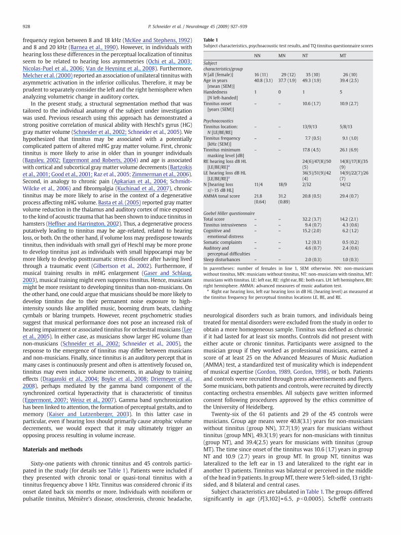
Table 1Subject characteristics, psychoacoustic test results, and TQ tinnitus questionnaire scores
NN MN NT MT
Subjectcharacteristics/groupN [all (female)] 16 (11) 29 (12) 35 (10) 26 (10)Age in years[mean (SEM)]
40.8 (3.1) 37.7 (1.9) 49.3 (1.9) 39.4 (2.5)
Handedness[N left-handed]
1 0 1 5
Tinnitus onset[years (SEM)]
– – 10.6 (1.7) 10.9 (2.7)
PsychoacousticsTinnitus location:N [LE/BE/RE]
– – 13/9/13 5/8/13
Tinnitus frequency[kHz (SEM)]
– – 7.7 (0.5) 9.1 (1.0)
Tinnitus minimummasking level [dB]
– – 17.8 (4.5) 26.1 (6.9)
RE hearing loss dB HL[LE/BE/RE]a
24(6)/47(8)/50(5)
14(8)/17(8)/35(9)
LE hearing loss dB HL[LE/BE/RE]a
36(5)/51(9)/42(4)
14(9)/22(7)/26(7)
N [hearing loss≤/N15 dB HL]
11/4 18/9 2/32 14/12
AMMA tonal score 21.8(0.64)
31.2(0.89)
20.8 (0.5) 29.4 (0.7)
Goebel Hiller questionnaireTotal score – – 32.2 (3.7) 14.2 (2.1)Tinnitus intrusiveness – – 9.4 (0.7) 4.3 (0.6)Cognitive andemotional distress
– – 15.2 (2.0) 6.2 (1.2)
Somatic complaints – – 1.2 (0.3) 0.5 (0.2)Auditory andperceptual difficulties
– – 4.6 (0.7) 2.4 (0.6)
Sleep disturbances – – 2.0 (0.3) 1.0 (0.3)
In parentheses: number of females in line 1, SEM otherwise. NN: non-musicianswithout tinnitus, MN:musicians without tinnitus, NT: non-musicians with tinnitus, MT:musicians with tinnitus. LE: left ear, RE: right ear, BE: both ears. LH: left hemisphere, RH:right hemisphere. AMMA: advanced measures of music audiation test.
a Right ear hearing loss, left ear hearing loss in dB HL (hearing level) as measured atthe tinnitus frequency for perceptual tinnitus locations LE, BE, and RE.
928 P. Schneider et al. / NeuroImage 45 (2009) 927–939
frequency region between 8 and 18 kHz (McKee and Stephens, 1992)and 8 and 20 kHz (Barnea et al., 1990). However, in individuals withhearing loss these differences in the perceptual localization of tinnitusseem to be related to hearing loss asymmetries (Ochi et al., 2003;Nicolas-Puel et al., 2006; Van de Heyning et al., 2008). Furthermore,Melcher et al. (2000) reported an association of unilateral tinnituswithasymmetric activation in the inferior colliculus. Therefore, it may beprudent to separately consider the left and the right hemispherewhenanalyzing volumetric change in auditory cortex.
In the present study, a structural segmentation method that wastailored to the individual anatomy of the subject under investigationwas used. Previous research using this approach has demonstrated astrong positive correlation of musical ability with Heschl's gyrus (HG)gray matter volume (Schneider et al., 2002; Schneider et al., 2005). Wehypothesized that tinnitus may be associated with a potentiallycomplicated pattern of altered mHG gray matter volume. First, chronictinnitus is more likely to arise in older than in younger individuals(Baguley, 2002; Eggermont and Roberts, 2004) and age is associatedwith cortical and subcortical graymatter volume decrements (Bartzokiset al., 2001; Good et al., 2001; Raz et al., 2005; Zimmerman et al., 2006).Second, in analogy to chronic pain (Apkarian et al., 2004; Schmidt-Wilcke et al., 2006) and fibromyalgia (Kuchinad et al., 2007), chronictinnitus may be more likely to arise in the context of a degenerativeprocess affecting mHG volume. Basta et al. (2005) reported gray mattervolume reduction in the thalamus and auditory cortex of mice exposedto the kind of acoustic trauma that has been shown to induce tinnitus inhamsters (Heffner and Harrington, 2002). Thus, a degenerative processputatively leading to tinnitus may be age-related, related to hearingloss, or both. On the other hand, if volume loss may predispose towardstinnitus, then individuals with small gyri of Heschl may be more proneto develop tinnitus just as individuals with small hippocampi may bemore likely to develop posttraumatic stress disorder after having livedthrough a traumatic event (Gilbertson et al., 2002). Furthermore, ifmusical training results in mHG enlargement (Gaser and Schlaug,2003), musical training might even suppress tinnitus. Hence, musiciansmight be more resistant to developing tinnitus than non-musicians. Onthe other hand, one could argue that musicians should bemore likely todevelop tinnitus due to their permanent noise exposure to high-intensity sounds like amplified music, booming drum beats, clashingcymbals or blaring trumpets. However, recent psychometric studiessuggest that musical performance does not pose an increased risk ofhearing impairment or associated tinnitus for orchestral musicians (Leeet al., 2005). In either case, as musicians show larger HG volume thannon-musicians (Schneider et al., 2002; Schneider et al., 2005), theresponse to the emergence of tinnitus may differ between musiciansand non-musicians. Finally, since tinnitus is an auditory percept that inmany cases is continuously present and often is attentively focused on,tinnitus may even induce volume increments, in analogy to trainingeffects (Draganski et al., 2004; Boyke et al., 2008; Driemeyer et al.,2008), perhaps mediated by the gamma band component of thesynchronized cortical hyperactivity that is characteristic of tinnitus(Eggermont, 2007; Weisz et al., 2007). Gamma band synchronizationhas been linked to attention, the formation of perceptual gestalts, and tomemory (Kaiser and Lutzenberger, 2003). In this latter case inparticular, even if hearing loss should primarily cause atrophic volumedecrements, we would expect that it may ultimately trigger anopposing process resulting in volume increase.
Materials and methods
Sixty-one patients with chronic tinnitus and 45 controls partici-pated in the study (for details see Table 1). Patients were included ifthey presented with chronic tonal or quasi-tonal tinnitus with atinnitus frequency above 1 kHz. Tinnitus was considered chronic if itsonset dated back six months or more. Individuals with noisiform orpulsatile tinnitus, Ménière's disease, otosclerosis, chronic headache,
neurological disorders such as brain tumors, and individuals beingtreated for mental disorders were excluded from the study in order toobtain a more homogeneous sample. Tinnitus was defined as chronicif it had lasted for at least six months. Controls did not present witheither acute or chronic tinnitus. Participants were assigned to themusician group if they worked as professional musicians, earned ascore of at least 25 on the Advanced Measures of Music Audiation(AMMA) test, a standardized test of musicality which is independentof musical expertise (Gordon, 1989, Gordon, 1998), or both. Patientsand controls were recruited through press advertisements and flyers.Somemusicians, both patients and controls, were recruited by directlycontacting orchestra ensembles. All subjects gave written informedconsent following procedures approved by the ethics committee ofthe University of Heidelberg.
Twenty-six of the 61 patients and 29 of the 45 controls weremusicians. Group age means were 40.8(3.1) years for non-musicianswithout tinnitus (group NN), 37.7(1.9) years for musicians withouttinnitus (group MN), 49.3(1.9) years for non-musicians with tinnitus(group NT), and 39.4(2.5) years for musicians with tinnitus (groupMT). The time since onset of the tinnitus was 10.6 (1.7) years in groupNT and 10.9 (2.7) years in group MT. In group NT, tinnitus waslateralized to the left ear in 13 and lateralized to the right ear inanother 13 patients. Tinnitus was bilateral or perceived in the middleof the head in 9 patients. In groupMT, therewere 5 left-sided,13 right-sided, and 8 bilateral and central cases.
Subject characteristics are tabulated in Table 1. The groups differedsignificantly in age (F[3,102]=6.5, pb0.0005). Scheffé contrasts
929P. Schneider et al. / NeuroImage 45 (2009) 927–939
(pb0.05) showed that group NT was significantly older than groupsMTandMN. Accordingly, agewas entered as a covariate in all analysesof variance that were computed. There were no significant differencesbetween the groups with regard to the proportion of males andfemales (χ2(3)=6.5, n.s.) or with regard to handedness (Fisher–Freeman–Halton test: c(3)=5.3, n.s.). Among tinnitus patients,musicians did not differ from non-musicians in the time since onsetof the tinnitus (F[1,47]=0.74, n.s.).
All participants were investigated for the extent of hearing lossusing pure-tone audiograms. Tinnitus patients were tested for thefrequency and the minimum masking level of their tinnitus. Theywere interviewed as to their perceived location of the tinnitus.Perceived tinnitus location was coded on a five-point scale (exclu-sively in the left ear, predominantly in the left ear, in both ears, orcentralized in the middle of the head, predominantly in the right ear,exclusively in the right ear). Patients were also given the Germanversion of the Tinnitus Questionnaire (TQ,Hallam et al., 1988, Hallam,1996) published by Goebel and Hiller (Goebel and Hiller, 1994; Goebeland Hiller, 1998).
For the audiometric and psychoacoustic tests stimuli werepresented monaurally using a Hammerfall DSP Multiface Systemand closed dynamic headphones (Sennheiser HDA 200) designed forextended high frequency testing. The absolute threshold of hearingwas determined for twelve frequencies between 0.125 kHz and 15 kHzusing a computer-based standard three-interval forced choiceprocedure implemented on the basis of the MATLAB AFC softwarepackage developed at the University of Oldenburg (Ewert and Dau,2004). Both the tones and the intervals between the tones were500 ms in duration; the tones had 20 ms onset and offset ramps.
Threshold values were obtained in dB full scale (dB FS) and thenrescaled in terms of dB hearing level (dB HL). Initially, the rescalingcoefficients were obtained from a subgroup of 30 subjects for whomclinical audiograms with extended frequency range up to 10 kHzwere available (Audiomaster CA 540/1, Hortmann Neuro-Otometrie,a division of GN Otometrics GmbH and Co KG, Neckartenzlingen,Germany). The rescaling coefficient for 15 kHz was obtained bypiecewise cubic extrapolation from this set of rescaling coefficientsusing the matlab interp1 procedure with the “cubic” and “extrap”options. This is feasible because the frequency response both of theHammerfall DSP Multiface and the Sennheiser HDA 200 is linear andflat between 10 and 16 kHz. When the test system was calibrated todeliver measurements scaled in dB sound pressure level (dB SPL) forthe frequency range between 0.125 kHz and 10 kHz (setup: Brüeland Kjaer type 2231 sound level meter coupled with a Brüel andKjaer type 4152 artificial ear and a Brüel and Kjaer type 4930artificial mastoid) and the dB HL values were calculated from thecalibrated dB SPL values using the IEC 318 correction, the meandifference between these and our previously obtained dB HL valueswas 2.9 dB (1.2 dB).
The frequencies tested were spaced equidistantly on the Bark scale(Zwicker, 1961, Zwicker and Terhardt, 1980), with the exception thatspacingwas twice as large for the six lower (0.125… 2.87 kHz) than forthe six higher frequencies (3.67 … 15.0 kHz). Following the criterionused in the selection of tinnitus patients, a (quasi-) tonal tinnituswith atinnitus frequency above 1 kHz, four summary indices of hearing losswere computed by averaging across frequencies, i.e. right and left earlow frequency (125 Hz … 746 Hz) and high frequency (1.183 kHz …
15 kHz) hearing loss. Additionally, left (right) ear hearing loss at thetinnitus frequency was computed by interpolating between thehearing loss values actually measured for the left (right) ear thatwere closest to the tinnitus frequencyon the frequencyaxis. For each ofthe twelve frequencies tested an index of hearing loss asymmetry wascomputed in terms of the difference between right ear (RE) and left ear(LE) hearing loss (HL) scaled in dB: δHL=HLRE−HLLE [dB]. Because ofdivision-by-zero problems, the raw difference score HLRE−HLLE ratherthan the standardized difference score (HLRE−HLLE)/(HLRE+HLLE) was
used and HLRE+HLLE was entered as a covariate into statisticalprocedures that included the raw difference score as a variable.
The tinnitus frequency was determined by a recursive two-intervalforced-choice procedure (Diesch et al., 2004), a reliable measure oftinnitus frequency (Henry et al., 2000, Henry and Meikle, 2000). Thefrequency interval of interest was bisected into two subintervals thatwere equal on the Bark scale and thus perceptually equivalent. Forboth the low and the high frequency subinterval it was determined byrepeated testingwhether the tinnitus wasmore similar to its low or itshigh frequency end. Depending on the outcome, bisection and two-interval forced-choice testing were reapplied to the low subinterval,the high subinterval, or rather a new middle interval that was boundby the midpoints of the low and the high subinterval. The procedurewas terminated if responses to repeated tests within a subinterval fellbelow a preset level of consistency or if the results from the low andthe high subinterval were inconsistent. Patients were included in thestudy if and only if they were able to reliably home in on their tinnitusor a salient tonal component thereof.
For the measurement of the minimal masking level of the tinnitus(TMML), a narrow-band “low-noise noise” (Dau et al., 1999;Kohlrausch et al., 1997) with a relative bandwidth of 0.7 criticalbands was presented as a masker stimulus. Low-noise noise exhibits asmooth temporal envelope without excessive peaks and troughs andthus lends itself optimally to themeasurement of the tinnitus minimalmasking level. The center frequency of the masker was equated withthe tinnitus frequency. TMML was determined as the differencebetween the intensity level that was just sufficient for masking thetinnitus and the threshold intensity level at which themasker was justaudible.
Tinnitus severity was measured using the German TQ version(Goebel and Hiller, 1994; Goebel and Hiller, 1998). This instrumentincludes several subscales: Intrusiveness of the tinnitus, cognitive andemotional distress, auditory and perceptual difficulties, sleep dis-turbances, and somatic complaints.
As summarized in Table 1, musicians with tinnitus did not differfrom non-musicians with tinnitus with regard to the tinnitusfrequency (F[1,57]=0.80, n.s.), the distribution of perceived tinnituslocations (Fisher–Freeman–Halton test: c(4)=3.2, n.s.), and thetinnitus minimum masking level (F[1,39]=0.34, n.s., regression onage: F[1,39]=11.4, pb0.002, with older subjects exhibiting lowertinnitus minimum masking levels). Musicians attained significantlylower scores than did non-musicians on the Goebel and Hiller (1994,1998) tinnitus questionnaire (F[1,45]=12.9, pb0.001) and its subscalesfor “tinnitus intrusiveness” (F[1,55]=21.6, pb0.0005), “cognitive andemotional distress” (F[1,55]=10.9, pb0.002), and “somatic com-plaints” (F[1,55]=5.9, pb0.05). There was no significant groupdifference on the subscales of “auditory and perceptual difficulties”(F[1,55]=2.6, n.s.) and “sleep disturbances” (F[1,55]=2.43, n.s.). Asexpected, musicians earned significantly higher tonal AMMA scoresthan non-musicians (F[1,78]=133.7, pb0.0005), but the AMMA scoresof tinnitus patients did not differ significantly from those of controls (F[1,78]=3.3, n.s.).
To obtain volumetric measures of the medial Heschl's gyri (mHG)of the left and the right hemisphere, the three-dimensional graymatter surface of individual auditory cortices was reconstructed fromT1-weighted MRI data (Siemens, Trio, 3 T, MPRAGE, 176 sagittal slices,slice thickness 1 mm, TR 1930 ms, TE 4.38 ms) after semi-automatedsegmentation by the BrainVoyager software program (BrainvoyagerQX, version 1.8, Brain Innovation). The images were corrected forinhomogeneity, rotated in the direction of the anterior–posteriorcommissural line, and normalized by unfiltered transformation inTalairach space (Talairach and Tournoux, 1988). Using standarddefinitions of anatomical landmarks of the auditory cortex (Leonardet al., 1998; Morosan et al., 2001; Penhune et al., 1996; Penhune et al.,2003; Rademacher et al., 2001; Westbury et al., 1999), the sagittalMRI slices of individual auditory cortices were segmented along the
930 P. Schneider et al. / NeuroImage 45 (2009) 927–939
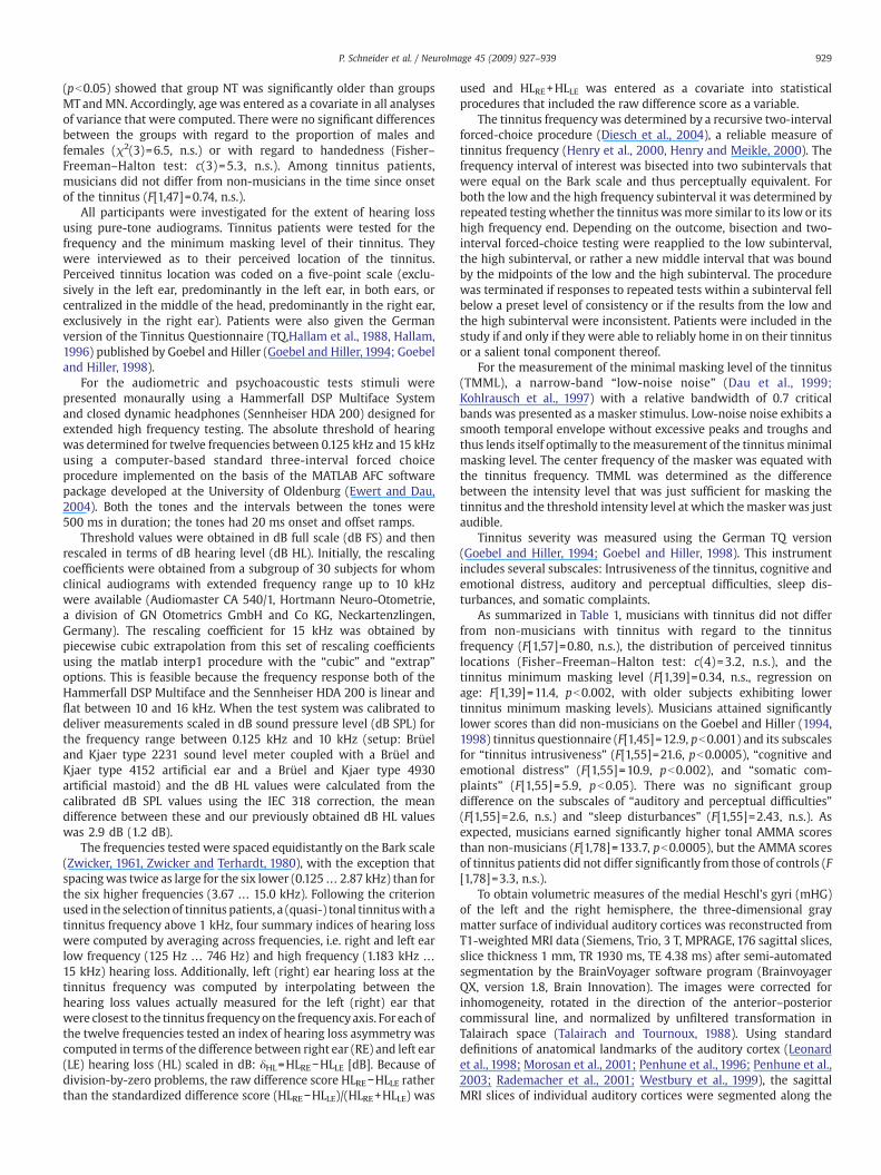
Sylvian fissure including the planum temporale (PT), Heschl's gyrus(HG), and the anterior supratemporal gyrus (aSTG) (Fig. 1). Prior tomorphometric analysis, the HG was identified by the first completeHeschl sulcus (HS) as its posterior and the first transverse sulcus (FTS)as its anterior boundary (Morosan et al., 2001; Rademacher et al.,2001). The medial boundary of the HG was identified in terms of theline connecting the medial end of FTS to the medial end of the mostanterior HS. The medial-to-lateral extent of mHG was defined by afixed distance of 24 mm in order to confine the region of interest tothe primary auditory cortex (PAC) and to fully include it. The distancechosen represents a compromise between the known averagelocation of primary auditory cortex in the medial two-thirds ofHeschl's gyrus and the known interindividual variability of itslocation (Hackett et al., 2001; (Leonard et al., 1998; Patterson et al.,2002; Schneider et al., 2005). Five major types of HG morphologywere distinguished with respect to gyral duplications (Fig. 2, Table 2):presence (1) of a single HG, (2) of a HG with a common stemduplication reaching the lateral but not the medial end of HG, (3) alocal intermediate sulcus (SI) reaching neither its medial nor itslateral end, (4) of a HG with a medial duplication, reaching themedial, not the lateral end (5) presence of a complete posteriorduplication (PD) with a second, posterior HG (Schneider et al., 2005).In several rare cases, a multiple gyration with three or four transverseHeschl's gyri was observed. The medial and complete posteriorduplications (cases 4 and 5), reaching both the medial end of mHG,were considered to be part of the planum temporale and were notincluded in the computation of mHG volume.
A cuboid-shaped region of interest was defined comprising the leftand right auditory cortex in which the inclusion range of gray matterintensity values was calculated. The inclusion range of gray matterintensity values was derived from the intensity histograms of graymatter values: The half-side slope of gray matter peak distribution (i)towards cerebral spinal fluid and (ii) the saddle point between thegray and white matter peak was identified for each subject. All voxelswithin this inclusion range for gray matter intensities were markedand used for three-dimensional reconstruction. In both hemispheres,gray matter volumes of mHG were calculated in eight successivecross-sectional 3 mm-slices that were orientated perpendicularly tothe major axis of the individual mHG. For each of the resulting eighthomologous segment pairs, an index of volumetric asymmetry was
Fig. 1.Morphology of the auditory cortex (AC). (A) Individual 3D-reconstruction of the right aof the segmented AC illustrating the anatomy of Heschl's gyrus (left: blue-colored, right: red-(PT) posterior to the HG. The posteromedial two-thirds of the HG are highlighted, comprisgradient of the underlying tonotopic organization, ranging from high to low frequencies inmedio-lateral extent of mHG for which the gray matter volume segments have been compu
computed in terms of the standardized difference between the rightand the left hemisphere volume: δS=(VRH−VLH)/(VRH+VLH). Severalsummary indices of mHG volume and volume asymmetry werecomputed: right and left hemisphere postero-medial (segments 1 …
4) and antero-lateral (segments 5 … 8) volume, and postero-medial(segments 1 … 4) and antero-lateral (segments 5 … 8) volumeasymmetry.
Results
Morphology and volumetry of Heschl's gyrus
As reported previously (Schneider et al., 2002, 2005), the 3D-reconstruction of individual auditory cortices revealed large varia-bility with respect to size, gyration, and HG duplication. Therewere nosignificant group differences with respect to the distribution of singleHGs, intermediate sulci, HGs with medial duplication, and completeHG duplications (left hemisphere: Fisher–Freeman–Halton c(9)=13.1,n.s., right hemisphere: Fisher–Freeman–Halton c(9)=11.2, n.s.). ThemHG volume of the left hemisphere was larger than the mHG volumeof the right hemisphere both for tinnitus patients (χ2(3)=13.7,pb0.005) and controls (χ2(3)=9.7, pb0.025). Tinnitus patients andcontrols showed comparable hemispheric asymmetries with respectto the frequency of HG duplications, i.e. a more frequent single HG inthe left hemisphere (tinnitus patients: LH 39.3%, RH 19.7%, controls:LH 46.7%, RH 17.8%) and a more frequent intermediate sulcus (SI) andcommon stem duplication (CS) in the right hemisphere (see Table 2).
Fig. 3 and Table 3 present a conspectus of the gray matter volumedata subdivided by tinnitus status, lateralization of perceived tinnituslocation,musicality, and hearing status.Multivariate analysis of variance(MANOVA, with tinnitus status and musicality as grouping factors andhemisphere and volume segment as repeatedmeasures factors showedthat graymatter volumeof the investigatedmedial part ofHeschl's gyrus(mHG) was smaller for tinnitus patients than for healthy controls (F[1,101]=15.9, pb0.0005) and larger for musicians than for non-musicians (F[1,101]=80.9, pb0.00001). Volume was larger in the leftthan in the right hemisphere (F[1,102]=50.5, pb0.00001). More lateralsegments were larger than more medial segments (T2[7,96]=9.7,pb0.00001). This effect was stronger in the left than in the righthemisphere (segment×hemisphere: T2[7,96]=0.83, pb0.00001) and
nd left AC, embedded in a transverse T1-weighted MRI section of the brain. (B) Top viewcolored), the anterior part of the supratemporal gyrus (aSTG) and the planum temporaleing major parts of the primary auditory core fields. The arrow on the left indicates themedio-lateral direction. The arrow on the right indicates the 24 mm range along theted.
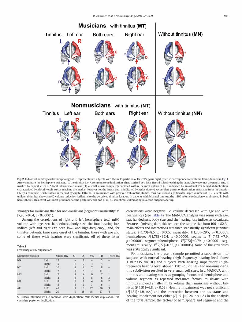
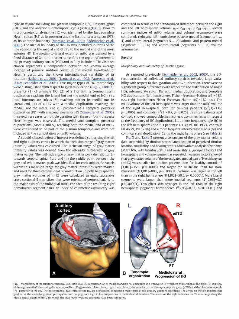
Fig. 2. Individual auditory cortex morphology of 16 representative subjects with the mHG partition of Heschl's gyrus highlighted in correspondence with the frame defined in Fig. 1.Arrows indicate the hemisphere ipsilateral to the tinnitus ear. A common stem duplication, characterized by a local Heschl sulcus reaching the lateral, however not the medial end, ismarked by capital letter C. A local intermediate sulcus (SI), a small sulcus completely enclosed within the most anterior HG, is indicated by an asterisk (⁎). A medial duplication,characterized by a local Heschl sulcus reaching the medial, however not the lateral end, is indicated by a plus sign (+). A complete posterior duplication, separated from the anteriorHG by a complete Heschl sulcus, is marked by capital letter D. In accordance with previous volumetric studies, musicians show significantly larger volumes of HG. Patients withunilateral tinnitus show a mHG volume reduction ipsilateral to the perceived tinnitus location. In patients with bilateral tinnitus, the mHG volume reduction was observed in bothhemispheres. This effect was most prominent at the posteromedial end of mHG, sometimes culminating in a cone-shaped tapering.
931P. Schneider et al. / NeuroImage 45 (2009) 927–939
stronger formusicians than for non-musicians (segment×musicality: T2
[7,96]=0.64, pb0.00001).Among the correlations of right and left hemisphere total mHG
volume with age, sex, handedness, body size, the four hearing lossindices (left and right ear, both low- and high-frequency), and, fortinnitus patients, time since onset of the tinnitus, those with age andsome of those with hearing were significant. All of these latter
Table 2Frequency of HG duplications
Duplication/group Single HG SI CS MD PD Three HG
NN Left 12 – 1 – 3 –
Right 5 2 2 3 4 –
NT Left 16 2 2 7 8 –
Right 7 6 4 7 11 –
MN Left 9 2 4 6 7 1Right 3 6 6 5 6 3
MT Left 8 3 1 4 8 2Right 5 5 6 3 6 1
All Left 45 7 8 17 26 3Right 20 19 18 18 27 4
SI: sulcus intermedius; CS: common stem duplication; MD: medial duplication; PD:complete posterior duplication.
correlations were negative, i.e. volume decreased with age and withhearing loss (see Table 4). The MANOVA analysis was rerun with age,sex, handedness, body size, and the hearing loss indices as covariates.Because of missing data, this reduced the sample size from 106 to 82 Allmain effects and interactions remained statistically significant (tinnitusstatus: F[1,70]=8.5, pb0.005, musicality: F[1,70]=29.7, pb0.00001,hemisphere: F[1,78] =37.4, pb0.00001, segment: T2[7,72] =7.9,pb0.00001, segment×hemisphere: T2[7,72]=0.79, pb0.00001, seg-ment×musicality: T2[7,72]=0.53, pb0.00005). None of the covariateswas statistically significant.
For musicians, the present sample permitted a subdivision intosubjects with normal hearing (high-frequency hearing level above1 kHz≤15 dB HL) and subjects with hearing impairment (high-frequency hearing level above 1 kHz N15 dB HL). For non-musicians,this subdivision resulted in very small cell sizes. In a MANOVA withtinnitus and hearing status as grouping factors and hemisphere andvolume segment as repeated measures factors, musicians withtinnitus showed smaller mHG volume than musicians without tin-nitus (F[1,51]=6.8, pb0.02). Hearing impairment was not significant(F[1,51]=1.9, n.s.) and the interaction between tinnitus status andhearing impairment not either (F[1,51]=0.24, n.s.). As in the analysisof the total sample, the factors of hemisphere and segment and the
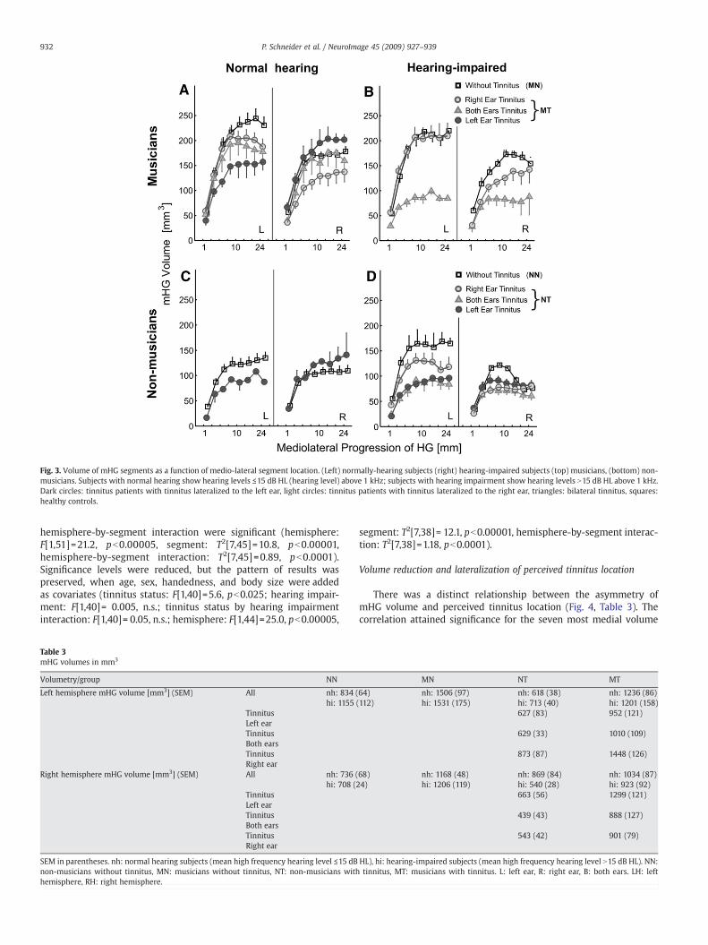
Fig. 3. Volume of mHG segments as a function of medio-lateral segment location. (Left) normally-hearing subjects (right) hearing-impaired subjects (top) musicians, (bottom) non-musicians. Subjects with normal hearing show hearing levels ≤15 dB HL (hearing level) above 1 kHz; subjects with hearing impairment show hearing levels N15 dB HL above 1 kHz.Dark circles: tinnitus patients with tinnitus lateralized to the left ear, light circles: tinnitus patients with tinnitus lateralized to the right ear, triangles: bilateral tinnitus, squares:healthy controls.
932 P. Schneider et al. / NeuroImage 45 (2009) 927–939
hemisphere-by-segment interaction were significant (hemisphere:F[1,51]=21.2, pb0.00005, segment: T2[7,45]=10.8, pb0.00001,hemisphere-by-segment interaction: T2[7,45]=0.89, pb0.0001).Significance levels were reduced, but the pattern of results waspreserved, when age, sex, handedness, and body size were addedas covariates (tinnitus status: F[1,40]=5.6, pb0.025; hearing impair-ment: F[1,40]= 0.005, n.s.; tinnitus status by hearing impairmentinteraction: F[1,40]= 0.05, n.s.; hemisphere: F[1,44]=25.0, pb0.00005,
Table 3mHG volumes in mm3
Volumetry/group NN
Left hemisphere mHG volume [mm3] (SEM) All nh: 834 (hi: 1155
TinnitusLeft earTinnitusBoth earsTinnitusRight ear
Right hemisphere mHG volume [mm3] (SEM) All nh: 736 (hi: 708 (2
TinnitusLeft earTinnitusBoth earsTinnitusRight ear
SEM in parentheses. nh: normal hearing subjects (mean high frequency hearing level ≤15 dBnon-musicians without tinnitus, MN: musicians without tinnitus, NT: non-musicians withhemisphere, RH: right hemisphere.
segment: T2[7,38]= 12.1, pb0.00001, hemisphere-by-segment interac-tion: T2[7,38]=1.18, pb0.0001).
Volume reduction and lateralization of perceived tinnitus location
There was a distinct relationship between the asymmetry ofmHG volume and perceived tinnitus location (Fig. 4, Table 3). Thecorrelation attained significance for the seven most medial volume
MN NT MT
64) nh: 1506 (97) nh: 618 (38) nh: 1236 (86)(112) hi: 1531 (175) hi: 713 (40) hi: 1201 (158)
627 (83) 952 (121)
629 (33) 1010 (109)
873 (87) 1448 (126)
68) nh: 1168 (48) nh: 869 (84) nh: 1034 (87)4) hi: 1206 (119) hi: 540 (28) hi: 923 (92)
663 (56) 1299 (121)
439 (43) 888 (127)
543 (42) 901 (79)
HL), hi: hearing-impaired subjects (mean high frequency hearing level N15 dB HL). NN:tinnitus, MT: musicians with tinnitus. L: left ear, R: right ear, B: both ears. LH: left
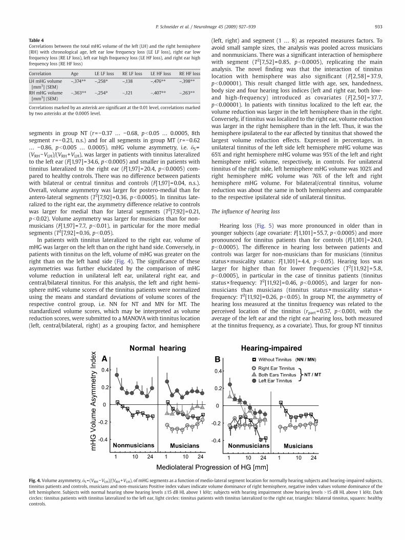
Table 4Correlations between the total mHG volume of the left (LH) and the right hemisphere(RH) with chronological age, left ear low frequency loss (LE LF loss), right ear lowfrequency loss (RE LF loss), left ear high frequency loss (LE HF loss), and right ear highfrequency loss (RE HF loss)
Correlation Age LE LF loss RE LF loss LE HF loss RE HF loss
LH mHG volume[mm3] (SEM)
–,374⁎⁎ –,258⁎ –,138 –,476⁎⁎ –,398⁎⁎
RH mHG volume[mm3] (SEM)
–,363⁎⁎ –,254⁎ –,121 –,407⁎⁎ –,263⁎⁎
Correlations marked by an asterisk are significant at the 0.01 level, correlations markedby two asterisks at the 0.0005 level.
933P. Schneider et al. / NeuroImage 45 (2009) 927–939
segments in group NT (r=−0.37 … −0.68, pb0.05 … 0.0005, 8thsegment r=−0.21, n.s.) and for all segments in group MT (r=−0.62… −0.86, pb0.005 … 0.0005). mHG volume asymmetry, i.e. δS=(VRH−VLH)/(VRH+VLH), was larger in patients with tinnitus lateralizedto the left ear (F[1,97]=34.6, pb0.0005) and smaller in patients withtinnitus lateralized to the right ear (F[1,97]=20.4, pb0.0005) com-pared to healthy controls. There was no difference between patientswith bilateral or central tinnitus and controls (F[1,97]=0.04, n.s.).Overall, volume asymmetry was larger for postero-medial than forantero-lateral segments (T2[7,92]=0.36, pb0.0005). In tinnitus late-ralized to the right ear, the asymmetry difference relative to controlswas larger for medial than for lateral segments (T2[7,92]=0.21,pb0.02). Volume asymmetry was larger for musicians than for non-musicians (F[1,97]=7.7, pb0.01), in particular for the more medialsegments (T2[7,92]=0.16, pb0.05).
In patients with tinnitus lateralized to the right ear, volume ofmHG was larger on the left than on the right hand side. Conversely, inpatients with tinnitus on the left, volume of mHG was greater on theright than on the left hand side (Fig. 4). The significance of theseasymmetries was further elucidated by the comparison of mHGvolume reduction in unilateral left ear, unilateral right ear, andcentral/bilateral tinnitus. For this analysis, the left and right hemi-sphere mHG volume scores of the tinnitus patients were normalizedusing the means and standard deviations of volume scores of therespective control group, i.e. NN for NT and MN for MT. Thestandardized volume scores, which may be interpreted as volumereduction scores, were submitted to a MANOVAwith tinnitus location(left, central/bilateral, right) as a grouping factor, and hemisphere
Fig. 4. Volume asymmetry, δS= (VRH−VLH)/(VRH+VLH), of mHG segments as a function of meditinnitus patients and controls, musicians and non-musicians Positive index values indicate vleft hemisphere. Subjects with normal hearing show hearing levels ≤15 dB HL above 1 kHzcircles: tinnitus patients with tinnitus lateralized to the left ear, light circles: tinnitus patiencontrols.
(left, right) and segment (1 … 8) as repeated measures factors. Toavoid small sample sizes, the analysis was pooled across musiciansand nonmusicians. There was a significant interaction of hemispherewith segment (T2[7,52]=0.85, pb0.0005), replicating the mainanalysis. The novel finding was that the interaction of tinnituslocation with hemisphere was also significant (F[2,58]=37.9,pb0.00001). This result changed little with age, sex, handedness,body size and four hearing loss indices (left and right ear, both low-and high-frequency) introduced as covariates (F[2,50] = 37.7,pb0.00001). In patients with tinnitus localized to the left ear, thevolume reduction was larger in the left hemisphere than in the right.Conversely, if tinnitus was localized to the right ear, volume reductionwas larger in the right hemisphere than in the left. Thus, it was thehemisphere ipsilateral to the ear affected by tinnitus that showed thelargest volume reduction effects. Expressed in percentages, inunilateral tinnitus of the left side left hemisphere mHG volume was65% and right hemisphere mHG volume was 95% of the left and righthemisphere mHG volume, respectively, in controls. For unilateraltinnitus of the right side, left hemisphere mHG volume was 102% andright hemisphere mHG volume was 76% of the left and righthemisphere mHG volume. For bilateral/central tinnitus, volumereduction was about the same in both hemispheres and comparableto the respective ipsilateral side of unilateral tinnitus.
The influence of hearing loss
Hearing loss (Fig. 5) was more pronounced in older than inyounger subjects (age covariate: F[1,101]=55.7, pb0.0005) and morepronounced for tinnitus patients than for controls (F[1,101]=24.0,pb0.0005). The difference in hearing loss between patients andcontrols was larger for non-musicians than for musicians (tinnitusstatus×musicality status: F[1,101]=4.4, pb0.05). Hearing loss waslarger for higher than for lower frequencies (T2[11,92] =5.8,pb0.0005), in particular in the case of tinnitus patients (tinnitusstatus×frequency: T2[11,92]=0.46, pb0.0005), and larger for non-musicians than musicians (tinnitus status×musicality status×frequency: T2[11,92]=0.26, pb0.05). In group NT, the asymmetry ofhearing loss measured at the tinnitus frequency was related to theperceived location of the tinnitus (rpart=0.57, pb0.001, with theaverage of the left ear and the right ear hearing loss, both measuredat the tinnitus frequency, as a covariate). Thus, for group NT tinnitus
o-lateral segment location for normally hearing subjects and hearing-impaired subjects,olume dominance of right hemisphere, negative index values volume dominance of the; subjects with hearing impairment show hearing levels N15 dB HL above 1 kHz. Darkts with tinnitus lateralized to the right ear, triangles: bilateral tinnitus, squares: healthy
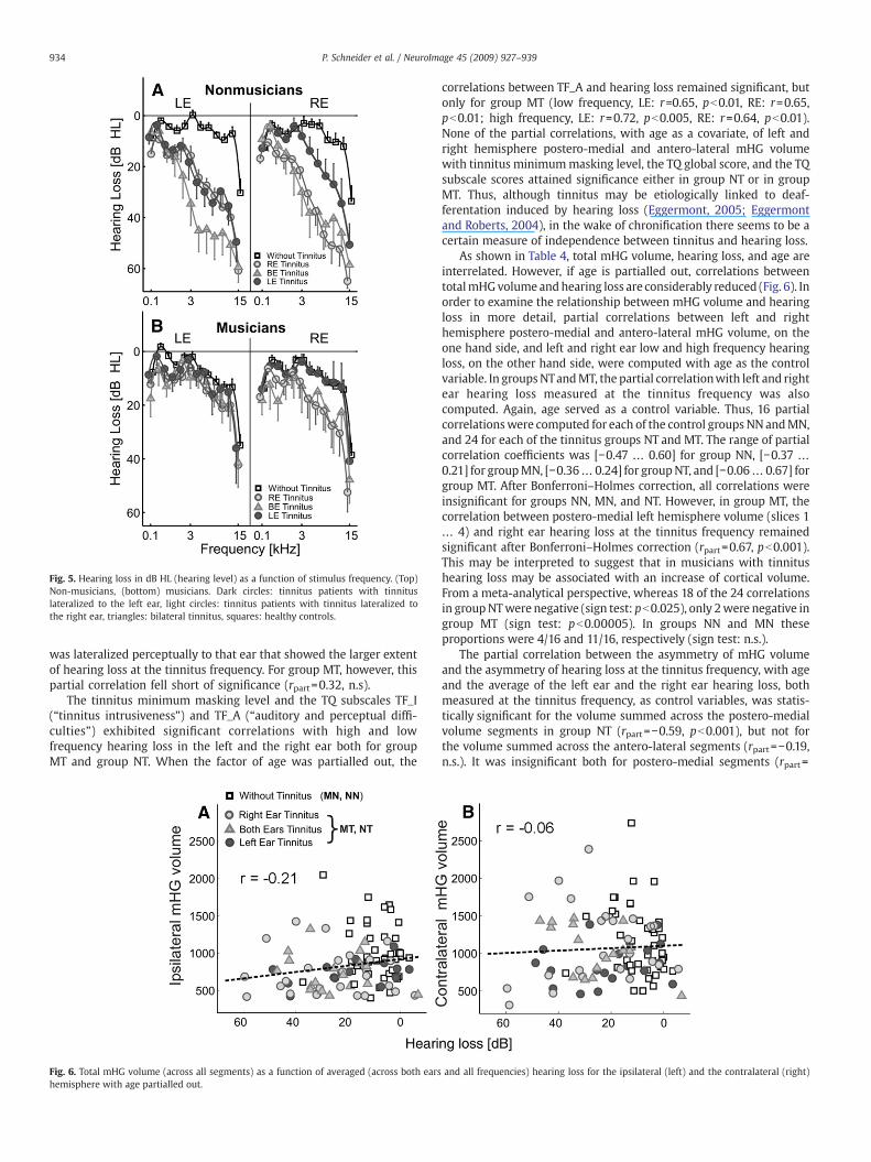
Fig. 5. Hearing loss in dB HL (hearing level) as a function of stimulus frequency. (Top)Non-musicians, (bottom) musicians. Dark circles: tinnitus patients with tinnituslateralized to the left ear, light circles: tinnitus patients with tinnitus lateralized tothe right ear, triangles: bilateral tinnitus, squares: healthy controls.
934 P. Schneider et al. / NeuroImage 45 (2009) 927–939
was lateralized perceptually to that ear that showed the larger extentof hearing loss at the tinnitus frequency. For group MT, however, thispartial correlation fell short of significance (rpart=0.32, n.s).
The tinnitus minimum masking level and the TQ subscales TF_I(“tinnitus intrusiveness”) and TF_A (“auditory and perceptual diffi-culties”) exhibited significant correlations with high and lowfrequency hearing loss in the left and the right ear both for groupMT and group NT. When the factor of age was partialled out, the
Fig. 6. Total mHG volume (across all segments) as a function of averaged (across both earshemisphere with age partialled out.
correlations between TF_A and hearing loss remained significant, butonly for group MT (low frequency, LE: r=0.65, pb0.01, RE: r=0.65,pb0.01; high frequency, LE: r=0.72, pb0.005, RE: r=0.64, pb0.01).None of the partial correlations, with age as a covariate, of left andright hemisphere postero-medial and antero-lateral mHG volumewith tinnitus minimummasking level, the TQ global score, and the TQsubscale scores attained significance either in group NT or in groupMT. Thus, although tinnitus may be etiologically linked to deaf-ferentation induced by hearing loss (Eggermont, 2005; Eggermontand Roberts, 2004), in the wake of chronification there seems to be acertain measure of independence between tinnitus and hearing loss.
As shown in Table 4, total mHG volume, hearing loss, and age areinterrelated. However, if age is partialled out, correlations betweentotalmHGvolume andhearing loss are considerably reduced (Fig. 6). Inorder to examine the relationship between mHG volume and hearingloss in more detail, partial correlations between left and righthemisphere postero-medial and antero-lateral mHG volume, on theone hand side, and left and right ear low and high frequency hearingloss, on the other hand side, were computed with age as the controlvariable. In groupsNTandMT, the partial correlationwith left and rightear hearing loss measured at the tinnitus frequency was alsocomputed. Again, age served as a control variable. Thus, 16 partialcorrelationswere computed for each of the control groups NN andMN,and 24 for each of the tinnitus groups NT and MT. The range of partialcorrelation coefficients was [−0.47 … 0.60] for group NN, [−0.37 …
0.21] for groupMN, [−0.36… 0.24] for groupNT, and [−0.06… 0.67] forgroup MT. After Bonferroni–Holmes correction, all correlations wereinsignificant for groups NN, MN, and NT. However, in group MT, thecorrelation between postero-medial left hemisphere volume (slices 1… 4) and right ear hearing loss at the tinnitus frequency remainedsignificant after Bonferroni–Holmes correction (rpart=0.67, pb0.001).This may be interpreted to suggest that in musicians with tinnitushearing loss may be associated with an increase of cortical volume.From a meta-analytical perspective, whereas 18 of the 24 correlationsin groupNTwere negative (sign test: pb0.025), only 2were negative ingroup MT (sign test: pb0.00005). In groups NN and MN theseproportions were 4/16 and 11/16, respectively (sign test: n.s.).
The partial correlation between the asymmetry of mHG volumeand the asymmetry of hearing loss at the tinnitus frequency, with ageand the average of the left ear and the right ear hearing loss, bothmeasured at the tinnitus frequency, as control variables, was statis-tically significant for the volume summed across the postero-medialvolume segments in group NT (rpart=−0.59, pb0.001), but not forthe volume summed across the antero-lateral segments (rpart=−0.19,n.s.). It was insignificant both for postero-medial segments (rpart=
and all frequencies) hearing loss for the ipsilateral (left) and the contralateral (right)
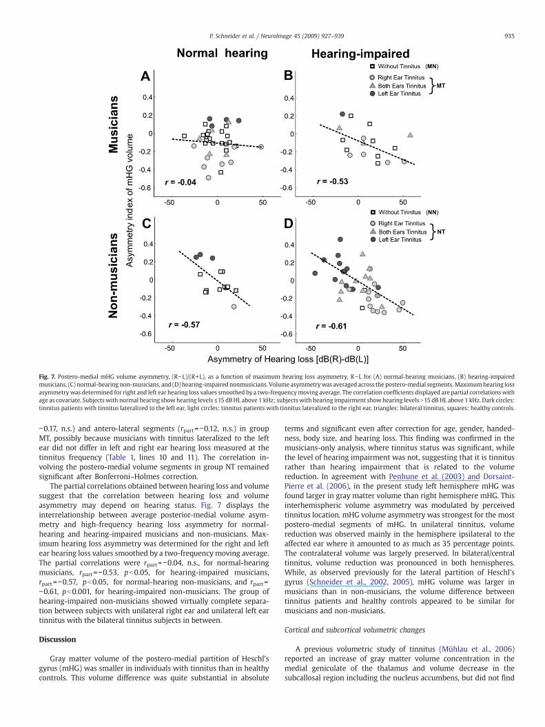
Fig. 7. Postero-medial mHG volume asymmetry, (R−L)/(R+L), as a function of maximum hearing loss asymmetry, R−L for (A) normal-hearing musicians, (B) hearing-impairedmusicians, (C) normal-hearing non-musicians, and (D) hearing-impairednonmusicians. Volume asymmetrywas averaged across the postero-medial segments.Maximumhearing lossasymmetrywas determined for right and left ear hearing loss values smoothed by a two-frequencymoving average. The correlation coefficients displayed are partial correlationswithage as covariate. Subjects with normal hearing showhearing levels ≤15 dBHL above 1 kHz; subjectswith hearing impairment showhearing levels N15 dBHL above 1 kHz. Dark circles:tinnitus patients with tinnitus lateralized to the left ear, light circles: tinnitus patients with tinnitus lateralized to the right ear, triangles: bilateral tinnitus, squares: healthy controls.
935P. Schneider et al. / NeuroImage 45 (2009) 927–939
−0.17, n.s.) and antero-lateral segments (rpart=−0.12, n.s.) in groupMT, possibly because musicians with tinnitus lateralized to the leftear did not differ in left and right ear hearing loss measured at thetinnitus frequency (Table 1, lines 10 and 11). The correlation in-volving the postero-medial volume segments in group NT remainedsignificant after Bonferroni–Holmes correction.
The partial correlations obtained between hearing loss and volumesuggest that the correlation between hearing loss and volumeasymmetry may depend on hearing status. Fig. 7 displays theinterrelationship between average posterior-medial volume asym-metry and high-frequency hearing loss asymmetry for normal-hearing and hearing-impaired musicians and non-musicians. Max-imum hearing loss asymmetry was determined for the right and leftear hearing loss values smoothed by a two-frequency moving average.The partial correlations were rpart=−0.04, n.s., for normal-hearingmusicians, rpart=−0.53, pb0.05, for hearing-impaired musicians,rpart=−0.57, pb0.05, for normal-hearing non-musicians, and rpart=−0.61, pb0.001, for hearing-impaired non-musicians. The group ofhearing-impaired non-musicians showed virtually complete separa-tion between subjects with unilateral right ear and unilateral left eartinnitus with the bilateral tinnitus subjects in between.
Discussion
Gray matter volume of the postero-medial partition of Heschl'sgyrus (mHG) was smaller in individuals with tinnitus than in healthycontrols. This volume difference was quite substantial in absolute
terms and significant even after correction for age, gender, handed-ness, body size, and hearing loss. This finding was confirmed in themusicians-only analysis, where tinnitus status was significant, whilethe level of hearing impairment was not, suggesting that it is tinnitusrather than hearing impairment that is related to the volumereduction. In agreement with Penhune et al. (2003) and Dorsaint-Pierre et al. (2006), in the present study left hemisphere mHG wasfound larger in gray matter volume than right hemisphere mHG. Thisinterhemispheric volume asymmetry was modulated by perceivedtinnitus location. mHG volume asymmetry was strongest for the mostpostero-medial segments of mHG. In unilateral tinnitus, volumereduction was observed mainly in the hemisphere ipsilateral to theaffected ear where it amounted to as much as 35 percentage points.The contralateral volume was largely preserved. In bilateral/centraltinnitus, volume reduction was pronounced in both hemispheres.While, as observed previously for the lateral partition of Heschl'sgyrus (Schneider et al., 2002, 2005), mHG volume was larger inmusicians than in non-musicians, the volume difference betweentinnitus patients and healthy controls appeared to be similar formusicians and non-musicians.
Cortical and subcortical volumetric changes
A previous volumetric study of tinnitus (Mühlau et al., 2006)reported an increase of gray matter volume concentration in themedial geniculate of the thalamus and volume decrease in thesubcallosal region including the nucleus accumbens, but did not find
936 P. Schneider et al. / NeuroImage 45 (2009) 927–939
an effect at the level of the auditory cortex. The study employed themethod of voxel-based morphometry (VBM, Ashburner and Friston,2000). However, the high interindividual anatomical variability of thegyri of Heschl with respect to gyration and angulation may constitutea disadvantage for VBM approaches, because interindividual anato-mical variability is likely to translate into regionally fluctuatingstatistical power to detect group differences (Tisserand et al., 2004).For instance, the hemisphere difference in HG gray matter volume infavor of the left hemisphere obtained in the present study has alsobeen reported by Dorsaint-Pierre et al. (2006), but for individuallyguided volume measurements, not for VBM. Using diffusion tensorimaging (DTI), Yoo et al. (2006) analyzed white matter anisotropy inpatients with tinnitus. Their preliminary results suggest the possibleinvolvement of changes in white matter structures in tinnitus.However, controls were not included in this study.
Is volume reduction caused by hyperactivity-related graymatter atrophy?
Could the smaller gray matter mHG volume found in tinnitusrepresent gray matter loss brought about by atrophy or programmedcell death? In some respects tinnitus is similar to pain (Folmer et al.,2001, Moller, 2000). Tinnitus is associated with spontaneous hyper-activity and increased evoked response amplitudes in severalstructures of the auditory pathway including auditory cortex (Dieschet al., 2004; Eggermont and Roberts, 2004; Eggermont, 2005). It couldbe hypothesized that chronic hyperactivation might generate excito-toxic levels of glutamate receptor activation (Mattson, 2003) that aresufficient to generate the volumetric changes observed in the tinnitussample investigated in this study. There have been several reports ofcortical gray matter volume reduction in various pain conditions thathave been interpreted as indicative of pathology-related atrophy. Atthe cortical level, chronic back pain has been shown to be associatedwith smaller gray matter volume of dorsolateral prefrontal cortex(Apkarian et al., 2004) and primary somatosensory cortex (Schmidt-Wilcke et al., 2006). In fibromyalgia, graymatter volume reduction hasbeen found to be particularly pronounced in the medial frontal cortex,the left hemisphere insula, the middle and posterior cingulate gyrus,and the left parahippocampal gyrus (Kuchinad et al., 2007). BothApkarian et al. (2004) and Kuchinad et al. (2007) have invokedatrophy secondary to excitotoxicity and/or as a consequence of theexposure to inflammatory agents, such as cytokines, as one possibleexplanation of volume reduction in terms of volume loss. However,the present data on the interrelationship of hearing loss and corticalvolume are merely correlational. Furthermore, volumetric MRI dataare ambiguous both with regard to the origin of group differences andthe cellular and subcellular mechanisms underlying volume change(Weinberger and McClure, 2002). As has been argued for volumetricstudies of pain conditions (Schmidt-Wilcke et al., 2006), longitudinaldesigns and appropriate treatment protocols will be needed toinvestigate whether all or part of the cortical volume reductionobserved in tinnitus is produced by neurodegeneration rather thantissue shrinkage.
Does small mHG volume predate tinnitus rather than followfrom tinnitus?
However, the observation that, in unilateral tinnitus, volumereduction effects are largely constrained to the hemisphere ipsilateralto the affected ear may present a problem to the idea that volumetricchanges could be mediated by, for instance, excitotoxic levels ofglutamate receptor activation purportedly generated by hyperactivityand hyperresponsivity of the afferent auditory pathway. Bothhyperactivity and hyperresponsivity are not constrained to theipsilateral projection. It is also important to note that in the presentstudy there were no significant interactions involving the group
variable of tinnitus status and the repeated-measurement variable ofsegment-within-mHG. The volume difference observed betweenpatients and controls probably affects the full extent of primaryauditory cortex, as primary auditory cortex tends to be located withinthe confines of the investigated partition of mHG (Galaburda andSanides, 1980, Hackett et al., 2001, Rademacher et al., 1993). Thisrenders it unlikely that the volume difference represents volume lossgenerated in the wake of deafferentation. If hearing loss were morepronounced for the higher frequencies, as it actually was, then, giventhe tonotopic organization of primary auditory cortex (Formisanoet al., 2003), any volume-loss-by-deafferentation hypothesis wouldhave to predict a significant mHG segment by tinnitus statusinteraction which did not obtain. If the processes that generatehyperactivation and tinnitus as sequelae of cochlear damage andhearing loss also generated mHG volume loss, a significant negativecorrelation between hearing loss and mHG volume would have to beexpected. However, of all correlations computed between mHGvolume and hearing loss, only the correlation of left hemispherepostero-medial mHG volume and right ear tinnitus-frequency hearingloss in group MT was statistically significant. And the sign of thatcorrelation was positive. Similarly, a significant positive correlationwould have to be expected between mHG volume asymmetry andhearing loss asymmetry. However, this correlation was significantlynegative for non-musicians with tinnitus when hearing loss asym-metry at the tinnitus frequency was considered and it wassignificantly negative for non-musicians and hearing-impaired musi-cians. Therefore, it seems likely that the volume difference betweenpatients and controls was present before the patients ever developedtinnitus. Small mHG volumemay thus constitute a vulnerability factor.An individual with a small mHG may carry a higher risk to developtinnitus in the wake of cochlear damage. Gray matter mHG volumewas not significantly correlated with time-since-onset of the tinnitus,tinnitus minimum masking level, and the TQ subscale of tinnitusintrusiveness in tinnitus patients. Thus, although tinnitus may beetiologically linked to deafferentation induced by hearing loss(Eggermont, 2005; Eggermont and Roberts, 2004), after chronificationthere seems to be a certain measure of independence betweentinnitus and hearing loss. The absence of these interrelationships iscompatible with a vulnerability hypothesis. However, any form ofvulnerability hypothesis can only be substantiated by studies invol-ving longitudinal designs or studies of monozygotic twins.
Is unilateral tinnitus related to a lack of transcallosal inhibition?
In unilateral tinnitus, mHG volume reduction occurred almostexclusively ipsilateral to the affected ear. How can we make sense ofthis lateralization effect? If it were the hemisphere contralateral to theear that the tinnitus is lateralized to that actually generated thesensation of tinnitus, then potentially because the reduced-sizeipsilateral mHG does not generate the amount of tonic transcallosalinhibition necessary to prevent his from happening. Homotopic andheterotopic auditory cortical areas of the left and the right hemisphereare interconnected by the corpus callosum. Other than in vision andsomatosensation where it is only the midline zone that featurescallosal fiber connections, in audition, callosal projections aredistributed across the full extent of auditory cortex (Bamiou et al.,2007). While all callosal fibers originating from pyramidal cells areneurochemically excitatory, they may terminate on inhibitory inter-neurons. As a result, interhemispheric projections are both excitatoryand inhibitory functionally (Bloom and Hynd, 2005). Under someconditions, the callosal influence is mainly inhibitory (Clarey et al.,1996; Kitzes and Doherty, 1994; Pluto et al., 2005; Tang et al., 2007). Itis easier to observe transcallosal inhibition than transcallosal facilita-tion of primary auditory cortex single unit responses to auditorystimulation (Kitzes and Doherty, 1994). Transcallosal projections seemto combine focused facilitation with widespread lateral inhibition
937P. Schneider et al. / NeuroImage 45 (2009) 927–939
(Tang et al., 2007). In the primary somatosensory cortex, widening ofreceptive fields, i.e. reduction of surround inhibition, has beenobserved in the wake of a reduction of the transcallosal influence(Clarey et al., 1996; Pluto et al., 2005). As there is an inverserelationship between callosal connectivity and hemispheric asymme-try (Aboitiz et al., 1992a, b; Dorion et al. 2000), a small mHG ipsilateralto the ear affected by tinnitus may generate a lesser amount of tonicinhibition of the contralateral mHG and thus facilitate the develop-ment of tinnitus-related activity. The positive correlation of lefthemisphere postero-medial mHG volumewith hearing loss, measuredat the tinnitus frequency, in the right ear in the musician group, thenegative correlation between postero-medial mHG volume asymme-try and the asymmetry of hearing loss at the tinnitus frequencyobserved in group NT, and the negative correlation between postero-medial mHG volume asymmetry and maximum hearing loss asym-metry in non-musicians and hearing impaired musicians are compa-tible with this hypothesis. To further explore the transcallosalinhibition hypothesis, it would be interesting to study the corpuscallosum in patients with unilateral tinnitus.
Does unilateral tinnitus generate volume increments in thecontralateral hemisphere?
The above scenario presumes that ipsilateral volume reductionactually predates the onset of tinnitus. While this remains apossibility, there may be alternatives. In acquired unilateral deafness,an enhancement of the amplitude of the ipsilateral cortical response,relative to the intact ear, to auditory stimulation of the intact ear hasbeen observed using single unit recordings (Kitzes, 1984, Reale et al.,1987), magnetoencephalography (Li et al., 2006, Vasama and Makela,1995), and functional magnetic resonance imaging (Firszt et al., 2006,Langers et al., 2005; Scheffler et al., 1998). For the present sample, thehemisphere contralateral to the tinnitus ear is the hemisphere that isipsilateral to the audiometrically less impaired ear. It seems possiblethat cortical volume increments similar to the ones observed afterenvironmental enrichment (Van et al., 2000) and sensorimotortraining (Draganski et al., 2004; Boyke et al., 2008; Driemeyer et al.,2008) may manifest themselves in the wake of structural andfunctional changes of the afferent auditory pathway. In particular,the positive correlation of left hemisphere postero-medial mHGvolume with the hearing loss, measured at the tinnitus frequency, inthe right ear in the musician group, as well as the negative correlationbetween postero-medial volume asymmetry and asymmetry ofhearing loss, again measured at the tinnitus frequency, in the non-musician group, and finally the negative correlation between postero-medial mHG volume asymmetry and maximum hearing loss asym-metry in non-musicians and in hearing-impaired musicians suggestthat in tinnitus there may be frequency-selective and therefore place-selective volume increments that may take place in a prior setting ofglobally reduced volume and that are correlated with and possiblybrought about by auditory deafferentation. In our sample, thesevolume increments assumed different forms for musicians and non-musicians which may mirror the fact that, at the tinnitus frequency,musicians with tinnitus lateralized to the left ear did not differ inhearing loss.
Does musical training protect against tinnitus?
Overall, musicians with tinnitus demonstrated lower TQ scores onthe subscales of tinnitus intrusiveness, cognitive and emotional dis-tress, and somatic complaints and a milder degree of high-frequencyhearing loss than non-musicians with tinnitus. This raises thequestion if extensive musical training and practice may protectagainst tinnitus. Frequency discrimination training exerts an influenceon frequency discrimination thresholds (Delhommeau et al., 2002;Demany, 1985; Irvine et al., 2000) and the tonotopic organization of
primary auditory cortex (Polley et al., 2006; Rutkowski and Weinber-ger, 2005; Recanzone et al., 1993; but see Brown et al., 2004, for anegative finding). Frequency discrimination trainingmay be beneficialin the treatment of tinnitus (Flor et al., 2004). Longitudinal studiesmay clarify if (i) reduced mHG volume constitutes a vulnerabilityfactor and (ii) musical training and performance, even if accompaniedwith extensive exposure to high levels of sound density, may have aprotective effect against auditory impairment.
Acknowledgments
We would like to thank Steffen Ritter for the help with both MEGand psychoacoustic measurements. We would also like to thank twoanonymous reviewers for their very helpful comments on themanuscript. This research was supported by the Deutsche Forschungs-gemeinschaft (grant Di 497/3-2 to E.D. and A.R.).
References
Aboitiz, F., Scheibel, A.B., Fisher, R.S., Zaidel, E., 1992a. Individual differences in brainasymmetries and fiber composition in the human corpus callosum. Brain Res. 598,154–161.
Aboitiz, F., Scheibel, A.B., Zaidel, E., 1992b. Morphometry of the Sylvian fissure and thecorpus callosum, with emphasis on sex differences. Brain 115 (Pt 5), 1521–1541.
Andersson, G., Lyttkens, L., Hirvela, C., Furmark, T., Tillfors, M., Fredrikson, M., 2000.Regional cerebral blood flow during tinnitus: a PET case study with lidocaine andauditory stimulation. Acta Otolaryngol. 120, 967–972.
Apkarian, A.V., Sosa, Y., Sonty, S., Levy, R.M., Harden, R.N., Parrish, T.B., et al., 2004.Chronic back pain is associated with decreased prefrontal and thalamic gray matterdensity. J. Neurosci. 24, 10410–10415.
Ashburner, J., Friston, K.J., 2000. Voxel-based morphometry—the methods. Neuroimage11, 805–821.
Baguley, D.M., 2002. Mechanisms of tinnitus. Br. Med. Bull. 63, 195–212.Bamiou, D.E., Sisodiya, S., Musiek, F.E., Luxon, L.M., 2007. The role of the interhemi-
spheric pathway in hearing. Brain Res. Rev. 56, 170–182.Barnea, G., Attias, J., Gold, S., Shahar, A., 1990. Tinnitus with normal hearing sensitivity:
extended high-frequency audiometry and auditory-nerve brain-stem-evokedresponses. Audiology 29, 36–45.
Bartels, H., Staal, M.J., Albers, F.W., 2007. Tinnitus and neural plasticity of the brain. Otol.Neurotol. 28, 178–184.
Bartzokis, G., Beckson, M., Lu, P.H., Nuechterlein, K.H., Edwards, N., Mintz, J., 2001. Age-related changes in frontal and temporal lobe volumes in men: a magneticresonance imaging study. Arch. Gen. Psychiatry 58, 461–465.
Basta, D., Tzschentke, B., Ernst, A., 2005. Noise-induced cell death in the mouse medialgeniculate body and primary auditory cortex. Neurosci. Lett. 381, 199–204.
Bloom, J.S., Hynd, G.W., 2005. The role of the corpus callosum in interhemispherictransfer of information: excitation or inhibition? Neuropsychol. Rev. 15, 59–71.
Boyke, J., Driemeyer, J., Gaser, C., Buchel, C., May, A., 2008. Training-induced brainstructure changes in the elderly. J. Neurosci. 28, 7031–7035.
Brown, M., Irvine, D.R., Park, V.N., 2004. Perceptual learning on an auditory frequencydiscrimination task by cats: association with changes in primary auditory cortex.Cereb. Cortex 14, 952–965.
Clarey, J.C., Tweedale, R., Calford, M.B., 1996. Interhemispheric modulation ofsomatosensory receptive fields: evidence for plasticity in primary somatosensorycortex. Cereb. Cortex 6, 196–206.
Dau, T., Verhey, J., Kohlrausch, A., 1999. Intrinsic envelope fluctuations and modulation-detection thresholds for narrow-band noise carriers. J. Acoust. Soc. Am. 106,2752–2760.
Delhommeau, K., Micheyl, C., Jouvent, R., Collet, L., 2002. Transfer of learning acrossdurations and ears in auditory frequency discrimination. Percept. Psychophys. 64,426–436.
Demany, L., 1985. Perceptual learning in frequency discrimination. J. Acoust. Soc. Am.78, 1118–1120.
De Ridder, D., Verstraeten, E., Van der, K.K., De Mulder, G., Sunaert, S., Verlooy, J., et al.,2005. Transcranial magnetic stimulation for tinnitus: influence of tinnitusduration on stimulation parameter choice and maximal tinnitus suppression.Otol. Neurotol. 26, 616–619.
Diesch, E., Struve,M., Rupp, A., Ritter, S., Hulse,M., Flor, H., 2004. Enhancement of steady-state auditory evoked magnetic fields in tinnitus. Eur. J. Neurosci. 19, 1093–1104.
Dorion, A.A., Chantome, M., Hasboun, D., Zouaoui, A., Marsault, C., Capron, C., et al.,2000. Hemispheric asymmetry and corpus callosum morphometry: a magneticresonance imaging study. Neurosci. Res. 36, 9–13.
Dorsaint-Pierre, R., Penhune, V.B., Watkins, K.E., Neelin, P., Lerch, J.P., Bouffard, M., et al.,2006. Asymmetries of the planum temporale and Heschl's gyrus: relationship tolanguage lateralization. Brain 129, 1164–1176.
Draganski, B., Gaser, C., Busch, V., Schuierer, G., Bogdahn, U., May, A., 2004.Neuroplasticity: changes in grey matter induced by training. Nature 427, 311–312.
Driemeyer, J., Boyke, J., Gaser, C., Buchel, C., May, A., 2008. Changes in gray matterinduced by learning—revisited. PLoS ONE 3, e2669.
Eggermont, J.J., 2005. Tinnitus: neurobiological substrates. Drug Discov. Today 10,1283–1290.
938 P. Schneider et al. / NeuroImage 45 (2009) 927–939
Eggermont, J.J., 2007. Correlated neural activity as the driving force for functionalchanges in auditory cortex. Hear. Res. 229, 69–80.
Eggermont, J.J., Roberts, L.E., 2004. The neuroscience of tinnitus. Trends Neurosci. 27,676–682.
Ewert, S.D., Dau, T., 2004. External and internal limitations in amplitude-modulationprocessing. J. Acoust. Soc. Am. 116, 478–490.
Firszt, J.B., Ulmer, J.L., Gaggl, W., 2006. Differential representation of speech sounds inthe human cerebral hemispheres. Anat. Rec. A Discov. Mol. Cell Evol. Biol. 288,345–357.
Flor, H., Hoffmann, D., Struve, M., Diesch, E., 2004. Auditory discrimination training forthe treatment of tinnitus. Appl. Psychophysiol. Biofeedback 29, 113–120.
Folmer, R.L., Griest, S.E., Martin, W.H., 2001. Chronic tinnitus as phantom auditory pain.Otolaryngol. Head Neck Surg. 124, 394–400.
Folmer, R.L., Carroll, J.R., Rahim, A., Shi, Y., Hal, M.W., 2006. Effects of repetitivetranscranial magnetic stimulation (rTMS) on chronic tinnitus. Acta Otolaryngol.Suppl. 96–101.
Formisano, E., Kim, D.S., Di Salle, F., van de Moortele, P.F., Ugurbil, K., Goebel, R., 2003Nov 13. Mirror-symmetric tonotopic maps in human primary auditory cortex.Neuron 40 (4), 859–869.
Galaburda, A., Sanides, F., 1980. Cytoarchitectonic organization of the human auditorycortex. J. Comp. Neurol. 190, 597–610.
Gaser, C., Schlaug, G., 2003. Gray matter differences between musicians andnonmusicians. Ann. N. Y. Acad. Sci. 999, 514–517.
Gilbertson, M.W., Shenton, M.E., Ciszewski, A., Kasai, K., Lasko, N.B., Orr, S.P., et al., 2002.Smaller hippocampal volume predicts pathologic vulnerability to psychologicaltrauma. Nat. Neurosci. 5, 1242–1247.
Goebel, G., Hiller, W., 1994. The tinnitus questionnaire. A standard instrument forgrading the degree of tinnitus. Results of a multicenter study with the tinnitusquestionnaire. HNO 42, 166–172.
Goebel, G., Hiller, W., 1998. Der Tinnitus-Fragebogen. Hogrefe, Göttingen.Good, C.D., Johnsrude, I.S., Ashburner, J., Henson, R.N., Friston, K.J., Frackowiak, R.S.,
2001. A voxel-based morphometric study of ageing in 465 normal adult humanbrains. Neuroimage 14, 21–36.
Gordon, E., 1989. Advanced Measures of Music Audiation. GIA Publications, Chikago.Gordon, E., 1998. Introduction to Research and the Psychology of Music. GIA
Publications, Chikago.Hackett, T.A., Preuss, T.M., Kaas, J.H., 2001. Architectonic identification of the core region in
auditorycortexofmacaques, chimpanzees, andhumans. J. Comp.Neurol. 441,197–222.Hallam, R.S., 1996. Manual of the Tinnitus Questionnaire (TQ). Psychological Corpora-
tion, London.Hallam, R.S., Jakes, S.C., Hinchcliffe, R., 1998. Cognitive variables in tinnitus annoyance.
Br. J. Clin. Psychol. 27 (Pt 3), 213–222.Hazell, J., 1990. Tinnitus and disability with ageing: adaptation and management. Acta
Otolaryngol. Suppl 476, 202–208.Heffner, H.E., Harrington, I.A., 2002. Tinnitus in hamsters following exposure to intense
sound. Hear. Res. 170, 83–95.Henry, J.A., Meikle, M.B., 2000. Psychoacoustic measures of tinnitus. J. Am. Acad. Audiol.
11, 138–155.Henry, J.A., Fausti, S.A., Flick, C.L., Helt, W.J., Ellingson, R.M., 2000. Computer-automated
clinical technique for tinnitus quantification. Am. J. Audiol. 9, 36–49.Irvine, D.R., Martin, R.L., Klimkeit, E., Smith, R., 2000. Specificity of perceptual learning
in a frequency discrimination task. J. Acoust. Soc. Am. 108, 2964–2968.Jackler, R.K., Whinney, D., 2001. A century of eighth nerve surgery. Otol. Neurotol. 22,
401–416.Jackson, P., 1985. A comparison of the effects of eighth nerve section with lidocaine on
tinnitus. J. Laryngol. Otol. 99, 663–666.Jastreboff, P.J., 1990. Phantom auditory perception (tinnitus): mechanisms of generation
and perception. Neurosci. Res. (N.Y.) 8, 221–254.Kaiser, J., Lutzenberger, W., 2003. Induced gamma-band activity and human brain
function. Neuroscientist 9, 475–484.Kitzes, L.M., 1984. Some physiological consequences of neonatal cochlear destruction in
the inferior colliculus of the gerbil, Meriones unguiculatus. Brain Res. 306, 171–178.Kitzes, L.M., Doherty, D., 1994. Influence of callosal activity on units in the auditory
cortex of ferret (Mustela putorius). J. Neurophysiol. 71, 1740–1751.Kleinjung, T., Eichhammer, P., Langguth, B., Jacob, P., Marienhagen, J., Hajak, G., et al.,
2005. Long-term effects of repetitive transcranial magnetic stimulation (rTMS) inpatients with chronic tinnitus. Otolaryngol. Head Neck Surg. 132, 566–569.
Kohlrausch, A., Fassel, R., van der Heijden, M., Kortekaas, R., van de Par, S., Oxenham, A.,et al., 1997. Detection of tones in low-noise noise: further evidence for the role ofenvelope fluctuations. Acustica - Acta Acustica 83, 659–669.
Kuchinad, A., Schweinhardt, P., Seminowicz, D.A., Wood, P.B., Chizh, B.A., Bushnell, M.C.,2007. Accelerated brain gray matter loss in fibromyalgia patients: premature agingof the brain? J. Neurosci. 27, 4004–4007.
Langers, D.R., van, D.P., Backes, W.H., 2005. Lateralization, connectivity and plasticity inthe human central auditory system. Neuroimage. 28, 490–499.
Langguth, B., Eichhammer, P., Kreutzer, A., Maenner, P., Marienhagen, J., Kleinjung, T., et al.,2006. The impact of auditory cortex activity on characterizing and treating patientswith chronic tinnitus— first results from a PET study. Acta Otolaryngol. Suppl. 84–88.
Lee, J., Behar, A., Kunov, H., Wong, W., 2005. Musicians' noise exposure in orchestra pit.Appl. Acoust. 66, 919–931.
Leonard, C.M., Puranik, C., Kuldau, J.M., Lombardino, L.J., 1998. Normal variation in thefrequency and location of human auditory cortex landmarks. Heschl's gyrus: whereis it? Cereb. Cortex 8, 397–406.
Li, L.P., Shiao, A.S., Chen, L.F., Niddam, D.M., Chang, S.Y., Lien, C.F., et al., 2006. Healthy-side dominance of middle- and long-latency neuromagnetic fields in idiopathicsudden sensorineural hearing loss. Eur. J. Neurosci. 24, 937–946.
Lockwood, A.H., Wack, D.S., Burkard, R.F., Coad, M.L., Reyes, S.A., Arnold, S.A., et al., 2001.The functional anatomy of gaze-evoked tinnitus and sustained lateral gaze.Neurology 56, 472–480.
Lockwood, A.H., Salvi, R.J., Burkard, R.F., 2002. Tinnitus. N. Engl. J. Med. 347, 904–910.Mattson, M.P., 2003. Excitotoxic and excitoprotectivemechanisms: abundant targets for
the prevention and treatment of neurodegenerative disorders. NeuromolecularMed. 3, 65–94.
McKee, G.J., Stephens, S.D., 1992. An investigation of normally hearing subjects withtinnitus. Audiology 31, 313–317.
Melcher, J.R., Sigalovsky, I.S., Guinan Jr., J.J., Levine, R.A., 2000. Lateralized tinnitusstudied with functional magnetic resonance imaging: abnormal inferior colliculusactivation. J. Neurophysiol. 83, 1058–1072.
Moller, A.R., 2000. Similarities between severe tinnitus and chronic pain. J. Am. Acad.Audiol. 11, 115–124.
Moller, A.R., 2003. Pathophysiology of tinnitus. Otolaryngol. Clin. North Am. 36 249–2vi.Morosan, P., Rademacher, J., Schleicher, A., Amunts, K., Schormann, T., Zilles, K., 2001.
Human primary auditory cortex: cytoarchitectonic subdivisions andmapping into aspatial reference system. Neuroimage 13, 684–701.
Mühlau, M., Rauschecker, J.P., Oestreicher, E., Gaser, C., Rottinger, M., Wohlschlager, A.M., et al., 2006. Structural brain changes in tinnitus. Cereb. Cortex 16, 1283–1288.
Mühlnickel, W., Elbert, T., Taub, E., Flor, H., 1998. Reorganization of auditory cortex intinnitus. Proc. Natl. Acad. Sci. U.S.A 95, 10340–10343.
Nicolas-Puel, C., Akbaraly, T., Lloyd, R., Berr, C., Uziel, A., Rebillard, G., et al., 2006.Characteristics of tinnitus in a population of 555 patients: specificities of tinnitusinduced by noise trauma. Int. Tinnitus. J. 12, 64–70.
Ochi, K., Ohashi, T., Kenmochi, M., 2003. Hearing impairment and tinnitus pitch inpatients with unilateral tinnitus: comparison of sudden hearing loss and chronictinnitus. Laryngoscope 113, 427–431.
Patterson, R.D., Uppenkamp, S., Johnsrude, I.S., Griffiths, T.D., 2002. The processing oftemporal pitch and melody information in auditory cortex. Neuron 36, 767–776.
Penhune, V.B., Zatorre, R.J., MacDonald, J.D., Evans, A.C., 1996. Interhemisphericanatomical differences in human primary auditory cortex: probabilistic mappingand volume measurement from magnetic resonance scans. Cereb. Cortex 6,661–672.
Penhune, V.B., Cismaru, R., Dorsaint-Pierre, R., Petitto, L.A., Zatorre, R.J., 2003. Themorphometry of auditory cortex in the congenitally deaf measured using MRI.Neuroimage 20, 1215–1225.
Plewnia, C., Reimold, M., Najib, A., Brehm, B., Reischl, G., Plontke, S.K., et al., 2007.Dose-dependent attenuation of auditory phantom perception (tinnitus) byPET-guided repetitive transcranial magnetic stimulation. Hum. Brain Mapp. 28,238–246.
Pluto, C.P., Chiaia, N.L., Rhoades, R.W., Lane, R.D., 2005. Reducing contralateral SI activityreveals hindlimb receptive fields in the SI forelimb-stump representation ofneonatally amputated rats. J. Neurophysiol. 94, 1727–1732.
Polley, D.B., Steinberg, E.E., Merzenich, M.M., 2006. Perceptual learning directs auditorycorticalmap reorganization through top-down influences. J. Neurosci. 26, 4970–4982.
Rademacher, J., Caviness Jr., V.S., Steinmetz, H., Galaburda, A.M., 1993. Topographicalvariation of the human primary cortices: implications for neuroimaging, brainmapping, and neurobiology. Cereb. Cortex 3, 313–329.
Rademacher, J., Morosan, P., Schormann, T., Schleicher, A., Werner, C., Freund, H.J., et al.,2001. Probabilistic mapping and volume measurement of human primary auditorycortex. Neuroimage 13, 669–683.
Raz, N., Lindenberger, U., Rodrigue, K.M., Kennedy, K.M., Head, D., Williamson, A., et al.,2005. Regional brain changes in aging healthy adults: general trends, individualdifferences and modifiers. Cereb. Cortex 15, 1676–1689.
Reale, R.A., Brugge, J.F., Chan, J.C., 1987. Maps of auditory cortex in cats reared afterunilateral cochlear ablation in the neonatal period. Brain Res. 431, 281–290.
Recanzone, G.H., Schreiner, C.E., Merzenich, M.M., 1993. Plasticity in the frequencyrepresentation of primary auditory cortex following discrimination training inadult owl monkeys. J. Neurosci. 13, 87–103.
Rutkowski, R.G., Weinberger, N.M., 2005. Encoding of learned importance of sound bymagnitude of representational area in primary auditory cortex. Proc. Natl. Acad. Sci.U.S.A 102, 13664–13669.
Scheffler, K., Bilecen, D., Schmid, N., Tschopp, K., Seelig, J., 1998. Auditory corticalresponses in hearing subjects and unilateral deaf patients as detected by functionalmagnetic resonance imaging. Cereb. Cortex 8, 156–163.
Schmidt-Wilcke, T., Leinisch, E., Ganssbauer, S., Draganski, B., Bogdahn, U., Altmeppen,J., et al., 2006. Affective components and intensity of pain correlate with structuraldifferences in gray matter in chronic back pain patients. Pain 125, 89–97.
Schneider, P., Scherg, M., Dosch, H.G., Specht, H.J., Gutschalk, A., Rupp, A., 2002.Morphology of Heschl's gyrus reflects enhanced activation in the auditory cortex ofmusicians. Nat. Neurosci. 5, 688–694.
Schneider, P., Sluming, V., Roberts, N., Scherg, M., Goebel, R., Specht, H.J., et al., 2005.Structural and functional asymmetry of lateral Heschl's gyrus reflects pitchperception preference. Nat. Neurosci. 8, 1241–1247.
Smits, M., Kovacs, S., De, R.D., Peeters, R.R., Van, H.P., Sunaert, S., 2007. Lateralization offunctional magnetic resonance imaging (fMRI) activation in the auditory pathwayof patients with lateralized tinnitus. Neuroradiology 49, 669–679.
Talairach, J., Tournoux, P., 1988. Co-planar Stereotaxic Atlas of the Human Brain. Thieme,New York.
Tang, J., Xiao, Z., Suga, N., 2007. Bilateral cortical interaction: modulation of delay-tunedneurons in the contralateral auditory cortex. J. Neurosci. 27, 8405–8413.
Tisserand, D.J., Van Boxtel, M.P., Pruessner, J.C., Hofman, P., Evans, A.C., Jolles, J., 2004. Avoxel-based morphometric study to determine individual differences in graymatter density associated with age and cognitive change over time. Cereb. Cortex14, 966–973.
939P. Schneider et al. / NeuroImage 45 (2009) 927–939
Van, P.H., Kempermann, G., Gage, F.H., 2000. Neural consequences of environmentalenrichment. Nat. Rev. Neurosci. 1, 191–198.
Van de Heyning, H.P., Vermeire, K., Diebl, M., Nopp, P., Anderson, I., De, R.D., 2008.Incapacitating unilateral tinnitus in single-sided deafness treated by cochlearimplantation. Ann. Otol. Rhinol. Laryngol. 117, 645–652.
Vasama, J.P., Makela, J.P., 1995. Auditory pathway plasticity in adult humans afterunilateral idiopathic sudden sensorineural hearing loss. Hear. Res. 87, 132–140.
Weinberger, D.R., McClure, R.K., 2002. Neurotoxicity, neuroplasticity, and magneticresonance imaging morphometry: what is happening in the schizophrenic brain?Arch. Gen. Psychiatry 59, 553–558.
Weisz, N., Moratti, S., Meinzer, M., Dohrmann, K., Elbert, T., 2005. Tinnitus perceptionand distress is related to abnormal spontaneous brain activity as measured bymagnetoencephalography. PLoS Med. 2, e153.
Weisz, N., Muller, S., Schlee, W., Dohrmann, K., Hartmann, T., Elbert, T., 2007. The neuralcode of auditory phantom perception. J. Neurosci. 27, 1479–1484.
Westbury, C.F., Zatorre, R.J., Evans, A.C., 1999. Quantifying variability in the planumtemporale: a probability map. Cereb. Cortex 9, 392–405.
Wienbruch, C., Paul, I., Weisz, N., Elbert, T., Roberts, L.E., 2006. Frequency organization ofthe 40-Hz auditory steady-state response in normal hearing and in tinnitus.Neuroimage 33, 180–194.
Yoo, D., Choi, W.Y., Lee, S.Y., Jeong, J., Lee, J.W., Kim, S., Chang, Y., 2006. QuantitativeAnalysis of White Matter on DTI Images of Patients with Tinnitus: preliminaryreport. Conf. Proc. IEEE Eng. Med. Biol. Soc. 1, 1870–1872.
Zimmerman, M.E., Brickman, A.M., Paul, R.H., Grieve, S.M., Tate, D.F., Gunstad, J., et al.,2006. The relationship between frontal gray matter volume and cognition variesacross the healthy adult lifespan. Am. J. Geriatr. Psychiatry 14, 823–833.
Zwicker, E., 1961. Subdivision of the audible frequency range into critical bands(Frequenzgruppen). J. Acoust. Soc. Am. 60, 198–212.
Zwicker, E., Terhardt, E., 1980. Analytical expression for critical band rate and criticalbandwidth as a function of frequency. J. Acoust. Soc. Am. 68, 1523–1525.
Related Documents