AMERICAN JOURNAL OF PHYSICAL ANTHROPOLOGY 54~499-545 (1981) Upper Pleistocene Human Remains From Vindija Cave, Croatia, Yugoslavia MILFORD H. WOLPOFF, FRED H. SMITH, MIRKO MALEZ, JAKOV Department of Anthropology, University of Michigan, Ann Arbor, Michigan 48109 (MH.W.1, Department of Anthropology, University of Tennessee, Kmxuille, Tennessee 37916 (FHS.), and Institute for Paleontology and Quaternary Geology, Yugoslav Academy of Sciences and Arts, 41000 Zagreb, Yugoslavia (M.M., J.R., D.R.) RADOVCIC, AND DARKO RUKAVINA KEY WORDS Modern Homo sapiens origin, Evolution Vindija, Neandertal, South Central Europe, ABSTRACT Human remains excavated from Vindija cave include a large although fragmentary sample of late Mousterian-associated specimens and a few additional individuals from the overlying early Upper Paleolithic levels. The Mousterian-associated sample is similar to European Neandertals from other regions. Compared with earlier Neandertals from south central Europe, this sam- ple evinces evolutionary trends in the direction of Upper Paleolithic Europeans. Compared with the western European Neandertals, the same trends can be demon- strated, although the magnitude of difference is less, and there is a potential for confusing temporal with regional sources of variation. The early Upper Paleo- lithic-associated sample cannot be distinguished from the Mousterian-associated hominids. We believe that this site provides support for Hrdlicka’s “Neandertal phase” of human evolution, as it was originally applied in Europe. The Pannonian Basin and surrounding val- leys of south central Europe have yielded a large and significant series of Upper Pleisto- cene fossil hominids (e.g. Jelinek, 1969) as well as extensive evidence of their cultural behavior (e.g. Valoch, 1968). One of the most informative sub-regions in this area is the semi-mountain- ous Hrvatsko Zagorje in northwestern Croatia (Yugoslavia), where several localities (caves, rockshelters, and open sites) have already yielded important information concerning the evolution of Upper Pleistocene hominids and their culture, in addition to patterns of faunal and climatic change (Malez, 1978b,c,d). The sites of Krapina, Velika Pecina, and Veternica are especially significant because each has pro- duced remains of Upper Pleistocene fossil hominids. The hominids from Krapina are un- questionably archaic European Homo sapiens (Neandertals) and have contributed greatly to our conception of the morphology, variation, and population structure of European Nean- dertals (Gorjanovic-Kramberger, 1906; Smith, 1976b; Wolpoff, 1979).The specimensfrom Vel- ika Pecina and Veternica are clearly modern Homo sapiens, with the former specimen being the earliest chronometrically dated Upper Paleolithic-associated hominid in Europe (Smith, 1976a). This report presents a detailed comparative description of a sample of fossil hominids re- cently excavated at the cave of Vindija, (Malez et al., 1980; Wolpoff, 19801, as well as a brief discussion of the stratigraphy and chronology of the site. THE VINDIJA SITE Vindija cave is located approximately 55 km NNE of Zagreb, 20 km W of Varaidin, and not far from the regional administrative center of Ivanec. It is situated in what is best termed semi-mountainous terrain, within a 50 km ra- dius of all three previously mentioned hom- inid-bearing sites. The cave is formed in Tor- tonian conglomerated limestone and consists of a single large chamber measuring some 50 m in length, 28 m in maximum breadth, and over 10 m in height. Its mouth opens at an elevation Address reprint requests to Dr. M.H. Wolpoff, Department of An- Received January 30, 1980; accepted September 24, 1980. thropology, University of Michigan, Ann Arbor, Michigan 48109. 0002-9483/81/5404-0499$12.50 0 1981 ALAN R. LISS, INC.

Welcome message from author
This document is posted to help you gain knowledge. Please leave a comment to let me know what you think about it! Share it to your friends and learn new things together.
Transcript
AMERICAN JOURNAL OF PHYSICAL ANTHROPOLOGY 54~499-545 (1981)
Upper Pleistocene Human Remains From Vindija Cave, Croatia, Yugoslavia
MILFORD H. WOLPOFF, FRED H. SMITH, MIRKO MALEZ, JAKOV
Department of Anthropology, University of Michigan, Ann Arbor, Michigan 48109 (MH.W.1, Department of Anthropology, University of Tennessee, Kmxuille, Tennessee 37916 (FHS.) , and Institute for Paleontology and Quaternary Geology, Yugoslav Academy of Sciences and Arts, 41000 Zagreb, Yugoslavia (M.M., J.R., D.R.)
RADOVCIC, AND DARKO RUKAVINA
KEY WORDS Modern Homo sapiens origin, Evolution
Vindija, Neandertal, South Central Europe,
ABSTRACT Human remains excavated from Vindija cave include a large although fragmentary sample of late Mousterian-associated specimens and a few additional individuals from the overlying early Upper Paleolithic levels. The Mousterian-associated sample is similar to European Neandertals from other regions. Compared with earlier Neandertals from south central Europe, this sam- ple evinces evolutionary trends in the direction of Upper Paleolithic Europeans. Compared with the western European Neandertals, the same trends can be demon- strated, although the magnitude of difference is less, and there is a potential for confusing temporal with regional sources of variation. The early Upper Paleo- lithic-associated sample cannot be distinguished from the Mousterian-associated hominids. We believe that this site provides support for Hrdlicka’s “Neandertal phase” of human evolution, as it was originally applied in Europe.
The Pannonian Basin and surrounding val- leys of south central Europe have yielded a large and significant series of Upper Pleisto- cene fossil hominids (e.g. Jelinek, 1969) as well as extensive evidence of their cultural behavior (e.g. Valoch, 1968). One of the most informative sub-regions in this area is the semi-mountain- ous Hrvatsko Zagorje in northwestern Croatia (Yugoslavia), where several localities (caves, rockshelters, and open sites) have already yielded important information concerning the evolution of Upper Pleistocene hominids and their culture, in addition to patterns of faunal and climatic change (Malez, 1978b,c,d). The sites of Krapina, Velika Pecina, and Veternica are especially significant because each has pro- duced remains of Upper Pleistocene fossil hominids. The hominids from Krapina are un- questionably archaic European Homo sapiens (Neandertals) and have contributed greatly to our conception of the morphology, variation, and population structure of European Nean- dertals (Gorjanovic-Kramberger, 1906; Smith, 1976b; Wolpoff, 1979). The specimens from Vel- ika Pecina and Veternica are clearly modern Homo sapiens, with the former specimen being
the earliest chronometrically dated Upper Paleolithic-associated hominid in Europe (Smith, 1976a).
This report presents a detailed comparative description of a sample of fossil hominids re- cently excavated at the cave of Vindija, (Malez et al., 1980; Wolpoff, 19801, as well as a brief discussion of the stratigraphy and chronology of the site.
THE VINDIJA SITE
Vindija cave is located approximately 55 km NNE of Zagreb, 20 km W of Varaidin, and not far from the regional administrative center of Ivanec. It is situated in what is best termed semi-mountainous terrain, within a 50 km ra- dius of all three previously mentioned hom- inid-bearing sites. The cave is formed in Tor- tonian conglomerated limestone and consists of a single large chamber measuring some 50 m in length, 28 m in maximum breadth, and over 10 m in height. Its mouth opens at an elevation
Address reprint requests to Dr. M.H. Wolpoff, Department of An-
Received January 30, 1980; accepted September 24, 1980.
thropology, University of Michigan, Ann Arbor, Michigan 48109.
0002-9483/81/5404-0499$12.50 0 1981 ALAN R. LISS, INC.
500 M.H. WOLPOFF ET AL.
of 275 m above sea level on the southwestern side of Kriinjak peak and commands an excel- lent view of a small valley below. An average of approximately 8 m of stratified Pleistocene de- posit was originally present in the cave, in addition to approximately 2 m of post-Pleisto- cene deposits. Most of the post-Pleistocene de- posits were investigated and removed during earlier excavations by Vukovic (see bibliogra- phy in Malez, 1978a,b). Renewed excavation began at Vindija in 1974 (Malez, 1975) and has concentrated on the Upper Pleistocene sed- iments.
STRATIGRAPHY
Detailed discussion of the Vindija stratig- raphy is neither possible nor appropriate here. The most detailed stratigraphic consideration is found in Malez and Rukavina (19791, but additional analyses are presented in Malez and Rukavina (1975), Rukavina (1978), and Malez (1978~).
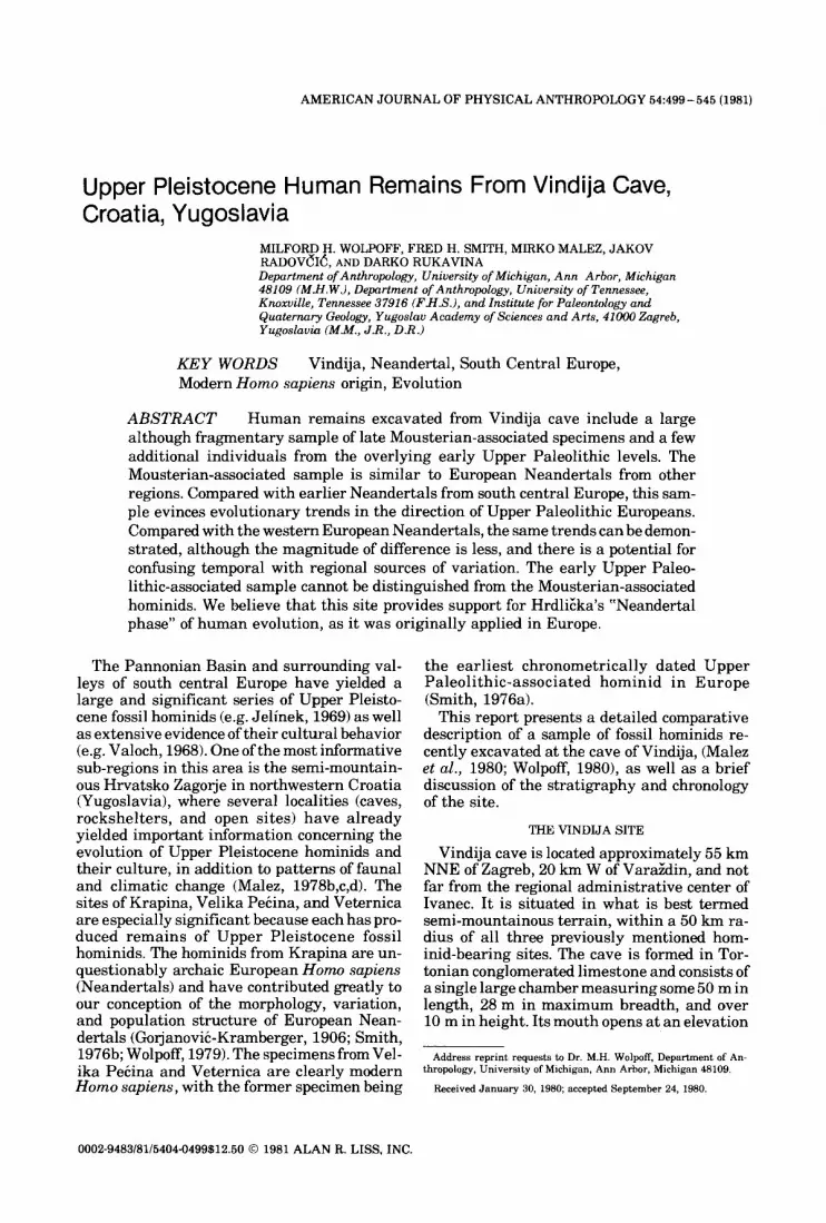
The Vindija deposits (Fig. 1) are divided into 13 primary stratigraphic units. The lower 10, D-M (M is not shown in Fig. 11, are Pleistocene, while the upper 3 are post-Pleistocene and will not be described. Three of the primary Pleisto- cene units (F, G, K) require further subdivision and are referred to here as complexes.
Level D, the youngest of the Pleistocene de- posits, is a yellowish-brown, fine sandy sedi- ment with a loess-like habitus. It ranges from 50 to 150 cm in thickness and probably is aeo- lian in origin. Level E is a grayish sandy sedi- ment with numerous small (< 2 cm), often cor- roded pieces of limestone. Complex F is further divided into 5 subunits and varies between ap- proximately 30 and 150 cm in thickness. The complex is basically characterized by the pres- ence of angular pieces of limestone of various sizes ranging from blocks to small particles in a sandy sediment. One distinct level, Fd/s, con- sists of a pasty loam with no rock fragments. It should be noted that the precise composition of the F complex varies in different parts of the cave, due to varying sedimentological pattern- ing.
Complex G is made up of generally sandy sediment, containing only rather small (often corroded) stone fragments, but in some places larger, angular fragments are present as well. However, there are markedly fewer stone fragments than in levels E, F, and H. Thickness varies between approximately 60 and 150 cm, and near the mouth of the cave the units of
Fig. 1. Schematic profile of the Pleistocene deposits of Vindija. Thickness and descriptions of individual strata are given in the text, but relative thickness of the strata is accurate. Revised from Malez and Rukavina (1979).
units are recognized within complex G. Level G, , the superior-most of these, is a very distinc- tive, reddish-colored clay or loam. It contains very few stone fragments, and those that are present are rounded, markedly corroded, and
complex G are disturbed by cryoturbation (Malez and Rukavina, 1975). Five distinct
colored by manganese. Level GI is from 8 to 20 cm thick and is present throughout the cave.

VINDIJA REMAINS 501
Level G,'is a grayish clay measuring 1 to 30 cm in thickness. It contains numerous small stones and is present only in certain portions of the cave. Level G, is sandy sediment, loess-like in habitus, and varies between approximately 10 to 30 cm in thickness. It is quite distinctive in color (light green) and contains relatively few rocks, which are usually rounded and corroded. Level G4 is a darker green deposit, again of loess-like habitus, but containing more rock fragments. Level G, is a sandy deposit in which rocks are rare and, if present, corroded.
Level H is a thick (100-200 cm) layer of sandy deposits containing numerous large, an- gular rocks exhibiting no evidence of corrosion. When freshly exposed, these sediments are olive to grayish-yellow in color. Level I is a sandy deposit, reddish-brown in color, and ranging from 30 to 100 cm in thickness. Few rocks are present, and these tend to be corroded. Level J is a sandy deposit containing a consid- erable density of large rock fragments. This layer is olive-gray when the sediment is fresh and varies between 100 and 200 m in thickness. Complex K isdivisible into 3 subunits. Level K, is sandy, dark brown in color with many small corroded limestone fragments. K, is similar but contains virtually no rock fragments, while K, is sandy and dark yellow to brown in color. Cryoturbation processes are observable in both K, and K,. Level L is characterized by the pres- ence of very large blocks of limestone as well as smaller fragments. There is a bone breccia ap- proximately 50 cm below contact with level K. Below this, the sediments are compacted and hard. Level L (and also M) is still largely unex- cavated.
ARCHAEOLOGY, CORRELATION, AND DATING
The Pleistocene strata a t Vindija contained rich Paleolithic assemblages (approximately 1,000 pieces in level G alone), which are still under study. Preliminary analysis of the lithic artifacts reveals that levels G2 through K con- tain remains of the Mousterian. Some of the upper Mousterian levels (e.g. G,) do contain elements most commonly associated with early Upper Paleolithic, but this phenomenon is common in the late Mousterian of central Europe (Valoch, 1968) and does not alter the basic Mousterian character of the assemblage. A single split-based bone point is the onlydiag- nostic artifact from the early Upper Paleolithic level GI, and it is from the top of the stratum. This indicates, though not conclusively, that level G, is Aurignacian. According to Valoch (personal communication), the lithic material
in G, is neither Szeletian nor Mousterian. Above GI, the remaining Pleistocene strata (D, E, F) contain only Upper Paleolithic as- semblages. Level D is characterized by a Gravettian assemblage, while the artifacts from E and F appear to represent the Aurig- nacian.
Based on the nature of the deposits described above, levels G, H, J, and complex F (and also L) represent long periods of cold. This is suggested chiefly by the presence of large angular blocks resulting from deterioration of cave walls by frost action. So-called paleosoils, which are most likely the result of mild or warm climates and therefore of chemical decomposition, are seen in levels G,, Ge, I, and the K complex. The habitus of the K complex sediment is very simi- la r to t h e Riss-Wurm levels at Krapina (Gorganovic-Kramberger, 19131, and the fauna a r e similar (particularly the presence of Merck's rhinocerous). Based on these factors, its stratigraphic location, and archeological content, it appears reasonable to correlate the K complex to the Riss-Wurm interglacial. The strata below the K complex (L and M) are tenta- tively correlated to the Riss.
Although charcoal is preserved in several levels from the cave, only one radiocarbon date has been obtained thus far. This date, associ- ated with the Aurignacian at Vindija, is from a charcoal sample taken a t the Fd and Fd/a inter- face and approximates 26,970 years B.P. (Malez, 1978c; 349). It is comparable to other Aurignacian dates from south central Europe (Frayer, 1978; Movius, 19601, and thus helps secure the upper end of the Vindija Pleistocene sequence.
Level GI, which is tentatively considered an early Aurignacian level, appears to represent a warm period and is quite similar (Valoch, per- sonal communication) to the layers represent- ing the Podhradem interstadial in the Mora- vian Karst (Musil and Valoch, 1966). This would make sense relative to its position be- tween a definitely Mousterian level (G,) and a definitely Aurignacian one (Fc,/,J and the date of ca. 34,000 B.P. for probably early Aurig- nacian at Velika Pecina (Malez, 1978d; Smith, 1976a).
If one then compares the entire Vindija Upper Plesitocene sequence to that determined for the not-too-distant Moravian Karst (Musil and Valoch, 1966; Valoch, 19681, also on the Pannionian Basin rim, there appears to be a reasonably acceptable correlation. We believe that the data strongly support this correlation, based independently on both quaternary geol- ogy and archaeology.

502 M.H. WOLPOFF ET AL
THE HOMINIDS
The hominid remains from Vindija fall into three stratigraphic samples. The most recent consists of some 34 specimens excavated from level D. These specimens exhibit a modern Homo sapiens morphology in every relevant respect and are not described here. Their mor- phology is consistent with their date of younger than 27,000 B.P. The second sample consists of four specimens from G, and three isolated teeth from the F complex. These are discussed here as Aurignacian-associated hominids. The remainder of the hominids (except for a few without certain provenience) are derived from level G,. These hominids are clearly represen- tatives of archaic European Homo sapiens (Neandertals) and are the stratigraphically earliest hominid sample yet excavated at Vin- dija. Based on the correlations discussed above, level G3 would be encompassed within the Lower Wiirm stadial, which dates between ap- proximately 40,000 and 59,000 years B.P. ac- cording to Valoch (1968). It is not possible to ascertain a more precise radiometric date for the Vindija G3 hominids a t present. Valoch, who examined some of the archeological and faunal materials from G3, believes this level to represent the latest portion of the Lower Wiirm stadial.
Regardless of their exact position within the Lower Wiirm stadial, it is quite certain that they date later than the Riss-Wiirm intergla- cial and are therefore chronologically more re- cent than the remains from the “Homo” zone at Krapina, if not more recent than virtually all of the Krapina specimens.
In the following sections the hominids are described by morphological area and by numeric order within each area. Beside each specimen number are references to the appro- priate figure(s) and the level designation (G:$, G,, F(,). Absence of a level designation means that provenience is not conclusively known. These specimens are most likely from level G3. Summary sections conclude the descriptions of each skeletal region represented, and general comparisons and conclusions are presented a t the end of the report.
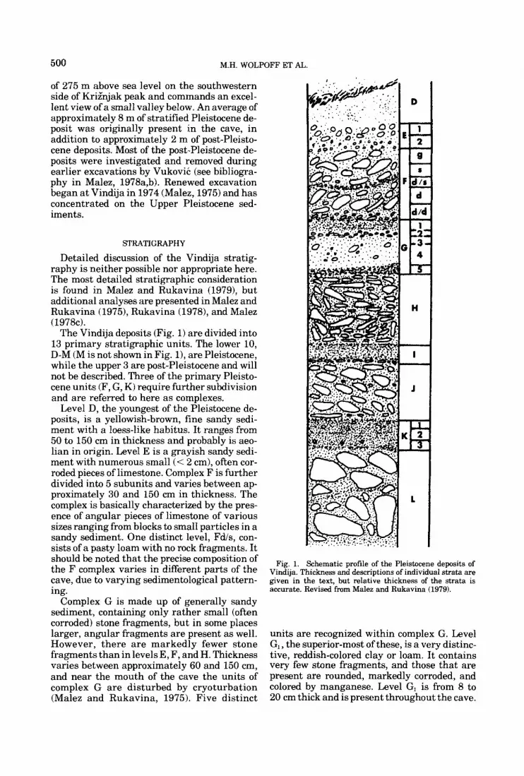
Maxillas Vi 225 Right maxilla (Fig. 2): G:j
This edentulous specimen is broken at the midline and posteriorly a t the middle of the M2 socket. It includes the lower portion of the lat- eral nasal aperture border and the base of the maxillary sinus. The base of a prominent nasal spine is also present, but the spine itself is bro-
ken away. The nasal margin is sharply defined. A double line forms the margin resulting in what appears to be an inner and outer lower nasal border (the same morphology as in the Vi 259 maxilla but less distinctly expressed). The lines are separated by a shallow groove, but there is a transverse ridge connecting them beginning on the lower line at the midline and extending superiorly and laterally to the upper line. Nasal breadth is determined by doubling the distance from the right lateral nasal mar- gin to the midline. It is narrow compared with Neandertals from Krapina and western Europe. Compared with the later sample, the nasal breadth is 3.2 standard deviations below the mean (Table 1). It is smaller than the single nasal breadth known for Krapina.
The maxillary sinus is in a low position rela- tive to the molar roots. Because of the break in this region exposing the sinus, there is not enough of the maxilla left to determine whether there was a canine fossa. A slight but- tress surrounds the canine root. Unfortunately, the size of the canine socket cannot be accu- rately measured because the labial wall is bro- ken away. At 7.5 mm below the alveolar mar- gin, the breadth of the socket is 11.3 mm, and it is likely that the canine was quite large. The socket extends a t least 20 mm superiorly from the alveolar margin. Length and breadth of the I’ and I2 sockets are, respectively, 8.2 x 8.8 mm and 7.0 x 9.5 mm. The suggestion that the lat- eral incisor was broader than the central pro- vides a similarity to the condition in some (al- though not all) western European Neandertals. While the anterior teeth were lost, dimensions incorporating the anterior tooth row (such as prosthion-post canine, Table 1) show no evi- dence of reduction from the Neandertal condi- tion. In fact, this distance is greater than in any known Neandertal maxilla.
The subnasal area is uncurved, dropping straight downward from the nasal border. Al- veolar height is quite small, lying 2.5 standard deviations below the western European Nean- dertal mean as well as below the range from Krapina (Table 1). Alveolar height is one of the features reflecting marked sexual dimorphism in the western European Neandertals. The Ne- andertal sex ratio for this feature (ME’) is 121%. This would suggest that the Vi 225 maxilla (as well as the Vi 259 maxilla, see below) is female. Even if so, the specimen is markedly smaller than western European Ne- andertal females like Gibraltar 1 and La Fer- rassie 2.
The palate is shallow compared with Nean- dertals. Although variation in Neandertals is great enough for the difference not to attain

Fig. 2. Maxillae Vi 225 (left) and Vi 259 in inferior, frontal, and lateral views. Specimens are shown to the same approximate scale. Note t he relative size of t he incisor roots, the morphology ofthe lower nasal margin, and the low position of the maxillary sinus.
statistical significance, measurements of the palate wall height between P4 and MI, and M1 and M2 are well below the Neandertal mean and at the lower end or below the range (Table 1). There is no palatine torus.
Vi 259 Left maxilla (Fig. 2): G3
The fragment is broken almost identically to 225, except for the fact that the entire alveolar margin and an M2 remain. Vindija 259 is the
504 M.H. WOLPOFF ET A L
TAEiLE 1 . Maxillary dimensions in millimeters for the Vindija, Krapina, and European Neandertal maxillae'
Internal palate wall Prosthion
Alveolar Nasal height to height breadth P/M' M'IM' post-canine
Vindija 225 259
Krapina E F
Western European Neandertals La Ferrassie 2 Gibraltar 1 Saccopastore 1 La Quina 5 SPY 1 Monsempron 1 La Chapelle Saccopastore 2 SPY 2 Arcy-sur-Cure 9 La Ferrassie 1 Kfilna Mt Circeo 1
Western European Neandertal - X U
Neandertal male - X U
Neandertal female - X U
17.5 16.3
22.5 24.2
22.2 22.5 23.0 25.0 25.3
26.5 27.9
28.5 29.9 30.4
26.1 3.0
28.6 1.6
23.6 1.4
28.5 26.2
30.5
34.1 32.9
33.1 33.7
33.3 34.0 30.0 35.1
33.3 1.5
33.2 1.7
33.5
8.4 9.6
23.9 12.0
12.5
18.7 11.1 13.6 12.9 12.5
14.7 4.4
13.8 2.9
16.1 6.7
10.5 9.6
12.7 20.9
12.7
17.1 10.1 12.7 13.2
16.5
14.5 3.4
13.9 2.9
15.4 4.7
27.3 24.3
25.0 25.8
25.0
26.2
23.0 23.9 25.1
24.9 1 .I
24.6 1.4
25.3 0.5
)The Saccopastore crania are included in this Neandertal sample because of the likelihood that their temporal position corresponds to Wurm I of France and Belgium, Kdlna, although published as a juvenile specimen, is actually adult since the M' tcmthwear exceeds that of Krapina maxilla E which has an erupted M'. Alveolar height is the nasospinale-alveolar point dimension, the postcanine position is the maximum distal point on the posterior surface ofthe root at the alveolar margin, and the internal palate wall heights were taken at the palate wall, perpendicular to the roof of the palate. All measurements were taken on the original specimens. In this and all other tables, measurements were taken by MHW unless otherwise indicated.
smaller of the two maxillas (Table 1) and is too small to fit any of the preserved mandibles. Also, none of the unassociated maxillary teeth fit this specimen. The maxillary sinus floor is in a low position. While the contour of the maxilla suggests the possibility of a shallow canine fossa, not enough of the bone is preserved to be sure this feature was present. Although bro- ken, the remaining portion of the nasal spine is prominent. A marked ridge extends 14.7 mm posteriorly from it onto the floor of the nasal cavity, terminating a t the opening for the inci- sive canal. The lower nasal margin is similar to that of Vi 225 in that there are two distinct ridges separated by a deep groove with a trans- verse ridge connecting them. Below the nasal
margin, the profile of the maxilla is somewhat concave. A moderate amount of alveolar prog- nathism is indicated.
The breadths of the central and lateral in- cisor sockets are 7.7 and 7.8 mm, respectively. The canine was probably not large. The maximum breadth of the canine socket a t the alveolar margin is 9 mm and the socket depth is 17.7 mm. No canine buttress extends onto the lateral maxillary face.
The M2 is markedly smaller than the isolated Vi 229 M2 (see Table 6). It is moderately worn and the roots are fused. The wear slope is lin- gual over the major portion of the crown; a second more distinctly angled facet covers the lingual 115. There is a moderate area of dentin
VINDIJA REMAINS 505
exposed by this facet, and the grooves have been obliterated over the entire lingual occlusal surface. On the buccal side, about V z of the original crown height remains. There is little interproximal attrition mesially. The facet measures 4.3 mm in breadth. The pres- ence of a faint distal interproximal facet indi- cates that the M 3 had erupted.
Alveolar height, nasal breadth, and the measures of palate depth are uniformly smaller than any western European Neandertal (Table l), probably indicating that Vi 259 is female. However, relative anterior tooth expansion is suggested by the fact that the prosthion-post- canine distance is about the size of the La Fer- rassie female (it was probably somewhat smaller, but the mesial migration of the ante- rior teeth of the much older La Ferrassie female has obscured the difference). The general size difference expressed by the other comparable dimensions is significant. Nasal breadth (de- termined also by doubling the measurement from the left lateral nasal border to the mid- line) lies 4.7 standard deviations below the western European Neandertal mean, and al- veolar height is 2.8 standard deviations below this mean. Even when the comparison is lim- ited to females, these dimensions are extra- ordinarily small.
Summary. The unusual morphology of the lower nasal border, as well as the more general similarities found in other features, suggest that these two individuals from level G3 may have been related, at least in a populational sense.
These maxillas seem to combine both Nean- dertal-like and more modern features. Their small size underlies many of the contrasts with western European Neandertals. Yet, size itself is an important feature, since facial reduction is one of the most dramatic differences between Neandertal populations and more modern pop- ulations that followed them in time. The most archaic-appearing features reflect the size and proportions of the anterior teeth. Both have a I' root that is broader than the I' root. The marked prosthion-postcanine distance is even more distinctive when the generally small size of the maxillas is taken into account. If the maxillas were drawn from the same sample as the postcranial remains, their small size might partially reflect a small average body size. However, even taking this into account, the fact remains that these specimens are much smaller than the most diminutive Neandertal female, and the size difference involves fea- tures that are taxonomically distinctive of Ne-
andertals; facial height, palate depth, and nasal breadth. Since facial size reduction is one of the major morphological alterations associ- ated with the transition from Neandertals to anatomically modern Homo sapiens (Brace, 1979; Howells, 1975; Brose and Wolpoff, 1971; Smith, 1976b), the fact that the Vindija G3 hominids tend to approach the early modern condition is indeed significant. Also, though it is not possible to be certain because of the spec- imen's fragmentary condition, the possible presence of a shallow canine fossa in Vi 259 could be another feature indicating a reduced face relative to other Neandertals.
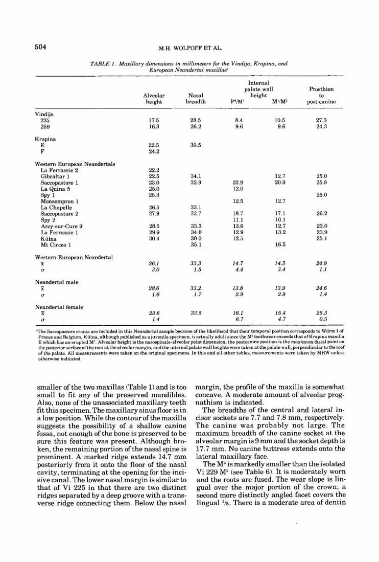
Mandibles Vi 206 symphysis (Fig. 3,4): G,
This specimen extends from the middle of the left canine socket to the approximate horizon- tal midpoint of the right ramus and includes the right canine and molar series. The general appearance of the corpus is similar to many of the late Neandertals from western Europe. There is a distinct, although not prominent, mental eminence and a rather vertical sym- physis. A single mental foramen lies directly under the center of the first molar; the anterior ramus border crosses the alveolar margin some 3 mm behind the distal face of M3, and the corpus is fairly uniform in height (Table 2).
There is a gentle concavity on the external face of the symphysis, beginning just below the incisor sockets and extending inferiorly about halfway down the bone. Here, the contour be- comes convex and forms a moderately devel- oped mental eminence. Oriented in the basal plane the eminence is more anterior than the incisor sockets at their superior margin, and in the occlusal plane orientation these are equally anterior. Laterally the concavity extends to the canine roots. The symphyseal angle for Vi 206, measured from the basal margin (c.f. Olivier 1969) is 87", considerably below the overall western European Neandertal and Krapina means of 95" and 99.6", respectively (Table 3). This value underscores the presence of a men- tal eminence and vertical symphysis for Vi 206. The symphyseal region of Vi 206 is extraordi- narily similar to Neandertal specimens such as Circeo 3 and Spy 1.
The right digastric fossa is extremely well excavated and is angled in the sagittal plane a t about 45" t o the corpus base. The excavated portion is 18.2 mm long and 10 mm in breadth. The position of gnathion lies some 3 mm above the fossa. On the internal symphyseal surface
Right mandibular corpus with
506 M.H. WOLPOFF ET AL
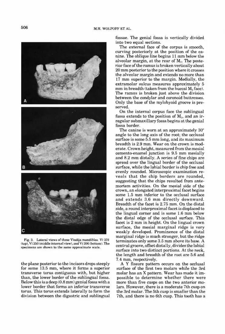
Fig. 3. Lateral views of three Vindija mandibles. Vi 231 (top), Vi 250 (middle internal view), and Vi 206 (bottom). The specimens are shown to the same approximate scale.
the plane posterior to the incisors drops steeply for some 13.5 mm, where i t forms a superior transverse torus contiguous with, but higher than, the lower border of the sublingual fossa. Below this is a deep (0.8 mm) genial fossa with a lower border that forms an inferior transverse torus. This torus extends laterally to form the division between the digastric and sublingual
fossae. The genial fossa is vertically divided into two equal sections.
The external face of the corpus is smooth, curving posteriorly a t the position of the ca- nine. The oblique line begins ll mm below the alveolar margin, at the rear of M,. The poste- rior face of the ramus is broken vertically about 20 mm posterior to the position where it crosses the alveolar margin and extends no more than 17 mm superior to the margin. Medially, the extramolar sulcus measures approximately 5 mm in breadth (taken from the buccal M, face). The ramus is broken just above the division between the condylar and coronoid buttresses. Only the base of the mylohyoid groove is pre- served.
On the internal corpus face the sublingual fossa extends to the position of M,, and an ir- regular submaxillary fossa begins at the genial fossa border.
The canine is worn at an approximately 30" angle to the long axis of the root; the occlusal surface is some 5.5 mm long, and its maximum breadth is 2.9 mm. Wear on the crown is mod- erate. Crown height, measured from the mesial cemento-enamel junction is 9.5 mm mesially and 8.2 mm distally. A series of fine chips are spread over the lingual border of the occlusal surface, while the labial border is chip free and evenly rounded. Microscopic examination re- veals t h a t the chip borders are rounded, suggesting that the chips resulted from ante- mortem activities. On the mesial side of the crown, an elongated interproximal facet begins some 1.5 mm inferior to the occlusal surface and extends 3.6 mm directly downward. Breadth of the facet is 2.75 mm. On the distal side, a round interproximal facet is displaced to the lingual corner and is some 1.6 mm below the distal edge of the occlusal surface. This facet is 2 mm in height. On the lingual crown surface, the mesial marginal ridge is very weakly developed. Prominence of the distal marginal ridge is much stronger, but the ridge terminates only some 3.5 mm above its base. A central groove, offset distally, divides the labial surface into two distinct portions. At the neck, the length and breadth of the root are 5.6 and 7.4 mm, respectively.
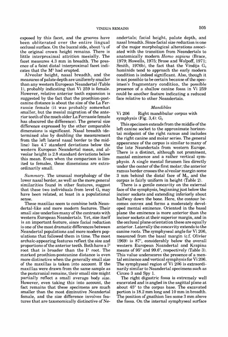
A Y fissure pattern occurs on the occlusal surface of the first two molars while the 3rd molar has an X pattern. Wear has made it im- possible to determine whether there were more than five cusps on the two anterior mo- lars. However, there is a moderate 7th cusp on the 3rd molar. The 5th cusp is smaller than the 7th, and there is no 6th cusp. This tooth has a
VINDIJA REMAINS 507
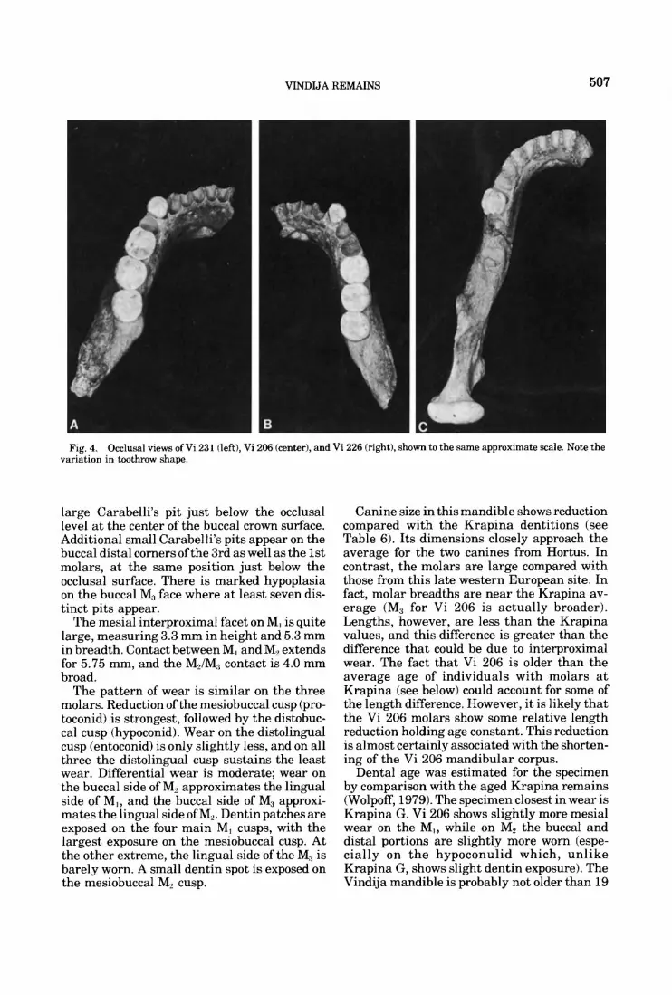
Fig. 4. Occlusal views of Vi 231 (left), Vi 206 (center), and Vi 226 (right), shown to the same approximate scale. Note the variation in toothrow shape.
large Carabelli’s pit just below the occlusal level at the center of the buccal crown surface. Additional small Carabelli’s pits appear on the buccal distal corners of the 3rd as well as the 1st molars, at the same position just below the occlusal surface. There is marked hypoplasia on the buccal M1 face where at least seven dis- tinct pits appear.
The mesial interproximal facet on MI is quite large, measuring 3.3 mm in height and 5.3 mm in breadth. Contact between MI and M2 extends for 5.75 mm, and the MJM, contact is 4.0 mm broad.
The pattern of wear is similar on the three molars. Reduction of the mesiobuccal cusp (pro- toconid) is strongest, followed by the distobuc- cal cusp (hypoconid). Wear on the distolingual cusp (entoconid) is only slightly less, and on all three the distolingual cusp sustains the least wear. Differential wear is moderate; wear on the buccal side of M, approximates the lingual side of MI , and the buccal side of M3 approxi- mates the lingual side of M,. Dentin patches are exposed on the four main MI cusps, with the largest exposure on the mesiobuccal cusp. At the other extreme, the lingual side of the M3 is barely worn. A small dentin spot is exposed on the mesiobuccal M, cusp.
Canine size in this mandible shows reduction compared with the Krapina dentitions (see Table 6). Its dimensions closely approach the average for the two canines from Hortus. In contrast, the molars are large compared with those from this late western European site. In fact, molar breadths are near the Krapina av- erage (M, for Vi 206 is actually broader). Lengths, however, are less than the Krapina values, and this difference is greater than the difference that could be due to interproximal wear. The fact that Vi 206 is older than the average age of individuals with molars a t Krapina (see below) could account for some of the length difference. However, it is likely that the Vi 206 molars show some relative length reduction holding age constant. This reduction is almost certainly associated with the shorten- ing of the Vi 206 mandibular corpus.
Dental age was estimated for the specimen by comparison with the aged Krapina remains (Wolpoff, 1979). The specimen closest in wear is Krapina G. Vi 206 shows slightly more mesial wear on the MI, while on M, the buccal and distal portions are slightly more worn (espe- cially on t h e hypoconulid which, unlike Krapina G, shows slight dentin exposure). The Vindija mandible is probably not older than 19
TAB
LE 2
. C
orpu
s di
men
sion
s fi
n m
illim
eter
s) fo
r th
e V
indi
ja a
nd K
rapi
na s
ampl
es'
Men
tal
fora
men
Sy
mph
ysis
M
JM,
posi
tion
M,N
, po
sitio
n M
3 to
ant
erio
r to
tal
ramu
s at
B
read
th
Hei
ght
Inde
x B
read
th
Hei
ght
Inde
x B
read
th
Hei
ght
Inde
x al
veol
ar m
argi
n N
umbe
r A
rea
Posi
tion
Vin
dija
20
6 15
.3
207
226
13.9
23
1 16
.8
250
Ave
rage
15
.3
U
1.5
Kra
pina
D
13
.7
E
13.5
F
14.6
G
14
.5
H
15.5
J
14.5
K
R
63
R64
R
66
U
.72 .ll
Ave
rage
14
.4
P (A
x =
0)
32.6
26.2
34
.6
31.1
4.
4
32.8
36
.5
31.5
31
.2
40.8
42
.4
35.9
4.
9 .10
46.9
15
.3
53.1
15
.4
48.6
17
.9
15.2
49.6
16
.0
3.2
1.3
41.8
14
.5
37.0
15
.6
46.3
16
.1
46.5
14
.4
38.0
16
.9
34
2
15.7
40.6
15
.5
5.1
1 .o
.01
.29
31.9
26.5
32
.5
30.3
3.
3
28.0
26
.6
30.2
35
.6
33.5
30.8
3.
8 .43
48.0
16
.1
58.1
15
.1
55.1
16
.9
15.9
53.7
16
.0
5.2
.7
51.8
58
.6
17.4
47.7
16
.0
47.5
15
.2
46.9
15
.5
14.4
50.5
15
.7
4.9
1 .I
20
.33
30.2
25
.6
25.3
32
.3
29.9
26.7
3.
1
25.5
28.5
33
.2
31.1
28
.3
24.1
28.5
3.
4 .46
53.3
59.7
52
.3
53.2
54.6
3.
4
68.2
56.1
45
.8
49.8
50
.9
54.2
8.
7 .46
- 3.
0 ~
4.9
- 8.
3 -
5.6
0.1
- 4
.3
3.1
~ 3.
0 - 3.
0
-11.
0 - 6.
9 ~
4.0
~ 2.
0
- 4
.3
3.6
~ 1.
00
1
1.50
Mi
2 14
.1
M, M
, 2
26.5
M
, fro
nt o
f M,
1
13.8
Fr
ont o
fM,
17.4
5:
6.
1 g s $
1 34
.5
P,M
, 0
2 32
.7
M, M
,/MZ
4 1
21.6
M
i h
1
51.9
B
acko
fM,
-3 $ c(I
1
24.6
M
,
1
23.7
B
acko
fM,
31.5
11
.3
.03
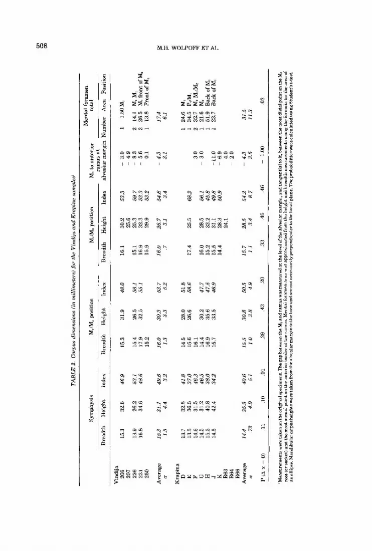
'Mea
sure
men
ts w
ere
take
n on
the
orlg
mal
spec
imen
s, T
he g
ap b
etw
een
the M, an
d ra
mus
was
mea
sure
d at
the
leve
l of t
he a
lveo
lar m
argi
n, a
nd ta
ngen
tial
to it
, bet
wee
n th
e m
ost d
ista
l poi
nt o
n th
e M
a ro
ot (
or so
cket
) and
the
mos
t mes
ial p
ant o
n th
e an
teri
or b
orde
r of
the
ram
us. M
enta
l fo
ram
en a
rea
was
app
roxi
mat
ed fr
om th
e he
ight
and
bre
adth
mea
sure
men
ts u
sing
the
form
ula
for t
he a
rea
of
anel
lips
e. M
andi
hula
rcor
push
eigh
ts w
eret
aken
from
thea
lveo
lar
mar
gin
to th
e haseandarenotnecessarilyperpendiculartothe hasa
lpla
ne. T
heprohahilitieswerecalculatedusingStudent's t-
test
.
VINDIJA REMAINS 509
TABLE 3. Symphyseal angles for Vindija, Krapina, and western European Neandertal
mandibles'
Specimen
Vindija 206 Vindija 231 Vindija 226I265 Vindija average (u)
Krapina D Krapina E Krapina G Krapina H Krapina J Krapina average (u)
La Ferrassie 1 La Chapelle-aux-Saints La Naulette Regourdou SPY 1 La Quina H9 Banolas Hortus 5 Western European
Neandertal average (u)
P (A Z = 0 ) VIK .OOO V/EN ,015 KIEN ,101
Symphyseal angle' SourceJ
87" 1 89" 1 85" 1 87" (1.6)
98" 1 98" 1
102" 1 101" 1 99" 1 99.6" (1.8)
85" 2 95" 2 95" 2
103" 2 106" 2 93" 2 92" (cast) 2 95" 3
95.5" (6.5)
~ ~~ ~~
'Although Hortus 5 has been regarded as a juvenile, the M, had just come into occlusion at the time of death. Means are compared using Student's t-test.
'The symphyseal angle is taken after t he method described by Olivier 1'69).
.'1 = measurements by FHS; 2 = Helm ('76); 3 = de Lumley ('73).
(Krapina G was aged at 18). The amount of wear on the M:, is equal to molars with 3-4 years of wear at Krapina (for instance, the M, of Krapina E). Thus, early MI, eruption (i.e. ap- proximately 15 years) is indicated for this Vin- dija mandible.
Vi 207 Right ramus and posterior mandibular corpus (Figs. 3 3 ) : G,
This edentulous specimen consists of a com- plete ramus, broken only at the tip of the coronoid, and a corpus extending to the mid M, position buccally but only to the mid M3 lin- gually.
The corpus is low (Table 2) and relatively thick. The oblique line begins a t the position of the M:, socket center. Judging from the condi- tion of the socket, the tooth was erupted.
The root of the ramus appears at about the mid M, position, and the anterior border of the ramus does not cross the alveolar margin until some 4.9 mm posterior to the distal border of the M:, socket. The ramus is small (Table 4) and
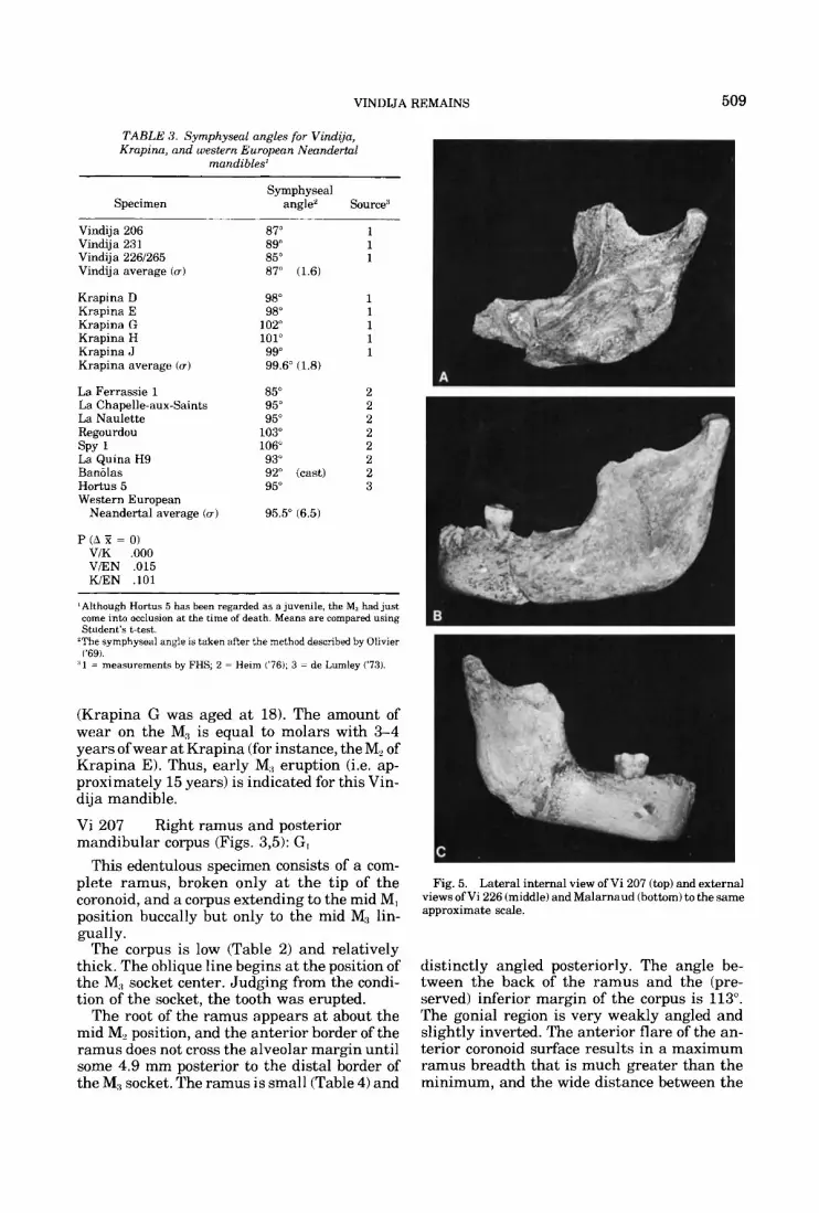
Fig. 5. Lateral internal view of Vi 207 (top) and external views of Vi 226 (middle) and Malarnaud (bottom) to the same approximate scale.
distinctly angled posteriorly. The angle be- tween the back of the ramus and the (pre- served) inferior margin of the corpus is 113". The gonial region is very weakly angled and slightly inverted. The anterior flare of the an- terior coronoid surface results in a maximum ramus breadth that is much greater than the minimum, and the wide distance between the

TABL
E 4.
Ram
us m
easu
rem
ents
and
pro
port
ions
for
the
Vin
dGa,
Kra
pina
, and
wes
tern
Eur
opea
n N
eand
erta
l sa
mpl
es'
Vin
dija
20
7 22
6 A
vera
ge
Kra
pina
J R
63
R64
R
65
R 66
R
68
R 70
R
73
R 7
4 A
vera
ge
Wes
tern
Eur
opea
n W
iirm
Nea
nder
tal
La C
hape
lle
La
Ferr
assi
e 1
La
Qui
na 5
L
a Q
uina
9
Mt C
irceo
3
Reg
ourd
ou
5
Ave
rage
5
Ram
us h
eigh
t
From
bas
e With
R
amus
bre
adth
In
dex
Con
dyle
In
oe
clus
al
~ M
inim
um r
amus
bre
adth
Len
gth
Bre
adth
pl
ane
hori
zont
al
goni
on
Min
imum
m
argi
n R
amus
hei
ght f
rom
bas
e re
stin
g pl
ane
From
A
t alv
eola
r ~
7.6
10.1
8.
9
14.5
12
.5
9.2
12.7
10
.4
9.0
9.8
9.3
10.9
2.
1
10.6
10
.2
9.0
12.0
10
.5
12
21.6
21
.6
28.4
21
.5
21.1
23
.1
23.5
23.5
2.
9
28.9
29
.4
26.5
25.0
27
.5
2.1
51.9
60
.5
56
2
62.4
57
.7
60.5
65
.5
61.5
3.
3
64.8
66
.4
65.1
64
.8
55.1
63
.2
4.6
59.5
68
.1
63.8
78.9
64
.9
71.9
84.2
81
.0
89.0
70.1
81
.1
8 .O
51.5
58
.8
55.2
59.8
58
.6
58.5
62
.4
59.8
1.
8
64.0
60
.7
62.4
38.0
41
.7
39.9
37.1
35
.0
34.8
35.6
1.
3
45.0
43
.7
43.7
42.2
37
.4
42.4
3 .O
45.0
46
.1
45.6
41.3
39
.5
43.0
39.5
40.8
1.
7
55.5
53
.5
47.5
46
.4
42.9
49
.2
5.2
73
69
71
60
61
58
59
69
66
67
68
68 1.5
'All
rnet
rics
tin
mil
lim
eter
s) w
ere
take
n on
the
ori
gina
l sp
ecim
ens.
VINDIJA REMAINS 511
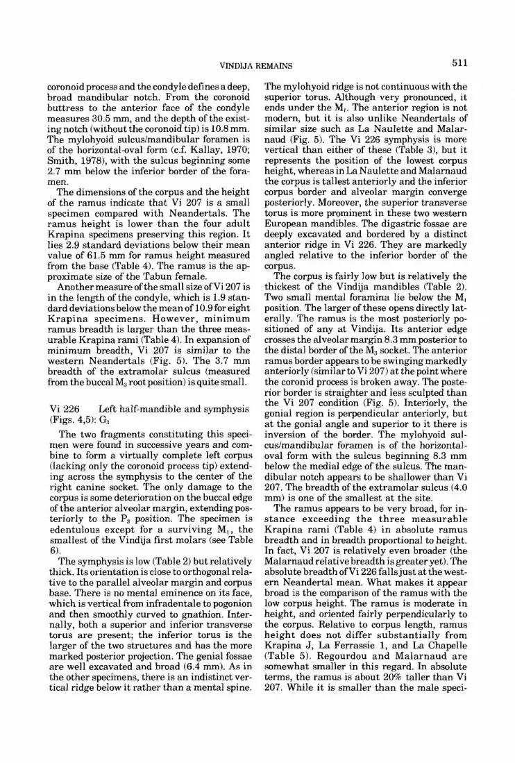
coronoid process and the condyle defines a deep, broad mandibular notch. From the coronoid buttress to the anterior face of the condyle measures 30.5 mm, and the depth of the exist- ing notch (without the coronoid tip) is 10.8 mm. The mylohyoid sulcus/mandibular foramen is of the horizontal-oval form (c.f. Kallay, 1970; Smith, 1978), with the sulcus beginning some 2.7 mm below the inferior border of the fora- men.
The dimensions of the corpus and the height of the ramus indicate that Vi 207 is a small specimen compared with Neandertals. The ramus height is lower than the four adult Krapina specimens preserving this region. I t lies 2.9 standard deviations below their mean value of 61.5 mm for ramus height measured from the base (Table 4). The ramus is the ap- proximate size of the Tabun female.
Another measure of the small size ofVi 207 is in the length of the condyle, which is 1.9 stan- dard deviations below the mean of 10.9 for eight Krapina specimens. However, minimum ramus breadth is larger than the three meas- urable Krapina rami (Table 4). In expansion of minimum breadth, Vi 207 is similar to the western Neandertals (Fig. 5). The 3.7 mm breadth of the extramolar sulcus (measured from the buccal M3 root position) is quite small.
Vi 226 (Figs. 4,5): GR
Left half-mandible and symphysis
The two fragments constituting this speci- men were found in successive years and com- bine to form a virtually complete left corpus (lacking only the coronoid process tip) extend- ing across the symphysis to the center of the right canine socket. The only damage to the corpus is some deterioration on the buccal edge of the anterior alveolar margin, extending pos- teriorly to the P3 position. The specimen is edentulous except for a surviving M, , the smallest of the Vindija first molars (see Table 6).
The symphysis is low (Table 2) but relatively thick. Its orientation is close to orthogonal rela- tive to the parallel alveolar margin and corpus base. There is no mental eminence on its face, which is vertical from infradentale to pogonion and then smoothly curved to gnathion. Inter- nally, both a superior and inferior transverse torus are present; the inferior torus is the larger of the two structures and has the more marked posterior projection. The genial fossae are well excavated and broad (6.4 mm). As in the other specimens, there is an indistinct ver- tical ridge below it rather than a mental spine.
The mylohyoid ridge is not continuous with the superior torus. Although very pronounced, it ends under the M,. The anterior region is not modern, but it is also unlike Neandertals of similar size such as La Naulette and Malar- naud (Fig. 5). The Vi 226 symphysis is more vertical than either of these (Table 3), but it represents the position of the lowest corpus height, whereas in La Naulette and Malarnaud the corpus is tallest anteriorly and the inferior corpus border and alveolar margin converge posteriorly. Moreover, the superior transverse torus is more prominent in these two western European mandibles. The digastric fossae are deeply excavated and bordered by a distinct anterior ridge in Vi 226. They are markedly angled relative to the inferior border of the corpus.
The corpus is fairly low but is relatively the thickest of the Vindija mandibles (Table 2). Two small mental foramina lie below the M, position. The larger of these opens directly lat- erally. The ramus is the most posteriorly po- sitioned of any at Vindija. Its anterior edge crosses the alveolar margin 8.3 mm posterior to the distal border of the M3 socket. The anterior ramus border appears to be swinging markedly anteriorly (similar to Vi 207) at the point where the coronid process is broken away. The poste- rior border is straighter and less sculpted than the Vi 207 condition (Fig. 5). Interiorly, the gonial region is perpendicular anteriorly, but a t the gonial angle and superior to it there is inversion of the border. The mylohyoid sul- cus/mandibular foramen is of the horizontal- oval form with the sulcus beginning 8.3 mm below the medial edge of the sulcus. The man- dibular notch appears to be shallower than Vi 207. The breadth of the extramolar sulcus (4.0 mm) is one of the smallest a t the site.
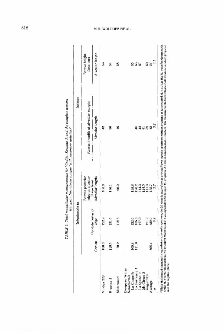
The ramus appears to be very broad, for in- s tance exceeding the three measurable Krapina rami (Table 4) in absolute ramus breadth and in breadth proportional to height. In fact, Vi 207 is relatively even broader (the Malarnaud relative breadth is greater yet). The absolute breadth ofVi 226 falls just at the west- ern Neandertal mean. What makes it appear broad is the comparison of the ramus with the low corpus height. The ramus is moderate in height, and oriented fairly perpendicularly to the corpus. Relative to corpus length, ramus height does not differ substantially from Krapina J, La Ferrassie 1, and La Chapelle (Table 5). Regourdou and Malarnaud are somewhat smaller in this regard. In absolute terms, the ramus is about 20% taller than Vi 207. While it is smaller than the male speci-
TA
BL
E 5
. To
tal m
andi
bula
r m
easu
rem
ents
for
Vin
dija
, Krapina J
, and
the
com
plet
e w
este
rn
Eur
opea
n N
eand
erta
l sa
mpl
e (w
ith
sum
mar
y st
atis
tics)
’
Infr
aden
tale
to
Inde
xes
Ram
us p
oste
rior
R
amus
hei
ght
edge
at
alve
olar
R
amus
bre
adth
at
alve
olar
mar
gin
from
bas
e C
ondy
le p
oste
rior
pl
ane
leve
l G
onio
n ed
ge
(alv
eola
r le
ngth
) A
lveo
lar
leng
th
Alv
eola
r le
ngth
3
% r V
indi
ja 2
26
100.
3 12
2.9
108.
3 43
56
Kra
pina
J
Mal
arna
ud
Eur
opea
n W
iirm
N
eand
erta
ls
La C
hape
lle
La
Fer
rass
ie 1
L
a Q
uina
9
Mt
Cir
ceo
3 R
egou
rdou
A
vera
ge
I7
110.
5 13
1.0
79.9
11
8.5
105.
8 13
2.0
111.
0 13
5.0
127.
0
121.
9 10
8.4
129.
0 5.
8
116.
1
90.0
118.
8 12
0.2
114.
3 11
4.0
111.
3 11
5.7
3.7
36
46
46
42
41
39
42 3.2
54
58
55
55
57
50
54 3.1
‘Whi
le M
alar
naud
isge
nera
lly re
gard
ed a
saju
veni
lesp
ecim
en, t
he M
, wea
r is
equi
vale
ntto
arc
haic
Hom
o sa
pren
s sp
ecim
ensw
ith
aner
upti
ngor
just
erup
ted
M3 1
i.e.. l
ike
theM
, wea
r for
Mon
tmau
rin
or th
e M, w
ear f
or R
egou
rdou
). We
inte
rpre
t Mal
arna
ud a
s a yo
ung
adul
t with
del
ayed
M3 e
rupt
ion.
All
dim
ensi
onsa
re in
mill
imet
ers.
The
mea
sure
men
ts fr
om in
frad
enta
le a
redi
rect
and
not
prq
ject
ed
into
the
sag
ittal
pla
ne.
VINDIJA REMAINS 513
mens La Chapelle, La Ferrassie 1, and Krapina J, it is substantially taller than Malarnaud (Fig. 5).
The condyle is moderate in size; its dimen- sions lie just below the Krapina average, al- though the divergence from the western Nean- dertal average is somewhat greater. Compared with these specimens, the greatest difference is in breadth. The Vi 226 condyle is relatively narrower.
Generally, the dimensions of the total man- dible are short compared with either Krapina or the western Neandertals. The total pro- portions, however, differ little from these sam- ples, and there is no combination of features that suggests the specimen be regarded as any- thing but Neandertal in its affinities. Sex de- termination for the specimen is somewhat more equivocal. While ramus height and symphysis height are generally accurate sex-distinguish- ing criteria in the Neandertals, only symphysis height seems to indicate clearly that the indi- vidual was a female. Ramus height is interme- diate between the male and female Neandertal distributions. Even a t Vindija these pro- portions stand out; the ramus is taller than that of Vi 207, while the symphysis is the shortest of the three preserved. We regard the bulk of the features as indicating the specimen was female, although this is not suggested with great certainty.
The first molar is worn quite flatly, except for the slight prominence of the mesiolingual cusp. Dentin is exposed at the positions of the five main cusps, which form a Y pattern as indi- cated by the remaining central fissures. Dentin exposure on the two mesial cusps is about three times greater than on the other three. Inter- proximal facets are quite large; mesial and dis- tal facet breadths are 4.6 and 5.1 mm, re- spectively. There are distinct buccal pits near the occlusal surface under the positions of the buccal and distobuccal grooves. The wear is greater than in the Vi 206 M, and most closely matches the M, of Vi 231. An approximate age of 20 years is suggested. The tooth is smaller than the other Vindija first molars. It is slightly under the Hortus average.
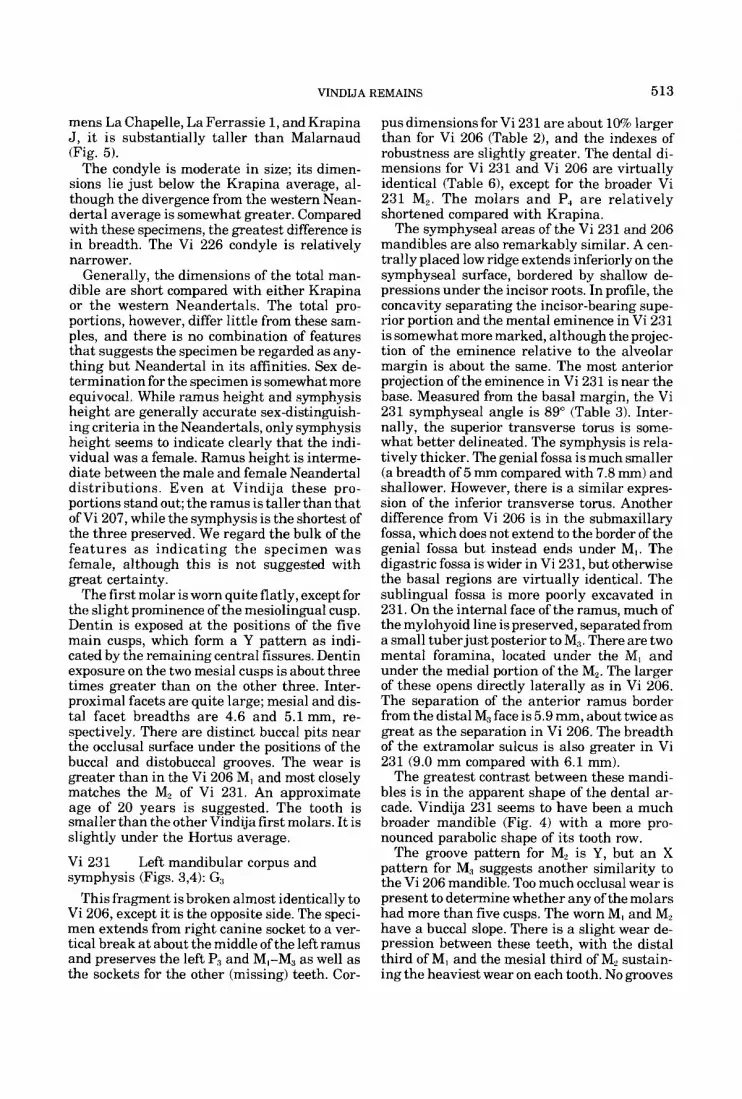
Vi 231 symphysis (Figs. 3,4): G3
This fragment is broken almost identically to Vi 206, except it is the opposite side. The speci- men extends from right canine socket to a ver- tical break a t about the middle of the left ramus and preserves the left P3 and M,-M, as well as the sockets for the other (missing) teeth. Cor-
Left mandibular corpus and
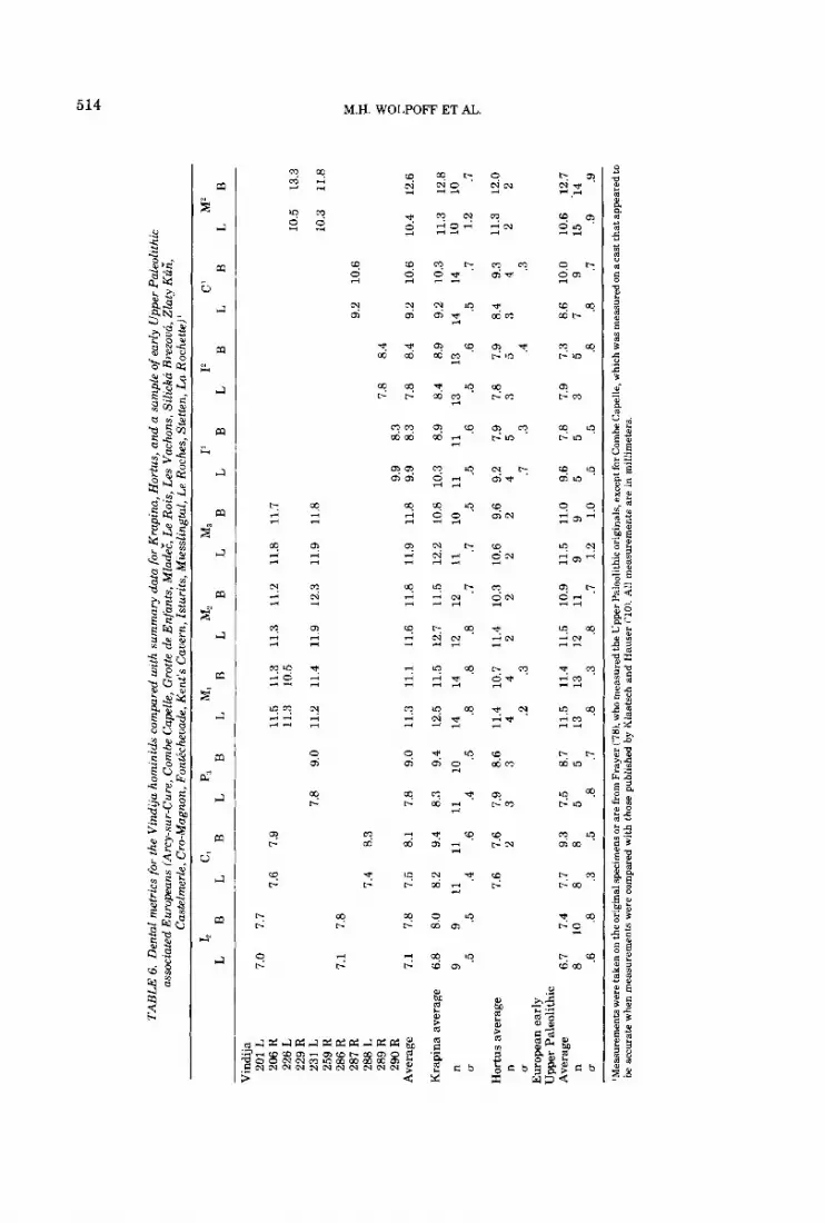
pus dimensions for Vi 231 are about 10% larger than for Vi 206 (Table 21, and the indexes of robustness are slightly greater. The dental di- mensions for Vi 231 and Vi 206 are virtually identical (Table 61, except for the broader Vi 231 Me. The molars and P, are relatively shortened compared with Krapina.
The symphyseal areas of the Vi 231 and 206 mandibles are also remarkably similar. A cen- trally placed low ridge extends inferiorly on the symphyseal surface, bordered by shallow de- pressions under the incisor roots. In profile, the concavity separating the incisor-bearing supe- rior portion and the mental eminence in Vi 231 is somewhat more marked, although the projec- tion of the eminence relative to the alveolar margin is about the same. The most anterior projection of the eminence in Vi 231 is near the base. Measured from the basal margin, the Vi 231 symphyseal angle is 89” (Table 3). Inter- nally, the superior transverse torus is some- what better delineated. The symphysis is rela- tively thicker. The genial fossa is much smaller (a breadth of 5 mm compared with 7.8 mm) and shallower. However, there is a similar expres- sion of the inferior transverse torus. Another difference from Vi 206 is in the submaxillary fossa, which does not extend to the border of the genial fossa but instead ends under MI. The digastric fossa is wider in Vi 231, but otherwise the basal regions are virtually identical. The sublingual fossa is more poorly excavated in 231. On the internal face of the ramus, much of the mylohyoid line is preserved, separated from a small tuberjust posterior to M3. There are two mental foramina, located under the MI and under the medial portion of the M,. The larger of these opens directly laterally as in Vi 206. The separation of the anterior ramus border from the distal M3 face is 5.9 mm, about twice as great as the separation in Vi 206. The breadth of the extramolar sulcus is also greater in Vi 231 (9.0 mm compared with 6.1 mm).
The greatest contrast between these mandi- bles is in the apparent shape of the dental ar- cade. Vindija 231 seems to have been a much broader mandible (Fig. 4) with a more pro- nounced parabolic shape of its tooth row.
The groove pattern for M, is Y, but an X pattern for M:% suggests another similarity to the Vi 206 mandible. Too much occlusal wear is present to determine whether any of the molars had more than five cusps. The worn M, and M, have a buccal slope. There is a slight wear de- pression between these teeth, with the distal third of MI and the mesial third of M, sustain- ing the heaviest wear on each tooth. No grooves
TA
BL
E 6
. Den
tal
met
rics
for
the
Vin
dija
hom
inid
s co
mpa
red
with
sum
mar
y da
ta fo
r Kra
pina
, Hor
tus,
and
a s
ampl
e of
ear
ly U
pper
Pal
eolit
hic
asso
ciat
ed E
urop
eans
(A
my-
sur-
Cur
e, C
ombe
Cap
elle
, Gro
tte d
e E
nfan
ts, M
lade
;, Le
Roi
s, L
es V
ach
ns,
Sili
cka
Bre
zoua
, Zln
ty K
S;,
Cas
telm
erle
, Cro
-Mag
non,
Fon
tech
euad
e. K
ent's
Cau
ern,
Istu
rits
. Mte
sslin
stal
. Le
Roc
hes.
Ste
tten.
La
Roc
hette
i'
LB
L B
L
Vin
dija
20
1 L
20
6 R
22
6 L
229
R 23
1 L
259
R
286
R
287
R 28
8 L
289
R 29
0 R
A
vera
ge
Kra
pina
ave
rage
n G-
Hor
tus
aver
age
n u
Eur
opea
n ea
rly
Upp
er P
aleo
lithi
c A
vera
ge
n u
7.0
7.7
7.6
7.1
7.8
7.4
7.1
6.8
9 .5
6.7
8 .6
7.8
7.5
8.0
8.2
9 11
.5
.4
7.6
7.4
7.7
.8
.3
10
8
7.9
8.3
8.1
9.4 .6
7.6
2 11 9.3
8 .5
7.8
7.8
8.3 .4
7.9
3 11 7.5
5 .a
BL
BL
BL
BL
BL
BL
BL
B
9.0
9.0
9.4 .5
8.6
3 10 8.7
5 .7
11.5
11
.3
11.3
11
.2
11.8
11
.7
11.3
10
.5
11.2
11
.4
11.9
12
.3
11.9
11
.8
9.9
11.3
11
.1
11.6
11
.8
11.9
11
.8
9.9
12.5
11
.5
12.7
11
.5
12.2
10
.8
10.3
14
14
12
12
11
10
11
.8
.8
.8
.7
.7
.5
.5
11.4
10
.7
11.4
10
.3
10.6
9.
6 9.
2 4
42
22
24
.2
.3
.7
10.5
13
.3
10.3
11
.8
9.2
10.6
7.8
8.4
8.3
8.3
7.8
8.4
9.2
10.6
8.9
8.4
8.9
9.2
10.3
.6
.5
.6
.5
.7
7.9
7.8
7.9
8.4
9.3
53
53
4
.3
.4
.3
11
13
13
14
14
11.5
11
.4
11.5
10
.9
11.5
11
.0
9.6
13
13
12
11
9 9
5 .
.8
.3
.8
.7
1.2
1.0
.5
.5
.a .a
.7
7.8
7.9
7.3
8.6
10.0
5
35
79
P T 4 s 10
.4
12.6
%
m % crl e
11.3
12
.8
10
10
F 1.
2 .7
11.3
12
.0
22
10.6
.1
2.7
15
14
.9
.9
'Mea
sure
men
ts w
ere
take
n on
the
orig
inal
spec
imen
s or
are f
rom
Fra
yer (
'781,
who
mea
sure
d th
e U
pper
Pal
eolit
hic
orig
inal
s, ex
cept
for C
ombe
Cap
elle
, whi
ch w
as m
easu
red
on a
cast
that
app
eare
d to
be a
ccur
ate
whe
n m
easu
rem
ents
wer
e co
mpa
red
with
tho
se p
ublis
hed
by K
laat
sch
and
Hau
ser
('10)
. All
mea
sure
men
ts a
re in
mill
imet
ers

VINDIJA REMAINS 515
remain on the M, occlusal surface, and the dis- tal portion is cupped in a predominantly trans- verse but partially mesiolingual direction. There is also a large dentin exposure on the mesiobuccal quadrant. Enamel perforation oc- curs on the five main cusps of M, (more me- sially). Third molar wear, however, is fairly flat. Dentin is exposed only on the mesial cusps, and the exposure is greater lingually. Wear on P3 is entirely buccal. There is a large dentin exposure on this side, which is flat and slightly angled buccally. In contrast, there is only slight polishing on the lingual portion of the occlusal surface. The buccal cusp was probably quite high, and the buccal face of the crown was fairly well curved.
Interproximal wear is marked on MI and M,, with mesial facet breadths of 6.0 and 5.0 mm. There is a smaller (3.3 mm) facet on M,, and a very small, centrally located facet on the mesial P, face. The teeth show neither hypoplasia nor the presence of any Carabelli’s features.
Finally, the specimen is somewhat older than Vi 206. The M3 is slightly more worn than the M, on the Vi 206 mandible. Assuming a 15 year date for the M, eruption, the estimated age is greater than 26 years. The molars are not much more worn than those of Krapina H, aged to 23 years (Wolpoff, 1979), and it is likely that Vi 231 is not much older than the 26-year minimum estimate.
Vi 250 and ramus (Fig. 3): G:,
This edentulous specimen extends from the distal border of the P3 root to the ramus, poste- rior to the mandibular foramen. This posterior break is approximately vertical up to the posi- tion of the mandibular foramen. A small seg- ment of the most inferior border of the notch also remains. The alveolar margin is intact buccally from the M, root and lingually from the M, root, although the alveoli up to the distal P, border remain. There are a number of simi- larities to Vi 207 and Vi 226, although the corpus appears slightly taller where compara- ble (Table 2). The base of the gonial region is slightly inverted, the mylohyoid sulcus is sepa- rated from the mandibular foramen (of hori- zontal-oval form) by 12.3 111111, anathe internal fossae are not well excavated. While the region is imperfectly preserved, the ramus seems t o have a distinct posterior orientation (perhaps as much as Vi 207). Unlike the other Vindija mandibles, however, the anterior border of the ramus crosses the alveolar margin a t about the back of the M, socket. A small lateral flange
Right portion of posterior corpus
beginning a t the M, position on the buccal corner of the corpus base is broken away ante- riorly.
The small mental foramen opens almost exactly laterally. Breadth of the extramolar sulcus (4.6 mm) is also small. In contrast, ramus dimensions are generally large.
The corpus is narrower than Vindija 226 a t the MJM, position, and only slightly taller and broader behind M2.
Summary. The mandibular sample consists of four Mousterian-associated specimens and a 5th from the early Aurignacian level. The lat- ter differs in no significant way from the former, but for its generally smaller size.
Considering the sample as a whole, speci- mens range in completeness from Vi 226 to Vi 250. The Vi 226 mandible is short when com- pared with Krapina J or the fairly complete Wurm Neandertals from western Europe (Table 5 ) . However, the RisdWunn specimen from Malarnaud is even smaller. Measures of its mandibular body length are similar to Re- gourdou.
Dimensions of the ramus of Vi 226 are also diminished, again only approached by Regour- dou (Table 4). Vi 226 resembles the (approxi- mately contemporary) western Neandertals and differs from the Krapina specimens in the breadth of the ramus relative to its height. The difference is mainly the result of relative broadening in the later Vindija specimens. When ramus breadth a t the alveolar margin and ramus height are compared with alveolar length (Table 5) , the breadth ratio for Krapina J is the smallest of all the mandibles compared, while the height ratio differs little. The Aurig- nacian-associated mandible, Vi 207, has the relatively broadest ramus of any of these spec- imens. I ts ramus height, however, is the smallest, as is the length of its condyle. The two Vindija rami have the semi-sculpted ap- pearance of the western European Neander- tals. Where the ramus base can be observed, it is consistently inverted.
Corpus dimensions of the Vindija mandibles do not differ significantly from the Krapina average (Table 2). However, a t the symphysis the average breadth for Vindija is greater and the average height is less, resulting in a signifi- cant difference for the breadth/height index. The indices for the three Vindija specimens preserving the symphysis lie above the Krapina range. The thickness is enhanced by the development of both superior and inferior transverse tori on the internal symphyseal
516 M.H. WOLPOFF ET AL.
faces of these three specimens, as well as a mental eminence on two of the three (see below).
There are certain aspects of the symphyseal region in the three Vindija specimens preserv- ing it (Vi 206, Vi 226, and Vi 231) which ap- proach the early modern sapiens condition more closely than the majority of other Ne- andertals do. Basically the Vindija G3 man- dibular symphyses exhibit relatively more for- ward projection of the base than the alveolar region, and in two (Vi 206 and Vi 231) there are slight concave depressions separating the two regions. This results in a weakly pronounced but distinct mental eminence and infers that the alveolus was somewhat less prognathic than most Neandertals. Typically, the alveolus of Neandertal mandibles projects anteriorly more than the base, and the contour between the areas is essentially linear (Wolpoff, 1975; Howells, 1975; Weidenreich, 1936; Smith, 1976b). A few western European Neandertal mandibles (Spy 1, La Ferrassie 1, and Circeo 31, however, exhibit external symphyseal mor- phologies similar to the Vindija G3 specimens.
The relative reduction in the projection of the alveolus relative to the base in the Vindija mandibles can also be illustrated by examina- tion of their symphyseal angles (Table 3). The Vindija G3 mean of 85" lies 1.2 and 6.7 standard deviations below the western European Ne- andertal and Krapina means, respectively. At Vindija, all symphyseal angles are below 90". Thus, as a group, the Vindija G3 hominids are closer to the modern European pattern i n symphyseal morphology than the western Ne- andertal sample in general, and the late sites, such as Hortus, in specific.
Interestingly, the reduction of the alveolar projection relative to the mandibular base at Vindija does not seem to reflect a general poste- rior repositioning of the tooth row. This can be seen at the corpus posterior where, except for Vi 250, the anterior border of the Vindija rami generally crosses the alveolar margin posterior to the last molar. In fact, the average distance between the crossing and the back of the tooth is identical to the Krapina average, and the distance for Vi 207 is near the average for both samples. A related feature, the position of the mental foramen (or foramina) also does not differ between these two samples.
Similarly, both samples include specimens with multiple foramina. However, there is a marked difference in foramen size. The average foramen (or foramina) area for the Vindija sample is only 55% that of Krapina.
In considering the Vindija mandibular sam- ple as a whole (including the Aurignacian-as- sociated Vi 207), we believe that there are a number of important similarities to the west- ern European Neandertal mandibles. How- ever, while we do not wish to overemphasize the differences, the fact remains that they are present. Some, we believe, are due to the late date of the sample. These provide the basis for similarity to the latest of the western European sample. Other differences, however, may be idiosyncratic or perhaps geographic, and it may not be possible at present to fully distinguish these from the temporal component of varia- tion, For instance, what is the meaning of the fact that the Vindija specimens tend to have a corpus that is shallower anteriorly and slightly deeper posteriorly while on the western Eu- ropean sample the opposite is the case? The fact that central European mandibles from Ochoz and Subalyuk tend to be more similar to the Krapina sample while the late Sipka juvenile more closely approximates the Vindija speci- mens might help provide evidence for temporal trends in central Europe. However, accurate dates for all of the sites concerned would present a firmer basis for this contention.
Isolated teeth Vi 290 Right I' (Fig. 6): G,
Except for a slight chip from the apex of its root, this tooth is a perfectly preserved maxil- lary central incisor with a total height of 30.6 mm. Vi 290 is not the same individual asVi 289 though both exhibit strong shoveling (see below), due to the latter's much more extensive occlusal wear. Root height for Vi 290 is 19.1 mm and crown height is 12.6 mm, measured from the center of the cemento-enamel junction. Crown dimensions (Table 6) are large and ap- proach the Krapina mean values. Length and breadth of the crown lie respectively 0.7 and 1.1 standard deviations below the Krapina means. The root measures 7.7 mm in length and 6.7 mm in breadth.
Occlusal wear is slight to moderate, with only very slight dentin exposure along the occlusal surface. Wear on the occlusal surface is flat in the labiolingual direction but convex mesiodistally. The wear surface is sharply in- clined lingually and continues about halfway down the marginal ridges of the tooth. Both interproximal facets are centrally located on their respective margins and begin at the occlusal surface. They are only slightly devel- oped, with breadths of 1.9 mm for the mesial facet and 2.4 nun for the distal facet.
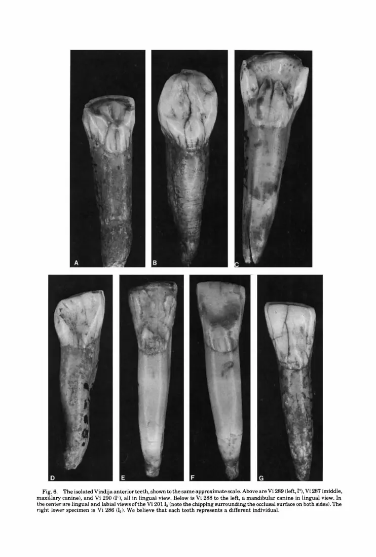
Fig. 6. The isolated Vindija anterior teeth, shown to the same approximate scale. Above are Vi 289 (left, I?, Vi 287 (middle, maxillary canine), and Vi 290 (I1), all in lingual view. Below is Vi 288 to the left, a mandibular canine in lingual view. In the center are lingual and labial views ofthe Vi 201 I, (note the chipping surrounding the occlusal surface on both sides). The right lower specimen is Vi 286 (L). We believe that each tooth represents a different individual.

518 M.H. WOLPOFF ET AL.
A number of chips occur on the occlusal sur-. face and the scar for a large step flake occurs on the labiodistal corner. For three of the chips, the edges a r e worn and ra ther rounded, suggesting that they represent antemortem damage.
Like most Neandertal maxillary incisors, Vi 290 is markedly shoveled. The basal tubercle is doubled, with the mesial tubercle measuring 1.8 mm in breadth at its base and the distal tubercle measuring 2.3 mm. Finger-like exten- sions of both extend up the lingual face. The marginal ridges are very well developed and measure over 2 mm in breadth each. The distal ridge is separated from the labial surface by a distinct groove, but the internal margin for the mesial ridge is less distinct.
Vi 289 Right I’ (Fig. 6): Fd This lateral maxillary incisor is very heavily
shoveled and markedly worn. Maximum tooth height and height of the center of the crown are 25.6 mm and 9.3 mm, respectively, but a t least half the original crown height is lost due to the extensive occlusal attrition. Root height is 17.5 mm, and the root measures 8.2 mm in labio- lingual diameter by 5.3 mm in mesiodistal di- ameter. Crown dimensions are moderate by Krapina standards, although well within the observed range (Table 6). Crown breadth is about one standard deviation below t h e Krapina mean.
Occlusal wear is very heavy for this speci- men, as well as slightly concave labiolingually and lingual in slope. The angle of the occlusal surface is about 30” to the horizontal defined by the long axis of the tooth. Dentin is exposed along the entire occlusal surface, including the marginal ridges, forming a continuous “U” or horseshoe pattern. A large chip occurs at the distolabial corner, with step flaking continuing down the labial face. The rounded edges suggest that this occurred antemortem. The interproximal facets are about 2 mm in breadth and are both centrally located on the labio- lingual plane. Interproximal wear is moderate in degree for this specimen.
Morphologically, Vi 289 is very similar to several lateral maxillary incisors a t Krapina. The basal tubercle and marginal ridges are all very strongly developed on the lingual aspect of this specimen, resulting in a marked degree of shoveling. The tubercle is 3.5 mm high and exhibits a well-excavated lingual pit superi- orly. The mesial and distal marginal ridges, only remainingfor 2.5 mm above their base due to the extensive occlusal attrition, are 2.5 mm
and 3.4 mm in breadth, respectively. The root is stout and oval in shape, with a moderate mesial groove. There is a distinct distal curve to its lower half.
Vi 201 This lateral mandibular incisor was the first
hominid specimen found at Vindija. It is a per- fectly preserved tooth measuring 27.3 mm in height. Root height is 17.8 mm, and crown height is 10.5 mm from the labial aspect of the cemento-enamel junction. The crown size of this tooth is very close to the western Neander- tal mean. Using breadth for comparison. since it is unaffected by interproximal attrition, the 7.7 mm value for this specimen lies at the mean of 7.8 mm calculated for 15 western Neander- tals (u = 0.69). Breadth for this specimen is 1.4 standard deviations below the mean of 8.4 mm for the three western specimens dated to Wurm 1 (2 from Arcy-sur-Cure and 1 from Regourdou) and is also less than the 8.0 mm mean value for nine measurable individuals from Krapina (Table 6). Crown length is 7.0 mm and exceeds the Krapina mean. The maximum labiolingual and mesiodistal diameters of the root are 8.4 mm and 4.6 mm, respectively.
Occlusal wear is moderately flattened and slightly convex in both the mesiodistal and labiolingual planes. The occlusal facet meas- ures 7.0 mm in length and 2.4 mm in maximum breadth. Only a slight exposure of dentin is present along this plane. The borders of the incisal surface are rounded, and there is exten- sive chipping on both the labial and lingual faces. (Fig. 6). Microscopic examination indi- cates only some small chips with rounded bor- ders, indicating they were clearly antemortem.
Interproximal wear is slight. The mesial in- terproximal facet measures some 4.5 mm in length and drops vertically from the occlusal plane. The distal facet is placed to the labial edge of the distal surface. I t begins some 2 mm below the occlusal surface and extends inferi- orally and slightly labially for 3 mm.
Morphologically, the tooth is weakly shov- eled with a broad basal tubercle, and a pattern of slight horizontal bands covering the labial crown surface. The root is mesiodistally com- pressed with strong labial and lingual pillars.
Left I, (Fig. 6)
Vi 286 Right I, (Fig. 6): F,,
This tooth is remarkably similar to Vi 201 in size (Table 61, morphology, and wear, except for the absence of chipping and crown damage. Total tooth height is 26.1 mm, root height is
VINDIJA REMAINS 519
17.2 mm, and crown height is 10.6 mm. Crown breadth is 7.8 mm, and the length and maximum breadth of the occlusal surface (inci- sal plane) are 7.1 and 2.7 mm, respectively. The labiolingual diameter of the root is 8.6 nun, and the mesiodistal diameter is 4.6 mm.
Occlusal wear is moderate and flat, with a distinct distal slope. Dentin is exposed along almost the entire surface length, but breadth of the exposure is never greater than 0.6 mm.
Interproximal wear is slight. The mesial con- tact facet (breadth = 2.2 mm) lies a t the occlusal margin, while the distal facet (breadth = 2.2 mm) is somewhat lower. Its superior edge is 2 mm below the occlusal margin. Both facets are centrally located on a labiolingual plane.
Like Vi 201, Vi 286 is weakly shoveled with very weak marginal ridges on its lingual crown surface. The basal tubercle is quite well devel- oped and extends about ?h of the way up the crown. A distally positioned vertical groove lies between the tubercle and the weak mar- ginal ridge. The root is strongly compressed mesiodistally, with well-developed mesial root concavities and pillars.
Vi 287 This canine shows only slight occlusal wear,
but in two distinctly separate planes. An occlusal facet occurs a t the tip, resulting in a worn surface 3.4 m in length and 3.6 mm broad. This facet is interrupted mesially by a large chip extending down the labial face with sharp edges that do not allow a determination of whether it occurred post- or ante-mortem. The central facet is convex in both labiolingual and mesiodistal directions and has a marked lingual slope. There is a very slight area of dentin exposure at its center. The second facet is contiguous with the first, extending halfway down the distal marginal edge of the lingual side. It is flat and only slightly angled relative to the length axis of the crown, resulting in a sharpened distal edge.
Total tooth height for this specimen is 33.0 mm and is probably only slightly reduced by occlusal wear. Root height and crown height are 21.8 mm and 12.5 mm. Crown dimensions are virtually identical to the Krapina means (Table 6). Maximum root dimensions are 6.7 mm (mesiodistal) and 10.1 mm (labiolingual).
Occlusal wear is moderate on this specimen, while there is but a trace of interproximal wear. A small mesial interproximal facet (breadth = 1.7 mm) is found about halfway up from the crown base and is displaced lingually viewed from the mesial aspect. An identically sized
Right C' (Fig. 6): G,
distal facet is located somewhat lower on the crown but more centrally positioned in labio- lingual aspect.
The basal tubercle continues on the mesial surfaces as a weak marginal ridge, marked by a distinct groove distal to it. The mesial pit is well developed and is connected to a second pit just mesial to the tubercle by a very weak groove. Additional small pits are found near the mesial and distal margins.
Vi 288 This canine exhibits more occlusal wear and
a different interproximal wear facet size than either Vi 201 or Vi 286, suggesting that it rep- resents a different individual than either of the unassociated mandibular incisors in the sam- ple. Total tooth height for this specimen is 25.4 mm. The root height is 16.5 mm, and the mesial and distal heights of the crown are 11.0 mm and 9.8 mm, reflecting the marked distal slope of the flat occlusal surface. Crown length lies 2.2 standard deviations below the Krapina mean, and the breadth difference is about the same (Table 6). The occlusal surface does not extend to the distal edge of the tooth and its maximum length and breadth diameters are 6.2 and 3.0 mm, respectively. Exposed dentin occupies ap- proximately ?h of the occlusal surface breadth. Maximum root diameters are 8.8 mm (labiolin- gual) and 5.0 mm (mesiodistal).
The lingual surface of the tooth is moderately shoveled, with the distal marginal ridge being the more pronounced and exhibiting a more distinct groove following its edge. The margi- nal ridges extend to within 2.1 mm of the cemento-enamel junction. There are some weak hypoplastic features near the base on the labial side. The root is robust and slightly com- pressed mesiodistally.
Interproximal wear is slight. The mesial in- terproximal facet measures 1.7 mm in breadth and is centrally located. A very weakly devel- oped distal facet parallels the distal marginal ridge.
Vi 229 Right M2 This upper molar exhibits a distal inter-
proximal facet (breadth = 4.1 mm) indicating that i t is not a third molar. It is suggested to be a second molar on the basis of the marked angu- lation of the roots and their lack of divergence. Adult status is indicated by the distal facet, fusion of the distal roots, and the amount of interproximal and occlusal wear.
Occlusal wear is moderate and fairly flat, with a mesiolingual slope. The most wear is on
Left C, (Fig. 6): Fd
520 M.H. WOLPOFF ET AL.
the mesiolingual cusp (protocone), where den- tin exposure is advanced. The least degree of wear is on the distobuccal cusp (metacone), where no dentin is exposed, although the grooves are virtually obliterated. Occlusal wear on the other cusps are intermediate be- tween the two. Hypocone development is mod- erate.
Dimensionally, the tooth is broad and mesio- distally short (Table 6). The mesial interproxi- ma1 facet is large, measuring some 5.6 mm in breadth.
Summary. All of the isolated Vindija ante- rior teeth with known provenience are from the Aurignacian levels. These teeth are somewhat reduced compared with the Krapina averages. However, they are markedly larger than the late Neandertal anterior teeth from Hortus but also are larger than the early Upper Paleo- lithic sample, primarily representing western Europe (Table 6). While intermediate between Krapina and the early Upper Paleolithic sam- ple, the specimens fall within both ranges. The fact that different individuals seem to be repre- sented by these teeth suggests that this inter- mediate position may be regarded with more certainty than the small sample sizes might suggest. Since anterior dental reduction is one of the critical evolutionary trends associated with early modern Homo sapiens populations (Frayer, 19781, the large size of the Vindija Aurignacian-associated anteriors may be im- portant.
Some morphological features resemble Ne- andertals. These include the marked shoveling of the maxillary incisors and their lingual basal tubercle development. Even the I, specimens are slightly shoveled, and there are distinct marginal ridges on the mandibular canine. Yet, the ranges of variation overlap with more modern populations. Moreover, the pronounced lingual angulation of occlusal wear on the maxillary incisors is generally found in more modern populations.
The posterior teeth with provenience (includ- ing those in jaws) are Mousterian associated. However, like the anteriors discussed above, they are generally between the Krapina sam- ple and the early Upper Paleolithic sample in size. They are larger than the Hortus teeth, paralleling the canine comparison. Compared with Krapina the main reduction is in length, and as a result the Vindija posterior teeth are squarer. Some of this difference is surely due to age; the Krapina average age at death is much lower than any estimate that could be made for
Vindija, and consequently there is less oppor- tunity for length reduction due to interproxi- ma1 attrition a t Krapina. However, if the Krapina interproximal wear rate can be applied to both sites, it is too low to account for the differential reduction of posterior tooth length (Wolpoff, 1979).
Frontals with supraorbitals As a preface to the discussion of the individ-
ual frontals from Vindija, it is necessary to con- sider some points pertinent to our analysis of supraorbital torus morphology. Two basic measures of torus size are used in this study. These are thickness and projection, taken at specific points along each torus (defined below). Thickness is defined as the cranial-caudal di- mension of the torus, and measurements of thickness are taken as chords from the ante- rior, superior orbital wall (the inferior aspect of the torus) to the superior toral margin. Projec- tion is defined as the anterior-posterior dimen- sion of the torus. Measurements of projection are taken as minimum chords from the anterior internal margin of the frontal just above the orbital roof to the most anterior point on the torus a t that level.
Depending on preservation of the specimen, thickness and projection are taken at up to three points above each orbit. The first, or me- dial, point is slightly lateral and superior to the medial orbital margin. In this sample, the di- mension here was invariably the thickest point on the entire torus. The second, or lateral, point is defined as the thickest point on the supraor- bital trigone portion of the torus. The third point, or mid-orbit, is positioned slightly lateral to the middle of the orbit. In this sample it is also the minimum thickness between the first two points. Projections were taken at each of these points as well. Very little asymmetry was noted for specimens possessing a complete torus. Therefore, when both right and left sides of a single torus were available for measure- ment, the values for corresponding meas- urements were averaged, and this value was used in computations.
A second point we believe to be relevant con- cerns the fact that there is a significant amount of age change in torus size in Neandertals (c.f. VlEek 1967; Smith 1976b). Infant and juvenile Neandertals exhibit very little torus develop- ment (VlEek 19701, and it is not until full adult- hood is reached that the torus achieves its maximum size. Apparently, to judge from Le Moustier and the Krapina remains, a good deal of growth and remodeling of the torus occurs
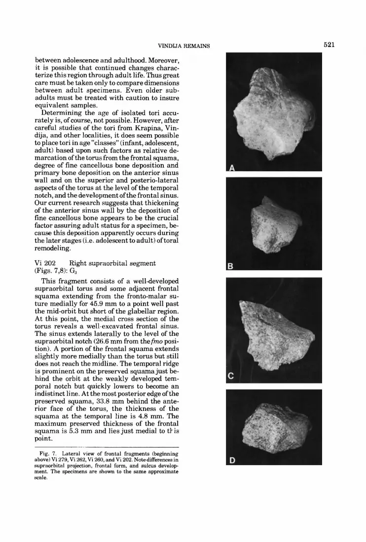
VINDIJA REMAINS 521
between adolescence and adulthood. Moreover, it is possible that continued changes charac- terize this region through adult life. Thus great care must be taken only to compare dimensions between adult specimens. Even older sub- adults must be treated with caution to insure equivalent samples.
Determining the age of isolated tori accu- rately is, of course, not possible. However, after careful studies of the tori from Krapina, Vin- dija, and other localities, it does seem possible to place tori in age “classes” (infant, adolescent, adult) based upon such factors as relative de- marcation of the torus from the frontal squama, degree of fine cancellous bone deposition and primary bone deposition on the anterior sinus wall and on the superior and posterio-lateral aspects of the torus at the level of the temporal notch, and the development ofthe frontal sinus. Our current research suggests that thickening of the anterior sinus wall by the deposition of fine cancellous bone appears to be the crucial factor assuring adult status for a specimen, be- cause this deposition apparently occurs during the later stages (i.e. adolescent to adult) oftoral remodeling.
Vi 202 Right supraorbital segment (Figs. 7,8): G3
This fragment consists of a well-developed supraorbital torus and some adjacent frontal squama extending from the fronto-malar su- ture medially for 45.9 mm to a point well past the mid-orbit but short of the glabellar region. At this point, the medial cross section of the torus reveals a well-excavated frontal sinus. The sinus extends laterally to the level of the supraorbital notch (26.6 mm from the fmo posi- tion). A portion of the frontal squama extends slightly more medially than the torus but still does not reach the midline. The temporal ridge is prominent on the preserved squama just be- hind the orbit a t the weakly developed tem- poral notch but quickly lowers to become an indistinct line. At the most posterior edge of the preserved squama, 33.8 mm behind the ante- rior face of the torus, the thickness of the squama a t the temporal line is 4.8 111111. The maximum preserved thickness of the frontal squama is 5.3 mm and lies just medial to tl-is point.
Fig. 7. Lateral view of frontal fragments (beginning above) Vi 279, Vi 262, Vi 260, and Vi 202. Note differences in supraorbital projection, frontal form, and sulcus develop- ment. The specimens are shown to the same approximate scale.
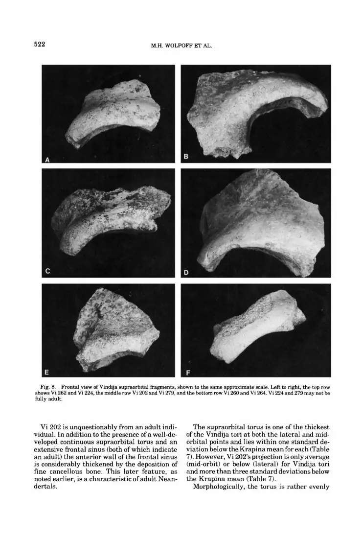
522 M.H. WOLPOFF ET AL.
Fig. 8. Frontal view of Vindija supraorbital fragments, shown to the same approximate scale. Left to right, the top row shows Vi 262 and Vi 224, the middle row Vi 202 and Vi 279, and the bottom row Vi 260 and Vi 264. Vi 224 and 279 may not be fully adult.
Vi 202 is unquestionably from an adult indi- vidual. In addition to the presence of a well-de- veloped continuous supraorbital torus and an extensive frontal sinus (both of which indicate an adult) the anterior wall of the frontal sinus is considerably thickened by the deposition of fine cancellous bone. This later feature, as noted earlier, is a characteristic of adult Nean- dertals.
The supraorbital torus is one of the thickest of the Vindija tori a t both the lateral and mid- orbital points and lies within one standard de- viation below the Krapina mean for each (Table 7). However, Vi 202’s projection is only average (mid-orbit) or below (lateral) for Vindija tori and more than three standard deviations below the Krapina mean (Table 7).
Morphologically, the torus is rather evenly

TABLE
7. T
hick
ness
and
pro
ject
ion
of
Vin
dija
supr
aorb
itals
com
pare
d w
ith K
mpi
nn a
nd
with
a s
ampl
e of
ear
ly U
pper
Pal
eolit
hic
spec
imen
s fr
om
cent
ral
and
east
ern
Eur
ope
(&no
1,
2, D
olni
V&
toni
ce,
Mla
deF
12,5
,6 P
aulo
u, S
tare
Mes
to, V
elik
a Pe
cina
, and
Zla
ty-K
&)I
Proj
ectio
n T
hick
ness
Lat
eral
M
id-o
rbit
Med
ial
Lat
eral
M
id-o
rbit
Med
ial
Vin
dija
V
i 202
V
i 22
4*
Vi 2
60
Vi
261
Vi
262
Vi
27
P
Vi
284
Adu
lt a
vera
ge
(U)
Sam
ple
aver
age
(U)
Kra
pina
A
vera
ge
(a)
n R
ange
Cen
tral
and
eas
tern
Eur
opea
n E
arly
Upp
er P
aleo
lithi
c A
vera
ge
(U)
n R
ange
19.5
19
.5
22.5
24
.5
22.0
16
.0
22.0
22
.1
(1.8
) 20
.9
(2.8
)
24.3
(1
.4)
8 23
.0-2
7 .O
20.3
(2
.6)
9 15
.0-2
3.0
19.0
17
.5
21.0
23
.0
16.0
13
.0
16.0
19
.0
(3.1
) 17
.9
(3.4
)
23.9
(1
.2)
11
23.0
-26.
0
16.1
(3
.4)
9 8.
0-19
.0
-
17 -
-
-
11.0
-
-
-
20.3
(2
.3)
4 17
.5-2
3.0
13.0
(3
.0)
9 8.
0-17
.5
11.3
9.
1 11
.0
10.5
10
.0
7.6
10.4
10
.6
(0.5
) 10
.0
(1.3
)
12.5
(1
5)
11
10.%16.0
8.1
(1.4
) 11
6.
0-10
.1
9.5
9.7
8.5
8.3
8.6
7.0
8.0
8.6
10.6
) 8.
5 (0
.9)
10.7
(1
.8)
13
7.CL14.3
5.4
(1.7
1 11
4.
4-7.
7
-
15.5
-
-
-
15.1
-
-
-
17.6
(3
.0)
4 15
.8-2
2.0
16.6
(3
.3)
11
11.E
-23.
7
The
mea
sure
men
ts, i
n m
illim
eter
s, w
ere
take
n by
FH
S an
d ar
e de
fine
d in
the
text
.
-Mis
sing
da
ta (
area
mis
sing
or
not m
easu
rabl
e).
'Pos
sibl
y ju
veni
le.

524 M.H. WOLPOFF ET AL
distributed over the orbit. In spite of the pres- ence of a supraorbital notch (breadth = 4 mm), there is no division of the torus into the distinct medial and lateral elements characteristic for modern Europeans (c.f. Cunningham, 1908). There is, however, a tendency toward more thinning of the mid-orbital segment of the torus relative to the lateral segment. This pattern is characteristic for all the Vindija adult tori. Sur- face bone characterized by the “vermiculate pattern” (c.f. Tappen, 1978; Oyenet al., 1979) is present on the anterior torus along its entire preserved length and on its superior aspect from the level of the supraorbital notch later- ally. Finally, a shallow sulcus separates the torus from the frontal squama.
Vi 224 Supraorbital region (Figs. 8,9): G3
The specimen consists of an entire left supra- orbital region preserved from the frontomalar suture past the midline and including a portion of the nasofrontal region. Virtually none of the frontal squama is preserved. Maximum preser- vation of the squama is a t the midline, extend- ing 33.1 mm behind glabella. Some damage was sustained by the lateral ?h of the anterior torus face, which is reminiscent of Le Moustier, Krapina 23, and Vi 279 in form. Although evenly developed, it barely projects anterior to the frontal squama and is only separated from the squama by a shallow sulcus laterally. At the midline, there is no sulcus separating the frontal squama from the glabellar region. The anterior face of the torus is not vertical. In- stead, it is evenly curved between the superior orbital margin and the frontal squama. While this morphology may reflect a young age for the specimen, the presence of a moderately marked temporal ridge and a fairly large frontal sinus extending laterally to within 27 mm of the fmo position argue against juvenile status. One fur- ther factor, however, suggests that Vi 224 may not be an adult. The anterior wall of the frontal sinus does not exhibit significant build-up of fine cancellous bone, which is found in all spec- imens from Krapina and Vindija that are un- equivocally adult, Thus, since it cannot be defi- nitely ascertained whether the torus of this specimen has reached its maximum growth level, it will not be included as an adult in the Vindija sample for purposes of metric compari- sons. The specimen is probably older than Le Moustier (13 years).
Laterally, the torus of Vi 224 is one of the thinnest a t Vindija, although at mid-orbit its thickness is the maximum at the site (Table 7).
Fig. 9. Vi 224 (above) and Vi 261 in lateral view. Note the marked differences in supraorbital form and projection, and in the position of the frontal squama relative to the torus.
Projection at mid-orbit is moderate. The mid- orbit and lateral thicknesses are 0.4 and 2.1 standard deviations below the Krapina mean. Vi 224 is one of the two Vindija specimens pre- serving glabella and the most medial extent of the torus. Here the thickness of the Vindija specimen lies 0.7 standard deviations below the Krapina mean.
Measures of torus projection provide inter- esting results. Supraorbital length as defined by Weidenreich is one of the smallest at Vin- dija, reflecting the shallow supra-toral sulcus. Torus projection measured from the internal cranial surface is 0.5 standard deviations below

VINDIJA REMAINS 525
TABLE 8. Projection of nasion anterior to the bi-fmt line for VindLj‘a and all other available
Neandertal specimens‘
bi-fmt breadth
Nasion to fmt
Vindija 224
Sala Krapina E Krapina C SPY 1 Le Moustier Mt Circeo Saccopastore 1 La Chapelle Neandertal La Quina 5 Gibraltar La Ferrassie 1 Saccopastore 2
Western European Neandertal - X U
A Vindija U
113.5
114.3 117.9 118.9 121.8 115.4 120.0 118.0 121.0 118.4 106.5 114.8 121.4 124.3
117.9 4.5
1.0
60.3
61.8 63.5 64.6 66.4 63.7 66.2 65.5 67.1 66.1 61.2 65.5 69.4 71.7
65.5 2.9
1.8
Nasion projection
20.4
23.5 23.6 25.6 26.5 27.0 28.0 28.4 29.0 29.4 30.2 31.6 33.6 34.5
28.5 3.4
2.4
‘The projection is defined as the perpendicular height of a triangle with the base being the bi-fmt chord and the sides being the nasion-fmt chord. The measurements, in millimeters, were taken on the original specimens.
the Vindija mean a t mid-orbit and 1.5 standard deviations below the Vindija mean laterally. We believe these data may reflect the in- complete frontal sinus development. Medial, mid-orbit, and lateral projections for Vi 224 are, respectively, 1.0,5.5, and 3.5 standard de- viations below the appropriate Krapina means. This contrasting development of supraorbital projection (i.e. relatively less lateral and mid- orbit than medial projection) may also reflect the juvenile status of Vi 224. Full remodeling and maximum torus development had not yet occurred in the lateral region.
One modern aspect of the torus lies in the relative projection of nasion as measured to a line drawn between the fmt points (Table 8). The normal Neandertal condition of mid-facial prognathism is reflected by a marked perpen- dicular projection of nasion anterior to this line. In Vi 224, nasion is in a very high position, approaching glabella, which would tend to in- crease its projection. Yet, the projection of the Vi 224 frontal is below the Neandertal range and 2.3 standard deviations below the Ne- andertal mean. This suggests a reduction in the extent of mid-facial prognathism for the Vin- dija specimen.
Aligning the superior orbital roof horizon- tally indicates a fairly high angle for the fore- head. Squama thickness a t the midline, 33 mm behind glabella, is 8.5 mm. This is 13.6 mm behind the posterior wall of the frontal sinus.
The intraorbital region is fairly broad. Poste- rior intraorbital breadth is estimated a t 30.5 mm, and the length of the nasofrontal suture from nasion to the medial orbital margin is 21 mm. Nasion is 19.6 mm anterior to the crzstu frontulis and 11.7 mm in front of the anterior sinus wall.
Vi 227 Frontal fragment with left portion of supraorbital: G3
This fragment unquestionably is from an in- fant. It extends from the left frontomalar su- ture to a point 15 mm to the right of the midline and sagitally no more than 39.5 mm posterior to the posterior border of the frontal sinus at the midline. While there is no actual torus devel- opment, a shallow sulcus extends posterior to the left orbital edge up to the somewhat el- evated midline region, This creates a distinct “outline” of where the torus will develop later. Similar morphology is exhibited by several in- fant and young juvenile Neandertals (VlEek 1967, 1970; Smith 1976b). There is no develop- ment of the vermiculate pattern on the frontal of this specimen.
The vault bone is thin (4.4 mm at the most posterior point on the midline and 3 mm at a point 26 mm behind the midorbit), probably a reflection of the specimen’s age. Yet, the poste- rior-superior portions of a t least two distinct sinus cavities are preserved on the left, and there is a corresponding single cavity on the right which terminates a t the broken edge of the fragment. The left cavities extend 15.6 mm from the midline. This suggests that the frontal sinuses begin to develop very early in these hominids. Midline curvature of the squama suggests a bulging (bossed) profile similar to some of the adults. Based on a horizontal orien- tation for the superior orbital roof, the forehead slope is moderate.
Vi 260 Right supraorbital torus fragment (Figs. 7,8): G,
This specimen extends some 32.5 mm along the torus from the frontomalar suture to a point just short of the mid-orbit. The frontal squama is preserved as far as 49.7 mm posterior to the superior orbital margin, and its thickness a t this point is 3.6 mm.
At its lateral border the torus is about aver- age in thickness for Vindija (Table 7). Mid-orbit

526 M.H. WOLPOFF ET AL
thickness lies 1.2 standard deviations below the Krapina mean. In contrast the torus projec- tion is marked, and there is a deep sulcus sepa- rating it from the fairly straight preserved por- tion of the frontal squama. At the medial break, frontal sinus is exposed. The anterior sinus border is 12 mm behind the anterior torus edge, but laterally it extends toabout 20 mmfrom the fmo position. The anterior wall exhibits signifi- cant thickening due to fine cancellous bone de- position. Also, the external bone table on the superior and anterior aspects of the torus ex- hibits the vermiculate pattern over the entire preserved segment. Thus, Vi 260 appears to be an adult specimen.
The temporal line is prominent, extending as a ridge for 25.5 mm behind the orbit. In con- trast, however, the squama is very thin com- pared with other Vindija specimens (see Table 11) or with the Krapina sample.
Vi 261 Frontal (Figs. 9,lO): Gi3 The specimen consists of three pieces. Two of
these (discovered in 1977 and 1978, re- spectively), combine to form a large fragment that extends from the border with the zygo- matic on the lateral left side to a point 36.5 mm to the right of the midline. On its lateral left face, the frontal squama extends to the coronal suture, some 59.3 mm posterior to the supra- orbital torus. The torus is preserved in its en- tirety for 36 mm medial to the frontozygomatic suture, and the top of the structure is preserved to the midline. The anterior and inferior por- tions of the torus are missing medial to mid-or- bit. A frontal sinus is exposed by the broken orbital roof. On the left side the complete sinus is 29 mm in transverse breadth, and its supe- rior surface extends 23 mm behind the anterior face of the torus. There is significant deposition of fine cancellous bone in the anterior wall of the sinus. The vermiculate pattern is present over the entire external surface of the anterior aspect of the torus but is limited on the superior aspect to the region lateral to the sinus.
This is the most archaic-appearingof the pre- served tori, resembling the Ehringsdorf H fron- tal in its marked projection. It is the most pro- jecting Vindija specimen both at mid-orbit and laterally, but it is still below the Krapina mean at mid-orbit and only slightly above it laterally (Table 7). The torus is continuous but unevenly developed, closely following the orbital contour and dipping significantly a t glabella. While there is thickening at both the lateral position and the most medial extension, the torus is thinnest a t a point about midway between
these. In fact, laterally and a t mid-orbit the thicknesses of Vi 261 are below the Vindija means and far below Krapina.
There is no supraorbital notch or groove. As seen from the top, there is a significant posteri- or-lateral angulation to the torus.
The extension of the torus anterior to the frontal squama is marked. The squama has a fairly high angle to the orbital roof and is dis- tinctly rounded in lateral view. There is a very deep sulcus between the squama and the su- praorbital (Fig. 91, which is accentuated by the marked boss immediately above it. This sulcus is shallowest a t the midline but deepens signif- icantly laterally. Measured at the position of the lateral eminence, the squama is the thick- est of the Vindija specimens (Table 11) and is slightly above the Krapina average. At this position, just behind the sinus and 29 mm from the midline, the thickness attains 8.7 mm. The temporal line forms a prominent ridge, extend- ing for most of the preserved length of the squama. Finally, the temporal notch is deep. Combined, these features suggest the likeli- hood that the specimen was a male. It is un- questionably adult.
A third piece is Vindija 278, a centrally lo- cated posterior fragment of frontal squama with a 6.7 mm length of coronal suture. This is almost certainly the same individual. Unfortu- nately, the badly eroded posterior border of the larger supraorbital-bearing fragment makes a clear contact impossible to establish. Vi 278 is maximally 64.8 mm in length (approximately parallel to midline) and 44.8 mm transversely. A marked endocranial groove, almost certainly representing the posterior extension of the sagittal sulcus, occurs along the long edge of the fragment. This edge is the approximate midline and consequently the bregma position is preserved on the coronal suture. Along the midline, thickness expands from 5.9 mm at bregma to 8.2 mm at the most anteriorally pre- served point. Combining the two pieces in their approximate relationship indicates a glabel- la-bregma distance of 113- 116 mm. In midline profile the complete squama is markedly curved.
Vi 262 Right supraorbital torus fragment (Figs. 7,8): G3
The lateral and central portions of the right side are present in this specimen and extend from the frontomalar suture to a position just medial to the mid-orbit. The frontal squama is preserved to a maximum of 30.8 mm posterior to the superior orbital margin. The torus is only
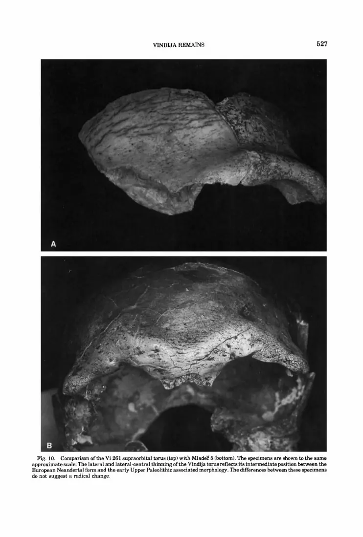
VINDIJA REMAINS 527
Fig. 10. Comparison of the Vi 261 supraorbital torus (top) with Mlad& 5 (bottom). The specimens are shown to the same approximate scale. The lateral and lateral-central thinning of the Vindija torus reflects its intermediate position between the European Neandertal form and the early Upper Paleolithic associated morphology. The differences between these specimens do not suggest a radical change.
528 M.H. WOLPOFF ET AL.
moderately developed, curving smoothly backwards from the superior orbital margin and presenting no steep anterior face. There is a great similarity to Vi 224 in this region. A shallow sulcus separates the torus from the squama. The torus itself is continuous and seems to thicken medially. The venniculate pattern is present both on the superior and an- terior aspect of the torus. No frontal sinus is exposed at the medial break. The overall form of the torus suggests an adult or older juvenile.
The temporal line is weakly developed, swinging directly posteriorly from the poorly excavated temporal notch. The frontal seems moderately angled relative t o a horizontal plane defined by the orbital roof.
Vi 279 Right supraorbital fragment (Figs. 7,8): GCj
This fragment extends from the frontozygo- matic suture, over the entire superior orbital margin, to a point that cannot be more than several millimeters short of the midline. Maximum frontal squama preservation is a t about the mid-orbit position and extends 41.7 mm behind the superior orbital margin. The very weakly developed torus does not present a vertical face above this margin. Actually, there is virtually no torus up to the position of the distinct supraorbital notch (breadth = 2.6 mm), located 34.9 mm medial to the fmt position. There is no groove extending from this notch, while moderate torus development appears medial to it. A well-developed frontal sinus, measuring 16.2 mm in height and 8.4 mm in length, is visible a t the medial break. The ex- posed sinus extends to the supraorbital notch, 26 mm from the fmo position, and has very thin walls with virtually no anterior thickening. As was the case for Vi 224, this feature, along with the generally weak torus development, iden- tifies Vi 279 as a juvenile, perhaps of the ap- proximate age of Le Moustier.
In both thickness and projection anterior to the internal surface, the torus is the most poorly developed of any specimen from Vindija, excepting Vi 227 (Table 7). Medial thickness is only 0.8 standard deviations below the Krapina mean; however, the virtual absence of torus development at the mid-orbital and lateral re- gions contrasts markedly with the Krapina condition. Mid-orbit projection from the inter- nal surface is more than 9 standard deviations below the Krapina mean, and lateral projection is 6 standard deviations below the Krapina mean.
Behind the torus there is a broad groove sep- arating i t from the frontal squama. The squama thickness is 5.3 mm at its most poste- rior preserved portion. Although one of the steepest foreheads from the site, horizontal an- gulation of the superior orbital border indicates that the forehead was far from vertical.
The temporal notch is not deep, and the weak temporal line has virtually disappeared by some 36 mm behind the superior orbital mar- gin.
The similarity of this specimen to Vindija 224 is marked. At first glance, it appears that the two fragments almost meet and could rep- resent a single individual. However, a number of factors convince us that two individuals are involved. The fragments do not actually con- tact. Moreover, there is marked asymmetry in the development of supraorbital torus thick- ness (especially centrally) and the projection of the torus a t this position (Table 7). Also, there is the presence of a supraorbital notch on Vi 279, whereas only a small foramen on the su- praorbital surface just medial to the anterior- medial orbital corner appears on Vi 224. Fi- nally, the temporal notch differs between these specimens insofar as its medial face is more vertical below the temporal line in Vi 224.
Vi 284 Right supraorbital (Fig. 8): G3
This fragment extends from the frontomalar suture to approximately the mid-orbit position, 40.4 mm medial to fmt. The most posterior ex- tent is a t the temporal notch, where the frag- ment is 26 mm behind the anterior supraorbital border. Beginning at the temporal notch and extending 22 mm medially, there is extensive damage to the superior supraorbital region and frontal squama.
A frontal sinus is exposed by the medial break, extending to within 28.5 nun from fmo. At its lateral wall, the sinus is 11 mm in its anterior-posterior dimension and 7.8 mm in height. The anterior sinus wall is thickened by fine cancellous bone deposition. At the mid-or- bit, there is virtually no sulcus between the frontal squama and the supraorbital.
Mid-orbital and lateral supraorbital thick- nesses (Table 7) are moderate to small com- pared to the other Vindija and Krapina speci- mens. Mid-orbit projection from the internal surface is very small, lying 6.8 standard devia- tions below the Krapina mean. Both the tem- poral ridge and notch are moderately pro- nounced.
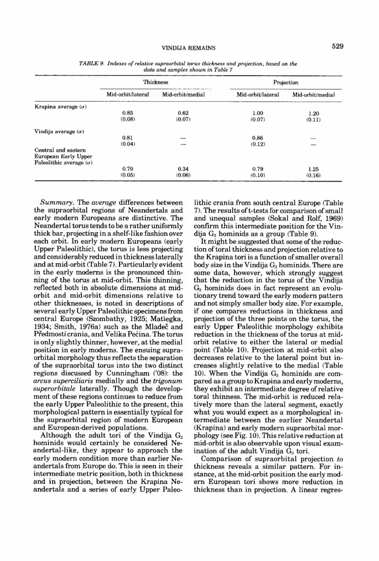
VINDIJA REMAINS 529
TABLE 9. Indexes of relative supraorbital torus thickness and projection, based on the data and samples shown in Table 7
Thickness Projection
Mid-orbit/lateral Mid-orbit/medial Mid-orbit'lateral Mid-orbitlmedial ~-
Krapina average (u) 0.85
(0.08)
Vindija average (0)
0.81 (0.04)
Central and eastern European Early Upper Paleolithic average (u)
0.70 (0.05)
0.62 (0.07)
- -
0.34 (0.06)
1.00 (0.07)
0.86 (0.12)
0.79 (0.10)
1.20 (0.11)
- -
1.25 (0.16)
Summary. The average differences between the supraorbital regions of Neandertals and early modern Europeans are distinctive. The Neandertal torus tends to be a rather uniformly thick bar, projecting in a shelf-like fashion over each orbit. In early modern Europeans (early Upper Paleolithic), the torus is less projecting and considerably reduced in thickness laterally and at mid-orbit (Table 7). Particularly evident in the early moderns is the pronounced thin- ning of the torus at mid-orbit. This thinning, reflected both in absolute dimensions at mid- orbit and mid-orbit dimensions relative to other thicknesses, is noted in descriptions of several early Upper Paleolithic specimens from central Europe (Szombathy, 1925; Matiegka, 1934; Smith, 1976a) such as the MladeE and PFedmosti crania, and Velika Pecina. The torus is only slightly thinner, however, at the medial position in early moderns. The ensuing supra- orbital morphology thus reflects the separation of the supraorbital torus into the two distinct regions discussed by Cunningham ('08): the arcus superciliaris medially and the trigonum superorbitale laterally. Though the develop- ment of these regions continues to reduce from the early Upper Paleolithic to the present, this morphological pattern is essentially typical for the supraorbital region of modern European and European-derived populations.
Although the adult tori of the Vindija G3 hominids would certainly be considered Ne- andertal-like, they appear to approach the early modern condition more than earlier Ne- andertals from Europe do. This is seen in their intermediate metric position, both in thickness and in projection, between the Krapina Ne- andertals and a series of early Upper Paleo-
lithic crania from south central Europe (Table 7). The results of t-tests for comparison of small and unequal samples (Sokal and Rolf, 1969) confirm this intermediate position for the Vin- dija G3 hominids as a group (Table 9).
It might be suggested that some of the reduc- tion of toral thickness and projection relative to the Krapina tori is a function of smaller overall body size in the Vindija G3 hominids. There are some data, however, which strongly suggest that the reduction in the torus of the Vindija G3 hominids does in fact represent an evolu- tionary trend toward the early modern pattern and not simply smaller body size. For example, if one compares reductions in thickness and projection of the three points on the torus, the early Upper Paleolithic morphology exhibits reduction in the thickness of the torus a t mid- orbit relative to either the lateral or medial point (Table 10). Projection at mid-orbit also decreases relative to the lateral point but in- creases slightly relative to the medial (Table 10). When the Vindija G3 hominids are com- pared as a group to Krapina and early moderns, they exhibit a n intermediate degree of relative toral thinness. The mid-orbit is reduced rela- tively more than the lateral segment, exactly what you would expect as a morphological in- termediate between the earlier Neandertal (Krapina) and early modern supraorbital mor- phology (see Fig. 10). This relative reduction a t mid-orbit is also observable upon visual exam- ination of the adult Vindija G3 tori.
Comparison of supraorbital projection to thickness reveals a similar pattern. For in- stance, at the mid-orbit position the early mod- ern European tori shows more reduction in thickness than in projection. A linear regres-

530 M.H. WOLPOFF ET AL.
I
sion fit for thickness and projection has a lower slope in the early modern sample when com- pared with Krapina. A corresponding slope cal- culated for the Vindija specimens is intermedi- ate, although closely approaching the early modern European value.
Interestingly, other aspects of the Vindija G3 hominid total morphological pattern suggest that reduced supraorbital tori might be ex- pected. Vi 224, for example, indicates that mid- facial prognathism may be reduced at Vindija relative to other Neandertals. Furthermore, at least two adult (Vi 261 and Vi 262) and two possibly juvenile (Vi 224 and Vi 279) frontals appear to possess more vertical frontal squamae than most Neandertals. Thus, based on either Endo's (1966) or Moss and Young's (1960) hypothesis to explain the presence and degree of development of a supraorbital torus, the Vindija G3 hominids might well be expected to exhibit reduced tori relative to other Nean- dertals, no matter what their body size.
Vindija is the only European site besides Krapina to preserve a large enough sample of Neandertal supraorbitals to estimate var- iability. In almost every case, the Vindija coef- ficients of variation exceed those at Krapina for browridge projections but are somewhat less for thicknesses. The same pattern appears in a comparison of Vindija for the averages of the 11 adult western Neandertals with supraorbitals. Vindija is more variable in projections and less so in thicknesses. This pattern of variability change represents another area in which the Vindija sample is intermediate between earlier Neandertals and the early modern European specimens. This more recent sample is even more variable than Vindija in supraorbital projections and less variable in thicknesses.
Finally, the Vindija tori represent the lat- est Neandertal supraorbital sample from anywhere in Europe. We believe it is signifi- cant that there is evidence for evolutionary change (contra Trinkaus and Howells, '791, and that the evolutionary trends they reveal, when compared with the earlier Neandertal sample from the same region, suggests change in the direction of early modern European popu- lations. The fact that the SBla frontal exhibits a parallel pattern of supraorbital thinning may not be coincidental, although it is unclear whether this better supports a grade or re- gional clade interpretation of the variation. Yet, we believe it is necessary to restate the fact that these data suggest an evolutionary trend within a morphological complex that must be regarded as Neandertal.
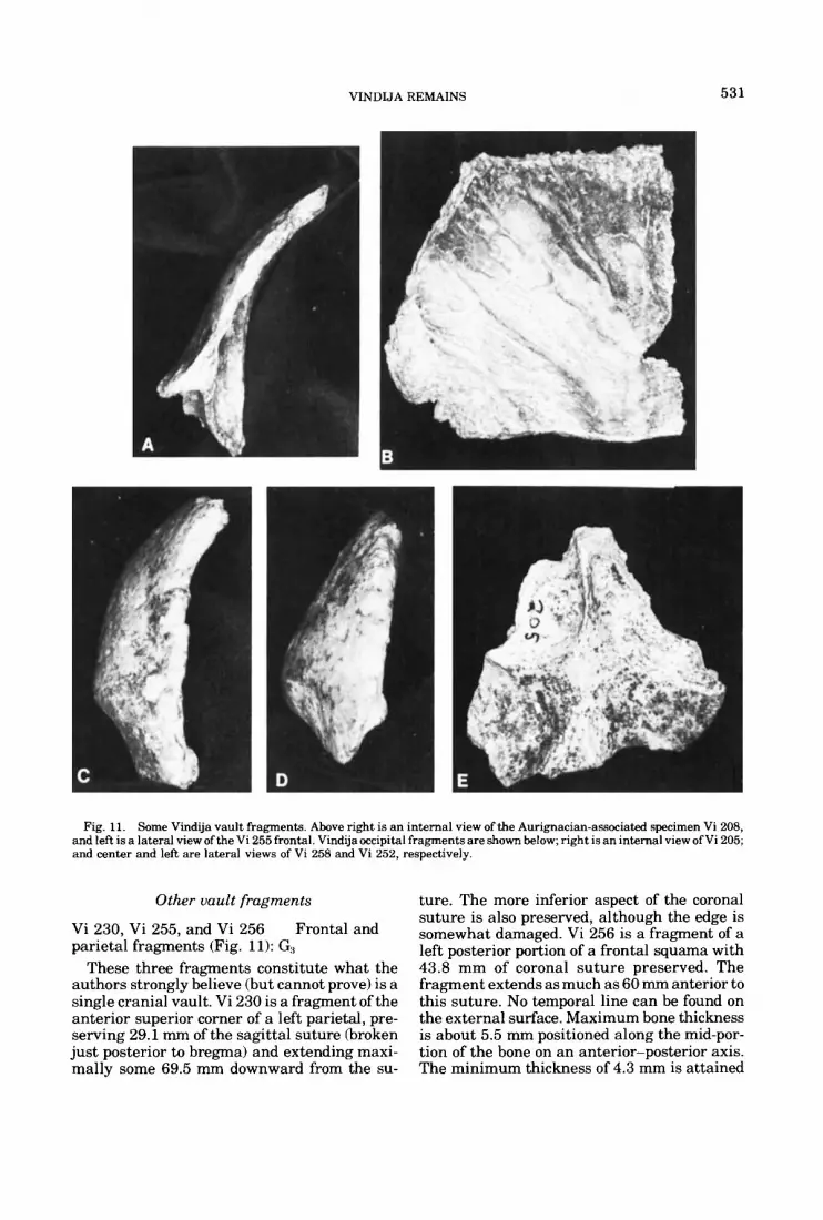
VINDIJA REMAINS 531
Fig. 11. Some Vindija vault fragments. Above right is an internal view of the Aurignacian-associated specimen Vi 208, and left is a lateral view of the Vi 255 frontal. Vindija occipital fragments are shown below; right is an internal view ofVi 205; and center and left are lateral views of Vi 258 and Vi 252, respectively.
Other vault fragments
Vi 230, Vi 255, and Vi 256 parietal fragments (Fig. 11): G:,
These three fragments constitute what the authors strongly believe (but cannot prove) is a single cranial vault. Vi 230 is a fragment of the anterior superior corner of a left parietal, pre- serving 29.1 mm of the sagittal suture (broken just posterior to bregma) and extending maxi- mally some 69.5 mm downward from the su-
Frontal and
ture. The more inferior aspect of the coronal suture is also preserved, although the edge is somewhat damaged. Vi 256 is a fragment of a left posterior portion of a frontal squama with 43.8 mm of coronal suture preserved. The fragment extends as much as 60 mm anterior to this suture. No temporal line can be found on the external surface. Maximum bone thickness is about 5.5 mm positioned along the mid-por- tion of the bone on an anterior-posterior axis. The minimum thickness of 4.3 mm is attained
532 M.H. WOLPOFF ET AL.
at the anterior-medial corner. The match with Vi 230 is possible along part of the coronal suture, although the damage on Vi 230 makes an exact fit impossible to establish. Finally, Vi 255 is a large diamond-shaped portion of cen- trally located frontal squama exposing the pos- terior wall of a large (breadth = 15.6 mm) cen- tral lobe and several lateral lobes of frontal sinus on each side. Except for behind glabella, there is a moderately expressed supraorbital sulcus, and the forehead bulges visibly above it. The anterior-posterior length of the exposed sinus is a t least 24.5 mm behind the broken anterior edge. The sinus appears to have been better developed on the left side.
The frontal is thin compared with the Krapina average (Table 111, although the dif- ference is less than a standard deviation and the value lies within the Krapina range. Parietal thickness is virtually identical with the Krapina average. The condition of the su- tures suggests that the individual was fairly young, almost certainly a young adult. A lack of marked coronal curvature in the preserved parietal indicates that the vault was probably fairly large. Internally, the parietal preserves impressions of the frontoparietal branch of the middle meningeal artery. There is no trace of either Breschet’s sinus or Pacchioni’s fossa.
Vi 264 Frontal fragment This fragment preserves the anterior mid-
portion of the frontal squama, extending later- ally to about the mid-portion of the left side. No sutural borders remain. Anteriorly, the most posterior border of the frontal sinus is pre- served. The sagittal sulcus remaining just su- perior to this is very shallow.
Vi 280 Posterior fragment of frontal squama: G:3
This fragment preserves bregma and extends for 63.3 mm anterior to it without any trace of crista frontalis endocranially. The maximum transverse breadth is 45 mm. On the posterior border 23.4 mm of coronal suture remain on the left side and 10 mm remain on the right. The bone is fairly thin; thickness varies from 3.6 mm a t bregma to 6.8 mm at the most anterior point. While the fragment is curved over its full anterior-posterior length, the curvature in- creases over the anterior half. Condition of the coronal suture indicates that the specimen probably was not especially young.
Vi 204 Posterior superior right parietal fragment
This fragment is roughly rectangular in shape, with 63 mm of the sagittal suture and
51.5 mm of the lambdoidal border preserved (the suture is broken away from the most infe- rior 15.5 mm of the border). The lambdoidal region bears no evidence of flattening, and the parietal is evenly curved in the sagittal plane. The suture itself appears to be unfused both internally and on the more complex external surface. The transverse curvature is also even but is rather marked, suggesting that the parietal breadth may not have been great. No parietal foramen is present.
The contour of the bone is not evenly rounded as seen from the rear; instead, the side is fairly gabled, with a pronounced angle between the superior and inferior portions, Rounding is the common, but not invariant, morphology for Neandertals.
No temporal line is visible on the external surface. On the posterior inferior corner there is an approximately 17 mm square area with outer bone table loss on the external surface (at the corner itself the bone thins to 4 mm). Ob- served microscopically, the external bone sur- face in this region is irregular, giving the ap- pearance of randomly distributed low swells. In cross section, all of the bone bordering this re- gion is compact. The internal surface appears undisturbed. Terminal portions of the grooves formed by the middle meningeal artery are present.
Parietal thicknesses differ little from the Krapina mean values (Table 11). The central dimensions are slightly under the Krapina mean, and the posterior dimensions are slightly above it.
Vi 208 left parietal (Fig. 11): G,
This fragment is roughly pentagonal (the posterior surface has an angle), with 65.7 mm of the coronal suture and 44.7 mm of the sagittal suture preserved and intersecting at bregma. The sutures appear to have been unfused, and it is unlikely that the specimen was an adult. There is little sagittal or transverse curvature to the preserved fragment, which might suggest a rather broad vault. Vault thickness decreases markedly along the sagittal suture, varying from 6.8 mm at bregma to 5.0 mm at the most posterior preserved point. Generally the vault thicknesses are moderate compared with other Vindija specimens (Table 111, but they are not significantly below the Krapina mean values. No trace of the temporal line is visible,
Internally, the fronto-parietal branch of the middle meningeal artery shows a distinct stem just posterior (10 mm) to the coronal suture a t the fragment’s base. Breschet’s sinus appears
Anterior superior fragment of

VINDIJA REMAINS 533
as a broad shallow sulcus (5.7 mm), which pro- ceeds vertically from this point, ending in a shallow pit (Pacchioni’s fossa) 21.5 mm inferior to the sagittal suture. Bregmatic and obelionic branches extend superiorly and posteriorly from this sulcus, and a second larger branch of Breschet’s sinus 8.3 mm in breadth also pro- ceeds in this direction and terminates in an even larger fossa, which is centered 10.6 mm inferior to the sagittal suture and 34 mm poste- rior to the coronal suture.
Vi 251 Left parietal The specimen is a small fragment (40 x 35
mm), which is difficult to identify with cer- tainty. The presence of two shallow internal pits, the relative positions of minimum and maximum thickness, and the curvature sug- gest that it is a fragment of left parietal very near the anterior-superior corner. There is no sutural preservation on its edges. Thickness varies from 5 mm to 6.4 mm.
Vi 257 Left parietal fragment This fragment is mainly composed of the
mastoid angle of a left parietal, preserving 35 mm of lambdoidal suture and 37.7 mm of the beveled squamosal border. The fragment dis- tinctly thickens anteriorly, varying from 7.3 mm on the most superior point preserved on the lambdoidal suture to 8.1 mm on the most ante- rior point preserved, about 10.5 mm superior to the squamosal border. A t the corner of the mas- toid angle, the bone is thinner than the Krapina parietals, lying below the range and one standard deviation below the Krapina mean (Table 11). Although the temporal line should appear on this fragment, its position cannot be ascertained.
Internally, grooves representing sporadic terminal branches of the middle meningeal ar- tery are preserved.
Vi 263 Right parietal fragment The specimen consists of a 64.7 mm long
portion of the right squamosal border of a pari- etal, extending to the anterior corner of the bone, where 16.7 mm ofthe squamosal suture is preserved. The maximum height above the squamosal suture is 41 mm. The thickness along the squamosal border is almost identical to Vi 257, and it is possible that the same indi- vidual is represented. Parietal thickness a t the approximate position of the middle eminence is the smallest at Vindija (Table 11) and is 2 stan- dard deviations below the Krapina mean. The bevel for the temporal squama does not extend far up the parietal, reaching 10.8 mm above the inferior border at the maximum.
On the internal surface, a very marked ex- pression of Breschet’s sinus is centered 11 mm posterior to the coronal suture, paralleling it for the preserved extent of the bone. The sinus breadth is 6.8 mm, and its maximum depth is 1.5 mm. It is identical in form to the expressions noted for Vi 208 and Vi 293.
Vi 285 Left parietal fragment: G3 The fragment is roughly rectangular, pre-
serving 38.9 mm of coronal suture from about the midpoint of its length, and extending poste- riorly for a maximum of 41.3 mm. Vault thick- ness ranges from 3.8 mm at the most superior point on the coronal suture to 5.7 mm at its most inferior point. The thinness of the vault and the condition of the coronal suture suggest the specimen is juvenile. Endocranially, a t least three arterial impressions remain. There is a very shallow indistinct anterior sulcus par- alleling the lower portion of the coronal suture. Its posterior edge is 8.7 mm behind the suture.
Vi 293 Left parietal fragment This specimen is roughly triangular in shape
and preserves the lower anterior corner of a thick parietal, broken anteriorly just short of the coronal suture. Basal length and height of the specimen are 68.3 mm and 69.7 mm, re- spectively. The inferior border has a 29 mm length of beveled edge for the temporal. The height of this bevel is 12.3 mm.
Externally a distinct temporal line is present 56 mm above the base of the temporal bevel. In the transverse plane, the curvature of this fragment is rounded evenly, lacking the more angled appearance of a modern parietal. Here there is no distinction between the top and sides of the vault. Thickness varies from 9.1 mm at the apex to 5.1 mm at the posterior inferior corner. The maximum breadth is at the approx- imate position of the anterior eminence. It is markedly greater than the other Vindija spec- imens, above the Krapina range, and 2.7 stan- dard deviations above the mean (Table 11). Thick parietals are characteristic of much later hominids from Vindija. Two Gravettian-asso- ciated specimens from layer D, Vi 210 and Vi 220, measure 7.5 and 9.6 mm in thickness a t the anterior eminence.
Internally, the arterial impressions are in- distinct. However, a broad (6.8 mm), deep (1.4 mm) Breschet’s sinus extends along the full length of the preserved anterior margin.
Vi 205 This fragment is centrally located and
roughly triangular, reaching from about 18 mm superior to inion (the apex) to no more than
Occipital fragment (Fig. 11)

534
3
B .3
.3 a
M.H. WOLPOFF ET AL.
-! t-
3
r-:
-! h
4

VINDIJA REMAINS 535
9 3 r(
c? m
gqc? m m l n
9 s a! m
9 0 9 9 3
$14 r3 i 3
2
-! W
ON091 m w t - t -
9 9 lnln
? ? m m
b
536 M.H. WOLPOFF ET AL.
41 mm inferior to it (the base). The maximum transverse breadth is 65.6 mm. No distinct prominence marks the position of inion. The point was defined at the intersection of the mid- line with the low nuchal torus. The fragment has very little curvature, either sagitally or transversely. Above the torus appears a supra- iniac depression, a small concavely depressed area that is characteristic of some Neandertal (and other) occiputs. The external bone table in this depression exhibits a surface pitting char- acteristic for similar depressions in the Krapina occipitals. There is a wide but very shallow sulcus below the low torus. In all, the region is quite similar to known Neandertal occiputs.
The inferior nuchal line is 22.5 mm below inion. The angle a t inion formed by chords to this line and to the maximum superior exten- sion of the fragment is 160". While this is not analogous to the occipital angle, it can be dupli- cated on both Neandertal and more modern crania. Not enough of the occipital plane is pre- served to determine whether the occipit was bunned. However, from the angulation and lack of marked curvature in the sagittal plane, it is clear that the position of inion lay well below opistocranion. Compared to Krapina specimens, the nuchal plane of Vi 205 exhibits moderate rugosity in the area for insertion of the nuchal musculature.
Internally, the sagittal and transverse crest intersection (endinion) forms a cross, and there is little apparent asymmetry of the cerebral fossae (the left may be slightly larger). The separate ridges of the inferior cruciate emi- nence are well divided by the base of the frag- ment (they are 14 mm apart a t the break), sug- gesting that the break is not far short of the foramen magnum posterior border. If so, the nuchal plane of the specimen was short (probably no longer than 46 mm). The superior branch of the cruciate eminence is very promi- nent. The position of endinion is some 12 mm inferior to inion.
Bone thickness a t inion and endinion is marked (Table 11). In both cases the Vindija values fall above the Krapina range, about 2 standard deviations from the mean. However, the marked thickness is more a reflection of the prominence of the cruciate eminence than of an unusual cortical bone development. The minimum thickness of the occiput is 1.8 mm.
Vi 252 Right occipital fragment (Fig. 11)
This fragment represents the superior right corner of an occiput, beginning at asterion and extending along the lambdoidal suture for 52.7
mm. Medially the fragment reaches the ap- proximate midline position. Eroded remnants of the sagittal sinus are present, and the top of the transverse sinus is visible along the frag- ment's base. Externally, the nuchal line dips inferiorly as it extends medially from asterion. While there is no nuchal torus on the fragment, it shows a marked angulation at the nuchal ridge as seen in sagittal profile. Below the nu- chal ridge, a marked sulcus parallels it and almost reaches asterion. Above the nuchal ridge is a roughened, shallow concave area (su- prainiac depression) a t least 23.7 mm in height and extending 22.5 mm laterally from the ap- parent midline. The nuchal ridge and trans- verse sinus reach the lambdoidal suture at the same point, which appears to represent asteri- on. If the break approximates the midline posi- tion, the occiput was rather narrow. Position- ing the fragment on a cast of La Ferrassie 1 leaves the sagittal sulcus border far short of the cast's midline.
The orientation of the nuchal line and the fact that inion appears to be in a low position because of the marked inferior dip at the (ap- proximate) midline suggests that the specimen is female. A feature supporting this suggestion is the lack of marked thickening at asterion (Table 11).
Vi 258 The specimen, like Vi 252, also represents a
right occiput in the vicinity of asterion, al- though less of the bone remains (the maximum height and breadth dimensions of the fragment are 38 mm x 41 mm). The morphological de- tails preserved are similar to those of Vi 252, but the individuals are far from identical.
The fragment is thicker (Table 11) at aste- rion, and the difference is even greater medi- ally. The nuchal line is in a higher position, relative to the preserved comparable land- marks. For instance, no transverse sinus is preserved, showing that the nuchal line and the sinus do not reach the lambdoidal suture at the same position. Moreover, parallel orienta- tion of the lambdoidal sutures of Vi 258 and Vi 252 shows that, as it extends toward the mid- line, the nuchal line of Vi 258 curves superiorly prior to its less marked inferior curvature. The sulcus inferior to the nuchal line does not seem as pronounced in Vi 258. Enough remains to show that Vi 258 does not represent the same individual as Vi 205.
Right occipital fragment (Fig. 11)
Vi 281 The specimen consist of a fairly flat nuchal
plane fragment including asterion, 17.6 mm of
Right occipital fragment: G.3
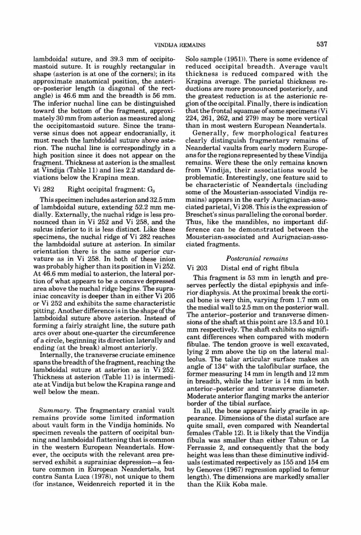
VINDIJA REMAINS 537
lambdoidal suture, and 39.3 mm of occipito- mastoid suture. It is roughly rectangular in shape (asterion is a t one of the corners); in its approximate anatomical position, the anteri- or-posterior length (a diagonal of the rect- angle) is 46.6 mm and the breadth is 56 mm. The inferior nuchal line can be distinguished toward the bottom of the fragment, approxi- mately 30 mm from asterion as measured along the occipitomastoid suture. Since the trans- verse sinus does not appear endocranially, it must reach the lambdoidal suture above aste- rion. The nuchal line is correspondingly in a high position since it does not appear on the fragment. Thickness a t asterion is the smallest a t Vindija (Table 11) and lies 2.2 standard de- viations below the Krapina mean.
Vi 282 This specimen includes asterion and 32.5 mm
of lambdoidal suture, extending 52.2 mm me- dially. Externally, the nuchal ridge is less pro- nounced than in Vi 252 and Vi 258, and the sulcus inferior to it is less distinct. Like these specimens, the nuchal ridge of Vi 282 reaches the lambdoidal suture a t asterion. In similar orientation there is the same superior cur- vature as in Vi 258. In both of these inion was probably higher than its position in Vi 252. At 46.6 mm medial to asterion, the lateral por- tion of what appears to be a concave depressed area above the nuchal ridge begins. The supra- iniac concavity is deeper than in either Vi 205 or Vi 252 and exhibits the same characteristic pitting. Another difference is in the shape of the lambdoidal suture above asterion. Instead of forming a fairly straight line, the suture path arcs over about one-quarter the circumference of a circle, beginning its direction laterally and ending (at the break) almost anteriorly.
Internally, the transverse cruciate eminence spans the breadth of the fragment, reaching the lambdoidal suture at asterion as in Vi 252. Thickness at asterion (Table 11) is intermedi- ate a t Vindija but below the Krapina range and well below the mean.
Right occipital fragment: G3
Summary. The fragmentary cranial vault remains provide some limited information about vault form in the Vindija hominids. No specimen reveals the pattern of occipital bun- ning and lambdoidal flattening that is common in the western European Neandertals. How- ever, the occiputs with the relevant area pre- served exhibit a suprainiac depression-a fea- ture common in European Neandertals, but contra Santa Luca (1978), not unique to them (for instance, Weidenreich reported it in the
Solo sample (1951)). There is some evidence of reduced occipital breadth. Average vault thickness is reduced compared with the Krapina average. The parietal thickness re- ductions are more pronounced posteriorly, and the greatest reduction is a t the asterionic re- gion of the occipital. Finally, there is indication that the frontal squamae of some specimens (Vi 224, 261, 262, and 279) may be more vertical than in most western European Neandertals.
Generally, few morphological features clearly distinguish fragmentary remains of Neandertal vaults from early modern Europe- ans for the regions represented by these Vindija remains. Were these the only remains known from Vindija, their associations would be problematic. Interestingly, one feature said to be characteristic of Neandertals (including some of the Mousterian-associated Vindija re- mains) appears in the early Aurignacian-asso- ciated parietal, Vi 208. This is the expression of Breschet's sinus paralleling the coronal border. Thus, like the mandibles, no important dif- ference can be demonstrated between the Mousterian-associated and Aurignacian-asso- ciated fragments.
Postcranial remains Vi 203 Distal end of right fibula
This fragment is 53 mm in length and pre- serves perfectly the distal epiphysis and infe- rior diaphysis. At the proximal break the corti- cal bone is very thin, varying from 1.7 mm on the medial wall to 2.5 mm on the posterior wall. The anterior-posterior and transverse dimen- sions of the shaft at this point are 13.5 and 10.1 mm respectively. The shaft exhibits no signifi- cant differences when compared with modern fibulae. The tendon groove is well excavated, lying 2 mm above the tip on the lateral mal- leolus. The talar articular surface makes an angle of 134" with the talofibular surface, the former measuring 14 mm in length and 12 mm in breadth, while the latter is 14 mm in both anterior-posterior and transverse diameter. Moderate anterior flanging marks the anterior border of the tibia1 surface.
In all, the bone appears fairly gracile in ap- pearance. Dimensions of the distal surface are quite small, even compared with Neandertal females (Table 12). It is likely that the Vindija fibula was smaller than either Tabun or La Ferrassie 2, and consequently that the body height was less than these diminutive individ- uals (estimated respectively as 155 and 154 cm by Genoves (1967) regression applied to femur length). The dimensions are markedly smaller than the Kiik Koba male.
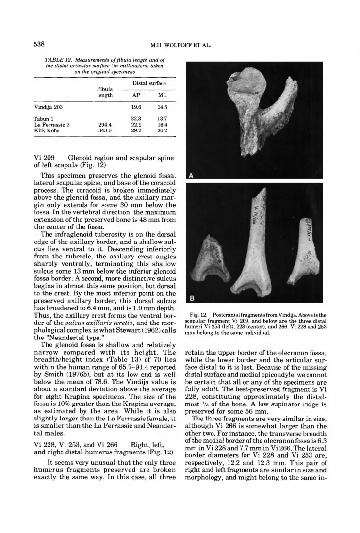
538 M.H. WOLPOFF ET A L
TABLE 12. Measurements of fibula length and of the distal articular surface (in millimeters) taken
on the originnl specimens
Distal surface Fibula length AP ML
Vindija 203 19.6 14.5
Tabun 1 22.3 13.7 La Ferrassie 2 294.4 22.1 16.4 Kiik Koba 343.0 29.2 20.2
Vi 209 Glenoid region and scapular spine of left scapula (Fig. 12)
This specimen preserves the glenoid fossa, lateral scapular spine, and base of the coracoid process. The coracoid is broken immediately above the glenoid fossa, and the axillary mar- gin only extends for some 30 mm below the fossa. In the vertebral direction, the maximum extension of the preserved bone is 48 nun from the center of the fossa.
The infraglenoid tuberosity is on the dorsal edge of the axillary border, and a shallow sul- cus lies ventral to it. Descending inferiorly from the tubercle, the axillary crest angles sharply ventrally, terminating this shallow sulcus some 13 mm below the inferior glenoid fossa border. A second, more distinctive sulcus begins in almost this same position, but dorsal to the crest. By the most inferior point on the preserved axillary border, this dorsal sulcus has broadened to 6.4 mm, and is 1.9 mm depth. Thus, the axillary crest forms the ventral bor- der of the sulcus axillaris teretis, and the mor- phological complex is what Stewart (1962) calls the “Neandertal type.”
The glenoid fossa is shallow and relatively narrow compared with its height. The breadth/height index (Table 13) of 70 lies within the human range of 65.7-91.4 reported by Smith (1976b), but a t its low end is well below the mean of 78.6. The Vindija value is about a standard deviation above the average for eight Krapina specimens. The size of the fossa is 10% greater than the Krapina average, as estimated by the area. While it is also slightly larger than the La Ferrassie female, it is smaller than the La Ferrassie and Neander- tal males.
Vi 228, Vi 253, and Vi 266 and right distal humerus fragments (Fig. 12)
It seems very unusual that the only three humerus fragments preserved are broken exactly the same way. In this case, all three
Right, left,
Fig. 12. Postcranial fragments from Vindija. Above is the scapular fragment Vi 209, and below are the three distal humeri Vi 253 (left), 228 (center), and 266. Vi 228 and 253 may belong to the same individual.
retain the upper border of the olecranon fossa, while the lower border and the articular sur- face distal to it is lost. Because of the missing distal surface and medial epicondyle, we cannot be certain that all or any of the specimens are fully adult. The best-preserved fragment is Vi 228, constituting approximately the distal- most % of the bone. A low supinator ridge is preserved for some 56 mm.
The three fragments are very similar in size, although Vi 266 is somewhat larger than the other two. For instance, the transverse breadth of the medial border of the olecranon fossa is 6.3 mm in Vi 228 and 7.7 mm in Vi 266. The lateral border diameters for Vi 228 and Vi 253 are, respectively, 12.2 and 12.3 mm. This pair of right and left fragments are similar in size and morphology, and might belong to the same in-
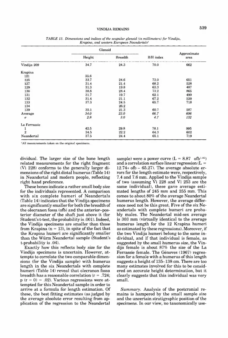
VINDIJA REMAINS 539
TABLE 13. Dimensions and indices of the scapular glenoid (in millimeters) for Vindija, Krapina, and western European Neandertals'
Glenoid Approximate
Height Breadth BIH index area
Vindija 209
Krapina 121 125 127 129 130 131 132 133 134 139
Average U
La Ferrassie 1 2
Neandertal
34.7
35.6 33.7 31.4 31.3 38.8 31.7 31.4 37.3
35.1 34.0 2.8
42.5 34.5 37.5
24.3
24.6 21.4 19.8 28.4 19.7 21.1 24.5 26.2 21.3 23.0
3.0
29.8 22.2 24.4
70.0
73.0 68.2 63.3 73.2 62.1 67.2 65.7
60.7 66.7
4.7
70.1 64.3 65.1
662
651 528 487 865 490 520 718
587 606 132
995 602 719
'All measurements taken on the original specimens.
dividual. The larger size of the bone length related measurements for the right fragment (Vi 228) conforms to the generally larger di- mensions of the right distal humerus (Table 14) in Neandertal and modern people, reflecting right hand preference.
These bones indicate a rather small body size for the individuals represented. A comparison with six complete humeri of Neandertals (Table 14) indicates that the Vindija specimens are significantly smaller for both the breadth of the olecranon fossa (ofb) and the anterior-pos- terior diameter of the shaft just above it (for Student's t-test, the probability is .001). Indeed, the Vindija specimens are smaller than those from Krapina (n = 131, in spite of the fact that the Krapina humeri are significantly smaller than the Wiirm Neandertal sample (Student's t-probability is .04).
Exactly how this reflects body size for the Vindija specimens is uncertain. However, at- tempts to correlate the two comparable dimen- sions (for the Vindija sample) with humerus length in the six Neandertals with complete humeri (Table 14) reveal that olecranon fossa breadth has a reasonable correlation (r = ,724; p (r = 0) = .02). Various regressions were at- tempted for this Neandertal sample in order to arrive a t a formula for length estimation. Of these, the best fitting estimates (as judged by the average absolute error resulting from ap- plication of the regression to the Neandertal
sample) were a power curve (L = 8.87 o f b l Y and a correlation surface linear regression (L = 12.74-x ofb - 65.27). The average absolute er- rors for the length estimate were, respectively, 7.4 and 7.6 mm. Applied to the Vindija sample of two (assuming Vi 228 and Vi 253 are the same individual), these gave average esti- mated lengths of 245 mm and 255 mm. This comes to about 80% of the average Neandertal humerus length. However, the average differ- ence need not be this great. Five of the six Ne- andertals with complete humeri are proba- bly males. The Neandertal mid-sex average is 303 mm (virtually identical to the average humerus length for the 12 Krapina humeri as estimated by these regressions). Moreover, if the two Vindija humeri belong to the same in- dividual, and if that individual is female, as suggested by the small humerus size, the Vin- dija female is about 87% the size of the La Ferrassie female. The Gnoves (1967) regres- sion for a female with a humerus of this length suggests a height of 135-138 cm. There are too many estimates involved for this to be consid- ered an accurate height determination, but it clearly suggests that this individual was very small.
Summary. Analysis of the postcranial re- mains is hampered by the small sample size and the uncertain stratigraphic position of the specimens. In our view, no taxonomically use-
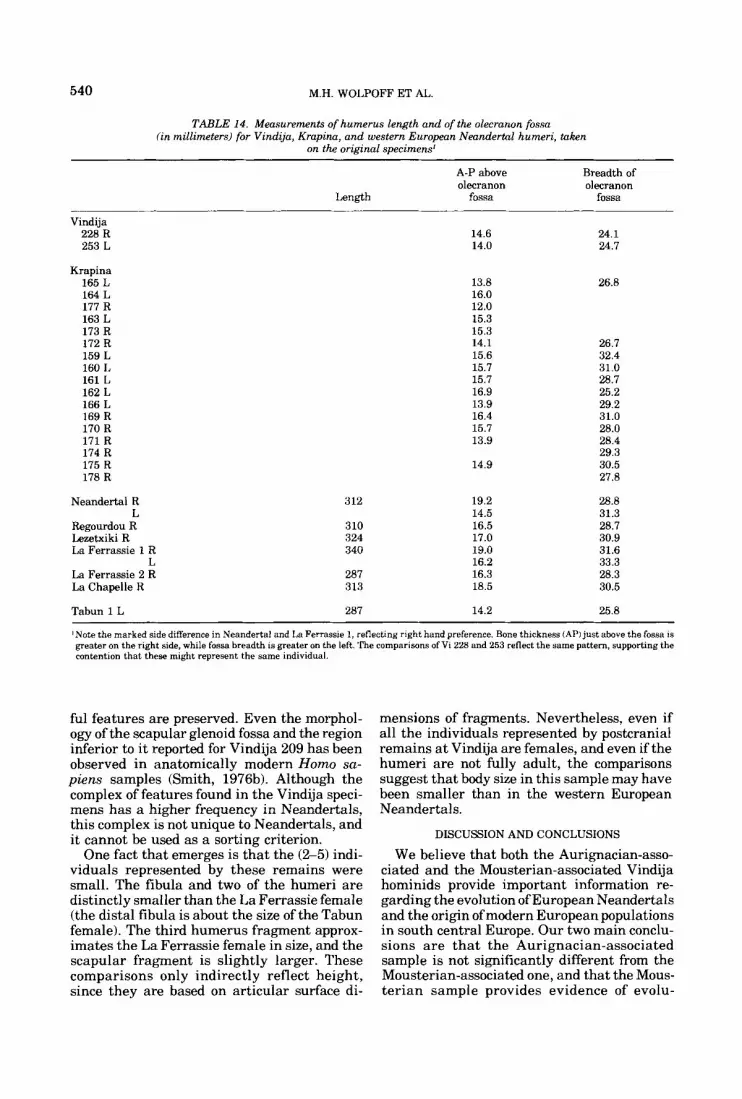
540 M.H. WOLPOFF ET AL
TABLE 14. Measurements of humerus length and of the olecranon fossa (in millimeters) for Vindija, Krapina, and western European Neandertal humeri, taken
on the original specimens'
A-P above Breadth of olecranon olecranon
Length fossa fossa
Vindija 228 R 253 L
KraDina 165 L 164 L 177 R 163 L 173 R 172 R 159 L 160 L 161 L 162 L 166 L 169 R 170 R 171 R 174 R 175 R 178 R
Neandertal R L
Regourdou R Lezetxiki R La Ferrassie 1 R
L La Ferrassie 2 R La Chapelle R
Tabun 1 L
312
310 324 340
287 313
287
14.6 14.0
13.8 16.0 12.0 15.3 15.3 14.1 15.6 15.7 15.7 16.9 13.9 16.4 15.7 13.9
14.9
19.2 14.5 16.5 17.0 19.0 16.2 16.3 18.5
14.2
24.1 24.7
26.8
26.7 32.4 31.0 28.7 25.2 29.2 31.0 28.0 28.4 29.3 30.5 27.8
28.8 31.3 28.7 30.9 31.6 33.3 28.3 30.5
25.8 ~~~ ~ ~
'Note the marked side difference in Neandertal and La Ferrassie 1, reflecting right hand preference. Bone thickness (AP) just above the fossa is greater on the right side, while fossa breadth is greater on the left. The comparisons of Vi 228 and 253 reflect the same pattern, supporting the contention that these might represent the same individual.
ful features are preserved. Even the morphol- ogy of the scapular glenoid fossa and the region inferior to it reported for Vindija 209 has been observed in anatomically modern Homo sa- piens samples (Smith, 1976b). Although the complex of features found in the Vindija speci- mens has a higher frequency in Neandertals, this complex is not unique to Neandertals, and it cannot be used as a sorting criterion.
One fact that emerges is that the (%5) indi- viduals represented by these remains were small. The fibula and two of the humeri are distinctly smaller than the La Ferrassie female (the distal fibula is about the size of the Tabun female). The third humerus fragment approx- imates the La Ferrassie female in size, and the scapular fragment is slightly larger. These comparisons only indirectly reflect height, since they are based on articular surface di-
mensions of fragments. Nevertheless, even if all the individuals represented by postcranial remains at Vindija are females, and even if the humeri are not fully adult, the comparisons suggest that body size in this sample may have been smaller than in the western European Neandertals.
DISCUSSION AND CONCLUSIONS
We believe that both the Aurignacian-asso- ciated and the Mousterian-associated Vindija hominids provide important information re- garding the evolution of European Neandertals and the origin of modern European populations in south central Europe. Our two main conclu- sions are that the Aurignacian-associated sample is not significantly different from the Mousterian-associated one, and that the Mous- terian sample provides evidence of evolu-
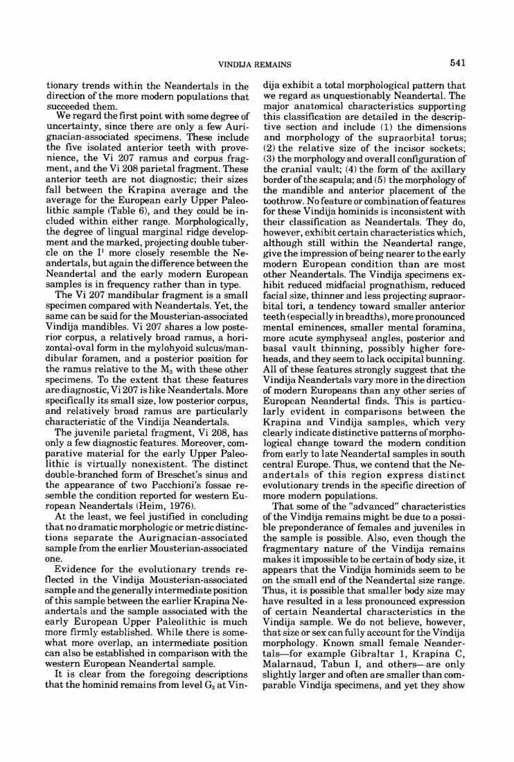
VINDIJA REMAINS 541
tionary trends within the Neandertals in the direction of the more modern populations that succeeded them.
We regard the first point with some degree of uncertainty, since there are only a few Auri- gnacian-associated specimens. These include the five isolated anterior teeth with prove- nience, the Vi 207 ramus and corpus frag- ment, and the Vi 208 parietal fragment. These anterior teeth are not diagnostic; their sizes fall between the Krapina average and the average for the European early Upper Paleo- lithic sample (Table 6), and they could be in- cluded within either range. Morphologically, the degree of lingual marginal ridge develop- ment and the marked, projecting double tuber- cle on the I’ more closely resemble the Ne- andertals, but again the difference between the Neandertal and the early modern European samples is in frequency rather than in type.
The Vi 207 mandibular fragment is a small specimen compared with Neandertals. Yet, the same can be said for the Mousterian-associated Vindija mandibles. Vi 207 shares a low poste- rior corpus, a relatively broad ramus, a hori- zontal-oval form in the mylohyoid sulcus/man- dibular foramen, and a posterior position for the ramus relative to the M3 with these other specimens. To the extent that these features are diagnostic, Vi 207 is like Neandertals. More specifically its small size, low posterior corpus, and relatively broad ramus are particularly characteristic of the Vindija Neandertals.
The juvenile parietal fragment, Vi 208, has only a few diagnostic features. Moreover, com- parative material for the early Upper Paleo- lithic is virtually nonexistent. The distinct double-branched form of Breschet’s sinus and the appearance of two Pacchioni’s fossae re- semble the condition reported for western Eu- ropean Neandertals (Heim, 1976).
At the least, we feel justified in concluding that no dramatic morphologic or metric distinc- tions separate the Aurignacian-associated sample from the earlier Mousterian-associated one.
Evidence for the evolutionary trends re- flected in the Vindija Mousterian-associated sample and the generally intermediate position of this sample between the earlier Krapina Ne- andertals and the sample associated with the early European Upper Paleolithic is much more firmly established. While there is some- what more overlap, an intermediate position can also be established in comparison with the western European Neandertal sample.
It is clear from the foregoing descriptions that the hominid remains from level G3 at Vin-
dija exhibit a total morphological pattern that we regard as unquestionably Neandertal. The major anatomical characteristics supporting this classification are detailed in the descrip- tive section and include (1) the dimensions and morphology of the supraorbital torus; (2) the relative size of the incisor sockets; (3) the morphology and overall configuration of the cranial vault; (4) the form of the axillary border of the scapula; and (5) the morphology of the mandible and anterior placement of the toothrow. No feature or combination of features for these Vindija hominids is inconsistent with their classification as Neandertals. They do, however, exhibit certain characteristics which, although still within the Neandertal range, give the impression of being nearer to the early modern European condition than are most other Neandertals. The Vindija specimens ex- hibit reduced midfacial prognathism, reduced facial size, thinner and less projecting supraor- bital tori, a tendency toward smaller anterior teeth (especially in breadths), more pronounced mental eminences, smaller mental foramina, more acute symphyseal angles, posterior and basal vault thinning, possibly higher fore- heads, and they seem to lack occipital bunning. All of these features strongly suggest that the Vindija Neandertals vary more in the direction of modern Europeans than any other series of European Neandertal finds. This is particu- larly evident in comparisons between the Krapina and Vindija samples, which very clearly indicate distinctive patterns of morpho- logical change toward the modern condition from early to late Neandertal samples in south central Europe. Thus, we contend that the Ne- andertals of this region express distinct evolutionary trends in the specific direction of more modern populations.
That some of the “advanced characteristics of the Vindija remains might be due to a possi- ble preponderance of females and juveniles in the sample is possible. Also, even though the fragmentary nature of the Vindija remains makes it impossible to be certain of body size, it appears that the Vindija hominids seem to be on the small end of the Neandertal size range. Thus, it is possible that smaller body size may have resulted in a less pronounced expression of certain Neandertal characteristics in the Vindija sample. We do not believe, however, that size or sex can fully account for the Vindija morphology. Known small female Neander- tals-for example Gibraltar 1, Krapina C , Malarnaud, Tabun I, and others-are only slightly larger and often are smaller than com- parable Vindija specimens, and yet they show

542 M.H. WOLPOFF ET AL.
no more indication of the early modern condi- tion than do the larger Neandertals. For exam- ple, the index of thickness between the lateral and mid-orbital points on the supraorbital torus of Gibraltar 1 is 0.86, which is above the Krapina mean and above the Vindija G:, mean of 0.81 (Table 3). Similarly, the Malarnaud mandible, one of the smallest western Euro- pean Neandertal mandibles, has one of the highest symphyseal angles (100’) in the entire sample. None of the faces of skeletally small Neandertals (such as La Ferrassie 2 or Tabun I) have as narrow a nose or as small a lower face as the Vindija maxillae would indicate, though the Vindija specimens match them in certain other dimensions. Based on these examples, it appears unlikely that the modern-like aspects of the Vindija G3 hominids can be purely attri- buted to allometric or other effects of size andlor sexual dimorphism. Furthermore, the reduced supraorbital torus, reduced facial size and prognathism, emergence of the mental emi- nence and of a more vertical symphysis, smaller anterior teeth, and alteration of the vault architecturemake good sense as an evolv- ing complex relating to reducing functional stress in the facial region. Thus, at this time, the most reasonable explanation for these characteristics in the Vindija hominids is sim- ply that they reflect an evolutionary trend in the direction of modern Europeans, when com- pared with other known European Neandertal samples. We believe this contradicts the claim of a “relatively constant physical pattern” for the European Neandertals made recently by Trinkaus and Howells (1979).
Such a n evolutionary trend might not be un- expected given the probable chronological posi- tion of the Vindija G3 sample, the late portion of the lower Wurm stadia1 as defined by Musil and Valoch (’66), if one accepts the proposal that the late Neandertals of south central Europe were in the process of evolving into the early Upper Paleolithic-associated populations that fol- lowed them in time, such as those represented by Mladei, Brno, and PFedmosti. The generally archaic if not specifically Neandertal-like mor- phology common in these Upper Paleolithic samples does not argue against this proposal.
The alternative explanation for the observa- ble sequence in south central Europe would regard the evolutionary trends as reflecting the influx of more modern populations from “somewhere in the east.” This hypothesis has probably been best expressed recently by Trinkaus and Howells (19791, and Howells (1974, 1975), and has been consistently sup- ported by Vandermeersch (1977). Theoreti-
cally, we agree t h a t peristent geneflow probably could not be distinguished from evolutionary change due to selection. However, we are troubled by the former explanation for several reasons, not the least of which is the absence of archaeological data suggesting the influx of populations from other regions during this timespan (Valoch, 19681, which is required by the migration model presented by Howells (1976).
In proposing that geneflow did not play an important role in the immediate origin of mod- ern European populations, we do not wish to discount or discredit the importance of migra- tion in human evolution (again contra Howells, 1976). The idea that this portion of the Euro- pean sequence can be explained by the action of selection on existing populations does not re- sul t from a n “antimigration assumption.” Rather, it rests on archaeological and morpho- logical grounds, and acts as a specific explana- tion for the case discussed. Howells proposes that the European Neandertals should be re- garded as a test case in the general conflict between a migrational and local evolutionary explanation of Modern Homo sapiens origins. We believe this proposal is more eurocentric than is necessary and point to the evidence for regional continuity between archaic and mod- ern Homo sapiens in other areas as supplemen- tary support for our analysis of the European situation. The fact is that recognizable se- quences in Sub-saharan Africa (Omo Kibish/ Laetolil + Border CaveD’lorisbad), North Af- rica (Jebel Irhoud + Dar-eg-Soltane -+
AfalouISinga), Mainland China (Choukoutein + Mapa/Dali + LiujiangNpper Cave), and Australasia (Sangiran Homo erectus -+ Sam- bungmachen + Solo + Kow SwampNungo) show parallel, distinct developments reflecting local continuity and extend well before the ap- pearance of modernHomo sapiens populations. A general migratory scheme must involve re- gions other than Europe and is required to ac- count for the migrants taking on attributes of the local archaic sapiens populations they re- place in a variety of different places. We find such a model to be unacceptable as a general explanation for the worldwide appearance of modern Homo sapiens populations; although it might apply to some local regional sequences, in our view, south central Europe is not one of these. Moreover, because of the geographic po- sition of this region within Europe, we question whether migration was an important factor in the evolutionary changes taking place any- where in Europe at this time (50,00@30,000 years ago).
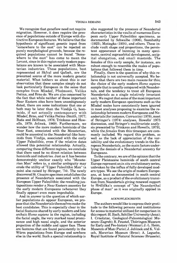
VINDIJA REMAINS 543
We recognize that geneflow need not require migration. However, it does require the pres- ence of populations outside of Europe with dis- tinctive European features. We believe that the hypothesis of significant genetic input from “somewhere to the east” can be rejected on purely morphological grounds, because the re- quired populations cannot be found. “Some- where to the east” has come to mean the Levant, since in this region early modern popu- lations are known to be associated with Mous- terian industries. These populations, best represented a t Skhul and Qafzeh, are the presumed source of the more modern genetic material. What bothers us about this is our observation that these samples simply do not look particularly European in the sense that samples from MladeE, Pledmosti, Velika Pecina, and Brno do. We are also concerned by the fact that while neither of these important Near Eastern sites have been unambiguously dated, there are some indications that one or both may be later than the Vindija G3 hom- inids, if not actually contemporary with Mladet, Brno, and Velika Pecina (Smith, 1977; Bada and Helfman, 1976; Trinkaus and How- ells, 1979; Jelinek, 1969). Furthermore, we wonder how the more modern hominids of the Near East, associated with the Mousterian, could be ancestral to the Neandertal-like hom- inids from Vindija, associated with the early Upper Paleolithic, even if the relative dates allowed this potential relationship. Actually, comparing these different regions, we conclude that there need be no direct relation between hominids and industries. Just as it has become demonstrably unclear exactly who “Mouste- rian Man” refers to, a similar ambiguity may erode the utility of “Upper Paleolithic Man” (a point also raised by Stringer, ’74). The newly discovered St. Cesaire specimen establishes the presence of Neandertals associated with the European Upper Paleolithic; the resulting jux- tapositions render a Near Eastern ancestry for the early modern Europeans (whenever they finally appear) even more improbable.
And in answer to the question of which ear- lier populations do appear European, we pro- pose that the Neandertals themselves make the best candidates. This is especially clear in the facial features shared by early modern and late archaic Homo sapiens in the region, including the facial angle, the very marked nasal promi- nence and high nasal angle, and the anterior projection of the middle and upper face. These are features that are found persistently in the Wiirm populations from Europe and nowhere else in the world. Such a special relationship is
also suggested by the presence of Neandertal characteristics in the vaults of numerous Euro- pean early Upper Paleolithic specimens, as documented by Schwalbe (19061, Szombathy (19251, Matiegka (19341, and others. These in- clude vault shape and proportions, the persis- tent appearance of bunning in many speci- mens, central supraorbital development, occip- ital elongation, and small mastoids. The females of this early sample, for instance, are robust enough to resemble the males of popu- lations that followed them in time.
Finally, there is the question of why this re- lationship is not universally accepted. We be- lieve that there are two main reasons for this; the choice of the early modern Homo sapiens sample that is usually compared with Neander- tals, and the tendency to treat all European Neandertals as a single relatively invariant unit. We regret that some of the most important early modern European specimens such as the Mladec‘ males have consistently been ignored in most analyses proposing to examine the re- lationship between modern Europeans and Ne- andertals [for instance, Corruccini (19761, most of Stringer’s (1974) analyses, Howells’ 1975 discussion, and Stringer’s multivariate analy- sis presented by Trinkaus and Howells, 19791, while the females from this timespan are com- monly included. We regard this problem, as well as the lack of appreciation of the var- iability and the evolutionary trends in the Eu- ropean Neandertals, as the main factors under- lying the denials of a Neandertal ancestry for Europeans.
To the contrary, we are of the opinion that the Upper Pleistocene hominids of south central Europe represent an in situ evolutionary series, unbroken by the influx of fully developed mod- ern types. We see the origin of modern Europe- ans, a t least as documented in south central Europe, as a product of the evolutionary transi- tion from Neandertals giving renewed support t o Hrdlic‘ka’s concept of “the Neanderthal phase of man” as it was originally applied in Europe.
ACKNOWLEDGMENTS
The authors would like to express their grati- tude to the following persons and institutions for access to material utilized for comparison in this report: H. Bach, Schiller University (Jena); I. Crnolatac, Geological-Paleontological Mu- seum (Zagreb); R. Feustel, Thiiringen Museum for Early and Pre-history (Weimar); J.-L. Heim, Museum of Man (Paris); J . Jeliinek and K. Val- och, Moravian Museum (Brno); A. Leguebe, Royal Institute of Natural Sciences (Brussels);
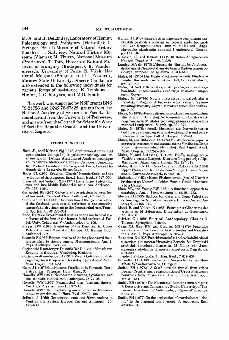
544 M.H. WOLPOFF ET A L
M.-A. and H. DeLumley, Laboratory of Human Paleontology and Prehistory (Marseille); C. Stringer, British Museum of Natural History (London); J. Szilvassy, Natural History Mu- seum (Vienna); M. Thurzo, National Museum (Bratislava); T. Toth, Historical-Natural Mu- seum of Hungary (Budapest); B. Vander- meersch, University of Paris; E. VlEek, Na- tional Museum (Prague); and U. Yakamov, Moscow State University. Sincere thanks are also extended to the following individuals for various forms of assistance: E. Trinkaus, J. Hinton, G.C. Ranyard, and M.O. Smith.
This work was supported by NSF grants BNS 75-21756 and BNS 76-87829, grants from the National Academy of Sciences, a Faculty Re- search grant from the University of Tennessee, and grants from the Council for Scientific Work of Socialist Republic Croatia, and the Univer- sity of Zagreb.
LITERATURE CITED
Bada, JL, and Helfman, PM (1976) Application ofamino acid racemization datings + in paleoanthropology and ar-
en Prehistoire: Methods et Limites. (Colloque I. Union Int. Sci. Prehist. Protohist., 9th Congress). Paris, Centre Na- tionale de la Recherche Scientifique.
Brace, CL (1979) Krapina, “Classic” Neanderthals, and the evolution of the European face. J. Hum. Evol., 8:527-550.
Brose, DS and Wolpoff, MH (1971) Early Upper Paleolithic man and late Middle Paleolithic tools. Am. Anthropol., 73: 115G1194.
Corruccini, RS (1976) Calvarial shape relations between fos- sil hominids. Yearbook Phys. Anthropol., 18:8%109.
Cunningham, DJ (1908) The evolution of the eyebrow region of the forehead, with special reference to the excessive supraorbital development in the Neanderthal race. Trans. R. Soc. Edinb., 46:28&311.
Endo, B (1966) Experimental studies on the mechanical sig- nificance of the form of the human facial skeleton. J. Fae. Sci. Univ. Tokyo, sec. V (Anthropol.), 3:l-106.
Frayer, DW (1978) Evolution of the Dentition in Upper Paleolithic and Mesolithic Europe. U. Kansas Publ. Anthropol., 10.
Genoves, S (1967) Proportionality of the long bones and their relationship to stature among Mesoamericans. Am. J. Phys. Anthropol., 26t67-78.
Gojanovic-Kramberger, D (1906) Der Diluviale Mensch von Krapina in Kroatien. Wiesbaden Kreidels.
Gorjanovic-Kramberger, D (1913) h o t i kultura diluvijal- noga Eovjeka iz Krapine ur Hrvatskoj. Djela Jugosl. Akad. Znan. Umjetn., 23:l-54.
Heim, J-L (1976) Les Hommes Fossiles de la Ferrassie. Tome I. Arch. Inst. Paleontol. Hum. Mem., 35.
Howells, WW (1974) Neanderthals: names, hypotheses, and the scientific method. Am. Anthropol., 76:24-38.
Howells, WW (1975) Neanderthal man: facts and figures. Yearbook Phys. Anthropol., 18:7-16.
Howells, WW (1976) Explaining modern man: evolutionists versus migrationists. J. Hum. Evol., 5:477-496.
Jelinek, J (1969) Neanderthal man and Homo sapiens in Central and Eastern Eurooe. Current Anthroool.. 10:
chaeology. In: Dations Q bsolutes e t Analyses Isotopique
Kallay, J (1970) Komparativne napomene o &ljustima kra- pinskih praljudi s obzirom na poloiaj medu homonid- ima. In: Krapina: 1899-1969 M. Malez (ed), Jugo- slavenska akademija znanosti i umjetnosti, Zagreb. pp. 153-164.
Klaatsch, H, and Hauser, 0 (1910) Homo Aurignacensis Hauseri. Praehist. Z., 1:273-338.
Lumley, MA de (1973) L’Homme de 1’Hortus. In: Anthean- dertaliens et Neindertaliens du bassin MaiterranBen oc- cidental europeen. Et. Quatern., 2t311-550.
Malez, M (1975) Die Hohle Vindija-ine neue Fundstelle fossiler Hominiden in Kroatien. Bull, Sci. (Yugoslavie), 20:139-140.
Malez, M (ed) (1978a) Krapinski prarovjek i evolucija hominida. Jugoslavenska akademija znanosti i umjet- nosti, Zagreb.
Malez, M (1978b) Novija instraiivanja paleolitika u Hrvatskom Zagoju. Arheolozka istraiivanja u Sjevero- zapadnoj Hrvatskoj, Zagreb, Hrvatsko arheolozko druztvo. pp. 9-69.
Malez, M (1978~) Populacije neandertalaca i neandertalcima sli&& ljudi u Hrvatskoj. In: Krapinski prarovjek i e vol- ucija hominida. M. Malez, (ed). Jugoslavenska akad-emija znanosti i umjetnosti, Zagreb. pp. 331-371.
Malez, M (1978d) Fossile Menschen aus Nonvestkroatien und ihre quarargeologische, palaontologische und palao- lithische Grundlage. Coll. Anthropol., 2t29-41.
Malez, M, and Rukavina, D (1975) Krioturbacijske pojave u gornjopleistocenskim naslagama pecine Vindije kcd Donje VoCe u sjeverozapadnoj Hrvatskoj. Rad Jugosl. Akad. Znan. Umjetn., 371:245265.
Malez, M, and Rukavina, D (1979) Poloiaj naslaga spilje Vindije u sustau Elanjenja Kvartara Fireg podurzja Alpa. Rad Jugosl. Akad. Znan. Umjetn. 383:187-218.
Malez, M, Smith, FH, RadovZic, J, and Rukavina, D (1980) Upper Pleistocene hominids from Vindija, Croatia, Yugo- slavia. Current Anthropol., 21:36%367.
Matiegka, J (1934) Homo PFedmostensis. Fosilni Clovek z Eedmosti na Morav;. I. Lebky. Prague, Eeska Akademie V&l a Urn%.
Moss, ML, and Young, RW (1960) A functional approach to craniology. Am. J. Phys. Anthropol., 18:281-292.
Movius, H (1960) Radiocarbon dates and Upper Paleolithic archaeology in Central and Western Europe. Current An- thropol., 1.355391.
M u d , R, and Valoch, K (1966) Beitrag zur Gliedemng des Wurms in Mitteleuropa. Eiszeitalter u. Gegenwart, 17:131-38.
Olivier, G (1969) Practical Anthropology. Charles C. Thomas, Springfield, Illinois.
Oyen, OJ, Rice, RW, and Cannon, MS (1979) Browridge structure and function in extant primates and Neander- thals. Am. J. Phys. Anthropol., 51:83-96.
Rukavina, D (1978) PaleoklimatoloEki i paleoekolozki odnosi u gornjem pleistocenu Hrvatskog Zagorja. In: Krapinski proEovjek i evolucija hominida. M. Malez (ed). Jugo- slavenska adademija zhanosti i urnjetnosti. Zagreb. pp. 319330. anderthal-like fossils. J. Hum. Evol., 7:619-636.
Schwalbe, G (1906) Studien zur Vorgeschichte des Men- schen. Schweizerbartsche, Stuttgart.
Smith, FH (1976a) A fossil hominid frontal from velika Pecina (Croatia) and a consideration of Upper Pleistocene hominids from Yugoslavia. Am. J. Phys. Anthropol., 44:127-134.
Smith, FH (1976b) The Neandertal Remains from Krapina: A Descriptive and Comparative Study. University of Ten- nessee Department of Anthropology, Report of Investiga- tions, 15.
Smith, FH (1977) On the application of morphological “dat- ing‘‘ to the hominid fossil record. J. Anthropol. Res., . ,
475503. 33t302-316.
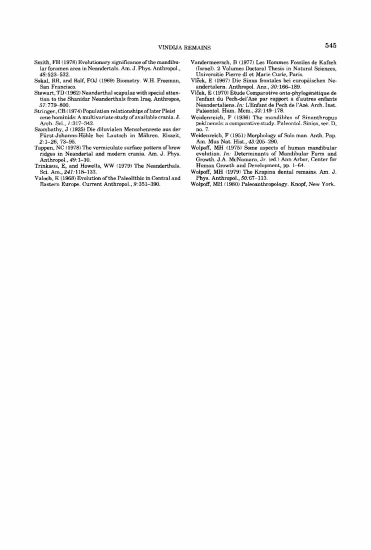
VINDIJA REMAINS 545
Smith, FH (1978) Evolutionary significance of the mandibu- lar foramen area in Neandertals. Am. J. Phys. Anthropol., 48:52%532.
Sokal, RR, and Rolf, FOJ (1969) Biometry. W.H. Freeman, San Francisco.
Stewart, TD (1962) Neanderthal scapulae with special atten- tion to the Shanidar Neanderthals from Iraq. Anthropos, 57:77%800.
cene hominids: A multivariate study of available crania. J. Arch. Sci., 2:317-342.
Szombathy, J (1925) Die diluvialen Menschenreste aus der FiirstJohanns-Hohle bei Lautsch in Mahren. Eiszeit, 2:l-26, 7%95.
Tappen, NC (1978) The vermiculate surface pattern of brow ridges in Neandertal and modern crania. Am. J. Phys. Anthropol., 49: 1- 10.
Trinkaus, E, and Howells, WW (1979) The Neanderthals. Sci. Am., 241:11%133.
Valoch, K (1968) Evolution of the Paleolithic in Central and
Stringer, CB (1974) Population relationships of later Pleisl
Eastern Europe. Current Anthropol., 9:351-390.
Vandermeersch, B (1977) Les Hommes Fossiles de Kafzeh (Israel). 2 Volumes Doctoral Thesis in Natural Sciences, Universitie Pierre dl et Marie Curie, Paris.
Vlbk, E (1967) Die Sinus frontales bei europaischen Ne- andertalern. Anthropol. Anz., 30: 16G189.
VlZek, E (1970) Etude Comparative onto-phylogenetique de I'enfant du Pech-del'Aze par rapport a dautres enfants Neandertaliens. In: L'Enfant de Pech de 1'&. Arch. Inst. Palbntol. Hum. Mem., 33:14%178.
Weidenreich, F (1936) The mandibles of Sinanthropus pekinensis: a comparative study. Paleontol. Sinica, ser. D, no. 7.
Weidenreich, F (1951) Morphology of Solo man. Anth. Pap. Am. Mus Nat. Hist., 43:205-290.
Wolpoff, MH (1975) Some aspects of human mandibular evolution. In: Determinants of Mandibular Form and Growth. J.A. McNamara, J r . (ed.) Ann Arbor, Center for Human Growth and Development, pp. 1-64.
Wolpoff, MH (1979) The Krapina dental remains. Am. J. Phys. Anthropol., 50:67-113.
Wolpoff, MH (1980) Paleoanthropology. Knopf, New York.
Related Documents