Atherosclerosis 191 (2007) 11–21 Unsaturated fatty acids suppress the expression of the ATP-binding cassette transporter G1 (ABCG1) and ABCA1 genes via an LXR/RXR responsive element Yoshinari Uehara a,b,∗ , Shin-ichiro Miura b , Arnold von Eckardstein c , Satomi Abe b , Akihiro Fujii a , Yoshino Matsuo b , Stephan Rust d , Stefan Lorkowski d , Gerd Assmann d , Tatsuo Yamada a , Keijiro Saku b a Department of Neurology and Healthcare, Fukuoka University Hospital, 7-45-1 Nanakuma, Jonan-ku, 814-0180 Fukuoka, Japan b Department of Cardiology, Fukuoka University School of Medicine, 7-45-1 Nanakuma, Jonan-ku, 814-0180 Fukuoka, Japan c Institute of Clinical Chemistry, University Hospital Z¨ urich, R¨ amistrasse 100, CH-8091 Z¨ urich, Switzerland d Institute of Arteriosclerosis Research,University of M¨ unster, Domagkstrasse 3, D-48149 M¨ unster, Germany Received 6 May 2005; received in revised form 11 April 2006; accepted 13 April 2006 Available online 30 May 2006 Abstract ATP-binding cassette transporters (ABC) G1 and ABCA1 are membrane cholesterol transporters and have been implicated to mediate cholesterol efflux from cells in the presence of high density lipoproteins and its major protein constituent apolipoprotein A-I, respectively. We previously demonstrated that unsaturated fatty acids suppress the stimulatory effects of oxysterols and retinoids on ABCA1 gene transcription. We here demonstrate that unsaturated fatty acids significantly suppress the stimulatory effects of oxysterols and retinoids on the expression of ABCG1 mRNA and protein and the activity of the wild-type human ABCG1 promoter as well as ABCA1. Mutation or deletion of the DR4 element within the ABCG1 or ABCA1 promoters, to which the transcriptional inducers LXR and RXR bind, abolished the suppressive effects of unsaturated fatty acids. Our observations provide the first evidence that unsaturated fatty acids suppress ABCG1 gene expression by a mechanism which involves the binding of LXR/RXR to the promoters. Suppression of both the ABCA1 and ABCG1 genes may indicate that unsaturated fatty acids suppress not only cholesterol efflux to apoA-I and thereby nascent HDL formation but also HDL-dependent cholesterol efflux from vascular cells. © 2006 Elsevier Ireland Ltd. All rights reserved. Keywords: ATP-binding cassette transporter; Cholesterol transport; Diabetes; Unsaturated fatty acid; Highdensity lipoprotein; LXR; RXR; Direct repeat 4 Abbreviations: ABC, ATP-binding cassette transporter; apo, apolipopro- tein; ATP, adenosine triphosphate; BSA, bovine serum albumin; CETP, cholesterol ester transfer protein; CYP7A, 7-hydroxylase; DR4, direct repeat 4; EPA, eicosapentaenoic acid; LA, linoleic acid; AraA, arachidonic acid; OA, oleic acid; PA, palmitic acid; SA, stearic acid; HDL, high density lipoprotein; LXR, liver-X-receptor alpha; LXR, liver-X-receptor beta; RXR, retinoid-X-receptor alpha; USFA, unsaturated fatty acid; TG, triglyc- eride; 9-cis-RA, 9-cis retinoic acid; 22R-HC, 22(R)-hydroxycholesterol ∗ Corresponding author. Tel.: +81 92 801 1011x3525; fax: +81 92 865 7900. E-mail address: [email protected] (Y. Uehara). 1. Introduction Dyslipidemia, notably hypertriglyceridemia and low plasma HDL cholesterolemia, is a common phenomenon in diabetes mellitus type 2 and insulin resistance and increases the risk of cardiovascular disease in these patients [1–3]. HDL exerts various potentially anti-atherogenic effects. For example, HDL particles transport cholesterol from cells in the arterial wall to the liver and to steroidogenic organs, in which cholesterol is used for the synthesis of bile acids, lipoproteins, vitamin D, and steroid hormones [2]. The details and the pathogenesis of low HDL cholesterol and the 0021-9150/$ – see front matter © 2006 Elsevier Ireland Ltd. All rights reserved. doi:10.1016/j.atherosclerosis.2006.04.018

Welcome message from author
This document is posted to help you gain knowledge. Please leave a comment to let me know what you think about it! Share it to your friends and learn new things together.
Transcript

A
cpWAeomue©
K
tcralRe
f
0d
Atherosclerosis 191 (2007) 11–21
Unsaturated fatty acids suppress the expression of the ATP-bindingcassette transporter G1 (ABCG1) and ABCA1 genes via an
LXR/RXR responsive element
Yoshinari Uehara a,b,∗, Shin-ichiro Miura b, Arnold von Eckardstein c, Satomi Abe b,Akihiro Fujii a, Yoshino Matsuo b, Stephan Rust d, Stefan Lorkowski d,
Gerd Assmann d, Tatsuo Yamada a, Keijiro Saku b
a Department of Neurology and Healthcare, Fukuoka University Hospital, 7-45-1 Nanakuma, Jonan-ku, 814-0180 Fukuoka, Japanb Department of Cardiology, Fukuoka University School of Medicine, 7-45-1 Nanakuma, Jonan-ku, 814-0180 Fukuoka, Japan
c Institute of Clinical Chemistry, University Hospital Zurich, Ramistrasse 100, CH-8091 Zurich, Switzerlandd Institute of Arteriosclerosis Research,University of Munster, Domagkstrasse 3, D-48149 Munster, Germany
Received 6 May 2005; received in revised form 11 April 2006; accepted 13 April 2006Available online 30 May 2006
bstract
ATP-binding cassette transporters (ABC) G1 and ABCA1 are membrane cholesterol transporters and have been implicated to mediateholesterol efflux from cells in the presence of high density lipoproteins and its major protein constituent apolipoprotein A-I, respectively. Wereviously demonstrated that unsaturated fatty acids suppress the stimulatory effects of oxysterols and retinoids on ABCA1 gene transcription.e here demonstrate that unsaturated fatty acids significantly suppress the stimulatory effects of oxysterols and retinoids on the expression ofBCG1 mRNA and protein and the activity of the wild-type human ABCG1 promoter as well as ABCA1. Mutation or deletion of the DR4
lement within the ABCG1 or ABCA1 promoters, to which the transcriptional inducers LXR and RXR bind, abolished the suppressive effectsf unsaturated fatty acids. Our observations provide the first evidence that unsaturated fatty acids suppress ABCG1 gene expression by a
echanism which involves the binding of LXR/RXR to the promoters. Suppression of both the ABCA1 and ABCG1 genes may indicate thatnsaturated fatty acids suppress not only cholesterol efflux to apoA-I and thereby nascent HDL formation but also HDL-dependent cholesterolfflux from vascular cells.
2006 Elsevier Ireland Ltd. All rights reserved.
s; Unsa
1
pdtHe
eywords: ATP-binding cassette transporter; Cholesterol transport; Diabete
Abbreviations: ABC, ATP-binding cassette transporter; apo, apolipopro-ein; ATP, adenosine triphosphate; BSA, bovine serum albumin; CETP,holesterol ester transfer protein; CYP7A, 7�-hydroxylase; DR4, directepeat 4; EPA, eicosapentaenoic acid; LA, linoleic acid; AraA, arachidoniccid; OA, oleic acid; PA, palmitic acid; SA, stearic acid; HDL, high densityipoprotein; LXR�, liver-X-receptor alpha; LXR�, liver-X-receptor beta;
XR�, retinoid-X-receptor alpha; USFA, unsaturated fatty acid; TG, triglyc-ride; 9-cis-RA, 9-cis retinoic acid; 22R-HC, 22(R)-hydroxycholesterol∗ Corresponding author. Tel.: +81 92 801 1011x3525;ax: +81 92 865 7900.E-mail address: [email protected] (Y. Uehara).
tild
021-9150/$ – see front matter © 2006 Elsevier Ireland Ltd. All rights reserved.oi:10.1016/j.atherosclerosis.2006.04.018
turated fatty acid; Highdensity lipoprotein; LXR; RXR; Direct repeat 4
. Introduction
Dyslipidemia, notably hypertriglyceridemia and lowlasma HDL cholesterolemia, is a common phenomenon iniabetes mellitus type 2 and insulin resistance and increaseshe risk of cardiovascular disease in these patients [1–3].DL exerts various potentially anti-atherogenic effects. For
xample, HDL particles transport cholesterol from cells in
he arterial wall to the liver and to steroidogenic organs,n which cholesterol is used for the synthesis of bile acids,ipoproteins, vitamin D, and steroid hormones [2]. Theetails and the pathogenesis of low HDL cholesterol and the
1 rosclero
ind[mgifc[
pllwhcefictpdsgt[
tbtbhotrc
Acreiutbhpct[
(awA
2
Paowef
RcfawmsatatoDtam
2
2
amArwrpa(
2
−haApAfg
2 Y. Uehara et al. / Athe
ncreased risk for atherosclerosis in insulin resistance areot well understood. Another typical abnormality in type IIiabetic patients is elevated plasma levels of free fatty acids4]. Free fatty acids play an important physiological role inany tissues, for example by inhibiting insulin-mediated
lucose uptake and glycogen synthesis. Furthermore, its well known that the dietary intake of polyunsaturatedatty acids exerts lipid-lowering effects on all lipoproteinlasses including LDL, VLDL (i.e. triglycerides) and HDL5,6].
The ATP-binding cassette transporter (ABC) A1 hasreviously been identified as a pivotal gene in the regu-ation of both plasma HDL cholesterol levels and cellu-ar cholesterol homeostasis, which is defective in patientsith Tangier disease [2,7–9]. In these patients and theireterozygous relatives, mutations in the ABCA1 geneause gene-dosage-dependent decreases in plasma lev-ls of HDL cholesterol and in the capacity of skinbroblasts and monocyte-derived macrophages to releaseholesterol in the extracellular presence of apolipopro-ein (apo) A-I or apoE [2,7–9]. Clinically, homozygousatients with Tangier disease accumulate macrophage-erived foam cells in various tissues. We previously demon-trated that unsaturated fatty acids downregulate ABCA1ene expression and inhibit apoA-I-induced cellular choles-erol efflux in cultivated macrophages and hepatocytes10].
ABCG1 (formerly termed ABC8) is another member ofhe ATP-binding cassette transporter superfamily which haseen mapped to chromosome 21q22.3 [11–16]. By contrasto ABCA1, which is a full ABC transporter with two ATPinding cassettes and transmembrane domains, ABCG1 is aalf transporter containing only one ATP binding cassette andne transmembrane domain [14,15]. It is therefore believedo need a dimeric partner for being active. Wang et al. recentlyeported ABCG1 contributes to HDL2- and HDL3-dependentellular cholesterol efflux [17].
Several ABC lipid transporters including ABCA1 andBCG1 have been shown to be strongly regulated by
holesterol-derived oxysterols. They activate nuclear liver Xeceptors (LXRs) LXR� and LXR� [18]. LXR� is mainlyxpressed in the liver, spleen, kidney, adipose tissue, smallntestine and macrophages [19], while LXR� is expressedbiquitously [20]. LXRs form obligate heterodimers withhe retinoid X receptor (RXR). The LXR/RXR heterodimerinds to lipogenic target gene promoters such as 7�-ydroxylase (CYP7A), apoE, cholesterol ester transferrotein (CETP), SREBP1c, ABCA1 and ABCG1, which allontain a direct repeat 4 (DR4) element in their promoter, i.e.wo direct hexameric repeats separated by four nucleotides19].
We demonstrate here that free unsaturated fatty acids
USFAs) strongly suppress the activity of both the ABCG1nd ABCA1 promoters, and elucidate the mechanism byhich USFAs suppress transcription of the ABCG1 andBCA1 genes.2
aa
sis 191 (2007) 11–21
. Materials and methods
Materials. 22(R)-hydroxycholesterol, 9-cis-retinoic acid,horbol 12-myristate 13-acetate (PMA), eicosapentaenoiccid (EPA), linoleic acid (LA), arachidonic acid (AraA),leic acid (OA), palmitic acid (PA), and stearic acid (SA)ere purchased from Sigma (St. Louis, MO). Fatty acid- and
ndotoxin-free bovine serum albumin (BSA) was purchasedrom Calbiochem.
Cell culture. Cells from the murine macrophage cell lineAW264 and THP-1 human monocyte (RIKEN, Japan) wereultured in DMEM or RPMI1640 medium containing 10%etal bovine serum (GIBCO-BRL) 100 units/ml penicillin Gnd 100 �g/ml streptomycin. The THP-1 cells were treatedith PMA for 72 h before experiment for differentiation toacrophage. For all experiments, cells were maintained in
erum-free medium which contained 10 g/l bovine serumlbumin (BSA) and was supplemented with or without addi-ives (22(R)-hydroxycholesterol, 9-cis-retinoic acid, or fattycids) at concentrations and time periods described in Sec-ion 3 and in the figure legends. A 100 mmol/l stock solutionf each fatty acid was prepared by diluting the fatty acid inMSO. Fatty acid was added from the stock solution bound
o 100 times volume of 10% BSA/medium solution and wasdjusted to 10 g/l of BSA at final concentration by addingedium.
.1. Reporter plasmid constructs
.1.1. Human ABCG1 promoter constructsFirefly luciferase reporter constructs were generated by
mplifying a 1141-bp fragment of the human ABCG1 pro-oter A (−1104/+37) and a 1330-bp fragment of the humanBCG1 promoter B (−1264/+66) with KpnI and XhoI
estriction sites out of genomic DNA by PCR amplificationith primer hABCG1 promoter A and hABCG1 promoter B,
espectively (primer sequences are given in Table 1). PCRroducts were digested with KpnI and XhoI, gel-purified,nd ligated to KpnI- and XhoI-digested pGL3 Basic vectorsPromega, Madison, WI).
.1.2. Truncated human ABCG1 promoter constructsThe deleted constructs, −607/+37, −530/+37, −413/+37,
303/+37, −233/+37, −163/+37 and −233/−122 of theuman ABCG1 promoter A upstream of exon 1 were gener-ted by PCR with hABCG1 promoter A-607, A-530, A-413,-303, A-233 or A-163 for the sense primer and hABCG1romoter A for the antisense primer, and hABCG1 promoter-233 for the sense primer and hABCG1 promoter A-122
or the antisense primer, respectively (primer sequences areiven in Table 1).
.1.3. Human ABCA1 promoter constructsFirefly luciferase reporter constructs were generated by
mplifying a 980-bp fragment of human ABCA1 promoters described previously [10].
Y. Uehara et al. / Atherosclerosis 191 (2007) 11–21 13
Table 1Sequences of PCR primers
Primer name Primer sequence
hABCG1 promoter A sense 5′-aag ggg tac cat gaa tga aag aag cca gac aca aa-3′hABCG1 promoter A-607 sense 5′-aag ggg tac ctt gtt acc agg ttt ctg cta agc tc-3′hABCG1 promoter A-530 sense 5′-aag ggg tac cgc tgc aat aat cat tgg cta gag g-3′hABCG1 promoter A-413 sense 5′-aag ggg tac ccc gta att gtt ttc aat gtg tgt t-3′hABCG1 promoter A-303 sense 5′-aag ggg tac ccc ttc tgt gga cag gta cta ggt g-3′hABCG1 promoter A-233 sense 5′-aag ggg tac cta acc ctg ctc act tcc tgt ttt c-3′hABCG1 promoter A-163 sense 5′-aag ggg tac ctc atg ctg att tta aaa caa tcc-3′hABCG1 promoter A antisense 5′-cct atc cgc tcg agc aca aac ata ggt agt cca gct gc-3′hABCG1 promoter A-122 antisense 5′-cct atc cgc tcg agg tgt aat gct aca ggg agg attg-3′hABCG1 promoter B sense 5′-aag ggg tac cgg atg act ctt gag aca aca cca tt-3′hABCG1 promoter B antisense 5′-cct atc cgc tcg agc ggt gcc gac cga gaa agc-3′hABCG1 DR4mut sense 5′-ggg agg gaa cag aac ccc cct tta gaa acc ctg ctc ac-3′hABCG1 DR4mut antisense 5′-gtg agc agg gtt tct aaa ggg ggg ttc tgt tcc ctc cc-3′hABCA1 DR4mut sense 5′-gag agc aca ggc ttt gtg tga tag tac tat ctg cgc tcg g-3′hABCA1 DR4mut antisense 5′-ccg agc gca gat agt act atc aca caa agc ctg tgc tct c-3′ABCG1 sense 5′-gga gaa tgc gaa gct gta cc-3′ABCG1 antisense 5′-gga ggc ggt ttt tac ctc tc-3′LXR� sense 5′-gat ctg gga tgt cca cga gt-3′LXR� antisense 5′-ccc ttc tca gtc tgc tcc ac-3′RXR� sense 5′-tcc ttc tcc cac cgc tcc atc-3′RGG
2
pb(sssamTTTwoAttoTtwtttoAc
2
g
fsowfoCLwaw(a(
2
2
uirtsrMwtt
XR� antisenseAPDH senseAPDH antisense
.1.4. Site-directed mutagenesisMutant plasmids were obtained for the human ABCG1
romoter A (1141 bp) and human ABCA1 promoter (980 bp)y using the QuickChange® Site-Directed Mutagenesis KitStratagene, La Jolla, CA) and each of the following double-tranded oligonucleotides: ABCG1; hABCG1 DR4mut sen-e, hABCG1 DR4mut antisense, ABCA1; hABCA1 DR4mutense, hABCA1 DR4mut antisense (the DNA sequencesre shown in Table 1). The following point mutations wereade at the DR4 site (underlined), ABCG1: TGCCCTTAG TAACCC → CCCCCT TTAG AAACCC; ABCA1:GACCG ATAG TAACCT → TGTGTG ATAG TACTAT.he ABCA1 promoter variant containing a mutated DR4 siteas obtained as described by Costet et al. [21]. Briefly, 25 ngf the wild-type human ABCG1 promoter A (1141 bp) andBCA1 promoter (980 bp) plasmids were used in a PCR reac-
ion containing 2.5 �l of Pfu polymerase buffer, 1.25 U Pfuurbo DNA polymerase, 10 mmol/l dNTPs, 62.5 ng of eachligonucleotide and water to give a reaction volume of 25 �l.he cycle setting for mutagenesis was selected according
o the manufacturer’s protocol, and the sample was treatedith 5 U of DpnI to cleave parental DNA and to improve
he efficiency of mutant plasmid screening. The reaction wasransferred to XL-1 Blue competent cells, and the transforma-ion mixture was plated on LB ampicillin plates. The identityf all DNA constructs was confirmed by DNA sequencing.ll plasmids were purified using Hi-speed Plasmid Midi kit
olumns (Qiagen).
.2. Reporter assay
For transfection, 50–80% confluent RAW 264 cells wererown on 24-well plates in DMEM supplemented with 10%
sfcl
5′-cag ctc cgt ctt gtc cat ctg-3′5′-aac gac ccc ttc att gac-3′5′-tcc acg aca tac tca gca c-3′
etal bovine serum, 100 units/ml penicillin G and 100 �g/mltreptomycin. Cells were transiently transfected with 1 �gf the firefly luciferase reporter plasmids and co-transfectedith 5 ng of phRL-SV40 vector (Promega, Madison, WI)
or the control Renilla luciferase by lipofection with 2 �lf LipofectAMINETM 2000 reagent (Invitrogen, Carlsbad,A) using 5 × 10⁵ cells per well. After incubation withipofectAMINETM 2000 for 6 h, the cells were incubatedith 22(R)-hydroxycholesterol, 9-cis-retinoic acid or fatty
cids for 6–24 h, as indicated. The luciferase activitiesere measured using the Dual-Luciferase Assay SystemTM
Promega). Firefly luciferase activity was divided by Renillactivity to obtain a normalized value: relative luciferase unitRLU).
.3. Quantitative real-time PCR assay
.3.1. RNA isolation and RT-PCRTotal RNA was extracted from cells in 9.6 cm2 dishes
sing the RNeasy kit (QIAGEN, Hilden, Germany) accord-ng to the manufacturer’s protocol, and treated with DNase toemove residual contaminating DNA. The RNA was reverse-ranscribed and cDNA was amplified by PCR using Super-cript II reverse transcriptase and Oligo-dT primer from Invit-ogen. The sequences of the primers are listed in Table 1.
ouse ABCG1, LXR�, RXR� and GAPDH gene expressionas quantified by real-time PCR using the Applied Biosys-
ems GeneAmp 5700 sequence detection system. Amplifica-ion was detected with SYBR Green as a fluorogenic probe
pecific for double-stranded DNA by using a qPCR-core kitor SYBR-Green (Eurogentec, Herstal, Belgium). Thresholdycle, Ct, which correlates inversely with the target mRNAevel, was measured as the cycle number at which the reporter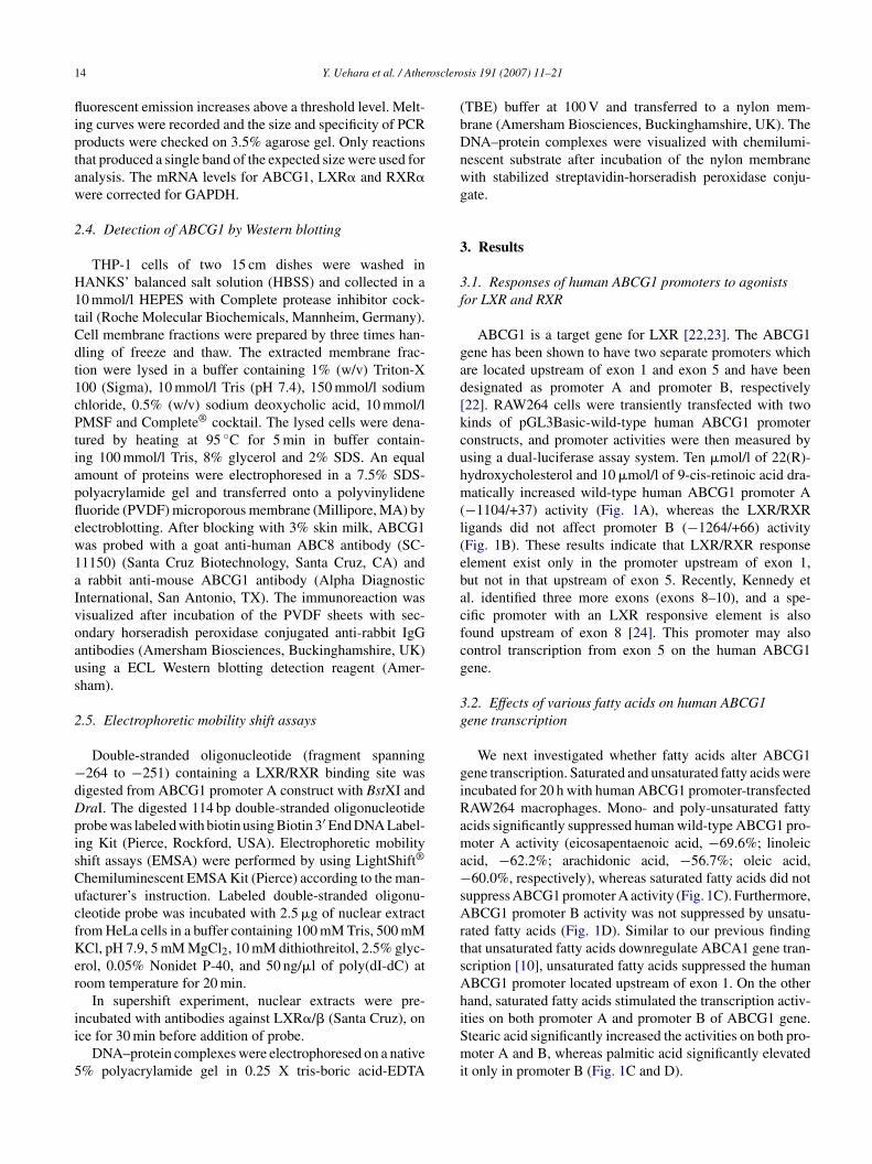
1 rosclero
fliptaw
2
H1tCdt1cPtiapflew1aIvoaus
2
−dDpisCucfKer
ii
5
(bDnwg
3
3f
gad[kcuhm(l(ebacfcg
3g
giRama−sArtsAh
4 Y. Uehara et al. / Athe
uorescent emission increases above a threshold level. Melt-ng curves were recorded and the size and specificity of PCRroducts were checked on 3.5% agarose gel. Only reactionshat produced a single band of the expected size were used fornalysis. The mRNA levels for ABCG1, LXR� and RXR�ere corrected for GAPDH.
.4. Detection of ABCG1 by Western blotting
THP-1 cells of two 15 cm dishes were washed inANKS’ balanced salt solution (HBSS) and collected in a0 mmol/l HEPES with Complete protease inhibitor cock-ail (Roche Molecular Biochemicals, Mannheim, Germany).ell membrane fractions were prepared by three times han-ling of freeze and thaw. The extracted membrane frac-ion were lysed in a buffer containing 1% (w/v) Triton-X00 (Sigma), 10 mmol/l Tris (pH 7.4), 150 mmol/l sodiumhloride, 0.5% (w/v) sodium deoxycholic acid, 10 mmol/lMSF and Complete® cocktail. The lysed cells were dena-
ured by heating at 95 ◦C for 5 min in buffer contain-ng 100 mmol/l Tris, 8% glycerol and 2% SDS. An equalmount of proteins were electrophoresed in a 7.5% SDS-olyacrylamide gel and transferred onto a polyvinylideneuoride (PVDF) microporous membrane (Millipore, MA) bylectroblotting. After blocking with 3% skin milk, ABCG1as probed with a goat anti-human ABC8 antibody (SC-1150) (Santa Cruz Biotechnology, Santa Cruz, CA) andrabbit anti-mouse ABCG1 antibody (Alpha Diagnostic
nternational, San Antonio, TX). The immunoreaction wasisualized after incubation of the PVDF sheets with sec-ndary horseradish peroxidase conjugated anti-rabbit IgGntibodies (Amersham Biosciences, Buckinghamshire, UK)sing a ECL Western blotting detection reagent (Amer-ham).
.5. Electrophoretic mobility shift assays
Double-stranded oligonucleotide (fragment spanning264 to −251) containing a LXR/RXR binding site was
igested from ABCG1 promoter A construct with BstXI andraI. The digested 114 bp double-stranded oligonucleotiderobe was labeled with biotin using Biotin 3′ End DNA Label-ng Kit (Pierce, Rockford, USA). Electrophoretic mobilityhift assays (EMSA) were performed by using LightShift®
hemiluminescent EMSA Kit (Pierce) according to the man-facturer’s instruction. Labeled double-stranded oligonu-leotide probe was incubated with 2.5 �g of nuclear extractrom HeLa cells in a buffer containing 100 mM Tris, 500 mMCl, pH 7.9, 5 mM MgCl2, 10 mM dithiothreitol, 2.5% glyc-
rol, 0.05% Nonidet P-40, and 50 ng/�l of poly(dI-dC) atoom temperature for 20 min.
In supershift experiment, nuclear extracts were pre-
ncubated with antibodies against LXR�/� (Santa Cruz), once for 30 min before addition of probe.DNA–protein complexes were electrophoresed on a native% polyacrylamide gel in 0.25 X tris-boric acid-EDTA
iSmi
sis 191 (2007) 11–21
TBE) buffer at 100 V and transferred to a nylon mem-rane (Amersham Biosciences, Buckinghamshire, UK). TheNA–protein complexes were visualized with chemilumi-escent substrate after incubation of the nylon membraneith stabilized streptavidin-horseradish peroxidase conju-ate.
. Results
.1. Responses of human ABCG1 promoters to agonistsor LXR and RXR
ABCG1 is a target gene for LXR [22,23]. The ABCG1ene has been shown to have two separate promoters whichre located upstream of exon 1 and exon 5 and have beenesignated as promoter A and promoter B, respectively22]. RAW264 cells were transiently transfected with twoinds of pGL3Basic-wild-type human ABCG1 promoteronstructs, and promoter activities were then measured bysing a dual-luciferase assay system. Ten �mol/l of 22(R)-ydroxycholesterol and 10 �mol/l of 9-cis-retinoic acid dra-atically increased wild-type human ABCG1 promoter A
−1104/+37) activity (Fig. 1A), whereas the LXR/RXRigands did not affect promoter B (−1264/+66) activityFig. 1B). These results indicate that LXR/RXR responselement exist only in the promoter upstream of exon 1,ut not in that upstream of exon 5. Recently, Kennedy etl. identified three more exons (exons 8–10), and a spe-ific promoter with an LXR responsive element is alsoound upstream of exon 8 [24]. This promoter may alsoontrol transcription from exon 5 on the human ABCG1ene.
.2. Effects of various fatty acids on human ABCG1ene transcription
We next investigated whether fatty acids alter ABCG1ene transcription. Saturated and unsaturated fatty acids werencubated for 20 h with human ABCG1 promoter-transfectedAW264 macrophages. Mono- and poly-unsaturated fattycids significantly suppressed human wild-type ABCG1 pro-oter A activity (eicosapentaenoic acid, −69.6%; linoleic
cid, −62.2%; arachidonic acid, −56.7%; oleic acid,60.0%, respectively), whereas saturated fatty acids did not
uppress ABCG1 promoter A activity (Fig. 1C). Furthermore,BCG1 promoter B activity was not suppressed by unsatu-
ated fatty acids (Fig. 1D). Similar to our previous findinghat unsaturated fatty acids downregulate ABCA1 gene tran-cription [10], unsaturated fatty acids suppressed the humanBCG1 promoter located upstream of exon 1. On the otherand, saturated fatty acids stimulated the transcription activ-
ties on both promoter A and promoter B of ABCG1 gene.tearic acid significantly increased the activities on both pro-oter A and B, whereas palmitic acid significantly elevatedt only in promoter B (Fig. 1C and D).
Y. Uehara et al. / Atherosclerosis 191 (2007) 11–21 15
Fig. 1. Effects of 22(R)-hydroxycholesterol, 9-cis-retinoic acid and various fatty acids on the activity of human ABCG1 promoters in RAW264 cells. Wild-type 1141 bp human ABCG1 promoter A upstream of exon 1 (A and C). Wild-type 1330 bp human ABCG1 promoter B upstream of exon 5 (B and D). Forexperimental details, see Section 2. After transfection, the cells were incubated with 10 �M of 22(R)-hydroxycholesterol and 9-cis-retinoic acid in the presenceof 0.5% BSA for 24 h (A and B), and 100 �M of eicosapentaenoic acid (EPA), linoleic acid (LA), arachidonic acid (AraA), oleic acid (OA), palmitic acid (PA)or stearic acid (SA) in the presence of 10 g/l BSA for 20 h (C and D). Control cells were treated with 0.1% DMSO. The luciferase activities were measured usingthe Dual-Luciferase Assay SystemTM (Promega). Firefly luciferase activity was divided by Renilla activity to obtain a normalized value as a relative luciferaseu sfectiona
3a
tetalgaft
3f
wamf
n−r
3p
RiLAc3psathan 7.7-fold over the control value for the five longer pro-
nit (RLU). Results represent means ± S.D. of at least four independent trannd B); one-way ANOVA (C and D).
.3. mRNA suppressions of ABCG1 by unsaturated fattycid
Unsaturated fatty acids appeared to suppress ABCG1 generanscription. To analyse the effects of unsaturated fatty acids,ndogenous mRNA levels of ABCG1 were determined inhe presence or absence of eicosapentaenoic acid or linoleiccid by real time RT-PCR. Both eicosapentaenoic acid andinoleic acid significantly (p < 0.01) suppressed the ABCG1ene expression (eicosapentaenoic acid, −48.6% and linoleiccid, −49.6%, respectively) (Fig. 2A). Mono-unsaturatedatty acid, oleic acid slightly, but not significantly, decreasedhe ABCG1 mRNA level.
.4. Protein suppressions of ABCG1 by unsaturatedatty acid
ABCG1 proteins were analysed by Western blot analysis
ith two kind of ABCG1 antibodies. Both specific antibodiesgainst ABCG1 were detected ABCG1 proteins in the sameolecular weight (Fig. 2B). Mono- and poly-unsaturated
atty acids significantly (p < 0.01) suppressed the endoge-
marA
experiments. **p < 0.01, *p < 0.05 vs. control; unpaired Student’s t-test (A
ous ABCG1 protein expressions (eicosapentaenoic acid,40.5%; linoleic acid, −45.8% and oleic acid, −35.6%,
espectively) (Fig. 2B).
.5. LXR/RXR responsible element on human ABCG1romoter
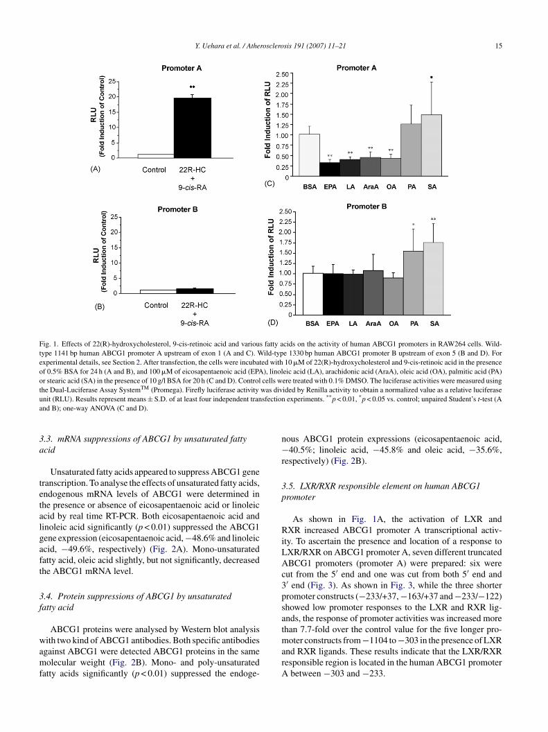
As shown in Fig. 1A, the activation of LXR andXR increased ABCG1 promoter A transcriptional activ-
ty. To ascertain the presence and location of a response toXR/RXR on ABCG1 promoter A, seven different truncatedBCG1 promoters (promoter A) were prepared: six were
ut from the 5′ end and one was cut from both 5′ end and′ end (Fig. 3). As shown in Fig. 3, while the three shorterromoter constructs (−233/+37, −163/+37 and −233/−122)howed low promoter responses to the LXR and RXR lig-nds, the response of promoter activities was increased more
oter constructs from −1104 to −303 in the presence of LXRnd RXR ligands. These results indicate that the LXR/RXResponsible region is located in the human ABCG1 promoter
between −303 and −233.

1 rosclero
3t
mdmiaoE
fiL−−−t−a
6 Y. Uehara et al. / Athe
.6. Effects of unsaturated fatty acids on severalruncated human ABCG1 promoters
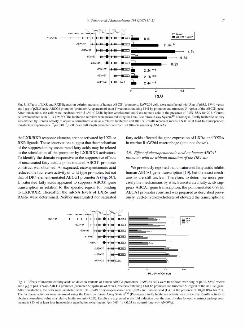
To determine the region within the human ABCG1 pro-oter A that is responsible for unsaturated fatty acid-
ependent transcriptional repression, several ABCG1 pro-oter constructs were prepared and transiently transfected
n murine RAW264 macrophage. Two polyunsaturated fatty
cids, EPA and LA, were incubated with cells in the presencef 10 g/l of BSA for 20 h after transfection. Interestingly,PA and LA reduced the transcriptional activities of the(
3h
s−sp
baXtdGsoaAeL
3p
AmA(
FAbwaAt(W2Sut1wt(
sis 191 (2007) 11–21
ve longer promoter constructs, −1104/+37 (EPA, −48.3%;A, −25.3%), −607/+37 (EPA, −49.9%; LA, −17.3%),530/+37 (EPA, −77.1%; LA, −54.0%), −413/+37 (EPA,70.4%; LA, −50.3%) and −303/+37 (EPA, −64.8%; LA,49.0%). In contrast, EPA and LA did not decrease the activi-
ies of the three shorter truncated promoters: −233/+37 (EPA,5.3%; LA, +33.8%), −163/+37 (EPA, +6.3%; LA, +29.3%)
nd −233/±122 (EPA, −0.1%; LA, +3.1%), respectivelyFig. 4).
.7. LXR/RXR responsive element on point-mutateduman ABCG1 promoter
As shown in Fig. 3, we investigated the LXR/RXR respon-ive region located in human ABCG1 promoter A between303 and −233. To determine more precisely the nucleotide
equence required for binding of LXR/RXR heterodimer, werepared a point-mutated ABCG1 promoter A construct.
The DR4 element, LXR/RXR binding site is characterizedy two direct hexameric repeats as AGGTCA or its similarrrangement separated by four nucleotides, i.e. AGGTCAXXX AGGTCA. Therefore, the full-length 1141 bp wild-
ype and mutant promoter constructs (−1104/+37) areescribed in Fig. 5A. The sequence −243ccC CCT TTAa−233 has three single mutations compared to the wild-type
equence −243TGC CCT TTA GT−233 to leave the functionut completely. 22(R)-hydroxycholesterol and 9-cis-retinoiccid stimulated the luciferase activity of wild-type humanBCG1 promoter A. However, DR4 (an LXR/RXR binding
lement)-mutated promoter construct did not respond to theXR and RXR ligands (Fig. 5B).
.8. Effect of eicosapentaenoic acid on theoint-mutated human ABCG1 promoter
As mentioned above, unsaturated fatty acids suppressed
BCG1 promoter activity of wild-type and truncated pro-oter constructs containing a DR4 element. Moreover,BCG1 promoter B or deleted promoter A constructs−233/+37, −163/+37 and −233/−122), which do not have
ig. 2. Effects of saturated and unsaturated fatty acids on the expressions ofBCG1 mRNA and protein. RAW264 and THP-1 cells (A and B) were incu-ated for 24 h with serum-free medium in the presence of 10 g/l BSA with orithout 100 �mol/l of eicosapentaenoic acid (EPA), linoleic acid (LA), oleic
cid (OA), palmitic acid (PA) or stearic acid (SA). The mRNA expression ofBCG1 was demonstrated by quantitative real-time PCR. Results represent
he means ± S.D. from at least three experiments. †p < 0.01 vs. BSA groupone-way ANOVA) (A). ABCG1 protein was analysed by SDS-PAGE and
estern blotting of proteins from PMA-treated THP-1 cells. Lanes 1 andrepresent ABCG1 proteins detected by using the ABCG1 antibody fromantacruz Biotechnology. Lanes 3 and 4 show ABCG1 proteins detected bysing the ABCG1 antibody from Alpha Diagnostic International. The PMA-reated cells were incubated in the presence of 10 g/l BSA with or without00 �mol/l of EPA, LA, OA, PA or SA. The ABCG1 proteins were blottedith the specific antibody of Santacruz Biotechnology. Results represent
he means ± S.D. from at least three experiments. †p < 0.01 vs. BSA groupone-way ANOVA) (B).
Y. Uehara et al. / Atherosclerosis 191 (2007) 11–21 17
Fig. 3. Effects of LXR and RXR ligands on deletion mutants of human ABCG1 promoters. RAW264 cells were transfected with 5 ng of phRL-SV40 vectorand 1 �g of pGL3 basic-ABCG1 promoter (promoter A; upstream of exon 1) vectors containing 1141 bp promoter and truncated 5′-region of the ABCG1 gene.After transfection, the cells were incubated with 5 �M of 22(R)-hydroxycholesterol and 9-cis-retinoic acid in the presence of 0.5% BSA for 20 h. Controlc d using TM
w ciferast truct, −
tRotTocrtUttR
fi
3p
ha
FaATom
ells were treated with 0.1% DMSO. The luciferase activities were measureas divided by Renilla activity to obtain a normalized value as a relative lu
ransfection experiments. **p < 0.01, *p < 0.05 vs. full length promoter cons
he LXR/RXR response element, are not activated by LXR orXR ligands. These observations suggest that the mechanismf the suppression by unsaturated fatty acids may be relatedo the stimulation of the promoter by LXR/RXR activators.o identify the domain responsive to the suppressive effectsf unsaturated fatty acid, a point-mutated ABCG1 promoteronstruct was obtained. As expected, eicosapentaenoic acideduced the luciferase activity of wild-type promoter, but nothat of DR4 element-mutated ABCG1 promoter A (Fig. 5C).
nsaturated fatty acids appeared to suppress ABCG1 generanscription in relation to the specific region for bindingo LXR/RXR. Thereafter, the mRNA levels of LXR� andXR� were determined. Neither unsaturated nor saturated
cpAo
ig. 4. Effects of unsaturated fatty acids on deletion mutants of human ABCG1 pnd 1 �g of pGL3 basic-ABCG1 promoter (promoter A; upstream of exon 1) vectorfter transfection, the cells were incubated with 100 �mol/l of eicosapentaenoic ahe luciferase activities were measured using the Dual-Luciferase Assay SystemT
btain a normalized value as a relative luciferase unit (RLU). Results are expressedeans ± S.D. of at least four independent transfection experiments. †p < 0.01, *p <
the Dual-Luciferase Assay System (Promega). Firefly luciferase activitye unit (RLU). Results represent means ± S.D. of at least four independent1104/+37 (one-way ANOVA).
atty acids affected the gene expression of LXR� and RXR�n murine RAW264 macrophage (data not shown).
.9. Effect of eicosapentaenoic acid on human ABCA1romoter with or without mutation of the DR4 site
We previously reported that unsaturated fatty acids inhibituman ABCA1 gene transcription [10], but the exact mech-nisms are still unclear. Therefore, to determine more pre-
isely the mechanisms by which unsaturated fatty acids sup-ress ABCA1 gene transcription, the point-mutated 0.98 kbBCA1 promoter construct was prepared as described previ-usly. 22(R)-hydroxycholesterol elevated the transcriptionalromoters. RAW264 cells were transfected with 5 ng of phRL-SV40 vectors containing 1141 bp promoter and truncated 5′-region of the ABCG1 gene.cid (EPA) and linoleic acid (LA) in the presence of 10 g/l BSA for 20 h.
M (Promega). Firefly luciferase activity was divided by Renilla activity toas the fold induction over the control value for each construct and represent0.05 vs. control (one-way ANOVA).

18 Y. Uehara et al. / Atherosclerosis 191 (2007) 11–21
Fig. 5. Mutational analysis of hABCG1 promoter. RAW264 cells weretransfected with wild-type hABCG1 promoter (from −1104 to +37 bp) orDR4-mutated promoter. The mutations are shown in Fig. 5A and Section2. After transfection, the cells were incubated with 10 �mol/l of 22(R)-hydroxycholesterol and 9-cis-retinoic acid in the presence of 0.5% BSAfor 20 h. Control cells were treated with 0.2% DMSO (B), the cells wereincubated with 100 �mol/l of eicosapentaenoic acid (EPA) in the pres-ence of 10 g/l BSA for 20 h. Control cells were treated with 10 g/l BSAand 0.1% DMSO (C). The luciferase activities were measured using theDual-Luciferase Assay SystemTM (Promega). Firefly luciferase activitywas divided by Renilla activity to obtain a normalized value as a relativeluciferase unit (RLU). The RLU are shown in fold change compared withctt
ablspA
Fig. 6. Mutational analysis of hABCA1 promoter. RAW264 cells were trans-fected with wild-type hABC1 promoter (from −811 to +172 bp) or itsmutated version at the DR4 site. The mutations are presented in Section2. After transfection, the cells were incubated with 10 �mol/l of 22(R)-hydroxycholesterol in the presence of 0.5% BSA for 20 h. Control cells weretreated with 0.1% DMSO (B), the cells were incubated with 100 �mol/l ofeicosapentaenoic acid (EPA) in the presence of 10 g/l BSA for 20 h. Controlcells were treated with 10 g/l BSA and 0.1% DMSO (C). The luciferase activ-ities were measured using the Dual-Luciferase Assay SystemTM (Promega).Firefly luciferase activity was divided by Renilla activity to obtain a nor-malized value as a relative luciferase unit (RLU). The RLU are shown infold change compared with control (DMSO or BSA). The results repre-sent three independent transfection experiments of duplicates. Bars indicatemd
A(
3p
o
ontrol (DMSO or BSA). The results represent four independent transfec-ion experiments of duplicates. Bars indicate mean ± S.D. †p < 0.001 vs.reatment with BSA and DMSO (unpaired Student’s t-test).
ctivity of the wild-type human ABCA1 promoter constructut not of a mutant human ABCA1 promoter construct
acking the DR4 element (Fig. 6A). Similar to the resultshown for the ABCG1 promoter, eicosapentaenoic acid sup-ressed the transcriptional activity of the human wild-typeBCA1 promoter construct but not the activity of a variantfcoI
ean ± S.D. †p < 0.01 vs. treatment with BSA and DMSO (unpaired Stu-ent’s t-test).
BCA1 promoter construct containing a mutated DR4 siteFig. 6B).
.10. LXR protein binds to a DR4 element on ABCG1romoter located upstream of exon 1
To determine whether the DR4 element binds to LXR,ligonucleotide probes containing DR4 site were used to per-
orm electrophoretic mobility shift assay. The DNA–proteinomplex was detected as the shifted bands in the presencef nuclear extracts from HeLa cells (Fig. 7, lanes 2 and 3).nterestingly, the nuclear extracts incubated with polyclonal
Y. Uehara et al. / Atherosclero
Fig. 7. Electrophoretic mobility shift assay of human ABCG1 promoter frag-ment. Double-stranded oligonucleotide (fragment spanning −264 to −251)containing a LXR/RXR binding site was digested from ABCG1 promoterA construct. The digested 114 bp double-stranded oligonucleotide probewas labeled with biotin. Labeled double-stranded oligonucleotide probe wasincubated with (lanes 2–4) or without (lane 1) HeLa nuclear extract. A sam-ple was pre-incubated with antibodies against LXR�/� before addition ofprobe (lane 4). DNA–protein complexes were electrophoresed on a native 5%polyacrylamide gel and transferred to a nylon membrane. The DNA–proteincomplexes were visualized with chemiluminescent substrate after incubationof the nylon membrane with stabilized streptavidin-horseradish peroxidasecc
Ll
4
iptmvdtatpc
A[Aetnowm
ofitnAiffmh
uedt1aAt(epTwwties
mmptlwuteth
onjugate. The arrow and arrow head indicate the band for DNA–proteinomplex and complex of DNA–protein with LXR�/� antibody, respectively.
XR�/� antibodies produced supershifted band (Fig. 7,ane 4).
. Discussion
Patients with diabetes mellitus have a tremendouslyncreased risk of atherosclerotic vascular disease which inart appears to be related to low HDL cholesterol levels. ABCransporters A1 and G1 play an important role in both HDL
etabolism and the regulation of cholesterol homeostasis inarious cells, notably in macrophages which unlike other cellso not limit cholesterol uptake. It has been well documentedhat ABCA1 plays a pivotal role in mediating phospholipid
nd cholesterol efflux to lipid-free apoA-I and, thereby, inhe formation of discoidal HDL precursors. Mature HDLarticles, which are spherical and transport almost all HDLholesterol, appear to induce cholesterol efflux via two others
am
sis 191 (2007) 11–21 19
BC transporters, ABCG1 and ABCG4, rather than ABCA117,25]. In a positive feed-forward regulation process, bothBCA1 and ABCG1 gene expression and thereby cholesterol
fflux are strongly induced by oxidized cholesterol deriva-ives, so-called oxysterols. Oxysterols are activators of theuclear transcription factor LXR� which together with itsbligatory heterodimeric partner RXR binds to DR4 elementshich are present in both the ABCA1 and the ABCG1 pro-oters.We have previously shown that the stimulatory effect
f oxysterols on ABCA1 gene transcription is inhibited byree unsaturated fatty acids which interestingly accumulaten patients with diabetes mellitus and insulin resistance. Inhe present study we extend these observations by threeovel findings: first, unsaturated fatty acids suppress theBCG1 mRNA and protein expressions. Second, the activ-
ty of the ABCG1 promoter is also suppressed by unsaturatedatty acids. Third, the suppressive effect of free unsaturatedatty acids depends on the presence of an intact DR4 ele-ent and probably due to the binding of the LXR�/RXR
eterodimer.The human ABCG1 gene has at least two promoters, one
pstream of exon 1 and another upstream of exon 5 andncodes for several transcripts [22,23]. A previous studyemonstrated that the activation of LXR� and RXR increaseshe expression of ABCG1 transcripts starting from both exonand exon 5 [22]. Interestingly we found that ligands for LXRnd RXR dramatically increase activity of ABCG1 promoter
located upstream of exon 1 (Fig. 1A), but do not changehe activity of ABCG1 promoter B located within intron 4Fig. 1B). These findings suggest that the transcription ofxon 5 and subsequent exons may be controlled, at least inart, by the ABCG1 promoter A located upstream of exon 1.o confirm this finding, electrophoretic mobility shift assayas performed. Existence of DNA-binding nuclear proteinsere ascertained on the extracted ABCG1 promoter A con-
aining DR4 element. In addition, one of the proteins wasdentified as LXRs by using the supershifted assay on EMSAxperiment. We also show that unsaturated fatty acids do onlyuppress the activity of ABCG1 promoter A.
In agreement with these findings only the ABCG1 pro-oter A located upstream of exon 1 contains a DR4 ele-ent which is needed for LXR�/RXR binding. In fact, the
romoter response to LXR/RXR ligands was not seen inruncated and mutated ABCG1 promoter constructs whichack a functional DR4 site (Figs. 3 and 5B). These mutantsere neither responsive to the suppressive effects of freensaturated fatty acids (Fig. 5C). Likewise the knock-out ofhe LXR/RXR response element in the ABCA1 promoterliminated the suppressive effect of USFA on ABCA1 generanscription (Fig. 6B). Our observation suggests that USFAas a potential for suppressive effect of ABCG1 gene tran-
cription.Although we show that the suppressive effects of free fattycids depend on the presence of a LXR/RXR binding site, theechanism by which free fatty acids suppress the promoter
2 rosclero
aitLstgruLwwtttRmt
tsmrp
ittTooimtm
A
StDRNav
R
[
[
[
[
[
[
[
[
[
[
[
[
[
0 Y. Uehara et al. / Athe
ctivities of ABCA1 and ABCG1 is not resolved. Neithern this nor in our previous study, we found any transcrip-ional effect of unsaturated fatty acids on the expression ofXR� or RXR. By contrast other researchers found eithertimulatory effects of USFAs on LXR� but not LXR� in cul-ivated hepatoma cells [26] or suppressive effects on LXR�ene expression [27]. Furthermore, fatty acids also play aole in the activation of PPAR� and � [28] which indirectlypregulate ABCA1 and ABCG1 gene expression by elevatingXR gene expression. On the posttranslational level, USFAsere reported to activate RXR [29] to form a heterodimerith LXR. However, USFAs were also found to compete
he binding of oxysterols to LXR� and thereby to suppresshe oxysterol-induced upregulation of SREBP1c [30]. Onhe other hand, DNA binding transcription factors, LXR andXR are capable of interacting with coregulators [31,32]. Itay be possible that free fatty acids regulate these coregula-
ors of LXR or RXR.In the present results, saturated fatty acids stimulated
he ABCG1 transcription activities. Whereas, the effects oftearic acid did not change on ABCG1 promoter A with DR4utation (data not shown). These results suggest that satu-
ated fatty acids stimulate the ABCG1 transcriptions inde-endent of LXR or RXR.
In conclusion, this study shows that ABCG1, like ABCA1,s suppressed by unsaturated fatty acids at the transcrip-ional level, and describes the structural requirements forhis suppressive effect within the human ABCG1 promoter.he suppressive effects of USFAs on promoter activitiesf both ABCA1 and ABCG1 indicate that elevated levelsf unsaturated fatty acids inhibit both apoA-I and HDL-nduced cholesterol efflux from macrophages. This impair-
ent of reverse cholesterol transport may contribute tohe increased cardiovascular risk of patients with diabetes
ellitus.
cknowledgments
This work was supported by a grant-in-aid for Youngcientists (B) (No. 16790517) from the Ministry of Educa-
ion, Culture, Sports, Science and Technology of Japan tor. Yoshinari Uehara and in part by funds from the Centralesearch Institute of Fukuoka University (No. 046002 ando. 041001) to Dr. Yoshinari Uehara and Dr. Keijiro Saku
nd by an HDL Research Award from Pfizer to Dr. Arnoldon Eckardstein.
We would like to thank Ms. Yuko Naito for her assistance.
eferences
[1] Gordon DJ. Rifkind BM: high-density lipoprotein—the clinical impli-cations of recent studies. N Engl J Med 1989;321:1311–6.
[2] von Eckardstein A, Nofer JR, Assmann G. High density lipoproteinsand arteriosclerosis. Role of cholesterol efflux and reverse cholesteroltransport. Arterioscler Thromb Vasc Biol 2001;21:13–27.
[
sis 191 (2007) 11–21
[3] Rohrer L, Hersberger M, von Eckardstein A. High density lipoproteinsin the intersection of diabetes mellitus, inflammation and cardiovasculardisease. Curr Opin Lipidol 2004;15:269–78.
[4] Reaven GM, Hollenbeck C, Jeng CY, et al. Measurement of plasmaglucose, free fatty acid, lactate, and insulin for 24 h in patients withNIDDM. Diabetes 1988;37:1020–4.
[5] Shepherd J, Packard CJ, Patsch JR, et al. Effects of dietary polyunsat-urated and saturated fat on the properties of high density lipoproteinsand the metabolism of apolipoprotein A-I. J Clin Invest 1978;61:1582–92.
[6] Sacks FM, Katan M. Randomized clinical trials on the effects of dietaryfat and carbohydrate on plasma lipoproteins and cardiovascular disease.Am J Med 2002;113(Suppl 9B):13S–24S.
[7] Brooks-Wilson A, Marcil M, Clee SM, et al. Mutations in ABC1 inTangier disease and familial high-density lipoprotein deficiency. NatGenet 1999;22:336–45.
[8] Bodzioch M, Orso E, Klucken J, et al. The gene encoding ATP-binding cassette transporter 1 is mutated in Tangier disease. Nat Genet1999;22:347–51.
[9] Rust S, Rosier M, Funke H, et al. Tangier disease is caused by mutationsin the gene encoding ATP-binding cassette transporter 1. Nat Genet1999;22:352–5.
10] Uehara Y, Engel T, Li Z, et al. Polyunsaturated fatty acids and ace-toacetate downregulate the expression of the ATP-binding cassettetransporter A1. Diabetes 2002;51:2922–8.
11] Croop JM, Tiller GE, Fletcher JA, et al. Isolation and characteriza-tion of a mammalian homolog of the Drosophila white gene. Gene1997;185:77–85.
12] Chen H, Rossier C, Lalioti MD, et al. Cloning of the cDNA for a humanhomologue of the Drosophila white gene and mapping to chromosome21q22.3. Am J Human Genet 1996;59:66–75.
13] Savary S, Denizot F, Luciani M, et al. Molecular cloning of a mam-malian ABC transporter homologous to Drosophila white gene. MammGenome 1996;7:673–6.
14] Walker JE, Saraste M, Runswick MJ, et al. Distantly related sequencesin the alpha- and beta-subunits of ATP synthase, myosin, kinases andother ATP-requiring enzymes and a common nucleotide binding fold.EMBO J 1982;1:945–51.
15] Dean M, Rzhetsky A, Allikmets R. The human ATP-binding cas-sette (ABC) transporter superfamily. Genome Res 2001;11:1156–66.
16] Klucken J, Buchler C, Orso E, et al. ABCG1 (ABC8), the humanhomolog of the Drosophila white gene, is a regulator of macrophagecholesterol and phospholipid transport. Proc Natl Acad Sci USA2000;97:817–22.
17] Wang N, Lan D, Chen W, et al. ATP-binding cassette transporters G1and G4 mediate cellular cholesterol efflux to high-density lipoproteins.Proc Natl Acad Sci USA 2004;101:9774–9.
18] Alberti S, Steffensen KR, Gustafsson JA. Structural characterisation ofthe mouse nuclear oxysterol receptor genes LXRalpha and LXRbeta.Gene 2000;243:93–103.
19] Willy PJ, Umesono K, Ong ES, et al. LXR, a nuclear recep-tor that defines a distinct retinoid response pathway. Genes Dev1995;9:1033–45.
20] Song C, Kokontis JM, Hiipakka RA, et al. Ubiquitous receptor: a recep-tor that modulates gene activation by retinoic acid and thyroid hormonereceptors. Proc Natl Acad Sci USA 1994;91:10809–13.
21] Costet P, Luo Y, Wang N, et al. Sterol-dependent transactivation ofthe ABC1 promoter by the liver X receptor/retinoid X receptor. J BiolChem 2000;275:28240–5.
22] Lorkowski S, Rust S, Engel T, et al. Genomic sequence and structureof the human ABCG1 (ABC8) gene. Biochem Biophys Res Commun
2001;280:121–31.23] Langmann T, Porsch-Ozcurumez M, Unkelbach U, et al. Genomicorganization and characterization of the promoter of the human ATP-binding cassette transporter-G1 (ABCG1) gene. Biochim Biophys Acta2000;1494:175–80.
rosclero
[
[
[
[
[
[
[
Y. Uehara et al. / Athe
24] Kennedy MA, Venkateswaran A, Tarr PT, et al. Characterization of thehuman ABCG1 gene: liver X receptor activates an internal promoterthat produces a novel transcript encoding an alternative form of theprotein. J Biol Chem 2001;276:39438–47.
25] Engel T, Lorkowski S, Lueken A, et al. The human ABCG4gene is regulated by oxysterols and retinoids in monocyte-derived macrophages. Biochem Biophys Res Commun 2001;288:483–8.
26] Tobin KA, Steineger HH, Alberti S, et al. Cross-talk between fatty acid
and cholesterol metabolism mediated by liver X receptor-alpha. MolEndocrinol 2000;14:741–52.27] Pawar A, Botolin D, Mangelsdorf DJ, et al. The role of liver X receptor-alpha in the fatty acid regulation of hepatic gene expression. J BiolChem 2003;278:40736–43.
[
[
sis 191 (2007) 11–21 21
28] Desvergne B, Wahli W. Peroxisome proliferator-activated receptors:nuclear control of metabolism. Endocrinol Rev 1999;20:649–88.
29] de Urquiza AM, Liu S, Sjoberg M, et al. Docosahexaenoic acid, aligand for the retinoid X receptor in mouse brain. Science 2000;290:2140–4.
30] Ou J, Tu H, Shan B, et al. Unsaturated fatty acids inhibit transcription ofthe sterol regulatory element-binding protein-1c (SREBP-1c) gene byantagonizing ligand-dependent activation of the LXR. Proc Natl AcadSci USA 2001;98:6027–32.
31] Hu X, Li S, Wu J, et al. Liver X receptors interact with corepressors toregulate gene expression. Mol Endocrinol 2003;17:1019–26.
32] Seol W, Choi HS, Moore DD. An orphan nuclear hormone receptor thatlacks a DNA binding domain and heterodimerizes with other receptors.Science 1996;272:1336–9.
Related Documents


