IMMUNE CROSS-REACTIVITY BETWEEN INFECTIOUS BOVINE RHINOTRACHEITIS VIRUS AND HUMAN CYTOMEGALOVIRUS. Item Type text; Thesis-Reproduction (electronic) Authors Abraham, Kristin Marie. Publisher The University of Arizona. Rights Copyright © is held by the author. Digital access to this material is made possible by the University Libraries, University of Arizona. Further transmission, reproduction or presentation (such as public display or performance) of protected items is prohibited except with permission of the author. Download date 24/06/2018 04:59:52 Link to Item http://hdl.handle.net/10150/275153

Welcome message from author
This document is posted to help you gain knowledge. Please leave a comment to let me know what you think about it! Share it to your friends and learn new things together.
Transcript
IMMUNE CROSS-REACTIVITY BETWEENINFECTIOUS BOVINE RHINOTRACHEITISVIRUS AND HUMAN CYTOMEGALOVIRUS.
Item Type text; Thesis-Reproduction (electronic)
Authors Abraham, Kristin Marie.
Publisher The University of Arizona.
Rights Copyright © is held by the author. Digital access to this materialis made possible by the University Libraries, University of Arizona.Further transmission, reproduction or presentation (such aspublic display or performance) of protected items is prohibitedexcept with permission of the author.
Download date 24/06/2018 04:59:52
Link to Item http://hdl.handle.net/10150/275153

INFORMATION TO USERS
This was produced from a copy of a document sent to us for microfilming. While the most advanced technological means to photograph and reproduce this document have been used, the quality is heavily dependent upon the quality of the material
submitted.
The following explanation of techniques is provided to help you understand markings or notations which may appear on this reproduction.
1.The sign or "target" for pages apparently lacking from the document photographed is "Missing Page(s)". If it was possible to obtain the missing page(s) or section, they are spliced into the film along with adjacent pages. This may have necessitated cutting through an image and duplicating adjacent pages to assure you of complete continuity.
2. When an image on the film is obliterated with a round black mark it is an indication that the film inspector noticed either blurred copy because of movement during exposure, or duplicate copy. Unless we meant to delete copyrighted materials that should not have been filmed, you will find a good image of the page in the adjacent frame. If copyrighted materials were deleted you will find a target note listing the pages in the adjacent frame.
3. When a map, drawing or chart, etc., is part of the material being photographed the photographer has followed a definite method in "sectioning" the material. It is customary to begin filming at the upper left hand corner of a large sheet and to continue from left to right in equal sections with small overlaps. If necessary, sectioning is continued again—beginning below the first row and continuing on until complete.
4. For any illustrations that cannot be reproduced satisfactorily by xerography,
photographic prints can be purchased at additional cost and tipped into your xerographic copy. Requests can be made to our Dissertations Customer
Services Department.
5. Some pages in any document may have indistinct print. In all cases we have filmed the best available copy.
University Microfilms
International 300 N. ZEEB RD„ ANN ARBOR, Ml 48106

1318016
! ABRAHAM# KRISTIN MARIE IMMUNE CROSS-REACTIVITY BETWEEN INFECTIOUS BOVINE RHINOTRACHEITIS VIRUS AND HUMAN CYTOMEGALOVIRUS,
THE UNIVERSITY OF ARIZONA, M.S., 1982
University Microfilms
International 300 N. ZEEB RD„ ANN ARBOR. Ml 48106

PLEASE NOTE:
In all cases this material has been filmed in the best possible way from the available copy. Problems encountered with this document have been identified here with a check mark V .
1. Glossy photographs or pages
2. Colored illustrations, paper or print ^
3. Photographs with dark background »/
4. Illustrations are poor copy
5. Pages with black marks, not original copy
6. Print shows through as there is text on both sides of page
7. Indistinct, broken or small print on several pages i/
8. Print exceeds margin requirements
9. Tightly bound copy with print lost in spine
10. Computer printout pages with indistinct print
11. Page(s) lacking when material received, and not available from school or author.
12. Page(s) seem to be missing in numbering only as text follows.
13. Two pages numbered . Text follows.
14. Curling and wrinkled pages
15. Other
University Microfilms
International

IMMUNE CROSS-REACTIVITY BETWEEN INFECTIOUS
BOVINE RHINOTRACHEITIS VIBDS AND
HUMAN CYTOMEGALOVIRUS
by
Kristin Marie Abraham
A Thesis Submitted to the Faculty of the
DEPARTMENT OF MICROBIOLOGY
In Partial Fulfillment of the Requirements For the Degree of
MASTER OF SCIENCE WITH A MAJOR IN MICROBIOLOGY
In the Graduate College
THE UNIVERSITY OF ARIZONA
1 9 8 2
4

STATEMENT BY AUTHOR
This thesis has been submitted in partial fulfillment of requirements for an advanced degree at The University of Arizona and is deposited in the University Library to be made available to borrowers under rules of the Library.
Brief quotations from this thesis are allowable without special permission, provided that accurate acknowledgment of source is made. Requests for permission for extended quotation from or reproduction of this manuscript in whole or in part may be granted by the head of the major department of the Dean of the Graduate College when in his judgment the proposed use of the material is in the interests of scholarship. In all other instances, however, permission must be obtained from the author.
SIGNED:.
APPROVAL BY THESIS DIRECTOR
This thesis has been approved on the date shown below:
RANDALL F. PRITCHETT Date Assistant Professor of Microbiology
ACKNOWLEDGMENTS
The author is Indebted to Dr. Randall F. Pritchett for his
guidance and time during this study.
The author would also like to thank Dr. Wayne R. Ferris for
his assistance with the electron microscopy, and Dr. B. Sue Criswell
for her advice and expertise.
Finally, the author wishes to express her appreciation to her
parents, Mr. and Mrs. Donald Olson, and her husband, Mr. John Abraham,
f
for their continued encouragement and support.
ill
TABLE OF CONTENTS
Page
LIST OF TABLES vi
LIST OF ILLUSTRATIONS . vll
ABSTRACT viii
INTRODUCTION 1
MATERIALS AND METHODS 8
Animals 8 Cell Cultures 8 Virus Propagation and Assay 9 Radioisotope Labeling of Virus 10 Purification of Virus 10 Preparation of Antigens 11 Ultraviolet Sterilization of Antigens 12 Protein Concentration Determination of Antigens 12
Electron Microscopy of Virus Preparations 12 Sensitization of Animals 13 Mitogens 13 Lymphocyte Culture Medium 13 Preparation of Peripheral Blood Lymphocytes for Assay 14
Lymphocyte Stimulation by Antigens and Mitogens 14
Statistical Analysis of Data 15
RESULTS 16
Purification of CMV and IBRV Antigens 16 Electron Microscopic Examination of Purified Virus 18
Kinetics of the Mitogenic Response to Con A and PHA 18
Lymphocyte Blastogenlc Response of Rabbits Immunized with IBRV and CMV 22
DISCUSSION 33
iv

TABLE OF CONTENTS—Continued
Page
SUMMARY 37
REFERENCES 39
v
LIST OF TABLES
Table Page
3 1. H-Thymidlne uptake of peripheral lymphocytes at
one week post-immunization as a function of virus antigen concentration 25
3 2. H-Thymidine uptake of peripheral lymphocytes in
response to cellular control antigens 30
3. Significance of test cpm vs control cpm at optimum response as determined by a paired-t test 32
vi

LIST OF ILLUSTRATIONS
Figure Page
3 1. H-Thymidine distribution in 10%-60% sucrose
gradients and preformed CsCl gradients after centrifugation of labeled IBRV 17
2. Electronmicrograph of the CMV virus preparation 19
3. Electronmicrograph of the IBRV virus preparation 20
4. Mitogenic responses of rabbit lymphocytes to Concanavalin A and Phytohaemeagglutinin as a function of in vitro culture time for four test rabbits 21
5. Changes in mitogenic response of rabbit lymphocytes to Concanavalin A and Phytohaemeagglutinin as a function of concentration of mitogen 23
6. Response of lymphocytes from rabbits immunized with IBRV to homologous and heterologous antigenic stimulation 26
7. Response of lymphocytes from rabbits immunized with CMV to homologous and heterologous antigenic stimulation 29
vii
ABSTRACT
Antigen specific lymphocyte blastogenesis assays can be used
to indicate in vivo immune reactivity to viral antigens after immuni
zation. This study Investigated the immune cross-reactivity of two
herpesviruses, human Cytomegalovirus and Infectious Bovine Rhino-
tracheitis Virus, as demonstrated by blastogenic assay of rabbit pe
ripheral blood lymphocytes.
In all cases, statistically significant lymphocyte responses
to homologous antigens were observed, as well as significant responses
to cross-reacting viral antigens. Cellular control antigen prepara
tions were stimulatory only in the case of lymphocytes taken from
animals immunized with CMV antigen preparations when stimulated by
antigens derived from the host cell used to propagate CMV. The abil
ity of lymphocytes committed to one virus to react with or be stimu
lated by a similar but different herpesvirus may lead to clinical
applications of this cross-reactivity.
viii
INTRODUCTION
Human Cytomegalovirus (CMV) and Infectious Bovine Rhino-
tracheitis Virus (IBRV; also referred to as Bovine Herpesvirus Type
1), two viruses of the herpesvirus group, are ubiquitous important
pathogens. Herpesviruses range in size from 180 nm to 200 nm in diam
eter. They consists of an innermost section or "core" which is 76 nm
in diameter that contains the double stranded DNA genome. The molecu
lar weight of the genome among individuals of the group ranges from 82
x 10^ daltons to 150 x 10^ daltons. The core is enclosed in an icosa-
hedral capsid 95-105 nm in diameter which is made up of 162 capsomers.
The capsid and enclosed core are referred to as the nucleocapsid. The
nucleocapsid is surrounded by a granular zone, or tegument, which in
turn is enclosed by an envelope from which small spikes project. Dur
ing morphogenesis nucleocapsids assemble in the nucleus and acquire
envelopes via budding through the inner nuclear membrane. Host mem
brane glycoproteins may be incorporated into the envelope during the
budding process. The typical herpesvirus virion consists of approxi
mately 70% protein, 7% DNA, 22% lipid (phospholipid - cellular in ori
gin) and 2% carbohydrate (1).
CMV is individually characterized by its ability to cause en
largement of cells or "cytomegaly", by the extraordinarily high molec
ular weight of its genome (150 x 10^ daltons), and by its narrow host
range. CMV infections are also characterized by the production of
aberrant forms of the virus referred to as dense bodies.

2
Dense bodies are spherical forms containing homogeneous
electron-dense material which is enclosed within membranes. These
forms contain 22 of the 23 polypeptides found in the complete virus,
but do not contain any DNA. The 22 structural polypeptides, including
8 glycoproteins, range in molecular weight from 24,000 to 171,000
daltons (2). Dense bodies which are synthesized in large amounts in
infected cells possibly serve as immunogens in the naturally infected
host when little extracellular virus is produced (3).
IBRV, a bovine pathogen, has not been reported to cause human
disease. However, it does replicate in human cells. IBRV has a wide
host range, and with respect to CMV, a short replication cycle. Un
like CMV, dense body forms are not seen in cells infected with IBRV.
Electrophoretic analysis of purified IBRV revealed the presence of 25
to 33 structural polypeptides, eleven of which were found to be glyco
proteins (4).
Herpesviruses are characterized by their ability to produce
latent infections, with reactivations possible after variable periods
of quiescence. CMV is thought to become latent in human peripheral
blood lymphocytes, as evidenced by IgM antibody levels to CMV develop
ing in seronegative patients who receive transfusions of blood from
seropositive healthy donors. The form of CMV in these cells is un
clear, as the virus is neither found complete, free in the plasma, nor
whole within the lymphocytes (5,6). Latent herpesvirus infections
typically occur after acute primary infections. In the case of CMV,
primary infection often occurs without overt disease and the virus re
mains In the host as an endogenous latent infection.
Primary CMV infections and reactivations of latent endogenous
CMV result In significant and often severe clinical manifestations in
the human population. Currently, prenatal and neonatal CMV infection
constitutes a greater danger to children than does rubella (7). From
0.5% to 1.0% of all children born excrete CMV in their urine, or have
antibodies against CMV which persist longer than maternal antibodies
(8). Cytomegalic inclusion disease (CMID), is the best characterized
and most severe form of CMV caused disease contracted in utero. The
clinical manifestations of CMID include microencephaly, cerebral cal
cifications, chorioretinitis, hepatosplenomegaly, thrombocytopenic
purpura, deafness and hemolytic anemia (9). Other congenital infec
tions may be inapparent in the newborn, but are activated in later
life.
Most exogenous childhood and adult infections are thought to
be spread via the respiratory route. Transmission via urine is also
possible, as those children with apparent and inapparent viral infec
tions contracted in utero may have protracted periods of viral shed
ding in the urine (10).
While most adults who become infected with the virus elicit
little or no symptomatology, in children younger than 4 months of age
the disease can progress and lead to hepatic and renal Insufficiency,
pneumonia, neurological symptoms and eventual death (11). The most
likely mode of transmission in these cases appears to be via mothers'
milk during breast feeding (12). In children over 4 months of age,
the symptomatology is generally less severe, and the CNS is spared,
although hepatitis or pneumonitis may result (13). Heterophile

4
negative mononucleosis Is an exogenous Infection caused by CMV, which
Is observed in CMV seronegative people who are recipients of
multiple blood transfusions from latently infected donors (14).
Active infections with symptomatology in adults are generally
due to the presence of a latent, endogenous virus contracted at an
earlier age, which is reactivated due to allogeneic stimulation or
Immunosuppressive therapy. CMV infection is the most common and most
Important infection that occurs after allogeneic marrow transplanta
tion. The most significant result of CMV disease after marrow trans
plantation is disseminated Infection with pneumonia, although
arthritis, hepatitis and leukopenia may result (15).
In patients receiving renal allografts, CMV and Herpes Simplex
Virus (HSV) reactivations commonly occur (16). CMV infections follow
ing renal allografts have been reported In association with several
syndromes including pneumonitis (17), fever and hepatitis (18), a
mononucleosis-type syndrome, and hemolytic anemia accompanied by
"transplant lung" syndrome and hepatic dysfunction (19). Humoral im
munity appears to be unimpaired in infected transplant patients, as
their neutralizing antibody titers are not significantly different
from controls. Thus, it is likely that impairment of cell-mediated
immune mechanisms are responsible for this complication of renal allo
graft surgery (20).
In the case of viral infections it has been shown that circu
lating antibody may be highly effective in protecting against infec
tion and in decreasing the possibility of development of systemic
5
disease through limiting viremia. However, certain virus infections
remain unresponsive to antibodies and may thrive in the face of high
titers (21). Through the study of patients with T lymphocyte defects,
it has been possible to elucidate the importance of the T cell in re
covery from viral infection. This recovery is thought to be dependent
on the elimination or restriction of virus infected cells (22).
In several cases of herpesvirus infections, the cellular re
sponse may be nonspecifically or specifically depressed in its level
of activity in vivo (23). The latter is found in striking clarity in
the case of cytomegalovirus infections, in which the reactivity of
peripheral lymphocytes in infected patients is specifically suppressed
to the CMV antigen (24). Thus, either through effects which result in
elimination of the virus, or through suppressive effects brought about
as a result of virus infection, the response of greatest consequence
to the outcome of virus infection and production of disease is the
cell-mediated response.
Treatment and preventative measures against CMV infection have
been of great interest for the last several years. The use of anti
viral chemotherapeutic drugs such as adenine arabinoside have not
shown much promise (25), and the use of Interferon during therapy has
also been fairly ineffective as in most cases both substances caused
only a transient decrease in urinary virus excretion (26).
The development of viral vaccines utilizing different labora
tory strains of CMV began with the use of the AD-169 strain, which was
originally isolated from a young girl undergoing adenoidectomy (27).
Secondary efforts utilizing the Towne strain, isolated from.an infant
6
with the signs of cytomegalic inclusion disease have also been under
taken (28). Both vaccines have undergone limited trials, with some
success in the development of an antibody response and some indica
tions of a cell-mediated response being elicited to the vaccine (29).
The largest drawback to the use of these attenuated strains is their
apparent ability to infect, and initiate latency in the vaccinated
individual (30). As CMV, along with IBRV and other herpesviruses are
suspected oncogenic agents (31,32), the long range suitability of
live, whole virus vaccines is under some doubt (33). Trials with sub-
unit vaccines have generally been disappointing, as vaccination re
sulted in poor protection when tested In an animal model system (34),
and did not prevent the appearance of viremia in these animals.
An alternative approach to treatment of CMV infections in
volves the use of an antigenically related virus or preparation to
cross-stimulate cell-mediated Immune (CMI) responses. IBRV has been
reported to cross-react Immunologically with human CMV. Using sero
logical methods such as immunofluorescence it was shown that CMV
immune sera will react with IBRV infected cells (35). A preliminary
report has suggested that oral administration of bovine transfer fac
tor prepared from lymphocytes of an IBRV sensitized cow was able to
stimulate a CMI response to CMV in a human patient with CMV disease
(36). Also, IBRV has recently been shown in isolated cases to stimu
late a blastogenic response in lymphocytes of donors with a history of 1'
infection with human herpesviruses (37). The possibility of using
this related virus in some capacity to augment recovery from CMV
caused diseases has stressed the importance for documentation of its
immune cross-reactivity with CMV.
This study was designed to establish whether antigenic compo
nents of CMV and IBRV are capable of cross-stimulating a CMI response
The experimental approach was first to immunize individual rabbits
with either CMV or IBRV. The in vitro blastogenic responses of pe
ripheral blood lymphocytes to the homologous and heterologous viruses
were then assayed and the results compared.
MATERIALS AMD METHODS
Animals
Adult male New Zealand White rabbits each weighing approxi
mately 2 kilograms were maintained with rabbit pellets and tap water
ad libitum throughout the experimental procedure.
Cell Cultures
Human foreskin fibroblast (HFF) cell cultures were prepared
from human foreskins in stationary flasks. Subsequently the HFF cells
were cultivated in 260 mm glass roller bottles (Bellco Glass, Inc.,
Vineland, N.J.). Cells were grown in Eagle's minimal essential medium
-(EMEM, Flow Laboratories, Rockville, Md.) supplemented with 10% fetal
calf serum (6IBC0 Laboratories, Grand Island, N.Y.), 2 mM L-glutamine
(GIBCO), 0.01 M Tricine, 0.075% NaHC03, and antibiotics (100 U Peni
cillin per ml and 100 yg of Streptomycin per ml). Cells were subcul-
tured after reaching confluency at a split ratio of 1:3 once per week.
Cells of passage levels 5 through 16 were used for virus propagation.
The Madin-Darby Bovine Kidney (MDBK) infinite cell line was
obtained from Dr. John Mare' (University of Arizona, Tucson, Az.) and
cultivated in 32 oz. glass prescription bottles. The MDBK cells were
grown in the same medium as HFF cell cultures and were subcultured
after reaching confluency at a split ratio of 1:4 twice per week.
8
9
Virus Propagation and Assay
The Towne strain of human CMV originally isolated from the
urine of an infant with cytomegalic inclusion disease was obtained
from Dr. M. F. Stinski (University of Iowa, Iowa City, la.)• Virus
was adsorbed onto roller bottle cultures of HFF cells at a concentra
tion of 0.1 plaque forming units (PFU) per cell. After an adsorption
period of 90 minutes at 37C, cultures were overlayed with 80 ml of
EMEM supplemented with 2% fetal calf serum, 2 mM L-glutamine, 0.01 M
Tricine, 0.075% NaHCO^, and antibiotics. Infected cell cultures were
incubated at 37C and rolled at 0.25 rpm. At 4 days post-infection,
the culture medium was replaced and L-arginine was added to a final
concentration of 1.2 mM. Virus was harvested at 9 days post-infection
from the extracellular culture fluid.
A stock preparation of IBRV was obtained from Dr. John Mare'.
Virus was adsorbed onto 32 oz. prescription bottle cultures of MDBK
cells at a concentration of approximately 20 PFU per cell. After an
adsorption period of 60 minutes at 37C, cultures were overlaid with
15 ml of EMEM supplemented with 2% fetal calf serum, 2 mM L-glutamine,
0.01 M Tricine, 0.075% NaHCO^, and antibiotics. At 24 hours post
infection, virus was harvested from the extracellular culture fluid.
Infectivity titers of CMV in HFF cells and IBRV in MDBK cells
were determined by the plaque technique under methyl cellulose over
lays (38). Plaques of CMV were counted at 9 days post-infection and
those of IBRV were counted at 4 days post-infection.
10
Radioisotope Labeling of Virus
To monitor virus purification, the DNA of IBRV was labeled by
3 the addition of 15 yCi/ml of H-thymldine (71 Ci/mmol, Amersham Corp.,
Arlington Heights, 111.) to MDBK cell cultures immediately after virus
adsorption.
Purification of Virus
All steps of the purification procedure were conducted at 4C
or on ice. Pooled extracellular culture fluid (200 ml containing 2 x
10® PFU per ml of IBRV or 1 x 10^ PFU per ml of CMV) was centrifuged
in a Sorvall 6SA rotor (DuPont Instruments, Newtown, Conn.) at 1500 x
g for 15 minutes to pellet cells and debris. The virus was subse
quently pelleted from the supernatant by centrifugation in a Sorvall
SS-34 rotor at 27,000 x g for 90 minutes at 4C.
Virus pellets were resuspended in 3 ml of virus resuspending
buffer (0.5 mM NaPO^, pH 7.1) and homogenized with 20-30 strokes of a
Dounce homogenizer. Suspensions were then clarified by centrifugation
at 1100 x g in a SS-34 rotor for 15 minutes and layered onto a 10% to
60% sucrose (wt/wt, in virus buffer: 0.15 M NaCl-50 mM Tris-hydro-
chloride, pH 7.2) gradient. Gradients were centrifuged at 64,000 x g
for 1 hour in a Beckman SW 25.1 rotor (Beckman Instruments, Inc., Palo
Alto, Ca.). In the case of CMV gradients, two light scattering bands
were observed in the gradients at the end of centrifugation: an upper
distinct band consisting of complete virions and a lower diffuse band
containing dense bodies. Both fractions were collected and diluted in
virus buffer. In the case of IBRV gradients, only one diffuse light
11
scattering band was observed, collected and diluted In virus buffer.
Virus preparations were subsequently pelleted by centrifugation in a
Beckman SW 25.1 rotor at 83,000 x g for 90 minutes. Virus pellets
were stored at -70C.
Virus pellets were resuspended in sterile deionized water and
the virus further purified by centrifugation through a preformed
3 3 cesium chloride gradient of density range 1.16 g/cm - 1.37 g/cm in
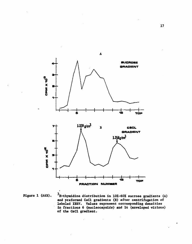
a Sorvall AH627 rotor, at 100,000 x g for 4 hours at 20C. Gradients
were subsequently fractionated into 0.5 ml fractions and the elution
3 profile of the H-thymidine labeled IBRV was obtained. CMV fractions
3 3 corresponding to densities of 1.263 g/cm and 1.219 g/cm (38), and
IBRV fractions corresponding to densities of 1.329 g/cm and 1.258
3 g/cm were pooled together independently, diluted with virus buffer
and pelleted by centrifugation at 110,000 x g at 4C for 2 hours in a
Sorvall AH627 rotor. Virus pellets were stored at -70C.
Preparation of Antigens
Virus pellets were resuspended in sterile deionized water and
treated with a 1:4000 dilution of 37% formaldehyde at 37C for three
hours. Preparations were then held at 4C for 18 hours and subsequent
ly pelleted by centrifugation in a Sorvall SS-34 rotor at 27,000 x g
for 60 minutes at 4C. Loss of infectivity of formalin-treated virus
was assured by the inability of an undiluted portion to produce cyto-
pathology in appropriate cell cultures. The formalin-inactivated
purified preparations of CMV and IBRV served as virus antigens.
12
Freeze-thaw lysates of cells which were clarified by centri-
fugatlon at 1100 x g for 15 minutes In a Sorvall SS-34 rotor served
as cellular control antigens.
Ultraviolet Sterilization of Antigens
Preparations to be used as antigens for animal injections and
lymphocyte stimulation assays were sterilized by exposure to ultra
violet light (UA-3GE 360W) for 5 minutes at a distance of 35 cm.
Sterility was assured by absence of growth after inoculation of pre
parations onto blood agar plates (5% sheep red blood cell; G1BC0).
Protein Concentration Determination of Antigens
Protein concentrations were determined for viral antigen and
cellular control antigen suspensions using the Bio-Rad colorimetric
technique (Bio-Rad Laboratories, Richmond, Ca.). Suspensions were
then adjusted to 150 yg of protein per ml of sterile deionized water.
Electron Microscopy of Virus Preparations
Virus preparations were spread on Formvar treated, carbon
coated copper electron microscopy grids, and negatively stained with
a 2% phosphotungstate solution. Virus preparations were examined and
photographs taken at various magnifications using a Phillips EH 200
electron microscope.

13
Sensitization of Animals
Rabbits were sensitized by injecting a total of 450 Jig of
purified CMV or IBRV antigen. Each of three injections was given sub-
cutaneously and prepared as an equal volume of antigen to Freunds in
complete adjuvant (Difco Laboratories, Detroit, Ml.), with a total
volume per injection of 2 ml. The three Injections were given within
15 days, each 5 days apart.
Mitogens
Phytohaemeagglutinin (PHA-P, GIBCO) was prepared as a 10 mg/ml
stock solution in sterile deionized water. Concanavalin A (Con A,
Sigma Chemical Co., St. Louis, Mo.) was prepared as a 1 mg/ml stock
solution in sterile deionized water. The stock solutions were filter
sterilized through .45 ym filters and then diluted to appropriate con
centrations in Roswell Park Memorial Institute 1640 medium (EPMI-1640,
GIBCO).
Lymphocyte Culture Medium
RPMI-1640 was buffered with 10 mM HEPES and supplemented with
10 mM Gentamycin Sulfate and 250 mM Fungizone (GIBCO). Pooled normal
rabbit serum which was heat inactivated at 56C for 30 minutes was
added to a final concentration of 5%. Complete medium was stored at
-20C.
14
Preparation of Peripheral Blood Lymphocytes for Assay
Approximately 18 ml of blood was collected via cardiac punc
ture into syringes containing 2 ml of 100 IU of heparin per ml. The
heparinized blood was diluted 1:2 with sterile RPMI-1640, layered over
Ficoll-Paque (Pharmacia, Piscataway, N.J.) and centrifuged at 400 x g
for 30 minutes at room temperature. Peripheral blood lymphocytes were
collected from the interface, washed in RPMI-1640 and pelleted by
centrifugation at 100 x g for 20 minutes at 4C. Cell pellets were re-
suspended in RPMI-1640, viability assessed by trypan blue dye exclu
sion, and the concentration adjusted to 1.0 x 10^ viable cells per ml
in complete media.
Lymphocyte Stimulation by Antigens and Mitogens
Cells were dispensed in 0.2 ml volumes into wells of a 96 well
flat bottom microtiter plate (Costar, Cambridge, Mass.). Ten, 15 or
20 pi amounts of test antigen or mitogen were added to appropriate
wells and plates were incubated in a humidified atmosphere of 5% CC>2
at 37C. After 48 hours of incubation (unless otherwise indicated) in
mitogen wells, and 5 days of incubation in antigen wells, 1 yCi of
3 3 1&-thymidine (5.0 Ci/mmol H-6-thymidine; Amersham Corp.) was added.
After a pulse labeling period of 24 hours, cells were harvested onto
glass fiber filters (Microbiological Associates, Walkersville, Md.)
using a Mini-MASH multiple automated sample harvester (Microbiological
Associates), and washed with 0.15 M NaCl. Filter pads were dried
15
under a heat lamp and transferred into plastic disposable scintilla
tion vials (New England Nuclear, Boston, Mass.)* Scintillation cock
tail (Betamax; Westchem Products, San Diego, Ca.) was added to each
vial in 5 ml amounts, and radioactivity in the vials was counted in a
Tri-Carb liquid scintillation spectrophotometer (Model 332, Packard
Instrument Co., LaGrange, 111.) at a gain of 6.5 and window settings
of 50 and 1000.
Statistical Analysis of Data
Data were analyzed using a Student's paired-t test on abso
lute counts per minute recorded on the Tri-Carb liquid scintillation
spectrophotometer.
17
SUCROSE
ORADIENT
1--
16 TOP
1-329 {/en? 7--CBCL
QRADIENT
S<
1--
FRACTION NUMBER
Figure 1 (A&B). H-thyroidine distribution in 10%-60% sucrose gradients (A) and preformed CsCl gradients (B) after centrifugation of labeled 1BRV. Values represent corresponding densities in fractions 6 (nucleocapsids) and 14 (enveloped virions) of the CsCl gradient.
18
3 3 for CUV are 1.263 g/cm for naked nucleocapslds and 1.219 g/cm for
enveloped virions and dense bodies (39).
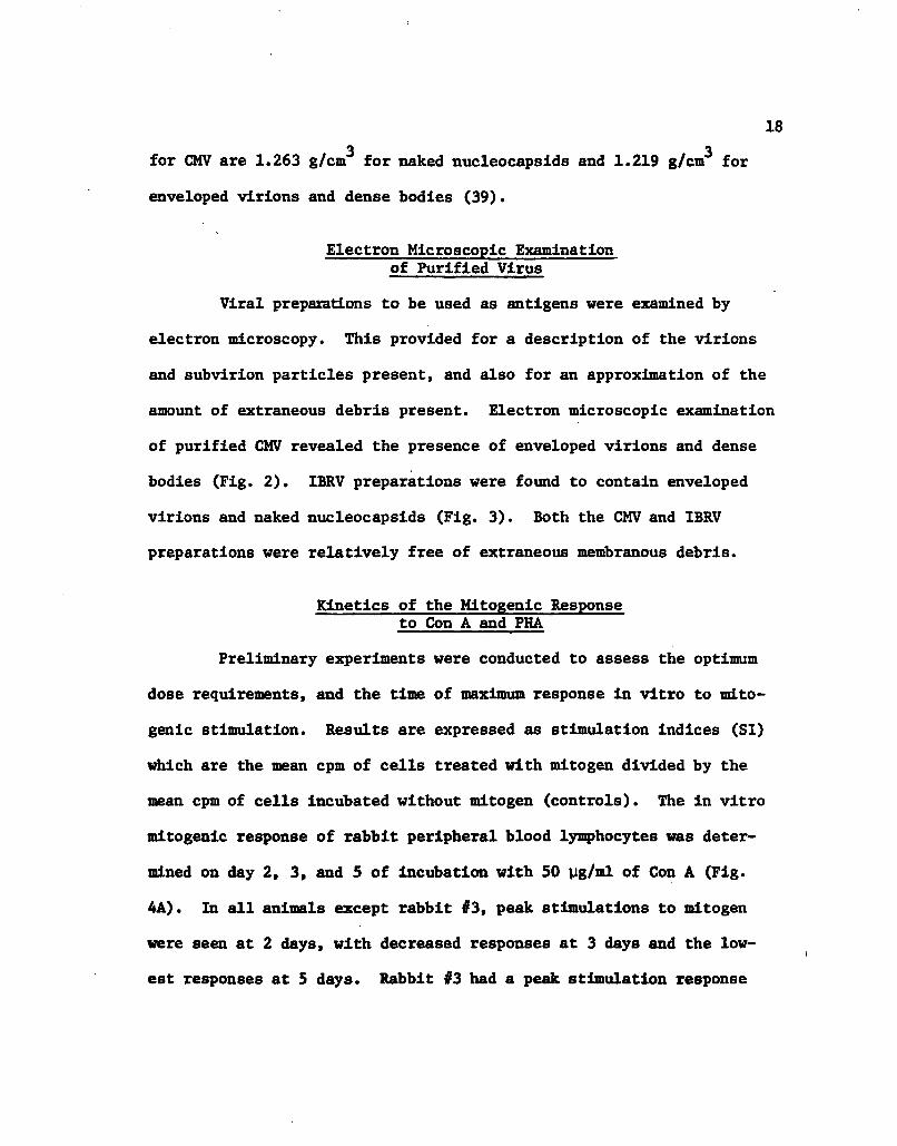
Electron Microscopic Examination of Purified Virus
Viral preparations to be used as antigens were examined by
electron microscopy. This provided for a description of the virions
and subvirion particles present, and also for an approximation of the
amount of extraneous debris present. Electron microscopic examination
of purified CMV revealed the presence of enveloped virions and dense
bodies (Fig. 2). IBRV preparations were found to contain enveloped
virions and naked nucleocapslds (Fig. 3). Both the CMV and IBRV
preparations were relatively free of extraneous membranous debris.
Kinetics of the Mitogenlc Response to Con A and PHA
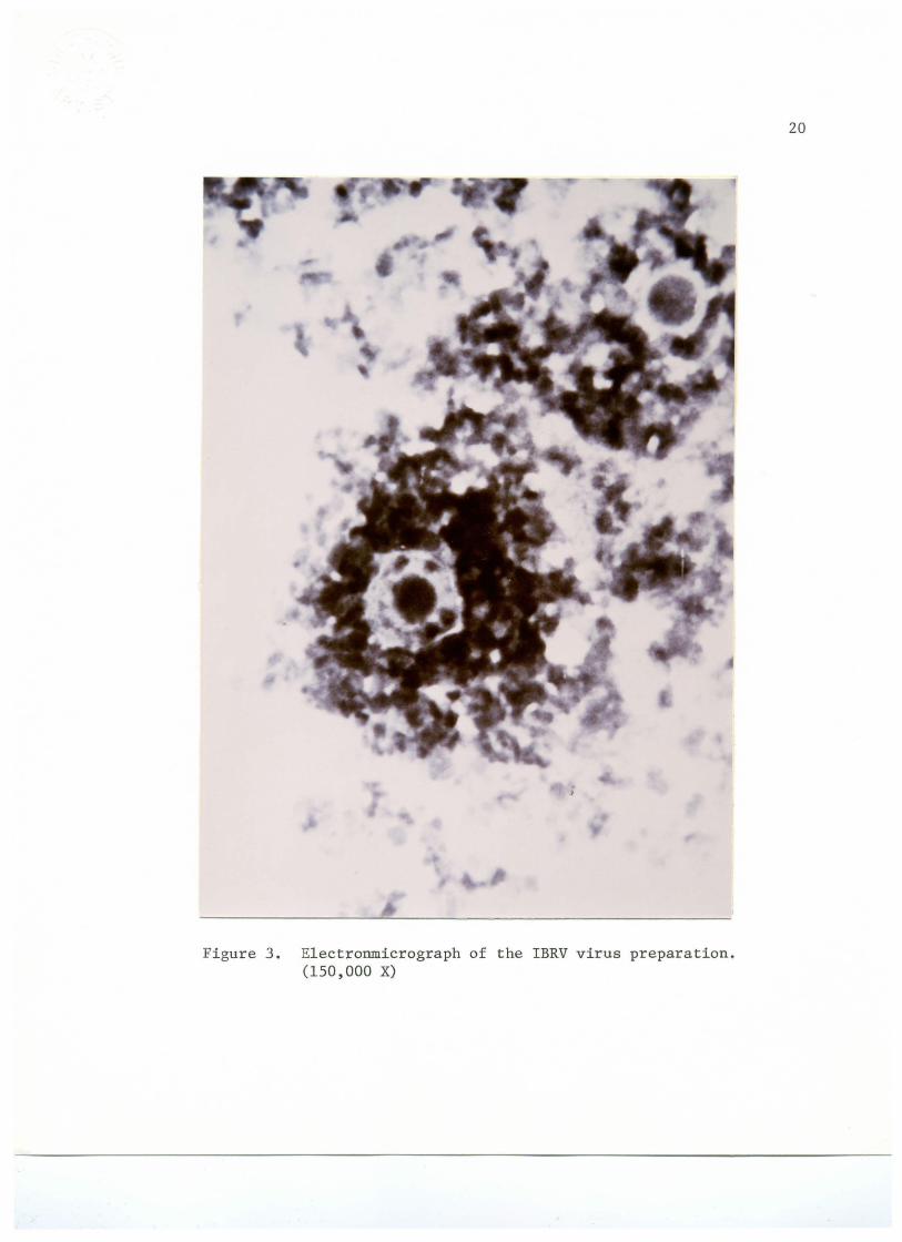
Preliminary experiments were conducted to assess the optimum
dose requirements, and the time of maximum response in vitro to mito
genlc stimulation. Results are expressed as stimulation indices (SI)
which are the mean cpm of cells treated with mitogen divided by the
mean cpm of cells incubated without mitogen (controls). The in vitro
mitogenlc response of rabbit peripheral blood lymphocytes was deter
mined on day 2, 3, and 5 of incubation with 50 yg/ml of Con A (Fig.
4A). In all animals except rabbit #3, peak stimulations to mitogen
were seen at 2 days, with decreased responses at 3 days and the low
est responses at 5 days. Rabbit #3 had a peak stimulation response

Figure 2. E1ectronmicrograph of the CMV virus preparation. (150,000 X)
19
j
Figure 3, Electronmicrograph of the IBRV virus preparation. (150,000 X)
20
X II D !
Concanavalin A 51 pi/Ill
A
Phytohaema&glutinin P B
25 PI/Ill
3
2d 3d 5dlrs 1 2 . 3 4
·A-IT NUM-A
4
Figure 4 (A&B). Mitogenic response of rabbit lymphocytes to Concanavalin A and Phytohaemeagglutinin as a function of in vitro culture time for four test rabbits. Stimulation index -= cpm in mitogen stimulated cultures + cpm in control cultures.
21

22
to Con A at 3 days, with lower responsiveness at 2 days and 5 days.
The mitogenic responses to PHA (25 yg/ml) on day 2, 3, and 5 of in
cubation are shown in Fig. 4B. For all rabbits except #3, peak re
sponses were seen at 2 days, with lower responses at 3 days and 5
days. Lymphocytes of rabbit #3 had a peak response at 3 days, with
lower responsiveness at 2 days and 5 days. In subsequent experi-
3 ments, mitogen stimulated cultures were pulsed with H-thymidine at
2 days of incubation.
The stimulation of peripheral lymphocytes as a function of
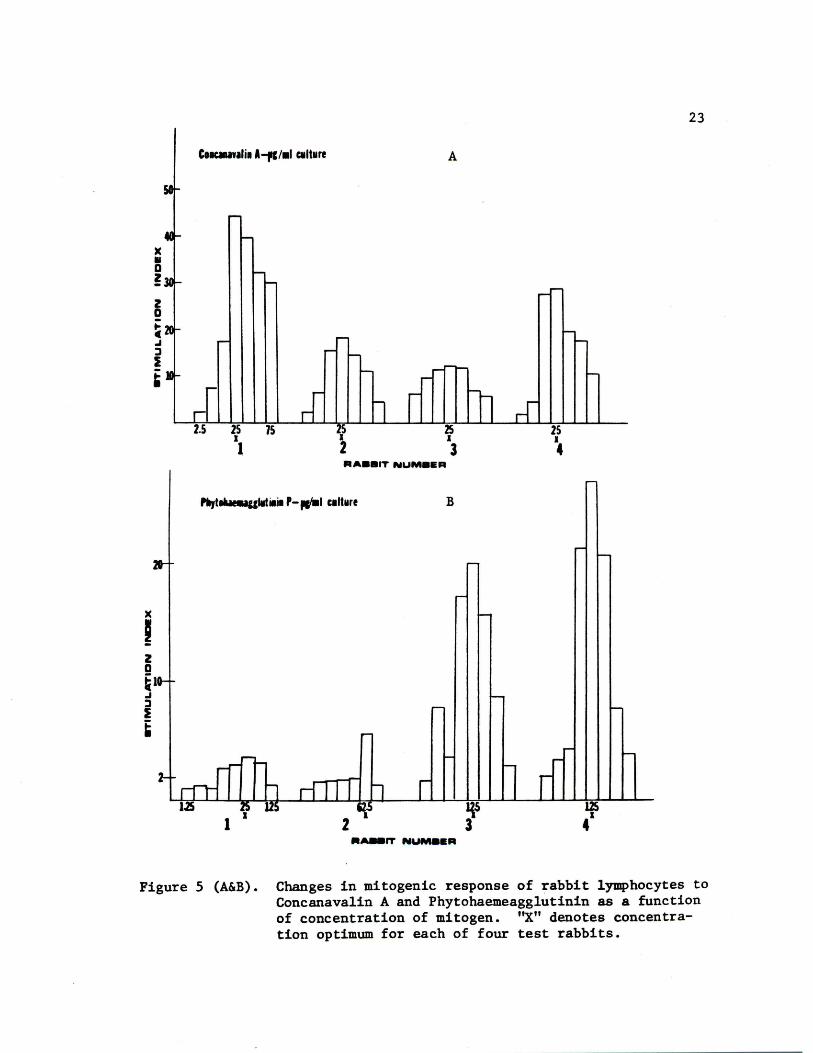
Con A concentration is illustrated in Fig. 5A. Although the lympho
cytes of rabbits #2 and #3 had quantitatively lower responses to the
mitogen than those of rabbits #1 and #4, the peak stimulation in
lymphocytes of all 4 rabbits occurred at a concentration of 25 yg/ml
of culture. Shown in Fig. 5B are the responses of the peripheral
blood lymphocytes as a function of concentration of PHA. The optimum
response of rabbits #3 and #4 was at a dose of 12.5 yg/ml, while
rabbit #1 responded best at 25 yg/ml and rabbit #2 at 62.5 yg/ml.
Rabbits #1 and #2 had a quantitatively lower response than rabbits
#3 and #4 to the PHA mitogen.
Lymphocyte Blastogenic Response of Rabbits Immunized with IBRV and CMV
To determine whether a CMI response would detect antigenic
relatedness between IBRV and CMV, the in vitro lymphocyte blastogenic
responses of four rabbits (two immunized with IBRV and two Immunized
with CMV) to homolgous and heterologous antigens were studied. At
)(
' z a ~1 .I :l I ~
75
J I
A
B
2 qs 3
125 41
23
Figure 5 (A&B). Changes in mitogenic response of rabbit lymphocytes to Concanavalin A and Phytohaemeagglutinin as a function of concentration of mitogen. "X" denotes concentration optimum for each of four test rabbits.
24
every weekly bleeding period, six concentrations of both CMV and IBRV
antigens were Incubated with peripheral blood lymphocytes in culture.
The mean cpm of triplicate samples was calculated, as well as stan
dard deviations for each concentration. The response of peripheral
lymphocytes of each rabbit at 1 week post-immunization is shown in
Table 1. In the case of rabbit #1, optimal response to CMV was at a
dilution of 1:2 of the original 150 yg/ml Injection preparation, when
10 yl was added to the culture. Response to IBRV was optimal at 1:2
(lOyl). Rabbit #2 had lymphocytes which responded best in culture
to a concentration of 1:10 (lOyl) of CMV and 1:2 (20yl) of IBRV.
The amount of antigen found to give the greatest stimulatory re
sponse in lymphocytes of rabbits #3 and #4 was 20 yl of a 1:2 dilu
tion of the 150 yg/ml injection preparation for both the CMV and
IBRV antigens. The responses at the above antigen concentrations
were plotted as a function of time (Figs. 6 and 7).
The mitogenic responses of rabbit peripheral lymphocytes
were used to assess the in vitro cultivation technique. This tech
nique was to be employed in subsequent assays to determine reactivity
of the lymphocytes to viral antigens. The results of studies to
determine optimum mitogen concentrations and proper timing of pulse-
labeling were utilized in a standard procedure at each weekly bleed
ing to assess the culturing technique.
The in vitro lymphocyte blastogenic responses to IBRV and
CMV antigens for two rabbits immunized with IBRV are shown in Figs.
6A (rabbit #1) and 6B (rabbit #2). The data plotted are mean cpm of
25
3 Table 1. H-Thymidine uptake of peripheral lymphocytes at one week
post-immunization as a function of virus antigen concentration.
1BRV Immunized CMV Immunized
Rabbit //I Rabbit #2 Rabbit 03 Rabbit #4
Antigen Concentration
1683(367)b 363(147)
3293(527) 492(192)
3729(365) 5324(1246)
3311(1099) 3390(1663)
3392(364) 4779(1960)
2629(315) 1185(1379)
1163(143) 262(33)
1927(1121) 212(9)
f
6020(396) 3242(506)
3780(5282) 1162(211)
1652(253) 307(8)
1768(784) 364(94)
1016(309) 245(20)
995(716) 308(29)
2355(414) 263(46)
CMV Antigen 1:2 20 yl
10 yl
CSV Antigen 1:10 20 yl 1466(333) 398(73)
10 yl 2690(176) 560(65)
CMV Antigen 1:100 20 yl 990(249) 234(121)
10 yl 2290(576) 278(43)
IBRV Antigen 1:2 20 yl 5067(990) 990(14)
10 yl 6723(724) 925(91)
IBRV Antigen 1:100 20 yl 3119(1002) 751(47)
10 yl 4009(850) 578(108)
IBRV Antigen 1:100 20 yl 1611(321) 295(47)
10 yl 2815(140) 369(156)
CONTROLS 3778(142) 582(111)
dilution of stock 150 yg/ml antigen solution
^Arithmetic mean of cpm of triplicate samples (standard deviation)
" D ~
X ~ 11. u
l\1 ·a c-
X
~ a. u
RABBIT~ ·IBRV A
PRE E!
RABBIT e ·IBRV B
J •,
' ' . ' ' ...
PRE
' ' ' " ' ' '
--- I ""-----t'
I I
I
I I
I I
I
D-IBRV
8-CMV
·-CONTROL
3
D•IBRV O·CMV
···CONTROL
3
WEEKS POST· IMMUNIZATION
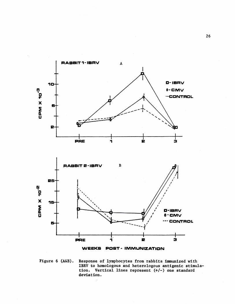
Figure 6 (A&B). Response of lymphocytes from rabbits immunized with IBRV to homologous and heterologous antigenic stimulation. Vertical lines represent (+/-) one standard deviation.
26
27
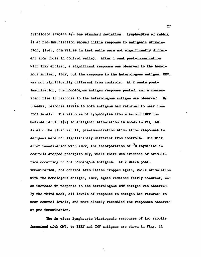
triplicate samples +/- one standard deviation. Lymphocytes of rabbit
#1 at pre-immunization showed little response to antigenic stimula
tion, (i.e., cpm values in test wells were not significantly differ
ent from those in control wells). After 1 week post-immunization
with IBRV antigen, a significant response was observed to the homol-
gous antigen, IBRV, but the response to the heterologous antigen, CMV,
was not significantly different from controls. At 2 weeks post-
immunization, the homologous antigen response peaked, and a concom
itant rise in response to the heterologous antigen was observed. By
3 weeks, response levels to both antigens had returned to near con
trol levels. The response of lymphocytes from a second IBRV im
munized rabbit (#2) to antigenic stimulation is shown in Fig. 6B.
As with the first rabbit, pre-immunizatlon stimulation responses to
antigens were not significantly different from controls. One week
3 after immunization with IBRV, the incorporation of H-thymidine in
controls dropped precipitously, while there was evidence of stimula
tion occurring to the homologous antigens. At 2 weeks post-
immunization, the control stimulation dropped again, while stimulation
with the homologous antigen, IBRV, again remained fairly constant, and
an increase in response to the heterologous CMV antigen was observed.
By the third week, all levels of response to antigen had returned to
near control levels, and more closely resembled the responses observed
at pre-immunization.
The in vitro lymphocyte blastogenic responses of two rabbits
immunized with CMV, to IBRV and CMV antigens are shown in Figs. 7A
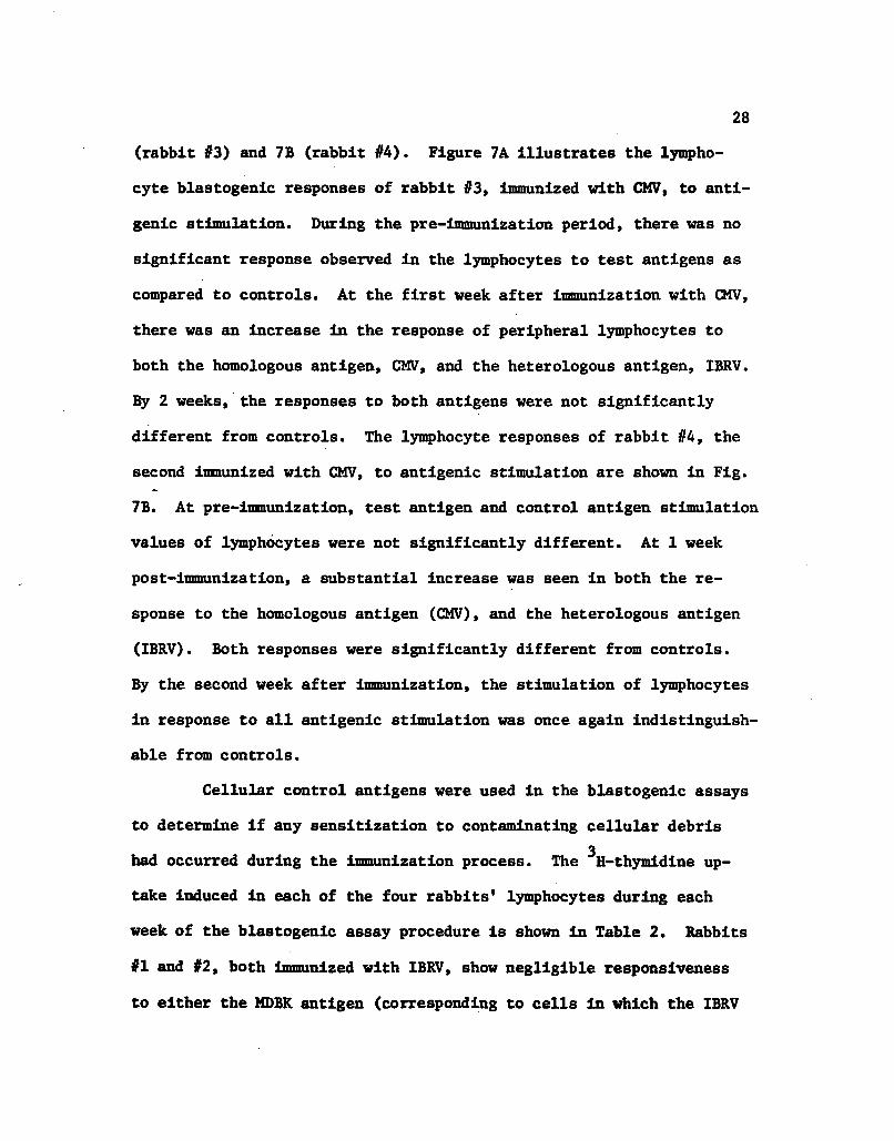
28
(rabbit #3) and 7B (rabbit #4). Figure 7A illustrates the lympho
cyte blastogenic responses of rabbit #3, immunized with CMV, to anti
genic stimulation. During the pre-immunization period, there was no
significant response observed in the lymphocytes to test antigens as
compared to controls. At the first week after immunization with CMV,
there was an Increase In the response of peripheral lymphocytes to
both the homologous antigen, CMV, and the heterologous antigen, IBRV.
By 2 weeks, the responses to both antigens were not significantly
different from controls. The lymphocyte responses of rabbit #4, the
second imnunized with CMV, to antigenic stimulation are shown in Fig.
7B. At pre-iimninization, test antigen and control antigen stimulation
values of lymphocytes were not significantly different. At 1 week
post-immunization, a substantial increase was seen in both the re
sponse to the homologous antigen (CMV), and the heterologous antigen
(IBRV). Both responses were significantly different from controls.
By the second week after immunization, the stimulation of lymphocytes
in response to all antigenic stimulation was once again indistinguish
able from controls.
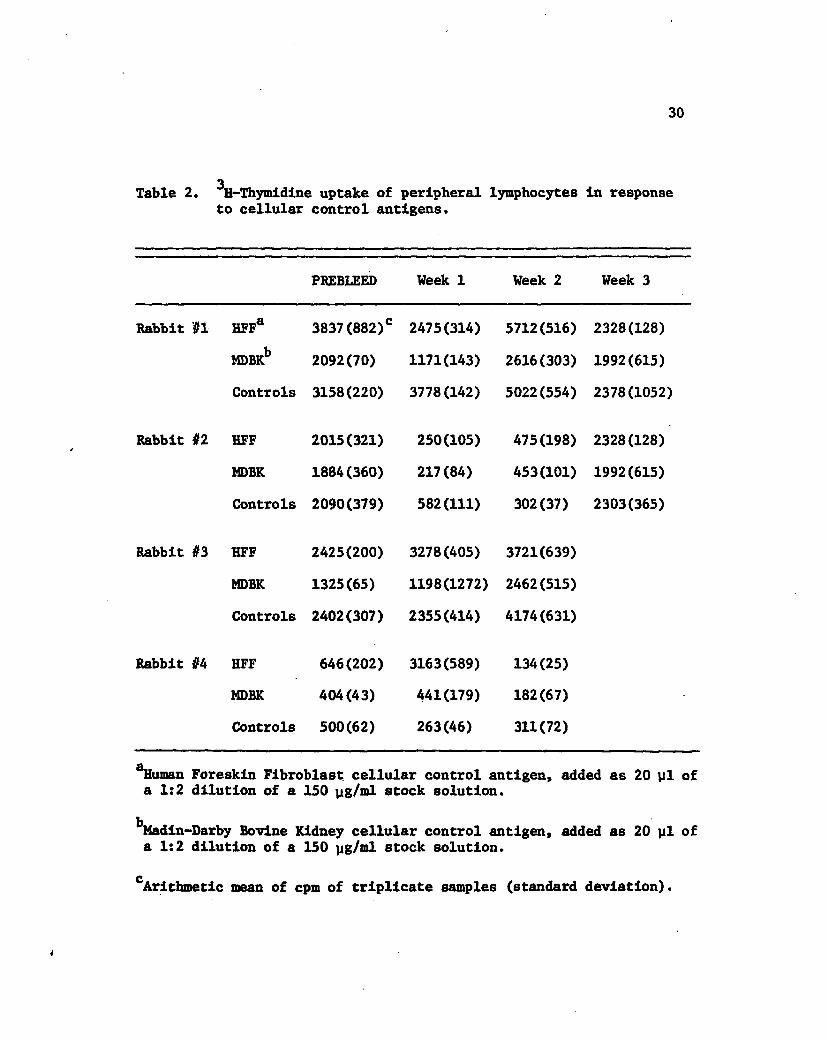
Cellular control antigens were used in the blastogenic assays
to determine if any sensitization to contaminating cellular debris
3 had occurred during the immunization process. The H-thymidine up
take induced in each of the four rabbits' lymphocytes during each
week of the blastogenic assay procedure is shown in Table 2. Rabbits
#1 and #2, both immunized with IBRV, show negligible responsiveness
to either the MDBK antigen (corresponding to cells in which the IBRV
)(
~ D. u
RABBIT 3·CMV A
--- -----~/~--
p E
RABBIT 4· CMV B
C·IBRV O·CMV
---CONTROL
2
29
D·IBRV
O·CMV
··-coNTROL
------ ' _ ... _.----r-- ---
WEEKS POST· IMMUNIZATION
Figure 7 (A&B). Response of lymphocytes from rabbits immunized with CMV to homologous and heterologous antigenic stimulation. Vertical lines represent (+/-) one standard deviation.
30
3 Table 2. H-Thymidine uptake of peripheral lymphocytes in response to cellular control antigens.
PREBLEED Week 1 Week 2 Week 3
Rabbit #1 HFFa 3837(882)c 2475(314) 5712(516) 2328(128)
MDBKb 2092(70) 1171(143) 2616(303) 1992(615)
Controls 3158(220) 3778(142) 5022(554) 2378(1052)
Rabbit $2 HFF 2015(321) 250(105) 475(198) 2328(128)
MDBK 1884(360) 217(84) 453(101) 1992(615)
Controls 2090(379) 582(111) 302(37) 2303(365)
Rabbit if3 HFF 2425(200) 3278(405) 3721(639)
MDBK 1325(65) 1198(1272) 2462(515)
Controls 2402(307) 2355(414) 4174(631)
Rabbit U HFF 646(202) 3163(589) 134(25)
MDBK 404(43) 441(179) 182(67)
Controls 500(62) 263(46) 311(72)
^uman Foreskin Fibroblast cellular control antigen, added as 20 yl of a 1:2 dilution of a 150 yg/ml stock solution.
^Madin-Darby Bovine Kidney cellular control antigen, added as 20 yl of a 1:2 dilution of a 150 yg/ml stock solution.
£ Arithmetic mean of cpm of triplicate samples (standard deviation).
31
was propagated), or the HFF antigen (derived from the cells used to
propagate CMV). The response seen against antigens in these rabbits
can be said to be virus-specific, and not reflective of sensitization
against contaminating cellular antigenic determinants. The lympho
cytes from rabbits immunized with CMV (#3 and #4), show no responsive
ness to MDBK antigens. However, at week 1, significant responses to
the HFF cellular antigen control were observed.
A summary of statistical evaluations of the data is shown in
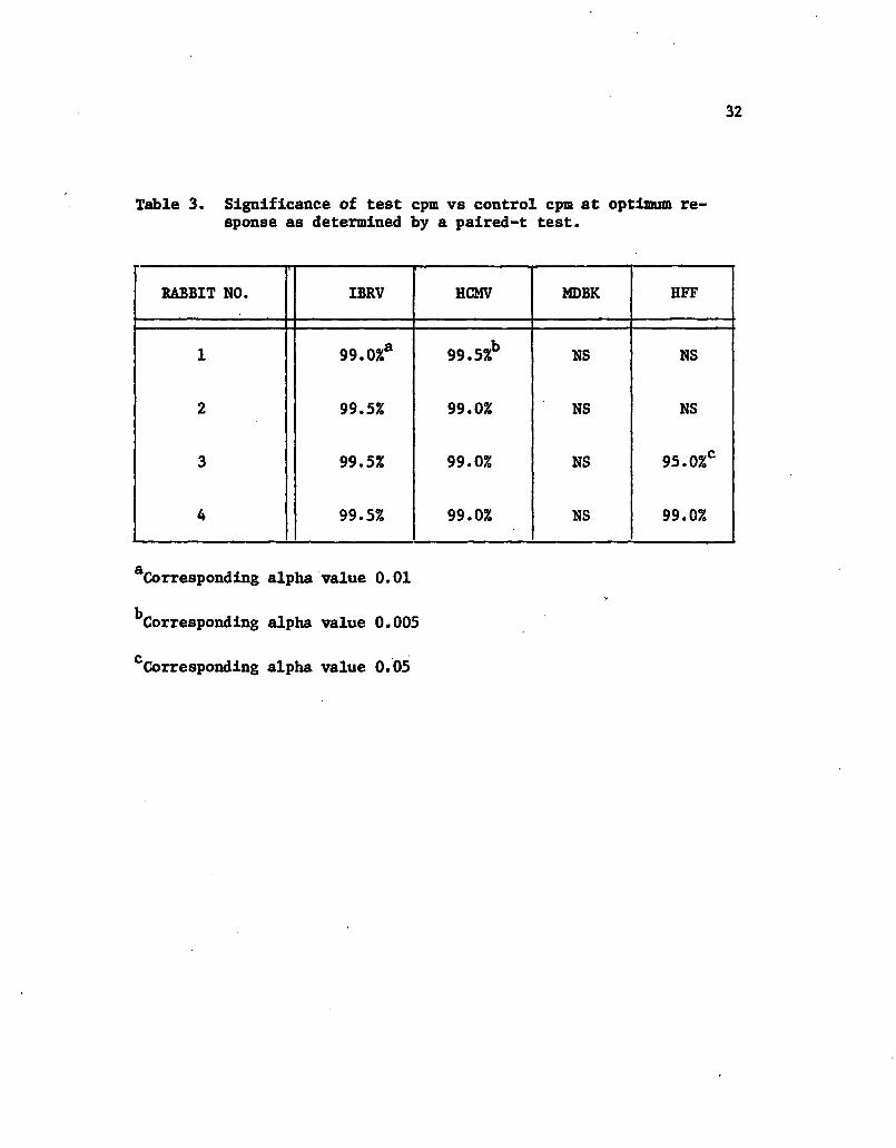
Table 3. As evaluated by a paired-t test, all rabbits gave statisti
cally significant reactions to the homologous antigen (IBRV: rabbits
#l~and #2, and CMV: rabbits #3 and #4). Lymphocytes of all rabbits
reacted significantly to the heterologous, or cross-reacting antigen
in vitro (CMV: rabbits #1 and #2, and IERV: rabbits #3 and #4).
Babbits #1 and #2 showed no statistically significant reactivity to
cellular control antigens in vitro. Rabbits #3 and //4 showed signifi
cant reactivity to the HFF celluar control antigen, but not to the
MDBK cellular control antigen.
32
Table 3. Significance of test cpm vs control cpm at optimum response as determined by a palred-t test.
BABBIT NO. IBRV HCMV MDBK HFF
1 99.0%a 99.5%b NS NS
2 99.5% 99.0% NS NS
3 99.5% 99.0% NS 95.0%°
4 99.5% 99.0% MS 99.0%
Corresponding alpha value 0.01
^Corresponding alpha value 0.005
cCorresponding alpha value 0.05
DISCUSSION
As a means of obtaining virus preparations which were rela
tively free of contaminating cellular debris, a double gradient tech
nique was utilized during purification. First, virus and subviral
components were separated from cellular debris on the basis of size
by velocity sedimentation through sucrose gradients. This technique
also separated naked nucleocapsids, enveloped virions, and dense
bodies. Second, the isopycnic banding of virus preparations in CsCl
separated virus from cellular material on the basis of density, and
enabled the estimation of densities of naked nucleocapsids and en
veloped virions of IBRV.
Electron microscopic examination of virus preparations re
vealed the presence of virions with typical herpesvirus morphology
in both the CMV and IBRV preparations. The virus suspensions ap
peared by visual inspection to be relatively free of cellular membran
eous materials. The majority of material present in the preparations
was viral in nature.
The response of lymphocytes to mitogenic stimulation by Con A
and PHA was used to assess the in vitro cultivation technique. This
basic technique was used in subsequent assays to determine the re
sponse of lymphocytes to antigenic stimulation. Examination of stimu
lation at three time Intervals revealed in most cases, optimum
response to both Con A and PHA occurred after 2 days of in vitro
33
34
culture. The optimum concentration of mitogen was also assessed.
The concentrations giving optimum stimulation were used throughout
all antigenic stimulation assays to assess the reactivity of cqlls in
culture. Con A and PHA have been shown to stimulate separate sub-
populations of T cells in the murine system (40), and it is thought
that through their joint use the majority of T cells present in
peripheral blood can be monitored. Throughout the procedure, all
animals maintained acceptable mitogenic stimulation levels (SI of
greater than 3), indicating the culturing technique was maintained
satisfactorily, and reactivity of rabbit lymphocytes was near levels
found at pre-imnunization. In the case of rabbit #2, mitogenic stimu
lation dropped at week 1, and returned to near normal levels at week
3. This trend was very similar to the pattern found in this rabbit
with antigenic stimulation.
As a means of determining the concentration of antigen which
would give the maximum response in sensitized lymphocytes, responses
to several concentrations of antigen were determined throughout the
procedure. These concentrations appeared to be fairly individual, as
most of the rabbits' lymphocytes reacted at different concentration
optimums.
Rabbit #1 (Immunized with IBRV), gave a rapid response to
antigenic stimulation with IBRV which was significant at week 1. The
response to CMV, the heterologous antigen, lagged a week behind and
did not become significant until week 2, when the response to IBRV
peaked. The lymphocytes of this rabbit injected with IBRV, could be
stimulated by both the homologous antigen (IBRV) and the heterologous

35
antigen (CMV). This response indicates that lymphocytes sensitized
against IBRV recognized certain similar or cross-reacting antigens of
CMV.
Rabbit //2, also immunized with IBRV, had lymphocytes which
gave a significant response to both IBRV and CMV at 2 weeks post-
immunization. The basal reactivity rate of this rabbit's lympho
cytes, along with their mitogenic reactivity, decreased dramatically
at one week post-immunization.
A lymphocytic response to both the CMV and IBRV antigens was
also detected in rabbits immunized with CMV (#3 and #4). Although
significant responses to both the homologous (CMV) and heterologous
(IBRV) antigens were detected in rabbit #3, the response to the heter
ologous antigen was statistically higher than the response to the
homologous antigen. This could possibly be attributed to the time of
stimulation of the cells in vitro. All antigenic lymphocyte responses
were pulsed after 5 days of incubation with the antigen. The previous
mitogen response experiments had revealed that lymphocytes of rabbit
#3 had a different optimum time of response than the other 3 rabbits.
Therefore, it is possible that the optimum response time of rabbit #3's
lymphocytes in vitro to stimulation by antigen could also be different
from the other animals.
The response in rabbit #4 was very brisk, peaking at one week
post-immunization to both CMV and IBRV antigens. This response indi
cates that after immunization with the CMV antigen preparation, cells
of the rabbit were sensitized against CMV, and were reactive against
certain antigens found in the IBRV preparation.
36
Through the purification technique, an IBRV antigen was pre
pared which appeared to be relatively free of extraneous debris from
the MDBK cells in which it was propagated, as lymphocytes from
rabbits injected with IBRV gave no significant reaction to MDBK cellu
lar antigens. The CMV preparation on the other hand, appeared to have
a lesser degree of purity with respect to HFF cellular contamination,
since the lymphocytes of the 2 rabbits immunized with CMV did give
significant responses to HFF cellular control antigens.
All rabbits in the study showed responsiveness to the homolo
gous antigen with which they were injected, indicating sensitization
had occurred during immunization. Each rabbit also had lymphocytes
which would react in vitro to the heterologous antigen, indicating
that the two viruses, IBRV and CMV, share some similar or identical
antigenic determinants. The reactivity of lymphocytes in the case of
IBRV immunized animals can be said to be virus specific, as no reacti
vity to cellular antigens was demonstrated. The stimulation of CMV
immunized rabbits' lymphocytes being virus specific is highly likely,
but can be disputed as there was some reactivity elicited to host
cellular antigens as well as viral antigens. Although the immunizing
antigen preparation appears to be contaminated with HFF cellular mater
ial, it cannot account for the significant cross-reactivity observed
between CMV and IBRV antigens. This is because the lymphocytes of CMV
Immunized rabbits were not stimulated by MDBK cellular antigens.

SUMMARY
Cytomegalovirus (CMV) is a human herpesvirus of current impor
tance as an ubiquitous human pathogen. The need for therapeutic
methods of control of cytomegaloviral diseases has brought to light
the use of Infectious Bovine Rhinotracheitis Virus (IBRV), a bovine
pathogen, to facilitate or augment the stimulation of an immune re
sponse against CMV. The immune cross-reactivity of these two viruses
within the humoral immune system has been previously documented using
immunofluorescent techniques, and recent reports also indicate that
this cross-reactivity may be detected by cell-mediated immune responses
as well.
In an effort to document or illustrate this cross-reactivity
within the cell-mediated Immune system, rabbits were injected with
either CMV or IBRV. Blastogenic assays were used to assess the reacti
vity of the rabbit peripheral blood lymphocytes to both homologous and
heterologous antigens.
Initial experiments characterizing the basic methodology
necessary for in vitro cultivation of rabbit peripheral blood lympho
cytes were accomplished using mitogenic stimulation. These basic
conditions were maintained for antigenic blastogenesis assays. All
animals were found, as a result of antigenic stimulation in the blasto
genesis assays, to have lymphocytes which could be stimulated by the
viral antigen which was used for immunization. The heterologous
37

38
antigen was also stimulatory in all cases, indicating that antigens
of the two viruses which are recognized by cell-mediated immune
mechanisms were cross-reactive.
This study highlighted trends which had been previously ob
served relating to the immune cross-reactivity of CMV and IBRV, and
indicated such cross-reactivity can be determined and defined in the
blastogenesis assay. The rabbit proved to be an appropriate host
with which to study this phenomenon. The study encourages the further
characterization of this cross-reactivity, and enhances the feasibility
for the use of IBRV antigen, in some form, as a protective or thera
peutic agent in CMV Infections.
REFERENCES
1. Dulbecco, R. 1973. The Herpesviruses. In: Microbiology. Harper & Row Publishers, Inc., Hagerstown, Md. Chap. 53, p. 1242.
2. Sarov, I. and I. Abady. 1975. The morphogenesis of human cytomegalovirus. Isolation and characterization of cytomegalo-virions and dense bodies. Virology 66:464.
3. Craighead, J.E., Kanish, R.E. and J.D. Almeida. 1972. Non-viral microbodies with viral antigenicity produced in cytomegalovirus infected cells. J. Virology 10:766.
4. Misra, V., Blumenthal, R.M., and L.A. Babiuk. 1981. Proteins specified by Bovine Herpesvirus 1 (Infectious Bovine Rhino-tracheitis Virus). J. Virology 40:367.
5. Joncas, J.H., Menezas, J. and E.S. Huang. 1975. Persistence of cytomegalovirus genomes in lymphoid cells after congenital infection. Nature 258:432.
6. Lans, D.J. and B. Noren. 1968. Cytomegaloviremia following congenital infection. J. Pediatrics 73:812.
7. Mars, J. 1975. Cytomegalovirus: a major cause of birth defects. Science 190:1184.
•
8. Stern, H. 1975. Intrauterine infection with cytomegalovirus. Proc. Roy. Soc. Med. 68:367.
9. Reynolds, D.W., Stagno, S., Stubbs, K.G., Dahle, A.J., Livingston, M.M., Saxon, S.S., and C.A. Alford. 1974. Inapparent congenital cytomegalovirus infection with elevated cord IgM levels: causal relation with auditory and mental deficiency. New England J. Med. 290:291.
10. Armstrong, D., Haghbin, M., Balakrishman, S., and L. Murphy. 1971. Asymptomatic cytomegalovirus infection in children with leukemia. Amer. J. Dis. Child. 122:404.
11. Heller, T.H. 1971. The cytomegaloviruses: ubiquitous agents with protean clinical manifestations (Part Two). New England J. Med. 285:267.
39
40
12. Stagno, S., Reynolds, D.N., Pass, R.F., and C.A. Alford. 1980. Breast milk and the risk of cytomegalovirus Infection. New England J. Med. 302;1073.
13. Weller, T.H. 1971. The cytomegaloviruses: ubiquitous agents with protean clinical manifestations (Part One). New England J. Med. 285;203.
14. Kantor, G.L. and L.S. Goldberg. 1971. Cytomegalovirus induced post-perfusion syndrome. Sem. Hemat. 8^:261.
15. Meyers, J.D., Spencer Jr., H.C., Watts, J.C., Gregg, M.B., Stewart, J.A., Troupin, R.H., and E.D. Thomas. 1975. Cytomegalovirus pneumonia after human marrow transplantation. Ann. Intern. Med. 82: 181.
16. Haahr, S., Larsen, A.M., Andersen, H.K., and E.S. Spencer. 1979. Cell-mediated and humoral immune responses to herpes simplex virus and cytomegalovirus in renal transplant patients. J. Clin. Microbiol. 10:267.
17. Fiala, M., Payne, J.E., Berne, T.V., Moore, T.C., Mbntgomerie, J.Z., Chatterjee, S.N., and L.B. Guze. 1975. Epidemiology of cytomegalovirus infection after transplantation and Immunosuppression. J. Infect. Dis. 132;421.
18. Fine, R.N., Grushkin, C.M., Anand, S., Lieberman, E., and H.T. Wright Jr. 1970. Cytomegalovirus in children. Amer. J. Dis. Child. 120:197.
19. Fine, R.N., Grushkin, C.M., Malekzadeh, M., and H.T. Wright Jr. 1972. Cytomegalovirus syndrome following renal transplantations. Arch. Surgery 105;564.
20. Rytel, M.W., Aguillar-Torres, F.G., Balay, J. and L.R. Helm. 1978. Assessment of the status of CMI in cytomegalovirus-infected renal allograft patients. Cellular Immunology 37;31.
21. Lang, D.J. and B. Noren. 1968. Cytomegaloviremia following congenital infection. J. Pediatrics 73;812.
22. Bloom, B.R., and B. Rager-Zisman. 1975. Cell-mediated inmunity in viral infections. In: Viral Immunology and Immunopathology. Academic Press, Inc., New York, Chap. 7, p. 113.
23. Howard, R.J., Miller, J. and J.S. Najarian. 1974. Cytomegalovirus Induced immune suppression II. Cell-Mediated Immunity. Clin. Exp. Immunol. 18;119.
24. Linner, K.M., Cinroy, M.M., McCue, S.A. and H.H. Balfur, Jr. 1981. Cytomegalovirus-specific humoral and cellular immune responses in human pregnancy. J. Infect. Dis. 143:391.
41
25. Pollard, R.B., Egbert, P.R., Gallagher, J.G. and T.C. Merigan. 1980. Cytomegalovirus retinitis in immunosuppressed hosts: 1. Natural history and effects of treatment with adenine arabino-side. Ann. Intern.. Med. 93;655.
26. Falcoff, E., Falcoff, R., Fournier, F. 1966. Production en masse, purification partielle et characterisation d'un interferon destine a des essais the'rapeutiques humains. Ann. Inst. Pasteur (Paris) 111:562.
27. Elek, S.D., and H. Stern. 1974. Development of a vaccine against mental retardation caused by cytomegalovirus infection in utero. Lancet ,1:1.
28. Starr, S.E., Glazer, J.P., Friendman, H.M., Farquhar, J.D., and Stanley Plotkin. 1981. Specific-cellular and humoral immunity after Immunization with live Towne strain Cytomegalovirus vaccine. J. Infect. Dis. 143:585.
29. Starr, S.E., Barker, D.F., Perloff, L.J., Huang, E.-S., and S.A. Plotkin. 1979. Live cytomegalovirus vaccination of renal transplant candidates: preliminary trial. Ann. Intern. Med. 91:676.
30. Sarov, I., Larsen, A.M., Heron, I., and H.K. Andersen. 1978. Stimulation of human lymphocytes by cytomegalovirions and dense bodies. Med. Microbiol. Immunol. 166:81.
31. Albrecht, T., and F. Rapp. 1973. Malignant transformation of hamster embryo fibroblasts following exposure to ultraviolet-irradiated human cytomegalovirus. Virology 55:53.
32. Geder, L., Ladda, R.L., Kreider, J.H., Sanford, E.J. and F. Rapp. ,1979. Properties of human epitheliod cells established in vitro by a herpesvirus (IBRV-HMC) isolated from cytomegalovirus-transformed human cells. J. Nat. Cancer Inst. 63:1313.
33. Osborn, J.E. 1981. Cytomegaloviruses: Pathogenicity, Immunology, and Vaccine Initiatives. J. Infect. Dis. 143:618.
34. Bia, F.J., Griffith, B.P., Tarsio, M., and G.D. Hsiung. 1980. Vaccination for the prevention of maternal and fetal infection with guinea pig cytomegalovirus. J. Infect. Dis. 142:732.
35. Geder, L., Hyman, R.W., Figueroa, M., Oakes, J.E., litis, J.P., Dawson, M.S. and F. Rapp. 1978. Identification of a herpesvirus isolated from cytomegalovirus-transformed human cells. J. Virology 27:713.
42
36. Jones, J.F., Jeter, W.S., Fulginiti, V.A., Minnich, L.L., Pritchett, R.F., and R.J. Wedgewood. 1981. Treatment of childhood combined Epsteln-Barr virus/Cytomegalovirus infection with oral bovine transfer factor. Lancet 2^:122.
37. Zaia, J.A., Leary, F.L. and M.J. Levin. 1978. Specificity of the blastogenlc response of human mononuclear cells to herpesvirus antigens. Infec. & Immun. 20:646.
38. Plummer, G., and M. Benyesh-Melnick. 1964. A plaque reduction neutralization test for human cytomegalovirus. Proc. Soc. Exp. Biol, Med. 117:145.
39. Hung, E.-S., Chen, S.-T., and J.S. Pagano. 1973. Human Cytomegalovirus: I. Purification and characterization of viral DNA. J. Virology 12:1473.
40. Stobo, J.D., and W.E. Paul. 1973. Functional heterogeneity of murine lymphoid cells. J. Immunol. 110:362.
Related Documents


![Microfilms Drouin - Québec - [A]institutdrouin.com/microfilms/MF-Quebec.pdf · 2015. 1. 16. · Microfilms Drouin - Québec A Abbotsford (comté de Rouville) (Saint-Paul) 1856-12-15](https://static.cupdf.com/doc/110x72/60afb93291213a1e803bfaca/microfilms-drouin-qubec-a-2015-1-16-microfilms-drouin-qubec-a-abbotsford.jpg)


