Protoplasma (1999) 206:73-86 PROTOPLASMA Springer-Verlag 1999 Printed in Austria Ultrarapid endocytotic uptake of large molecules in Dunaliella species M. Ginzburg t'2'*, B. Z. Ginzburg 1'2and R. Wayne 1 ISection of Plant Biology, Cornell University, Ithaca, New York, and 2Institute of Life Sciences, The Hebrew University of Jerusalem, Jerusalem Received July 29, 1998 Accepted December 16, 1998 Summary. This paper describes the uptake of Lucifer Yellow carbo- hydrazide and fluorescent dextrans labeled with fluorescein isothio- cyanate or Sodium Green 0nolecular masses ranging from 522 to 2 x 10⁶ Da) by Dunaliella spp. halotolerant unicellular green algae iso- lated from salt pools in the Sinai peninsula. The fluorescent dyes were taken up into a set of vesicles around the nucleus and just above the chloroplast. It proved impossible to inhibit uptake of the fluores- cent compounds in cells treated with a large variety of metabolic and other inhibitors. Cell labeling was complete within half a minute of addition of fluorescent compounds to the outside medium; efflux was equally rapid. The results are interpreted in terms of an endocy- totic process whereby the outside medium, together with any sub- stance dissolved in it, remains within vesicles enclosed within the cell body but cycles rapidly between the plasma membrane and the interior of the cell. The outside medium does not pass across the vesicular membrane, nor enters the cytosol. Keywords: Algae; Dunaliella sp.; Endocytosis; Halophilism. Abbreviations: LYCH Lucifer Yellow carbohydrazide; FITC fluo- rescein-5-isothiocyanate; TCA trichloroacetic acid; DMSO dimethylsulfoxide; NEM N-ethyl maleimide; DNP dinitrophenol; CCCP m-chlorocarbonyl-cyanide phenylhydrazone; APM ami- prophos-methyl. Introduction Dunaliella is a genus of green microalgae distributed throughout the world in seas, lakes, and ponds vary- ing from brackish to highly saline. They are distin- guished by their ability to grow at high, changeable salt concentrations. Unlike most other plant cells, *Correspondence and reprints: Plant Biophysical Laboratory, Botany Department, Institute of Life Sciences, The Hebrew Univer- sity of Jerusalem, Givat Ram, Jerusalem 91904, Israel. Dunaliella cells do not have a rigid wall composed of cellulose or of any other inert material. Instead, the cell membrane is exposed directly to the bathing medium and is coated by glycoprotein patches which do not interfere with its mechanical properties (Oliveira etal. 1980). Thus the cells are able to change in volume when there are alterations in outside salt concentration (Trezzi etal. 1965). Two models have been put forward to explain the mechanism of volume regulation and exceptional halotolerance of Dunaliella cells. The most popular model (Ben-Amotz and Avron 1973) treats the cell as a single compartment enclosed by a plasma mem- brane highly impermeable to salts and small organic solutes such as glycerol and glucose. According to this model, the "osmotic pressure" of the salt in the outside medium is balanced within the cell by glyc- erol (Craigie and McLachlan 1964, Ben-Amotz and Avron 1973, Borowitzka and Brown 1974) and the glycerol is distributed uniformly throughout the intra- cellular compartment where its activity equals the activity of the outside salt solution. Glycerol acts as a "compatible solute"; this means that it does not inhib- it the cells' enzymes (Borowitzka and Brown 1974). Thus, according to this model, halotolerance is equiv- alent to osmoregulation because salt is effectively excluded from the cell and therefore cannot influence it. Results obtained by Ben-Amotz (1974), Bental et al. (1988), Pick etal. (1986), Karni and Avron (1988), and Fisher et al. (1997) have been interpreted in the light of this model.

Welcome message from author
This document is posted to help you gain knowledge. Please leave a comment to let me know what you think about it! Share it to your friends and learn new things together.
Transcript

Protoplasma (1999) 206:73-86
PROTOPLASMA �9 Springer-Verlag 1999 Printed in Austria
Ultrarapid endocytotic uptake of large molecules in Dunaliella species
M. Ginzburg t'2'*, B. Z. Ginzburg 1'2 and R. Wayne 1
ISection of Plant Biology, Cornell University, Ithaca, New York, and 2Institute of Life Sciences, The Hebrew University of Jerusalem, Jerusalem
Received July 29, 1998
Accepted December 16, 1998
Summary. This paper describes the uptake of Lucifer Yellow carbo- hydrazide and fluorescent dextrans labeled with fluorescein isothio- cyanate or Sodium Green 0nolecular masses ranging from 522 to 2 x 10⁶ Da) by Dunaliella spp. halotolerant unicellular green algae iso- lated from salt pools in the Sinai peninsula. The fluorescent dyes were taken up into a set of vesicles around the nucleus and just above the chloroplast. It proved impossible to inhibit uptake of the fluores- cent compounds in cells treated with a large variety of metabolic and other inhibitors. Cell labeling was complete within half a minute of addition of fluorescent compounds to the outside medium; efflux was equally rapid. The results are interpreted in terms of an endocy- totic process whereby the outside medium, together with any sub- stance dissolved in it, remains within vesicles enclosed within the cell body but cycles rapidly between the plasma membrane and the interior of the cell. The outside medium does not pass across the vesicular membrane, nor enters the cytosol.
Keywords: Algae; Dunaliella sp.; Endocytosis; Halophilism.
Abbreviations: LYCH Lucifer Yellow carbohydrazide; FITC fluo- rescein-5-isothiocyanate; TCA trichloroacetic acid; DMSO dimethylsulfoxide; NEM N-ethyl maleimide; DNP dinitrophenol; CCCP m-chlorocarbonyl-cyanide phenylhydrazone; APM ami- prophos-methyl.
Introduction
Dunaliella is a genus of green microalgae distributed throughout the world in seas, lakes, and ponds vary- ing from brackish to highly saline. They are distin- guished by their ability to grow at high, changeable salt concentrations. Unlike most other plant cells,
*Correspondence and reprints: Plant Biophysical Laboratory, Botany Department, Institute of Life Sciences, The Hebrew Univer- sity of Jerusalem, Givat Ram, Jerusalem 91904, Israel.
Dunaliella cells do not have a rigid wall composed of cellulose or of any other inert material. Instead, the cell membrane is exposed directly to the bathing medium and is coated by glycoprotein patches which do not interfere with its mechanical properties (Oliveira etal. 1980). Thus the cells are able to change in volume when there are alterations in outside salt concentration (Trezzi etal. 1965). Two models have been put forward to explain the mechanism of volume regulation and exceptional halotolerance of Dunaliella cells. The most popular model (Ben-Amotz and Avron 1973) treats the cell as a single compartment enclosed by a plasma mem- brane highly impermeable to salts and small organic solutes such as glycerol and glucose. According to this model, the "osmotic pressure" of the salt in the outside medium is balanced within the cell by glyc- erol (Craigie and McLachlan 1964, Ben-Amotz and Avron 1973, Borowitzka and Brown 1974) and the glycerol is distributed uniformly throughout the intra- cellular compartment where its activity equals the activity of the outside salt solution. Glycerol acts as a "compatible solute"; this means that it does not inhib- it the cells' enzymes (Borowitzka and Brown 1974). Thus, according to this model, halotolerance is equiv- alent to osmoregulation because salt is effectively excluded from the cell and therefore cannot influence it. Results obtained by Ben-Amotz (1974), Bental et al. (1988), Pick etal. (1986), Karni and Avron (1988), and Fisher et al. (1997) have been interpreted in the light of this model.

74 M. Ginzburg et al.: Endocytosis in Dunaliella species
However, this model ignores properties unique to DunaIiella spp., especially the cells' ability to take up large organic molecules and Na +, apparently through the plasma membrane. Accordingly, a second model was proposed (M. Ginzburg 1969, Ben-Amotz and Ginzburg 1969, B. Ginzburg 1978, M. Ginzburg and Richman 1985). This model treats the Dunaliella cell as consisting of two major thermodynamic compart- ments, the outer is bounded by a membrane apparent- ly readily permeable to Na + and C1-, glycerol, glucose and even inulin (molecular mass 5,500 Da), while the inner compartment, which comprises 50-70% of the cell volume and within which volume regulation is osmotic, is surrounded by typical membranes of low permeability. According to this model, the activity of the Na + of the outer compartment is roughly equal to that of the outside medium. The osmotic pressure of the inner compartment is maintained largely by glyc- erol, which rises and falls with changes in the outside salt concentration. Strong experimental support for the model was obtained by Ehrenfeld and Cousin (1982, 1984), who, in the course of a study of ion regulation in D. terti- olecta, centrifuged cell suspensions in the presence of a marker assumed to be impermeative to the cell membrane, to correct for medium trapped inside cell pellets during centrifugation. The markers used were 14C-sorbitol, and 14C-dextrans with molecular masses of 20 and 70 kDa. The volumes occupied by 14C-sor- bitol and 14C-dextran (molecular mass 20 kDa) within cell pellets of equal size were larger than that occu- pied by 14C-dextran-70,000, leading them to think that the two smaller molecules had actually entered the cells. They were encouraged in this belief by find- ing that the volume of individual cells, calculated by dividing pellet volume by cell number, after correc- tion for intercellular space by 14C-dextran-70,000, agreed with dimensions found by direct microscopic measurement, whereas calculations of cell Volume using corrections made with the two smaller com- pounds yielded values smaller than those obtained by direct observation. It should be noted that the time given for mixing the sorbitol or dextran with the cell suspension, although not stated, is likely to have been very short. While experimental results supported the two-com- partment model, the main difficulty posed by the model still remained. This difficulty was expressed by M. Ginzburg (1969: p. 376): "The postulation of large pores [in the outer membrane] poses a difficult ques- tion in that it is hard to see how small essential mole-
cules such as amino-acids and nucleotides are retained within the c e l l . . . The answer needs further research." The present paper demonstrates that endocytotic processes in Dunaliella cells bring about a rapid and extensive uptake of the extracellular milieu. Once in the cells, components of the medium are located in vesicles restricted to a zone surrounding the nucleus. Endocytosis includes various engulfing processes (de Duve 1963) in which a region of the cell membrane becomes invaginated and finally forms a vesicle with- in, though not forming a part of, the cytosol. It has already been demonstrated in several plants (Tanchak etal. 1984, 1988; O'Driscoll etal. 1993; Roszak and Rambour 1997; D. Miller etal. 1997) and in microal- gae with walled cells (Clayton and Ashburner 1994, Domozych and Nimmons 1992, Beech and Wether- bee 1988, O'Neil and La Claire 1988, McFadden et al. 1986). Dunaliella cells are highly suited to perform endocytosis since they have no continuous cell wall and no turgor pressure to inhibit endocytosis. There is, therefore, no barrier preventing large molecules from coming into contact with the plasma membrane. In addition, the presence of clathrin-coated vesicles, putatively involved in endocytosis, has been demon- strated both biochemically and cytologically in D. salina (Zhang etal. 1993). We suggest that the present work, demonstrating endocytosis in Dunaliella cells, serves to harmonize the two cell models described above. The one-compartment model posited correctly that the plasma membrane is semipermeable and that Na + and C1- are largely excluded from the cytosol. However, the fact that the plasma membrane is capa- ble of rapidly forming endocytotic vesicles explains why it could formerly be called "highly permeable". These vesicles account for the detection of large amounts of Na + and C1- inside the cell, and the move- ment of large molecules into the cell interior. We emphasize that these substances, though separated from the cytosol by vesicular membranes, are physi- cally contained within the cell. A short account of this work has appeared previously (M. Ginzburg etal. 1997).
Material and methods Organisms used
Most of the work was done on Dunaliella sp. vat. D13, closely akin to D. salina, which was isolated by us some years ago from salt pools in the Sinai peninsula. The cells of this variety are of medium size ( -760 x 10 -18 m 3) and were green under the conditions of culture
used.
M. Ginzburg et al.: Endocytosis in Dunaliella species
Dunaliella salina 1644 (UTEX), C9AA, and E1 were also used. C9AA was isolated from salt pools near the Dead Sea and E1 from saltern pools near Eilat, Israel; both species were isolated by us from single cells and are kept as sterile cultures in our Dunaliella collec- tion. In all examples tested, cells of these isolates behaved in a man- ner identical to cells of D13.
Methods of culture
Algal suspensions were grown in a growth chamber (Cryo-Fridge; Revco, Asheville, N.C., U.S.A.), maintained at 28 ~ during light periods and 20 ~ during the dark. Photoperiods consisted of 14 h light and 10 h dark. Light intensity was 25 gmol/m 2. s as measured with a quantum sensor (model LI-189; LICOR Quantum Radiome- ter/Photometer, Lincoln, Nebr., U.S.A.). These conditions were arranged to keep cell division synchronized during the beginning of the dark period. The culture medium consisted of modified Artificial Seawater Medium (ASI00) with an initial pH of 8 (Wayne et al. 1991) and a final NaC1 concentration of 1.7 M NaC1.
Microscopic examination of algae
Cells were observed with an Olympus BH-2 microscope equipped with a xl00 (SPLANAPO 100, N.A. 1.4) objective lens and either a x3.3 or x5 projection lens connected to a video camera (CCD-72 or CCD-100; Dage MTI, Michigan City, Ind., U.S.A.) and a monitor (PVM-13420; Sony, Ichinomiya, Japan). This microscope was equipped with epifluorescence optics, including a BP490 excitation filter, a DM500 chromatic beam splitter, and a 0515 barrier filter. An additional barrier filter (D605/55; Chroma Technology, Brattleboro, Vt., U.S.A.) was used to suppress chlorophyll autofluorescence. Images were digitized and analyzed with an image processor (Image 1/AT; Universal Imaging, West Chester, Pa., U.S.A.). Color photographs of cell suspensions containing Lucifer Yellow carbohydrazide (LYCH) were taken after observation through Zeiss Axioplan 2/Axiophot 2 microscopes using the fluorescein isothio- cyanate (FITC) filter set. The video camera was an Optronix DEI- 470 Peltier-cooled color CCD camera. Prints were printed with a Sony Mavigraph color video printer. Stock solutions of LYCH (0.2 mg/ml), FITC-dextrans (4 mg/mi), and Sodium Green dextran (4 mg/ml) were prepared in growth medium. Freshly prepared N-propyl-gallate (0.05%, w/v) was used as an antioxidant. Equal volumes of dye, cell suspension, and N-propyl- gallate were mixed prior to examination of cells under the micro- scope, the time of mixing depending on the experiment. Final dye concentrations were 67 gg/ml for LYCH and 1.3 mg/ml for dextrans. In order to ensure that the addition of dye did not significantly change the osmotic pressure of the solutions surrounding the cells, we measured the osmotic pressure of the various solutions of LYCH and the FITC-dextrans with a freezing-point depression osmometer (Model One-Ten; Fiske Associates, Needham Heights, Mass., U.S.A.). Since the range of our osmometer only goes up to 2000 mOsrrdkg, we diluted all the solutions by a factor of two. We then multiplied this value by two to estimate the osmotic pressure of the working solution. Measurements were made in triplicate. The osmot- ic pressure of the medium with 0.05% (w/v) N-propyl-gallate given as the mean with the standard error was 3792 _+ 5 mOsm/kg H20. The osmotic pressure of this medium plus LYCH (67 mg/ml), FD4 (1.3 mg/ml), FD10 (1.3 mg/mi), FD20 (1.3 mg/ml), FD40 (1.3 mg/ml), FD70 (1.3 mg/ml), FD150 (1.3 mg/ml), FD250 (1.3 mg/ml), FD500 (1.3 mg/ml), and FD2000 (1.3 mg/mi) was 3794• 3796_+ 13, 3799
75
+ 10, 3798 _+ 10, 3807 __. 4, 3837 -+ 22, 3803 _+ 2, 3802 + 5, 3794 _+ 10 mOsm/kg HzO respectively. Since the osmotic pressure of these solutions varied by only 1% compared with the control, and none of them was statistically significantly different from the control (by a t- test), we are confident that we are not observing osmocytosis, which is induced by hypertonic media in plant cells (Oparka et al. 1990; Diekmann et al. 1993), but another form of endocytosis (de Duve 1963).
ATP measurements
ATP was measured by the luciferin-luciferase method. Light emis- sion was measured with an FP-750 spectrofluorometer used in the luminescence mode (Jasco, Inc., Easton, Md., U.S.A). A known number of cells was pelleted by centrifugation in a Cole- man micro-centrifuge (6-811; Coleman Instruments, Maywood, Ill., U.S.A.) in plastic tubes and every trace of moisture removed from the tube by careful wiping. The cells were resuspended in 50 gl 5% TCA (w/v) and enough Tris buffer was added to neutralize the TCA. ATP was measured on the supernatant which was separated from the residue by centrifugation in the cold. A nominal 1 mM solution of ATP was prepared and the true concen- tration of ATP was calculated from the extinction coefficient to be 0.177 mM after measurements with a Jasco V-530 spectrophotome- ter. The extinction coefficient of ATP at 259 nm is 15.4 mM (Bock et al. 1956).
Quantitative measurements of dye uptake
A method was devised for the reproducible extraction of fluorescent compounds from cell suspensions to which the compound had been added. The brightness of the extracted fluorescent solutions could be measured by means of the image processor described above and the amount of compound associated with the suspension could therefore be determined. Equal volumes of cell suspension and of fluorescent compound dissolved in 1.7 M NaC1 were mixed for various times depending on the experiment. Aliquots of the cell suspension were transferred to 300 ~1 polyethylene tubes and were centrifuged in a Coleman microcentrifuge, a process which takes only a few seconds. The supernatant (solution A) was removed and stored. The walls of the centrifuge tube were carefully wiped with absorbent tissue and 60 gl fresh medium added to the cell pellet. The cells were dispersed in this medium and allowed to equilibrate after which they were cen- trifuged again. After centrifugation, the supernatant contained the fluorescent compound derived from the extracellular space between the cells as well as any compound taken up by the cells and liberated into the medium. The brightness of this solution (solution B) could be measured and compared with that of solution A. Care was taken to dilute solution A so that the brightness of the diluted solution was close to that of solution B. At any rate, over the range used, the brightness of a given solution was proportional to the concentration of the fluorescent compound dissolved in it. The brightness of a giv- en solution was determined by pipetting 20 gl of solution onto a microscope slide with two coverslips stuck down on either side of the mid point serving as supports for a third coverslip placed on top of the drop. This occupied a circular area in the middle of the slide. It did not evaporate for periods of up to 6 h and its brightness could therefore be measured at leisure. Measurements remained constant throughout the 6 h period, although, in practice, they were made as soon as the slide was prepared.

76 M. Ginzburg et al.: Endocytosis in Dtmaliella species
Column chromatography
Columns of 1.5 ml Dowex 50WX8, a cation-exchange resin, equili- brated with medium were prepared in Pasteur pipettes according to the method of Pick et al. (1986). Suspensions of Dunaliella cells with LYCH were passed through the column and eluted with medium free of LYCH at a rate of 6.4 ml/min. 175 gl samples were collected and the cells examined for the presence of fluorescent vesicles.
Chemicals used
LYCH (molecular mass 522 Da) and FITC-dextrans were obtained from Sigma. Sodium Green dextran was obtained from Molecular Probes, Inc. Prior to use, FD4 and FD2000 were tested for free FITC by passing solutions down a column of Sephadex G25. The colored solutions passed down the column in a single hand: no free FITC was detected (data not shown). Before and after a 10min exposure to Dunaliella cells, the homogeneity of the entire series of FITC-dex- trans and of Sodium Green dextran was confirmed by subjecting 4 gg samples of the dextrans to thin-layer chromatography on silica plates (Whatman TLC plates with fluorescent indicator; Whatman International Ltd., Maidstone, England) with various concentrations of ethanol from 30 to 95% (data not shown). In view of the possibil- ity of hydrolysis (M. Ginzburg and Richman 1985), FD2000 (mole- cular mass 2 x 10⁶ Da) was incubated in the presence of cells for 30 rain and tested for breakdown by chromatography on a silica plate. No hydrolysis was found (data not shown).
Results
Qualitative observations with LYCH
Immedia te ly after cells of Dunaliella sp. were
exposed to a solut ion of LYCH, the LYCH appeared
in a col lect ion of bright yel low vesicles in the anteri-
or region of the cell (Fig. 1). In order to observe bet-
ter the precise locat ion of the vesicles in these rapidly
sw imming and spiral l ing cells, we arrested their
m o v e m e n t by reduc ing the total vo lume of l iquid on
the microscope slide to 7 gl. The cells then looked
slightly f lat tened which accounts for their rounded
appearance in Fig. 1 B. In s ide-view the f luorescent
vesicles are seen to form a hol low truncated cone in
the anterior part of the cell be tween the p lasma mem-
brane and the nucleus. The average diameter of the
vesicles is 40 gin. The bright yel low color is due to
LYCH since Dunal ie l la cells untreated with LYCH
showed no yel low autof luorescence in the anterior
part of the cell. Probenecid ( C u n n i n g h a m et al. 1981)
does not affect uptake of LYCH in Dunaliella sp.,
indica t ing that in this cell LYCH enters by endocyto-
sis and not by means of an anion transporter (Oparka
etal . 1991).
In order to see whether changes in the salt concentra-
t ion affected endocytosis , LYCH-sta ined ceils grown
in 1.7 M NaC1 were transferred to either 1.2 M NaC1
plus LYCH or 5 M NaC1 plus LYCH (Table 1). On
Fig. 1 A, B. Dunaliella cells with 100 gg of LYCH per ml and with- out N-propyl-gallate. The yellow-green color of the background is due to LYCH in process of quenching (unquenched LYCH is bright yellow). The bright yellow coloration within each cell is due to LYCH within the cell. Note that the bright yellow equatorial band in A appears as a section through a hollow truncated cone in B. The red color in the region of the chloroplast is due to autofluorescence of chlorophyll. Bar: A, 10gm; B, 5 gm
hypoosmot ic t reatment , the cells increased in size by
10%, as de termined by measurements of median-sec-
t ional area, and s imul taneous ly the area taken up by
the endocytot ic-exocytot ic vesicles decreased by
M. Ginzburg et al.: Endocytosis in Dunaliella species
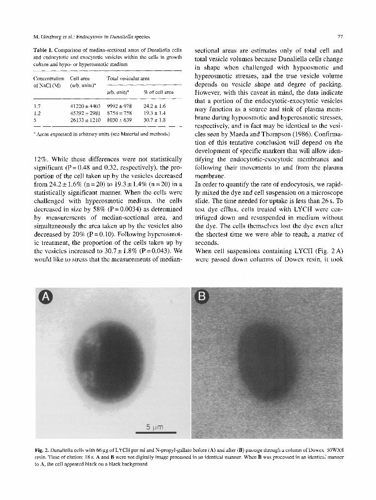
Table 1. Comparison of median-sectional areas of Dunaliella cells
and endocytotic and exocytotic vesicles within the cells in growth
culture and hypo- or hyperosmotic medium
Concentration Cell area Total vesicular area
of NaC1 (M) (arb. units)" arb. units a % of cell area
1.7 41220 + 4403 9992 + 978 24.2 + 1.6
1.2 45292 _+ 2981 8754 _+ 758 19.3 _+ 1.4
5 26133 • 1210 8030• 639 30.7_+ 1.8
Areas expressed in arbitrary units (see Material and methods)
12%. While these differences were not statistically significant (P = 0.48 and 0.32, respectively), the pro- portion of the cell taken up by the vesicles decreased from 24.2 _+ 1.6% (n = 20) to 19.3 + 1.4% (n = 20) in a statistically significant manner. When the cells were challenged with hyperosmotic medium, the cells decreased in size by 58% (P = 0.0034) as determined by measurements of median-sectional area, and simultaneously the area taken up by the vesicles also decreased by 20% (P = 0.10). Following hyperosmot- ic treatment, the proportion of the cells taken up by the vesicles increased to 30.7 __+ 1.8% (P = 0,043). We would like to stress that the measurements of median-
77
sectional areas are estimates only of total cell and total vesicle volumes because Dunaliella cells change in shape when challenged with hypoosmotic and hyperosmotic stresses, and the true vesicle volume depends on vesicle shape and degree of packing. However, with this caveat in mind, the data indicate that a portion of the endocytotic-exocytotic vesicles may function as a source and sink of plasma mem- brane during hypoosmotic and hyperosmotic stresses, respectively, and in fact may be identical to the vesi- cles seen by Maeda and Thompson (1986). Confirma- tion of this tentative conclusion will depend on the development of specific markers that will allow iden- tifying the endocytotic-exocytotic membranes and following their movements to and from the plasma membrane. In order to quantify the rate of endocytosis, we rapid- ly mixed the dye and cell suspension on a microscope slide. The time needed for uptake is less than 26 s. To test dye efflux, cells treated with LYCH were cen- trifuged down and resuspended in medium without the dye. The cells themselves lost the dye even after the shortest time we were able to reach, a matter of seconds. When cell suspensions containing LYCH (Fig. 2 A) were passed down columns of Dowex resin, it took
Fig. 2, Dunaliella cells with 66 gg of LYCH per ml and N-propyl-gallate before (A) and after (B) passage through a column of Dowex 50WX8
resin. Time of elution: l 8 s. A and B were not digitally image processed in an identical manner. When B was processed in an identical manner
to A, the cell appeared black on a black background
78 M. Ginzburg et al.: Endocytosis in Dunaliella species
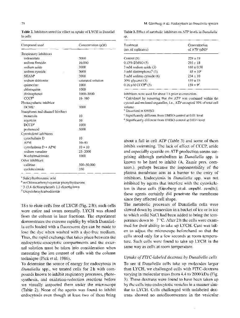
Table 2. Inhibitors tested for effect on uptake of LYCH in Dunaliel- la cells
Table 3. Effect of metabolic inhibitors on ATP levels in Dunaliella sp.
Treatment Concentration (no. of replicates) of ATP (gM) ~
Compound used Concentration (gM)
Respiratory inhibitors iodoacetate 3000 sodium fluoride 10,000 sodium azide 3000 sodium cyanide 3000 SHAM a 3000 sodium dithionite saturated solution quinacrine 1000 chloroquine 1000 dinitrophenol 1000-3000 CCCP b 16-160
Photosynthetic inhibitor DCMU ~ 3000
Ionophores and channel blockers monensin 10 nigericin 10 DCCD d 1000 probenecid 5000
Cytoskeletal inhibitors cytochalasin D 10 APM 10-40 cytochalasin D + APM 10 + 10 sodium vanadate 25-2000 N-ethylmaleimide 1000
Other inhibitors caffeine 500-50,000 cycloheximide 350
a Salicylhydroxamic acid b m-Chlorocarbonyl-cyanide phenylhydrazone ~ 3-(3,4-dichlorophenol)- 1,1-dimethylurea a Dicyclohexylcarbodiimide
18 s to elute cel ls free of L Y C H (Fig. 2 B); such cel ls
were ent ire and s w a m normal ly . LYCH was e lu ted
f rom the co lumn in la ter f ract ions. The expe r imen t
demons t ra t e s the ex t reme rap id i ty by which Duna l i e l -
la cel ls l oaded with a f luorescen t dye can be made to
lose the dye when washed wi th a dye - f r ee med ium.
Thus, the r ap id exchange that takes p lace be twee n the
endocy to t i c - exocy to t i c compa r tmen t s and the exter-
nal so lu t ion mus t be taken into cons ide ra t ion when
measur ing the ion conten t of cel ls wi th the co lumn
techn ique (P ick et al. 1986).
To de te rmine the source o f energy for endocy tos i s in
Dunaliella spp., we t rea ted cel ls for 2 h wi th com-
pounds k n o w n to inhibi t r e sp i ra to ry processes , photo-
synthesis , and ox ida t ion - reduc t ion reac t ions be fore
we v i sua l ly inspec ted them under the m i c r o s c o p e
(Table 2). None o f the agents was found to inhibi t
endocy tos i s even though at least two o f them br ing
Control (8) 229 +_ 19 0.13% DMSO (3) 293 + 18 3 mM sodium azide (3) 169 + 0.58 3 mM dinitrophenol b (3) 45 _+ 10 c 3 mM sodium cyanide (6) 234 + 10 30% glycerol (3) 159 _+ 33 0.16 gM CCCP b (3) 159 +_ 9 a
Inhibitors were used for about i h prior to extraction. a Calculated by assuming that the ATP was contained within the cytosol and enclosed organelles, i.e., ATP occupied 70% of total cell volume b Dissolved in DMSO c Significantly different from DMSO control at 0.01 level d Significantly different from DMSO control at 0.034 level
about a fal l in cel l ATP (Table 3) and some of them
inhibi t sw imming . The lack o f effect o f CCCP, az ide
and espec ia l ly cyan ide on ATP p roduc t ion seems sur-
p r i s ing a l though m e t a b o l i s m in Dunaliella spp. is
k n o w n to be hard to inhib i t (A. Z a m i r pers. com-
mun.) , pe rhaps because the i m p e r m e a b i l i t y of the
p l a s m a m e m b r a n e acts as a bar r ie r to the entry o f
inhibi tors . Endocy tos i s in DunaIiella spp. was not
inh ib i ted by agents that in ter fere wi th the cy toske le -
ton in these cel ls (Ginzburg e ta l . unpubl , resul ts) ;
these agents cer ta in ly d id pene t ra te the m e m b r a n e
s ince they affected cel l shape.
The me tabo l i c p rocesses o f Duna l i e l l a cel ls were
s lowed down b y i m m e r s i o n in a bucke t of ice or in ice
to which sol id NaC1 had been a d d e d to br ing the tem-
pera ture down to - 7 ~ Af te r 2 h the cel ls were exam-
ined for their ab i l i ty to take up LYCH. Care was tak-
en to adjus t the m i c r o s c o p e be fo r e ha nd so that the
cel ls s tood on ly for a few seconds at r o o m tempera-
ture. Such cel ls were found to take up LYCH in the
same way as cel ls at r o o m tempera ture .
Uptake of FITC-labeled dextrans by Dunaliella cells
To see i f Duna l i e l l a cel ls take up molecu le s larger
than LYCH, we cha l l enged cel ls wi th F IT C-de x t r ans
va ry ing in mo lecu l a r mass f rom 4.4 to 2000 k D a (Fig.
3). These dextrans were found to have been taken up
by the cel ls into endocy to t i c ves ic les in a manner s im-
i lar to LYCH. Cel ls cha l l enged wi th un labe led dex-
trans showed no au to f luorescence in the ves icu la r

M. Ginzburg et al.: Endocytosis in Dunaliella species 79
Fig. 3. Dunaliella cells with 2 mg of FITC-dextrans per ml, molecular masses: A 4.4 kDa, B and C 38.9 kDa, D 2000 kDa, showing presence of dye in the anterior region of the cell
region. The vesicles in the anterior part of the cell can
be seen with differential interference optics (Fig. 4).
The outline of the chloroplast can also be discerned with the pyrenoid within,
Quantitative fluorometric measurements
The brightness of the vesicular region occupied by
FITC-dextrans (FD4 through FD2000) was 82.3+
5.8% (n--- 90) of the background, suggesting that the
concentrations of dye within the vesicles and in the
outside medium are similar.
A major subject of concern was the time needed for
uptake of dextrans by the cells. The development of a
quantitative method for determining the content of a
given fluorescent compound within cell pellets made
it possible to determine max imum times of entry and
washout. Figure 5 shows that for FD4 these times are shorter than 29 s for uptake and 15 s for washout.

80 M. Ginzburg et al.: Endocytosis in Dunaliella species
Fig. 4 A-D. Dunaliella cells seen by differential interference optics. A and C Optical sections through the middle of the cells. B and D Optical sections taken close to the cell boundary
We were interested to know whether every dextran would be taken up to the same extent, regardless of size of molecule, so we measured uptake of the series of dextrans. 2-5 min was allowed for uptake in every case and Fig. 6 shows that the amount of dextran tak- en up in this time period falls with increase in molec- ular mass. It is evident that much of the dextran, espe- cially at the higher end of the scale, must be in the intercellular space though it is not possible to deter- mine how much.
Uptake of dextrans for longer periods varying from 15 to 20 rain was tested optically and quantified with a digital image processor (autofluorescence of chloro- phyll was suppressed with a filter). The advantage of this method is that measurements are made directly on individual cells; there is no need for a correction for intercellular fluid. The proportion of the cell area occupied by fluorescent material varied between 24.4 and 33.4% of total cell area and does not differ signif- icantly with molecular mass of the FITC-dextran

M. Ginzburg et al.: Endocytosis in Dunaliella species
20
18
16
~14 a) N12
.~10
4
2
0 , = + + r +k + , ,
0.5 1 1.5 2 2.5 3 3.5 4 minutes
Fig. 5. Rates of uptake and washout of 2 mg FITC-dextran (molecu-
lar mass 4.4 kDa) per ml by Dunaliella cells. Uptake (0 ) was deter-
mined at different times after addition of the FITC-dextran to the cell
suspension as the amount of FITC-dextran recovered from pellets of
centrifuged cells. The washout curve ( I ) was obtained from cells
treated with FITC-dextran for 50 min after resuspension in fresh
medium. Each point is the mean of two determinations
(Table 4). Thus, the larger ones are taken up by Dunaliella cells though more slowly than smaller ones: it takes less than 29 s for uptake of FD4 and ca. 15 rain for uptake of FD2000. The increase in time with size of molecule may be due to the slower diffu- sion of larger molecules through the medium.
Uptake of Sodium Green dextran by Dunaliella cells
Evidence that the endocytotic vesicles contain Na + was obtained by adding to a cell suspension Sodium Green dextran, an indicator that fluoresces after bind-
81
Table 4. Percentage of cell area occupied by fluorescent material
after addition of FITC-dextran to Dunaliella cell suspensions
Molecular mass of dextran (kDa) % vesicles of cell area a
4 28.0 + 1.1
10 24.4 + 2.6 20 33.4 _+ 3.8
40 30.5 + 1.0
70 26.5 + 1.9
150 24.7 + 1.5
250 24.8 + 6.0
500 29.1 _+ 1.0
2000 27.8 + 1.0
a Ratio of fluorescence of total cell to that of background, both cor- rected for the fluorescence of the chloroplast. Values are averages with SE of measurements on 10 cells
ing to Na + (Fig. 7): fluorescent vesicles were found in the same cell region as LYCH and fluorescent dex- trans. 29.9_+ 1.1% (n=30) of the total cell area was occupied by fluorescent vesicles containing Sodium Green dextran (cf. vesicular area obtained with FITC- dextrans in Table 4). The brightness of the vesicles containing the dextran was 73.4_+ 3.2% of the back- ground.
30
25 ==
.-= 15
t
0 I I
3 4 5 6 7 log tool mass of dextran
Fig. 6. Recovery of a graded series of FITC-dextrans (2 mg/ml) vary-
ing in molecular mass from 4.4 to 2000 kDa from pellets of cen- trifuged Dunaliella cells. 2-5 rain were allowed for mixing of the
compound with the cells before centrifugation. The volume of packed cells in the centrifuged pellet was the product of average cell
volume and cell number and equalled 10 gl. Uptake expressed in microgram of dextran. Each point is the mean of 2 -4 determinations
Fig. 7. Dunaliella cells containing Sodium Green dextran. Compare
with Figs. 1 and 3 to see that fluorescence is limited to same area of
cell. Note that in this preparation, concentrations of Na + cannot be determined with this dye, the Kd of which is 6.0 mM, since the Na + of
the medium is 1.7 M

82
Table 5. Time parameters in calculations of rates of endocytosis
M. Ginzburg et al.: Endocytosis in Dunaliella species
System measured Type of cell Time parameter used Reference by original author
Volume of fluid pinocytosed Acanthamoeba 25 % of cell volume/h L cells; many cultured cells 25% of cell volume/3-6 h mouse peritoneal M p 25% of cell volume/h
macrophages 25 % of cell volume/180 s
L cell fibroblasts 120 rain/cell surface" macrophages 33 mirdcell surface '~
elongating pollen tubes 30-80% of produced membrane recycled
BHK cells "c = 33 min b chromaffin-coated vesicles z = 15 min
nerve cells "c = several tens of seconds
mast cells ~ = 3 h chromaffin cells "c = 4 min pituitory cells "c = 4 s
Filling of pinocytosis vacuoles
Membrane internalization
Clathrin-mediated endocytosis
Retrieval of exocytosed membrane
Bowers and Olsewski 1972 Steinman et al. 1976 Steinman et al. 1976
Steinman et al. 1976
Steinman et al. 1976 Steinman et al. 1976
Picton and Steer 1983
Marsh and Helenius 1980 Patzak et al. 1987
T. Miller and Heuser 1984
Thilo 1985 yon Grafenstein et al. 1986 Thomas et al. 1994
Time needed for recycling of entire cell surface b Time coefficient "c is a measure of rate of endocytosis and depends on the experimental conditions and the kinetic model. For details, see yon Grafenstein et al. (1986)
Discuss ion
Dunal ie l la cells const i tu t ively take up and re lease
large f luorescent mo lecu le s to the external m e d i u m
by a process o f f lu id-phase endocytos is and exocyto-
sis. This conc lus ion fo l lows f rom the observa t ion that
f luorescent dyes taken up by the cells are not distrib-
uted evenly throughout the cell, but are l imi ted to
vesicles . Uptake is ent i rely nonspec i f ic and noncon-
centrat ing.
Endocytos i s is t radi t ional ly cons idered to be a slow
process (Watts and Marsh 1992). This is i l lustrated in
Table 5 which shows that the t ime needed to recyc le
the cel l surface of ten takes several hours. On the oth-
er hand, studies with several cel l types, inc luding
synaptic ves ic les of the goldf ish ret inal b ipolar cell,
have consis tent ly revea led a re la t ively rapid process
with a t ime constant of a few seconds (Betz and
Ang le son 1998). Endocytos i s in Dunal ie l la cells
be longs to this latter group of rapidly endocy tos ing
cells, s ince the t ime needed to in ternal ize the entire
cell membrane is less than 4 s (Table 6).
Cel l vo lume is ca lcula ted on the assumpt ion that the
cell is a prolate e l l ipsoid with major axis 2A and
minor axis 2B. The propor t ion of cell v o l u m e occu-
pied by ves ic les can be es t imated f rom the percen tage
o f cell area occup ied by ves ic les (Table 4) by mode l -
ing the ves icular reg ion as a ho l low truncated cone
with height a and width o f each side b: area of vesic-
ular region in median section, Av = 2ab/2; area o f the
cel l in med ian section, A~ = JrAB; vo lume of the vesic-
ular region,Vv = zcab2; v o l u m e of the cell , Vc = (4 / 3)
~. AB2; Vv / Vc = 3/r ab 2 / (4 / tAB 2) = [0.75ab / (AB)]
(b/B). Since A v / A c = ab/rcAB=0.3, then ab/AB= 0.37c. Hence , Vv / Vc= 0.75 �9 0.3Jr(b / B) =0.70b/B. W h e n b/B=0.5, then V v / V c = 0 . 3 5 . Therefore , pro-
v ided that b/B = 0.5, the ratio o f Av to Ac is approxi-
mate ly equal to Vv/Vc.
In Dunaliella spp. the rates o f endo- and exocytos is
for molecu les with a mass equal or less than 4000 Da,
as measured by v o l u m e flow, are at least one order of
magni tude faster than rates in other eucaryot ie cells
Table 6. Calculation of rate of endocytosis in Dunaliella sp., as- suming that 30% of the cell is occupied by endocytosed fluid and that this fluid is within spherical vesicles
Parameter Value
Diameter of vesicle, 2r 400. 10 -9 m Volume of vesicle, (4/3) zcr 3 = Vv 3.4 ' l0 20 m 3 Surface area of vesicle, 4zcr 2 = Sv 5.0 �9 10 6 m 2 Major cell axis, A 5.5. 10 -6 m Minor cell axis, B 6.0 - 10 6 m Cell volume, (4/3) JrAB 2 = Vc 7.6 " 10 16 m 3 Surface area of cell (= surface area of
plasma membrane), 4JrAB = Sc 4.6.10 -~~ m 2 Number of vesicles, n = 0.3 Vc/Vv 6.7 - 10 ~ Total surface area of vesicles, nSv 3.4 �9 10 -9 m 2 (nSv)/Sc 7.7 Time to fill endocytotic vesicles -30 s Time to internalize entire cell
membrane, 30/(nSJSc) 3.9 s

M. Ginzburg et al.: Endocytosis in Dunaliella species
Table 7. Endocytotic volume flow into various types of cells
83
Cell type Cell volume (10 18 m 3) Cell surface area (10 -~2 m 2) Volume flow (m/s) a Reference
Yeast 100 100 8.3 �9 10 -13 Lenhard et al. 1992
Human fibroblast 1765 2100 2 �9 10 12 van Deurs et al. 1989
Dictyostelium discoideum 500 314 7 . 10 -1~ Lenhard et al. 1992
Macrophage 385 825 2.5. 10 -9 Steinman et al. 1976 Dunaliella sp. var. D13 760 460 1.7. 10 -8 this work
a Volume flow was calculated from data on percentage of cell volume taken up by endocytotic vesicles, cell surface area, and time constants pre-
sented in the references cited. Volume flow is defined as volume of fluid (m 3) to cross one unit of cell surface area (m 2) per unit of time (s)
Table 8. Dimensions of types of endocytotic vesicles measured on thin sections by electron microscopy
Type of vesicle Source Diameter (l 0 -9 m) Reference
Coated vesicles; partially coated soybean protoplast 50-70 Tanchak et al. 1984, 1988
reticulum and dictyosomes
Coated vesicles
Pinocytosis primary vesicle
Primary endocytosis vesicle
Clathrin-coated vesicles
Nonclathrin-coated vesicles (a)
Nonclathrin-coated (b)
"Slow" endocytosis vesicles
"Fast" endocytotic vesicles
various plant cells 84-91
Lobelia root cells -100
soybean protoplasts - 100 various plant cells 72-96
Dunaliella salina 70-100
Acanthamoeba 250
macrophages and L-cells 150
human fibroblasts 100
smooth muscle cells, fibroblasts, 50-100
endothelial cells Vero cells, Hep-2 cells, A431 cells 150-300
BHK cells - 100
rat melantrophs 800
Coleman et al. 1987
Samuels and Bisapultra 1990
Fowke and Tanchak 1988
Emons and Traas 1986
Zhang et al. 1993
Bowers et al. 1981 Steinman et al. 1976
van Deurs et al. 1989
van Deurs et al. 1989
van Deurs et al. 1989
Thomas et al. 1994
Thomas et al. 1994
(Table 7). The high rates of endocytotic volume flow in Dunaliella spp. may be a consequence of the high specific rate of internalization per vesicle, the lack of a thick extracellular matrix and of turgor pressure, the large volume fraction of vesicles per cell (25-30% of cell volume as compared to 1-10% in other cells) and/or the relatively large size of the endo- and exo- cytotic vesicles (400 nm diameter as compared to 100 nm in other cells, see Tables 6 and 8). On the other hand, these very fast rates are limited to LYCH and the smaller dextrans; we found that FD2000 took several minutes to enter the cells. The difference in behavior between the smaller and larger molecules accounts for the fact that previous workers did not find larger dextrans to be taken up by Dunaliella cells (Ehrenfeld and Cousin 1982, M. Ginzburg and Richman 1985); these workers cen- trifuged cell suspensions after mixing their dextran markers with cell suspensions for not more than 2 min, thus avoiding the slow endocytotic uptake of
large molecules. It is unclear whether the vesicles once inside the cell remain independent, fuse with other vesicles to form an endosomal reticulum involved in membrane recy- cling (Hopkins etal. 1990) or interact with other inter- nal membrane systems in the cell such as the trans- Golgi network (Tanchak etal. 1984, Fowke eta1. 1991, Mukherjee et al. 1997, Low and Chandra 1994, Prescianotto-Baschong and Riezman 1998). Given the rapid rates of endo- and exocytosis, we favor the first, or first two of these possibilities for the majority of vesicles at least. Vesicle formation involved in membrane trafficking typically requires chemical energy coupled to dephos- phorylation of ATP and GTR In an attempt to search for the processes that provide the energy necessary for vesicle formation, we treated the cells with a wide variety of inhibitors, yet, in all cases we were unable to inhibit endocytosis, which was not stopped even in cells cooled to -7~ Thus, either endocytosis

84 M. Ginzburg et al.: Endocytosis in Dunaliella species
requires little energy or is energized by some unusual source. In this paper we present a new model that synthesizes the two antithetical models concerning halotolerance in Dunaliella spp. The discovery of the rapid consti- tutive processes of endocytosis and exocytosis in the Dunaliella cell solves a long-standing controversy concerning the structure and composition of this cell. The one-compartment model treated halotolerance as an osmotic problem and considered the plasma mem- brane to be of the usual semipermeable type. Several authors (e.g., Ben-Amotz and Avron 1973, Pick et al. 1986) have equated the cytosol with the cell and have not taken the 25-30% of the cell volume occupied by endocytotic vesicles into account. We consider the cell Na + measurements of those authors to character- ize the cytosol and nonendo- and nonexocytotic organelles rather than the entire cell. On the other hand, the two-compartment model, based on static and dynamic measurements of total cell glycerol, Na +, K +, and C1-, assumed that the plasma membrane was unusually permeable to salts and glycerol, although it was also assumed that typically semiper- meable membranes enclosed the organelles. Neither model took the possibility of endo- or exocytosis into account. We suggest that the so-called outer compart- ment described by the two-compartment model can be accounted for by the endo- and exocytotic vesicles. The high cell Na + concentrations found by several workers (summarized in M. Ginzburg 1987) can be accounted for by NaC1 from the outside medium trapped within endocytotic vesicles. Is endocytosis related to the extraordinary capacities for salt regulation and osmoregu!ation found in Dunaliella spp.? Certainly the endo- and exocytotic membranes serve as a ready source and sink of plas- ma membrane for cells challenged by hypoosmotic and hyperosmotic stresses (Table 1) (Maeda and Thompson 1986). However, the amount of vesicular membrane present is far greater than is needed to account for any increase in surface area that has ever been observed to occur after a hypoosmotic stress. It is therefore possible that the vesicles have some addi- tional function. Cells living in high concentrations of salt (up to several molar) may also benefit from mem- brane systems through which there is a continuous flow of large volumes of highly concentrated NaC1 solutions. We suggest that the enormous membrane area created by the system could serve to synthesize ATP by using the large differences in Na + or C1- exist- ing across the cell membrane: molar concentrations in
the outside medium and cytosolic Na + kept to a low millimolar concentration by light-driven ion pumps. Reversible Na+-ATPases "form a large group of enzymes found in all kingdoms of living organisms" (Skulachev 1994). Na§ ATP synthesis may help to mitigate the stress experienced by cells living in highly saline environments.
Acknowledgements We thank Professor M.V. Parthasarathy who took the photographs shown in Fig. 1. We thank Professors Roger Spanswick and Tom Owen for stimulating discussions. This work was partially supported by grants from NASA.
References Beech PL, Wetherbee R (1988) Observations on the flagellar appara-
tus and peripheral endoplasmic reticulum of the coccol- ithophorid Pleurochrysis carterae. Phycologia 27:142-158
Ben-Amotz A (1974) Osmoregulation mechanism in the halophylic alga Dunaliella parva, In: Zimmermann U, Dainty J (eds) Mem- brane transport in plants. Springer, Berlin Heidelberg New York. pp 95-100
- Avron M (1973) The role of glycerol in the osmotic regulation of the halophilic alga Dunaliella parva. Plant Physiol 51:875-878
- Ginzburg BZ (1969) Light-induced proton uptake in whole cells of DunalielIa parva. Biochim Biophys Acta 183:144-154
Bental M, Degani H, Avron M (1988) Z3Na-NMR studies of the intracellular sodium ion concentration in the halotolerant alga Dunaliella salina. Plant Physiol 87:813- 817
Betz WJ, Angleson JK (1998) The synaptic vesicle cycle. Annu Rev Physiol 60:347-363
Bock RM, Ling N-S, Morell SA, Lipton SH (1956) Ultraviolet absorption spectra of adenosine-5'-triphosphate and related 5'- ribonucleotides. Arch Biochem Biophys 62:253-264
Borowitzka LJ, Brown AD (1974) The salt relations of marine and halophylic species of the unicellular green alga Dunaliella.
Arch Microbiol 96:37--52 Bowers B, Olszewski TE (1972) Pinocytosis in Acanthamoeba:
kinetics and morphology. J Cell Biol 53:681-694 Clayton MN, Ashburner CM (1994) Secretion of phenolic bodies
fallowing fertilization in DurviIIaea potatorum (Durvillaeales, Phaeophyta). Eur J Phycol 29:1-9
Coleman J, Evans D, Hawes C, Horsley D, Cole L (1987) Structure and molecular organization of higher plant coated vesicles. J Cell Sci 88:35-45
Craigie JS, McLachlan J (1964) Glycerol as a photosynthetic product in Dunaliella tertiolecta Butcher. Can J Bot 42:777~78
Cunningham RF, Israili ZH, Dayton PG (1981) Clinical pharmacoki- netics of probenecid. Clin Pharmacol Kinet 6:135-151
de Duve C (1963) "Endocytosis', a note. In: de Reuck AVS, Cameron MP (eds) Lysosomes. Little, Brown and Co, Boston, p 126
Diekmann W, Hedrich R, Raschke K, Robinson DG (1993) Osmo- cytosis and vacuolar fragmentation in guard cell protoplasts:
M. Ginzburg et al.: Endocytosis in Dunaliella species
their relevance to osmotically-induced volume changes in guard cells. J Exp Bot 44:1569-1577
Domozych DS, Nimmous TT (1992) The contractile vacuole as an endocytic organelle of the chlamydomonad flagellate Gloeomonas kupferi (Volvocales, Chlorophyta). J Phycol 28: 809-816
Ehrenfeld J, Cousin J-L (1982) Ionic regulation of the unicellular green alga Dunaliella tertiolecta. J Membr Biol 70:47-57
- - (1984) Ionic regulation of the unicellular green alga Dunaliel-
la tertiolecta: response to hypertonic shock. J Membr Biol 77: 45-55
Emons, AMC, Traas JA (1986) Coated pits and coated vesicles on the plasma membrane of plant cells. Eur J Cell Biol 41: 57- 64
Fisher M, Gokhman I, Pick U, Zamir A (1997) A structurally novel transferrin-like protein accumulates in the plasma membrane of the unicellular green alga Dunaliella salina grown in high salin- ities. J Biol Chem 272:1565-1570
Fowke LC, Tanchak MA (1988) The structure and function of plant coated vesicles. In: Pais MSS, Mavituna F, Novais JM (eds) Plant cell biotechnology. Springer, Berlin Heidelberg New York Tokyo, pp 153-163 (NATO ASI Series, series H, vol 18)
- - Galway ME (1991) Ultrastructurat cytology of the endocytotic pathway in plants. In: Hawes CR, Coleman JOD, Evans DE (eds) Endocytosis, exocytosis and vesicle traffic in plants. Cam- bridge University Press, Cambridge, pp 15-40 (Society for Experimental Biology Seminar series, vol 45)
Ginzburg BZ (1978) Regulation of cell volume and osmotic pressure in Dunaliella. In: Caplan SR, Ginzburg M (eds) Energetics and structure of halophilic microorganisms. Elsevier, Amsterdam, pp 543-558
Ginzburg M (1969) The unusual permeability of two halophilic uni- cellular organisms. Biochim Biophys Acta 173:370-5/6
- (1987) Dunaliella: a green alga adapted to salt. Adv Bot Res 14: 93-183
- Richman L (1985) Permeability of whole Dunaliella cells to glu- cose. J Exp Bot 36:1959-1968
- Ginzburg BZ, Wayne R (1997) The plasma membrane of Dunaliella is more than a plasma membrane! Plant Physiol 114: $42, abstract 120
Hopkins CR, Gibson A, Shipman M, Miller K (1990) Movement of internalized ligand-receptor complexes along a continuous endo- somal reticulum. Nature 346:335-338
Karni L, Avron M (1988) Ion content of the halotolerant alga Dunaliella salina. Plant Cell Physiol 29:1311-1314
Lenhard JM, Mayorga L, Stahel PD (1992) Characterisation of endo- some-endosome fusion in a cell free system using Dictyostelium
discoideum. J Biol Chem 267:1896-1903 Low PS, Chandra S (1994) Endocytosis in plants. Annu Rev Plant
Physiol Plant Mol Biol 45:609-631 Maeda M, Thompson GA (1986) On the mechanism of rapid plasma
membrane and chloroplast envelope expansion in Dunaliella salina exposed to hyperosmotic shock. J Cell Biol 102:289-297
Marsh M, Helenius A (1980) Adsorptive endocytosis of semliki for- est virus. J Mol Biol 142:439-454
McFadden GI, Preisig HR, Melkonian M (1986) Golgi apparatus activity and membrane flow during scale biogenesis in the green flagellate Scherffelia dubia (Prasinophyceae) II: cell wall secre- tion and assembly. Protoplasma 131:174-184
Miller DM, de Ruijter CA, Emons AMC (1997) From signal to form:
85
aspects of the cytoskeleton-plasma membrane-cell wall continu- um in root hair tips. J Exp Bot 48:1881-1896
Miller TM, Heuser JE (1984) Endocytosis of synaptic vesicle mem- brane at the frog neuromuscular junction. J Cell Biol 98: 685- 698
Mukherjee S, Ghosh RN, Maxfield FR (1997) Endocytosis. Physiol Rev 77:760-803
O'Driscoll D, Hann C, Read SM, Steer MW (1993) Endocytotic uptake of fluorescent dextrans by pollen tubes grown in vitro. Protoplasma 175:126-130
O'Neil RM, La Claire JW II (1988) Endocytosis and membrane dynamics during the wound response of the green alga Boergen- esia. Cytobios 53:113-125
Oliviera L, Bisalputra T, Antia NJ (1980) Ultrastructural observation of the surface coat of Dunaliella tertiolecta from staining with cationic dyes and enzyme treatments. New Phytol 85:385-392
Oparka K J, Prior DAM, Harris N (1990) Osmotic induction of fluid- phase endocytosis in onion epidermal cells. Planta 180: 555- 561
- Murant EA, Wright KM, Prior DAM, Harris N (1991) The drug probenecid inhibits the vacuolar accumulation of fluorescent anions in onion epidermal cells. J Cell Sci 99:557-563
Patzak A, Annis D, Langley K (1987) Membrane recycling after exo- cytosis: an ultrastructure study of cultured chromaffin cells. Exp Cell Res 171:346-356
Picton JM, Steer MW (1983) Membrane recycling and the control of secretory activity in pollen tubes. J Cell Sci 63:303-310
Pick U, Karni L, Avron M (1986) Determination of ion content and ion fluxes in the halotolerant alga Dunaliella salina. Plant Phys- iol 81:92 - 96.
Prescianotto-Baschong C, Riezman H (1998) Morphology of the yeast endocytotic pathway. Mol Biol Cell 9:173-189
Roszak R, Rambour S (1997) Uptake of Lucifer Yellow by plant cells in the presence of endocytotic inhibitors. Protoplasma 199: 198-207
Samuels AL, Bisapultra T (1990) Endocytosis in elongating root cells ofLobelia erinus. J Cell Sci 97:157-165
Skulachev VP (1994) The latest news from the sodium world. Biochim Biophys Acta 1187:216-221
Steinman RM, Brodie SE, Cohn ZA (1983) Membrane flow during pinocytosis: a stereologic analysis. J Cell Biol 68:665-687
Tanchak M, Griffing L, Mersey B, Fowke L (1984) Endocytosis of cationized ferritin by coated vesicles of soybean protoplasts. Planta 162:481-486
- Rennie PJ, Fowke LC (1988) Ultrastructure of the partially coat- ed reticulum and dictyosomes during endocytosis by soybeans protoplasts. Planta 175:433-441
Thilo L (1985) Selective internalization of granule membrane after secretion in mast cells. Proc Natl Acad Sci USA 82: 1711- 1715
Thomas P, Lee A, Wong J, Almers W (1994) A triggered mechanism retrieves membrane in seconds after Ca2+-stimulated exocytosis in single pituitary cells. J Cell Biol 124:667-675
Trezzi F, Galli MG, Bellini E (1965) L'osmoresistenza di Dunaliel- la salina: richerche ultrastrutturali. G Bot Ital 72:255-263
van Deurs B, Petersen O, Olsnes S, Sandvig K (1989) The ways of endocytosis. Int Rev Cytol 117:131-177
yon Grafenstein H, Roberts CS, Baker PF (1986) Kinetic analysis of the triggered exocytosis/endocytosis secretory cycle in cultured bovine adrenal medullary cells. J Cell Biol 103:2343-2352

86
Watts C, Marsh M (1992) Endocytosis: what goes in and how? J Cell Sci 103:1-8
Wayne R, Kadota A, Watanabe M, Furuya M (1991) Photomove- ment in DunalieIla salina fluence rate: response curves and action spectra. Planta 184:515-524
M. Ginzburg et al.: Endocytosis in Dunaliella species
Zhang XQ, Dubacq J-P, Alfsen A (1993) Biochemical and cytologi- cal evidence for the stimulation of clathrin-coated pit (vesicle) formation by exogenous folic acid in Dunaliella salina (Chloro- phyta). J Phyco129:302-209
Related Documents