Ultrananocrystalline Diamond Thin Films Functionalized with Therapeutically Active Collagen Networks Houjin Huang, †,‡ Mark Chen, § Paola Bruno, | Robert Lam, ‡ Erik Robinson, †,‡ Dieter Gruen, | and Dean Ho* ,†,‡,⊥ Department of Biomedical Engineering, Northwestern UniVersity, EVanston, Illinois 60208, Department of Mechanical Engineering, Northwestern UniVersity, EVanston, Illinois 60208, Departments of Chemistry and Biological Sciences, Northwestern UniVersity, EVanston, Illinois 60208, Materials Science DiVision, Argonne National Laboratory, Argonne, Illinois 60439, and Robert H. Lurie ComprehensiVe Cancer Center, Northwestern UniVersity, Chicago, Illinois 60611 ReceiVed: January 14, 2009; ReVised Manuscript ReceiVed: February 4, 2009 The fabrication of biologically amenable interfaces in medicine bridges translational technologies with their surrounding biological environment. Functionalized nanomaterials catalyze this coalescence through the creation of biomimetic and active substrates upon which a spectrum of therapeutic elements can be delivered to adherent cells to address biomolecular processes in cancer, inflammation, etc. Here, we demonstrate the robust functionalization of ultrananocrystalline diamond (UNCD) with type I collagen and dexamethasone (Dex), an anti-inflammatory drug, to fabricate a hybrid therapeutically active substrate for localized drug delivery. UNCD oxidation coupled with a pH-mediated collagen adsorption process generated a comprehensive interface between the two materials, and subsequent Dex integration, activity, and elution were confirmed through inflammatory gene expression assays. These studies confer a translational relevance to the biofunctionalized UNCD in its role as an active therapeutic network for potent regulation of cellular activity toward applications in nanomedicine. Introduction The emergence of implantable medical devices has prompted the development of a spectrum of materials that are capable of suppressing inflammatory responses and other processes that can preclude long-term implant activity. For example, polymeric, lipid-based, and metallic nanomaterials have been explored as drug delivery agents for applications in inflammatory suppres- sion and chemotherapeutic release, among others. 1-6 Among these classes of therapeutic delivery platforms, ultrananocrys- talline diamond (UNCD)-based substrates have been character- ized as a promising technology for biological applications owing to their low cytotoxicity, scalable fabrication parameters, chemical reactivity, and biocompatible physical properties. 7 Utilizing these favorable properties as a foundation for bioint- erface applications, the addition of strategies to attenuate surface- mediated inflammatory cytokine expression and release will further enhance the medical significance of UNCD. The fusion of biology and nanotechnology in the form of nanodiamond- based materials has created novel strategies for the fabrication of medically relevant nanodiamond hybrids. 8-16 The facile nature of UNCD deposition on silicon surfaces, and the straightforward approach toward functionalizing the UNCD films with thera- peutically active collagen networks enables UNCD interfacing with a broad array of emerging implant technologies, particularly microfabricated devices on silicon substrates. Owing to the scalability and broad relevance of these devices toward the treatment of a spectrum of physiological disorders, they are thus finding applicability in the neurological, cardiovascular, ortho- pedic, and real-time health monitoring domains, among others. As such, UNCD coatings may play an important role toward enhancing the bioamenability of an important class of advanced implantation technologies. Here, we demonstrate the ability to generate a biologically relevant interface between soft and hard materials, by depositing and interfacing collagen fibrils integrated with the anti-inflam- matory element dexamethasone (Dex) atop UNCD thin films, resulting in a therapeutically relevant active substrate. This novel hybrid technology is based upon a facile strategy for nanodia- mond film biofunctionalization that can provide potent and requisite inflammatory attenuation toward translational applica- tions. The UNCD film exhibits favorable biocompatibility and has previously been functionalized with DNA oligonucleotides for immobilizing biomolecules and proteins for biosensors. 17-19 Through the particular process presented, collagen fibrils were adsorbed to the UNCD film surface, and cross-linkages between collagen fibrils were observed to form via atomic force microscopy (AFM). In addition, biomolecular studies were performed to confirm the successful conjugation and elution of dexamethasone from the collagen matrix via suppression of * Corresponding author. Address: 2145 Sheridan Road, Evanston, IL 60208. Phone: (847) 467-0548. Fax: (847) 491-3915. E-mail: d-ho@ northwestern.edu. † Department of Biomedical Engineering, Northwestern University. ‡ Department of Mechanical Engineering, Northwestern University. § Departments of Chemistry and Biological Sciences, Northwestern University. | Argonne National Laboratory. ⊥ Robert H. Lurie Comprehensive Cancer Center, Northwestern University. 2966 10.1021/jp9004086 CCC: $40.75 2009 American Chemical Society Published on Web 02/18/2009 2009, 113, 2966–2971

Welcome message from author
This document is posted to help you gain knowledge. Please leave a comment to let me know what you think about it! Share it to your friends and learn new things together.
Transcript
Ultrananocrystalline Diamond Thin Films Functionalized with Therapeutically ActiveCollagen Networks
Houjin Huang,†,‡ Mark Chen,§ Paola Bruno,| Robert Lam,‡ Erik Robinson,†,‡ Dieter Gruen,|
and Dean Ho*,†,‡,⊥
Department of Biomedical Engineering, Northwestern UniVersity, EVanston, Illinois 60208, Department ofMechanical Engineering, Northwestern UniVersity, EVanston, Illinois 60208, Departments of Chemistry andBiological Sciences, Northwestern UniVersity, EVanston, Illinois 60208, Materials Science DiVision, ArgonneNational Laboratory, Argonne, Illinois 60439, and Robert H. Lurie ComprehensiVe Cancer Center,Northwestern UniVersity, Chicago, Illinois 60611
ReceiVed: January 14, 2009; ReVised Manuscript ReceiVed: February 4, 2009
The fabrication of biologically amenable interfaces in medicine bridges translational technologies with theirsurrounding biological environment. Functionalized nanomaterials catalyze this coalescence through the creationof biomimetic and active substrates upon which a spectrum of therapeutic elements can be delivered to adherentcells to address biomolecular processes in cancer, inflammation, etc. Here, we demonstrate the robustfunctionalization of ultrananocrystalline diamond (UNCD) with type I collagen and dexamethasone (Dex),an anti-inflammatory drug, to fabricate a hybrid therapeutically active substrate for localized drug delivery.UNCD oxidation coupled with a pH-mediated collagen adsorption process generated a comprehensive interfacebetween the two materials, and subsequent Dex integration, activity, and elution were confirmed throughinflammatory gene expression assays. These studies confer a translational relevance to the biofunctionalizedUNCD in its role as an active therapeutic network for potent regulation of cellular activity toward applicationsin nanomedicine.
Introduction
The emergence of implantable medical devices has promptedthe development of a spectrum of materials that are capable ofsuppressing inflammatory responses and other processes thatcan preclude long-term implant activity. For example, polymeric,lipid-based, and metallic nanomaterials have been explored asdrug delivery agents for applications in inflammatory suppres-sion and chemotherapeutic release, among others.1-6 Amongthese classes of therapeutic delivery platforms, ultrananocrys-talline diamond (UNCD)-based substrates have been character-ized as a promising technology for biological applications owingto their low cytotoxicity, scalable fabrication parameters,chemical reactivity, and biocompatible physical properties.7
Utilizing these favorable properties as a foundation for bioint-erface applications, the addition of strategies to attenuate surface-mediated inflammatory cytokine expression and release willfurther enhance the medical significance of UNCD. The fusionof biology and nanotechnology in the form of nanodiamond-based materials has created novel strategies for the fabricationof medically relevant nanodiamond hybrids.8-16 The facile natureof UNCD deposition on silicon surfaces, and the straightforward
approach toward functionalizing the UNCD films with thera-peutically active collagen networks enables UNCD interfacingwith a broad array of emerging implant technologies, particularlymicrofabricated devices on silicon substrates. Owing to thescalability and broad relevance of these devices toward thetreatment of a spectrum of physiological disorders, they are thusfinding applicability in the neurological, cardiovascular, ortho-pedic, and real-time health monitoring domains, among others.As such, UNCD coatings may play an important role towardenhancing the bioamenability of an important class of advancedimplantation technologies.
Here, we demonstrate the ability to generate a biologicallyrelevant interface between soft and hard materials, by depositingand interfacing collagen fibrils integrated with the anti-inflam-matory element dexamethasone (Dex) atop UNCD thin films,resulting in a therapeutically relevant active substrate. This novelhybrid technology is based upon a facile strategy for nanodia-mond film biofunctionalization that can provide potent andrequisite inflammatory attenuation toward translational applica-tions. The UNCD film exhibits favorable biocompatibility andhas previously been functionalized with DNA oligonucleotidesfor immobilizing biomolecules and proteins for biosensors.17-19
Through the particular process presented, collagen fibrils wereadsorbed to the UNCD film surface, and cross-linkages betweencollagen fibrils were observed to form via atomic forcemicroscopy (AFM). In addition, biomolecular studies wereperformed to confirm the successful conjugation and elution ofdexamethasone from the collagen matrix via suppression of
* Corresponding author. Address: 2145 Sheridan Road, Evanston, IL60208. Phone: (847) 467-0548. Fax: (847) 491-3915. E-mail: [email protected].
† Department of Biomedical Engineering, Northwestern University.‡ Department of Mechanical Engineering, Northwestern University.§ Departments of Chemistry and Biological Sciences, Northwestern
University.| Argonne National Laboratory.⊥ Robert H. Lurie Comprehensive Cancer Center, Northwestern University.
2966
10.1021/jp9004086 CCC: $40.75 2009 American Chemical Society
Published on Web 02/18/2009
2009, 113, 2966–2971
inflammatory gene expression shown through significant reduc-tion in interleukin-6 (IL-6) and tumor necrosis factor-alpha(TNF-R).
Collagen is a major protein within the extracellular matrixthat provides a good foundation for tissue growth on implantsand other devices for cellular interrogation studies. The collagenmolecule has a length of 3000 Å, a width of 15 Å, and amolecular weight of 300 kDa and is composed of threepolypeptide chains of a common repeating amino acid sequenceintertwined in a triple helix structure.20,21 The amino acidsglycine, proline, and hydroxyproline are held together bycovalent bonding and hydrogen bonding between -CO and-NH groups.20 Due to the great tensile strength achievedthrough bundling, collagen is a major component in musclesand bones.21,22 As such, collagen is an ideal material forbiomedical applications with its innate biocompatibility, strengththrough cross-linking, self-assembly, and aggregation, as wellas easily modifiable characteristics.21 This work merges thebenefits afforded from both the innately medically relevantUNCD properties23,24 and collagen to generate a resultant hybridfilm with Dex-based therapeutic activity. The soft interfaceserves as a versatile platform that can be functionalized withvirtually any drug or effector molecule for translational ap-plications that merge technology with biology and medicine.
Experimental Methods
UNCD Film Synthesis and Deposition. The UNCD thinfilms under study were grown using a commercially availableIPlas 2.45 GHz microwave plasma enhanced chemical vapordeposition system. A nominal gas composition of 1% CH4 and99% Ar with 100 sccm total flow rate made up the synthesisgas. Silicon wafer substrates were mechanically polished withfine diamond powder, which yielded a surface roughness of5-10 nm in order to provide a high density of nucleation sitesfor UNCD film growth.25 During the deposition process, thesubstrate temperature was independently set at 800 °C, whilethe total ambient pressure and input power were kept at 150mbar and 1200 W, respectively. UNCD films prepared in thisway have been extensively characterized by a variety oftechniques.23,24 Their microstructure consists of randomlyoriented 3-5 nm diamond crystallites strongly held togetherby largely sp2 bonded carbons at 0.2-0.3 nm wide grainboundaries. UNCD films are distinct from nanocrystallinediamond (ND) films in that the latter are composed of diamondcrystallites that are about an order of magnitude or more larger.The unique microstructure of UNCD films is responsible fortheir being smooth on the nanometer scale. Such films aretherefore exceptionally well suited for the production ofconformal coatings of biomaterials that can display nanoscaleroughness properties.
Surface Characterization and Collagen Adhesion. Thesurfaces of UNCD films were oxidized in concentrated HNO3
at 60-70 °C for 24 h. This oxidation reaction transformed theface of the film from a hydrophobic surface to a hydrophilicsurface by adding carboxylate groups to the film. Subsequently,the surfaces were washed with nanopure water, and 50 µL ofaqueous type I rabbit collagen (pH 8, Sigma-Aldrich, St. Louis,MO) adjusted with glacial acetic acid, sodium hydroxide, andnanopure water was pipetted onto a 2 × 3 cm2 oxidized UNCDfilm and allowed to spread across the surface of the film bymeans of diffusion. The coated UNCD film was then dehydratedin an oven at 40 °C for 30 min under a vacuum to promotesolvent evaporation. Following dehydration, the samples werecooled and washed with nanopure water and dried with air to
remove any weakly bound collagen fibrils on the surface. Thecollagen-Dex samples were composed of a solution of 45 µLof aqueous type I rabbit collagen (pH 8) and 5 µL of Dex thatwas coated on the UNCD film based on the procedure detailedabove. The final concentration of Dex in cell culture solutionwas 1.67 µg/mL.
Atomic Force Microscopy. An Asylum MFP3D atomic forcemicroscope (Santa Barbara, CA) was used to image the surfaceof the UNCD thin film deposited on silicon, a UNCD filmdeposited in silicon and oxidized in concentrated HNO3, as wellas collagen-functionalized UNCD films. Imaging parametersincluded the application of tapping mode atomic force micros-copy at a scan rate of 1 Hz. Image sizes included 1 µm × 1 µmand 5 µm × 5 µm.
XPS Analysis. XPS analysis was performed on UNCD,oxidized UNCD, collagen, and UNCD-collagen using anOmicron ESCA probe equipped with an EA125 energy analyzer.Spectra were obtained using monochromatized Al K a1 radiation(1486.6 eV) at 10 kV with a current of 7.74 mA under UHV.EIS software (Omicron,Taunusstein, Germany) was used tocollect and evaluate survey and high resolution scans. Theelectron gun was operated at 8 eV and 0.005 mA to compensatefor the low electrical conductivity of both collagen samples.
Cell Culture. RAW 264.7 (ATCC, Manassas, VA) murinemacrophage cells were cultured in 1× DMEM (Cellgro,Herndon, VA) containing 10% FBS (ATCC) and 1% penicillin/streptomycin (Cambrex, East Rutherford, NJ) at 37 °C. Afterthe cell culture reached 70-80% confluence, the macrophagecultures were split and plated on the four substrates: glass,UNCD film, UNCD film with collagen, and UNCD film withcollagen-Dex. After 20 h of growth, lipopolysaccharide (LPS)was added to a concentration of 5 ng/mL in each cell/substrateculture. LPS stimulation lasted for 4 h. The cells were allowedto grow on the substrates for a total of 24 h at 37 °C and thenharvested for gene expression studies.
Quantitative Real-Time Polymerase Chain Reaction (RT-PCR). RNA was purified via cell lysis with 1 mL of TRIzol(Invitrogen, Carlsbad, CA) followed by extraction with 200 µLof chloroform. The samples were then centrifuged for 15 minat 14 000 RPM and 4 °C, and the supernatant was transferredinto isopropyl alcohol for RNA isolation and overnight storageat -80 °C to promote nucleic acid precipitation. Following RNApurification, the samples were thawed and centrifuged for 30min at 14 000 RPM and 4 °C to form an RNA pellet. Thesupernatant was removed and the pellets were washed with 70%ethanol to remove isopropyl alcohol and salts, and thencentrifuged for 5 min at 14 000 RPM and 4 °C. Aftercentrifugation, the supernatant was removed and the pellets weredried in air for 5 min. Nucleic acid analysis with a UV-visspectrophotometer (Beckman Coulter, Fullerton, CA) was thenperformed on the RNA samples to determine the amounts ofRNA present in each sample, and to normalize RNA concentra-tions for cDNA synthesis. Using the appropriate RNA volumesbased off of A260 values, cDNA was synthesized using aniScript Select cDNA Synthesis Kit (Bio-Rad, Hercules, CA) withreverse transcriptase. The samples were incubated in a waterbath at 37 °C for 90 min to complete cDNA synthesis.
RT-PCR was run with the iCycler thermocycler (Bio-Rad)on 25 µL samples comprised of DEPC water, SYBR GreenSupermix (Bio-Rad), the forward and reverse primers for therespective cytokines, and the cDNA sample dissolved in DEPCwater. The amplification conditions were the following0: 95 °C(3 min), 45 cycles of 95 °C (20 s), 55 °C (30 s). The �-actin,mTNF-R, and mIL-6 primer sets were purchased from Integrated
Letters J. Phys. Chem. B, Vol. 113, No. 10, 2009 2967
DNA Technologies (Coralville, IA). The primer sequences usedare as follows: TNF-R, 5′-GGTGCCTATGTCTCAGCCTCTT-3′ and 5′-CGATCACCCCGAAGTTCAGTA-3′; IL-6, 5′-CA-CAGAGGATAC-CACTCCCAACA-3′ and 5′ TCCACGATTTC-CCAGAGAACA-3′;�-actin,5′-TGGAATCCTGTGGCATCCATGAAAC-3′ and 5′-TAAAACGCAGCTCAGTAACAGTCCG-3′.
Results and Discussion
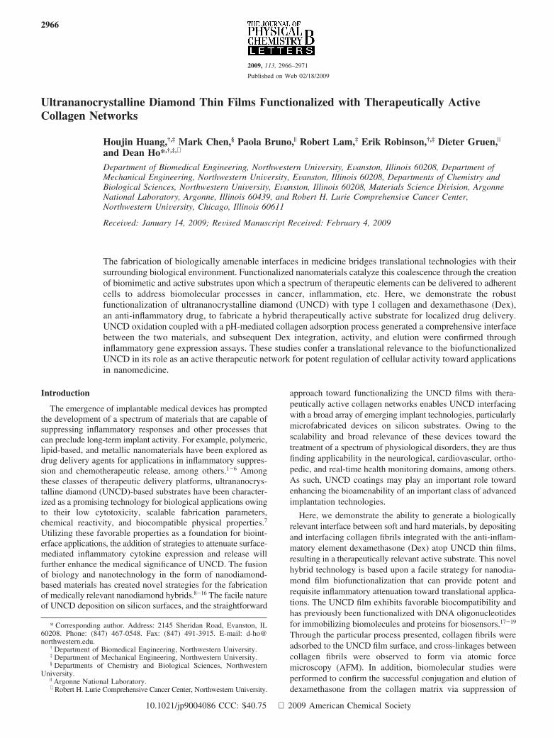
Formation of a uniform fibrous collagen layer on an UNCDsurface is not a trivial issue. The surface properties of UNCD,the complicated conformation and aggregation states of collagenin solution, as well as the delicate interaction between UNCDand collagen are all needed to be considered. The functional-ization of UNCD films can be facilitated by the functionalgroupspresentontheUNCDfilmsurface.Successfulbiotic-abioticinterfaces such as the UNCD-collagen integrated film are basedon interactions that can take advantage of surface propertiesfrom both components of the interface. In this case, enhancedwetting on the UNCD surface can be coupled with the soft/therapeutically active soft surface properties presented by thecollagen. Experiments conducted using NDs as catalysts foroxidation reactions have shown that UNCD surfaces aresuccessfully saturated in atomic oxygen.26 In addition, theoxidation of UNCD surfaces has produced hydroxyl andcarboxylic acid groups that can enhance surface wettability toimprove collagen protein adhesion.16,27 The hydroxide ions inthe aqueous collagen (pH 8) act as the deprotonating agent forthe carboxyl group when the aqueous collagen is added to thesurface of the oxidized UNCD film. This reaction generatescarboxylate anions on the surface of the UNCD that can enhancecharged-based interactions with the collagen protein. Forexample, maximum coating and retention of collagen on thesurface of the film was observed at pH 8. Dehydration in anoven was required for successful UNCD-collagen integrationtoaid inremovalofwaterduringformationof thecollagen-UNCDinterface. Therefore, promoting the rate by driving the mech-anism of dehydration in an oven near physiological temperatureswas crucial to interface formation. It has previously beensuggested that an entropy increase due to liberation of watermolecules drives mechanism of adsorption, and not collagenconformational changes.28 In addition, increased temperature inthe preparation of collagen has been shown to produce longer,thinner, and more flexible collagen fibers than at lower tem-peratures. The collagen/surface dehydration treatment at 40 °Cfor 30 min under a vacuum resulted in the potent adsorption ofcollagen fibril networks. Figure 2 shows the topographicalmodifications associated with UNCD oxidation that facilitatedrobust collagen-UNCD integration. Figure 1A represents aUNCD film deposited on a silicon wafer prior to surfaceoxidation. Surface features are more pronounced and larger indimension (e.g., diameter) compared to the oxidized UNCDfeatures shown in Figure 1B.
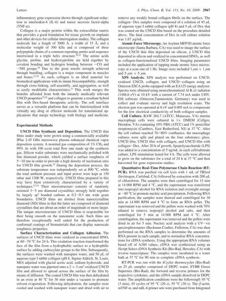
Collagen layers adsorbed on hydrophilic surfaces are morestable, possibly due to a lower affinity caused by increasedsurface hydration and/or electrostatic repulsion or deprotonatedcarboxyl functions on the surface.29 Hydrophilic surfaces witha lower rate of adsorption cause a conformal dense layer ofcollagen to form, whereas, in a hydrophobic surface, a highrate of adsorption/desorption causes loose segments to form andaggregate, albeit through multiple sources of individual fibrillinkage29 (Figure 2A). We found that the optimal collagenadherence occurred at a slightly basic pH (Figure 2B). Decreas-ing the pH of the solution produced little to no noticeablecollagen attachment and has been implicated in the formation
of micropores, increasing disentanglement due to decreasedinterfibril forces while forming a diffuse layer of globularsubunits.30-32 Moreover, collagen layers are more likely to formmeshes,31 increase in density through loss of water from collagenfibers, and have higher stability due to increased intermolecularforces caused by the deprotonation of histidine and ionizationof other primary acids at higher pH levels.33-36 In addition tothe clearly visible collagen fibrils shown in Figure 2, X-rayphotoelectron spectroscopy (XPS) was performed to furtheranalyze UNCD film oxidation as well as collagen presence onthe UNCD film.
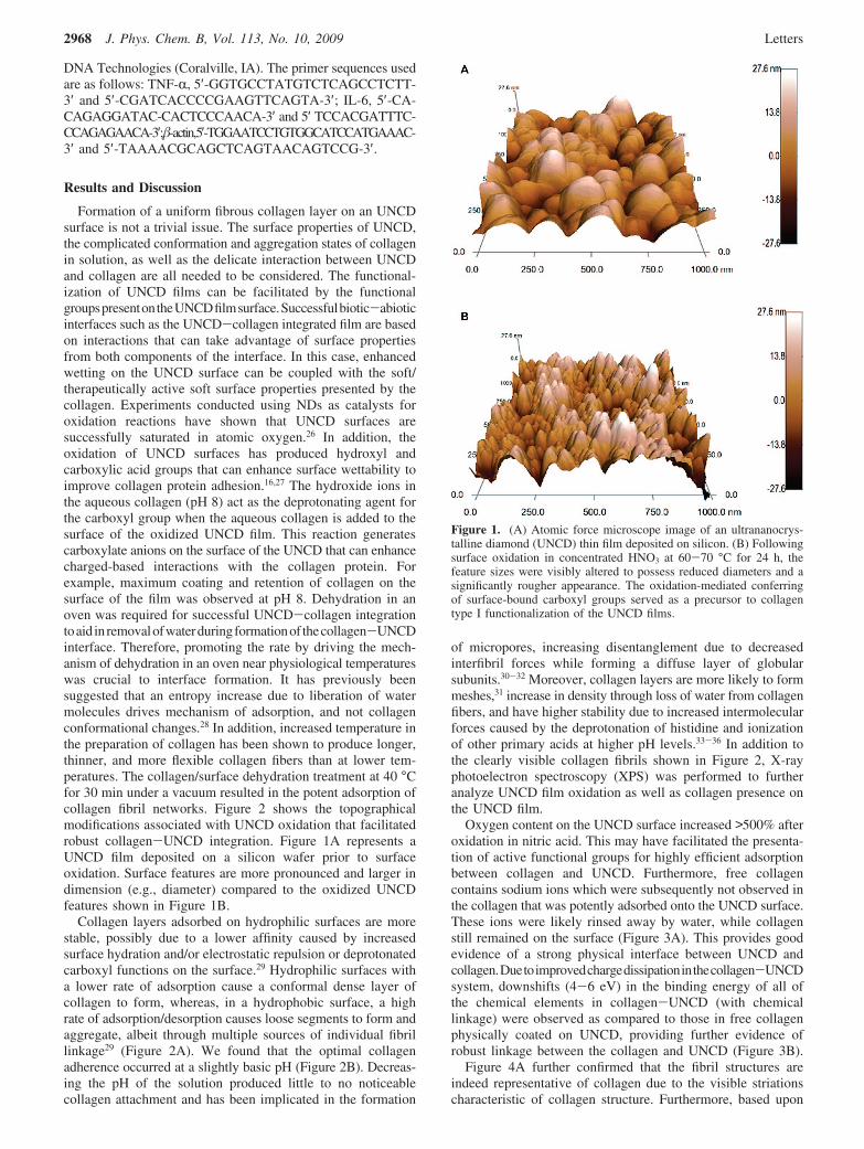
Oxygen content on the UNCD surface increased >500% afteroxidation in nitric acid. This may have facilitated the presenta-tion of active functional groups for highly efficient adsorptionbetween collagen and UNCD. Furthermore, free collagencontains sodium ions which were subsequently not observed inthe collagen that was potently adsorbed onto the UNCD surface.These ions were likely rinsed away by water, while collagenstill remained on the surface (Figure 3A). This provides goodevidence of a strong physical interface between UNCD andcollagen.Duetoimprovedchargedissipationinthecollagen-UNCDsystem, downshifts (4-6 eV) in the binding energy of all ofthe chemical elements in collagen-UNCD (with chemicallinkage) were observed as compared to those in free collagenphysically coated on UNCD, providing further evidence ofrobust linkage between the collagen and UNCD (Figure 3B).
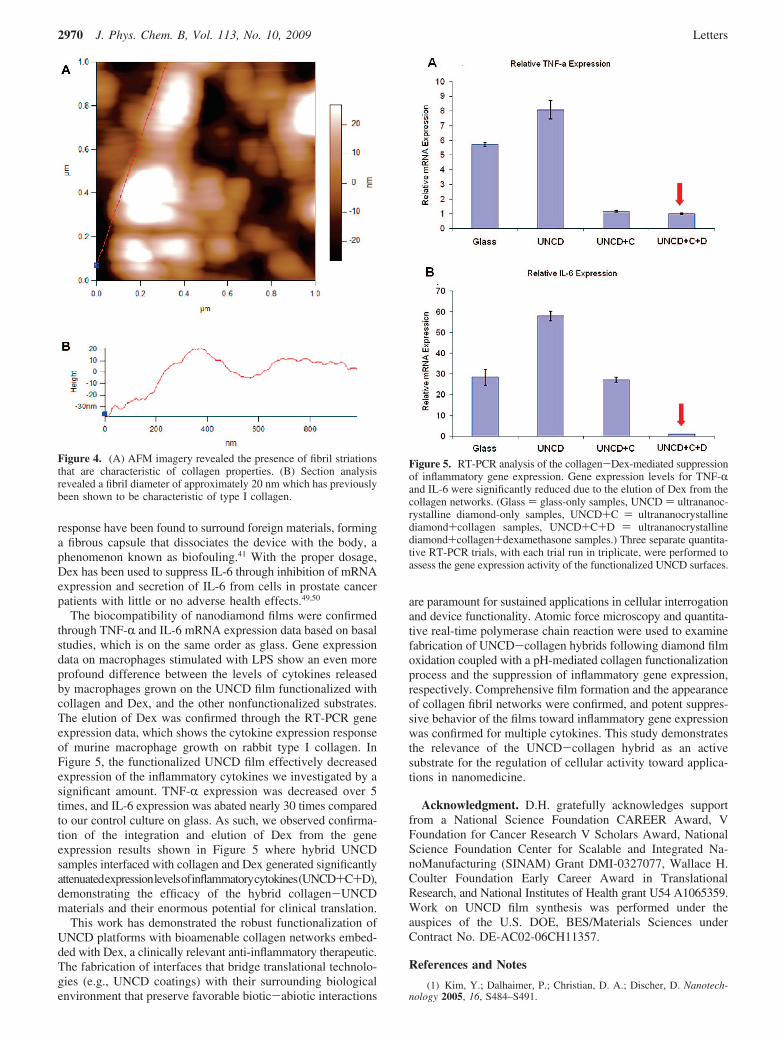
Figure 4A further confirmed that the fibril structures areindeed representative of collagen due to the visible striationscharacteristic of collagen structure. Furthermore, based upon
Figure 1. (A) Atomic force microscope image of an ultrananocrys-talline diamond (UNCD) thin film deposited on silicon. (B) Followingsurface oxidation in concentrated HNO3 at 60-70 °C for 24 h, thefeature sizes were visibly altered to possess reduced diameters and asignificantly rougher appearance. The oxidation-mediated conferringof surface-bound carboxyl groups served as a precursor to collagentype I functionalization of the UNCD films.
2968 J. Phys. Chem. B, Vol. 113, No. 10, 2009 Letters
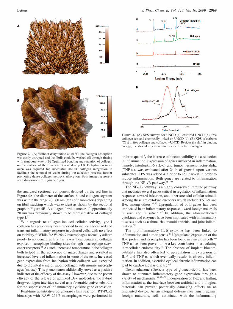
the analyzed sectional component denoted by the red line inFigure 4A, the diameter of the surface-bound collagen segmentwas within the range 20-60 nm (tens of nanometers) dependingon fibril stacking which was evident as shown by the sectionalgraph in Figure 4B. A collagen fibril diameter of approximately20 nm was previously shown to be representative of collagentype I.37
With regards to collagen-induced cellular activity, type Icollagen has previously been reported to induce a localized andtransient inflammatory response in cultured cells, with no effecton viability.35 While RAW 264.7 macrophages normally adherepoorly to nondenatured fibrillar layers, heat denatured collagenexposes macrophage binding sites through macrophage scav-enger receptors.31 As such, increased temperature in the collagenboth helped in the adherence of macrophages and resulted inincreased levels of inflammation in some of the tests. Increasedgene expression from incubation with collagen was expecteddue to the interfacing of rabbit collagen with murine macroph-ages (mouse). This phenomenon additionally served as a positiveindicator of the efficacy of the assay. However, due to the potentefficacy of the release of admixed Dex molecules, the hybriddrug-collagen interface served as a favorable active substratefor the suppression of inflammatory cytokine gene expression.
Real-time quantitative polymerase chain reaction (RT-PCR)bioassays with RAW 264.7 macrophages were performed in
order to quantify the increase in biocompatibility via a reductionin inflammation. Expression of genes involved in inflammation,namely, interleukin-6 (IL-6) and tumor necrosis factor-alpha(TNF-R), was evaluated after 24 h of growth upon varioussubstrates. LPS was added 4 h prior to cell harvest in order toinduce inflammation. Both genes are related to inflammationthrough the NF-κB pathway.38-40
The NF-κB pathway is a highly conserved immune pathwaythat mediates several genes critical in regulation of inflammation,responses toward infection, and other stressful cellular stimuli.Among these are cytokine encoders which include TNF-R andIl-6, among others.39,40 Upregulation of both genes has beenimplicated in an inflammatory response toward foreign materialsin ViVo and in Vitro.41,42 In addition, the aforementionedcytokines and enzymes have been implicated with inflammatorydiseases such as asthma, rheumatoid arthritis, and joint inflam-mation.38
The proinflammatory IL-6 cytokine has been linked toinflammation and tumorigensis.43 Upregulated expression of theIL-6 protein and its receptor has been found in cancerous cells.44
TNF-R has been proven to be a key contributor in articulatingintracellular endotoxicity.45 The absence of implant biocom-patibility has also often led to upregulation in expression ofIL-6 and TNF-R, which eventually results in chronic inflam-mation. In addition, extended cyclical chronic inflammation canlead to cardiovascular disease.46
Dexamethasone (Dex), a type of glucocorticoid, has beenshown to attenuate inflammatory gene expression through avariety of mechanisms.38,47-50 Incorporation of Dex and haltinginflammation at the interface between artificial and biologicalmaterials can prevent potentially damaging effects on animplanted device. As an integral defense mechanism againstforeign materials, cells associated with the inflammatory
Figure 2. (A) Without dehydration at 40 °C, the collagen adsorptionwas easily disrupted and the fibrils could be washed off through rinsingwith nanopure water. (B) Optimized bonding and retention of collagenon the surface of the film was observed at pH 8. Dehydration in anoven was required for successful UNCD-collagen integration tofacilitate the removal of water during the adhesion process, furtherpromoting dense collagen network adsorption. Both images representscan dimensions of 5 µm × 5 µm.
Figure 3. (A) XPS surveys for UNCD (a), oxidized UNCD (b), freecollagen (c), and chemically linked on UNCD (d). (B) XPS of carbons(C1s) in free collagen and collagen-UNCD. Besides the shift in bindingenergy, the shoulder peak is more evident in free collagen.
Letters J. Phys. Chem. B, Vol. 113, No. 10, 2009 2969
response have been found to surround foreign materials, forminga fibrous capsule that dissociates the device with the body, aphenomenon known as biofouling.41 With the proper dosage,Dex has been used to suppress IL-6 through inhibition of mRNAexpression and secretion of IL-6 from cells in prostate cancerpatients with little or no adverse health effects.49,50
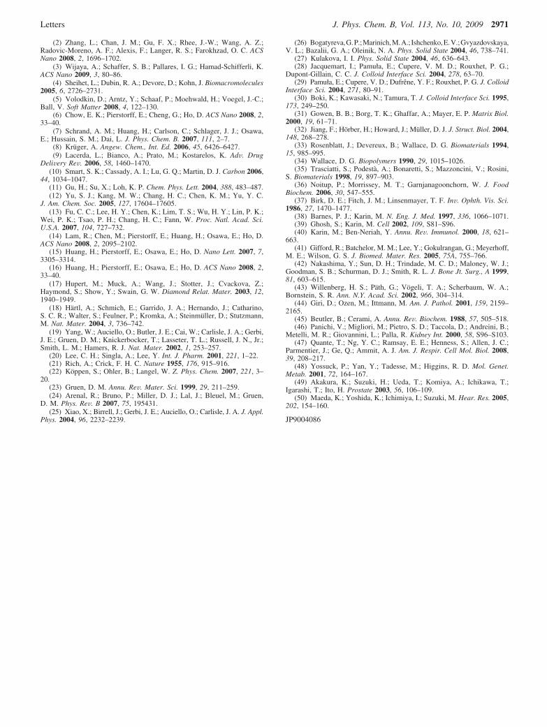
The biocompatibility of nanodiamond films were confirmedthrough TNF-R and IL-6 mRNA expression data based on basalstudies, which is on the same order as glass. Gene expressiondata on macrophages stimulated with LPS show an even moreprofound difference between the levels of cytokines releasedby macrophages grown on the UNCD film functionalized withcollagen and Dex, and the other nonfunctionalized substrates.The elution of Dex was confirmed through the RT-PCR geneexpression data, which shows the cytokine expression responseof murine macrophage growth on rabbit type I collagen. InFigure 5, the functionalized UNCD film effectively decreasedexpression of the inflammatory cytokines we investigated by asignificant amount. TNF-R expression was decreased over 5times, and IL-6 expression was abated nearly 30 times comparedto our control culture on glass. As such, we observed confirma-tion of the integration and elution of Dex from the geneexpression results shown in Figure 5 where hybrid UNCDsamples interfaced with collagen and Dex generated significantlyattenuatedexpressionlevelsofinflammatorycytokines(UNCD+C+D),demonstrating the efficacy of the hybrid collagen-UNCDmaterials and their enormous potential for clinical translation.
This work has demonstrated the robust functionalization ofUNCD platforms with bioamenable collagen networks embed-ded with Dex, a clinically relevant anti-inflammatory therapeutic.The fabrication of interfaces that bridge translational technolo-gies (e.g., UNCD coatings) with their surrounding biologicalenvironment that preserve favorable biotic-abiotic interactions
are paramount for sustained applications in cellular interrogationand device functionality. Atomic force microscopy and quantita-tive real-time polymerase chain reaction were used to examinefabrication of UNCD-collagen hybrids following diamond filmoxidation coupled with a pH-mediated collagen functionalizationprocess and the suppression of inflammatory gene expression,respectively. Comprehensive film formation and the appearanceof collagen fibril networks were confirmed, and potent suppres-sive behavior of the films toward inflammatory gene expressionwas confirmed for multiple cytokines. This study demonstratesthe relevance of the UNCD-collagen hybrid as an activesubstrate for the regulation of cellular activity toward applica-tions in nanomedicine.
Acknowledgment. D.H. gratefully acknowledges supportfrom a National Science Foundation CAREER Award, VFoundation for Cancer Research V Scholars Award, NationalScience Foundation Center for Scalable and Integrated Na-noManufacturing (SINAM) Grant DMI-0327077, Wallace H.Coulter Foundation Early Career Award in TranslationalResearch, and National Institutes of Health grant U54 A1065359.Work on UNCD film synthesis was performed under theauspices of the U.S. DOE, BES/Materials Sciences underContract No. DE-AC02-06CH11357.
References and Notes
(1) Kim, Y.; Dalhaimer, P.; Christian, D. A.; Discher, D. Nanotech-nology 2005, 16, S484–S491.
Figure 4. (A) AFM imagery revealed the presence of fibril striationsthat are characteristic of collagen properties. (B) Section analysisrevealed a fibril diameter of approximately 20 nm which has previouslybeen shown to be characteristic of type I collagen.
Figure 5. RT-PCR analysis of the collagen-Dex-mediated suppressionof inflammatory gene expression. Gene expression levels for TNF-Rand IL-6 were significantly reduced due to the elution of Dex from thecollagen networks. (Glass ) glass-only samples, UNCD ) ultrananoc-rystalline diamond-only samples, UNCD+C ) ultrananocrystallinediamond+collagen samples, UNCD+C+D ) ultrananocrystallinediamond+collagen+dexamethasone samples.) Three separate quantita-tive RT-PCR trials, with each trial run in triplicate, were performed toassess the gene expression activity of the functionalized UNCD surfaces.
2970 J. Phys. Chem. B, Vol. 113, No. 10, 2009 Letters
(2) Zhang, L.; Chan, J. M.; Gu, F. X.; Rhee, J.-W.; Wang, A. Z.;Radovic-Moreno, A. F.; Alexis, F.; Langer, R. S.; Farokhzad, O. C. ACSNano 2008, 2, 1696–1702.
(3) Wijaya, A.; Schaffer, S. B.; Pallares, I. G.; Hamad-Schifferli, K.ACS Nano 2009, 3, 80–86.
(4) Sheihet, L.; Dubin, R. A.; Devore, D.; Kohn, J. Biomacromolecules2005, 6, 2726–2731.
(5) Volodkin, D.; Arntz, Y.; Schaaf, P.; Moehwald, H.; Voegel, J.-C.;Ball, V. Soft Matter 2008, 4, 122–130.
(6) Chow, E. K.; Pierstorff, E.; Cheng, G.; Ho, D. ACS Nano 2008, 2,33–40.
(7) Schrand, A. M.; Huang, H.; Carlson, C.; Schlager, J. J.; Osawa,E.; Hussain, S. M.; Dai, L. J. Phys. Chem. B. 2007, 111, 2–7.
(8) Kruger, A. Angew. Chem., Int. Ed. 2006, 45, 6426–6427.(9) Lacerda, L.; Bianco, A.; Prato, M.; Kostarelos, K. AdV. Drug
DeliVery ReV. 2006, 58, 1460–1470.(10) Smart, S. K.; Cassady, A. I.; Lu, G. Q.; Martin, D. J. Carbon 2006,
44, 1034–1047.(11) Gu, H.; Su, X.; Loh, K. P. Chem. Phys. Lett. 2004, 388, 483–487.(12) Yu, S. J.; Kang, M. W.; Chang, H. C.; Chen, K. M.; Yu, Y. C.
J. Am. Chem. Soc. 2005, 127, 17604–17605.(13) Fu, C. C.; Lee, H. Y.; Chen, K.; Lim, T. S.; Wu, H. Y.; Lin, P. K.;
Wei, P. K.; Tsao, P. H.; Chang, H. C.; Fann, W. Proc. Natl. Acad. Sci.U.S.A. 2007, 104, 727–732.
(14) Lam, R.; Chen, M.; Pierstorff, E.; Huang, H.; Osawa, E.; Ho, D.ACS Nano 2008, 2, 2095–2102.
(15) Huang, H.; Pierstorff, E.; Osawa, E.; Ho, D. Nano Lett. 2007, 7,3305–3314.
(16) Huang, H.; Pierstorff, E.; Osawa, E.; Ho, D. ACS Nano 2008, 2,33–40.
(17) Hupert, M.; Muck, A.; Wang, J.; Stotter, J.; Cvackova, Z.;Haymond, S.; Show, Y.; Swain, G. W. Diamond Relat. Mater. 2003, 12,1940–1949.
(18) Hartl, A.; Schmich, E.; Garrido, J. A.; Hernando, J.; Catharino,S. C. R.; Walter, S.; Feulner, P.; Kromka, A.; Steinmuller, D.; Stutzmann,M. Nat. Mater. 2004, 3, 736–742.
(19) Yang, W.; Auciello, O.; Butler, J. E.; Cai, W.; Carlisle, J. A.; Gerbi,J. E.; Gruen, D. M.; Knickerbocker, T.; Lasseter, T. L.; Russell, J. N., Jr.;Smith, L. M.; Hamers, R. J. Nat. Mater. 2002, 1, 253–257.
(20) Lee, C. H.; Singla, A.; Lee, Y. Int. J. Pharm. 2001, 221, 1–22.(21) Rich, A.; Crick, F. H. C. Nature 1955, 176, 915–916.(22) Koppen, S.; Ohler, B.; Langel, W. Z. Phys. Chem. 2007, 221, 3–
20.(23) Gruen, D. M. Annu. ReV. Mater. Sci. 1999, 29, 211–259.(24) Arenal, R.; Bruno, P.; Miller, D. J.; Lal, J.; Bleuel, M.; Gruen,
D. M. Phys. ReV. B 2007, 75, 195431.(25) Xiao, X.; Birrell, J.; Gerbi, J. E.; Auciello, O.; Carlisle, J. A. J. Appl.
Phys. 2004, 96, 2232–2239.
(26) Bogatyreva,G.P.;Marinich,M.A.; Ishchenko,E.V.;Gvyazdovskaya,V. L.; Bazalii, G. A.; Oleinik, N. A. Phys. Solid State 2004, 46, 738–741.
(27) Kulakova, I. I. Phys. Solid State 2004, 46, 636–643.(28) Jacquemart, I.; Pamuła, E.; Cupere, V. M. D.; Rouxhet, P. G.;
Dupont-Gillain, C. C. J. Colloid Interface Sci. 2004, 278, 63–70.(29) Pamuła, E.; Cupere, V. D.; Dufrene, Y. F.; Rouxhet, P. G. J. Colloid
Interface Sci. 2004, 271, 80–91.(30) Boki, K.; Kawasaki, N.; Tamura, T. J. Colloid Interface Sci. 1995,
173, 249–250.(31) Gowen, B. B.; Borg, T. K.; Ghaffar, A.; Mayer, E. P. Matrix Biol.
2000, 19, 61–71.(32) Jiang, F.; Horber, H.; Howard, J.; Muller, D. J. J. Struct. Biol. 2004,
148, 268–278.(33) Rosenblatt, J.; Devereux, B.; Wallace, D. G. Biomaterials 1994,
15, 985–995.(34) Wallace, D. G. Biopolymers 1990, 29, 1015–1026.(35) Trasciatti, S.; Podesta, A.; Bonaretti, S.; Mazzoncini, V.; Rosini,
S. Biomaterials 1998, 19, 897–903.(36) Noitup, P.; Morrissey, M. T.; Garnjanagoonchorn, W. J. Food
Biochem. 2006, 30, 547–555.(37) Birk, D. E.; Fitch, J. M.; Linsenmayer, T. F. InV. Ophth. Vis. Sci.
1986, 27, 1470–1477.(38) Barnes, P. J.; Karin, M. N. Eng. J. Med. 1997, 336, 1066–1071.(39) Ghosh, S.; Karin, M. Cell 2002, 109, S81–S96.(40) Karin, M.; Ben-Neriah, Y. Annu. ReV. Immunol. 2000, 18, 621–
663.(41) Gifford, R.; Batchelor, M. M.; Lee, Y.; Gokulrangan, G.; Meyerhoff,
M. E.; Wilson, G. S. J. Biomed. Mater. Res. 2005, 75A, 755–766.(42) Nakashima, Y.; Sun, D. H.; Trindade, M. C. D.; Maloney, W. J.;
Goodman, S. B.; Schurman, D. J.; Smith, R. L. J. Bone Jt. Surg., A 1999,81, 603–615.
(43) Willenberg, H. S.; Path, G.; Vogeli, T. A.; Scherbaum, W. A.;Bornstein, S. R. Ann. N.Y. Acad. Sci. 2002, 966, 304–314.
(44) Giri, D.; Ozen, M.; Ittmann, M. Am. J. Pathol. 2001, 159, 2159–2165.
(45) Beutler, B.; Cerami, A. Annu. ReV. Biochem. 1988, 57, 505–518.(46) Panichi, V.; Migliori, M.; Pietro, S. D.; Taccola, D.; Andreini, B.;
Metelli, M. R.; Giovannini, L.; Palla, R. Kidney Int. 2000, 58, S96–S103.(47) Quante, T.; Ng, Y. C.; Ramsay, E. E.; Henness, S.; Allen, J. C.;
Parmentier, J.; Ge, Q.; Ammit, A. J. Am. J. Respir. Cell Mol. Biol. 2008,39, 208–217.
(48) Yossuck, P.; Yan, Y.; Tadesse, M.; Higgins, R. D. Mol. Genet.Metab. 2001, 72, 164–167.
(49) Akakura, K.; Suzuki, H.; Ueda, T.; Komiya, A.; Ichikawa, T.;Igarashi, T.; Ito, H. Prostate 2003, 56, 106–109.
(50) Maeda, K.; Yoshida, K.; Ichimiya, I.; Suzuki, M. Hear. Res. 2005,202, 154–160.
JP9004086
Letters J. Phys. Chem. B, Vol. 113, No. 10, 2009 2971
Related Documents