Tumorigenesis and Neoplastic Progression Tumor Progression Stage and Anatomical Site Regulate Tumor-Associated Macrophage and Bone Marrow-Derived Monocyte Polarization Elizabeth F. Redente,* Lori D. Dwyer-Nield,* Daniel T. Merrick, † Komal Raina,* Rajesh Agarwal,* William Pao, ‡ Pamela L. Rice, § Kenneth R. Shroyer, ¶ and Alvin M. Malkinson* From the Departments of Pharmaceutical Sciences,* and Pathology, † University of Colorado, Aurora, Colorado; the Department of Medicine, ‡ Memorial Sloan-Kettering Cancer Center, New York, New York; the Division of Pulmonary Biology, § Veterans Affairs Medical Center, Denver, Colorado; and the Departments of Medicine and Pathology, ¶ Stony Brook University, Stony Brook, New York Tumor-associated macrophages (TAMs) encourage and coordinate neoplastic growth. In late stage hu- man lung adenocarcinoma , TAMs exhibited mixed M1 (classical; argI low iNOS high ) and M2 (alternative; argI high iNOS low ) polarization based on arginine me- tabolism. In several murine cancer models including chemically and genetically-induced primary lung tu- mors, prostate tumors, colon xenografts, and lung metastases , TAMs expressed argI high iNOS low early during tumor formation; argI low iNOS high polarization also occurred during malignancy in some models. In a chemically-induced lung tumor model , macro- phages expressed argI high iNOS low within one week after carcinogen treatment , followed by similar polar- ization of bone marrow– derived monocytes (BDMCs) a few days later. TAMs surrounding murine prostate tumors also expressed argI high iNOS low early during tumorigenesis , indicating that this polarization is not unique to neoplastic lungs. In a human colon cancer xenograft model , the primary tumor was surrounded by argI high iNOS low -expressing TAMs , and BDMCs also expressed argI high iNOS low , but pulmonary macro- phages adopted argI high iNOS low polarization only af- ter tumors metastasized to the lungs. Persistence of tumors is required to maintain TAM polarization. In- deed , in both conditional mutant Kras- and FGF10- driven models of lung cancer, mice expressing the transgene develop lung tumors that regress rapidly when the transgene is silenced. Furthermore, pul- monary macrophages expressed argI high iNOS low on tumor induction, but then returned to argI low iNOS low (no polarization) after tumors regressed. Manipulating TAM function or depleting TAMs may provide novel therapeutic strategies for preventing and treating many types of cancer. (Am J Pathol 2010, 176:2972–2985; DOI: 10.2353/ajpath.2010.090879) Resident and newly infiltrating macrophages are key leu- kocyte populations that regulate production of inflamma- tory mediators to warn of injury and initiate tissue repair during cancer-associated inflammation. 1,2 The destiny of macrophages is at least partly determined during their ontogeny in the bone marrow. Monocyte precursors dif- ferentiate to become either macrophages that reside nor- mally in tissues or inflammatory macrophages that re- spond to chemokine signals and infiltrate into the affected tissue to mount a response. 3 During tumorigen- esis, macrophages infiltrate tumors and surrounding tis- sue. However, unlike scenarios where macrophages are involved in host defense, tumor associated macrophages (TAMs) support rather than hinder neoplastic growth, as shown by histopathological, epidemiological, pharmaco- logical, and genetic studies. 4 TAMs were first observed 150 years ago by Virchow, 5 and current evidence asso- ciates chronic inflammatory diseases such as bronchitis, inflammatory bowel disease, gastritis, and colitis with increased risk of cancer development. 2 This synergistic co-evolution of cancer and inflammation continues when tissues are damaged by aggressive cancer growth. Anti- Supported by National Cancer Institute 033497 and 132552 (to A.M.M.), CA113876 (to R.A.), and National Institutes of Health Special Program of Research Excellence program PA50CA058187 (to D.T.M.). Accepted for publication January 27, 2010. W.P. is a consultant for Molecular MD. Supplemental material for this article can be found on http://ajp. amjpathol.org. Address reprint requests to Lori D. Dwyer-Nield, Ph.D., Department of Pharmaceutical Sciences, University of Colorado Denver, C238-P15 RII, 12700 East 19th Avenue, Aurora, CO 80045. E-mail: Lori.Nield@ ucdenver.edu. The American Journal of Pathology, Vol. 176, No. 6, June 2010 Copyright © American Society for Investigative Pathology DOI: 10.2353/ajpath.2010.090879 2972

Welcome message from author
This document is posted to help you gain knowledge. Please leave a comment to let me know what you think about it! Share it to your friends and learn new things together.
Transcript

Tumorigenesis and Neoplastic Progression
Tumor Progression Stage and Anatomical SiteRegulate Tumor-Associated Macrophage andBone Marrow-Derived Monocyte Polarization
Elizabeth F. Redente,* Lori D. Dwyer-Nield,*Daniel T. Merrick,† Komal Raina,*Rajesh Agarwal,* William Pao,‡ Pamela L. Rice,§
Kenneth R. Shroyer,¶ and Alvin M. Malkinson*From the Departments of Pharmaceutical Sciences,* and
Pathology,† University of Colorado, Aurora, Colorado; the
Department of Medicine,‡ Memorial Sloan-Kettering Cancer
Center, New York, New York; the Division of Pulmonary Biology,§
Veterans Affairs Medical Center, Denver, Colorado; and the
Departments of Medicine and Pathology,¶ Stony Brook
University, Stony Brook, New York
Tumor-associated macrophages (TAMs) encourageand coordinate neoplastic growth. In late stage hu-man lung adenocarcinoma, TAMs exhibited mixedM1 (classical; argIlowiNOShigh) and M2 (alternative;argIhighiNOSlow) polarization based on arginine me-tabolism. In several murine cancer models includingchemically and genetically-induced primary lung tu-mors, prostate tumors, colon xenografts, and lungmetastases, TAMs expressed argIhighiNOSlow earlyduring tumor formation; argIlowiNOShigh polarizationalso occurred during malignancy in some models. Ina chemically-induced lung tumor model, macro-phages expressed argIhighiNOSlow within one weekafter carcinogen treatment, followed by similar polar-ization of bone marrow–derived monocytes (BDMCs)a few days later. TAMs surrounding murine prostatetumors also expressed argIhighiNOSlow early duringtumorigenesis, indicating that this polarization is notunique to neoplastic lungs. In a human colon cancerxenograft model, the primary tumor was surroundedby argIhighiNOSlow-expressing TAMs, and BDMCs alsoexpressed argIhighiNOSlow, but pulmonary macro-phages adopted argIhighiNOSlow polarization only af-ter tumors metastasized to the lungs. Persistence oftumors is required to maintain TAM polarization. In-deed, in both conditional mutant Kras- and FGF10-driven models of lung cancer , mice expressing thetransgene develop lung tumors that regress rapidlywhen the transgene is silenced. Furthermore, pul-
monary macrophages expressed argIhighiNOSlow
on tumor induction, but then returned to argIlow
iNOSlow (no polarization) after tumors regressed.Manipulating TAM function or depleting TAMs mayprovide novel therapeutic strategies for preventingand treating many types of cancer. (Am J Pathol 2010,
176:2972–2985; DOI: 10.2353/ajpath.2010.090879)
Resident and newly infiltrating macrophages are key leu-kocyte populations that regulate production of inflamma-tory mediators to warn of injury and initiate tissue repairduring cancer-associated inflammation.1,2 The destiny ofmacrophages is at least partly determined during theirontogeny in the bone marrow. Monocyte precursors dif-ferentiate to become either macrophages that reside nor-mally in tissues or inflammatory macrophages that re-spond to chemokine signals and infiltrate into theaffected tissue to mount a response.3 During tumorigen-esis, macrophages infiltrate tumors and surrounding tis-sue. However, unlike scenarios where macrophages areinvolved in host defense, tumor associated macrophages(TAMs) support rather than hinder neoplastic growth, asshown by histopathological, epidemiological, pharmaco-logical, and genetic studies.4 TAMs were first observed150 years ago by Virchow,5 and current evidence asso-ciates chronic inflammatory diseases such as bronchitis,inflammatory bowel disease, gastritis, and colitis withincreased risk of cancer development.2 This synergisticco-evolution of cancer and inflammation continues whentissues are damaged by aggressive cancer growth. Anti-
Supported by National Cancer Institute 033497 and 132552 (to A.M.M.),CA113876 (to R.A.), and National Institutes of Health Special Program ofResearch Excellence program PA50CA058187 (to D.T.M.).
Accepted for publication January 27, 2010.
W.P. is a consultant for Molecular MD.
Supplemental material for this article can be found on http://ajp.amjpathol.org.
Address reprint requests to Lori D. Dwyer-Nield, Ph.D., Department ofPharmaceutical Sciences, University of Colorado Denver, C238-P15 RII,12700 East 19th Avenue, Aurora, CO 80045. E-mail: [email protected].
The American Journal of Pathology, Vol. 176, No. 6, June 2010
Copyright © American Society for Investigative Pathology
DOI: 10.2353/ajpath.2010.090879
2972

inflammatory drugs such as sulindac and aspirin inhibitcolon polyp formation in colorectal cancer patients,6 whilepolymorphisms in genes regulating inflammation controlsusceptibility to lung cancer in both mice7 and humans.8
Overexpression of anti-inflammatory signals9,10 depressescancer growth, whereas mutations that enhance inflamma-tory responses augment tumorigenesis.11
Macrophages exhibit a wide phenotypic diversity andexert multiple pathobiological effects12 that support andencourage tumor growth, including the release of proan-giogenic factors such as VEGF,13 EGF,14 and proteasesthat digest normal tissue to provide a route for vesselintravasation and metastatic invasion of tumor cells. Mac-rophages also suppress the activities of CD4� and CD8�
lymphocytes by secreting chemokines that attract T-reg-ulatory cells to dampen adaptive immune surveillanceagainst tumors.15,16
Two macrophage activation phenotypes, M1 and M2,characterize extremes along a gradient of activation andare regulated by the relative abundance of TH1 and TH2cytokines. In vitro, mature macrophages demonstrateplasticity by shuttling back and forth between these ac-tivation pathways.17,18 Macrophages classically/M1 po-larized by exposure to interferon (IFN)-� or LPS produceinterleukin (IL)-6 and IL-12, and up-regulate induciblenitric oxide synthase (iNOS) to produce nitric oxide (NO)and citrulline. Alternative/M2 polarization is induced byexposure to IL-4 and IL-13, cytokines that bind to acommon receptor subunit, IL-4R�.19 M2 macrophagesproduce anti-inflammatory IL-10, up-regulate expressionof the mannose receptor and arginase I, as well as mac-rophage-specific genes including Ym1 and Fizz1. Poly-amines are produced, and the iNOS substrate, arginine,is depleted in M2 polarized macrophages.12,20–22 Thesechanges in macrophage phenotype have functional con-sequences. NO produced by M1 macrophages can killother cells by combining with oxygen radicals to formtoxic peroxynitrite.23 Ornithine produced by M2 macro-phages can be exported and taken up by tumor cells toserve as precursors for DNA synthesis.24 Some macro-phages express both arginase and iNOS enzymes (M1 �M2), which can act synergistically, as well as indepen-dently, to limit T cell function. A recent study illustratingthe clinical significance of M2 polarization by TAMs dem-onstrated that mice vaccinated against the asparaginylendopeptidase, legumain, which is highly and specifi-cally expressed in M2 macrophages, primed lympho-cytes against this M2 antigen. Subsequent killing of M2macrophages by CD8� lymphocytes inhibited the growthof breast cancer xenografts.25 The TH2 cytokines thatregulate M2 TAM activation are associated with manyneoplasias, including non-small cell lung (NSCLC),26
breast,25 and ovarian cancer.27,28
We recently characterized stromal cell compositionduring primary lung cancer progression induced in A/Jmice by urethane,29 an animal model of pulmonary ade-nocarcinoma (AC).30 Macrophages accumulated adja-cent to, but outside of, tumors. As tumors progressed tomalignancy, macrophage numbers increased, and theirpolarization changed.29 Pulmonary macrophage argininemetabolism was assessed to determine their polarization
state. Naïve A/J mice expressed very low levels of argi-nase I and iNOS (argIlowiNOSlow), constituting an unpo-larized phenotype. TAMs in mice bearing benign tumorsexpressed arginase I (argIhighiNOSlow; M2 polarized),whereas macrophages associated with malignant tumorsexpressed iNOS (argIlowiNOShigh; M1 polarized). Localpulmonary macrophages were not the only populationaffected by signals emanating from tumors in this study.Polarization of monocytes extruded from the bone mar-row (bone marrow–derived monocytes [BDMCs]) mir-rored that of TAMs, switching from argIlowiNOSlow in con-trol mice to argIhighiNOSlow in adenoma-bearing mice,and then to argIlowiNOShigh when carcinomas had devel-oped.29 BDMCs thus underwent tumor-directed differen-tiation while still in the bone marrow before entering thecirculation and infiltrating the lungs to become TAMs.This suggests that the differentiation status of circulatingmonocytes could be evaluated as a diagnostic tool formonitoring lung cancer progression.
Herein, we assess the arginine metabolic phenotype ofmacrophages and BDMCs during tumorigenesis to as-sess polarization in response to different carcinogens, atdifferent organ sites, and in genetic models, thus testingthe specificity of the macrophage response to tumors,their location, and at different stages of tumorigenesis.We find correlations between these experimental modelsand human disease.
Materials and Methods
Human Lung Cancer Sample Acquisition
Fourteen histological specimens of pulmonary AC wereselected on the basis of availability of adequate tissuefor immunohistochemical evaluation from archival col-lections maintained by the Department of Pathology,University of Colorado Denver under the auspices ofthe COMIRB. These tumors had been previously clas-sified as well-differentiated, moderately-differentiated,or poorly-differentiated.
Macrophage Polarization in Formalin-FixedTissue
Human tumor biopsy and surrounding stromal tissue andmouse lung tissue were examined for macrophage infil-tration and polarization status by immunofluorescenceusing antibodies against F4/80 to identify macrophages,iNOS to detect M1 polarization, and mannose receptor(human) or arginase I (mouse) to detect M2 polarization,using procedures described previously29 and summa-rized in Table 1. Briefly, sections were deparaffinized andrehydrated through a series of ethanol washes, and an-tigens retrieved in boiling 100 mmol/L citrate buffer fol-lowed by a 40-minute autofluorescence block with 10mg/ml sodium borohydride in PBS. Samples were firstblocked with avidin-biotin solution (Vector Laboratories,Burlingame, CA), followed by exposure to the primaryand biotinylated secondary antibodies (Table 1) and de-tection with fluorescent avidin conjugates.
Tumors Regulate Macrophage Polarization 2973AJP June 2010, Vol. 176, No. 6

Chemically Induced Lung Tumor Models
A/J, FVB, BALB/cByJ (hereafter, cBy), BALB/cJ, andC57BL/6J (hereafter, B6) male and female mice 4 to 6weeks of age were purchased from Jackson Laboratory(Bar Harbor, ME), housed on hardwood bedding with12-hour light/dark cycles, and fed standard rodent diet(Harlan Teklad, Madison, WI) at the Center for LaboratoryAnimal Care at the University of Colorado Denver under aprotocol approved by the University of Colorado Institu-tional Animal Care and Use Committee. For single agentcarcinogenesis, male A/J, FVB, and B6 mice were in-jected i.p. (intraperitoneally) with 1 mg urethane (ethylcarbamate; Sigma, St. Louis, MO)/g body wt dissolved insterile 0.9% NaCl as previously described31; control micereceived a single saline i.p. injection. Animals were sac-rificed at days 1, 2, 7, 14, and 21 and weeks 16, 24, 32,and 42 after urethane exposure as indicated in eachexperiment. Macrophages present in the bronchoalveo-lar lavage (BAL) and BDMCs were isolated (see below)from 4 to 5 mice per time point and lungs prepared forhistological examination from 4 to 5 additional mice per timepoint. To determine tumor multiplicity in A/J, FVB, and B6 at24 and 42 weeks after urethane exposure, lungs were re-moved from control and treated animals, and tumors weredissected from surrounding tissue and counted using adissecting microscope with fiberoptic lighting. BecauseBALB/c, cBy, and B6 mice are more resistant to carcino-genesis than A/J, these strains received multiple i.p. injec-tions, consisting of 1 mg/g urethane i.p. once per week for7 weeks to induce lung tumors; control mice received 7weekly saline vehicle injections. These mice were sacrificed24, 32, and 42 weeks after the initial injection, and sampleswere processed as described above.
We also examined the universality of macrophage po-larization in chemically-induced primary mouse lung tu-mors in which the proto-oncogene Kras is mutated at adifferent codon than in the urethane model and in whichmacrophage infiltration has been shown to augment tu-mor formation.32 In 2-stage initiation/promotion carcino-genesis studies, mice were injected once i.p. with 10mg/kg 3-methylcholanthrene (MCA; Toronto ResearchChemicals, Inc., Ontario, Canada) followed by six once-weekly injections of butylated hydroxytoluene (BHT;Sigma, 100 mg/kg BHT week one, 200 mg/kg weeks 2–6)in corn oil as previously described.32,33 Age-matchedcontrol mice were injected with equal volumes of corn oil
vehicle (Mazola�). Animals were sacrificed at 24, 32, and42 weeks after the initial MCA injection, and sampleswere prepared as described above.
Isolation of Pulmonary Macrophages by BAL
Mice were sacrificed by lethal i.p. pentobarbital injection,their tracheas cannulated, and lungs lavaged three timeswith 1 ml PBS containing 0.6 mmol/L EDTA.34 The super-natant from the first lavage was saved for cytokine anal-ysis. Inflammatory cell infiltration was determined bypooling lavaged samples from each mouse and countingcells using a hemocytometer. Differential cell counts werebased on cell morphology as determined by Wright/Giemsastaining, and infiltrating cells classified as monocytes/mac-rophages, lymphocytes, neutrophils, or eosinophils. Innaïve mice, 90 to 100% of BAL cells are macrophages, apercentage that decreases during tumor formation whenadditional inflammatory cell types infiltrate the lungs.29
Preparation of BDMCs
One femur was removed from each of the mice describedin the previous section and bone marrow cells harvestedby flushing 1 ml of sterile PBS through the bone marrowcavities with a 25 5/8 gauge syringe.29 Cell numberswere determined, 20,000 cells were cytospun onto aslide, and differential cell counts were performed asabove. On average, 2 to 4% of the extruded bone marrowcells are monocytes.
Isolation of Peritoneal Macrophages
Peritoneal macrophage harvest was performed beforeBAL in A/J mice 24 and 42 weeks after a single urethaneinjection. The abdominal cavity was lavaged with 10 ml ofsterile PBS injected i.p. for 1 minute, after which fluid wasremoved using a 10-ml syringe. Peritoneal cells werecounted and 20,000 cells cytospun onto a slide; differ-ential counts were performed as above.
Iron Prussian Blue Staining of Bone MarrowMacrophages
One femur from each A/J, cBy, and B6 control and ure-thane-treated mouse was removed and decalcified in 8%
Table 1. Antibodies and Conditions for Immunofluorescence
Antibody Dilution Secondary Sample type Fluorophore/color
F4/80–polycolonal (CaltagLaboratories, Carlsbad, CA)
1:100 Rat BAL, human and mouse pulmonary macrophages,peritoneal macrophages
Fluorescein/green
CD-68–monocolonal (Dako,Glostrup, Denmark)
1:100 Mouse BDMCs, xenograft TAMs, prostate TAMs Fluorescein/green
Arginase I–polyclonal (Santa CruzBiotech, Santa Cruz, CA)
1:100 Goat Murine M2 activation AMCA/blue
CD-206/Mannosereceptor–monoclonal (Serotec,Oxford, UK)
1:100 Human Human M2 activation AMCA/blue
iNOS–polyclonal (BD Pharmagen,San Diego, CA)
1:100 Rabbit Mouse and human M1 activation Rhodamine/red
2974 Redente et alAJP June 2010, Vol. 176, No. 6

formic acid solution for 3 hours and formalin fixed. Resi-dent bone marrow macrophages were stained with IronPrussian blue stain containing hydrochloric acid and po-tassium ferrocyanide. Sections were counterstained withnuclear fast red solution (University Hospital HistologyLaboratory, Denver, CO).
Immunofluorescence Analysis of the PolarizationStates of Macrophages and BDMCs
Immunofluoresence was performed on BAL macrophagesand BDMCs as described.29 Table 1 denotes the antibodyconditions, sources, and fluorophores used. Primary an-tibody binding to iNOS, arginase I, CD-68 (BDMCs),and F4/80 (BAL macrophages) was detected usingbiotin-conjugated IgG secondary antibody (Vector), afterincubation with rhodamine, 7-amino-4-methylcoumarin-3-acetic acid (AMCA) and fluorescein fluorescent avidinconjugates (Vector), respectively. A digital deconvolutionmicroscopy imaging system attached to a Zeiss Axioplan2 epi-Fluorescence upright microscope was used to im-age fluorescent staining.29 Fluorescent images are �630final magnification. For all samples, macrophages andmonocytes that display no detectable arginase I or iNOSstaining are designated as argIlow/iNOSlow. Macrophagesthat display argIhigh/iNOSlow staining are M2 or alterna-tively polarized, and macrophages that immunostainedargIlow/iNOShigh classified as M1 or classically polarized.ArgIhigh/iNOShigh stained macrophages are classified asM1 � M2. Colocalization of the binding of different anti-bodies to the same cell is detected by using multiplefluorescent stains. Semiquantitative estimates of proteinexpression were determined by comparing fluorescentstaining exposure times (msec) of treated and controlcells.
Quantitation of BAL IFN-� and IL-4 Contents byELISA
IFN-� and IL-4 contents in BAL fluid were assayed byELISA, according to the manufacturer’s instructions (R&DSystems, Minneapolis, MN).
Transgenic Adenocarcinoma of the MouseProstate Model
Transgenic adenocarcinoma of the mouse prostate(TRAMP) mice develop early spontaneous prostatic intra-epithelial neoplasia (PIN) lesions of the dorsolateral pros-tate that progress to late-stage metastatic AC over thecourse of six months. This models the progression ofhuman prostate cancer35 and is thus clinically relevant.Regulatory elements of the minimal rat probasin (rPB)promoter gene encode androgen related protein, whichspecifically drives expression of SV40 early genes (T/t;Tag) in prostatic epithelium.36 This hormonally regulatedtransgene is expressed at sexual maturity to effect neo-plastic epithelial transformation.37 Formalin-fixed pros-tate tissue specimens obtained from TRAMP B6 male
mice38 at different stages of neoplastic progression werehistologically characterized as PIN, well-, moderately-, orpoorly-differentiated AC. Four slides from each groupwere examined for macrophage infiltration and polariza-tion state by immunofluorescence as described above.
Mutant Kras Transgenic Lung Cancer Model
Bitransgenic FVB mice that express Doxycycline (Dox)-inducible K-Ras4b�G12D under the control of the CCSPpromoter39 were treated with Dox (0.5 mg/ml in the drink-ing water) to induce development of spontaneous prolif-erative foci that progressed to adenomas (4 weeks) and
Figure 1. Activation state of macrophages associated with human AC. A:H&E-stained human ACs have both TAMs (circles) and TIMs (squares). B:Immunofluorescent staining for macrophages (green, CD-68) expressingboth mannose receptor (blue) and iNOS (red). White circles indicate cor-responding areas of macrophage localization. T indicates tumor tissue. Mag-nification for H&E images is �400 and �630 for immunofluorescent staining.
Tumors Regulate Macrophage Polarization 2975AJP June 2010, Vol. 176, No. 6

AC (8 weeks). These lesions completely regressed within4 weeks of Dox withdrawal. Formalin-fixed lung sectionsfrom mice receiving Dox for 8 weeks and from micereceiving Dox for 8 weeks followed by Dox removal for 3days, 7 days, or 4 weeks (4 mice per group) were exam-ined by immunofluorescence for macrophage infiltrationand polarization state as described above.
FGF-10 Lung Tumor Model
An FGF-10 transgene was ligated to the CCSP promoterand is expressed in bronchial nonciliated Clara cells.11
Conditional expression of the transgene is induced byexposure to Dox in drinking water. After 12 days of Doxexposure, spontaneous pulmonary tumors develop ac-companied by a massive macrophage infiltration. Whentransgene expression is silenced, pulmonary tumors re-gress completely within 6 days, and the numbers ofpulmonary macrophages decrease.11
HT-29 Colon Cancer Xenograft Model
To examine the activation state of pulmonary macro-phages and BDMCs in a metastasis model, a luciferase-expressing human colon cancer cell line HT-29 (Xeno-gen, Cranbury, NJ) was injected subcutaneously (s.c.;1 � 106 cells in 200 �l PBS) into the flanks of 8 malesevere combined immunodeficient beige (SCID/bg) mice(Harlan Inc., Indianapolis, IN) to produce xenografts.40,41
Mice were housed in sterile microisolator cages and pro-vided with sterile water and food ad libitum. Forty-six daysafter inoculation when solid tumors were �1.5 cm3, micewere sacrificed and their primary tumors, lungs, andbone marrow harvested and fixed. Lung metastases wereobserved in six of the eight mice upon histopathological
and bioluminescence examination. Primary xenograft,formalin-fixed lungs, and BDMCs were characterized formacrophage infiltration and activation phenotypes as de-scribed above.
Statistics
Data are presented as means � SEM. Differences be-tween conditions at specific time points were examinedusing Student unpaired t test. One-way analysis of vari-ance with Newman–Keuls post hoc analysis comparedresults from more than two groups, with P � 0.05 con-sidered significant.
Results
Macrophages Exhibit MannoseReceptorhighiNOShigh Staining inHuman Pulmonary AC
In human lung cancer, the density of TAMs is inverselyproportional to patient survival, implying their pathophys-iologic significance.13 Macrophage polarization was ex-amined in tissue sections of pulmonary AC from 14 dif-ferent patients. Because of the abundance of arginase Iin human lung epithelial tissue and its lack of up-regula-tion by IL-4 and IL-13 in human monocyte-derived mac-rophages,42,43 it is not a suitable marker for alterna-tive/M2 polarization in human samples. Instead, weselected staining for mannose receptor as an indicator ofM2 polarization, based on biomarkers identified in genearrays.44 Each tissue section was analyzed by triple im-munofluorescent imaging as well as H&E staining. Figure1, A and B, shows a representative human pulmonary
Table 2. BAL Macrophage and BDMC Activation in Naïve and Tumor-Bearing Mice of Different Strains
Strain Carcinogenesis protocol Time after carcinogen (wks) Tumor multiplicity Activation state Tumor stage
A/J Age-matched control 24, 42 0.0 � 0 argIlowiNOSlow None1 Urethane 24 24.5 � 0.8 argIhighiNOSlow Ad
42 49.8 � 4.8 argIlowiNOShigh ACMCA/BHT 24 84 � 6.0 argIhighiNOSlow Ad
42 ND argIhighiNOSlow ACFVB Age-matched control 24 1.0 � 0.0 argIlowiNOSlow Ad
1 Urethane 24 10.8 � 2.8 argIhighiNOSlow AdBALB/cBy Age-matched control 24, 32 0.0 � 0 argIhighiNOShigh None
7 Urethane 24 17 � 5.0 argIhighiNOShigh Ad32 25 � 1.9 argIhighiNOShigh AC
MCA/BHT 24 33.5 � 9.0 argIhighiNOSlow Ad32 ND argIhighiNOSlow AC
BALB/c Age-matched control 24, 32 0.0 � 0 argIlowiNOSlow None7 Urethane 24 29.7 � 3.1 argIhighiNOSlow Ad
32 46.4 � 4.4 argIhighiNOSlow ACMCA/BHT 24 5.9 � 1.6 argIhighiNOSlow Ad
32 ND argIhighiNOSlow ACB6 Age-matched control 24, 42 0.0 � 0 argIlowiNOSlow None
1 Urethane 24 0.86 � 0.2 argIhighiNOSlow Ad42 4.6 � 0.6 argIhighiNOSlow Ad
7 Urethane 24 8.7 � 2.2 argIhighiNOSlow Ad42 13.0 � 0.1 argIhighiNOSlow Ad
ND indicates not determined; Ad, adenoma; AC, adenocarcinoma (these tumors invade adjacent tissue, are dysplastic, have high nuclear/cytoplasmicratios, and are cellularly heterogenous48).
2976 Redente et alAJP June 2010, Vol. 176, No. 6

AC. Tumor morphology varied, consisting of papillary-glandular or more solid growth patterns, and rangedfrom well- to poorly-differentiated. Both tumor infiltrat-ing macrophages (TIMs) and TAMs were detected ineach human sample. Despite morphological heterogene-ity among the different AC sections, 100% of TIMs andTAMs exhibited mannose receptor (MRP)high, indicatingalternative polarization. Macrophages in distal unin-volved human lung may exhibit different polarization phe-notypes, but these samples were not available for anal-ysis. In 12 of the 14 tumors examined, TIMs and TAMsexpressed MRPhighiNOShigh, indicating a mixed M1 � M2polarization state.
The Polarization Phenotype of PulmonaryMacrophages and BDMCs Is Dependent onGenetic Background and the Nature of theCarcinogen
We have previously shown29 that all pulmonary macro-phages are alternatively polarized during the adenomastage of lung tumor development and classically polar-ized when these evolve into carcinomas in A/J micetreated with a single injection of urethane. Because hu-man lung TAMs and TIMs were MRPhighiNOShigh, weexamined additional models of chemically-induced lungtumors in several strains of mice. We determined macro-phage phenotypes in both single agent carcinogenesis(urethane) and in a model of 2 stage carcinogenesis(MCA/BHT) wherein macrophage infiltration into thelungs augments tumor formation.32 In the following ex-periments, 100% of the macrophage population under-went the polarization changes indicated. BAL macro-phage and BDMC polarization states were examined infive inbred strains with differing sensitivities to chemicalcarcinogens (A/J � FVB � cBy � BALB/c � B6, with A/Jmice being most sensitive) at various times after the initialurethane or MCA injection (Table 2). In more resistantstrains (cBy, BALB/c and B6), multiple urethane injec-tions are required to produce a substantial number oflung tumors. Resistant B6 mice receiving one or multipleurethane injections do not progress to malignancy by 42weeks. All pulmonary macrophages and BDMCs pre-pared from naïve A/J, FVB, BALB/c, and B6 mice dis-played an argIlowiNOSlow phenotype. In contrast, naïvecBy mice exhibited argIhighiNOShigh expression in all BALmacrophages and BDMCs, constituting a mixed polar-ization phenotype (Table 2). cBy activation differs fromthat of the closely related BALB/c substrain, whose naïvemyeloid polarization phenotype was argIlowiNOSlow. Weestimated the magnitude of this substrain difference bythe exposure times needed to detect immunofluorescentstaining of iNOS and arginase I above background lev-els. By this semiquantitative technique, naïve cBy BALmacrophages contained more than three times as mucharginase I (150 ms versus 580 ms) and nearly eight timesas much iNOS (311 ms versus 2010 ms) as did naïveBALB/c macrophages. When mice were treated withMCA/BHT, all strains examined exhibited argIhighiNOSlow
polarized TAMs and BDMCs throughout tumor develop-ment (Table 2).
Although various myeloid polarization states wereobserved after carcinogen exposure, arginase I wasexpressed in 100% of BAL macrophages and BDMCsdetected in mice bearing benign lung tumors. Macro-phages and BDMCs in highly resistant B6 mice31,45
that had developed even a single lung tumor becameargIhighiNOSlow, but pulmonary macrophages remainedargIlowiNOSlow in age-matched non–tumor-bearing con-trols. cBy mice maintain their naïve argIhighiNOShigh
macrophage phenotype throughout urethane-inducedcarcinogenesis (as observed in human lung ACs, Fig-ure 1), but when cBy mice were subjected to an MCA/BHT carcinogenesis protocol, 100% of their BAL mac-
Figure 2. Time courses of macrophage activation and IL-4 and IFN-�production in urethane-treated A/J mice. BAL fluid, pulmonary macrophages,and BDMCs were isolated from urethane treated A/J mice at the indicatedtimes. A: BAL macrophage (filled triangle) and BDMC (open triangle)polarization were examined by arginase I and iNOS immunofluorescenceand compared with control naïve macrophage expression. *P � 0.05 versus1 day BAL macrophage, **P � 0.01 versus 1 day BDMC. B: IL-4 content inBAL from control (filled square) and urethane-treated (open square) A/Jmice. *P � 0.05 versus control. C: IFN-� expression in BAL from control(filled square) and urethane-treated (open square) A/J mice. *P � 0.05versus control. Cytokine content was assessed by ELISA.
Tumors Regulate Macrophage Polarization 2977AJP June 2010, Vol. 176, No. 6

rophages and BDMCs switched to a predominantlyargIhighiNOSlow phenotype, showing that classicalmacrophage polarization is both strain and carcinogendependent.
Quantitative analysis of pulmonary macrophage in-filtration and changes in the bone marrow macrophagepopulations were examined. The extent of BAL macro-phage infiltration in A/J, cBy, and B6 mice reflects therelative lung tumor susceptibilities of these strains, butthe number of resident bone marrow macrophagesdoes not correlate with these strain-dependent varia-tions (Supplemental Figure S1, available online athttp://ajp.amjpathol.org).
Alveolar Macrophage and BDMC PolarizationOccurs Early in Tumor Development
Pulmonary macrophage populations in A/J mice changedfrom argIlowiNOSlow in naïve mice to an argIhighiNOSlow state24 weeks after carcinogen exposure.29 To determine howearly during neoplastic progression pulmonary tumorsinfluence macrophage and monocyte polarization, BALmacrophages and BDMCs were isolated from urethane-treated and age-matched control mice 1, 2, 7, 14, and 21days after carcinogen exposure. BAL macrophages re-mained argIlowiNOSlow up to 7 days after urethane treat-ment (Figure 2A). Because urethane is rapidly metabo-lized and cleared within 24 hours of administration intomice,46 macrophages were not directly activated by thecarcinogen per se or any of its distal metabolites. By
seven days after urethane exposure, expression of argi-nase I increased in all BAL macrophages, whereas iNOSexpression was undetectable by immunofluorescencestaining (Supplemental Figure S2A, available online athttp://ajp.amjpathol.org). This alternative polarization wasconsistent within the entire population of pulmonarymacrophages.
The BDMC population expressed no arginase or iNOSuntil 14 days after urethane exposure when their arginaseI expression increased 2.4-fold compared with controls;again, no iNOS expression was detected, analogous tothe pulmonary macrophages (Figure 2, SupplementalFigure S2B, available online at http://ajp.amjpathol.org).This delay in BDMC polarization suggests that residentpulmonary macrophages respond to local signals ema-nating from early pulmonary lesions. The increased argi-nase I expression in BDMCs one week later implies thatsignals emitted by these early lesions or the already polar-ized pulmonary macrophages were systemically trans-ported to the bone marrow to activate BDMCs. IFN-� in-duces M1 polarization, whereas a mixture of IL-4 andIL-13 induces M2 polarization.17 Thus, IFN-� and IL-4cytokine contents were examined in lavage fluid from thesemice. IL-4 and IFN-� secretion into BAL fluid correspondedwith the timing of argIhighiNOSlow and argIlowiNOShigh BALmacrophage polarization, respectively (Figure 2, B andC, respectively). Pre-neoplastic lesions are thus capableof altering both the local microenvironment and can sys-temically influence macrophage populations in distantorgans.
Figure 3. Peritoneal macrophages (indicated by white arrowheads) in urethane-treated A/J mice. Peritoneal macrophages (CD-68, green) from naïve A/J miceand mice with benign (24 weeks after urethane) and malignant (42 weeks after urethane) lung tumors express arginase I (blue) but not iNOS (red). Magnification �630.
2978 Redente et alAJP June 2010, Vol. 176, No. 6
Polarization of Monocytes/Macrophages IsSpecific to the Tumor-Bearing Organ and BoneMarrow
Because BDMCs were polarized after pulmonary macro-phage polarization, we examined whether monocyte/macrophage pools in other extra-pulmonary organs weresimilarly affected. Peritoneal macrophages were isolatedfrom naïve and tumor bearing A/J mice 24 and 42 weeksafter urethane administration and assayed for arginase Iand iNOS expression (Figure 3). All peritoneal macro-phages were argIhighiNOSlow both in naïve mice andthroughout pulmonary tumor development, even thoughpulmonary macrophages and BDMCs in naïve mice wereargIlowiNOSlow and changed from argIhighiNOSlow toargIlowiNOShigh by 42 weeks after urethane (Table 2).Thus, polarized BDMCs specifically infiltrate tumor-bear-ing lungs and either do not infiltrate into the peritoneal
cavity or undergo a change in polarization state to matchphysiological signals present in those local microenviron-ments after they arrive.
Stage-Dependent Polarization of TAMs AlsoOccurs in a Mouse Model of Prostate Cancer
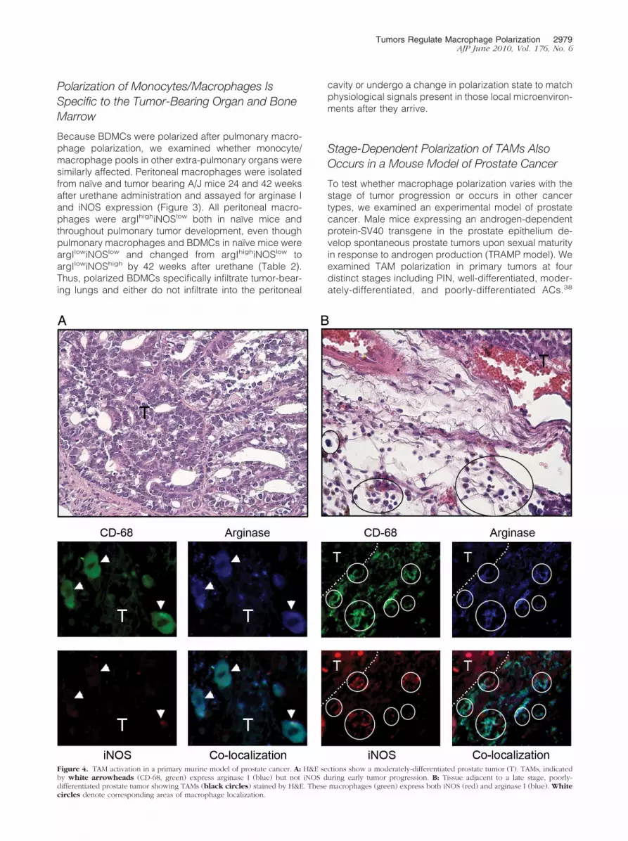
To test whether macrophage polarization varies with thestage of tumor progression or occurs in other cancertypes, we examined an experimental model of prostatecancer. Male mice expressing an androgen-dependentprotein-SV40 transgene in the prostate epithelium de-velop spontaneous prostate tumors upon sexual maturityin response to androgen production (TRAMP model). Weexamined TAM polarization in primary tumors at fourdistinct stages including PIN, well-differentiated, moder-ately-differentiated, and poorly-differentiated ACs.38
Figure 4. TAM activation in a primary murine model of prostate cancer. A: H&E sections show a moderately-differentiated prostate tumor (T). TAMs, indicatedby white arrowheads (CD-68, green) express arginase I (blue) but not iNOS during early tumor progression. B: Tissue adjacent to a late stage, poorly-differentiated prostate tumor showing TAMs (black circles) stained by H&E. These macrophages (green) express both iNOS (red) and arginase I (blue). Whitecircles denote corresponding areas of macrophage localization.
Tumors Regulate Macrophage Polarization 2979AJP June 2010, Vol. 176, No. 6

TAMs surrounding PIN were argIhighiNOSlow, indicatingM2 polarization (data not shown), as were macrophagesfrom well-differentiated and moderately differentiatedprostate ACs (Figure 4A). All macrophages associatedwith advanced poorly differentiated prostatic ACs wereargIhighiNOShigh (mixed M1 � M2 polarization; Figure4B). Macrophage expression of iNOS in late-stage can-cer was also demonstrated in the urethane-induced pul-monary ACs in A/J mice,29 urethane-induced ACs in cBymice (Table 2), and TIMs and TAMs in human pulmonaryAC (Figure 1).
Macrophage Polarization Returns to anargIlow/iNOSlow State upon Resolution ofPulmonary Tumors
Pulmonary macrophages were examined in two revers-ible genetic models of pulmonary cancer during tumordevelopment and after tumors had regressed to examinemacrophage polarization in a non–carcinogen-inducedlung tumor model and determine whether tumor regres-sion affects macrophage polarization. In the first model,tumors were induced by overexpressing human KRAS(mutated in codon 12 from G to D) under the control of theCCSP promoter in FVB mice. This specifically targetsmutant transgene expression to bronchiolar Clara cellsand alveolar type II cells with administration of Dox indrinking water. When Dox is removed, the mutated on-
cogene is not expressed, and tumors regress rapidly.46
In this model, macrophages not only localize outside themutant KRAS-induced tumor parenchyma as observedafter chemical carcinogenesis but are also found withinthe tumor as TIMs46 similar to that seen in human lungAC. Seven days after Dox administration, pulmonary le-sions appear.39 By 2 months after Dox exposure, adeno-mas with both TAMs and TIMs are apparent in H&E-stained sections (Figure 5A). All BDMCs and pulmonarymacrophages in naïve FVB mice are argIlowiNOSlow (Ta-ble 2), whereas 100% of TAMs and TIMs from this strainexpress argIhighiNOSlow, indicating alternative polariza-tion (Figure 5A). Three days after Dox was removed fromthe drinking water, tumors began to regress, but as longas small yet detectable tumor remnants remained, 100%of macrophages were argIhighiNOSlow. Seven days afterDox removal, small hyperplastic lesions and TAMs re-mained in the lungs but TIMs had disappeared (Figure5B). One month after Dox removal when lungs are tumor-free and histologically normal, all pulmonary macro-phages returned to an argIlowiNOSlow polarization state(Figure 5C), demonstrating that the presence of the tumoris necessary to maintain polarization state changes. Mac-rophage argIhighiNOSlow polarization was also demon-strated in FVB mice that develop lung tumors when theFGF10 transgene is induced by Dox11 (see SupplementalFigure S3, available online at http://ajp.amjpathol.org).Analogous to the conditional mutant KRAS model, when
Figure 5. Pulmonary macrophage activation in a mutant Kras transgenic lung cancer model. Conditional overexpression of mutant Kras induces pulmonarytumors. A: Two months after DOX treatment, H&E images show the location of macrophages peripheral to, TAMs (circles), and within, TIMs (squares),pulmonary tumors (T, border indicated by white dashed line). Macrophages identified with F4/80 (green) also stain for arginase I (blue) but not iNOS (red).B: Seven days after DOX removal, H&E staining indicates lesions and associated macrophages (circles) are still present in the lungs. TAMs express arginase I(blue). C: One month after DOX removal, tumors have regressed. Pulmonary macrophages (circles) expressed neither arginase I nor iNOS. Magnification �630for fluorescent images, �400 for H&E images.
2980 Redente et alAJP June 2010, Vol. 176, No. 6

expression of the FGF10 transgene was turned off, tu-mors regressed and macrophage polarization returned toargIlowiNOSlow.
Pulmonary Macrophage Polarization OccursOnly after Tumor Metastasis to the Lungs in aXenograft Model of Human Colon Cancer
A human xenograft colon cancer model allowed us toexamine the contribution of pulmonary metastasis tomacrophage activation and confirm tumor to marrow sig-naling in an extra-pulmonary cancer model. HT-29 hu-man colon cancer cells can metastasize from their pri-mary site of implantation to the lungs within 46 days afterinjection into the flank.40,41 We analyzed macrophage/monocyte activation in three different microenvironmentsin this model, macrophages associated with the primaryimplant (TAMs), macrophages associated with meta-static colon cancer deposits in the lungs, and BDMCsfrom animals with primary and metastatic tumors. Allmacrophages located in the stroma surrounding the pri-mary xenograft were argIhighiNOSlow, indicating M2 TAMactivation (Figure 6A). Consistent with the concept oftumor to marrow signaling, all BDMCs from mice withHT-29 flank tumors were also argIhighiNOSlow (Figure 6B).Alternative activation of BDMCs was not dependent on
the presence of tumor tissue in lungs, because this oc-curred both in mice where lung metastases had devel-oped as well as mice with no detectable metastases.Metastasis of HT-29 cells to the lungs occurred in six ofeight mice, as determined by serially sectioning fixedlung tissue. All pulmonary macrophages from lungscontaining HT-29 – derived metastases were argIhighi-NOSlow, regardless of their proximity to metastatic de-posits (Figure 6C). However, the polarization state ofpulmonary macrophages in the two mice lacking de-tectable lung metastases remained argIlowiNOSlow
(Figure 6D), indicating that pulmonary macrophagepolarization required the local presence of these coloncancer metastases.
Discussion
We demonstrate herein that TAMs and BDMCs expressarginase I, thus indicating alternative polarization dur-ing tumor formation regardless of organ site (lung,flank, or prostate) or carcinogen (see Figure 7). This isan early event, occurring before chemically-inducedtumors can be detected macroscopically. In somemodels (human NSCLC, A/J mice treated with ure-thane, cBy mice treated with urethane, and the TRAMPprostate cancer model) iNOShigh polarization of TAMs,
Figure 6. Activation status of macrophages in a human HT-29 colon cancer xenograft model. Macrophages and BDMCs are identified with anti-F4/80 oranti–CD-68 respectively (green). A: H&E staining shows that macrophages (black circles) surrounding the xenograft (T) express arginase I (blue) but not iNOS.White circles indicate areas of macrophage localization. B: BDMCs (green) isolated from xenograft-bearing mice express arginase I (blue) but not iNOS. C: H&Esection shows pulmonary macrophages (black circles) in lungs containing HT-29 metastases (black arrow). Macrophages positively stain for arginase I (blue)but not iNOS. White circles indicate areas of macrophage localization. D: Pulmonary macrophages (green, white circles) in lungs with no detectable metastasesstain for neither arginase-I nor iNOS. Magnification �630 for fluorescent images, �400 for H&E images.
Tumors Regulate Macrophage Polarization 2981AJP June 2010, Vol. 176, No. 6

TIMs, and BDMCs also occurs in late-stage disease.These dynamic changes in monocyte/macrophage po-larization are unique to TAMs and BDMCs, as perito-neal macrophages were not differentially polarized inresponse to lung tumor formation or progression. Ingenetic models in which lung tumors regress, pulmo-nary macrophages return to an argIlowiNOSlow pheno-type. When BDMCs are polarized in response toa flank tumor, pulmonary macrophages remain argIlow
iNOSlow unless tumor cells detach and metastasize to thelung.
Analysis of pulmonary macrophages from urethane-treated A/J mice indicates that macrophage polarizationis a very early event in lung cancer, presumably sup-porting and maintaining tumor development. A timecourse after urethane administration to A/J mice re-vealed argIhighiNOSlow polarization as early as 1 weekafter urethane, when microscopic tumors (referred to asmicroadenomas47 or atypical adenomatous hyperpla-sias48) are developing. Because TAMs were not polar-ized before this time, macrophage polarization is not adirect consequence of urethane itself or any of itsrapidly cleared metabolites. Polarization of BDMCs oc-curs soon after pulmonary macrophage polarization inthis model. Although macrophage polarization changedvery early in lung neoplasia, the number of macrophagesin the lungs did not increase significantly within the first24 weeks after urethane treatment, implying that eventhough BDMCs were polarized, they were not actively
recruited to the lungs until many weeks later. BMDCpolarization is not limited to a response to pulmonarytumorigenesis because BDMCs were also polarized bythe growth of a colon tumor xenograft. Detection of po-larized BDMCs in the circulation may thus imply the pres-ence of neoplastic lesions but not their anatomicalsource.
The exquisite sensitivity of the macrophage responseto pulmonary neoplasia is illustrated by argIhighiNOSlow
polarization in resistant B6 mice31,45 given a single injec-tion of urethane so that only a small percentage developlung tumors. BAL macrophages remained argIlowiNOSlow
in the mice that did not develop tumors, whereas pulmo-nary macrophages in mice with even one lung tumorexpressed arginase I. Because macrophage infiltrationhas been shown to be a necessary component of BHT-mediated lung tumor promotion, we also examined mac-rophage activation in mice treated with MCA�BHT.ArgIlowiNOSlow to argIhighiNOSlow polarization in pulmo-nary adenoma-bearing mice was also observed in thismodel. Unlike TAMs associated with urethane-inducedlung tumors in A/J mice, TAMs associated with tumorsinduced by MCA�BHT or urethane in other strains didnot switch to argIlowiNOShigh polarization when tumorsprogressed to AC, but remained argIhighiNOSlow through-out AC development. Macrophages isolated from naïvemice are tumoricidal but can be made ten times morepotent at killing tumor cells after M1 polarization withIFN-�,49 consonant with their classical role in host de-fense. Elevated arginase expression in M2 macrophagesreduces the amount of NO available to kill tumor cells,49
inhibits antigen presentation,21 and enhances the capac-ity of myeloid suppressor cells to inhibit T cell prolifera-tion.50 Polyamines secreted by M2 macrophages en-courage tumor cell growth in vitro, while inhibitingarginase activity reduces tumor growth in vivo.51 Cyto-kines such as TNF-�, IL-1�, and IL-6 released by M2macrophages enhance tumor cell survival.52 Hence,the association of M1 TAMs with AC would in theorycause tumor cell death, yet ACs in A/J mice are notdestroyed by the M1 polarized TAMs that surroundthem. A/J macrophages are defective in their ability tobecome tumoricidal both in vitro and in vivo, possiblyaccounting for this apparent discrepancy.53–55
The cBy substrain of BALB mice also exhibited an excep-tion to the trend of argIlowiNOSlow to argIhighiNOSlow polariza-tion of TAMs in early tumorigenesis. BDMCs and BAL macro-phages from naïve cBy mice were argIhighiNOShigh, a mixedM1 � M2 phenotype that persists in macrophages associ-ated with both benign and malignant tumors induced bymultiple urethane injections. In contrast, TAMs in cBy miceexposed to MCA/BHT carcinogenesis express argIhigh
iNOSlow throughout tumor development, showing thatTAM polarization is a consequence of both geneticbackground and tumor etiology. The mixed argIhigh
iNOShigh macrophage polarization in this strain is similarto that seen in human pulmonary AC and late stagemurine prostate tumors (Figures 1 and 4).
Strain-dependent variations in the degree of macro-phage infiltration during lung neoplasia do not arise fromthe genetic differences among strains in the number of
Figure 7. Schematic summarizing macrophage activation in mouse modelsof lung cancer, prostate cancer, tumor regression, and metastasis.
2982 Redente et alAJP June 2010, Vol. 176, No. 6

bone marrow macrophages (Supplemental Figure S1,available online at http://ajp.amjpathol.org). These differ-ences may reflect strain-dependent variations in theamount or kinds of chemokines secreted by TAMs thatrecruit BDMCs to the lungs or in the relative sensitivitiesof these BDMCs to such chemoattractants. We haveshown previously56 that BHT induces lung epithelial cellsto produce one such macrophage chemoattractant,CCL2, a few days before macrophages enter the lungs.Whether A/J, cBy, or B6 mice vary in chemokine produc-tion, expression of the relevant chemokine receptor, ordownstream signaling after chemokine binding to recep-tors is not known. Because both A/J and B6 BDMCs inadenoma-bearing mice are argIhighiNOSlow polarized whileshowing great disparity in the degree of lung infiltration bymacrophages, those signals that polarize BDMCs pre-sumably are different from recruitment signals.
Tumor to marrow signaling is not limited to murinecancers, because HT-29 colon tumor cells injected intoimmuno-compromised SCID mice induce argIhighiNOSlow
polarization in both TAMs associated with the growingxenograft and BDMCs. In those mice in which colontumor cells metastasized to the lungs, pulmonary macro-phages were also argIhighiNOSlow. Pulmonary macro-phages remained argIlowiNOSlow in mice without detect-able HT-29 pulmonary metastases, indicating localsignaling by metastatic lesions. Tumor cells exert localchanges on macrophages co-cultured with human ovar-ian cancer cells,28 as is indicated by the appearance ofargIhighiNOSlow pulmonary macrophages in lungs of ure-thane-treated A/J mice one week before argIhighiNOSlow
BDMCs are detectable. Because SCID mice lack matureB and T cells, we deduce that the argIlowiNOSlow toargIhighiNOSlow switch can occur without involvement ofTH2 cytokine production from pulmonary T-cells. Thesource of activating IFN-� and IL-4 cytokines found inBAL from tumor-bearing lungs was not determined, butthis study suggests that cells other than lymphocytesmay produce sufficient amounts to polarize both localmacrophages and distal BDMCs. Whether production ofthese cytokines is muted in SCID mice compared withimmuno-competent mice has not been determined but isof interest if manipulation of innate or adaptive immunityis used to treat cancer.
ArgIlowiNOSlow to ArgIhighiNOSlow polarization was alsoobserved in TAMs from lungs bearing tumors induced byoverexpressing mutant Kras38 or wild-type FGF-1010 inClara and type 2 cells. Conditional overexpression ofeither transgene was accompanied by argIhighiNOSlow
expression in pulmonary macrophages even though neo-plastic growth is greatly accelerated compared withchemically-induced tumorigenesis. Thus, whether tumorsappear within days (genetic models) or weeks (chemicalcarcinogenesis), early lung neoplasia is associated withalternative polarization of macrophages. When tumorsinduced by either transgene were allowed to regress byhalting transgene transcription, the pulmonary macro-phage population reverted back to argIlowiNOSlow. Aslong as even tiny lesions remained before complete tu-mor regression was achieved, macrophages continuedto be alternatively polarized. Macrophage polarization
thus accompanies lung tumor growth when KRAS is theonly epithelial mutation, or in the case of FGF-10 overex-pression, when tumors appear in the absence of anyknown epithelial mutation. We confirmed that macro-phages infiltrate the parenchyma of K-Ras4b�G12D-in-duced lung tumors, as was originally reported.39 Thisdiffers from other mouse lung tumor models where mac-rophages remain adjacent to, but outside of, the tumorparenchyma, but is similar to human lung cancer whereTIMs are detected. Both TIMs and TAMs detected in thistransgenic mutant KRAS model are argIhighiNOSlow,whereas TIMs and TAMs associated with human lungcancer are argIhighiNOShigh. Although TIMs and TAMsvary in other molecular characteristics, such as the rela-tive contents of adhesion molecules that allow TIMs tocross the tumor vasculature or the up-regulation of IL-10synthesis that has been reported to occur only in TAMs,57
they display similar activation phenotypes.Polarization of TAMs to an alternatively activated phe-
notype, first described by Mills,17,58 Mantovani,59 andtheir colleagues,12 has been demonstrated in severalhuman cancers and experimental mouse models. TAMs,however, can be classically or alternatively activated orexhibit a mixed M1 � M2 phenotype.52 In this study,primary mouse lung and prostate tumors, as well ashuman xenograft tumors, are surrounded by polarizedTAMs. It remains to be clarified how tumor cells andmacrophages co-evolve to favor tumor progression.60 Athorough understanding would refine therapeutic target-ing to the tumor microenvironment that could be used asan adjuvant along with standard cytotoxic interventionsdirected at solid carcinomas.
Acknowledgment
We are grateful to Dr. Jeffrey A. Whitsett (University ofCincinnati Children’s Hospital Medical Center) for thekind gift of FGF10tg lung sections.
References
1. Nathan C: Points of control in inflammation. Nature 2002, 420:846–852
2. Coussens LM, Werb Z: Inflammation and cancer. Nature 2002,420:860–867
3. Geissmann F, Jung S, Littman DR: Blood monocytes consist of twoprincipal subsets with distinct migratory properties. Immunity 2003,19:71–82
4. Malkinson AM: Role of inflammation in mouse lung tumorigenesis: areview. Exp Lung Res 2005, 31:57–82
5. Balkwill F, Mantovani A: Inflammation and cancer: back to Virchow?Lancet 2001, 357:539–545
6. Williams CS, Goldman AP, Sheng H, Morrow JD, Dubois RN: Sulindacsulfide, but not sulindac sulfone, inhibits colorectal cancer growth.Neoplasia 1999, 1:170–176
7. Bauer AK, Malkinson AM, Kleeberger SR: Susceptibility to neoplasticand non-neoplastic pulmonary diseases in mice: genetic similarities.Am J Physiol Lung Cell Mol Physiol 2004, 287:L685–L703
8. Engels EA, Wu X, Gu J, Dong Q, Liu J, Spitz MR: Systematic evalu-ation of genetic variants in the inflammation pathway and risk of lungcancer. Cancer Res 2007, 67:6520–6527
9. Keith RL, Miller YE, Hoshikawa Y, Moore MD, Gesell TL, Gao B,Malkinson AM, Golpon HA, Nemenoff RA, Geraci MW: Manipulation of
Tumors Regulate Macrophage Polarization 2983AJP June 2010, Vol. 176, No. 6

pulmonary prostacyclin synthase expression prevents murine lungcancer. Cancer Res 2002, 62:734–740
10. Bernert H, Sekikawa K, Radcliffe RA, Iraqi F, You M, Malkinson AM:Tnfa and Il-10 deficiencies have contrasting effects on lung tumorsusceptibility: gender-dependent modulation of IL-10 haploinsuffi-ciency. Mol Carcinog 2003, 38:117–123
11. Clark JC, Tichelaar JW, Wert SE, Itoh N, Perl AK, Stahlman MT,Whitsett JA: FGF-10 disrupts lung morphogenesis and causes pul-monary adenomas in vivo. Am J Physiol Lung Cell Mol Physiol 2001,280:L705–L715
12. Gordon S: Alternative activation of macrophages. Nat Rev Immunol2003, 3:23–35
13. Takanami I, Takeuchi K, Kodaira S: Tumor-associated macrophageinfiltration in pulmonary adenocarcinoma: association with angiogen-esis and poor prognosis. Oncology 1999, 57:138–142
14. Wyckoff J, Wang W, Lin EY, Wang Y, Pixley F, Stanley ER, Graf T,Pollard JW, Segall J, Condeelis J: A paracrine loop between tumorcells and macrophages is required for tumor cell migration in mam-mary tumors. Cancer Res 2004, 64:7022–7029
15. Terabe M, Berzofsky JA: Immunoregulatory T cells in tumor immunity.Curr Opin Immunol 2004, 16:157–162
16. Sica A, Bronte V: Altered macrophage differentiation and immunedysfunction in tumor development. J Clin Invest 2007, 117:1155–1166
17. Mills CD, Kincaid K, Alt JM, Heilman MJ, Hill AM: M-1/M-2 macro-phages and the Th1/Th2 paradigm. J Immunol 2000, 164:6166–6173
18. Stout RD, Jiang C, Matta B, Tietzel I, Watkins SK, Suttles J: Macro-phages sequentially change their functional phenotype in responseto changes in microenvironmental influences. J Immunol 2005,175:342–349
19. Kotowicz K, Callard RE, Friedrich K, Matthews DJ, Klein N: Biologicalactivity of IL-4 and IL-13 on human endothelial cells: functional evi-dence that both cytokines act through the same receptor. Int Immunol1996, 8:1915–1925
20. Munder M, Eichmann K, Moran JM, Centeno F, Soler G, Modolell M:Th1/Th2-regulated expression of arginase isoforms in murine macro-phages and dendritic cells. J Immunol 1999, 163:3771–3777
21. Mantovani A, Sica A, Sozzani S, Allavena P, Vecchi A, Locati M: Thechemokine system in diverse forms of macrophage activation andpolarization. Trends Immunol 2004, 25:677–686
22. Montaner LJ, da Silva RP, Sun J, Sutterwala S, Hollinshead M, VauxD, Gordon S: Type 1 and type 2 cytokine regulation of macrophageendocytosis: differential activation by IL-4/IL-13 as opposed to IFN-gamma or IL-10. J Immunol 1999, 162:4606–4613
23. Nair MG, Cochrane DW, Allen JE: Macrophages in chronic type 2inflammation have a novel phenotype characterized by the abundantexpression of Ym1 and Fizz1 that can be partly replicated in vitro.Immunol Lett 2003, 85:173–180
24. Morris SM Jr, Kepka-Lenhart D, Chen LC: Differential regulation ofarginases and inducible nitric oxide synthase in murine macrophagecells. Am J Physiol 1998, 275:E740–E747
25. Luo Y, Zhou H, Krueger J, Kaplan C, Lee SH, Dolman C, Markowitz D,Wu W, Liu C, Reisfeld RA, Xiang R: Targeting tumor-associatedmacrophages as a novel strategy against breast cancer. J Clin Invest2006, 116:2132–2141
26. Huang M, Wang J, Lee P, Sharma S, Mao JT, Meissner H, UyemuraK, Modlin R, Wollman J, Dubinett SM: Human non-small cell lungcancer cells express a type 2 cytokine pattern. Cancer Res 1995,55:3847–3853
27. Robinson-Smith TM, Isaacsohn I, Mercer CA, Zhou M, Van RN,Husseinzadeh N, Farland-Mancini MM, Drew AF: Macrophages me-diate inflammation-enhanced metastasis of ovarian tumors in mice.Cancer Res 2007, 67:5708–5716
28. Hagemann T, Wilson J, Burke F, Kulbe H, Li NF, Pluddemann A,Charles K, Gordon S, Balkwill FR: Ovarian cancer cells polarizemacrophages toward a tumor-associated phenotype. J Immunol2006, 176:5023–5032
29. Redente EF, Orlicky DJ, Bouchard RJ, Malkinson AM: Tumor signal-ing to the bone marrow changes the phenotype of monocytes andpulmonary macrophages during urethane-induced primary lung tu-morigenesis in A/J mice. Am J Pathol 2007, 170:693–708
30. Malkinson AM: Primary lung tumors in mice as an aid for understand-ing, preventing, and treating human adenocarcinoma of the lung.Lung Cancer 2001, 32:265–279
31. Malkinson AM, Beer DS: Major effect on susceptibility to urethan-
induced pulmonary adenoma by a single gene in BALB/cBy mice.J Natl Cancer Inst 1983, 70:931–936
32. Bauer AK, Dwyer-Nield LD, Keil K, Koski K, Malkinson AM: Butylatedhydroxytoluene (BHT) induction of pulmonary inflammation: a role intumor promotion. Exp Lung Res 2001, 27:197–216
33. Malkinson AM, Koski KM, Evans WA, Festing MF: Butylated hydroxy-toluene exposure is necessary to induce lung tumors in BALB micetreated with 3-methylcholanthrene. Cancer Res 1997, 57:2832–2834
34. Bauer AK, Dwyer-Nield LD, Hankin JA, Murphy RC, Malkinson AM:The lung tumor promoter, butylated hydroxytoluene (BHT), causeschronic inflammation in promotion-sensitive BALB/cByJ mice but notin promotion- resistant CXB4 mice. Toxicology 2001, 169:1–15
35. Gingrich JR, Barrios RJ, Foster BA, Greenberg NM: Pathologic pro-gression of autochthonous prostate cancer in the TRAMP model.Prostate Cancer Prostatic Dis 1999, 2:70–75
36. Greenberg NM, DeMayo F, Finegold MJ, Medina D, Tilley WD,Aspinall JO, Cunha GR, Donjacour AA, Matusik RJ, Rosen JM: Pros-tate cancer in a transgenic mouse. Proc Natl Acad Sci USA 1995,92:3439–3443
37. Greenberg NM, DeMayo FJ, Sheppard PC, Barrios R, Lebovitz R,Finegold M, Angelopoulou R, Dodd JG, Duckworth ML, Rosen JM:The rat probasin gene promoter directs hormonally and developmen-tally regulated expression of a heterologous gene specifically to theprostate in transgenic mice. Mol Endocrinol 1994, 8:230–239
38. Raina K, Singh RP, Agarwal R, Agarwal C: Oral grape seed extractinhibits prostate tumor growth and progression in TRAMP mice. Can-cer Res 2007, 67:5976–5982
39. Fisher GH, Wellen SL, Klimstra D, Lenczowski JM, Tichelaar JW,Lizak MJ, Whitsett JA, Koretsky A, Varmus HE: Induction and apo-ptotic regression of lung adenocarcinomas by regulation of a K-Rastransgene in the presence and absence of tumor suppressor genes.Genes Dev 2001, 15:3249–3262
40. Mitchell BS, Horny HP, Schumacher U: Immunophenotyping of hu-man HT29 colon cancer cell primary tumours and their metastases insevere combined immunodeficient mice. Histochem J 1997, 29:393–399
41. Jojovic M, Schumacher U: Quantitative assessment of spontaneouslung metastases of human HT29 colon cancer cells transplanted intoSCID mice. Cancer Lett 2000, 152:151–156
42. Raes G, Brys L, Dahal BK, Brandt J, Grooten J, Brombacher F,Vanham G, Noel W, Bogaert P, Boonefaes T, Kindt A, Van den BR,Leenen PJ, De BP, Ghassabeh GH: Macrophage galactose-typeC-type lectins as novel markers for alternatively activated macro-phages elicited by parasitic infections and allergic airway inflamma-tion. J Leukoc Biol 2005, 77:321–327
43. Rotondo R, Barisione G, Mastracci L, Grossi F, Orengo AM, Costa R,Truini M, Fabbi M, Ferrini S, Barbieri O: IL-8 induces exocytosis ofarginase 1 by neutrophil polymorphonuclears in nonsmall cell lungcancer. Int J Cancer 2009, 125:887–893
44. Martinez FO, Gordon S, Locati M, Mantovani A: Transcriptional pro-filing of the human monocyte-to-macrophage differentiation andpolarization: new molecules and patterns of gene expression. J Im-munol 2006, 177:7303–7311
45. Miller YE, Dwyer-Nield LD, Keith RL, Le M, Franklin WA, MalkinsonAM: Induction of a high incidence of lung tumors in C57BL/6 micewith multiple ethyl carbamate injections. Cancer Lett 2003, 198:139–144
46. Forkert PG, Jackson AC, Parkinson A, Chen S: Diminished expressionof CYP1A1 in urethane-induced lung tumors in strain A/J mice: anal-ysis by in situ hybridization and immunohistochemical methods. Am JRespir Cell Mol Biol 1996, 14:444–453
47. O’Donnell EP, Zerbe LK, Dwyer-Nield LD, Kisley LR, Malkinson AM:Quantitative analysis of early chemically-induced pulmonary lesionsin mice of varying susceptibilities to lung tumorigenesis. Cancer Lett2006, 241:197–202
48. Nikitin AY, Alcaraz A, Anver MR, Bronson RT, Cardiff RD, Dixon D,Fraire AE, Gabrielson EW, Gunning WT, Haines DC, Kaufman MH,Linnoila RI, Maronpot RR, Rabson AS, Reddick RL, Rehm S, RozengurtN, Schuller HM, Shmidt EN, Travis WD, Ward JM, Jacks T: Classificationof proliferative pulmonary lesions of the mouse: recommendations of themouse models of human cancers consortium. Cancer Res 2004,64:2307–2316
49. Dinapoli MR, Calderon CL, Lopez DM: The altered tumoricidal ca-pacity of macrophages isolated from tumor-bearing mice is related to
2984 Redente et alAJP June 2010, Vol. 176, No. 6

reduce expression of the inducible nitric oxide synthase gene. J ExpMed 1996, 183:1323–1329
50. Zea AH, Rodriguez PC, Atkins MB, Hernandez C, Signoretti S,Zabaleta J, McDermott D, Quiceno D, Youmans A, O’Neill A, Mier J,Ochoa AC: Arginase-producing myeloid suppressor cells in renal cellcarcinoma patients: a mechanism of tumor evasion. Cancer Res2005, 65:3044–3048
51. Chang CI, Liao JC, Kuo L: Macrophage arginase promotes tumor cellgrowth and suppresses nitric oxide-mediated tumor cytotoxicity.Cancer Res 2001, 61:1100–1106
52. Biswas SK, Sica A, Lewis CE: Plasticity of macrophage functionduring tumor progression: regulation by distinct molecular mecha-nisms. J Immunol 2008, 180:2011–2017
53. Boraschi D, Meltzer MS: Defective tumoricidal capacity of macro-phages from A/J mice. I. Characterization of the macrophage cyto-toxic defect after in vivo and in vitro activation stimuli. J Immunol1979, 122:1587–1591
54. Boraschi D, Meltzer MS: Defective tumoricidal capacity of macro-phages from A/J mice. II. Comparison of the macrophage cytotoxicdefect of A/J mice with that of lipid A-unresponsive C3H/HeJ mice.J Immunol 1979, 122:1592–1597
55. Boraschi D, Meltzer MS: Defective tumoricidal capacity of macro-phages from A/J mice. III. Genetic analysis of the macrophage de-fect. J Immunol 1980, 124:1050–1053
56. Meyer AM, Dwyer-Nield LD, Hurteau G, Keith RL, Ouyang Y, FreedBM, Kisley LR, Geraci MW, Bonventre JV, Nemenoff RA, MalkinsonAM: Attenuation of the pulmonary inflammatory response followingbutylated hydroxytoluene treatment of cytosolic phospholipase A2null mice. Am J Physiol Lung Cell Mol Physiol 2006, 290:L1260-L1266
57. Watkins SK, Egilmez NK, Suttles J, Stout RD: IL-12 rapidly alters thefunctional profile of tumor-associated and tumor-infiltrating macro-phages in vitro and in vivo. J Immunol 2007, 178:1357–1362
58. Mills CD, Shearer J, Evans R, Caldwell MD: Macrophage argininemetabolism and the inhibition or stimulation of cancer. J Immunol1992, 149:2709–2714
59. Mantovani A, Sozzani S, Locati M, Allavena P, Sica A: Macrophagepolarization: tumor-associated macrophages as a paradigm forpolarized M2 mononuclear phagocytes. Trends Immunol 2002,23:549 –555
60. Swann JB, Smyth MJ: Immune surveillance of tumors. J Clin Invest2007, 117:1137–1146
Tumors Regulate Macrophage Polarization 2985AJP June 2010, Vol. 176, No. 6
Related Documents