Tumor Induced Hepatic Myeloid Derived Suppressor Cells Can Cause Moderate Liver Damage Tobias Eggert 1 , Jose ´ Medina-Echeverz 1 , Tamar Kapanadze 1,2 , Michael J. Kruhlak 3 , Firouzeh Korangy 1 , Tim F. Greten 1 * 1 Gastrointestinal Malignancy Section, Center for Cancer Research, National Cancer Institute, National Institutes of Health, Bethesda, Maryland, United States of America, 2 Department of Gastroenterology, Hepatology and Endocrinology, Hannover Medical School, Hannover, Germany, 3 Experimental Immunology Branch, Center for Cancer Research, National Cancer Institute, National Institutes of Health, Bethesda, Maryland, United States of America Abstract Subcutaneous tumors induce the accumulation of myeloid derived suppressor cells (MDSC) not only in blood and spleens, but also in livers of these animals. Unexpectedly, we observed a moderate increase in serum transaminases in mice with EL4 subcutaneous tumors, which prompted us to study the relationship of hepatic MDSC accumulation and liver injury. MDSC were the predominant immune cell population expanding in livers of all subcutaneous tumor models investigated (RIL175, B16, EL4, CT26 and BNL), while liver injury was only observed in EL4 and B16 tumor-bearing mice. Elimination of hepatic MDSC in EL4 tumor-bearing mice using low dose 5-fluorouracil (5-FU) treatment reversed transaminase elevation and adoptive transfer of hepatic MDSC from B16 tumor-bearing mice caused transaminase elevation indicating a direct MDSC mediated effect. Surprisingly, hepatic MDSC from B16 tumor-bearing mice partially lost their damage-inducing potency when transferred into mice bearing non damage-inducing RIL175 tumors. Furthermore, MDSC expansion and MDSC- mediated liver injury further increased with growing tumor burden and was associated with different cytokines including GM-CSF, VEGF, interleukin-6, CCL2 and KC, depending on the tumor model used. In contrast to previous findings, which have implicated MDSC only in protection from T cell-mediated hepatitis, we show that tumor-induced hepatic MDSC themselves can cause moderate liver damage. Citation: Eggert T, Medina-Echeverz J, Kapanadze T, Kruhlak MJ, Korangy F, et al. (2014) Tumor Induced Hepatic Myeloid Derived Suppressor Cells Can Cause Moderate Liver Damage. PLoS ONE 9(11): e112717. doi:10.1371/journal.pone.0112717 Editor: Salvatore Papa, Institute of Hepatology - Birkbeck, University of London, United Kingdom Received June 20, 2014; Accepted October 14, 2014; Published November 17, 2014 This is an open-access article, free of all copyright, and may be freely reproduced, distributed, transmitted, modified, built upon, or otherwise used by anyone for any lawful purpose. The work is made available under the Creative Commons CC0 public domain dedication. Data Availability: The authors confirm that all data underlying the findings are fully available without restriction. All relevant data are within the paper and its Supporting Information files. Funding: The underlying research reported in the study was funded by the National Institutes of Health intramural research program. The funders had no role in study design, data collection and analysis, decision to publish, or preparation of the manuscript. Competing Interests: The authors have declared that no competing interests exist. * Email: [email protected] Introduction Infections, toxins, radiation, neoplasms, ischemia and trauma cause liver injury. The degree of liver injury depends on both, direct (agent dependent) and indirect (immune mediated) effects, since different cells of the innate immune system are rapidly recruited to the site of liver injury, where they aggravate liver damage [1–3]. On a molecular level, there are different mechanisms that can cause liver injury. For instance, detoxifica- tion of exogenous substances renders the liver susceptible to oxidative stress, which is produced during metabolism of toxic exogenous substances [4]. Acetaminophen [5] and alcohol [4] have been shown to exert a direct toxic effect through reactive oxygen species (ROS) or intermediate metabolites on hepatocytes. However, in addition to these mechanisms these agents also cause immune-mediated liver injury. The contribution of the innate immune system to liver injury is universally acknowledged and has been extensively reviewed [6– 10]. Not only the innate immune system in general, but more specifically the accumulation of neutrophils and macrophages can cause liver damage [8,11]. In alcoholic liver disease, activated Kupffer cells produce TNF-a, which induces apoptosis in hepatocytes through TNF-a receptor binding [12]; thereby contributing to hepatocyte cell death and hepatic inflammation [13,14]. This sterile cell death can trigger Kupffer cells to secrete the acute inflammatory response cytokine IL-1 [15], which can lead to recruitment of neutrophils to the liver. In acetaminophen induced liver injury, the depletion of these infiltrating neutrophils protects mice from severe hepatotoxicity [1]. These cells also play a pivotal role not only in drug-induced liver injury as described above, but also in liver damage caused by obesity, i.e. non- alcoholic steatohepatitis. In mouse models of dietary-induced non- alcoholic steatohepatitis, liver inflammation was aggravated by accumulation of immature myeloid cells or macrophages [16,17]. Immature myeloid cells with immune suppressive ability are also termed myeloid-derived suppressor cells (MDSC). These MDSC were initially found to accumulate in tumor bearing hosts [18]. More recently, they have also been identified in trauma and chronic infections [19]. MDSC are a heterogeneous population of immature myeloid cells and comprise myeloid progenitors at different stages of the differentiation, such as precursors of granulocytes, macrophages and dendritic cells (DC). They can be found as tumor infiltrating cells, in blood, bone marrow, spleen and liver. In tumor-bearing mice, MDSC are identified by their PLOS ONE | www.plosone.org 1 November 2014 | Volume 9 | Issue 11 | e112717

Welcome message from author
This document is posted to help you gain knowledge. Please leave a comment to let me know what you think about it! Share it to your friends and learn new things together.
Transcript
Tumor Induced Hepatic Myeloid Derived SuppressorCells Can Cause Moderate Liver DamageTobias Eggert1, Jose Medina-Echeverz1, Tamar Kapanadze1,2, Michael J. Kruhlak3, Firouzeh Korangy1,
Tim F. Greten1*
1 Gastrointestinal Malignancy Section, Center for Cancer Research, National Cancer Institute, National Institutes of Health, Bethesda, Maryland, United States of America,
2 Department of Gastroenterology, Hepatology and Endocrinology, Hannover Medical School, Hannover, Germany, 3 Experimental Immunology Branch, Center for Cancer
Research, National Cancer Institute, National Institutes of Health, Bethesda, Maryland, United States of America
Abstract
Subcutaneous tumors induce the accumulation of myeloid derived suppressor cells (MDSC) not only in blood and spleens,but also in livers of these animals. Unexpectedly, we observed a moderate increase in serum transaminases in mice with EL4subcutaneous tumors, which prompted us to study the relationship of hepatic MDSC accumulation and liver injury. MDSCwere the predominant immune cell population expanding in livers of all subcutaneous tumor models investigated (RIL175,B16, EL4, CT26 and BNL), while liver injury was only observed in EL4 and B16 tumor-bearing mice. Elimination of hepaticMDSC in EL4 tumor-bearing mice using low dose 5-fluorouracil (5-FU) treatment reversed transaminase elevation andadoptive transfer of hepatic MDSC from B16 tumor-bearing mice caused transaminase elevation indicating a direct MDSCmediated effect. Surprisingly, hepatic MDSC from B16 tumor-bearing mice partially lost their damage-inducing potencywhen transferred into mice bearing non damage-inducing RIL175 tumors. Furthermore, MDSC expansion and MDSC-mediated liver injury further increased with growing tumor burden and was associated with different cytokines includingGM-CSF, VEGF, interleukin-6, CCL2 and KC, depending on the tumor model used. In contrast to previous findings, whichhave implicated MDSC only in protection from T cell-mediated hepatitis, we show that tumor-induced hepatic MDSCthemselves can cause moderate liver damage.
Citation: Eggert T, Medina-Echeverz J, Kapanadze T, Kruhlak MJ, Korangy F, et al. (2014) Tumor Induced Hepatic Myeloid Derived Suppressor Cells Can CauseModerate Liver Damage. PLoS ONE 9(11): e112717. doi:10.1371/journal.pone.0112717
Editor: Salvatore Papa, Institute of Hepatology - Birkbeck, University of London, United Kingdom
Received June 20, 2014; Accepted October 14, 2014; Published November 17, 2014
This is an open-access article, free of all copyright, and may be freely reproduced, distributed, transmitted, modified, built upon, or otherwise used by anyone forany lawful purpose. The work is made available under the Creative Commons CC0 public domain dedication.
Data Availability: The authors confirm that all data underlying the findings are fully available without restriction. All relevant data are within the paper and itsSupporting Information files.
Funding: The underlying research reported in the study was funded by the National Institutes of Health intramural research program. The funders had no role instudy design, data collection and analysis, decision to publish, or preparation of the manuscript.
Competing Interests: The authors have declared that no competing interests exist.
* Email: [email protected]
Introduction
Infections, toxins, radiation, neoplasms, ischemia and trauma
cause liver injury. The degree of liver injury depends on both,
direct (agent dependent) and indirect (immune mediated) effects,
since different cells of the innate immune system are rapidly
recruited to the site of liver injury, where they aggravate liver
damage [1–3]. On a molecular level, there are different
mechanisms that can cause liver injury. For instance, detoxifica-
tion of exogenous substances renders the liver susceptible to
oxidative stress, which is produced during metabolism of toxic
exogenous substances [4]. Acetaminophen [5] and alcohol [4]
have been shown to exert a direct toxic effect through reactive
oxygen species (ROS) or intermediate metabolites on hepatocytes.
However, in addition to these mechanisms these agents also cause
immune-mediated liver injury.
The contribution of the innate immune system to liver injury is
universally acknowledged and has been extensively reviewed [6–
10]. Not only the innate immune system in general, but more
specifically the accumulation of neutrophils and macrophages can
cause liver damage [8,11]. In alcoholic liver disease, activated
Kupffer cells produce TNF-a, which induces apoptosis in
hepatocytes through TNF-a receptor binding [12]; thereby
contributing to hepatocyte cell death and hepatic inflammation
[13,14]. This sterile cell death can trigger Kupffer cells to secrete
the acute inflammatory response cytokine IL-1 [15], which can
lead to recruitment of neutrophils to the liver. In acetaminophen
induced liver injury, the depletion of these infiltrating neutrophils
protects mice from severe hepatotoxicity [1]. These cells also play
a pivotal role not only in drug-induced liver injury as described
above, but also in liver damage caused by obesity, i.e. non-
alcoholic steatohepatitis. In mouse models of dietary-induced non-
alcoholic steatohepatitis, liver inflammation was aggravated by
accumulation of immature myeloid cells or macrophages [16,17].
Immature myeloid cells with immune suppressive ability are
also termed myeloid-derived suppressor cells (MDSC). These
MDSC were initially found to accumulate in tumor bearing hosts
[18]. More recently, they have also been identified in trauma and
chronic infections [19]. MDSC are a heterogeneous population of
immature myeloid cells and comprise myeloid progenitors at
different stages of the differentiation, such as precursors of
granulocytes, macrophages and dendritic cells (DC). They can
be found as tumor infiltrating cells, in blood, bone marrow, spleen
and liver. In tumor-bearing mice, MDSC are identified by their
PLOS ONE | www.plosone.org 1 November 2014 | Volume 9 | Issue 11 | e112717
co-expression of CD11b and Gr-1. The hallmark of MDSC is their
ability to suppress both adaptive and innate immune responses
through multiple mechanisms. Their accumulation in livers has
been shown to protect from liver injury and to dampen T cell
mediated-hepatitis [20–23].
Recently, our group investigated antibody-mediated hepatic
MDSC depletion [24]. In addition to the finding, that anti-Gr-1
antibody failed to deplete MDSC in the liver, we observed an
increase in alanine aminotransferase (ALT) and aspartate amino-
transferase (AST) in EL4 subcutaneous tumor bearing mice.
Therefore, we set out to study the effect of hepatic MDSC in
different models of subcutaneous tumor-bearing mice in more
detail. Here, we provide evidence that hepatic MDSC accumu-
lation in tumor bearing mice can causes mild liver damage.
MDSC-induced liver damage was tumor specific as not all tumor
models investigated caused liver injury, although MDSC expan-
sion was observed in all models.
Materials and Methods
Mice and cell lines8–10 week-old female C57BL/6 and BALB/c were obtained
from NCI/Frederick (Frederick, USA). EL4 (lymphoma), RIL175
(hepatocellular carcinoma [25]) and B16 (melanoma) tumor cell
lines on C57BL/6 background and CT26 (colon carcinoma) and
BNL (hepatocellular carcinoma) tumor cell lines on BALB/c
background were used for subcutaneous tumor models. EL4 [26],
B16 [27] and CT26 [28] cell lines were a kind gift of Dr. Drew
Pardoll (The Johns Hopkins University, Baltimore, USA), The
BNL cell line was generously provided by Dr. Jesus Prieto
(University of Navarra, Spain; [29]) and the RIL175 cell line was
obtained from Dr. Lars Zender (University Hospital of Tubingen,
Germany; [25,30]). All experiments were performed according to
the institutional guidelines and approved by the National Cancer
Institute Bethesda Animal Care and Use Committee (Bethesda,
MD, USA).
Animal experiments16106 tumor cells were injected subcutaneously into the left
flank of 8–10 week-old female mice. Mice were sacrificed, when
subcutaneous tumors reached 15 mm or 20 mm mean diameter.
ALT and AST levels were determined in mouse sera and livers
were collected for immune cell analysis or fixed in 10%
Formaldehyde for histology and TUNEL assays. TUNEL
stainings were performed using the ApopTag Peroxidase In Situ
Apoptosis Detection Kit (Millipore, Billerica, USA) according to
manufacturer’s instructions. Mouse testis served as control tissue.
Liver histology slides stained with TUNEL were analyzed by
counting TUNEL positive cells in 20 non-overlapping visual fields
from individual specimens of 2 livers per group. Immunohisto-
chemistry images were collected using a Zeiss AxioObserver Z1
microscope equipped a 106 plan-apochromat (N.A. 0.45)
objective lens and a AxioCam MRc5 color CCD camera (Carl
Zeiss Microscopy, llc., Thornwood, NY, USA).
MDSC depletion was achieved as described previously [31].
Briefly, mice were treated with 5-FU (50 mg/g body weight) when
EL4 tumor surface was approximately 100 mm2. Saline treated
mice served as controls.
For hepatic MDSC transfer, a single cell suspension was
prepared from B16 subcutaneous tumor-bearing mouse livers by
density gradient centrifugation (Percoll; Fisher Scientific, Pitts-
burgh, USA) and red blood cell lysis (ACK Lysis Buffer; Quality
Biologicals), subsequently MACS-sorted using CD11b microbeads
(Miltenyi Biotec Inc., San Diego, USA) and injected (56107 cells)
intravenously into female C57BL/6 mice. Accumulation of
transferred cells in livers of recipient mice was confirmed in a
pilot experiment by transferring hepatic CD45.1+CD11b+ cells
from tumor-bearing mice into naıve C57BL/6 (CD45.2+) mice
and detection of CD45.1+CD11b+Gr-1+ cells in the recipient
mouse liver via flow cytometry. Purity of MACS-sorted cells was
assessed by flow cytometry. .95% of cells for transfer were
CD11b+ and 75% were CD11b+Gr1+. Mice were sacrificed 16 h
after transfer and serum ALT and AST levels were analyzed.
Flow cytometry analysisSingle cell suspensions were prepared as described earlier [24].
Briefly, livers were homogenized, passed through a nylon mesh
and liver-infiltrating cells were isolated by isotonic Percoll (Fisher
Scientific, Pittsburgh, USA) centrifugation. RBCs were lysed using
ACK lysis buffer (Quality Biological, Gaithersburg, USA). Cells
were stained with the following mouse antibodies against: CD11b
(Clone M1/70), Ly6G (1A8), Ly6C (HK1.4) CD3 (17A2), CD4
(GK1.5), CD8 (53–6.7), NK1.1 (PK136), CD19 (eBio1D3), CD11c
(N418), B220 (RA3-6B2) and CD244 (eBio244F4) (all from
eBioscience Inc., San Diego, USA) and Gr-1 (RB6-8C5;
BioLegend, San Diego, USA). Flow cytometry was performed
on BD FACS Calibur using BD CellQuest Pro software or LSRII
using BD FACSDiva software (BD Biosciences, San Diego, USA).
Data were analyzed using FlowJo software (Tree Star Inc.,
Ashland, USA). MDSC were defined as CD11b+Gr-1+, monocytic
MDSC (M-MDSC) as CD11b+Ly6G2Ly6Chigh, granulocytic
MDSC (PMN-MDSC) as CD11b+Ly6G+Ly6Clow, Macrophages
as CD11b+Gr-12F4/80+, conventional DC as CD11c+CD11b+,
plasmacytoid DC as CD11c+CD11b2B220+, CD4 T cells as
CD3highCD4+, CD8 T cells as CD3+CD4+, NK cells as
NK1.1+CD32, NKT cells as NK1.1+CD3low and B cells as
CD19+CD32.
Cytokine assayMouse serum samples and tumor-conditioned media, derived
from in vitro cultured tumor cell lines, were analyzed by Mouse
Cytokine/Chemokine Magnetic bead panel (Millipore, Billerica,
USA) according to manufacturer’s instructions. Serum samples
from tumor-bearing mice were normalized to naıve wild-type
mice.
Statistical analysisData were analyzed for statistical significance using Student’s t
test to compare two groups. When one control group was
compared to multiple groups, One-way ANOVA was used. (Prism
software; GraphPad); p,0.05 was considered to be statistically
significant.
Results
Tumor-bearing mice suffer from mild liver damageTo investigate liver damage in subcutaneous tumor-bearing
mice, we analyzed ALT and AST serum levels of BALB/c or
C57BL/6 mice bearing tumors of ectodermal (B16), mesodermal
(EL4) and endodermal (RIL175, BNL, CT26) origin (Figure 1A,
B). B16 and EL4 tumor bearing mice had elevated levels of both
liver enzymes, ALT and AST, whereas only subtle statistically not
significant ALT and AST elevation were noticed in mice with
other tumors. The highest increase was observed in B16 tumor-
bearing mice. Both macroscopic and microscopic evaluation of
livers from B16 and EL4 subcutaneous tumor-bearing mice
indicated no signs for the presence of liver metastasis as a possible
cause for elevated ALT and AST levels (data not shown). TUNEL
MDSC Can Cause Liver Damage
PLOS ONE | www.plosone.org 2 November 2014 | Volume 9 | Issue 11 | e112717

assays were performed to demonstrate that the increase in ALT
and AST levels in subcutaneous tumor-bearing mice was due to
hepatocyte injury, i.e. apoptosis. Indeed, more apoptotic hepato-
cytes were seen on sections from B16 tumor-bearing mice
compared to tumor-free controls (Figure 1C, D). Together, these
results show that subcutaneous growth of certain tumors causes
mild liver damage.
Subcutaneous tumors induce primarily expansion ofMDSC among liver immune cell subsets
Since immune cells are capable of exacerbating liver injury, we
hypothesized that the increase in ALT and/or AST in subcuta-
neous tumor-bearing mice is mediated by an accumulation of
immune cells in the liver. To this end, we analyzed the hepatic
immune subsets in mice with the highest (B16 and EL4) increase in
liver enzymes in C57BL/6 mice (Figure 2). In all tumor-bearing
mice the frequency and number of cells of the myeloid
compartment increased compared to naıve mice (Figure 2A, B
and D). Of all myeloid cells, the strongest increase was seen in
MDSC. On the other hand, cells of the lymphoid compartment
did not increase in frequency and only slightly increased in cell
number (Figure 2B). To confirm that CD11b+Gr-1+ cells repre-
sent MDSC rather than neutrophils in our tumor-bearing mice,
we studied whether CD11b+Gr-1+ cells were also positive for
CD244, which has been proposed as a marker to distinguish
neutrophils from granulocytic MDSC [32]. Indeed, CD11b+Gr1+
cells were also positive for CD244 in livers of B16 and RIL175
tumor-bearing mice (Figure 2C). Next, we analyzed the cell
number of MDSC and non-MDSC in all tumor models used
(Figure 2E). The increase of MDSC in tumor bearing vs. naıve
mice was higher than the increase of non-MDSC. In summary,
MDSC were the predominant immune subset expanding in livers
of mice with subcutaneous tumors.
Figure 1. Melanoma and lymphoma subcutaneous tumor-bearing mice suffer from mild liver damage. C57BL/6 and BALB/c micebearing indicated subcutaneous tumors were sacrificed, when tumor diameter reached 15 mm. ALT (A) and AST (B) levels were analyzed in mouseserum (N$8 mice per tumor, N$6 naıve mice, 3 independent experiments). Naıve C57BL/6 mice (C, left image) or mice bearing B16 subcutaneoustumors (C, right image) were sacrificed, when tumor diameter reached 20 mm. TUNEL assays were performed on liver specimen (C; scale bar= 100 mm; N = 2 mice per group, total of 5 TUNEL assays per group) and TUNEL positive cells were counted in 20 non-overlapping visual fields. Meansof TUNEL positive cells per liver section were plotted (D). C, Representative examples of visual fields are shown. Data are expressed as mean 6SEM.*p,0.05, ***p,0.001, ****p,0.0001 (by One-way ANOVA).doi:10.1371/journal.pone.0112717.g001
MDSC Can Cause Liver Damage
PLOS ONE | www.plosone.org 3 November 2014 | Volume 9 | Issue 11 | e112717
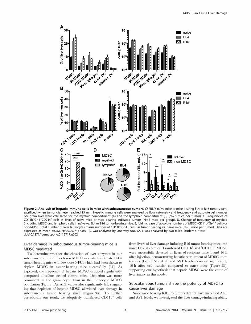
Liver damage in subcutaneous tumor-bearing mice isMDSC mediated
To determine whether the elevation of liver enzymes in our
subcutaneous tumor models was MDSC mediated, we treated EL4
tumor-bearing mice with low dose 5-FU, which had been shown to
deplete MDSC in tumor-bearing mice successfully [31]. As
expected, the frequency of hepatic MDSC dropped significantly
compared to saline treated control mice. Depletion was more
prominent in the granulocytic than in the monocytic MDSC
population (Figure 3A). ALT values also significantly fell; suggest-
ing that depletion of hepatic MDSC alleviated liver damage in
subcutaneous tumor bearing mice (Figure 3A). To further
corroborate our result, we adoptively transferred CD11b+ cells
from livers of liver damage-inducing B16 tumor-bearing mice into
naıve C57BL/6 mice. Transferred CD11b+Gr-1+CD45.1+ MDSC
were successfully detected in livers of recipient mice 1 and 16 h
after injection, demonstrating hepatic recruitment of MDSC upon
transfer (Figure S1). ALT and AST levels increased significantly
16 h after cell transfer compared to naıve mice (Figure 3B),
supporting our hypothesis that hepatic MDSC were the cause of
liver injury in this model.
Subcutaneous tumors shape the potency of MDSC tocause liver damage
Since mice bearing RIL175 tumors did not have increased ALT
and AST levels, we investigated the liver damage-inducing ability
Figure 2. Analysis of hepatic immune cells in mice with subcutaneous tumors. C57BL/6 naıve mice or mice bearing EL4 or B16 tumors weresacrificed, when tumor diameter reached 15 mm. Hepatic immune cells were analyzed by flow cytometry and frequency and absolute cell numberper gram liver were calculated for the myeloid compartment (A) and the lymphoid compartment (B) (N = 5 mice per tumor). C, Frequencies ofCD11b+Gr-1+CD244+ cells in livers of naıve mice or mice bearing indicated tumors (N = 3 mice per group). D, Change of frequency of myeloid(including MDSC) and lymphoid cells in naıve vs. EL4 or B16 tumor-bearing mice. E, fold increase of absolute numbers of MDSC (CD11b+Gr-1+ cells) ornon-MDSC (total number of liver leukocytes minus number of CD11b+Gr-1+ cells) in tumor bearing vs. naıve mice (N = 8 mice per tumor). Data areexpressed as mean 6SEM. *p,0.05, **p,0.01 (C was analyzed by One-way ANOVA. E was analyzed by two-tailed Student’s t test).doi:10.1371/journal.pone.0112717.g002
MDSC Can Cause Liver Damage
PLOS ONE | www.plosone.org 4 November 2014 | Volume 9 | Issue 11 | e112717
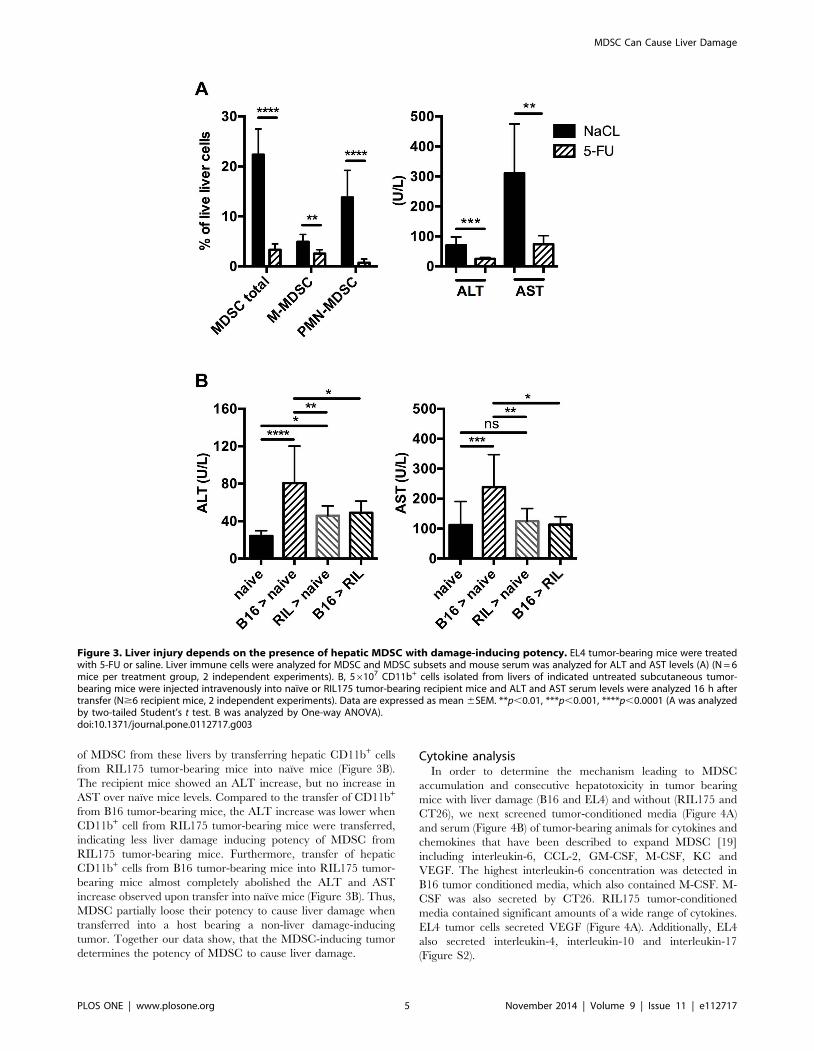
of MDSC from these livers by transferring hepatic CD11b+ cells
from RIL175 tumor-bearing mice into naıve mice (Figure 3B).
The recipient mice showed an ALT increase, but no increase in
AST over naıve mice levels. Compared to the transfer of CD11b+
from B16 tumor-bearing mice, the ALT increase was lower when
CD11b+ cell from RIL175 tumor-bearing mice were transferred,
indicating less liver damage inducing potency of MDSC from
RIL175 tumor-bearing mice. Furthermore, transfer of hepatic
CD11b+ cells from B16 tumor-bearing mice into RIL175 tumor-
bearing mice almost completely abolished the ALT and AST
increase observed upon transfer into naıve mice (Figure 3B). Thus,
MDSC partially loose their potency to cause liver damage when
transferred into a host bearing a non-liver damage-inducing
tumor. Together our data show, that the MDSC-inducing tumor
determines the potency of MDSC to cause liver damage.
Cytokine analysisIn order to determine the mechanism leading to MDSC
accumulation and consecutive hepatotoxicity in tumor bearing
mice with liver damage (B16 and EL4) and without (RIL175 and
CT26), we next screened tumor-conditioned media (Figure 4A)
and serum (Figure 4B) of tumor-bearing animals for cytokines and
chemokines that have been described to expand MDSC [19]
including interleukin-6, CCL-2, GM-CSF, M-CSF, KC and
VEGF. The highest interleukin-6 concentration was detected in
B16 tumor conditioned media, which also contained M-CSF. M-
CSF was also secreted by CT26. RIL175 tumor-conditioned
media contained significant amounts of a wide range of cytokines.
EL4 tumor cells secreted VEGF (Figure 4A). Additionally, EL4
also secreted interleukin-4, interleukin-10 and interleukin-17
(Figure S2).
Figure 3. Liver injury depends on the presence of hepatic MDSC with damage-inducing potency. EL4 tumor-bearing mice were treatedwith 5-FU or saline. Liver immune cells were analyzed for MDSC and MDSC subsets and mouse serum was analyzed for ALT and AST levels (A) (N = 6mice per treatment group, 2 independent experiments). B, 56107 CD11b+ cells isolated from livers of indicated untreated subcutaneous tumor-bearing mice were injected intravenously into naıve or RIL175 tumor-bearing recipient mice and ALT and AST serum levels were analyzed 16 h aftertransfer (N$6 recipient mice, 2 independent experiments). Data are expressed as mean 6SEM. **p,0.01, ***p,0.001, ****p,0.0001 (A was analyzedby two-tailed Student’s t test. B was analyzed by One-way ANOVA).doi:10.1371/journal.pone.0112717.g003
MDSC Can Cause Liver Damage
PLOS ONE | www.plosone.org 5 November 2014 | Volume 9 | Issue 11 | e112717
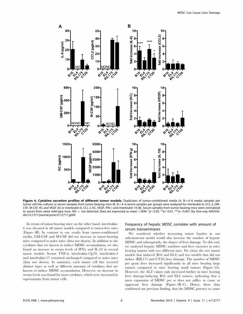
In serum of tumor-bearing mice on the other hand, interleukin-
6 was elevated in all tumor models compared to tumor-free mice
(Figure 4B). In contrast to our results from tumor-conditioned
media, GM-CSF and M-CSF did not increase in tumor-bearing
mice compared to naıve mice (data not shown). In addition to the
cytokines that are known to induce MDSC accumulation, we also
found an increase in serum levels of IFNc and IL-10 in several
tumor models. Serum TNF-a, interleukin-12p70, interleukin-4
and interleukin-17 remained unchanged compared to naıve mice
(data not shown). In summary, each tumor cell line secreted
distinct types as well as different amounts of cytokines that are
known to induce MDSC accumulation. However, no increase in
serum levels was found for most cytokines, which were increased in
supernatants from tumor cells.
Frequency of hepatic MDSC correlate with amount ofserum transaminases
We wondered whether increasing tumor burden in our
subcutaneous model would also increase the number of hepatic
MDSC and subsequently, the degree of liver damage. To this end,
we analyzed hepatic MDSC numbers and liver enzymes in mice
bearing tumors with two different sizes. We chose the two tumor
models that induced (B16 and EL4) and two models that did not
induce (RIL175 and CT26) liver damage. The number of MDSC
per gram liver increased significantly in all mice bearing large
tumors compared to mice bearing small tumors (Figure 5A).
However, the ALT values only increased further in mice bearing
liver damage-inducing B16 and EL4 tumors, indicating that a
mere expansion of MDSC per se does not suffice to cause or
aggravate liver damage (Figure 5B–C). Hence, these data
confirmed our previous finding, that the MDSC potency to cause
Figure 4. Cytokine secretion profiles of different tumor models. Duplicates of tumor-conditioned media (A, N = 4–6 media samples pertumor cell line culture) or serum samples from tumor-bearing mice (B, N = 4–6 serum samples per group) were analyzed for interleukin-6, CCL-2, GM-CSF, M-CSF, KC and VEGF (A) or interleukin-6, CCL-2, KC, VEGF, IFN-c and interleukin 10 (B). Serum samples from tumor-bearing mice were normalizedto serum from naıve wild-type mice. ND = not detected. Data are expressed as mean 6SEM. *p,0.05, **p,0.01, ***p,0.001 (by One-way ANOVA).doi:10.1371/journal.pone.0112717.g004
MDSC Can Cause Liver Damage
PLOS ONE | www.plosone.org 6 November 2014 | Volume 9 | Issue 11 | e112717
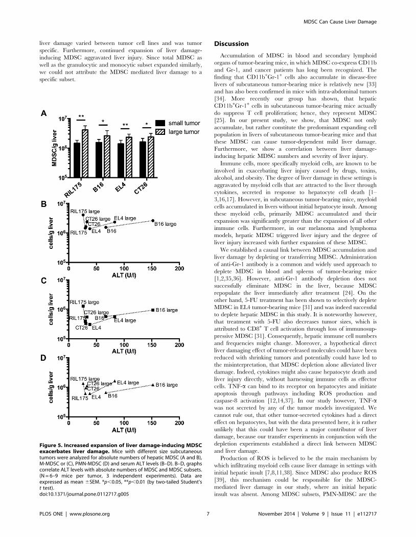
liver damage varied between tumor cell lines and was tumor
specific. Furthermore, continued expansion of liver damage-
inducing MDSC aggravated liver injury. Since total MDSC as
well as the granulocytic and monocytic subset expanded similarly,
we could not attribute the MDSC mediated liver damage to a
specific subset.
Discussion
Accumulation of MDSC in blood and secondary lymphoid
organs of tumor-bearing mice, in which MDSC co-express CD11b
and Gr-1, and cancer patients has long been recognized. The
finding that CD11b+Gr-1+ cells also accumulate in disease-free
livers of subcutaneous tumor-bearing mice is relatively new [33]
and has also been confirmed in mice with intra-abdominal tumors
[34]. More recently our group has shown, that hepatic
CD11b+Gr-1+ cells in subcutaneous tumor-bearing mice actually
do suppress T cell proliferation; hence, they represent MDSC
[25]. In our present study, we show, that MDSC not only
accumulate, but rather constitute the predominant expanding cell
population in livers of subcutaneous tumor-bearing mice and that
these MDSC can cause tumor-dependent mild liver damage.
Furthermore, we show a correlation between liver damage-
inducing hepatic MDSC numbers and severity of liver injury.
Immune cells, more specifically myeloid cells, are known to be
involved in exacerbating liver injury caused by drugs, toxins,
alcohol, and obesity. The degree of liver damage in these settings is
aggravated by myeloid cells that are attracted to the liver through
cytokines, secreted in response to hepatocyte cell death [1–
3,16,17]. However, in subcutaneous tumor-bearing mice, myeloid
cells accumulated in livers without initial hepatocyte insult. Among
these myeloid cells, primarily MDSC accumulated and their
expansion was significantly greater than the expansion of all other
immune cells. Furthermore, in our melanoma and lymphoma
models, hepatic MDSC triggered liver injury and the degree of
liver injury increased with further expansion of these MDSC.
We established a causal link between MDSC accumulation and
liver damage by depleting or transferring MDSC. Administration
of anti-Gr-1 antibody is a common and widely used approach to
deplete MDSC in blood and spleens of tumor-bearing mice
[1,2,35,36]. However, anti-Gr-1 antibody depletion does not
successfully eliminate MDSC in the liver, because MDSC
repopulate the liver immediately after treatment [24]. On the
other hand, 5-FU treatment has been shown to selectively deplete
MDSC in EL4 tumor-bearing mice [31] and was indeed successful
to deplete hepatic MDSC in this study. It is noteworthy however,
that treatment with 5-FU also decreases tumor sizes, which is
attributed to CD8+ T cell activation through loss of immunosup-
pressive MDSC [31]. Consequently, hepatic immune cell numbers
and frequencies might change. Moreover, a hypothetical direct
liver damaging effect of tumor-released molecules could have been
reduced with shrinking tumors and potentially could have led to
the misinterpretation, that MDSC depletion alone alleviated liver
damage. Indeed, cytokines might also cause hepatocyte death and
liver injury directly, without harnessing immune cells as effector
cells. TNF-a can bind to its receptor on hepatocytes and initiate
apoptosis through pathways including ROS production and
caspase-8 activation [12,14,37]. In our study however, TNF-awas not secreted by any of the tumor models investigated. We
cannot rule out, that other tumor-secreted cytokines had a direct
effect on hepatocytes, but with the data presented here, it is rather
unlikely that this could have been a major contributor of liver
damage, because our transfer experiments in conjunction with the
depletion experiments established a direct link between MDSC
and liver damage.
Production of ROS is believed to be the main mechanism by
which infiltrating myeloid cells cause liver damage in settings with
initial hepatic insult [7,8,11,38]. Since MDSC also produce ROS
[39], this mechanism could be responsible for the MDSC-
mediated liver damage in our study, where an initial hepatic
insult was absent. Among MDSC subsets, PMN-MDSC are the
Figure 5. Increased expansion of liver damage-inducing MDSCexacerbates liver damage. Mice with different size subcutaneoustumors were analyzed for absolute numbers of hepatic MDSC (A and B),M-MDSC or (C), PMN-MDSC (D) and serum ALT levels (B–D). B–D, graphscorrelate ALT levels with absolute numbers of MDSC and MDSC subsets.(N = 6–9 mice per tumor, 3 independent experiments). Data areexpressed as mean 6SEM. *p,0.05, **p,0.01 (by two-tailed Student’st test).doi:10.1371/journal.pone.0112717.g005
MDSC Can Cause Liver Damage
PLOS ONE | www.plosone.org 7 November 2014 | Volume 9 | Issue 11 | e112717
predominant subset and produce more ROS than their monocytic
counterpart [40]. Accordingly, in mice with growing tumor
burden and increasing ALT levels, we saw an expansion of this
MDSC subset. Nevertheless, M-MDSC expanded as well,
suggesting that this subtype might also contribute to MDSC-
mediated liver damage. MDSC not only produce ROS, but are
also known to produce a plethora of other immune suppressive
factors, e.g. transforming growth factor-b (TGF-b) [18,41].
However, TGF-b has also been recognized to induce apoptosis
in hepatocytes [42–44] and macrophage-derived TGF-b has been
shown to cause hepatocellular injury [45], providing another
potential mechanism by which MDSC might cause liver damage.
In summary, MDSC are equipped with means that have the
potential to cause hepatocyte injury.
Several cytokines and chemokines like IL-6, CCL2, GM-CSF,
M-CSF, KC and VEGF have been implicated in MDSC
expansion and migration [20,25,46–51]. In our study, every
tumor cell line secreted at least one of the aforementioned factors
and IL-6 elevation could also be detected in the serum of tumor-
bearing mice compared to tumor free controls. The combination
and secreted amount of these factors varied between all cell lines;
therefore, each cell line possessed an individual cytokine secretion
profile. Still, each individual cytokine profile was capable of
inducing hepatic MDSC expansion. Nevertheless, it is important
to distinguish between mechanisms of MDSC expansion and
MDSC activation, as factors that induce MDSC accumulation do
not necessarily confer functional activity [19]. Cytokines whose
signaling pathways converge on the transcription factor STAT3
have been reported to be the key mechanism of MDSC expansion
[52,53], while STAT1 and STAT6 signaling has been shown to be
important for MDSC activity [54–56]. Moreover, it has been
shown that the combination of GM-CSF with either G-CSF or
interleukin-6 gave rise to a more immunosuppressive phenotype of
MDSC than each cytokine alone, indicating that a secretion
pattern of different cytokines rather than one specific cytokine is
important for the function and activity of these cells [57]. Indeed,
our transfer experiments showed, that the liver damage-inducing
potency of MDSC was tumor-specific and our cytokine analysis
revealed, that each tumor had an individual cytokine secretion
profile, suggesting that these cytokine profiles determined the liver
damage-inducing potency. In summary, all tumor-specific cyto-
kine profiles in our study were capable of expanding hepatic
MDSC, yet with differing potencies to cause liver damage.
However, we could not establish a correlation between the
accumulation of liver damage-inducing MDSC and a specific
cytokine. Future experiments should dissect the role of candidate
cytokines in inducing MDSC with liver damaging potency.
The hallmark of MDSC is their immune suppressive function.
Therefore, it is not surprising that various studies provide evidence
of MDSC-mediated liver protection [20–23]. In these studies, the
immune cells causing liver injury were T cells and the degree of
liver damage was much more severe than in our study, where
MDSC only cause tumor-specific mild liver damage. Naturally,
the T cell mediated liver injury could be prevented through
MDSC-mediated T cell suppression. Therefore, we argue that the
moderate liver damage caused by hepatic MDSC accumulation
observed here is ‘collateral damage’, triggered by the same
mechanisms that are actually in place to prevent severe forms of
liver injury mediated by other immune cells.
Supporting Information
Figure S1 Adoptively transferred CD11b+ cells accumu-late in livers of recipient mice. 56107 MACS-sorted hepatic
CD45.1+CD11b+ cells from tumor-bearing mice were injected
intravenously into naıve C57BL/6 (CD45.2+) mice. Accumulation
of transferred cells in the liver of recipient mice was confirmed via
detection of CD45.1+CD11b+Gr-1+ cells in the recipient mouse
liver via flow cytometry. (N = 2 recipient mice per time point).
Data are expressed as mean 6SEM.
(TIFF)
Figure S2 Cytokine secretion profiles of different tumormodels. Duplicates of tumor-conditioned media (N = 4–6 media
samples per tumor cell line culture) were analyzed for interleukin-
4, interleukin-10 and interleukin-17 (A). ND = not detected. Data
are expressed as mean 6SEM.
(TIFF)
Acknowledgments
We would like to thank Dr. Leigh Samsel (National Heart, Lung, and
Blood Institute) for technical assistance with the luminex cytokine assays.
Author Contributions
Conceived and designed the experiments: TE JME FK TG. Performed the
experiments: TE TK. Analyzed the data: TE MJK. Contributed reagents/
materials/analysis tools: MJK. Wrote the paper: TE TG.
References
1. Liu ZX, Han D, Gunawan B, Kaplowitz N (2006) Neutrophil depletion protects
against murine acetaminophen hepatotoxicity. Hepatology 43: 1220–1230.
2. Bonder CS, Ajuebor MN, Zbytnuik LD, Kubes P, Swain MG (2004) Essentialrole for neutrophil recruitment to the liver in concanavalin A-induced hepatitis.
Journal of immunology 172: 45–53.
3. Jaeschke H, Hasegawa T (2006) Role of neutrophils in acute inflammatory liverinjury. Liver international: official journal of the International Association for
the Study of the Liver 26: 912–919.
4. Wu D, Cederbaum AI (2009) Oxidative stress and alcoholic liver disease.Seminars in liver disease 29: 141–154.
5. Nelson SD (1990) Molecular mechanisms of the hepatotoxicity caused byacetaminophen. Seminars in liver disease 10: 267–278.
6. Eksteen B, Afford SC, Wigmore SJ, Holt AP, Adams DH (2007) Immune-
mediated liver injury. Seminars in liver disease 27: 351–366.
7. Schwabe RF, Brenner DA (2006) Mechanisms of Liver Injury. I. TNF-alpha-induced liver injury: role of IKK, JNK, and ROS pathways. American journal of
physiology Gastrointestinal and liver physiology 290: G583–589.
8. Jaeschke H (2006) Mechanisms of Liver Injury. II. Mechanisms of neutrophil-
induced liver cell injury during hepatic ischemia-reperfusion and other acute
inflammatory conditions. American journal of physiology Gastrointestinal andliver physiology 290: G1083–1088.
9. Corazza N, Badmann A, Lauer C (2009) Immune cell-mediated liver injury.
Seminars in immunopathology 31: 267–277.
10. Adams DH, Ju C, Ramaiah SK, Uetrecht J, Jaeschke H (2010) Mechanisms ofimmune-mediated liver injury. Toxicological sciences: an official journal of the
Society of Toxicology 115: 307–321.
11. Jaeschke H, Smith CW (1997) Mechanisms of neutrophil-induced parenchymal
cell injury. Journal of leukocyte biology 61: 647–653.
12. Faubion WA, Gores GJ (1999) Death receptors in liver biology and
pathobiology. Hepatology 29: 1–4.
13. Adachi Y, Bradford BU, Gao W, Bojes HK, Thurman RG (1994) Inactivation of
Kupffer cells prevents early alcohol-induced liver injury. Hepatology 20: 453–
460.
14. Iimuro Y, Gallucci RM, Luster MI, Kono H, Thurman RG (1997) Antibodies to
tumor necrosis factor alfa attenuate hepatic necrosis and inflammation caused bychronic exposure to ethanol in the rat. Hepatology 26: 1530–1537.
15. Kono H, Karmarkar D, Iwakura Y, Rock KL (2010) Identification of thecellular sensor that stimulates the inflammatory response to sterile cell death.
Journal of immunology 184: 4470–4478.
16. Deng ZB, Liu Y, Liu C, Xiang X, Wang J, et al. (2009) Immature myeloid cells
induced by a high-fat diet contribute to liver inflammation. Hepatology 50:1412–1420.
17. Miura K, Yang L, van Rooijen N, Ohnishi H, Seki E (2012) Hepatic recruitmentof macrophages promotes nonalcoholic steatohepatitis through CCR2. Amer-
ican journal of physiology Gastrointestinal and liver physiology 302: G1310–
1321.
MDSC Can Cause Liver Damage
PLOS ONE | www.plosone.org 8 November 2014 | Volume 9 | Issue 11 | e112717
18. Gabrilovich DI, Ostrand-Rosenberg S, Bronte V (2012) Coordinated regulation
of myeloid cells by tumours. Nature reviews Immunology 12: 253–268.19. Gabrilovich DI, Nagaraj S (2009) Myeloid-derived suppressor cells as regulators
of the immune system. Nature reviews Immunology 9: 162–174.
20. Cheng L, Wang J, Li X, Xing Q, Du P, et al. (2011) Interleukin-6 induces Gr-1+CD11b+ myeloid cells to suppress CD8+ T cell-mediated liver injury in mice.
PloS one 6: e17631.21. Conrad E, Resch TK, Gogesch P, Kalinke U, Bechmann I, et al. (2014)
Protection against RNA-induced liver damage by myeloid cells requires type I
interferon and IL-1 receptor antagonist in mice. Hepatology 59: 1555–1563.22. Sarra M, Cupi ML, Bernardini R, Ronchetti G, Monteleone I, et al. (2013) IL-
25 prevents and cures fulminant hepatitis in mice through a myeloid-derivedsuppressor cell-dependent mechanism. Hepatology 58: 1436–1450.
23. Zuo D, Yu X, Guo C, Wang H, Qian J, et al. (2013) Scavenger receptor Arestrains T-cell activation and protects against concanavalin A-induced hepatic
injury. Hepatology 57: 228–238.
24. Ma C, Kapanadze T, Gamrekelashvili J, Manns MP, Korangy F, et al. (2012)Anti-Gr-1 antibody depletion fails to eliminate hepatic myeloid-derived
suppressor cells in tumor-bearing mice. Journal of leukocyte biology 92: 1199–1206.
25. Kapanadze T, Gamrekelashvili J, Ma C, Chan C, Zhao F, et al. (2013)
Regulation of accumulation and function of myeloid derived suppressor cells indifferent murine models of hepatocellular carcinoma. Journal of hepatology 59:
1007–1013.26. Gorer PA, Kaliss N (1959) The effect of isoantibodies in vivo on three different
transplantable neoplasms in mice. Cancer research 19: 824–830.27. Fidler IJ (1975) Biological behavior of malignant melanoma cells correlated to
their survival in vivo. Cancer research 35: 218–224.
28. Griswold DP, Corbett TH (1975) A colon tumor model for anticancer agentevaluation. Cancer 36: 2441–2444.
29. Drozdzik M, Qian C, Xie X, Peng D, Bilbao R, et al. (2000) Combined genetherapy with suicide gene and interleukin-12 is more efficient than therapy with
one gene alone in a murine model of hepatocellular carcinoma. Journal of
hepatology 32: 279–286.30. Zender L, Xue W, Cordon-Cardo C, Hannon GJ, Lucito R, et al. (2005)
Generation and analysis of genetically defined liver carcinomas derived frombipotential liver progenitors. Cold Spring Harbor symposia on quantitative
biology 70: 251–261.31. Vincent J, Mignot G, Chalmin F, Ladoire S, Bruchard M, et al. (2010) 5-
Fluorouracil selectively kills tumor-associated myeloid-derived suppressor cells
resulting in enhanced T cell-dependent antitumor immunity. Cancer research70: 3052–3061.
32. Youn JI, Collazo M, Shalova IN, Biswas SK, Gabrilovich DI (2012)Characterization of the nature of granulocytic myeloid-derived suppressor cells
in tumor-bearing mice. Journal of leukocyte biology 91: 167–181.
33. Ilkovitch D, Lopez DM (2009) The liver is a site for tumor-induced myeloid-derived suppressor cell accumulation and immunosuppression. Cancer research
69: 5514–5521.34. Connolly MK, Mallen-St Clair J, Bedrosian AS, Malhotra A, Vera V, et al.
(2010) Distinct populations of metastases-enabling myeloid cells expand in theliver of mice harboring invasive and preinvasive intra-abdominal tumor. Journal
of leukocyte biology 87: 713–725.
35. Li H, Han Y, Guo Q, Zhang M, Cao X (2009) Cancer-expanded myeloid-derived suppressor cells induce anergy of NK cells through membrane-bound
TGF-beta 1. Journal of immunology 182: 240–249.36. Xia S, Sha H, Yang L, Ji Y, Ostrand-Rosenberg S, et al. (2011) Gr-1+ CD11b+
myeloid-derived suppressor cells suppress inflammation and promote insulin
sensitivity in obesity. The Journal of biological chemistry 286: 23591–23599.37. Kaplowitz N (2002) Biochemical and cellular mechanisms of toxic liver injury.
Seminars in liver disease 22: 137–144.38. Teufelhofer O, Parzefall W, Kainzbauer E, Ferk F, Freiler C, et al. (2005)
Superoxide generation from Kupffer cells contributes to hepatocarcinogenesis:
studies on NADPH oxidase knockout mice. Carcinogenesis 26: 319–329.39. Corzo CA, Cotter MJ, Cheng P, Cheng F, Kusmartsev S, et al. (2009)
Mechanism regulating reactive oxygen species in tumor-induced myeloid-derived suppressor cells. Journal of immunology 182: 5693–5701.
40. Youn JI, Nagaraj S, Collazo M, Gabrilovich DI (2008) Subsets of myeloid-
derived suppressor cells in tumor-bearing mice. Journal of immunology 181:
5791–5802.
41. Terabe M, Matsui S, Park JM, Mamura M, Noben-Trauth N, et al. (2003)
Transforming growth factor-beta production and myeloid cells are an effector
mechanism through which CD1d-restricted T cells block cytotoxic T
lymphocyte-mediated tumor immunosurveillance: abrogation prevents tumor
recurrence. The Journal of experimental medicine 198: 1741–1752.
42. Black D, Lyman S, Qian T, Lemasters JJ, Rippe RA, et al. (2007) Transforming
growth factor beta mediates hepatocyte apoptosis through Smad3 generation of
reactive oxygen species. Biochimie 89: 1464–1473.
43. Shima Y, Nakao K, Nakashima T, Kawakami A, Nakata K, et al. (1999)
Activation of caspase-8 in transforming growth factor-beta-induced apoptosis of
human hepatoma cells. Hepatology 30: 1215–1222.
44. Schrum LW, Bird MA, Salcher O, Burchardt ER, Grisham JW, et al. (2001)
Autocrine expression of activated transforming growth factor-beta(1) induces
apoptosis in normal rat liver. American journal of physiology Gastrointestinal
and liver physiology 280: G139–148.
45. Hori Y, Takeyama Y, Ueda T, Shinkai M, Takase K, et al. (2000) Macrophage-
derived transforming growth factor-beta1 induces hepatocellular injury via
apoptosis in rat severe acute pancreatitis. Surgery 127: 641–649.
46. Bunt SK, Yang L, Sinha P, Clements VK, Leips J, et al. (2007) Reduced
inflammation in the tumor microenvironment delays the accumulation of
myeloid-derived suppressor cells and limits tumor progression. Cancer research
67: 10019–10026.
47. Huang B, Lei Z, Zhao J, Gong W, Liu J, et al. (2007) CCL2/CCR2 pathway
mediates recruitment of myeloid suppressor cells to cancers. Cancer letters 252:
86–92.
48. Filipazzi P, Valenti R, Huber V, Pilla L, Canese P, et al. (2007) Identification of
a new subset of myeloid suppressor cells in peripheral blood of melanoma
patients with modulation by a granulocyte-macrophage colony-stimulation
factor-based antitumor vaccine. Journal of clinical oncology: official journal of
the American Society of Clinical Oncology 25: 2546–2553.
49. Serafini P, Carbley R, Noonan KA, Tan G, Bronte V, et al. (2004) High-dose
granulocyte-macrophage colony-stimulating factor-producing vaccines impair
the immune response through the recruitment of myeloid suppressor cells.
Cancer research 64: 6337–6343.
50. Menetrier-Caux C, Montmain G, Dieu MC, Bain C, Favrot MC, et al. (1998)
Inhibition of the differentiation of dendritic cells from CD34(+) progenitors by
tumor cells: role of interleukin-6 and macrophage colony-stimulating factor.
Blood 92: 4778–4791.
51. Gabrilovich D, Ishida T, Oyama T, Ran S, Kravtsov V, et al. (1998) Vascular
endothelial growth factor inhibits the development of dendritic cells and
dramatically affects the differentiation of multiple hematopoietic lineages in vivo.
Blood 92: 4150–4166.
52. Nefedova Y, Huang M, Kusmartsev S, Bhattacharya R, Cheng P, et al. (2004)
Hyperactivation of STAT3 is involved in abnormal differentiation of dendritic
cells in cancer. Journal of immunology 172: 464–474.
53. Nefedova Y, Nagaraj S, Rosenbauer A, Muro-Cacho C, Sebti SM, et al. (2005)
Regulation of dendritic cell differentiation and antitumor immune response in
cancer by pharmacologic-selective inhibition of the janus-activated kinase 2/
signal transducers and activators of transcription 3 pathway. Cancer research 65:
9525–9535.
54. Kusmartsev S, Nagaraj S, Gabrilovich DI (2005) Tumor-associated CD8+ T cell
tolerance induced by bone marrow-derived immature myeloid cells. Journal of
immunology 175: 4583–4592.
55. Bronte V, Serafini P, De Santo C, Marigo I, Tosello V, et al. (2003) IL-4-
induced arginase 1 suppresses alloreactive T cells in tumor-bearing mice. Journal
of immunology 170: 270–278.
56. Rutschman R, Lang R, Hesse M, Ihle JN, Wynn TA, et al. (2001) Cutting edge:
Stat6-dependent substrate depletion regulates nitric oxide production. Journal of
immunology 166: 2173–2177.
57. Marigo I, Bosio E, Solito S, Mesa C, Fernandez A, et al. (2010) Tumor-induced
tolerance and immune suppression depend on the C/EBPbeta transcription
factor. Immunity 32: 790–802.
MDSC Can Cause Liver Damage
PLOS ONE | www.plosone.org 9 November 2014 | Volume 9 | Issue 11 | e112717
Related Documents