Tuberculosis Epidemiology and Selection in an Autochthonous Siberian Population from the 16 th -19 th Century Henri Dabernat 1 , Catherine The ` ves 1 , Caroline Bouakaze 1 , Dariya Nikolaeva 2 , Christine Keyser 1 , Igor Mokrousov 3 , Annie Ge ´ raut 1 , Sylvie Duchesne 1 , Patrice Ge ´ rard 1 , Anatoly N. Alexeev 5 , Eric Crube ´zy 1 *, Bertrand Ludes 1,4 1 Molecular Anthropology and Image Synthesis (AMIS) Laboratory, UMR 5288, CNRS, University of Toulouse (Paul Sabatier 3); University of Strasbourg, Toulouse, France, 2 Cultural History Centre of Contemporary Societies (CHCSC), University of Versailles Saint-Quentin-en-Yvelines, Versailles, France, 3 Laboratory of Molecular Microbiology, St. Petersburg Pasteur Institute, St. Petersburg, Russia, 4 Institute of Legal Medicine, University Paris Descartes, Paris, France, 5 Institute of the Humanities and the Indigenous Peoples of the North, Siberian Branch of the Russian Academy of Sciences, Yakutsk, Russia Abstract Tuberculosis is one of most ancient diseases affecting human populations. Although numerous studies have tried to detect pathogenic DNA in ancient skeletons, the successful identification of ancient tuberculosis strains remains rare. Here, we describe a study of 140 ancient subjects inhumed in Yakutia (Eastern Siberia) during a tuberculosis outbreak, dating from the 16 th –19 th century. For a long time, Yakut populations had remained isolated from European populations, and it was not until the beginning of the 17 th century that first contacts were made with European settlers. Subsequently, tuberculosis spread throughout Yakutia, and the evolution of tuberculosis frequencies can be tracked until the 19 th century. This study took a multidisciplinary approach, examining historical and paleo-epidemiological data to understand the impact of tuberculosis on ancient Yakut population. In addition, molecular identification of the ancient tuberculosis strain was realized to elucidate the natural history and host-pathogen co-evolution of human tuberculosis that was present in this population. This was achieved by the molecular detection of the IS6110 sequence and SNP genotyping by the SNaPshot technique. Results demonstrated that the strain belongs to cluster PGG2-SCG-5, evocating a European origin. Our study suggests that the Yakut population may have been shaped by selection pressures, exerted by several illnesses, including tuberculosis, over several centuries. This confirms the validity and necessity of using a multidisciplinary approach to understand the natural history of Mycobacterium tuberculosis infection and disease. Citation: Dabernat H, The ` ves C, Bouakaze C, Nikolaeva D, Keyser C, et al. (2014) Tuberculosis Epidemiology and Selection in an Autochthonous Siberian Population from the 16 th -19 th Century. PLoS ONE 9(2): e89877. doi:10.1371/journal.pone.0089877 Editor: David Caramelli, University of Florence, Italy Received September 29, 2013; Accepted January 27, 2014; Published February 26, 2014 Copyright: ß 2014 Dabernat et al. This is an open-access article distributed under the terms of the Creative Commons Attribution License, which permits unrestricted use, distribution, and reproduction in any medium, provided the original author and source are credited. Funding: This project was funded by the French archaeological Mission in Oriental Siberia (Ministe ` re des Affaires Etrange `res et Europe ´ ennes, France), the North- Eastern Federal University (Yakutsk, Sakha Republic), the program HUMAD from IPEV (Institut Polaire franA ˜ 1ais Paul Emile Victor). The funders had no role in study design, data collection and analysis, decision to publish, or preparation of the manuscript. Competing Interests: Co-author, Igor Mokrousov, is a PLOS ONE Editorial Board Member. This does not alter our adherence to PLOS ONE policies on sharing data and materials. * E-mail: [email protected] Introduction Tuberculosis (TB) is one of the oldest human diseases in the world, and even today, according to World Health Organization (WHO), it is second only to HIV/AIDS as the greatest killer worldwide due to a single infectious agent (http://www.who.int/ mediacentre/factsheets/fs104/en/). It is caused by a group of phylogenetically closely related bacteria, collectively known as the Mycobacterium tuberculosis Complex (MTBC). Understanding host- pathogen co-evolution in TB will help to develop better tools and strategies to control its expansion [1]. Concerning the host, recent studies have shown that the long-term association between MTBC and its human host has shaped the biology and epidemiology of human TB. Although several heritability, linkage and candidate gene association studies have investigated TB susceptibility, the exact causative genetic variants have not been characterized [1,2]. Different factors and biases could explain why some associations could not be validated [1,2], but it is important to note that all human TB genetic studies are impeded by our inability to determine the degree of exposure to M. tuberculosis [3], and the different degrees of natural selection on past populations. Concerning the pathogen, most of the strains that have infected past populations are unknown [4]. In this context, identifying ancient strains of TB and evaluating the strength and role of natural selection, in terms of mortality, will help to elucidate the natural history and host-pathogen co- evolution of human tuberculosis. We propose such an approach for a sample of 140 frozen bodies, dating from the 16 th –19 th century, from Yakutia in Eastern Siberia. For a long time this region had been isolated from contact with European populations, and it was not until the beginning of the 17 th century that first contacts were made with Slavic Russians. The value of this sample, apart from it representing a naı ¨ve population, is also related to its exceptional state of preservation under permafrost conditions, which has allowed the detection, not only of human sequences [5], PLOS ONE | www.plosone.org 1 February 2014 | Volume 9 | Issue 2 | e89877

Welcome message from author
This document is posted to help you gain knowledge. Please leave a comment to let me know what you think about it! Share it to your friends and learn new things together.
Transcript
Tuberculosis Epidemiology and Selection in anAutochthonous Siberian Population from the 16th-19th
CenturyHenri Dabernat1, Catherine Theves1, Caroline Bouakaze1, Dariya Nikolaeva2, Christine Keyser1,
Igor Mokrousov3, Annie Geraut1, Sylvie Duchesne1, Patrice Gerard1, Anatoly N. Alexeev5, Eric Crubezy1*,
Bertrand Ludes1,4
1 Molecular Anthropology and Image Synthesis (AMIS) Laboratory, UMR 5288, CNRS, University of Toulouse (Paul Sabatier 3); University of Strasbourg, Toulouse, France,
2 Cultural History Centre of Contemporary Societies (CHCSC), University of Versailles Saint-Quentin-en-Yvelines, Versailles, France, 3 Laboratory of Molecular Microbiology,
St. Petersburg Pasteur Institute, St. Petersburg, Russia, 4 Institute of Legal Medicine, University Paris Descartes, Paris, France, 5 Institute of the Humanities and the
Indigenous Peoples of the North, Siberian Branch of the Russian Academy of Sciences, Yakutsk, Russia
Abstract
Tuberculosis is one of most ancient diseases affecting human populations. Although numerous studies have tried to detectpathogenic DNA in ancient skeletons, the successful identification of ancient tuberculosis strains remains rare. Here, wedescribe a study of 140 ancient subjects inhumed in Yakutia (Eastern Siberia) during a tuberculosis outbreak, dating fromthe 16th–19th century. For a long time, Yakut populations had remained isolated from European populations, and it was notuntil the beginning of the 17th century that first contacts were made with European settlers. Subsequently, tuberculosisspread throughout Yakutia, and the evolution of tuberculosis frequencies can be tracked until the 19th century. This studytook a multidisciplinary approach, examining historical and paleo-epidemiological data to understand the impact oftuberculosis on ancient Yakut population. In addition, molecular identification of the ancient tuberculosis strain was realizedto elucidate the natural history and host-pathogen co-evolution of human tuberculosis that was present in this population.This was achieved by the molecular detection of the IS6110 sequence and SNP genotyping by the SNaPshot technique.Results demonstrated that the strain belongs to cluster PGG2-SCG-5, evocating a European origin. Our study suggests thatthe Yakut population may have been shaped by selection pressures, exerted by several illnesses, including tuberculosis,over several centuries. This confirms the validity and necessity of using a multidisciplinary approach to understand thenatural history of Mycobacterium tuberculosis infection and disease.
Citation: Dabernat H, Theves C, Bouakaze C, Nikolaeva D, Keyser C, et al. (2014) Tuberculosis Epidemiology and Selection in an Autochthonous SiberianPopulation from the 16th-19th Century. PLoS ONE 9(2): e89877. doi:10.1371/journal.pone.0089877
Editor: David Caramelli, University of Florence, Italy
Received September 29, 2013; Accepted January 27, 2014; Published February 26, 2014
Copyright: � 2014 Dabernat et al. This is an open-access article distributed under the terms of the Creative Commons Attribution License, which permitsunrestricted use, distribution, and reproduction in any medium, provided the original author and source are credited.
Funding: This project was funded by the French archaeological Mission in Oriental Siberia (Ministere des Affaires Etrangeres et Europeennes, France), the North-Eastern Federal University (Yakutsk, Sakha Republic), the program HUMAD from IPEV (Institut Polaire franA1ais Paul Emile Victor). The funders had no role in studydesign, data collection and analysis, decision to publish, or preparation of the manuscript.
Competing Interests: Co-author, Igor Mokrousov, is a PLOS ONE Editorial Board Member. This does not alter our adherence to PLOS ONE policies on sharingdata and materials.
* E-mail: [email protected]
Introduction
Tuberculosis (TB) is one of the oldest human diseases in the
world, and even today, according to World Health Organization
(WHO), it is second only to HIV/AIDS as the greatest killer
worldwide due to a single infectious agent (http://www.who.int/
mediacentre/factsheets/fs104/en/). It is caused by a group of
phylogenetically closely related bacteria, collectively known as the
Mycobacterium tuberculosis Complex (MTBC). Understanding host-
pathogen co-evolution in TB will help to develop better tools and
strategies to control its expansion [1]. Concerning the host, recent
studies have shown that the long-term association between MTBC
and its human host has shaped the biology and epidemiology of
human TB. Although several heritability, linkage and candidate
gene association studies have investigated TB susceptibility, the
exact causative genetic variants have not been characterized [1,2].
Different factors and biases could explain why some associations
could not be validated [1,2], but it is important to note that all
human TB genetic studies are impeded by our inability to
determine the degree of exposure to M. tuberculosis [3], and the
different degrees of natural selection on past populations.
Concerning the pathogen, most of the strains that have infected
past populations are unknown [4].
In this context, identifying ancient strains of TB and evaluating
the strength and role of natural selection, in terms of mortality, will
help to elucidate the natural history and host-pathogen co-
evolution of human tuberculosis. We propose such an approach
for a sample of 140 frozen bodies, dating from the 16th–19th
century, from Yakutia in Eastern Siberia. For a long time this
region had been isolated from contact with European populations,
and it was not until the beginning of the 17th century that first
contacts were made with Slavic Russians. The value of this sample,
apart from it representing a naıve population, is also related to its
exceptional state of preservation under permafrost conditions,
which has allowed the detection, not only of human sequences [5],
PLOS ONE | www.plosone.org 1 February 2014 | Volume 9 | Issue 2 | e89877

but also of the bacterial and viral genetic material present in each
subject [6,7]. The study takes a multidisciplinary approach [2,8];
we examine historical data, conduct macroscopic observations for
diagnosis of tuberculosis, and conduct genetic analyses in order to
test the possibility to amplify Mycobacterium tuberculosis, and thus,
confirm the presence of tuberculosis, and to identify the ancient
strains involved. Finally, we study the chronological distribution of
tuberculosis cases to determine how the disease spread.
Materials and Methods
Archaeological samplesFrom 2002 to 2012, a French-Russian team studying the
evolution of Yakut populations conducted excavations in the
regions of central Yakutia, Vilyuy and Verkhoyansk; ecologically
different areas that Yakuts inhabited when the Russians first
arrived in 1630. We have shown that contact between regions
were frequent, related to trade and the movement of people
(Figure 1; [9]). Excavations identified 134 isolated archaeological
graves, separated by distances of one to 900 km (Figure 1; [4]).
One hundred and forty subjects were unearthed (132 graves
contained one subject, 2 graves contained multiple burials); all
bodies were frozen due to inhumation in the permafrost (Figure 2)
and so this sample represents the largest frozen mummy sample in
the world. The wooden graves were dated precisely to the 16th to
the early/mid 19th century by dendrochronological analysis, the
burial construction technique and the artifacts held inside. During
the period studied [9], the sex ratio of the sample was balanced,
but child burials remained rare until the spread of Christianity in
the early 19th century [4]. Information on the age and sex of
subjects, along with their datations and geographic locations are
presented in Table 1.
Historical dataHistorical data available on tuberculosis in Russia [10–13] was
reviewed critically. Most data dated from the 19th and 20th
centuries; documents were either old or were written during the
Stalin era, and so had been interpreted or censured [14].
PathologyEach subject was autopsied according to the forensic medicine
guidelines for bodies presenting preservation of soft tissue by
freezing and/or natural mummification. All the bone and tissue
lesions (when remaining) were photographed, recorded and
sampled. Lesions were studied in different organs, soft tissues
and pieces of skeleton by the standard methods of forensic
medicine. Paleopathology was studied for certain subjects, by
radiological and histological examinations [5,6,15–17]. A full
radiological examination was made on one subject [4,18].
Diagnosis of bone tuberculosis using macroscopic dataThe diagnosis of bone tuberculosis was based on observations of
bone characteristics considered to be tuberculosis lesions in
children and adults during pre-antibiotic times, as well as in more
recent times [19–24]. Tuberculosis shows in its clinical evolution
specific and frequent bone localizations [23,25,26] whose charac-
teristics include: different lytic lesions localized on the vertebrae
(spondylitis leading to Pott’s disease), long bones and joints, and
cold abscesses on other skeletal bones. For each frozen body
radiological and forensic findings, including bloody pleurisy and
infectious lesions of the spine, sacroiliac joint, or pelvis, were
necessary to conclude that death was due to a disseminated
infection.
Ancient DNA analysisPrecautions against contamination. Samples were han-
dled taking into consideration the critical issue of pre-laboratory
contamination in ancient DNA (aDNA) studies. Ancient vertebrae
were sampled by a forensic scientist within a few minutes of being
exposed, and were transported to the laboratory in individual bags
and stored in controlled conditions until the molecular investiga-
tions. In the laboratory, the precautions recommended when
working with ancient DNA were followed: sample preparation and
all analytical steps prior to the first PCR amplification were
conducted in a specialized aDNA laboratory where no work with
modern DNA (human or M. tuberculosis) is permitted [7]. This pre-
PCR area is physically separated from the post-PCR laboratory.
Within the aDNA laboratory, strict precautions regarding
cleaning, equipment, reagents and clothing were taken [27].
Concerning analysis, blank extraction and PCR controls were
always included; at least two independent extractions were
performed from each sample and each PCR reaction was
reproduced twice to monitor cross-contamination.
Sample preparation and DNA extraction. To minimize
contamination, the outer surface of each vertebra was abraded
using a sanding machine (Dremel). The vertebral body of each
vertebra was then reduced to fine bone powder in liquid nitrogen
using a 6870 SamplePrep Freezer MillH (Fischer Bioblock). DNA
extraction was performed on 250 mg of bone powder using two
different silica filter column-based procedures as described
previously [27,28].
IS6110 MTBC analysis. Firstly, all samples were tested for
the presence of MTBC DNA by targeting a specific region of the
repetitive element IS6110, an insertion element used for the
identification of the MTBC strains. The commonly used two-tube
nested PCR, producing an 123 bp outer product and a 92 bp
inner product was performed using primer pairs published
previously ([29,30]; Table S1). PCRs were optimized for the
analysis of aDNA. The first PCR was conducted in a final volume
of 50 mL containing 1X of Buffer Gold (Applied Biosystems),
1.5 mM of MgCl2, 0.2 mM of each dNTP, 0.5 mM of each
external primer, 0.2 mg/ml of BSA (Sigma-Aldrich), 1.25 U of
AmpliTaq Gold Polymerase (Applied Biosystems) and 5 ml of
DNA extract. Thermal cycling conditions were 95uC for a 5 min
denaturation step, followed by 40 PCR cycles at 94uC for 1
minute, 66uC for 1 minute, 72uC for 1 minute and final extension
at 72uC for 7 minutes. The second PCR was conducted on 1 mL of
PCR products using the nested primers in the same conditions as
described above, but with only 25 cycles with an annealing step at
58uC. The specificity of the PCR products of expected size was
confirmed by direct sequencing in both directions using the Big
Dye Terminator 3.1 Kit (Applied Biosystems), according to
manufacturer’s protocol. Sequencing products were separated on
an ABI Prism 3100 or 3500 Genetic Analyzer (Applied
Biosystems) and the resulting sequences were edited using the
software Sequencher v.4.7 (Genecodes).
SNP MTBC genotyping. SNP genotyping further character-
ized the samples that successfully amplified the IS6110 region.
These samples were first examined for the presence of SNPs at
four positions: position 1410 of gyrB gene (CRT in M. bovis and M.
bovis BCG), three positions in codon 95 of gyrA gene, and codons
203 and 463 of katG gene whose combination provides discrim-
ination between the three principal genetic groups (PGG1 to
PGG3), as defined by Sreevatsan et al. [31]. Based on the results of
this first genotyping assay, four additional SNPs were tested to
determine specific downstream phylogenetic groupings to gain
further resolution. SNP genotyping was performed using the
SNaPshot minisequencing approach (Applied Biosystems) and
Tuberculosis and Ancient Autochthonous Population
PLOS ONE | www.plosone.org 2 February 2014 | Volume 9 | Issue 2 | e89877

Tuberculosis and Ancient Autochthonous Population
PLOS ONE | www.plosone.org 3 February 2014 | Volume 9 | Issue 2 | e89877
primers described previously [32]. As seen in Table S1, the
primers designed validated the first step of multiplex PCR1
(mPCR1), followed by SBE1 to validate species and principal
genetics groups, as discussed above. A second step of multiplex
PCR2 (mPCR2) followed by SBE2 was realized to validate lineage
specific SNPs. The SNapShot technique was performed from the
first classical PCR amplification; the sizes of the amplicons ranged
from 72 to 150 bp with mPCR1, and 81 to 141 bp with mPCR2.
In the second step, the minisequencing primers were tailed at the
59end with a non-homologuous sequence [32] and poly(C), if
necessary, to produce extension products that ranged in length
from 28 to 76 nucleotides (nt) with SBE1, and from 31 to 73 nt
with SBE2 (see Table S1). Thus, extension products differed in
length from each other by at least 6 nt to allow sufficient
separation by capillary electrophoresis. All conditions of mPCR,
SBE and capillary electrophoresis are given in [32]. The data
obtained from purified SBE products were analyzed and the alleles
were automatically called by the Gene Mapper ID software
(v.3.2.1 Applied Biosystems). SNaPshot results were validated by
directly sequencing segments containing SNP positions for some
selected amplicons obtained from mPCR1 when amplifications
were positive. For the SNapShot technique, however, targeted
segments are more or less small, and so the sequenced segments
are small in certain cases.
Human DNA amplification:. Human autosomal STR
typing was performed using the AmpFlSTR Identifiler kit (Applied
Biosystems), following the manufacturer’s recommendations, to
test for the presence of amplifiable DNA in extracts and to assure
that the PCR reaction was not inhibited.
Statistical analysisThe palaeoepidemiological study was undertaken after grouping
the TB and non-TB subjects into time periods [33]. Prevalence
was calculated as the number of individuals presenting the disease
divided by the number of individuals in the study population.
Crude Prevalence Rate (CPR) was calculated for each time period
sub-unit: the number of individuals with the disease was divided
by the number of individuals in each period sub-unit. The
Chi-squared (x 2) test was calculated using Statistica v.6.0 Graphs
were realized with Illustrator CS4.
Results
The archaeological samplesThe sample contained significantly less children than adults
(Table 1; x2 = 8.35, p,0.005), and for adults there was no
significant difference between the frequency of sexes (x2 = 0.91,
p = 0.18). Cases of tuberculosis were found in each of the three
regions (Table 2). It seems that for some males Y-chromosomal
STR and autosomal STR lineages may have been buried
preferentially over other lineages, as a function of their higher
social status [9]. Female autosomal STR and mtDNA lineages
showed great variability [5]. Therefore, this sample gives a
representative picture of the state of the population in studied
periods. There are two limitations of the sample: 1/The small
sample size leads to large confidence intervals (Table S2) when
exploring variation in the subjects with tuberculosis; and 2/
Modeling the zero point upstream of the epidemic (Figure 3) is
difficult to determine accurately, although historical data does
indicate the beginning of 17th century [10,14]. Note, however, that
for a population of the past, the quantity and quality of subjects
studied is exceptional.
Diagnosis and frequency of tuberculosis frommacroscopic data
During the excavation, the bodies and bones were observed to
be well-preserved, even for newborns. Subjects dated from the 16th
to 19th century. 13/140 subjects, presented skeletal injuries
compatible with the bony involvement of tuberculosis. These
subjects were over the age of one and originated from three
geographic areas: 1/Central Yakutia area (seven subjects); 2/
Verkhoyansk (three subjects); and 3/Nyurba Vilyuy (three
subjects) (Tables 2 and 3). All lesions are in agreement with the
morphological diversity of tuberculosis lesions during the pre-
antibiotic era [23].
Subjects dated from different time periods: two cases were prior
to the 18th century but after 1689, seven cases from the early 18th
century and four cases from the late 18th century. None of the 37
subjects found in Christian-type burials from the 19th century
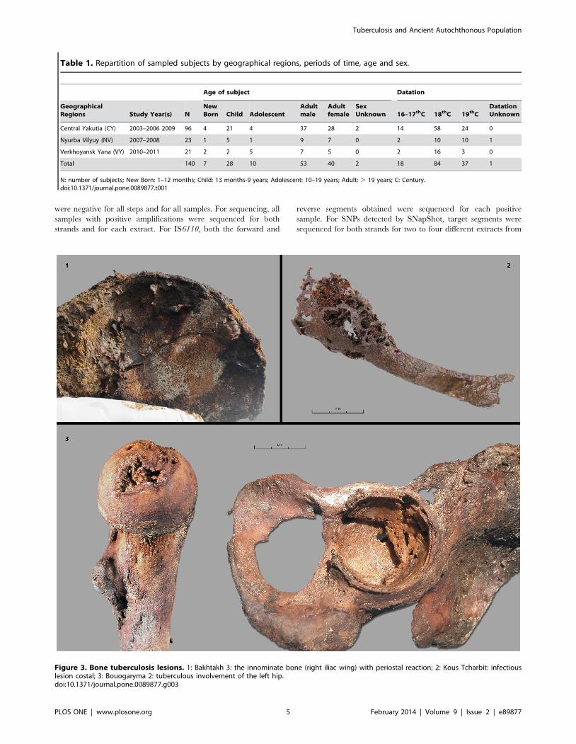
presented bone tuberculosis lesions (Figure 2). An older male
subject, Bakhtakh 3, presented multiple bone tuberculosis lesions,
including a pathological fracture of the femoral neck and
numerous lesions of innominate bone, suggesting a condition of
advanced tuberculosis (Figure 3.1).
CPR of bone lesions was 9.3% (13/140) for the studied
population as a whole (from 16th to 19th centuries). For the most
ancient cases to those from the end of the 18th century, CPR
varied from 11.1% (2/18) to 14% (7/50) (early 18th century), and
11.7% (4/34) (late 18th century). For subjects prior and during the
18th century, CPR was 12.7% (13/102). For subjects from the 18th
century only, CPR was 13.1% (11/84; see Figure 4).
DNA analysisInformation on blank controls, reproducibility and
consensus criteria. All samples were extracted with blank
controls. On average, two to four independent extractions were
performed for each of the 13 samples (Table 3). These blank
controls followed all the steps for MTBC DNA amplification,
PCR, sequencing and SNapShot techniques. All blank controls
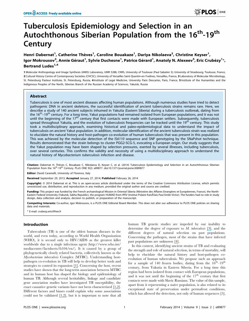
Figure 1. Map of archaeological regions of excavations. VY: Verkhoyansk Yana area; CY: the Central Yakutia area and NV: Nyurba Vilyuy area.doi:10.1371/journal.pone.0089877.g001
Figure 2. Graves of three frozen bodies from Yakutia. Bodieswere autopsied and diagnosed as having bone tuberculosis: Graves ofOkhtoubout 2, Kyys Ounhouogha and Bakhtakh 3 respectively.doi:10.1371/journal.pone.0089877.g002
Tuberculosis and Ancient Autochthonous Population
PLOS ONE | www.plosone.org 4 February 2014 | Volume 9 | Issue 2 | e89877
were negative for all steps and for all samples. For sequencing, all
samples with positive amplifications were sequenced for both
strands and for each extract. For IS6110, both the forward and
reverse segments obtained were sequenced for each positive
sample. For SNPs detected by SNapShot, target segments were
sequenced for both strands for two to four different extracts from
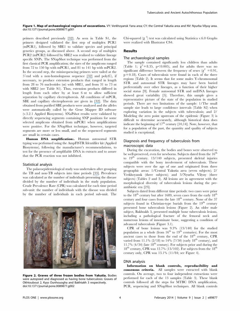
Table 1. Repartition of sampled subjects by geographical regions, periods of time, age and sex.
Age of subject Datation
GeographicalRegions Study Year(s) N
NewBorn Child Adolescent
Adultmale
Adultfemale
SexUnknown 16–17thC 18thC 19thC
DatationUnknown
Central Yakutia (CY) 2003–2006 2009 96 4 21 4 37 28 2 14 58 24 0
Nyurba Vilyuy (NV) 2007–2008 23 1 5 1 9 7 0 2 10 10 1
Verkhoyansk Yana (VY) 2010–2011 21 2 2 5 7 5 0 2 16 3 0
Total 140 7 28 10 53 40 2 18 84 37 1
N: number of subjects; New Born: 1–12 months; Child: 13 months-9 years; Adolescent: 10–19 years; Adult: . 19 years; C: Century.doi:10.1371/journal.pone.0089877.t001
Figure 3. Bone tuberculosis lesions. 1: Bakhtakh 3: the innominate bone (right iliac wing) with periostal reaction; 2: Kous Tcharbit: infectiouslesion costal; 3: Bouogaryma 2: tuberculous involvement of the left hip.doi:10.1371/journal.pone.0089877.g003
Tuberculosis and Ancient Autochthonous Population
PLOS ONE | www.plosone.org 5 February 2014 | Volume 9 | Issue 2 | e89877

each sample when mPCR1 results were positive. Reproducibility
of sequencing was obtained for at least two or more strands
(forward and/or reverse) for each positive sample, including
different extracts. Reproducibility of the SNapShot genotyping
technique was performed between two and four times. For
sequencing, consensus sequences were realized from at least two
sequences obtained for each sample. Thus, target SNPs were
assigned by at least two electrophoregrams, with unequivocal
peaks (see Table S3 for final sequences). The blank controls also
followed all the steps for the amplification human DNA [5].
Human DNA amplification. Human autosomal STR typing
was successfully performed to demonstrate that no PCR inhibition
had occurred in our DNA samples [5]. Negative amplifications of
MTBC DNA were not caused by inhibitions of PCR reactions, but
were most probably due to differential DNA conservation.
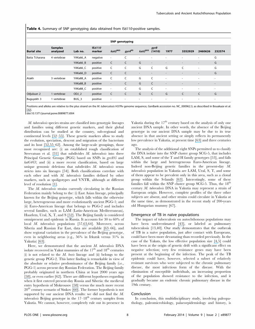
MTBC DNA amplification. All 13 TB subjects buried in the
17th and 18th centuries were analyzed for MTBC DNA. Sampling
was made from thoracic and lumbar vertebrae, which presented
lytic lesions (Table 3). The presence of MTBC DNA was
confirmed by the detection of the IS6110 sequence; DNA
fragments of expected size were observed repeatedly in the
vertebrae samples recovered from these four burials: three adults
(Batta Tcharana, Odjuluun 2, Atakh) and one adolescent
(Buguyekh 3, 15–18 years old; Table 4). Two subjects were from
central Yakutia from the late 17th– early 18th century (1741 by
dendrochronology). The other two subjects were from Nyurma
Vilyuy and Verkhoyansk Yana, buried in the late 18th century. As
revealed by direct sequencing, these fragment sequences were
identical to that of the laboratory strain M. tuberculosis H37Rv
(NC_000962.3; data not shown). These four samples were always
positive by single-stage PCR and nested PCR. The remaining
samples repeatedly failed to yield PCR products of the expected
size, even after multiple extraction and amplification attempts. A
failure to amplify this region was also observed for blank controls.
For three subjects, SNP typing by SNaPshot (as described in
Bouakaze et al. [32]) was carried out to compare the three TB
strains, which are geographically and temporally distant. As shown
in Table 4, SNP analysis failed for one sample (Buguyekh 3). None
of the samples that were analyzed successfully for the gyrB(1410)
position gene had the T allele characteristic of the M. bovis species,
indicating that the three individuals were not infected by a M. bovis
strain (Table 4). Analysis of positions katG203, katG463 and gyrA95
revealed that these samples were infected by M. tuberculosis strains
belonging to PGG2 (Table 4). To further characterize the strain
lineages that infected these individuals, we analyzed four
additional SNP positions, when sufficient DNA extract was
available. These included positions 1977, 3352929, 2460626 and
232574 in the H37Rv genome, as they enabled the PGG-2 strain
to be further divided into four genetic groups: SCG-3c, SCG3-b,
SCG-4 and SCG-5. The nucleotides at the targeted positions
could be unambiguously and reproducibly determined for only
one sample (Odjulunn 2), revealing the presence of a M. tuberculosis
strain belonging to PGG-2/SCG-5 (Table 4).
Replication of SNapShot results was evaluated by sequencing the
SNP positions (katG203, katG463, gyrA95 and gyrB(1410) for the three
subjects with positive SNP bases (Table 4). On average at least two
or more sequences were obtained to validate the SNapShot results,
demonstrating the presence of determinant SNPs at target positions
(Table S3). GyrB sequences of good quality could not be produced
for one subject (Batta Tcharana), but the amplified segments for the
other three subjects were analyzed and repeatedly confirmed the
target SNP positions. Mutations in other positions on the same
segment (for example on gyrA95 segment for three samples; Table
S3) were also confirmed on both strands and from different extracts
when available. However, information on deviating sequences
focuses on some positions in a very short region, and no specific
treatment of these sequences is available except the confirmation
that the muted positions are on both strands (Table S3).
Only samples with bone TB lesions were tested for MTBC
DNA genotyping to identify the type of strains which had infected
these Siberian subjects. Samples without typical lesions have not
yet been tested for MTBC markers.
Discussion
In this study we confirmed several cases of bone tuberculosis by
SNP genotyping to show that ancient samples had successfully
Table 2. Crude Prevalence Rate (CPR) of cases of tuberculosis according to time period and geographic area.
16–17thC (n = 18) 18thC 1st half (n = 50) 18thC 2nd half (n = 34) 19thC (n = 37)
Global (13) 11.1% (2/18) 14% (7/50) 11.7% (4/34) (0/37)
Central Yakutia (7) 14.3% (2/14) 11.1% (4/36) 4.5% (1/22) (0/24)
Verkhoyansk Yana (3) 0/2 25% (2/8) 12.5% (1/8) (0/3)
Nyurma Vilyuy (3) 0/2 16.6% (1/6) 25% (2/4) (0/10)
C: centurydoi:10.1371/journal.pone.0089877.t002
Figure 4. Temporal distribution of bone TB cases in Yakutia:evolution of CPR (%). Data values are presented in Table S1 (IC 95%).doi:10.1371/journal.pone.0089877.g004
Tuberculosis and Ancient Autochthonous Population
PLOS ONE | www.plosone.org 6 February 2014 | Volume 9 | Issue 2 | e89877
amplified the IS6110 region. The sample consisted of 140 frozen
bodies, from Yakutia (eastern Siberia), dating from the 16th –19th
centuries, in an exceptional state of preservation due to their burial
in the permafrost. For a long time this region had been isolated
from contact with European populations, and it was not until the
beginning of the 17th century that first contacts were made with
Europeans, including Slavic Russians. The ancient strain of
tuberculosis was identified to understand the impact of tubercu-
losis on the immunologically naıve Yakut population.
The evolution of tuberculosis frequencies through timesIn this study, the two oldest archaeological subjects with bone
tuberculosis date from the late 17th century (after 1689), and
originate from Central Yakutia where the Europeans settled first
[34]. In our sample no cases of tuberculosis were found in subjects
from the 19th century (Table 2). If the chronological development
of the sample over time and the distribution of bone tuberculosis
cases are correlated, we find a typical rise, peak and decline in the
number of tuberculosis cases over about one hundred years
(Figure 4). This distribution mirrors the theoretical plot of high
mortality from tuberculosis over time when tuberculosis first
emerges in a human population group [35]. From a theoretical
point of view [36], the shape of the epidemic curve for tuberculosis
is the same as that for any other infectious disease, but with much
longer time span [37]. The rate of bone tuberculosis differs among
populations, but it represents less than 25% of the maximal rate of
extra pulmonary tuberculosis [38,39]. The distribution of cases
with bone lesions compatible with tuberculosis is, however, very
specific, and it could reflect the evolution of tuberculosis
frequencies in the population.
Historical and epidemiological dataYakuts knew the signs of pulmonary tuberculosis prior to the
20th century, and were able to differentiate it from other
respiratory diseases [40,41]. During the ‘golden age’ of Yakutia,
between 1689 and 1728 [4], the prevalence of subjects in our
sample with bone tuberculosis at death increased from 11.1% to
14% (Figure 4). We can therefore suggest that at the time death
rates from TB (both pulmonary TB and extra-pulmonary TB)
were superior to this percentage. The spread of TB could have
been linked to the confinement of subjects in their homes during
winter [42,43]. As diagnosed by bone lesions (Figure 3) and
confirmed by molecular testing (Table 4), and since the strain
identified in our samples may be of European origin (see below), it
is highly likely that the disease appeared just a few years after the
first contact with Europeans in the early 17th century. Periods of
colonization have been favorable circumstances for the spread of
pathogenic agents and the emergence of infectious diseases, for
example during the conquest of the New World in the early 17th
century [44–46] and with the settlement of Japanese islands by
migrants from the Asian continent 2000 years ago [47]. Likewise,
the history of Siberia is marked by the impact of infectious diseases
during the Russian penetration and by the exploration of Siberia
[34,48]. The study of European subjects buried in the pioneer
town of Krasnoyarsk in Southwest Yakutia, showed that bone
tuberculosis also existed in this population during the same period,
but at a lower frequency [49]. We did not find cases of bone
tuberculosis in samples dating from the 19th century; it may have
become a rare disease at this time or changed its clinical form [50].
During the 19th century, a relatively low occurrence of tuberculosis
in the Yakut countryside was reported by the medical profession
[12,40]. The re-emergence of tuberculosis in the late 19th and
early 20th centuries appears to be linked to the arrival of new
migrants, including political exiles and prisoners, killing over 24–
63% of subjects between 1900 and 1904 [11]. There were also
many incidences of tuberculosis in the 1930s [10,51], when the
creation and collectivization of villages probably favored the
spread of the disease.
Molecular identification of the tuberculosis strainAs discussed above, the Eastern Siberian region had been
isolated for a long period, during which time autochthonous
populations, including Yakuts, did not have contact with
Europeans. First commercial exchanges began from the 17th
century, following contact with Slavic Russians. It is worth asking
whether the TB strain affecting the native population existed in
Yakutia prior to this time and emerged due to the deterioration of
Yakut standard of living. Alternatively, it could have been an
exogenous TB strain transmitted by the new settlers that infected
an immunologically naıve Yakut population.
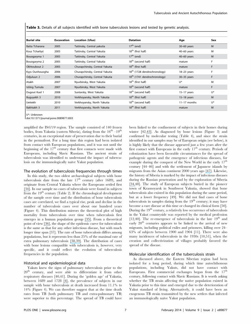
Table 3. Details of all subjects identified with bone tuberculosis lesions and tested by genetic analysis.
Burial site Excavation Location (Ulus) Datation Age Sex
Batta Tcharana 2005 Tattinsky, Central yakutia 17th (end) 30–60 years M
Kous Tcharbyt 2005 Tattinsky, Central Yakutia 18th (first half) 40–60 years M
Bouogaryma 1 2005 Tattinsky, Central Yakutia 17th (end) 40–60 years M
Bouogaryma 2 2005 Tattinsky, Central Yakutia 18th (second half) mature F
Okhtoubout 2 2005 Churapchinsky, Central Yakutia 18th (first half) mature F
Kyys Ounhouogha 2006 Churapchinsky, Central Yakutia 18th (1728 dendrochronology) 18–23 years F
Odjuluun 2 2006 Churapchinsky, Central Yakutia 18th (1741 dendrochronology) 30–35 years F
Atakh 2007 Nyurbinsky, West Yakutia 18th (first half) U* M
Istiing Tumula 2007 Nyurbinsky, West Yakutia 18th (second half) mature F
Ougout Kuel 1 2008 Suntarsky, West Yakutia 18th (second half) 15–17 years U*
Buguyekh 3 2010 Verkhoyansky, North Yakutia 18th (first half) 15–18 years M
Uettekh 2010 Verkhoyansky, North Yakutia 18th (second half) 11–17 months U*
Bakhtakh 3 2011 Verkhoyansky, North Yakutia 18th (first half) mature M
U*: Unknowndoi:10.1371/journal.pone.0089877.t003
Tuberculosis and Ancient Autochthonous Population
PLOS ONE | www.plosone.org 7 February 2014 | Volume 9 | Issue 2 | e89877
M. tuberculosis species strains are classified into genotypic lineages
and families using different genetic markers, and their global
distribution can be studied at the country, sub-regional and
continental levels [52–55]. These genetic markers allow to study
the evolution, speciation, descent and migration of the bacterium
and its host [52,55–62]. Among the large-scale groupings, those
most recognized are: (i) an established rough classification of
Sreevatsan et al. [31] that subdivides M. tuberculosis into three
Principal Genetic Groups (PGG) based on SNPs in gyrA95 and
katG463; and (ii) a more recent classification, based on large
unique genomic deletions that subdivides M. tuberculosis sensu
stricto into six lineages [54]. Both classifications correlate with
each other and with M. tuberculosis families defined by other
markers, such as spoligotypes and VNTR, although at different
level of resolution [1].
The M. tuberculosis strains currently circulating in the Russian
Federation mainly belong to the: (i) East Asian lineage, principally
known for the Beijing genotype, which falls within the otherwise
large, heterogeneous and more evolutionarily ancient PGG-1; and
(ii) Euro-American lineage that belongs to PGG-2 and includes
several families, such as LAM (Latin-American Mediterranean),
Haarlem, Ural, X, T, and S [52]. The Beijing family is considered
omnipresent and epidemic in Russia. It accounts for 30 to 60% of
local M. tuberculosis populations [57,63,64]. Moreover, across
Siberia and Russian Far East, data are available [63–66], and
show regional variation in the prevalence of the Beijing genotype,
even in neighboring areas (e.g., 56% in Irkutsk versus 31% in
Yakutia) [63].
Here, we demonstrated that the ancient M. tuberculosis DNA
isolate recovered in Yakut mummies of the 17th and 18th centuries
(i) is not related to the M. bovis lineage and (ii) belongs to the
genetic group PGG-2. This latter finding is remarkable in view of
the absolute or relative predominance of the Beijing family (i.e.,
PGG-1) across present day Russian Federation. The Beijing family
probably originated in northern China at least 2000 years ago
[58], or even earlier [62]. There are different hypotheses regarding
when it first entered present-day Russia and Siberia: the medieval
entry hypothesis of Mokrousov [58] versus the much more recent
20th century scenario of Sinkov [60]. The former hypothesis is not
supported by our ancient DNA results: we did not find the M.
tuberculosis Beijing genotype in the 17–18th century samples from
Yakutia. We cannot, however, completely rule out its presence in
Yakutia during the 17th century based on the analysis of only one
ancient DNA sample. In other words, the absence of the Beijing
genotype in our ancient DNA sample may be due to its true
absence in that ancient setting or simply reflects its permanently
low prevalence in Yakutia, at present time [63] and three centuries
ago.
The analysis of the additional eight SNPs permitted us to classify
the DNA isolate into the SNP cluster group SCG-5, that includes
LAM, S, and some of the T and H family genotypes [53], and falls
within the large and heterogeneous Euro-American lineage.
Indeed non-Beijing genetic families in the present-day M.
tuberculosis population in Yakutia are LAM, Ural, S, T, and some
of them appear to be prevalent only in this area, such as a clonal
group within the S-family [63]. Interestingly, some of these
families fall within the SNP cluster group SCG-5. Thus, the 18th
century M. tuberculosis DNA in Yakutia may represent a strain of
European origin. However, complete profiles of the three other
subjects are absent, and other strains could circulate in Yakutia at
the same time, as demonstrated in the recent study of 200-years
old Hungarian mummy [67].
Emergence of TB in naıve populationsThe impact of tuberculosis on autochthonous populations may
have been underestimated [43], or labeled as pulmonary
tuberculosis [13,40]. Our study demonstrates that the outbreak
of TB in a naıve population, just after contact with Europeans,
could have been more devastating than recorded previously. In the
case of the Yakuts, the low effective population size [4,5] could
have been at the origin of genetic drift with a significant effect on
negative selection; very few resistance genes may have been
present at the beginning of the infection. The peak of the TB
epidemic could have, however, selected a subset of relatively
resistant survivors who were subjected to the chronic pulmonary
disease, the most infectious form of the disease. With the
elimination of susceptible individuals, an increasing proportion
of the population showed resistance to the infection, and it
gradually became an endemic chronic pulmonary disease in the
19th century.
ConclusionIn conclusion, this multidisciplinary study, involving paleopa-
thology, paleomicrobiology, palaeoepidemiology and history, is
Table 4. Summary of SNP genotyping data obtained from IS6110-positive samples.
SNP genotyping
Burial siteSamplesanalyzed Lab no.
IS6110marker katG463 gyrA95 katG203
gyrB(1410) 1977 3352929 2460626 232574
Batta Tcharana 4 vertebrae YAKa66_A negative - C - - - - - G
YAKa66_B positive C C G - - - - G
YAKa66_C positive C C G C G C - G
YAKa66_D positive C C - - - - - G
Atakh 3 vertebrae YAKa88_A positive C C G C - - - -
YAKa88_B positive - C G C - - - -
YAKa88_C positive - C G C - - - -
Odjuluun 2 1 vertebrae ODJ_2 positive C C G C G C C G
Buguyekh 3 1 vertebrae BUG_3 positive - - - - - - - -
Positions and alleles are relative to the plus strand on the M. tuberculosis H37Rv genome sequence, GenBank accession no. NC_000962.3, as described in Bouakaze et al.[32].doi:10.1371/journal.pone.0089877.t004
Tuberculosis and Ancient Autochthonous Population
PLOS ONE | www.plosone.org 8 February 2014 | Volume 9 | Issue 2 | e89877

the first to be conducted with archaeological samples from Siberia.
It demonstrates the presence of tuberculosis in an autochthonous
population just a few years after the population’s first contact with
Europeans, and the highest prevalence of the disease to have been
observed. The ancient TB strain identified might be of European
origin, but its characterization needs to be specified to fully
understand its implication in human Yakut population and its
phylogeny related to modern TB strains. We can hypothesize that
this strain might have exerted some selective pressure on a small
population that was subsequently hit by epidemics caused by other
MTB strains centuries later. This favored the evolution of TB,
which we will continue to study. The sample material provides an
excellent opportunity to research the ancestral pattern of the
human populations and the presence of pathogens of parasitic and
infectious diseases during Siberian historical period from the
beginning of its colonization.
Supporting Information
Table S1 PCR primers used in this study.(DOC)
Table S2 Evolution of tuberculosis crude prevalencerate (CPR) in Yakut population from the modern tocontemporary era. Prevalence was calculated as the number of
individuals presenting the disease divided by the number of
individuals in the study population. Crude Prevalence Rate (CPR)
was calculated using the whole study population as the
denominator. CPR was calculated for each time period, dividing
the number of subjects with the disease by the number of
individuals in each period. C: Century.
(DOC)
Table S3 Final MTBC DNA sequences obtained andcompared to H37Rv (Sequence view is plus strand). The
characteristic positions for SNPs are in bold.
(DOC)
Acknowledgments
Administrative and research work were permitted through the program of
the France-Russia Associated International Laboratory (LIA COSIE
number 1029), associating the North-Eastern Federal University (Yakutsk,
Sakha Republic), the State Medical University of Krasnoyask, the Russia
Foundation for Fundamental Research (Moscow, Russia), the University of
Paul Sabatier Toulouse III (France), the University of Strasbourg I (France)
and the National Centre of Scientific Research (Paris, France). We thank
Marie-Therese Marty (Laboratoire TRACES, Toulouse, CNRS) and Olga
Melnichuk (North-Eastern Federal University) for the institutional
collaboration, University of Paul Sabatier for the stay of Dr. I. Mokrousov
as Invited Professor within AMIS Laboratory. We also wish to
acknowledge Becky Coles and Veronica Pereda-Loth who read the
manuscript and provided critical comments.
Author Contributions
Conceived and designed the experiments: HD CB CK EC. Performed the
experiments: HD CB DN AG SD PG. Analyzed the data: HD CB SD IM.
Contributed reagents/materials/analysis tools: BL CK EC ANA. Wrote
the paper: EC HD CB IM CT.
References
1. Gagneux S (2012) Host-pathogen coevolution in human tuberculosis. Philos
Trans R Soc Lond B Biol Sci 367850–859.
2. Stein CM (2011) Genetic epidemiology of tuberculosis susceptibility: impact ofstudy design. PLoS Pathog 7: e1001189.
3. Moller M, de Wit E, Hoal EG (2010) Past, present and future directions in
human genetic susceptibility to tuberculosis. FEMS Immunol Med Microbiol 58:
3–26.
4. Crubezy E, Alexeev A (2007) Chamane: Kyys, jeune fille des glaces. Paris:Errance. 167 p.
5. Crubezy E, Amory S, Keyser C, Bouakaze C, Bodner M, et al. (2010) Human
evolution in Siberia: from frozen bodies to ancient DNA. BMC Evol Biol 10: 25.
6. Biagini P, Theves C, Balaresque P, Geraut A, Cannet C, et al. (2012) Variolavirus in a 300-year-old Siberian mummy. N Engl J Med 367: 2057–2059.
7. Theves C, Senescau A, Vanin S, Keyser C, Ricaut FX, et al. (2011) Molecular
identification of bacteria by total sequence screening: determining the cause ofdeath in ancient human subjects. PLoS One 6: e21733.
8. Comas I, Gagneux S (2011) A role for systems epidemiology in tuberculosis
research. Trends Microbiol 19: 492–500.
9. Crubezy E, Alexeev A (2012)
.
(The world of ancient yakoutia). With an english summary (15 p). Yakuts: Yakuts
University. 225 p.
10. Boushkov PM (1928) Materialy po izysheniyu tubersuleza v Jakuti (data abouttuberculosis in Yakoutia). Yakutsk. 102 p.
11. Kaganovitch RB, editor (1952) Iz istorii borby s tyberskulezom. 318 p p.
12. Kon FA (1889) Fisiologisheskie i biologisheskie dannye o jakutakh (biological and
physiological data from Yakutia). Minousinsk. 87 p.
13. Mizkevitsj SM,editor (1969) Zapiski vrasha – obshestvennika. 239p p.
14. Andreıev EN (1953) Tuberculose I bor’ba s nim s Jakutskoia ASSR(Tuberculosis and the fight again tuberculosis in yakutia). Yakutsk: Yakuts
University. 334 p.
15. Aufderheide AC, Rodriguez-Martin C (1998) The Cambridge Encyclopedia of
Human Paleopathology. Cambridge: Cambridge University Press.
16. Ortner DJ (2003) Identification of Pathological Conditions in Human SkeletalRemains: Elsevier.
17. Waldron T (2008) Palaeopathology. Cambridge: Cambridge University Press.
18. Dedouit F, Geraut A, Baranov V, Ludes B, Rouge D, et al. (2010) Virtual and
macroscopical studies of mummies–differences or complementarity? Report of anatural frozen Siberian mummy. Forensic Sci Int 200: e7–13.
19. De Vuyst D, Vanhoenacker F, Gielen J, Bernaerts A, De Schepper AM (2003)
Imaging features of musculoskeletal tuberculosis. Eur Radiol 13: 1809–1819.
20. Huang CH (1996) Extra-articular tuberculosis osteomyelitis. Int Orthop 20:169–171.
21. Nathanson L, Cohen W (1941) A statistical analysis and roentgen analysis of two
hundred cases of bone and joint tuberculosis. Radiology 36: 550.
22. Rasool MN, Govender S, Naidoo KS (1994) Cystic tuberculosis of bone in
children. J Bone Joint Surg BR 76-B: 113–117.
23. Sorrel E, Sorrel-Dejerine (1932) Tuberculose osseuse et osteo-articulaire. Paris:
Masson.
24. Teo HEL, Peh WCG (2004) Skeletal tuberculosis in children. Pediatric
Radiology 34: 853–860.
25. Broca A, Mery H (1925) Tuberculose chirurgicale: I. Tuberculose des enfants.
Paris: Bailliere.
26. Lewis ME (2006) The Bioarchaeology of Children. Perspectives from Biological
and Forensic Anthropology: University of Reading. 266 p.
27. Keyser-Tracqui C, Ludes B (2005) Methods for the study of ancient DNA.
Methods Mol Biol 297: 253–264.
28. Mendisco F, Keyser C, Hollard C, Seldes V, Nielsen A, et al. (2011) Application
of the iPLEXTM Gold SNP genotyping method for the analysis of Amerindian
ancient DNA samples: benefits for ancient population studies. Electrophoresis
32: 386–393.
29. Fletcher HA, Donoghue HD, Taylor GM, van der Zanden AG, Spigelman M
(2003) Molecular analysis of Mycobacterium tuberculosis DNA from a family of
18th century Hungarians. Microbiology. 149 (1):143–151.
30. Taylor G, Crossey M, Saldanha J, Waldron T (1996) DNA from Mycobacterium
tuberculosis identified in mediaeval human skeletal remains using Polymerase
Chain Reaction. Journal of Archaeological Science 23: 789–798.
31. Sreevatsan S, Pan X, Stockbauer KE, Connel ND, Kreisswirth BN, et al. (1997)
Restricted structural gene polymorphism in the Mycobacterium tuberculosis
Complex indicates evolutionnarily recent global dissemination. Proc Natl Acad
Sci USA 94: 9869–9874.
32. Bouakaze C, Keyser C, de Martino S, Sougakoff W, Veziris N, et al. (2010)
Identification and Genotyping of Mycobacterium tuberculosis Complex species by
use of a SNaPshot mnisequencing-based assay. Journal of Clinical Microbiology:
1758–1766.
33. Waldron T (2007) Palaeoepidemiology: the measure of disease in the human
past. Walnut Creek CA: Left Coast Press.
34. Forsyth J (1992) A history of the peoples of Siberia. Russia’s north Asian colony
1581–1990. Cambridge: Cambridge University Press.
35. Grigg ER (1958) The arcana of tuberculosis with a brief epidemiologic history of
the disease in the U.S.A. Am Rev Tuberc 78: 151–172.
36. Aparicio J, Castillo-Chavez C (2009) Mathematical modelling of tuberculosis
epidemics. Math Biosci Eng 6: 209–237.
37. Tuberculosis Manual (2006.) Hong Kong. wwwinfogovhk/tb_chest/doc/
Tuberculosis_Manual2006pdf.
Tuberculosis and Ancient Autochthonous Population
PLOS ONE | www.plosone.org 9 February 2014 | Volume 9 | Issue 2 | e89877
38. Ussery XT, Valway SE, McKenna M, Cauthen GM, McCray E, et al. (1996)
Epidemiology of tuberculosis among children in the United States: 1985 to 1994.Pediatr Infect Dis J 15: 697–704.
39. Walls T, Shingadia D (2004) Global epidemiology of paediatric tuberculosis.
J Infect 48: 13–22.40. Tyrylgin MA (2010) Otsherki po istorii i ob organizazii borby s tuberkulezom v
Jakutii (The fights against tuberculosis in Yakoutia: a study). Yakutsk.41. Myglan VS, Vaganov EA (2005) Epidemii i Epizootii v Sibiri v XVII – pervoj
polovine XIX veka i dlitel’nye izmeneniya klimata. Arheologiya,
etnografiya i antropologiya Evrazii 4: 136–144.42. Popov NA (2009) Proshloe Jakutii (La Passe de la Iakoutie). Iakoutsk pp. p.219–
220.43. Bates JH, Stead WW (1993) The history of tuberculosis as a global epidemic.
Med Clin North Am 77: 1205–1217.44. Cook ND (1998) Born to die, disease and New World conquest, 1492–1650.
Cambridge: Cambridge Univ. Press. 248 p.
45. Cook ND, Lovell WG (1992) Secret judgments of God: Old world disease incolonial Spanish America.: Univ. Oklahoma Press. 285 p.
46. Kiple KFE (1999) The Cambridge world history of human disease. Cambridge:Cambridge Univ Press. 1176 p.
47. Suzuki T, Inoue T (2007) Earliest evidence of spinal tuberculosis from the
Aneolithic Yayoi period in Japan. International Journal of Osteoarchaeology 17:392–402.
48. Naumov IV (2006) The history of Siberia. London: Routledge. 242 p.49. Dabernat H, Reis TM, Tarasov A Y, Artyukhov IP, Nikolaev VG, et al. (2013)
Paleopathology of the population of Krasnoyarsk, Central Siberia, (Pokrovskiyand Voskressensko-Preobrajenskiy necropolises, XVIIth-early XXth centuries).
Archeologiya, etnografiya i antropologiya Evrazii in press.
50. Alcaıs A, Fieschi C, Abel L, Casanova J (2005) Tuberculosis in children andadults: two distinct genetic diseases. J Exp Med 202: 1617–1621.
51. Shreiber SE (1929) Predvaritelny ‘I otchet medico-sanitarnogo otriada Jakutskoiekspedizii Akademii Nayk SSSR 1925–1926 gg po obsledovaniyu Viluoskoi i
Olekminsloi okrougov//Kratkie otchety o rabotakh otriadov Jakutskoi eksde-
dizii AN SSSR. Leningrad. 23–84 p.52. Demay C, Liens B, Burguiere T, Hill V, Couvin D, et al. (2012) SITVITWEB–a
publicly available international multimarker database for studying Mycobacte-rium tuberculosis genetic diversity and molecular epidemiology. Infect Genet
Evol 12: 755–766.53. Filliol I, Motiwala AS, Cavatore M, Qi W, Hazbon MH, et al. (2006) Global
phylogeny of Mycobacterium tuberculosis based on single nucleotide polymor-
phism (SNP) analysis: insights into tuberculosis evolution, phylogenetic accuracyof other DNA fingerprinting systems, and recommandations for a minimal
standard SNP set. J Bacteriol 188: 759–772.
54. Gagneux S, DeRiemer K, Van T, Kato-Maeda M, de Jong BC, et al. (2006)
Variable host-pathogen compatibility in Mycobacterium tuberculosis. Proc Natl
Acad Sci USA 103: 2869–2873.
55. Gutacker MM, Mathema B, Soini H, Shashkina E, Kreiswirth BN, et al. (2006)
Single-nucleotide polymorphism-based population genetic analysis of Mycobac-
terium tuberculosis strains from 4 geographic sites. J Infect Dis 193: 121–128.
56. Hershberg R, Lipatov M, Small PM, Sheffer H, Niemann S, et al. (2008) High
functional diversity in Mycobacterium tuberculosis driven by genetic drift and
human demography. PLoS Biol 6: e311.
57. Mokrousov I, editor (2012) Human migratory history: Through the looking-glass
of genetic geography of Mycobacterium tuberculosis. In: Causes and
Consequences of Human Migration.: Cambridge University Press. 317–341 p.
58. Mokrousov I, Ly HM, Otten T, Lan NN, Vyshnevskyi B, et al. (2005) Origin
and primary dispersal of the Mycobacterium tuberculosis Beijing genotype: clues
from human phylogeography. Genome Res 15: 1357–1364.
59. Shabbeer A, Ozcaglar C, Yener B, Bennett KP (2012) Web Tools for mlecular
epidemiology of tuberculosis. Infection, Genetics and Evolution: 767–781.
60. Sinkov VV, Savilov ED, Ogarkov OB (2011) Reconstruction of the epidemic
history of the Beijing genotype of Mycobacterium tuberculosis in Russia and
former Soviet countries using spoligityping. Molecular Genetics, Microbiology
and Virology (Moscow) 26: 120–125.
61. Thorne N, Borrell S, Evans J, Magee J, de Viedma DG, et al. (2011) IS6110-
based globla phylogeny of Mycobacterium tuberculosis. Infection, Genetics and
evolution 11: 132–138.
62. Wirth T, Hildebrand F, Allix-Beguec C, Wolbeling F, Kubica T, et al. (2008)
Origin, spread and demography of the Mycobacterium tuberculosis complex.
PLoS Pathog 4: e1000160.
63. Zhdanova S, Heysell SK, Ogarkov O, Boyarinova G, Alexeeva G, et al. (2013)
Primary multidrug-resistant Mycobacterium tuberculosis in 2 regions, Eastern
Siberia, Russian Federation. Emerg Inf Dis 19: 1649–1652.
64. Dymova M, (2011) Analysis of the genetic diversity of M. tuberculosis in the NIS
countries. Novosibirsk.
65. Dymova M, Kinsht V, Cherednichenko A, Khrapov E, Svistelnik A, et al. (2011)
Highest prevalence of the Mycobacterium tuberculosis Beijing genotype isolates
in patients newly diagnosed with tuberculosis in the Novosibirsk oblast, Russian
Federation. J Med Microbiol 60: 1003–1009.
66. Matrakshin A, Mes’ko E, NK B, Andreevskaia S, Smirnova T, et al. (2004)
[Genotypic characteristics of Mycobacterium tuberculosis strains from the
Republic of Tyva]. Probl Tuberk Bolezn Legk 3: 37–40.
67. Chan J, Sergeant M, Lee O, Minnikin D, Besra G, et al. (2013) Metagenomic
analysis of tuberculosis in a mummy. N Engl J Med 369: 289–290.
Tuberculosis and Ancient Autochthonous Population
PLOS ONE | www.plosone.org 10 February 2014 | Volume 9 | Issue 2 | e89877
Related Documents