1

Treatment of caries - SHINY MOUNICA.P
Jul 16, 2015
Welcome message from author
This document is posted to help you gain knowledge. Please leave a comment to let me know what you think about it! Share it to your friends and learn new things together.
Transcript

1

Total no. of slides: 144
No. of illustrations: 48
Total time of presentation :90 min
2

REFERENCES
INTRODUCTION
DEFINITION OF DENTAL CARIES
CLASSIFICATION OF TECHNIQUES
HAND PIECE,BURS
SMART PREP BURS,
HAND EXCAVATION
AIR ABRASION
AIR POLISHING
ULTRASONIC INSTRUMENTATION
3

SONO ABRASION
CHEMOMECHANICAL CARIES REMOVAL
FACE
LASERS CONCLUSION
4

1. STURDEVANT’S Art and science of Operative dentistry 5th edition
2. Minimally invasive dentistry :The management of caries.N.Wilson
3. Current concepts and techniques for caries excavation and adhesion to residual dentinJAdhes Dent 2011;13; 7-22.
4. Dentine caries excavation: a review of current clinical tecniques. British dental journal 2000;188;476-482.
5.Current concepts in cariology DCNA 2010;54(3)
5

6.In vitro Evaluation of Five Alternative Methods of Carious Dentine Excavation.Caries Research 2000;34(2)
7.Scanning electron microscopic observations of human dentine after mechanical caries excavation. Journal of Dentistry 2000;28; 179–186.
8.A SEM of different caries removal tecniques on human dentin.Oper Dent 2002;27(4);360-6.
9. Performance of four dentine excavation methods in deciduous teeth. Caries Res 2006;40:117-123.
Efficacy of 4 caries excavtion methods compared .Oper Dent 2006;31;5
6

10.In vitro comparison of ceramic burs and conventional tungsten carbide bud burs in dentin caries excavation.Quintessence Int2008;39:495-499.
11.In Vivo Comparison of Reduction in Bacterial Countafter Caries Excavation with 3 Different Techniques .J Dent Child 2011;78:31-5
12.Microhardness as a predictor of sound and carious dentine removal using alumina air abrasion. CariesRes 2006;40:292-295
7

13.An in vitro investigation of the effect and retention of bioactive glass air-abrasive on sound and carious dentine.
J Dent 2008;36:214-218.
14.Comparative evaluation of the efficacy of chemomechanical caries removal agent(Papacarie) and conventional method of caries removal: An in vitro study J Indian soc Pedod Prevent Dent 2010 ; 2 ( 28 )73
8

15.Selective caries removal with air abrasion. Oper Dent 1998;23:236-243.
16.Efficacy of chemo-mechanical method (carisolv) of caries removal with that of hand cutting and rotary cutting instruments. Annals and essences of dentistry Dec 2011.
17.Effectiveness and Efficiency of Chemomechanical Carious Dentin Removal.Braz Dent J (2006) 17(1): 63-67
9

18.Self-limiting caries therapy with proteolytic agents. Am J Dent 2008;21:303-
312.
19.Human teeth with and without dental caries studied by visible luminescent spectroscopy. J Dent Res 1981;60:120-122
20.Diagnodent: An optical method for caries detection. J Dent Res 2004;83:80-83.
21.Residual caries detection using visible fluorescence. Caries Res 2002;36:315-319
10

22.Fluorescence-aided caries excavation (FACE) compared toconventional method. Oper Dent 2003;28:341-345.
23.Quantity of remaining bacteria and cavity size after excavation with FACE, caries detector dye and conventional excavation in vitro. OperDent2007;32:236-241
24.Essentials of preventive and community dentistry. 3rd edition Soben Peter.
25.Walsh LJ. The current status of laser applications in dentistry. Austr Dent J 2003;48:146-155
11

Dental caries is an infectious microbiologic disease of the teeth that results in localized dissolution and destruction of calcified tissues. -Sturdevant
12

Caries removal or rather treatment of the infected dentine, is best defined by outcome criteria, i.e., procedures that lead to local arrestment of the carious process.
GV Black, in 1893—the principle of “extension for prevention”
13

Term “caries excavation” was defined as a synonym for “cavity preparation”, which in turn consisted of
“mechanical treatment of the injuries to the teeth produced by dental caries, as would best fit the remaining part of the tooth to receive a filling”
14

Stability form
Retention form
15

CATEGORY TECHNIQUE
Mechanical, rotary Hand piece , burs
Mechanical, non rotary Hand excavators, air-abrasion ,air polishing, ultrasonics , sono-abrasion
Chemo-mechanical Caridex,carisolv,enzymes
Photo ablation lasers
16

Controlled selective rotary excavation Torque controlled motors
Carisolv power drive
Polymer burs
Smart prep burs
Ceramic burs
Fluorescence aided caries excavation
17

Conventional Excavation with Burs
Carbon-steel or tungsten-carbide burs
18

Enamel pit and fissure caries- No.1 or No.2 round bur.
Carious dentin – round steel excavating burs in a low speed contra-angled hand piece
19

A sharp round steel bur- large as lesion
Burs with a positive rake angle -used to cut softer, weaker substances, such as soft carious dentin.
20

Microscopic tungsten-carbide particles are held together in a matrix of cobalt or nickel at the head (working end) of the bur.
head- typical spiral-like cutting edges with or without additional cross cuts to improve cutting efficiency.
21

22

Greater number of flutes than carbide bur.
Smoother cutting action
Operator is provided with a better tactile sense.
same caries-removing properties as tungsten-carbide burs
less expensive,
but they are much more prone to corrosion and dulling
23

24

A light force with wiping motion- to discriminate between carious and normal dentin .
start carious dentin excavation from the periphery towards the center of the lesion in order to minimize the risk of infection in case of accidental pulp exposure.
25

Tungsten-carbide or carbon-steel burs in low-speed contra-angle handpieces are the most efficient method to excavate carious lesions in terms of time, and are therefore
still the most widely used caries-excavation method.
Performance of four dentine excavation methods in deciduous teeth. Caries Res 2006;40:117-123
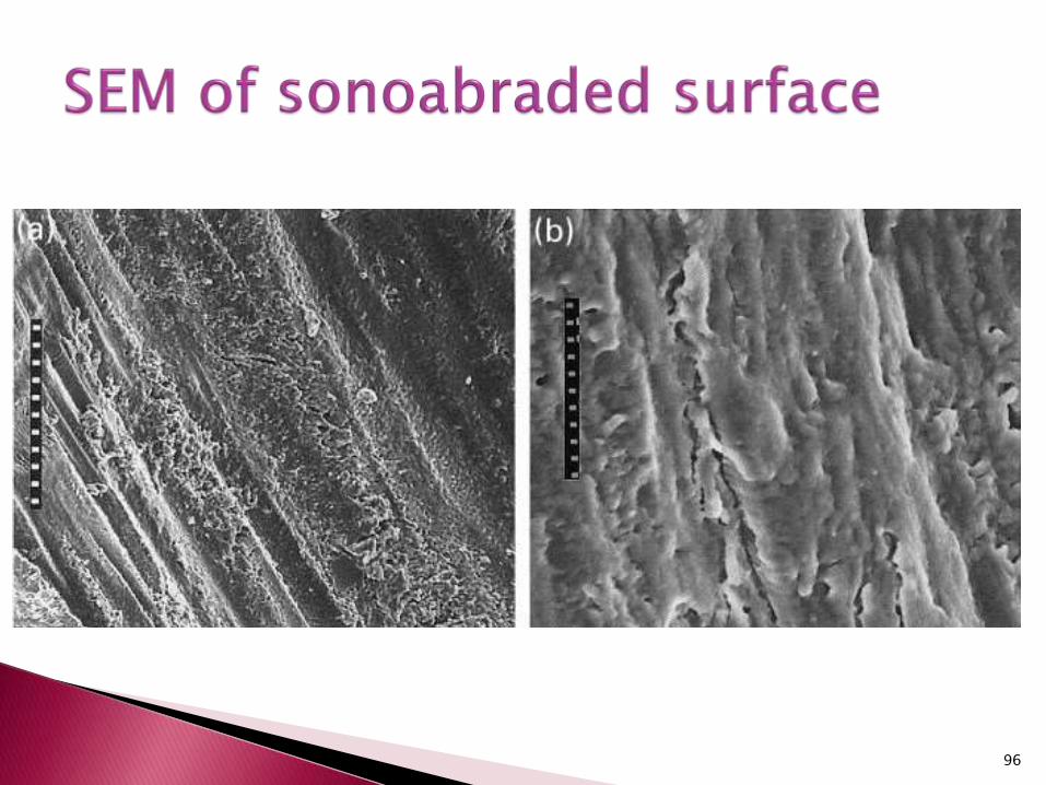
26

When studied by SEM,this method leaves a homogeneous smear layer with more or less uniform roughness, and dentinal tubules visibly obstructed with smear plugs.
A scanning electron microscopic study of different caries removal techniques on human dentin. Oper Dent
2002;27:360-366.
27

28
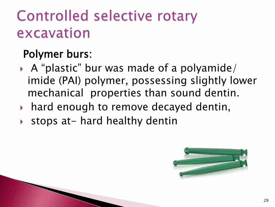
Polymer burs:
A “plastic” bur was made of a polyamide/ imide (PAI) polymer, possessing slightly lower mechanical properties than sound dentin.
hard enough to remove decayed dentin,
stops at- hard healthy dentin
29

self limiting –
The blade design was developed to remove dentin by locally depressing the carious tissue and pushing it forward along the surface until it ruptures and is carried out of the cavity
30

SmartPrep, SSWhite Burs; Lakewood, NJ, USA)
PEKK
Hardness -50 KHN
Higher than carious dentin (0 to 30 KHN)
Lower than sound dentin (70 to 90 KHN)
31

available in 3 sizes #2, #4, #6,
smaller than their carbide round bur counterparts
low speed i.e. 500-800 rpm , without water spray.
used with very light air brush type stroke.
32

Their cutting edges were not spiralled but straight.
Disadvantage:
To excavate caries from the center to the periphery in order to avoid contact with sound tooth tissue, the bur would
be prematurely and irreversibly damaged
33

More residual caries – smart prep burs.
Micro tensile bond strength to carious dentin- excavated with smart prep burs-lower.
TEM
34

SmartBurs
In primary teeth, resulted in the highest coincidence between the caries removal endpoint obtained by auto-fluorescence of carious dentin and the actual degree of caries removal.
surface hardness of the SmartBurs (26.6 KHN)
arrested carious dentin (39.2 KHN)
35

36

The CeraBurs are all-ceramic round burs made of alumina-yttria stabilized zirconia.
high cutting efficiency in infected, soft dentin.
replaces both the explorer and the spoon excavator by simultaneously providing tactile sensation, reducing preparation time.
37

CeraBurs with different diameters. From left to right: 10-,14-, 18-, and 23-mm diameter.
38

In vitro investigation of the caries-removal efficiency and efficacy did not show any significant difference between the ceramic and conventional tungsten-carbide burs.
In vitro comparison of ceramic burs and conventional tungsten carbide bud burs in dentin caries excavation.
Quintessence Int 2008;39:495-499.
39

Caries removal with a carbide bur, polymer bur, and spoon excavator produced significant reduction in viable count of both Streptococcus mutans and lactobacilli.
Carbide burs, however, produced greater reduction in the viable count of bacteria followed by polymer bur and spoon excavator.
In Vivo Comparison of Reduction in Bacterial Countafter Caries Excavation with 3 Different Techniques (J Dent Child 2011;78:31-5)
40

Mechanical Non-rotary: Hand excavators
Air abrasion
Air polishing
Ultrasonic instrumentation
Sono-abrasion
41

Spoon excavator and enamel hatchets –excavation of caries
Sharp excavators are effective and will reduce the force required for caries removal.
42

43

Advantages:
Long term observations have shown adequate tissue removal
Over excavation is unlikely
Accepted procedure especially in pedodonticsand anxious patient
Does not require any expensive equipment
Disadvantages:
High pressure causes pain
44
Banerjee, Kidd and Watson in 2000 –
conventional hand excavation appeared to offer the best combination of efficiency and effectiveness for carious dentine excavation.
In vitro Evaluation of Five Alternative Methods of Carious Dentine Excavation Caries Res 2000;34:144–150
.
45
Steel bur was the fastest method, followed by the polymer bur, hand excavator and laser.
Steel bur exhibited also the largest overpreparation area, followed by laser, hand excavator and polymer bur.
The largest underpreparation area was found using polymer bur, followed by laser, hand excavator and steel bur.
46

Overall, hand excavator seemed to be the most suitable method for carious dentine excavation in deciduous teeth, combining good excavation time with effective caries removal.
Performance of Four Dentine Excavation Methods in Deciduous Teeth Caries Res 2006;40:117-123
47

48

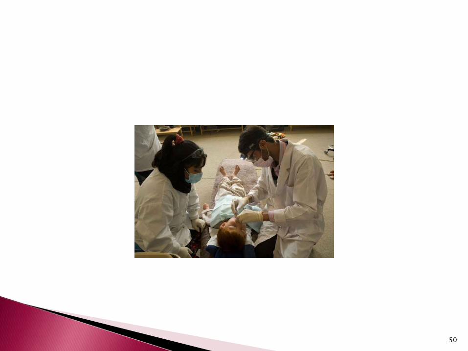
Procedure based on excavating carious cavities in teeth using hand instruments only and subsequent restoration with adhesive filling material (glass-ionomer).
innovative,
largely pain-free,
minimal intervention approach of
treating decayed teeth
49
50

The reasons for using hand instruments :
- it makes restorative care accessible for all population groups,
requires minimal cavity preparation that conserves sound tooth tissues
causes less trauma to the teeth,
- the low cost of hand instruments compared to electrically driven dental equipment,
51

the limitation of pain that reduces the need for local anaesthesia to a minimum and reduces psychological trauma
- simplified infection control.
Hand instruments can easily be cleaned and sterilized after every patient
52

The reasons for using glass-ionomer are
GIC sticks chemically to both enamel and dentine, the need to cut sound tooth tissue to prepare cavity is reduced,
- fluoride is released
- it is rather similar to hard oral tissues and does not inflame the pulp or gingiva.
53

- there is a cavity involving the dentine, and
- that cavity is accessible to hand instruments.
54

swelling (abscess) or fistula (opening from abscess to the oral cavity,
- the pulp exposed,
- teeth have been painful for a long time and there may be chronic inflammation of the pulp,
- there is an obvious carious cavity, but the opening is inaccessible to hand instruments
55

Mouth mirror
Explorer
A pair of tweezers
Spoon excavators
Enamel hatchet
Carver
Mixing pad and spatula
56

1.Place cotton wool rolls alongside the tooth to be treated.
2.Remove plaque from tooth surface with wet cotton wool pellets.
3.Dry the tooth surface with dry cotton wool pellets.
4.If necessary make the entrance of the cavity wider with a dental hatchet.
5.Remove the carious dentin with excavators starting at the enamel dentin junction.
57

6.Fracture off unsupported thin enamel with the hatchet. Make sure the enamel does not contain any carious spots.
7.Clean the cavity with wet and dry cotton wool pellets.
8.Remove the caries near the pulp carefully.
9.Clean the cavity again with cotton wool pellets.
10.Check the relation of the tooth to be restored with the opposing teeth by asking the patient to bite.
11.Complete the procedure by drying the cavity with dry cotton pellets.
58

Dr. Robert B. Black was the first to study air-abrasives technology in dentistry in 1943.
In 1951, S.S.White introduced the first air-abrasive system – Airdent
59

The principle employed by the airdent unit utilizes kinetic energy or inertia as a rapid and not unpleasant means of removing tooth structure by incorporating a fine abrasive material in a high velocity gaseous propellent.
EK= ½ m v2
60

Air abrasion is not a completely painless method of cavity preparation;
It eliminates
vibration,
bone-conducted noise,
pressure and heat.
The traumatic influence on tooth structure and periodontal tissue is reduced to a minimum.
61

Unit
Foot control
Hand piece- consists of a handle, a shaft – an adjustable contra-angle (ball and socket) and
a tip or nozzle in a 90 relationship to the shaft.
62

63
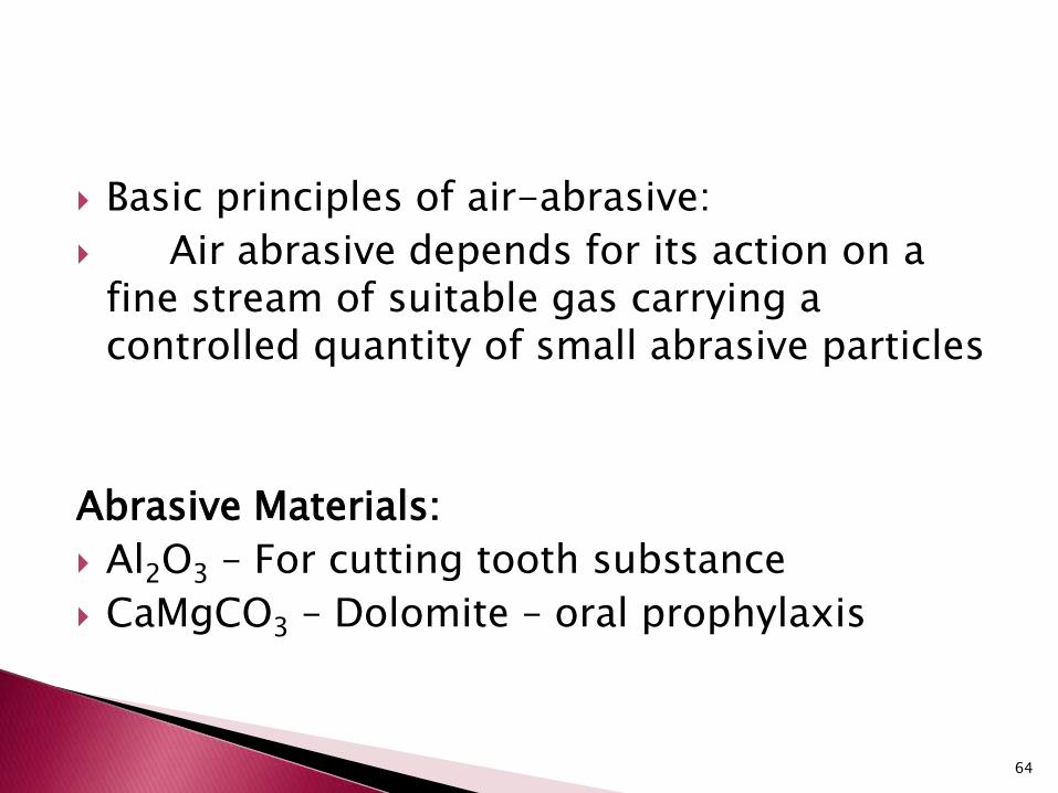
Basic principles of air-abrasive:
Air abrasive depends for its action on a fine stream of suitable gas carrying a controlled quantity of small abrasive particles
Abrasive Materials:
Al2O3 – For cutting tooth substance
CaMgCO3 – Dolomite – oral prophylaxis
64

US FDA approval for clinical use of 27.5 alumina particles
It possess a hardness of 9 on Moh’s scale and its particles possess sharp edges and pointed corners when properly prepared.
65

Propellants: CO2 was found to possess certain advantage
for this purpose. Practically free from moisture Non-toxic in low concentrations Convenient and almost universally available
The pressure of the liquid CO2 varies from 700 to 1300 pounds per square inch.
This pressure is reduced to app.115 pounds in the line and
80 to 45 pounds at the nozzle.
66

67

A nozzle tip distance of 1mm- the angulationis zero
At 2 mm total angulation - 7.
At 5mm it is 13.
At 10 mm it is 23
and at 15mm it is 35.
68

Action of air abrasive is influenced by factors
propellant pressure
type and particle size of the abrasive used,
abrasive mixture
nozzle bore and length,
nozzle distance from the enamel surface
nozzle angulation.
69

No.561chrome plated dental bur =6 mg of enamel _ 30 sec at 1725 rpm , pressure of 2 pounds.
Al2O3 _ 80 psi—a nozzle of 0.018 inch inside diameter and nozzle tip distance of 7
to 13 mm -90,
air abrasive is capable of removing 30 mg of enamel in 30 seconds.
70
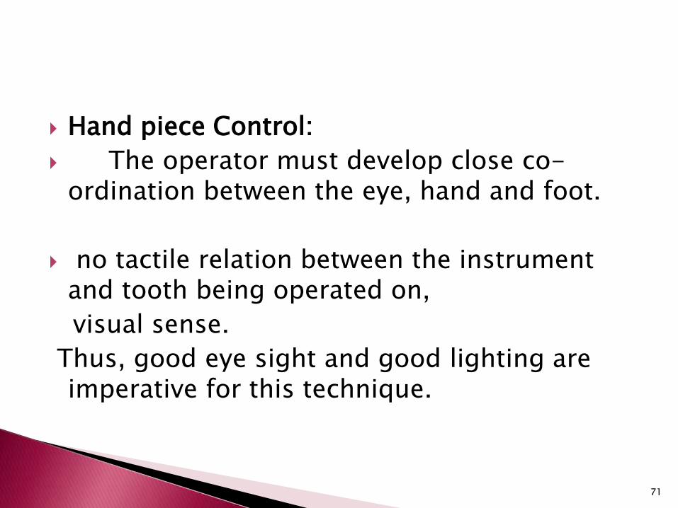
Hand piece Control:
The operator must develop close co-ordination between the eye, hand and foot.
no tactile relation between the instrument and tooth being operated on,
visual sense.
Thus, good eye sight and good lighting are imperative for this technique.
71

Hand piece grasp:
Air abrasive hand piece is held lightly in the pen grasp .
No pushing or pulling is necessary .
3rd or 4th finger is generally used not as a brace but as a rest for steadying the instrument.
72

Nozzle angulation must be correlated with nozzle tip distance.
Greater the nozzle tip distance the greater will be the angulation
73

Straight line cut:
high degree of definition is desired.
This type of cut utilizes close nozzle distances and is precise and narrow.
Angle cut:
greater nozzle distance, together with the required nozzle angulation.
As the nozzle distance from the substance being cut increases, the angle of the walls increases proportionately.
74

Advantages of angle cut–
greater cutting speed and
less visual interference
75
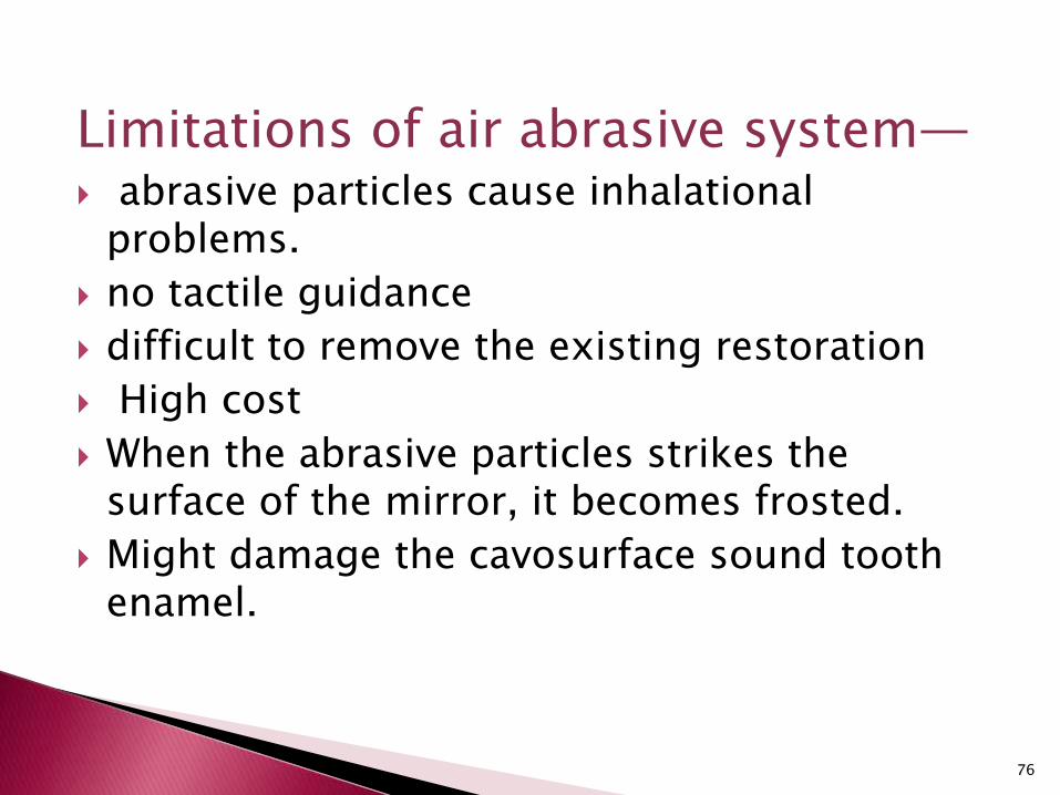
Limitations of air abrasive system— abrasive particles cause inhalational
problems.
no tactile guidance
difficult to remove the existing restoration
High cost
When the abrasive particles strikes the surface of the mirror, it becomes frosted.
Might damage the cavosurface sound tooth enamel.
76

The major drawback of air-abrasion excavation of carious dentin is that sound dentin is more efficiently removed than carious dentin.
Microhardness as a predictor of sound and carious dentine
removal using alumina air abrasion. CariesRes 2006;40:292-295.
77

High Speed Drills Air Abrasion
Rotary bur cause micro
fractures
No micro fractures
Excessive destruction of
tooth structure
Less destruction of tooth
structure
Heat, vibration,bone
conducted noise-patient
discomfort
Heatless, vibration less,
minimal sound
Patient Anxiety Patient friendly
78

Spherical glass beads
Polycarbonate resin-crushed powder removed artificially softened dentin more selectively without cutting sound dentin or enamel.
Selective caries removal with air abrasion. Oper Dent 1998;23:236-243.
79
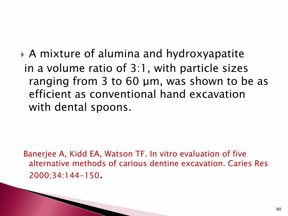
A mixture of alumina and hydroxyapatite
in a volume ratio of 3:1, with particle sizes ranging from 3 to 60 μm, was shown to be as efficient as conventional hand excavation with dental spoons.
Banerjee A, Kidd EA, Watson TF. In vitro evaluation of five alternative methods of carious dentine excavation. Caries Res
2000;34:144-150.
80

Bioactive glass powder (Bioglass, NovaminTechnology;Alachua, USA) with a particle diameter between 25 and 32μm was also explored.
Risk of unnecessary sound dentin removal was reduced because of the difference in cutting rate between sound and carious dentin.
An in vitro investigation of the effect and retention of bioactive glass air-abrasive on sound and carious dentine.
J Dent 2008;36:214-218.
81

82

Water soluble sodium bicarbonate and tricalcium phosphate
0.08% by weight
to improve the flow characteristics
air pressure,
concentric water jet.
As the abrasive is water soluble it does not escape too far from the operating field.
83

Razoog and Koka in 1994,
increasing the air-pressure beyond 90 psi actually reduced the abrasiveness of the microprophy system.
This was due to a phenomenon -choked flow.
as the air pressure exceeds the critical pressure, the mass flow of particles will reduce thus limiting the system’s abrasiveness.
84

85

Commercially recommended use of this technique is to
remove surface enamel stains,
plaque and calculus
overzealous use - remove healthy tooth structure
removal of carious dentine at the end of cavity preparation.
86
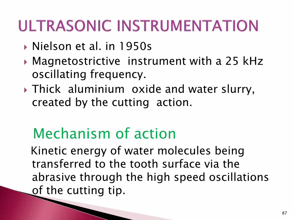
Nielson et al. in 1950s
Magnetostrictive instrument with a 25 kHz oscillating frequency.
Thick aluminium oxide and water slurry, created by the cutting action.
Mechanism of actionKinetic energy of water molecules being transferred to the tooth surface via the abrasive through the high speed oscillations of the cutting tip.
87

Nielson attempted to analyse the results from altering
the pressure applied,
the length of use of the instrument,
the powder water ratio in the slurry,
the nature of the material
cut and the type of abrasive used.
88

SONIC OSCILLATION (SONOABRASION)
89

Removal of carious dentin using high frequency ,sonic air scaler with modified abrasive tips
First Design
•Sonic micro unit designed by Dr.Hugo Unterbrink and
Mosele
•Based on Soniflex 2000L and 2000N Air scaler Hand
piece
•Oscillations - < 6.5 KHZ
90

Tips- elliptical motion - transverse distance of between 0.08 to 0.15 mm
longitudinal movement of between 0.55 –0.135mm
tips are diamond coated on one side using
40 grit diamond
Cooled using water irrigant at a flow rate of between 20-30 ml/min.
The operational air pressure -3.5 bar.
91

A lengthways
halved torpedo
shape
9.5mm
long,1.3mm wide
A small hemisphere
1.5 mm diameter
A large
hemisphere 2.2mm
diameter
92

93

Torque Applied – 2N More pressure - dampens oscillations –
cutting efficiency reduced.
Indications
•Carious dentin removal
•Finishing cavity preparations
More studies needed to prove its efficiency
94

Advantage
less over preparation than with rotary instruments
smaller access cavity is possible.
Disadvantage
unclear completeness of excavation
95
96

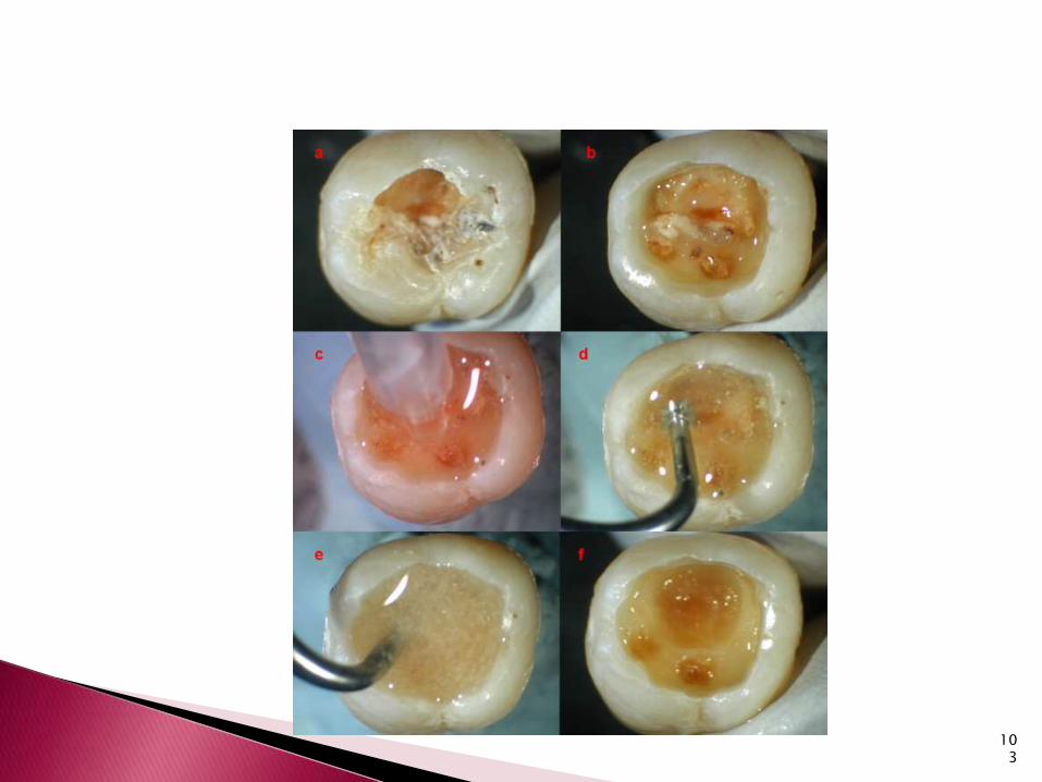
CHEMO MECHANICALCARIES REMOVAL (CMCR)
Chemical softening of carious dentin followed
by its removal by gentle excavation
97

Dentin
Inorganic – 70% Organic matrix - 20 % H2O – 10 %
18 % Collagen 2 % Non Collagen
Proline + Glycine - Polypeptides – Tropocollagen - Fibril
• Chlorination of Partially Degraded Collagen (Conversion of Hydroxyproline to Pyrrole-2-carboxylic acid)
98

CMCR limits the removal of sound tooth structure, the cutting of open dentinal tubules, pulpal irritation pain
99

Goldman and Kronman
Na0Cl + Sorenson’s Buffer
(Glycine ,NaOH,NaCl)
N Mono Chloro Glycine
(GK 1019)
Glycine replaced by
Amino Butyric
acid
N-Mono Chloro DL2 amino
butyric acid
(NMAB) –GK 101E
100

Chlorination of Partially Degraded Collagen (Conversion of Hydroxyproline to Pyrrole-2-carboxylic acid)
Cleavage by Oxidation of glycine residues –Disruption of collagen – more friable collagen- removed.
101

Solution 1: 1% NaOCl
Solution 2: glycine+amino butyric acid+
NaCl+NaOH
pH = 11
Delivery system- reservoir,
heater and pump,
handpiece,
applicator tips
102
103

104
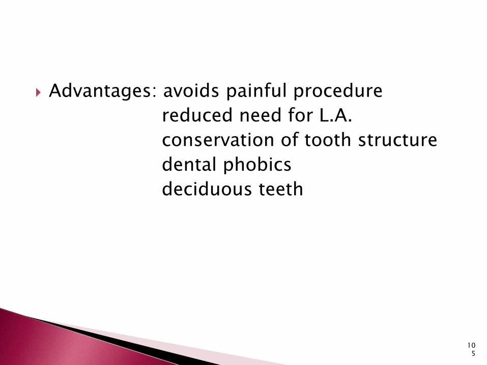
Advantages: avoids painful procedure
reduced need for L.A.
conservation of tooth structure
dental phobics
deciduous teeth
105

Rotary & hand instruments were still needed
Large volumes of solution
Slow
Long term studies were lacking
Short shelf life
Special delivery system was needed
106

carisolv107

2 Syringes
NaOCl
Pink Viscous gel ( Lysine,
Leucine, Glutamic Acid
+Carboxymethyl cellulose +
Erythrosine )
Max Volume of Gel – 0.2 – 1 ml
Cloudy - frosty
‘A silent revolution’
108

Multi mix
109

singlemix110

112

operative steps in chemomechanical caries excavation include:
(1) application of the solution, (2) scrapping off the carious dentin with
possible change of instrument size,(3)rinsing, and (4) repetition of the procedures until all caries
is removed.Time required 10-15 minVolume required 0.2 -1 ml
113

114

115

Carisolv power drive is a faster and easier way of working with carisolv.
Advantages:-
It has unique torque limitations and this helps to protect the healthy dentine.
It works at very low speed, thereby minimizing noise and pain.
Power drive is used with special star bur – 1.0, 1.5, 2.0. These burs work with power drive or a low speed handpiece of maximum 300 rpm.
116

117

CARIDEX CARISOLV
SOL I 1% NaOCl 0.5 % NaOCl
SOL II 0.1MAminobutyric acid
glyciene
0.1M NaCl,0.1 M NaOH
0.1M glutamic acid / leucine /
lysine, NaCl, NaOH
Dye - Erythrocyin
pH 11 11
Physical Nature Liquid gel
Volume 100-500ml 0.2 – 1ml
Time required 10-15 mins 10-15 mins
Instruments Applicator tips Specially designed
Active time 1 Hr 20 mins118

Painless
No need of local anesthesia
Conservation of sound tooth structure
Reduced risk of pulp exposure
Well suited for anxious
Better than Caridex
LIMITATION
•Rotary and hand instruments may still be needed
119

Complete removal of caries was achieved significantly in both the methods,( Papacarie, with conventional slow-speed rotary instrument(bur)
there was less marked destruction of dentinal tubules in chemomechanical caries removal method by Papacarie.
Comparative evaluation of the efficacy ofchemomechanicalcaries removal agent(Papacarie) and conventional method of caries removal: An in vitro study J INDIAN SOC PEDOD PREVENT DENT
2010( 28 )|
120

Removal of carious dentin with Carisolv is highly effective than that of Hand Excavation, but slightly less than round carbide bur.
It may be because of carisolv which removes only the infected dentin and not the affected dentin.
Efficacy of chemo-mechanical method (carisolv) of caries removal with that of hand cutting and rotary cutting instruments. Annals and essences of dentistry Dec 2011
121

Chemomechanical excavation using Carisolvgel was the slowest technique.
hand excavation presented higher efficiency and effectiveness than chemomechanicalexcavation.
Effectiveness and Efficiency of Chemomechanical Carious Dentin Removal. Braz Dent J (2006) 17(1): 63-67
122

123

Pepsin in a phosphoric acid/sodium biphosphate buffer- alternative to CMCR.
phosphoric acid dissolves the inorganic component of carious dentin.
pepsin - organic part of the carious biomass
denatured collagen
Self-limiting caries therapy with proteolytic agents. Am J Dent 2008;21:303-312
124
Advantage:
more specific by digesting only denatured collagen (after the triple-helix integrity is lost) than the sodium hypochlorite-based agents.
125

126

In 1989 Goldsberg and Keil
Achromobacter collagenase- did not affect the sound layers of dentin beneath the lesion.
In 1996 Norbo, Brown and Jan -Enzyme
Pronase –non specific proteolytic enzyme –
Streptomyces griseus
127
This technique was developed as a direct method to clinically differentiate between infected and affected carious dentin.
Changes in tooth fluorescence detects early tooth surface caries.
Lennon et al. in 2002 studied the residual caries detection using visible fluorescence.
128

Based on the fact that several oral microorganisms produce orange-red fluorophores as by-products of their metabolism (porphyrins), infected carious tissue will fluoresce especially in the red fraction of the visible spectrum due to the presence of proto- and meso-porphyrins.
Human teeth with and without dental caries studied by visible
luminescent spectroscopy. J Dent Res 1981;60:120-122
Diagnodent: An optical method for caries detection.
J Dent Res 2004;83:80-83.129

Carious dental tissue fluorescences more intensely in the red portion of the visible spectrum (>540 nm) than the sound dentine.
130

Violet light (370-420 nm) –
The operator can observe the cavity through a 530 nm – high pass filter.
Areas exhibiting orange-red fluorescence –caries -- removed by appropriate size bur.
131

Compared to Caries Detector or the visual-tactile method for establishing the caries removal endpoint, the FACE method showed the highest sensitivity, specificity, percentage correct score, and predictive values for residual caries detection, as evaluated using confocal microscopy.
Residual caries detection using visible fluorescence. Caries Res 2002;36:315-319
132

There was a significant reduction in the number of samples presenting residual bacteria after excavation with FACE, when compared to Carisolvor bur excavation guided by Caries Detector(1)
Histological examination after staining with ethidium bromide revealed fewer samples presenting bacteria in dentin when the FACE method was used than was the case with conventional bur excavation(2)
1.Efficiency of 4caries excavation methods compared. Oper Dent 2006;31:551-555.
2.Fluorescence-aided caries excavation (FACE) compared toconventional method. Oper Dent 2003;28:341-345.
133

Advantages: very efficient, with less time needed to
excavate caries and without a need to change instruments, apply chemical agents, or to test the cavity with an explorer.
FACE was apparently not associated with an increased cavity size or overexcavation.
Efficiency of 4 caries excavation methods compared. Oper
Dent 2006;31:551-555
Quantity of remaining bacteria and cavity size after excavation with FACE, caries detector dye and conventional excavation in vitro.
OperDent2007;32:236-241.
134

LASER THERAPY
135

Light Amplification by Stimulated Emission of Radiation
In 1960, Theodore Maiman developed the first working laser device which emitted a deep red-coloured beam from a ruby crystal applied to cutting both hard and soft tissues in the mouth
136

Efficacy of laser depends on •Pulse energy
•Optical properties of incident tissue
•Wavelength characteristics
Applications•Selective Hard Ablation•Selective Carious Dentin Removal•Destroy S.Mutans•Sealing of Fissures•Adjunctive treatment in caries prophylaxis•Modify structures of dentin and enamel tissue
137

Ablation:The absorption differences between
carious and healthy dentin were the highest at blue spectral range
0.4J/cm2 but below 1.8J/cm2.
The laser energy must be delivered uniformly to the lesion surface.
Murray et al. –remaining dentine thickness should be at least
0.5 mm to avoid pulp injury.
138
CO2 lasers and Nd :YAG produce surface changes in enamel such as roughness, cracking, fissuring, melting and recrystallisation.
generate markedly elevated surface and pulpal temperature.
139

ArF excimer lasers have been reported to remove dental caries.
Krypton F excimer laser has been shown to cut dentin;
however enamel is resistant to effective ablation.
140
Walsh LJ. The current status of laser applications in dentistry. Austr Dent J 2003;48:146-155

CO2 laser irradiation inhibits the progression of caries like lesion up to 85%.
Er : YAG -40%
Er, Cr : YSGG - 60% caries reduction
141
Er : YAG lasers, Er : YSGG and Er, Cr : YSGG lasers operate at wavelengths of 2940, 2790 and 2780 nm.
These wavelengths correspond to the peak absorption range of water in the infra red spectrum.
The efficiency of ablation is greatest for the Er : YAG laser.
142

Er-based laser systems - popping sound.
MECHANISM OF ACTION
A laser powered hydrokinetic system delivers photons into an air-water spray matrix with resultant microexplosive forces on water droplets.
The mechanism of hard tissue cutting is based on this process.
This system with its accompanying air water spray has been shown to cut enamel, dentine, cementum and bone efficiently and clearly without any deleterious thermal effects on dental pulp.
143

145

146
Related Documents


