Transcription Mapping as a Tool in Phage Genomics: The Case of the Temperate Streptococcus thermophilus Phage Sfi21 Marco Ventura, Sophie Foley, Anne Bruttin, Sandra Chibani Chennoufi, Carlos Canchaya, and Harald Bru ¨ssow 1 Nestle ´ Research Center, Nestec Ltd., Vers-chez-les-Blanc, CH-1000 Lausanne 26, Switzerland Received October 2, 2001; accepted December 31, 2001 For the lytic growth cycle of the temperate cos-site Streptococcus thermophilus phage Sfi21 a transcription map was developed on the basis of systematic Northern blot hybridizations. All deduced 5 ends were confirmed by primer extension analysis. Three time classes of transcripts were observed. Early transcripts were identified in four different genome regions. One prominent early mRNA of 4.8 kb length covered a group of 12 genes located between the origin of replication and the cos-site. Two short early mRNAs represented a single gene from the direct vicinity of the cos-site and the superinfection immunity gene from the lysogeny module, respectively. A fourth early transcript covered a group of four genes located between the lysin and the integrase gene. Middle transcripts of 2.1 and 5.8 kb length covered cro-like and ant-like repressor genes and the DNA replication module, respectively. Four types of late transcripts were identified. The transcripts covered the likely DNA packaging genes, the head morphogenesis module plus the major tail gene, the remainder of the tail genes, and the putative tail fiber plus lysis genes, respectively. Only the transcript from the head morphogenesis genes yielded defined late mRNA species. The transcription map concurred with most of the in silico predictions for the genome organization of phage Sfi21 except for the separation of the DNA replication module from a possible transcription regulation module. Most 5 ends of the transcripts determined in primer-extension experiments were not preceded by a consensus promoter sequence. The involvement of phage-encoded regulators for middle and late transcription was suggested by chloramphenicol-inhibition experiments. © 2002 Elsevier Science (USA) INTRODUCTION Streptococcus thermophilus phages are a major cause of fermentation failures in the dairy industry (Bru ¨s- sow, 1999). Due to this economic importance, S. ther- mophilus bacteriophage became one of the most care- fully sequenced phage groups (Bru ¨ssow and Desiere, 2001). All S. thermophilus phages belong to the Sipho- viridae family of tailed phages (Caudovirales). Closely related temperate and virulent S. thermophilus phages were described (Bru ¨ssow, 2001) that shared sequence similarity not only with phages from other important dairy starters (Lactococcus lactis, Lactobacillus spp.) (Desiere et al., 2001a), but also from important human pathogens such as S. pyogenes (Desiere et al., 2001b). Therefore S. thermophilus phages were chosen in our laboratory as reference phages for comparative phage genomics. However, simple database searches with phage se- quences were of limited use in the elucidation of the phage genome organization yielding few indications on transcription and expression pattern. For example, phage Sfi21, the prototype temperate cos-site S. ther- mophilus phage, has a 40-kb genome that potentially encodes 53 genes (Lucchini et al., 1999b). Database searches allowed attribution of a likely function to only 10 genes. For dairy phages this is a fairly typical case. Phage O1205, the prototype temperate pac-site S. ther- mophilus phage with a similar genome size, but a totally unrelated structural gene cluster, also showed only 10 genes with good matches to entries from the database (Stanley et al., 1997). It is currently unknown if this ob- servation reflects an intrinsically great variability of phage genes or simply a limitation of the database. At present, only about 100 complete phage genomes are deposited in the database corresponding to less than two Escherichia coli genome equivalents. Alternative approaches are therefore urgently needed to further our understanding of the genome organization in phages that are investigated for applied and not aca- demic purposes (e.g., phage resistance in dairy micro- biology, phage therapy in medical microbiology, and phage ecology in marine microbiology). One successful approach was the comparative genomic analysis of S. thermophilus phages that differed in phenotype (host range; susceptibility to phage resistance mechanisms; life-style: temperate vs virulent; DNA packaging mecha- nism: cos-site vs pac-site) (Lucchini et al., 1999a,b). These comparisons allowed a given phage phenotype to be assigned to single gene (putative antireceptor pro- tein) or larger DNA segments (putative lysogeny, DNA packaging, DNA replication modules, and structural 1 To whom correspondence and reprint requests should be addressed. Fax:41-21-785-8925. E-mail: [email protected]. Virology 296, 62–76 (2002) doi:10.1006/viro.2001.1331 0042-6822/02 $35.00 © 2002 Elsevier Science (USA) All rights reserved. 62

Welcome message from author
This document is posted to help you gain knowledge. Please leave a comment to let me know what you think about it! Share it to your friends and learn new things together.
Transcript
Virology 296, 62–76 (2002)
Transcription Mapping as a Tool in Phage Genomics: The Caseof the Temperate Streptococcus thermophilus Phage Sfi21
Marco Ventura, Sophie Foley, Anne Bruttin, Sandra Chibani Chennoufi, Carlos Canchaya, and Harald Brussow1
Nestle Research Center, Nestec Ltd., Vers-chez-les-Blanc, CH-1000 Lausanne 26, Switzerland
Received October 2, 2001; accepted December 31, 2001
For the lytic growth cycle of the temperate cos-site Streptococcus thermophilus phage Sfi21 a transcription map wasdeveloped on the basis of systematic Northern blot hybridizations. All deduced 5� ends were confirmed by primer extensionanalysis. Three time classes of transcripts were observed. Early transcripts were identified in four different genome regions.One prominent early mRNA of 4.8 kb length covered a group of 12 genes located between the origin of replication and thecos-site. Two short early mRNAs represented a single gene from the direct vicinity of the cos-site and the superinfectionimmunity gene from the lysogeny module, respectively. A fourth early transcript covered a group of four genes locatedbetween the lysin and the integrase gene. Middle transcripts of 2.1 and 5.8 kb length covered cro-like and ant-like repressorgenes and the DNA replication module, respectively. Four types of late transcripts were identified. The transcripts coveredthe likely DNA packaging genes, the head morphogenesis module plus the major tail gene, the remainder of the tail genes,and the putative tail fiber plus lysis genes, respectively. Only the transcript from the head morphogenesis genes yieldeddefined late mRNA species. The transcription map concurred with most of the in silico predictions for the genomeorganization of phage Sfi21 except for the separation of the DNA replication module from a possible transcription regulationmodule. Most 5� ends of the transcripts determined in primer-extension experiments were not preceded by a consensus
INTRODUCTION
Streptococcus thermophilus phages are a majorcause of fermentation failures in the dairy industry (Brus-sow, 1999). Due to this economic importance, S. ther-mophilus bacteriophage became one of the most care-fully sequenced phage groups (Brussow and Desiere,2001). All S. thermophilus phages belong to the Sipho-viridae family of tailed phages (Caudovirales). Closelyrelated temperate and virulent S. thermophilus phageswere described (Brussow, 2001) that shared sequencesimilarity not only with phages from other important dairystarters (Lactococcus lactis, Lactobacillus spp.) (Desiereet al., 2001a), but also from important human pathogenssuch as S. pyogenes (Desiere et al., 2001b). Therefore S.thermophilus phages were chosen in our laboratory asreference phages for comparative phage genomics.However, simple database searches with phage se-quences were of limited use in the elucidation of thephage genome organization yielding few indications ontranscription and expression pattern. For example,phage Sfi21, the prototype temperate cos-site S. ther-mophilus phage, has a 40-kb genome that potentiallyencodes 53 genes (Lucchini et al., 1999b). Database
1
62
searches allowed attribution of a likely function to only 10genes. For dairy phages this is a fairly typical case.Phage O1205, the prototype temperate pac-site S. ther-mophilus phage with a similar genome size, but a totallyunrelated structural gene cluster, also showed only 10genes with good matches to entries from the database(Stanley et al., 1997). It is currently unknown if this ob-servation reflects an intrinsically great variability ofphage genes or simply a limitation of the database. Atpresent, only about 100 complete phage genomes aredeposited in the database corresponding to less thantwo Escherichia coli genome equivalents.
Alternative approaches are therefore urgently neededto further our understanding of the genome organizationin phages that are investigated for applied and not aca-demic purposes (e.g., phage resistance in dairy micro-biology, phage therapy in medical microbiology, andphage ecology in marine microbiology). One successfulapproach was the comparative genomic analysis of S.thermophilus phages that differed in phenotype (hostrange; susceptibility to phage resistance mechanisms;life-style: temperate vs virulent; DNA packaging mecha-nism: cos-site vs pac-site) (Lucchini et al., 1999a,b).These comparisons allowed a given phage phenotype tobe assigned to single gene (putative antireceptor pro-
promoter sequence. The involvement of phage-encodedchloramphenicol-inhibition experiments. © 2002 Elsevier Scien
To whom correspondence and reprint requests should be addressed.Fax:�41-21-785-8925. E-mail: [email protected].
doi:10.1006/viro.2001.1331
0042-6822/02 $35.00
ors for middle and late transcription was suggested by)
tein) or larger DNA segments (putative lysogeny, DNApackaging, DNA replication modules, and structural
© 2002 Elsevier Science (USA)All rights reserved.
regulatce (USA
gene clusters). Another approach hinged on the obser-vation that S. thermophilus phages closely matched thegene map of lambdoid coliphages over the late generegion (Desiere et al., 1999). An evolutionary analysissuggested that this similarity reflected ancient phyloge-netic relationships between both phage groups (Brus-sow and Desiere, 2001). On the basis of the genome mapalignments with lambdoid phages (Casjens et al., 1992),a number of gene functions have been predicted. In fact,the identity of the major head and major tail proteinswere confirmed by electron microscopy combined withN-terminal sequencing (Desiere et al., 1999; Stanley etal., 1997). In addition, the tail tape protein (Pedersen etal., 2000), the tail absorption protein (Duplessis andMoineau, 2001), the holin, and the lysin genes (Sheehanet al., 1999) have been defined in dairy phages by acombination of genetic and biological experiments.
In the present article we report a transcription map forthe temperate cos-site S. thermophilus phage Sfi21 byusing 34 different DNA probes and confirming all de-duced transcript locations by primer-extension experi-ments. The transcription map was used to crosscheckthe in silico predictions of comparative phage genomics.The transcription map also provided a first insight intothe dynamics of gene expression of phage Sfi21. In con-trast to temperate lactococcal phages (Madsen andHammer, 1998) the transcription of the temperate phageSfi21 during the lytic growth was not initiated in thegenetic switch region predicted by in silico analysis. Thedivergently transcribed genes of the postulated geneticswitch region gave rise to middle transcripts. Chloram-phenicol-inhibition studies suggested that the prominentlytic transcript of this region was under positive controlby a phage protein encoded in early genes.
RESULTS
Lytic growth of the temperate S. thermophilus phageSfi21
S. thermophilus cell strain SfiI was infected with thetemperate phage Sfi21 at an OD (600nm) of 0.1 with amultiplicity of infection of 10. Intracellular progeny phagewas detected at 30 min postinfection (p.i.) and reachedmaximal titers at 50–60 min p.i., when the bacterial cul-ture underwent lysis. Extracellular phage titers followedthe intracellular phage titers with a 10- to 20-min time lag(data not shown). Until shortly before cell lysis, infectedand uninfected cells could not be distinguished by theirpolypeptide pattern on SDS–PAGE (data not shown).Cells infected with the temperate phage Sfi21, but notthose infected with a virulent S. thermophilus phage,resumed growth in milk with a time lag of about 4 h. Allsurvivors contained the Sfi21 prophage. Based on adoubling time of 25 min for S. thermophilus cells, we canestimate that approximately 1 of 1000 infected cells tookthe lysogenic instead of the lytic pathway. Under single-
step growth conditions a comparable growth curve wasobtained for phage Sfi21 (data not shown). The burst sizewas about 150 phage particles per cell. Comparison ofthe viable cell counts and infective centers demonstratedagain that about 1 in 1000 cells survived the infection.
Transcription mapping in phage Sfi21
Total RNA was isolated from the infected cells at 0 and2 min p.i., and then every 5 min until about 40 min p.i.Fifteen micrograms of total RNA were separated on adenaturing agarose gel, blotted, and phage transcriptswere revealed by Northern blot hybridization using spe-cific DNA probes. The precision of the localization of thephage mRNAs on the phage genome map is limited bythe number of DNA probes used in Northern blots andtheir spatial resolution. More than 30 DNA probes wereused to localize the transcripts on the genome map. Toincrease that resolution, the 5� ends of all transcriptswere mapped by primer-extension experiments. The 3�position of the mRNA was deduced from the estimatedsize of the mRNAs and from hybridization experimentswith probes corresponding to adjacent DNA segments.Rho-independent terminators were predicted by in silicoanalysis. A summary of the results is presented in thetranscription map shown in Fig. 1.
Transcription of the predicted genetic switch region
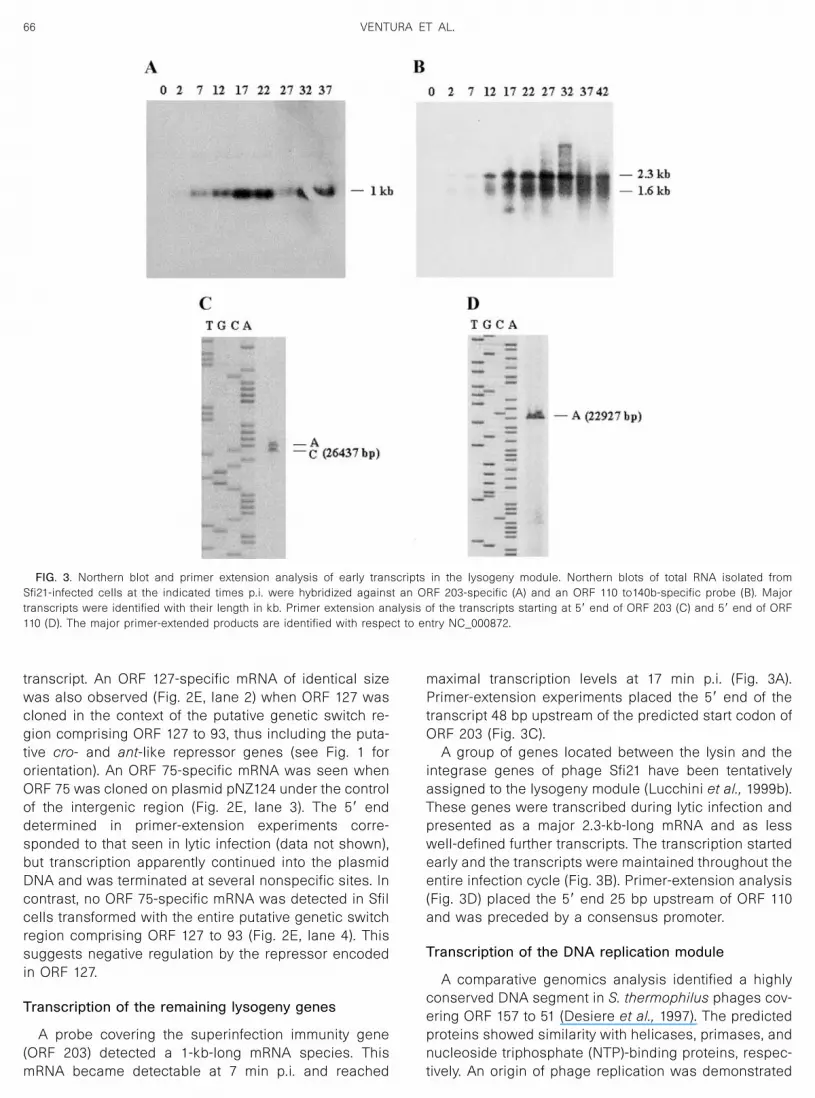
DNA sequence analysis of phage Sfi21 predicted agenetic switch region between ORF 127 and 75, twodivergently transcribed genes that encode repressor-likeproteins (Fig. 1) (Bruttin et al., 1997b). ORF 127 and thefollowing three genes represent the only leftward tran-scribed gene clusters of the phage Sfi21 genome andincluded a biologically proven superinfection immunitygene (ORF 203) (Bruttin et al., 1997b) and the phageintegrase (ORF 359) (Bruttin et al., 1997a) (Fig. 1). Whenthe Northern blots were probed with ORF 127, the pre-dicted cI-like repressor gene, only weak phage tran-scripts were detected (Fig. 2C) and no primer extensionproducts were obtained with ORF127-specific primersfrom RNA of lytically infected cells (data not shown). A1.6- and a 3.2 kb-long mRNA species was detected withthe ORF 127 probe only after longer exposure times (Fig.2C�). When Northern blots were probed with ORF 75, thepredicted cro-like repressor gene, a single 2.1-kb hybrid-ization signal was detected by autoradiography (Fig. 2A).The signal was clearly visible at 12 min p.i., increased inintensity until 27 min p.i., and decreased at later times ofinfection. The maintenance of a signal until late in infec-tion could indicate ongoing transcription or stability ofthe mRNA synthesized at an earlier time of infection.Primer-extension experiments located the 5� end 45 bpupstream of the start codon of ORF 75 (Fig. 2B). The ORF75 start codon is preceded by a good ribosomal bindingsite (RBS) and the 5� end of the transcript is preceded by
63TRANSCRIPTION MAPPING AS A TOOL IN PHAGE GENOMICS
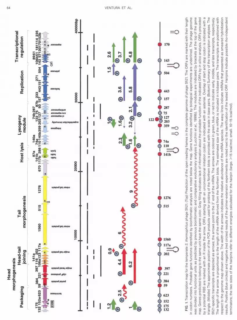
FIG.1.T
rans
crip
tion
map
for
the
tem
pera
teS.thermophilus
phag
eS
fi21.
(Top
)Pre
dict
ion
ofth
eop
enre
adin
gfr
ames
inth
eco
mpl
ete
geno
me
ofph
age
Sfi2
1.Th
eO
RFs
are
mar
ked
with
thei
rle
ngth
inco
don
num
bers
.Pro
babl
ege
nefu
nctio
nsid
entif
ied
bybi
oinf
orm
atic
anal
ysis
are
note
dbe
low
the
map
.Gen
efu
nctio
nsid
entif
ied
bybi
olog
ical
expe
rimen
tsar
eun
derli
ned.
The
phag
ege
nom
ew
asdi
vide
din
tofu
nctio
nalu
nits
aspr
edic
ted
bypr
evio
usbi
oinf
orm
atic
and
com
para
tive
evol
utio
nary
anal
ysis
(Luc
chin
ietal.,
1999
b).T
hem
odul
esar
ein
dica
ted
bybr
acke
tsat
the
top
ofth
ege
nem
ap.G
enes
pred
icte
dto
belo
ngto
the
sam
eun
itha
veth
esa
me
colo
r.G
ray
fillin
gin
dica
tes
lack
ofin
form
atio
nab
outt
hepr
obab
lefu
nctio
nof
the
indi
cate
dO
RFs
byinsilico
anal
ysis
.OR
Fspr
eced
edby
apo
tent
ialR
BS
are
mar
ked
with
anR
insi
deth
ear
row
.OR
Fsst
artin
gw
ithan
unco
nven
tiona
lini
tiatio
nco
don
are
indi
cate
dw
ithan
aste
risk.
Ove
rlap
ofst
art
and
stop
codo
nis
indi
cate
dw
itha
tria
ngle
.(M
iddl
e)Th
eap
prox
imat
epo
sitio
nof
the
PC
Rpr
oduc
tsus
edfo
rpr
obin
gof
the
Nor
ther
nbl
ots
ispr
ovid
edw
ithth
esc
ale
inba
sepa
irs.(
Bot
tom
)Sum
mar
yof
the
tran
scrip
tion
anal
yses
.The
Sfi2
1-sp
ecifi
ctr
ansc
ripts
are
depi
cted
asar
row
s;th
ear
row
spo
intt
oth
e3�
end
ofth
em
RN
A.T
hear
row
sar
eco
lore
din
gree
n,bl
ue,o
rre
dto
indi
cate
early
,mid
dle,
and
late
tran
scrip
ts,r
espe
ctiv
ely.
The
leng
thof
the
arro
wis
prop
ortio
nalt
oth
ele
ngth
ofth
em
RN
Ade
rived
from
the
Nor
ther
nbl
ots.
The
estim
ated
size
ofth
em
RN
Ais
indi
cate
din
kilo
base
pairs
.The
tran
scrip
tsar
epo
sitio
ned
with
resp
ectt
oth
ege
nom
em
apsh
own
abov
e.Th
ew
idth
ofth
ear
row
sin
dica
tes
the
rela
tive
abun
danc
eof
the
mR
NA
spec
ies.
The
wav
ylin
esin
dica
tem
RN
As
that
pres
ente
das
smea
red
hybr
idiz
atio
nre
sults
.Pos
itive
(blu
eci
rcle
s)an
dne
gativ
e(r
edci
rcle
s)re
sults
ofth
epr
imer
-ext
ensi
onex
perim
ents
are
note
dne
xtto
the
iden
tific
atio
nof
the
test
edO
RF.
Hai
rpin
sin
dica
tepo
ssib
lerh
o-in
depe
nden
tte
rmin
ator
s;th
etw
osi
zes
ofth
eha
irpin
sre
fer
todi
ffere
nten
ergi
esca
lcul
ated
for
the
hairp
in(la
rge:
�15
kcal
/mol
;sm
all:
10–1
5kc
al/m
ol).
64 VENTURA ET AL.

nearly perfect �10 and �35 consensus promoter sitesfor L. lactis, a close evolutionary relative of S. thermophi-lus.
Transcription of the cloned genetic switch region
When ORF 127 was cloned on plasmid pNZ124 underthe control of the intergenic region between ORF 127 and
75, an ORF 127-specific messenger was detected intransformed SfiI cells (Fig. 2E, lane 1) and the putativetranscription start site could be determined by primerextension (Fig. 2D). The 5� end suggested the use of aninternal in-frame start codon in ORF 127. Neither a goodRBS nor a consensus promoter structure, respectively,preceded the predicted start codon and the 5� end of the
FIG. 2. Northern blot of total RNA isolated from S. thermophilus SfiI at the indicated time after infection (0–42 min p.i., indicated at the top of theNorthern blots) with phage Sfi21 were hybridized against an ORF75-specific DNA (A) or an ORF127-specific DNA (C, C� shows a six-fold longerexposure of the autoradiography compared to A). The estimated size of the detected transcripts in kb are indicated to the right of each panel.Primer-extension analysis carried out with transcripts from Sfi21-infected cells with oligonucleotides placed near the 5� ends of the transcripts startingfrom ORF 75 (B) and from ORF 127 (D, RNA from SfiI cell transformed with plasmid pSFcI). The major primer-extended products are identified withrespect to entry NC_000872. E: ORF127-specific (lanes 1, 2) and ORF 75-specific (lanes 3, 4) mRNA expression in SfiI cells containing plasmids pSFcI(lane 1), pSFswi (lanes 2, 4), and pSFcro (lane 3). Molecular weights are indicated in kb.
65TRANSCRIPTION MAPPING AS A TOOL IN PHAGE GENOMICS
transcript. An ORF 127-specific mRNA of identical sizewas also observed (Fig. 2E, lane 2) when ORF 127 wascloned in the context of the putative genetic switch re-gion comprising ORF 127 to 93, thus including the puta-tive cro- and ant-like repressor genes (see Fig. 1 fororientation). An ORF 75-specific mRNA was seen whenORF 75 was cloned on plasmid pNZ124 under the controlof the intergenic region (Fig. 2E, lane 3). The 5� enddetermined in primer-extension experiments corre-sponded to that seen in lytic infection (data not shown),but transcription apparently continued into the plasmidDNA and was terminated at several nonspecific sites. Incontrast, no ORF 75-specific mRNA was detected in SfiIcells transformed with the entire putative genetic switchregion comprising ORF 127 to 93 (Fig. 2E, lane 4). Thissuggests negative regulation by the repressor encodedin ORF 127.
Transcription of the remaining lysogeny genes
A probe covering the superinfection immunity gene(ORF 203) detected a 1-kb-long mRNA species. ThismRNA became detectable at 7 min p.i. and reached
maximal transcription levels at 17 min p.i. (Fig. 3A).Primer-extension experiments placed the 5� end of thetranscript 48 bp upstream of the predicted start codon ofORF 203 (Fig. 3C).
A group of genes located between the lysin and theintegrase genes of phage Sfi21 have been tentativelyassigned to the lysogeny module (Lucchini et al., 1999b).These genes were transcribed during lytic infection andpresented as a major 2.3-kb-long mRNA and as lesswell-defined further transcripts. The transcription startedearly and the transcripts were maintained throughout theentire infection cycle (Fig. 3B). Primer-extension analysis(Fig. 3D) placed the 5� end 25 bp upstream of ORF 110and was preceded by a consensus promoter.
Transcription of the DNA replication module
A comparative genomics analysis identified a highlyconserved DNA segment in S. thermophilus phages cov-ering ORF 157 to 51 (Desiere et al., 1997). The predictedproteins showed similarity with helicases, primases, andnucleoside triphosphate (NTP)-binding proteins, respec-tively. An origin of phage replication was demonstrated
FIG. 3. Northern blot and primer extension analysis of early transcripts in the lysogeny module. Northern blots of total RNA isolated fromSfi21-infected cells at the indicated times p.i. were hybridized against an ORF 203-specific (A) and an ORF 110 to140b-specific probe (B). Majortranscripts were identified with their length in kb. Primer extension analysis of the transcripts starting at 5� end of ORF 203 (C) and 5� end of ORF110 (D). The major primer-extended products are identified with respect to entry NC_000872.
66 VENTURA ET AL.
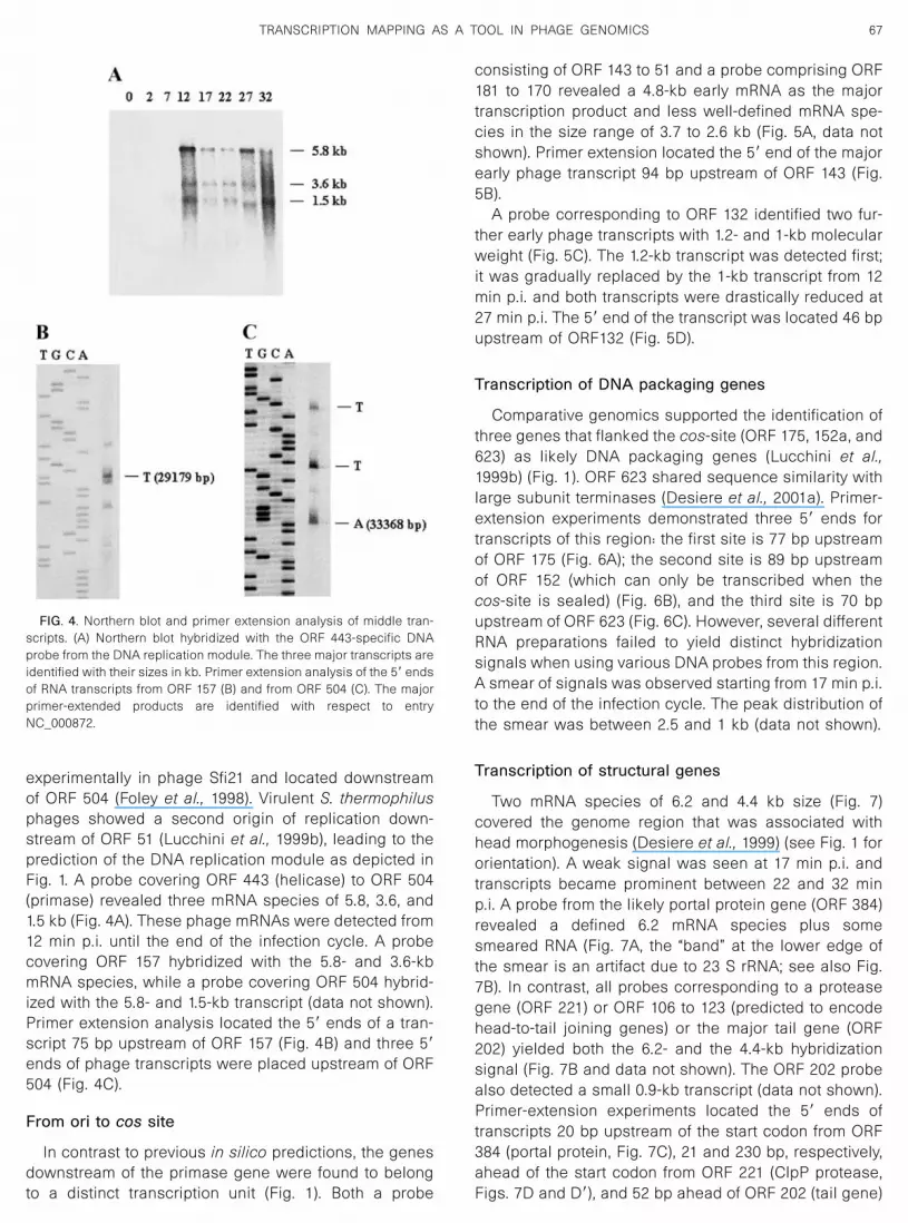
experimentally in phage Sfi21 and located downstreamof ORF 504 (Foley et al., 1998). Virulent S. thermophilusphages showed a second origin of replication down-stream of ORF 51 (Lucchini et al., 1999b), leading to theprediction of the DNA replication module as depicted inFig. 1. A probe covering ORF 443 (helicase) to ORF 504(primase) revealed three mRNA species of 5.8, 3.6, and1.5 kb (Fig. 4A). These phage mRNAs were detected from12 min p.i. until the end of the infection cycle. A probecovering ORF 157 hybridized with the 5.8- and 3.6-kbmRNA species, while a probe covering ORF 504 hybrid-ized with the 5.8- and 1.5-kb transcript (data not shown).Primer extension analysis located the 5� ends of a tran-script 75 bp upstream of ORF 157 (Fig. 4B) and three 5�ends of phage transcripts were placed upstream of ORF504 (Fig. 4C).
From ori to cos site
In contrast to previous in silico predictions, the genesdownstream of the primase gene were found to belongto a distinct transcription unit (Fig. 1). Both a probe
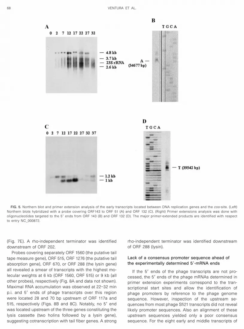
consisting of ORF 143 to 51 and a probe comprising ORF181 to 170 revealed a 4.8-kb early mRNA as the majortranscription product and less well-defined mRNA spe-cies in the size range of 3.7 to 2.6 kb (Fig. 5A, data notshown). Primer extension located the 5� end of the majorearly phage transcript 94 bp upstream of ORF 143 (Fig.5B).
A probe corresponding to ORF 132 identified two fur-ther early phage transcripts with 1.2- and 1-kb molecularweight (Fig. 5C). The 1.2-kb transcript was detected first;it was gradually replaced by the 1-kb transcript from 12min p.i. and both transcripts were drastically reduced at27 min p.i. The 5� end of the transcript was located 46 bpupstream of ORF132 (Fig. 5D).
Transcription of DNA packaging genes
Comparative genomics supported the identification ofthree genes that flanked the cos-site (ORF 175, 152a, and623) as likely DNA packaging genes (Lucchini et al.,1999b) (Fig. 1). ORF 623 shared sequence similarity withlarge subunit terminases (Desiere et al., 2001a). Primer-extension experiments demonstrated three 5� ends fortranscripts of this region: the first site is 77 bp upstreamof ORF 175 (Fig. 6A); the second site is 89 bp upstreamof ORF 152 (which can only be transcribed when thecos-site is sealed) (Fig. 6B), and the third site is 70 bpupstream of ORF 623 (Fig. 6C). However, several differentRNA preparations failed to yield distinct hybridizationsignals when using various DNA probes from this region.A smear of signals was observed starting from 17 min p.i.to the end of the infection cycle. The peak distribution ofthe smear was between 2.5 and 1 kb (data not shown).
Transcription of structural genes
Two mRNA species of 6.2 and 4.4 kb size (Fig. 7)covered the genome region that was associated withhead morphogenesis (Desiere et al., 1999) (see Fig. 1 fororientation). A weak signal was seen at 17 min p.i. andtranscripts became prominent between 22 and 32 minp.i. A probe from the likely portal protein gene (ORF 384)revealed a defined 6.2 mRNA species plus somesmeared RNA (Fig. 7A, the “band” at the lower edge ofthe smear is an artifact due to 23 S rRNA; see also Fig.7B). In contrast, all probes corresponding to a proteasegene (ORF 221) or ORF 106 to 123 (predicted to encodehead-to-tail joining genes) or the major tail gene (ORF202) yielded both the 6.2- and the 4.4-kb hybridizationsignal (Fig. 7B and data not shown). The ORF 202 probealso detected a small 0.9-kb transcript (data not shown).Primer-extension experiments located the 5� ends oftranscripts 20 bp upstream of the start codon from ORF384 (portal protein, Fig. 7C), 21 and 230 bp, respectively,ahead of the start codon from ORF 221 (ClpP protease,Figs. 7D and D�), and 52 bp ahead of ORF 202 (tail gene)
FIG. 4. Northern blot and primer extension analysis of middle tran-scripts. (A) Northern blot hybridized with the ORF 443-specific DNAprobe from the DNA replication module. The three major transcripts areidentified with their sizes in kb. Primer extension analysis of the 5� endsof RNA transcripts from ORF 157 (B) and from ORF 504 (C). The majorprimer-extended products are identified with respect to entryNC_000872.
67TRANSCRIPTION MAPPING AS A TOOL IN PHAGE GENOMICS
(Fig. 7E). A rho-independent terminator was identifieddownstream of ORF 202.
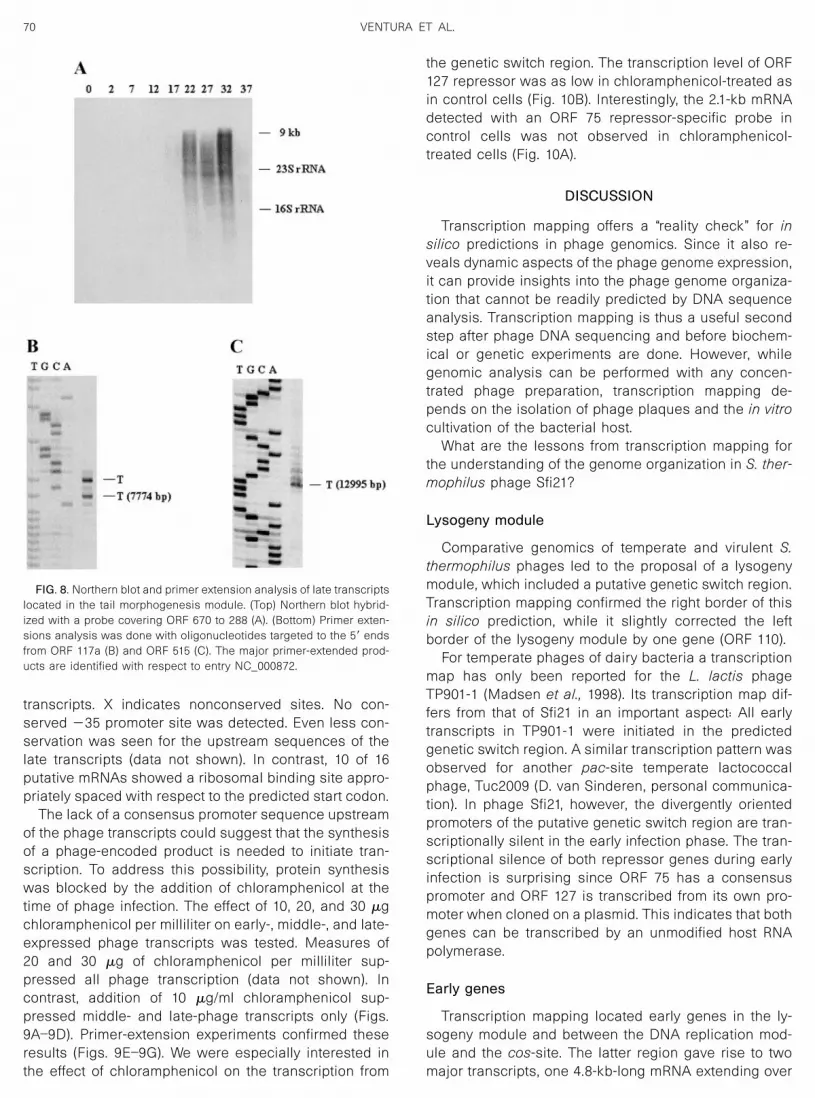
Probes covering separately ORF 1560 (the putative tailtape measure gene), ORF 515, ORF 1276 (the putative tailabsorption gene), ORF 670, or ORF 288 (the lysin gene)all revealed a smear of transcripts with the highest mo-lecular weights at 6 kb (ORF 1560, ORF 515) or 9 kb (allother probes), respectively (Fig. 8A and data not shown).Maximal RNA accumulation was observed at 22–32 minp.i. and 5� ends of phage transcripts over this regionwere located 28 and 70 bp upstream of ORF 117a and515, respectively (Figs. 8B and 8C). Notably, no 5� endwas located upstream of the three genes constituting thelysis cassette (two holins followed by a lysin gene),suggesting cotranscription with tail fiber genes. A strong
rho-independent terminator was identified downstreamof ORF 288 (lysin).
Lack of a consensus promoter sequence ahead ofthe experimentally determined 5�-mRNA ends
If the 5� ends of the phage transcripts are not pro-cessed, the 5� ends of the phage mRNAs determined inprimer extension experiments correspond to the tran-scriptional start sites and allow the identification ofphage promoters by reference to the phage genomesequence. However, inspection of the upstream se-quences from most phage Sfi21 transcripts did not reveallikely promoter sequences. Also an alignment of theseupstream sequences yielded only a poor consensussequence. For the eight early and middle transcripts of
FIG. 5. Northern blot and primer extension analysis of the early transcripts located between DNA replication genes and the cos-site. (Left)Northern blots hybridized with a probe covering ORF143 to ORF 51 (A) and ORF 132 (C). (Right) Primer extensions analysis was done witholigonucleotides targeted to the 5� ends from ORF 143 (B) and ORF 132 (D). The major primer-extended products are identified with respectto entry NC_000872.
68 VENTURA ET AL.

phage Sfi21, the following consensus was derived:AATxxAaAxAxaxxtxxAxaxaxatGTtaTaaaxAAaxt-first basepair of primer extension product. A potential extended
�10 promoter site is underlined. Uppercase indicatesconservation in more than or equal to five of the eighttranscripts, and lowercase indicates conservation in four
FIG. 6. Primer-extension experiments with the mRNAs from the putative DNA packaging module. The oligonucleotides were targeted to the 5� endsfrom ORF 175 (A), ORF 152 (B), and ORF 623 (C). The major primer-extended products are identified with respect to entry NC_000872.
FIG. 7. Northern blot and primer extension analysis of late transcripts located in the head morphogenesis module. (Top) Northern blot analysis usingprobes corresponding to ORF 384 (A) and to a region covering ORF 106 to ORF123 (B). (Bottom) Primer extensions analysis was done witholigonucleotides targeted to the 5� ends from ORF 384 (C), ORF 221 (D, D�), and ORF 202 (E). The major primer-extended products are identified withrespect to entry NC_000872.
69TRANSCRIPTION MAPPING AS A TOOL IN PHAGE GENOMICS
transcripts. X indicates nonconserved sites. No con-served �35 promoter site was detected. Even less con-servation was seen for the upstream sequences of thelate transcripts (data not shown). In contrast, 10 of 16putative mRNAs showed a ribosomal binding site appro-priately spaced with respect to the predicted start codon.
The lack of a consensus promoter sequence upstreamof the phage transcripts could suggest that the synthesisof a phage-encoded product is needed to initiate tran-scription. To address this possibility, protein synthesiswas blocked by the addition of chloramphenicol at thetime of phage infection. The effect of 10, 20, and 30 �gchloramphenicol per milliliter on early-, middle-, and late-expressed phage transcripts was tested. Measures of20 and 30 �g of chloramphenicol per milliliter sup-pressed all phage transcription (data not shown). Incontrast, addition of 10 �g/ml chloramphenicol sup-pressed middle- and late-phage transcripts only (Figs.9A–9D). Primer-extension experiments confirmed theseresults (Figs. 9E–9G). We were especially interested inthe effect of chloramphenicol on the transcription from
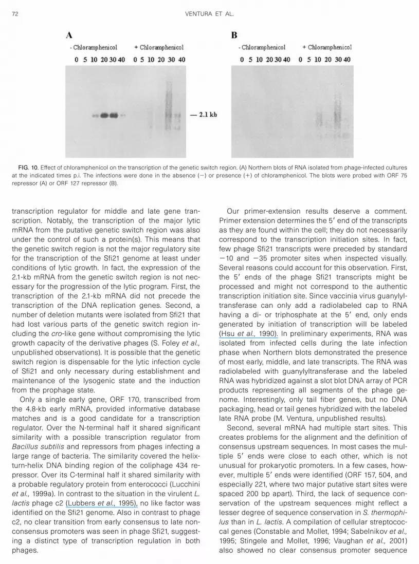
the genetic switch region. The transcription level of ORF127 repressor was as low in chloramphenicol-treated asin control cells (Fig. 10B). Interestingly, the 2.1-kb mRNAdetected with an ORF 75 repressor-specific probe incontrol cells was not observed in chloramphenicol-treated cells (Fig. 10A).
DISCUSSION
Transcription mapping offers a “reality check” for insilico predictions in phage genomics. Since it also re-veals dynamic aspects of the phage genome expression,it can provide insights into the phage genome organiza-tion that cannot be readily predicted by DNA sequenceanalysis. Transcription mapping is thus a useful secondstep after phage DNA sequencing and before biochem-ical or genetic experiments are done. However, whilegenomic analysis can be performed with any concen-trated phage preparation, transcription mapping de-pends on the isolation of phage plaques and the in vitrocultivation of the bacterial host.
What are the lessons from transcription mapping forthe understanding of the genome organization in S. ther-mophilus phage Sfi21?
Lysogeny module
Comparative genomics of temperate and virulent S.thermophilus phages led to the proposal of a lysogenymodule, which included a putative genetic switch region.Transcription mapping confirmed the right border of thisin silico prediction, while it slightly corrected the leftborder of the lysogeny module by one gene (ORF 110).
For temperate phages of dairy bacteria a transcriptionmap has only been reported for the L. lactis phageTP901-1 (Madsen et al., 1998). Its transcription map dif-fers from that of Sfi21 in an important aspect: All earlytranscripts in TP901-1 were initiated in the predictedgenetic switch region. A similar transcription pattern wasobserved for another pac-site temperate lactococcalphage, Tuc2009 (D. van Sinderen, personal communica-tion). In phage Sfi21, however, the divergently orientedpromoters of the putative genetic switch region are tran-scriptionally silent in the early infection phase. The tran-scriptional silence of both repressor genes during earlyinfection is surprising since ORF 75 has a consensuspromoter and ORF 127 is transcribed from its own pro-moter when cloned on a plasmid. This indicates that bothgenes can be transcribed by an unmodified host RNApolymerase.
Early genes
Transcription mapping located early genes in the ly-sogeny module and between the DNA replication mod-ule and the cos-site. The latter region gave rise to twomajor transcripts, one 4.8-kb-long mRNA extending over
FIG. 8. Northern blot and primer extension analysis of late transcriptslocated in the tail morphogenesis module. (Top) Northern blot hybrid-ized with a probe covering ORF 670 to 288 (A). (Bottom) Primer exten-sions analysis was done with oligonucleotides targeted to the 5� endsfrom ORF 117a (B) and ORF 515 (C). The major primer-extended prod-ucts are identified with respect to entry NC_000872.
70 VENTURA ET AL.

12 genes and another mRNA covering a single gene(ORF 132). Comparative genomics has correctly set apartORF 132 from the rest of the genome. The right border ofthe module defined by the 4.8-kb mRNA was also cor-rectly predicted (Lucchini et al., 1999b). However, the lefthalf of the 4.8-kb mRNA was erroneously assigned to theDNA replication module based on the observation of aduplicated origin of replication in the corresponding ge-nome region of virulent S. thermophilus phages. ORF 143to 51 are part of the 4.8-kb early transcript, which makes
the in silico prediction as DNA replication genes unten-able. The left border of the DNA replication module wascorrectly predicted by in silico analysis (Desiere et al.,1997). The newly defined right border of the DNA repli-cation module ends now with an experimentally provenorigin of replication (Foley et al., 1998).
The transcription of middle and late genes, but not ofearly genes, was suppressed when the cells were in-fected in the presence of a protein synthesis inhibitor.This data suggest that the early Sfi21 genes encode a
FIG. 9. Early, but not middle, and late transcripts are made in the presence of 10 �g/ml of the protein synthesis inhibitor chloramphenicol. (Top)Northern blots of mRNA isolated at the indicated times after infection of chloramphenicol-treated cultures were hybridized against the followingprobes. (A) Early genes ORF 181 to 170; (B) early genes ORF 110 to 140b; (C) middle gene 157 (DNA replication module); and (D) late gene 384 (portalprotein gene). (Bottom) Primer extensions analysis was done with oligonucleotides targeted to the 5� ends from early gene ORF 143 (E), middle geneORF 157 (F), and late gene ORF 117a (G). Three primer extension experiments are shown for RNA preparations recovered at 7, 12, and 17 min p.i. (E)and at 12, 17, and 22 min p.i. (F and G) from control (�) and chloramphenicol (�)-treated cultures.
71TRANSCRIPTION MAPPING AS A TOOL IN PHAGE GENOMICS
transcription regulator for middle and late gene tran-scription. Notably, the transcription of the major lyticmRNA from the putative genetic switch region was alsounder the control of such a protein(s). This means thatthe genetic switch region is not the major regulatory sitefor the transcription of the Sfi21 genome at least underconditions of lytic growth. In fact, the expression of the2.1-kb mRNA from the genetic switch region is not nec-essary for the progression of the lytic program. First, thetranscription of the 2.1-kb mRNA did not precede thetranscription of the DNA replication genes. Second, anumber of deletion mutants were isolated from Sfi21 thathad lost various parts of the genetic switch region in-cluding the cro-like gene without compromising the lyticgrowth capacity of the derivative phages (S. Foley et al.,unpublished observations). It is possible that the geneticswitch region is dispensable for the lytic infection cycleof Sfi21 and only necessary during establishment andmaintenance of the lysogenic state and the inductionfrom the prophage state.
Only a single early gene, ORF 170, transcribed fromthe 4.8-kb early mRNA, provided informative databasematches and is a good candidate for a transcriptionregulator. Over the N-terminal half it shared significantsimilarity with a possible transcription regulator fromBacillus subtilis and repressors from phages infecting alarge range of bacteria. The similarity covered the helix-turn-helix DNA binding region of the coliphage 434 re-pressor. Over its C-terminal half it shared similarity witha probable regulatory protein from enterococci (Lucchiniet al., 1999a). In contrast to the situation in the virulent L.lactis phage c2 (Lubbers et al., 1995), no like factor wasidentified on the Sfi21 genome. Also in contrast to phagec2, no clear transition from early consensus to late non-consensus promoters was seen in phage Sfi21, suggest-ing a distinct type of transcription regulation in bothphages.
Our primer-extension results deserve a comment.Primer extension determines the 5� end of the transcriptsas they are found within the cell; they do not necessarilycorrespond to the transcription initiation sites. In fact,few phage Sfi21 transcripts were preceded by standard�10 and �35 promoter sites when inspected visually.Several reasons could account for this observation. First,the 5� ends of the phage Sfi21 transcripts might beprocessed and might not correspond to the authentictranscription initiation site. Since vaccinia virus guanylyl-transferase can only add a radiolabeled cap to RNAhaving a di- or triphosphate at the 5� end, only endsgenerated by initiation of transcription will be labeled(Hsu et al., 1990). In preliminary experiments, RNA wasisolated from infected cells during the late infectionphase when Northern blots demonstrated the presenceof most early, middle, and late transcripts. The RNA wasradiolabeled with guanylyltransferase and the labeledRNA was hybridized against a slot blot DNA array of PCRproducts representing all segments of the phage ge-nome. Interestingly, only tail fiber genes, but no DNApackaging, head or tail genes hybridized with the labeledlate RNA probe (M. Ventura, unpublished results).
Second, several mRNA had multiple start sites. Thiscreates problems for the alignment and the definition ofconsensus upstream sequences. In most cases the mul-tiple 5� ends were close to each other, which is notunusual for prokaryotic promoters. In a few cases, how-ever, multiple 5� ends were identified (ORF 157, 504, andespecially 221, where two major putative start sites werespaced 200 bp apart). Third, the lack of sequence con-servation of the upstream sequences might reflect alesser degree of sequence conservation in S. thermophi-lus than in L. lactis. A compilation of cellular streptococ-cal genes (Constable and Mollet, 1994; Sabelnikov et al.,1995; Stingele and Mollet, 1996; Vaughan et al., 2001)also showed no clear consensus promoter sequence
FIG. 10. Effect of chloramphenicol on the transcription of the genetic switch region. (A) Northern blots of RNA isolated from phage-infected culturesat the indicated times p.i. The infections were done in the absence (�) or presence (�) of chloramphenicol. The blots were probed with ORF 75repressor (A) or ORF 127 repressor (B).
72 VENTURA ET AL.
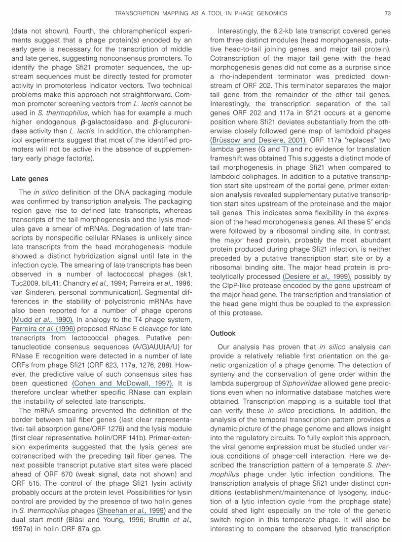
(data not shown). Fourth, the chloramphenicol experi-ments suggest that a phage protein(s) encoded by anearly gene is necessary for the transcription of middleand late genes, suggesting nonconsensus promoters. Toidentify the phage Sfi21 promoter sequences, the up-stream sequences must be directly tested for promoteractivity in promoterless indicator vectors. Two technicalproblems make this approach not straightforward. Com-mon promoter screening vectors from L. lactis cannot beused in S. thermophilus, which has for example a muchhigher endogenous �-galactosidase and �-glucuroni-dase activity than L. lactis. In addition, the chloramphen-icol experiments suggest that most of the identified pro-moters will not be active in the absence of supplemen-tary early phage factor(s).
Late genes
The in silico definition of the DNA packaging modulewas confirmed by transcription analysis. The packagingregion gave rise to defined late transcripts, whereastranscripts of the tail morphogenesis and the lysis mod-ules gave a smear of mRNAs. Degradation of late tran-scripts by nonspecific cellular RNases is unlikely sincelate transcripts from the head morphogenesis moduleshowed a distinct hybridization signal until late in theinfection cycle. The smearing of late transcripts has beenobserved in a number of lactococcal phages (sk1,Tuc2009, bIL41; Chandry et al., 1994; Parreira et al., 1996;van Sinderen, personal communication). Segmental dif-ferences in the stability of polycistronic mRNAs havealso been reported for a number of phage operons(Mudd et al., 1990). In analogy to the T4 phage system,Parreira et al. (1996) proposed RNase E cleavage for latetranscripts from lactococcal phages. Putative pen-tanucleotide consensus sequences (A/G)AUU(A/U) forRNase E recognition were detected in a number of lateORFs from phage Sfi21 (ORF 623, 117a, 1276, 288). How-ever, the predictive value of such consensus sites hasbeen questioned (Cohen and McDowall, 1997). It istherefore unclear whether specific RNase can explainthe instability of selected late transcripts.
The mRNA smearing prevented the definition of theborder between tail fiber genes (last clear representa-tive: tail absorption gene/ORF 1276) and the lysis module(first clear representative: holin/ORF 141b). Primer-exten-sion experiments suggested that the lysis genes arecotranscribed with the preceding tail fiber genes. Thenext possible transcript putative start sites were placedahead of ORF 670 (weak signal, data not shown) andORF 515. The control of the phage Sfi21 lysin activityprobably occurs at the protein level. Possibilities for lysincontrol are provided by the presence of two holin genesin S. thermophilus phages (Sheehan et al., 1999) and thedual start motif (Blasi and Young, 1996; Bruttin et al.,1997a) in holin ORF 87a gp.
Interestingly, the 6.2-kb late transcript covered genesfrom three distinct modules (head morphogenesis, puta-tive head-to-tail joining genes, and major tail protein).Cotranscription of the major tail gene with the headmorphogenesis genes did not come as a surprise sincea rho-independent terminator was predicted down-stream of ORF 202. This terminator separates the majortail gene from the remainder of the other tail genes.Interestingly, the transcription separation of the tailgenes ORF 202 and 117a in Sfi21 occurs at a genomeposition where Sfi21 deviates substantially from the oth-erwise closely followed gene map of lambdoid phages(Brussow and Desiere, 2001). ORF 117a “replaces” twolambda genes (G and T) and no evidence for translationframeshift was obtained This suggests a distinct mode oftail morphogenesis in phage Sfi21 when compared tolambdoid coliphages. In addition to a putative transcrip-tion start site upstream of the portal gene, primer exten-sion analysis revealed supplementary putative transcrip-tion start sites upstream of the proteinase and the majortail genes. This indicates some flexibility in the expres-sion of the head morphogenesis genes. All these 5� endswere followed by a ribosomal binding site. In contrast,the major head protein, probably the most abundantprotein produced during phage Sfi21 infection, is neitherpreceded by a putative transcription start site or by aribosomal binding site. The major head protein is pro-teolytically processed (Desiere et al., 1999), possibly bythe ClpP-like protease encoded by the gene upstream ofthe major head gene. The transcription and translation ofthe head gene might thus be coupled to the expressionof this protease.
Outlook
Our analysis has proven that in silico analysis canprovide a relatively reliable first orientation on the ge-netic organization of a phage genome. The detection ofsynteny and the conservation of gene order within thelambda supergroup of Siphoviridae allowed gene predic-tions even when no informative database matches wereobtained. Transcription mapping is a suitable tool thatcan verify these in silico predictions. In addition, theanalysis of the temporal transcription pattern provides adynamic picture of the phage genome and allows insightinto the regulatory circuits. To fully exploit this approach,the viral genome expression must be studied under var-ious conditions of phage–cell interaction. Here we de-scribed the transcription pattern of a temperate S. ther-mophilus phage under lytic infection conditions. Thetranscription analysis of phage Sfi21 under distinct con-ditions (establishment/maintenance of lysogeny, induc-tion of a lytic infection cycle from the prophage state)could shed light especially on the role of the geneticswitch region in this temperate phage. It will also beinteresting to compare the observed lytic transcription
73TRANSCRIPTION MAPPING AS A TOOL IN PHAGE GENOMICS

pattern to that obtained with the closely related virulentS. thermophilus phages and deletion/replacement deriv-atives of phage Sfi21.
MATERIALS AND METHODS
Bacterial strains, bacteriophages, and media
The E. coli strain XL1-Blue was cultivated aerobicallyin LB broth or on LB broth solidified with 1.5% (w/v) agarat 37°C. Streptococcus thermophilus strain SfiI andtransformants thereof were routinely subcultured at 42°Cin either LM17 (M17 supplemented with 0.5% lactose) orBelliker (DIFCO, Detroit, MI) media. Phage Sfi21 waspropagated on S. thermophilus strain SfiI in LM17 broth.Phage enumeration was achieved by plaque assay asdescribed by Foley et al. (1998).
Construction of plasmids
The cloning vector used in this study was the highcopy number E. coli/lactococcal/streptococcal shuttlevector pNZ124 (Platteeuw et al., 1994). A 2534-bp XbaIfragment from the lysogeny module of the S. thermophi-lus phage Sfi21 was cloned in the XbaI site of pUC19generating the construct pX6. The insert was then trans-ferred as a BamHI–HindIII fragment from pX6 and clonedin the respective sites of pNZ124 generating the con-struct pSFswi. The putative cI-like repressor gene, ORF127, was cloned independently of ORF 75, the putativecro repressor gene, by inserting the 664-bp BsrBI/BamHIfragment of pX6 in the EclI36II/BamHI sites of pNZ124,generating the construct pSFcI. pSFcro consists of a517-bp BamHI/XbaI PCR fragment (containing the puta-tive genetic switch region, ORF 75, and the 5� ends ofORF 127 and 287) cloned in the respective sites ofpNZ124. The PCR fragment was generated using phageSfi21 DNA as the template and primer pair 1F/R (5�-GGCTGCAGGATCCG ACA TGG AAC TGT TGT C-3� and5�-GC TCTAGA AAG TTC ATT CAT CGT CA-3�, containingBamHI and XbaI restriction sites, respectively).
DNA techniques
DNA samples were amplified in a Perkin–Elmer ther-mal cycler programmed for 30 cycles each consisting of94°C for 30 s, 55°C for 30 s, and 72°C for 1 min.Synthetic primers were designed according to the estab-lished phage Sfi 21 DNA sequence (Accession No.NC_000872) and used together with the relevant DNAtemplate and Taq polymerase Fementas. PCR productswere purified using the Qiagen PCR-purification kit fol-lowing the instruction of the supplier.
Total RNA isolation
Batch cultures (350 ml) were prepared by inoculatingBelliker broth with 3.5 ml of an overnight culture of S.thermophilus SfiI followed by incubation at 42°C until an
OD600nm of 0.2 was reached. CaCl2 was added to a finalconcentration of 10 mM before phage Sfi21 was added ata multiplicity of infection of 8.
Samples (20 ml) were taken at 0, 2, 7, 17, 22, 32, and 37min after the start of infection. Immediately after collec-tion, the cell samples were frozen in a ethanol–dry icebath.
Total RNA was isolated by resuspending the frozenbacterial cell pellets in 1 ml of TRIzol Reagent (Gibco-BRL, Gaithersburg, MD), adding 106 �m glass beads(Sigma Chemical Co., St. Louis, MO), and shearing theslurry with a Mini-Beadbeater-8 cell disruptor (BiospecProducts, Bartlesville, U.K.) as described by Walker et al.(1999). Standard procedure to minimize RNase contam-ination was used (Sambrook and Russel, 2001).
Northern blot analysis of phage transcripts were car-ried out on 15 �g aliquots of total RNA separated on a1.5% agarose-formaldehyde denaturing gel, transferredto a zeta-Probe blotting membrane (Bio-Rad Laborato-ries, Richmond, CA) by the method of Sambrook andRussel (2001) and fixed by UV cross-linking using aStratalinker 1800 (Stratagene).
Northern blots and hybridizations
Prehybridization and hybridization of the Northernblots were carried out at 65°C in 0.5 M NAHPO4 (pH7.2)–1.0 mM EDTA (pH 7.0)–7% sodium dodecyl sulfate(SDS). Following 18 h of hybridization, the membranewas rinsed twice (30 min) at 65°C in 0.1 M NaHPO4 (pH7.2)–1.0 mM EDTA–1% SDS, twice (30 min) at 65°C in 0.1M NaHPO4 (pH 7.2)–1.0 mM EDTA–0.1% SDS, and ex-posed to X-OMAT autoradiography film (Eastman KodakCo., Rochester, NY).
The probes for the Northern blot hybridization werelabeled with [�32P] by using the random-primed DNAlabeling system (Boehringer Mannheim GmbH) and pu-rified with Nuc Trap probe purification columns (Strat-agene). Their location on the phage genome are pro-vided in Fig. 1.
Primer extension analysis
The 5� ends of RNA transcripts were determined inprimer extension reactions conducted with 15 �g of totalRNA mixed with 1 pmol of primer (IRD800 labeled) and 2�l of Buffer H (2 M NaCl, 50 mM PIPES, pH 6.4). Themixture was denatured at 90°C for 5 min and then hy-bridized for 1 h at 42°C. After addition of 5 �l 1 MTris–HCl (pH 8.2), 10 �l 0.1 M DTT, 5 �l 0.12 M MgCl2, 20�l 2.5 mM dNTP mix, 0.4 �l (5 U) reverse transcriptase(Sigma), and 49.6 �l double-distilled water, the enzymaticreaction was incubated for 2 h at 42°C. The reaction wasstopped by adding 250 �l ethanol/acetone mix (1:1) andincubated at �70°C for 15 min followed by a centrifuga-tion at 10,000 rpm for 15 min. The pellets were dissolvedin 4 �l distilled water and mixed with 2.4 �l loading
74 VENTURA ET AL.
buffer from the sequencing kit (Thermo-sequenase fluo-rescence labeled, Amersham). The cDNA was separatedthrough 8% polyacrylamide–urea gels along with se-quencing reactions which were conducted using thesame primers employed for primer extensions and de-tected using the LiCor sequencer machine (MWG Bio-tech).
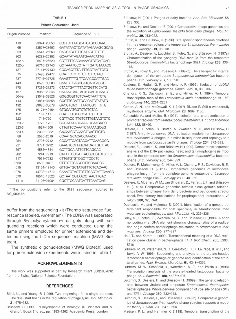
The synthetic oligonucleotides (MWG Biotech) usedfor primer extension experiments were listed in Table 1.
ACKNOWLEDGMENTS
This work was supported in part by Research Grant 5002-057832from the Swiss National Science Foundation.
REFERENCES
Blasi, U., and Young, R. (1996). Two beginnings for a single purpose:The dual-start holins in the regulation of phage lysis. Mol. Microbiol.21, 675–682.
Brussow, H. (1999). “Encyclopedia of Virology” (R. Webster and A.Granoff, Eds.), 2nd ed., pp. 1253–1262. Academic Press, London.
Brussow, H. (2001). Phages of dairy bacteria. Ann. Rev. Microbiol. 55,283–303.
Brussow, H., and Desiere, F. (2001). Comparative phage genomics andthe evolution of Siphoviridae: Insights from dairy phages. Mol. Mi-crobiol. 39, 213–222.
Bruttin, A., and Brussow, H. (1996). Site-specific spontaneous deletionsin three genome regions of a temperate Streptococcus thermophilusphage. Virology 219, 96–104.
Bruttin, A., Desiere, F., Lucchini, S., Foley, S., and Brussow, H. (1997a).Characterization of the lysogeny DNA module from the temperateStreptococcus thermophilus bacteriophage Sfi21. Virology 233, 136–148.
Bruttin, A., Foley, S., and Brussow, H. (1997b). The site-specific integra-tion system of the temperate Streptococcus thermophilus bacterio-phage Sfi21. Virology 237, 136–148.
Casjens, S., Hatfull, G. F., and Hendrix, R. (1992). Evolution of dsDNAtailed-bacteriophage genomes. Semin. Virol. 3, 383–397.
Chandry, P. S., Davidson, B. E., and Hillier, A. J. (1994). Temporaltranscription map of the Lactococcus lactis bacteriophage sk1. Mi-crobiology 140, 2251–2261.
Cohen, A. N., and McDowall, K. J. (1997). RNase E: Still a wonderfullymysterious enzyme. Mol. Microbiol. 23, 1099–1106.
Constable A., and Mollet, B. (1994). Isolation and characterisation ofpromoter regions from Streptococcus thermophilus. FEMS Microbiol.Lett. 122, 85–90.
Desiere, F., Lucchini, S., Bruttin, A., Zwahlen, M. C., and Brussow, H.(1997). A highly conserved DNA replication module from Streptococ-cus thermophilus phages is similar in sequence and topology to amodule from Lactococcus lactis phages. Virology 234, 372–382.
Desiere, F., Lucchini, S., and Brussow, H. (1999). Comparative sequenceanalysis of the DNA packaging, head, and tail morphogenesis mod-ules in the temperate cos-site Streptococcus thermophilus bacterio-phage Sfi21. Virology 260, 244–253.
Desiere, F., Mahanivong, C., Hiller, A. J., Chandry, P. S., Davidson, B. E.,and Brussow, H. (2001a). Comparative genomics of lactococcalphages: Insight from the complete genome sequence of Lactococ-cus lactis phage BK5-T. Virology 283, 240–252.
Desiere, F., McShan, W. M., van Sinderen, D., Ferretti, J. J., and Brussow,H. (2001b). Comparative genomics reveals close genetic relation-ships between phages from dairy bacteria and pathogenic strepto-cocci: Evolutionary implications for prophage-host interactions. Vi-rology 288, 325–341.
Duplessis, M., and Moineau, S. (2001). Identification of a genetic de-terminant responsible for host specificity in Streptococcus ther-mophilus bacteriophages. Mol. Microbiol. 41, 325–336.
Foley, S., Lucchini, S., Zwahlen, M. C., and Brussow, H. (1998). A shortnoncoding viral DNA element showing characteristics of a replica-tion origin confers bacteriophage resistance to Streptococcus ther-mophilus. Virology 250, 377–387.
Hsu, T., and Karam, J. (1990). Transcriptional mapping of a DNA repli-cation gene cluster in bacteriophage T4. J. Biol. Chem. 265, 5303–5316.
Lubbers, M. W., Waterfield, N. R., Beresfold, T. P. J., Le Page, R. W. F., andJarvis A. W. (1995). Sequencing and analysis of the prolate-headedlactococcal bacteriophage c2 genome and identification of the struc-tural genes. Appl. Environ. Microbiol. 61, 4348–4356.
Lubbers, M. W., Schofield, K., Waterfield, N. R., and Polzin K. (1998).Transcription analysis of the prolate-headed lactococcal bacterio-phage c2. J. Bacteriol. 180, 4487–4496.
Lucchini, S., Desiere, F., and Brussow, H. (1999a). The genetic relation-ship between virulent and temperate Streptococcus thermophilusbacteriophages: Whole genome comparison of cos-site phages SfiI9and Sfi21. Virology 260, 232–243.
Lucchini, S., Desiere, F., and Brussow, H. (1999b). Comparative genom-ics of Streptococcus thermophilus phage species supports a modu-lar theory. J. Virol. 73, 8647–8656.
Madsen, P. L., and Hammer K. (1998). Temporal transcription of the
TABLE 1
Primer Sequences Used
Oligonucleotide Positiona Sequence 5�3 3�
110 23074–23053 CCTTCTTTAGCATCAGCCCAAG65 23577–23602 GATATAACTCATATAGAAAGCGCAG
359 25547–25568 CAGCAGCCTTGATAGCTTCTG203 26282–26303 GCAATATAGAATGAAACATTG122-a 26497–26520 CGTTTTTCACAAAAGTCTCATCAC122-b 26716–27740 GGTAAATCCCTA TTGATGTAAACG127 27111–27130 CCCAGCTTTA TTTGCTAATTCTG
75 27498–27477 CCATTCTCTCTCTTGTTGTAC287 27749–27726 GAAGTTTTG TTCAACCCATTGAC443 30529–30508 CAATGTGAGCCATCACCACGG170 37296–37273 CTACTGATTTTACTGGTTCCATG157 29368–29346 CATAATCACTAGTCCAGTCAATC504-a 33548–33526 GGGGCGTTCTCAACTAATTCTG143 34881–34858 GCGTTGCATTGCACATCTATATG132 39685–36678 GACGTCACTTTAAGCGCTTGTG175 40171–40154 GTGCAATGGTCTCTCTAC152 167–147 CGATTTTGCGCCATGTTTCTC623 744–720 CGTTACC TTGTCTTTGTAAGTCTC623-2 1772–1751 GGGATATACGAAA CCAAGAGC623-3 1527–1505 CTTGTTCCAAATCTAAAAGCGG623-4 2003–1982 GACAACGTCAAGTGAGTTGTC
59 2536–2519 CCAATGCACACCAAACC384 2735–2712 CCCATTCACTACCATTCAAGGCG221 3781–3792 GAAGTCCTTATCATCATTGCTTAC397 4563–4544 GCTTGCA ATTCTTCAGCAC202 7244–7219 CATTTTGCGATTACGCCGTCTGTTG117 7951–7932 CTTGTGTGTCGCTTCCCTC
1560 8502–8481 CTTTCTTGAGCCTTCCAAGCG515 13164–13140 GTTGATGCTATGTTTCTCACAAC
1276 14738–14712 CAAATGTACTTGTTGAGTATTCAAGG670 18545–18523 GCTAATCGTAACCTAACTTGAC141b 21106–21084 CGCTTCCAATCATTTCAATCAG
a The bp positions refer to the Sfi21 sequence reported inNC_000872.
75TRANSCRIPTION MAPPING AS A TOOL IN PHAGE GENOMICS
lactococcal temperate phage TP901-1 and DNA sequence of theearly promoter region. Microbiology 144, 2203–2215.
Mudd, E. A., Carpousis, A. J., and Krisch, H. M. (1990). Escherichia coliRNase E has a role in the decay of bacteriophage T4 mRNA. GenesDev. 4, 873–881.
Parreira, R., Valyasevi, R., Lerayer, A. L. S., Ehrilch, S. D., and ChopinM. C. (1996). Gene organization and transcription of a late expressedregion of a Lactococcus lactis phage. J. Bacteriol. 178, 6158–6165.
Pedersen, M., Ostergaard, S., Bresciani, J., and Vogensen, F. K. (2000).Mutational analysis of two structural genes of temperate bacterio-phage TP901-1 involved in tail length determination assembly. Virol-ogy 276, 315–328.
Plateeuw, C., Simons, G., and de Vos, W. M. (1994). Use of the Esche-richia coli �-glucurouidase (gus A) gene as a reporter gene foranalyting promoters in Pactic acid bacteria. Appl. Environ. Microbiol.60, 587–593.
Sabelnikov, A. G., Greenberg, B., and Lacks, S. A. (1995). An extended�10 promoter alone directs transcription of the DpnII operon ofStreptococcus pneumoniae. J. Mol. Biol. 250, 144–155.
Sambrook, J., and Russel, D. W. (2001). “Molecular Cloning: A Labora-
tory Manual,” 3rd ed., Cold Spring Harbor Laboratory, Cold SpringHarbor, NY.
Sheehan, M. M., Stanley, E., Fitzgerald, G. F., and van Sinderen, D.(1999). Identification and characterization of a lysis module presentin a large proportion of bacteriophage infecting Streptococcus ther-mophilus. Appl. Environ. Microbiol. 65, 569–577.
Stanley, E., Fitzgerald, G. F., Le Marrec, C., Fayard, B., and van Sinderen,D. (1997). Sequence analysis and characterization of phi O1205, atemperate bacteriophage infecting Streptococcus thermophilusCNRZ1205. Microbiology 143, 3417–3429.
Stingele, F., and Mollet, B. (1996). Disruption of the gene encodingpenicillin-binding protein 2b (pbp2b) causes altered cell morphologyand cease in exopolisaccharide production in Streptococcus ther-mophilus Sfi6. Mol. Microbiol. 22, 357–366.
Vaughan, E. E., Bogaard, P. T. C., Catzeddu, P., Kuipers, O., and Vos W.(2001). Activation of silent gal genes in the lac-gal regulon of Strep-tococcus thermophilus. J. Bacteriol. 183, 1184–1194.
Walker, D. C., Girgis, H., and Klaenhammer, T. R. (1999). The groESLchaperone operon of Lactobacillus johnsonii. Appl. Environ. Micro-biol. 65, 3033–3041.
76 VENTURA ET AL.
Related Documents


![Cronicon · foods such as yogurt contains live beneficial bacteria of Lactobacillus bulgaricus and Streptococcus thermophilus [18]. In dietary supple-ments probiotics are available](https://static.cupdf.com/doc/110x72/5fa69a779751cf5d8e373fc1/cronicon-foods-such-as-yogurt-contains-live-beneficial-bacteria-of-lactobacillus.jpg)