1 ARTIK EL PENEL ITIAN Toxoplasma gondii Identification in Mother’s Blood and Fetal Tissue with Nested PCR Aloysius Suryawan 1 , Juliana A. Masengi-Rumopa 2 , Josef S. B. Tuda 3 , Theresia M. Rahardjo 4 , Mihoko Imada 5 1 Department of Obstetrics and Gynecology,Maranatha Christian University / Immanuel Hospital Bandung, 2 Department of Obstetrics and Gynecology, General Hospital Manado, 3 Department of Parasitology, 4 Department of Biology, Sam Ratulangi University, Manado, Indonesia and, 5 JICA (Japanese I nternational Cooperation Agency) Manado, Indonesia. Abstrak Toxoplasma gondii identification in mother’s blood and fetal tissue with nested PCR Objective: To examine the correlation between Toxoplasma gondii infection with spontaneous abortion on pregnant women based on nested PCR result from mother’s blood and fetal tissue. Methods: A prospective c linical diagnostic study using nested PCR performed on 30 cases of pregnant women with spontaneous abortion fulfilling the inclusion criteria, latex agglutination test (+) and exclusion criteria, latex agglutination test (-). Mother’s blood and fetal tissues samples, which gave positive result in serologic test, were analyzed with nested PCR using18S-rDNA gene primers. Results: Five of 30 mother’s bl ood samples (16.7%) and 9 of 30 fetal tissue sam ples (30%) gave positive PCR results. According to Fisher’s Exact test, PCR detected the presence of Toxoplasma gondii in significant value (P < .001). Conclusion: There is a strong correlation between Toxoplasma gondii infection with spontaneous abortion (P < 001). Keywords: nested PCR – Toxoplasma gondii infection – spontaneous abortion

Welcome message from author
This document is posted to help you gain knowledge. Please leave a comment to let me know what you think about it! Share it to your friends and learn new things together.
Transcript

7/21/2019 toxoplasmosis
http://slidepdf.com/reader/full/toxoplasmosis-56de754d01f71 1/16
1
ARTIKEL PENELITIAN
Toxoplasma gondii Identification in Mother’s Bloodand Fetal Tissue with Nested PCR
Aloysius Suryawan1 , Juliana A. Masengi-Rumopa 2 , Josef S. B. Tuda3 ,Theresia M. Rahardjo 4 , Mihoko Imada5
1Department of Obstetrics and Gynecology,Maranatha Christian University / ImmanuelHospital Bandung,2Department of Obstetrics and Gynecology, General Hospital Manado,
3Department of Parasitology, 4Department of Biology, Sam Ratulangi University, Manado,
Indonesia and, 5 JICA (Japanese International Cooperation Agency) Manado, Indonesia.
Abstrak Toxoplasma gondii identification in mother’s blood and fetal tissue with nested PCR
Objective: To examine the correlation between Toxoplasma gondii infection with spontaneousabortion on pregnant women based on nested PCR result from mother’s blood and fetal tissue.
Methods: A prospective clinical diagnostic study using nested PCR performed on 30cases of pregnant women with spontaneous abortion fulfilling the inclusion criteria, latexagglutination test (+) and exclusion criteria, latex agglutination test (-). Mother’s blood and
fetal tissues samples, which gave positive result in serologic test, were analyzed with nestedPCR using18S-rDNA gene primers.
Results: Five of 30 mother’s blood samples (16.7%) and 9 of 30 fetal tissue samples(30%) gave positive PCR results. According to Fisher’s Exact test, PCR detected the presenceof Toxoplasma gondii in significant value (P < .001). Conclusion: There is a strong correlationbetween Toxoplasma gondii infection with spontaneous abortion (P < 001).
Keywords: nested PCR – Toxoplasma gondii infection – spontaneous abortion

7/21/2019 toxoplasmosis
http://slidepdf.com/reader/full/toxoplasmosis-56de754d01f71 2/16
JKM.
Vol. 5, No. 2, Februari 2006
2
Toxoplasma gondii is an obli-gate intracellular protozoan pa-rasite that can infect an extremelywide host range, from birds tomammals, including humans. This
parasite can survive in all nucleat-ed cells, including blood cells inacute stage, forms a specificvacuole that protect the parasitefrom host cell immune system. Inthe chronic stage, the parasite canform a cyst in the central nervoussystem, skeletal muscle and eyetissue and can exist for the lifetimeof its host. The cysts can ruptureand release highly invasivetrophozoite, which may cause arecurrent infection and potentiallyfatal if the host is in a state ofimmune deficiency.1,2,3
Toxoplasma gondii infection isoften asymptomatic in healthy in-dividuals, but a primary Toxoplas-ma gondii infection in pregnantwomen may cause a range ofabnormalities including abortion,fetal death and congenital defect,depending on gestation age when
infection occurs. The fetus isinfected by Toxoplasma gondiithrough placental circulation. Thecyst can form in placental tissueand fetal brain. When the fetus isinfected at first trimester, abortioncan occur. In Norway between 1992to 1994, 10.9% women wereinfected before pregnancy and0.17% were infected during preg-nancy. In Indonesia, toxoplasmosis
prevalence is 14% and it is still highin pregnant women, about 5.5-84%.The high score of toxo-plasmosis inpregnant women, especially
asymptomatic or silent infection,will limit and cause difficulty inthe diagnosis process so theabnormality or mortality of thefetus will be increased.4,5,6,7
Current diagnosis of Toxo-plasma gondii is based on parasiteisolation and serological assay.Toxoplasma gondii can be isolated bymice inoculation or tissue cul-turebut this technique need longer time,about 3-6 weeks. Serological testcan overcome this problem anddetect Toxoplasma gondii anti-body.Positive serologic result wasdetermined by showing a serocon-version of immunoglobulin G anti-bodies for primary infection anddetection of specific immunoglo-bulin M. This method is also time-consuming and complicated by thepresence of crossreactive antibo-dies, and influenced by immuno-logy condition, especially inimmunosuppression or immuno-deficiency patients.8,9,10,11,12,13
Based on this fact, PCRbecomes a very important diag-
nostic tool because it has a veryhigh sensitivity and specificity indetecting Toxoplasma gondii infec-tion, both in acute and chronicstage. One of the PCR methodswhich has very high sensitivity andaccuracy is a nested PCR usingprimer which is very conservedand species-specific14, like 18S-rDNA primers used in this study.The PCR is performed both on
mother’s blood and aborted tissueand the result can be performed ina few hours.15-22

7/21/2019 toxoplasmosis
http://slidepdf.com/reader/full/toxoplasmosis-56de754d01f71 3/16
Toxoplasma gondii Identification in Mother’s Bloodand Fetal Tissue with Nested PCR
Aloysius Suryawan, Juliana A. Masengi-Rumopa, Josef S. B. Tuda, Theresia M. Rahardjo, Mihoko Imada
3
Materials And MethodsPatients’ characteristic and
selection. (i) Mothers. All pregnant
women with spon-taneous abortionwithin 20 weeks of gestation fromOctober 2002 to March 2003 wereidentified and characterized by age,gestational age, spontaneousabortion history and parity. Theywere offered serological test(agglutination test) and suggestedto perform PCR examination bothon mother’s blood and fetal tissue ifserological test gives positive result.After the informed consent wasgranted, 5 cc venous blood wasdrawn and centrifuged to separateits serum from blood, then theserum was serologically tested.Thirty preg-nant women withabortion and whose gave positiveresult in serologically test wereincluded in this study. The remainsof blood samples were preserved at-20oC for subsequent DNA isolationstep. (ii) Fetal tissue. Five cc fetal
tissues from all serologic positivemothers were taken and preservedat -20oC for subsequent DNAisolation step.22,23,24
Selection of Primer. Primerused for this nested PCR willamplify a DNA sequence from 18S-rDNA gene. The primer was usedin Keio University, Japan and pro-ven to give positive result in HIV-positive individuals with
encephalitis toxoplasmosis. Thefirst primer pair produces a 311-bpDNA segment from base 48 to 359and the second primer pair
produces a 290-bp DNA segmentfrom base 58 to 348. The firstprimer pair used are 5'-CCATGCATGTCTAAGTATAA GCand 5'-GTTACCCGTCACTG
CCAC. The second primer used are5'-CTAAGTATAAGCTTTTATACGGC and 5'-TGCCACGGTAGTCCAATAC.
Preparation of DNA tem-plate for PCR. DNA templates we-re prepared from mother’s blood
and fetal tissue. Fifty μl of eachsample was putt into 2.5 ml a
Eppendorf tube and 100 μl TritonX-100 was added into the tube.
Then the tube was boiled for 5minutes and stored at -20oC untilused. This technique was used inKeio University Japan and hasproven to be successful in PCRprocess.
Amplification protocol. Thereagents used for PCRamplification were given from KeioUniversity Japan. PCR mix-tures
were prepared for 50 μl react-ionvolume as following: distilled
water 33.25 μl, 10xPCR buffer 5 μl,2mM dNTPs 5μl, 25 mM MgCl2 4μl,
each primer 0.5μl, Taq poly-merase
0.5μl and DNA template 1.5 μl.DNA thermal cycler (Perkin-Elmer)was programmed for 40 cycles ofamplification, both for first andsecond round PCR. Parameters forthe first round PCR cycle consistedof 5 min at 95oC (initialdenaturation), 30 sec at 94oC
(denaturation), 1 min at 64oC(primer annealing), 2 min at 72oC(polymerization) and 5 min at 72oC

7/21/2019 toxoplasmosis
http://slidepdf.com/reader/full/toxoplasmosis-56de754d01f71 4/16
JKM.
Vol. 5, No. 2, Februari 2006
4
(extended polymerization). Para-meters for the second round PCRconsisted of 5 min at 95oC (initialdenaturation), 30 sec at 94oC (de-naturation), 1 min at 60oC (primer
annealing), 2 min at 72oC (poly-merization) and 5 min at 72oC(extended polymerization). Analiquot of the reaction mixture waselectrophoresis analyzed by 2%
agarose gel stained by 2 μlethidium bromide and visualizedunder UV light. A UV Camera wasused to take its photograph usinga 559 Polaroid film.
ResultThirty pregnant women
with spontaneous abortion within20 weeks of gestation, charac-terized by age, gestational age andspontaneous abortion history, whogave positive result in serologicaltest were submitted to nested PCR.Nested PCR showed 5 positiveresults from mother’s blood and 9positive results from fetal tissue.
(Figure 1, 2 and 3)Based on PCR result, wo-
men at age 20-29 had the largestpositive cases with 2 cases (6.67%)from mother’s blood and 3 cases(10%) from fetal tissue, including apositive case both on mother’sblood and its fetal tissue. Women atage below 20 showed 4 positivecases, each 2 cases from mother’sblood and their fetal tissue (6.67%).
In women aged 30-39, there werealso 4 positive cases, 1 case (3.33%)from mother’s blood and 3 cases(10%) from fetal tissue. There was
only 1 positive case from fetaltissue on women aged over 40.(Table 2)
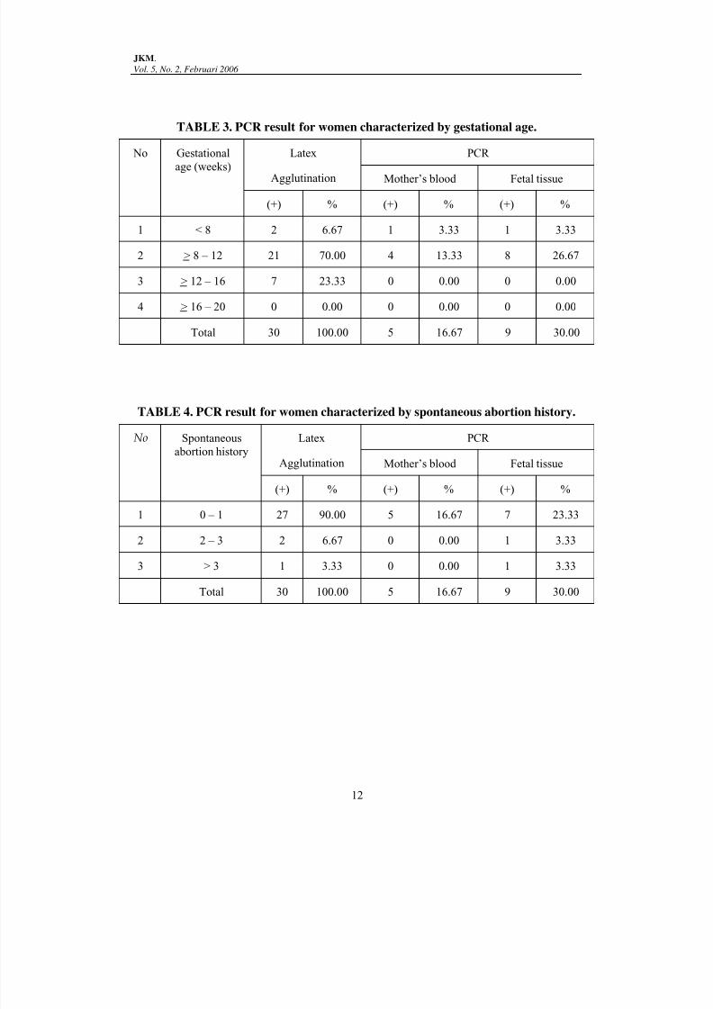
On gestational age, thelargest positive cases were found at
> 8-12 weeks pregnancy, with 4cases (13.33%) from mother’s bloodand 8 cases (26.67%) from fetaltissue. Four positive cases frommother’s blood consisted of each 2cases at 8-10 and 10-12 weekspregnancy, all of them showedpositives result in their fetal tissuesamples. Eight positive cases fromfetal tissue consisted of each 4cases at 10-12 and 8-10 weekspregnancy. At gestational agebelow 8 weeks, we found 1 positiveresult (3.33%), both on mother’sblood and fetal tissue, but there areno positive results at > 12-16 and >16-20 weeks pregnancy. (Table 3)
According to spontaneousabortion history, women with firstspontaneous abortion history gavethe largest positive results with 5positive cases (16.67%) from mo-ther’s blood and 7 positive cases
(23.33%) from fetal tissue, 5 positiveresults occurred both on mother’sblood and their fetal tissues. Eachfollowed by 1 posi-tive case fromfetal tissue for women with 2-3times and more of 3 timesspontaneous abortion history.(Table 4)
All data were analyzed withFischer’s Exact Test and showedsignificant relationship (P < .001,
95% CI) between fetal tissue andmother’s blood examination bynested PCR.

7/21/2019 toxoplasmosis
http://slidepdf.com/reader/full/toxoplasmosis-56de754d01f71 5/16
Toxoplasma gondii Identification in Mother’s Bloodand Fetal Tissue with Nested PCR
Aloysius Suryawan, Juliana A. Masengi-Rumopa, Josef S. B. Tuda, Theresia M. Rahardjo, Mihoko Imada
5
Discussion
This study has some advan-tages. First, it describes the charac-terization of women with spon-
taneous abortion, which wereinfected by Toxoplasma gondii andthe prevalence of toxoplasmosis atMalalayang General Hospital(RSUP) Manado, North Sulawesi,Indonesia. Second, it shows thesignificant advantage of nestedPCR to detect Toxoplasma gondii inmother’s blood and fetal tissue andestimate the stage of Toxoplasma gondii, whether it is in acute orchronic form. Third, the study alsoshows the correlation between fetaltissue and mother’s blood PCRresult. Beside some ad-vantagesabove, there is also a possibilitythat patients with positiveserological result had no fullprotection because they still had achance to get toxoplasmosis.
Based on the result, we candescribe the pattern of Toxoplasma gondii infection in women with
spontaneous abortion at Manado,who had a serological positiveresult, according to age, gestationalage, spontaneous abortion historyand parity.
The result showed that from30 pregnant women with spon-taneous abortion, women at age 20-29 had the higher risk for toxo-plasmosis with 2 positive result(6.67%) from mother’s blood
(samples 5 and 21) and 3 positiveresult (10%) from fetal tissue(samples 5, 20 and 21). The lowerincidence of toxoplasmosis was
obtained in women at age below 20with 4 positive results, each 2positive results (6.67%) from mo-ther’s blood and fetal tissue(samples 26 and 30). In women
aged 30-39, there were also 4positive results, 1 positive result(3.33%) from mother’s blood (sam-ple 24) and 3 positive results (10%)from fetal tissue (sample 11, 15 and24). In older women over 40, only 1positive result (3.33%) was foundfrom fetal tissue (sample 12).According to this, we can see thatthe highest Toxoplasma gondii infection occurs especially in activereproductive women, aged bet-ween 16 to 39 and the incidence ofinfection decreases with ageincrease. It gives a perception thatthere is a lower toxoplasmosisincidence in older patients (in thisstudy older than 40) because olderpatients have a bigger chance to geta contact with Toxoplasma gondii which gives a higher anti-body(IgG) protection to recurrentinfection. This assumption still
need further study with largersubject sample with ELISA assay asa quantitative screening tool. ELISAcan give more accurate result thanagglutination test be-cause it candescribe IgG antibody level, whichgives protection to recurrentinfection.25,26
According to gestationalage, the largest positive result wasfound in gestational age > 8-12
weeks, with 4 positive cases(13.33%) from mother’s blood and 8positive cases (26.67%) from fetaltissue. Four positive cases from
7/21/2019 toxoplasmosis
http://slidepdf.com/reader/full/toxoplasmosis-56de754d01f71 6/16
JKM.
Vol. 5, No. 2, Februari 2006
6
mother’s blood consisted of sam-ple 21 and 30 (8-10 weeks) and 24and 26 (10-12 weeks), while 8positive cases from fetal tissueconsisted of sample 11, 12, 24 and
26 (10-12 weeks) and sample 15, 20,21 and 30 (8-10 weeks). We onlyfound 1 positive case (3.33%) ingestational age < 8 weeks (sample5), both on mother’s blood and fetaltissue and no positive result atgestational age over 12 weeks. Thisresult shows that the highestspontaneous abortion occurred at 8-12 weeks gestational age, whichbecomes the most criti-cal period. Itsupport the suggest-ion made insome previous studies, which usedserological assay, thattoxoplasmosis has a significantcorrelation with spon-taneousabortion in the first trimes-ter ofpregnancy and the rate ofspontaneous abortion will increaseif toxoplasmosis occurs at earlygestational age.27,28,29 The correla-tion is so strong because the over-all positive result for mother’s
blood were 5 cases (16.66%) and 9cases for fetal tissue (30%). Furtherstudy with larger sample is neededto support the “evidence basedmedicine” principle.
Beside age and gestationalage, spontaneous abortion historyand parity were some parameterswe used. In spontaneous abortionhistory, the highest number ofpositive cases was on women with
first spontaneous abortion history,consisting of 5 positive cases(16.67%) from mother’s blood(sample 5, 21, 24, 26 and 40) and 7
positive cases (23.33%) from fetaltissue (sample 5, 11, 20, 21, 24, 26and 40). We found only 1 positivecase from fetal tissue (sample 15)from mother with more than 3 ti-
mes (5 times) spontaneous abort-ion history. Some previous studiesusing serology method suggestedthat it still need some further stu-dies with larger sample in toxo-plasmosis patients with recurrentabortion.12,25
In this study, there are 5positive cases on mother’s blood(16,67%) and 9 positive cases onfetal tissue (30%). In fact, those 5positive cases from mother’s bloodalso give positive result in theirfetal tissue samples and theremaining 4 positive cases onlyoccurred on fetal tissue. There aresome possibilities that could causethis result as following:1. Late phase of acute infection
(early phase of chronic infect-ion)In this phase, the antibody al-ready increased, reached the
peak state and started to goingdown. The higher level of anti-body will give a positive resultin serology test. The positiveresult in mother’s blood showsthat the parasite still is presentin circulation, while in tropho-zoite or bradyzoite form. Tro-phozoite is the acute form ofToxoplasma gondii while bra-dyzoite is the chronic from of
the parasite and evolve after theparasite penetrates the whiteblood cell and forms aprotective specific vacuole. The

7/21/2019 toxoplasmosis
http://slidepdf.com/reader/full/toxoplasmosis-56de754d01f71 7/16
Toxoplasma gondii Identification in Mother’s Bloodand Fetal Tissue with Nested PCR
Aloysius Suryawan, Juliana A. Masengi-Rumopa, Josef S. B. Tuda, Theresia M. Rahardjo, Mihoko Imada
7
positive result on fetal tissueshows that the parasite alreadypenetrated fetal tissue, in thiscase placenta, and is followedby cyste formation causing
abortion to occur.30,31,32
2. Toxoplasmosis reinfectionSome literature mentioned thatToxoplasma gondii infectiongives a total protection to a newinfection. Based on the re-sultabove, especially on 5 posi-tivecases on mother’s blood andfetal tissue, there is a possibilitythat the protection does workbut not totally. This gives achance for a new infect-ion tooccur. The previous To-xoplasma gondii infection wasshown by the cyst form inplacenta that gives a positiveresult on fetal tissue while thenew infection was shown by thepresence of the parasite inblood circulation giving apositive result on mother’sblood.3,12,23,24,27
3. Toxoplasmosis infection re-
lapseThe parasite cyst in tissue canrupture and release highly in-vasive trophozoite into bloodcirculation. This situation deve-lops in patients with decreas-ing immune reaction. Cystalready formed in tissue isshown by positive result in fe-tal tissue and newly develop-ing trophozoite in circulation
is shown by positive result inmother’s blood.2,3,35
The remaining 4 positive casesoccuring only in fetal tissue show-
ed that the parasite only presentedin cyst form and there are notrophozoite and bradyzoite insystemic circulation.
Nested PCR method used in
this study eliminated false positivepossibility because it has a bettersensitivity and specificity than anordinary PCR method. This studyhas no comparable stu-diesbecause there were no pre-viouspublished studies that used nestedPCR to detect Toxoplasma gondii inblood and tissue. This study founda quite higher positive result, 16.7%positive result was found inmother’s blood and 30% in fetaltissue. There is only one othersimilar study titled “Identifi-cationof Toxoplasma gondii B1 gene withPCR in aborted fetus preserv-ed informalin”, conducted by M. Assmaret al from Teheran, Iran, publishedin Irn J Med Sci 2000; 25 (1&2): 59-61. This study found a 20% positiveresult in fetal tissue.23
Summary
Based on the result, we no-ticed that the incidence of Toxo- plasma gondii infection in womenwith spontaneous abortion is veryhigh at RSUP Manado. From 30patients, there are 5 positive casesfrom mother’s blood (16,67%) and 9positive cases (30%) from fetaltissue that suggested a strongcorrelation between abortion inearly pregnancy with toxoplas-
mosis, evidence by a very highpositive PCR result in fetal tissue atearly gestational rate.
7/21/2019 toxoplasmosis
http://slidepdf.com/reader/full/toxoplasmosis-56de754d01f71 8/16
JKM.
Vol. 5, No. 2, Februari 2006
8
Furthermore, this study alsoshowed PCR capability to detectToxoplasma gondii both in mother’sblood and fetal tissue. It provedthat PCR has a very high sensitivity
and specificity because it can detectthe parasite in white blood cellsthat constitute only a small part ofthe total blood. It also showed thatthe PCR is enabling the clinician toestimate infection period, whetherit is in acute or chronic period.
In this study, serologicalassay and PCR were done at thesame time so the exact decisionwhether the patient had beeninfected or is still in the infectionperiod could not be made. Anotherproblem is the serological test usedto detect IgG antibody which wasnot a quantitative method. Basedon those problems, further studywith more subjects and larger sam-ple and more sensitive quantitativemethod like ELISA combined withPCR is needed. 12,36-38
Acknowledgments
This research was support-ed by Keio University Japan and JICA (Japan International Coope-ration Agency).
We thank all the peopleinvolved in this research especiallyMaeda, MD from Keio University Japan. We also thank the Parasito-logy Department, Medical Faculty,Sam Ratulangi University Manado,for research facilitation including
place, materials and equipment.
References1. Remington JS, McLeod R, Desmonts
G. Toxoplasmosis. In: Remington JS &Klein JO, eds. Infectious diseases of thefetus and newborn infant. 4th edition.Philadelphia: WB Saunders Co, 1995;
141-267.2. Christofer BW. Toxoplasmosis. In:
Sciarra JJ, ed. Gynecology andObstetrics. Revised Edition. Volume 3.Philadelphia-New York: Lippincott-Raven, 1997; 50: 1-6.
3. Mcleod R, Remington JS. Toxo-plasmosis. In: Nelson WE, ed.Textbook of Pediatrics, 15th edition.USA: WB Saunders Company, 1996;978-87.
4. Chandra G. Toxoplasma gondii: Biologi,Epidemiologi, Diagnosis dan Penata-laksanaannya. Disampaikan dalam
naskah lengkap pada Pertemuan Ilmi-ah Tahunan PIT POGI XIII, Malang2002.
5. Gandahusada S. Toksoplasmosis: Epi-demiologi, patologi dan diagnostik.Kumpulan Makalah Simposium Toxo-plasmosis, Jakarta, FK UI, 1990:1-10.
6. Widiasmoko S, Hadijanto B. Keja-dian toxoplasmosis pada wanita hamildi RSUP Dr. Kariadi Semarang, PIT XIPOGI, 1999.
7. Hartono T. Penemuan Toxoplasma gondii dari wanita keguguran di RSCMdan RSHS. Majalah Kesehatan
Masyarakat Indonesia, XXII, 1994; 12:793-9.
8. Widyantoro B. Antibodi toxoplasmapada kejadian abortus dan ibu hamilFK UI, Jakarta, 1989.
9. Januar Jak, J.A. Masengi, Loho ML.Infeksi toksoplasma pada wanita yangmengalami abortus di RSUP denganpemeriksaan serologi, Manado, 1999.
10. Chintana T, Sukhtana Y, Bunyakai etal. Toxoplasma gondii antibody inpregnant women with and withoutHIV infection. Southeast Asian J Trop
Med Public Health 1998; 29 (2): 383-6.11.
Gandahusada S. Diagnosis prenataltoksoplasmosis kongenital dan pence-gahannya. Bagian Parasitologi FKUI.

7/21/2019 toxoplasmosis
http://slidepdf.com/reader/full/toxoplasmosis-56de754d01f71 9/16
Toxoplasma gondii Identification in Mother’s Bloodand Fetal Tissue with Nested PCR
Aloysius Suryawan, Juliana A. Masengi-Rumopa, Josef S. B. Tuda, Theresia M. Rahardjo, Mihoko Imada
9
Maj Kedokt Indon, 1 Januari 1999; 49:15-8.
12. Desmonts G, Couvreur J. Congenitaltoxoplasmosis: a prospective study ofthe offspring of 542 women whoacquired toxoplasmosis during preg-
nancy. Pathophysiology of congenitaldisease. In: Thalhammer O,Baumgarten K, Pollak A, edis.Perinatal medicine. Proceedings of thesixth European Congress on PerinatalMedicine; 1978; Vienna, Austria.Stuttgart, Germany: Georg Thieme;1979; 51.
13. Miller D, Davis J, Rosa R, Diaz M,Perez E. Utility of Tissue Culture forDetection of Toxoplasma gondii in Vi-treous Humor of Patients Diagnosedwith Toxoplasmic Retinochoroiditis. Jof Clin Microbiol, October 2000; 38(10):
3840-2.14. Guay JM, Dubois D, Morency MJ,
Gagnon S, Mercier J, Levesque RC.Detection of the Pathogenic ParasiteToxoplasma gondii by Specific Ampli-fication of Ribosomal Sequences UsingComultiplex Polymerase Chain React-ion. J Clin Microbiol 1993; 31: 203-207.
15. Fricker-Hidalgo H, Pelloux H, Muet F,Racinet C, Bost M, Goullier-Fleuret A,et al. Prenatal diagnosis of congenitaltoxoplasmosis: compara-tive value offoetal blood and amniotic fluid usingserological techniques and cultures.
Prenat Diagn, 1997; 17 (9): 831-5.16. Grover CM, Thulliez P, Remington
JS, Boothroyd JC. Rapid prenataldiagnosis of congenital Toxoplasmainfection by using polymerase chainreaction and amniotic fluid. J ClinMicro-biol, 1990; 28: 2297-2301.
17. Gangneux FR, Gavinet MF, Ancelle T,Raymond J, Schaefer CT, Camet JD. Value of Prenatal Diagnosis and EarlyPostnatal Diagnosis of Congeni-talToxoplasmosis: Retrospective Stu-dyof 110 Cases. J Clin Microbiol, 1999; 37:2893-2898.
18.
Lin MH, Chen TC, Kuo TT, TsengCC, Tseng CP. Real-Time PCR forQuantitative Detection of Toxoplasma
gondii. J of Clin Microbiol, November2000; 38(11): 4121-5.
19. Burg JL, CM Grover, P Pouletty, JCBoothroyd. Direct and sensitive de-tection of a pathogenic protozoan, To-xoplasma gondii, by polymerase chain
reaction. J Clin Microbiol 1989; 27:1787-92.20. Christopher MG, Philippe T, Jack SR,
John CB. Rapid prenatal diagno-sis ofcongenital toxoplasma infection byusing polymerase chain reaction andamniotic fluid. J of Clin Microbiol 1990;28: 2297-2301.
21. Hohlfeld P, Daffos F, Costa JM,Thulliez P, Forestier F, Vidaud M. Prenatal diagnosis of congenital toxo-plasmosis with a polymerase-chain-reaction test on amniotic fluid. N Engl
J Med 1994; 331: 695-9.
22.
Value of PCR for Detection of Toxo- plasma gondii in Aquaeous Humor andBlood Samples from Immunocompe-tent Patients With Ocular Toxoplas-mosis. J of Clin Microbiol, November1999; 37(11): 3465-8.
23. Assmar M, Terhovanessian, Fajrak H,Naddaf S.R. Detection of Toxoplasma
gondii in Dead Fetus By PolymeraseChain Reaction (PCR). Irn J. Med Sci2000;25(1&2): 59-61.
24. Susanto L, Supali T, Gandahusada S.Deteksi Gen BI dan Gen P30 untukdiagnosis Toxoplasmosis dengan PCR.
Tesis. Program Pasca Sarjana IlmuBiomedik UI. 1999.
25. Recombinant Antigen to DetectToxoplasma gondii-Specific Immuno-globulin G and Immunoglobulin M inHuman Sera by Enzyme Immuno-assay. J of Clin Microbiol, March 2000;38(3): 1144-50.
26. Guerina NG, Hsu HW, Meissner C,Maguire JH, Lynfield R, StechenbergB. Neonatal serologic screening andearly treatment for congenital Toxo-
plasma gondii infection. N Engl J Med,1994; 330:1858-63.
27.
Wilson CB, Remington JS, Stagno S,Reynolds DW. Development of ad-verse sequelae in children born with

7/21/2019 toxoplasmosis
http://slidepdf.com/reader/full/toxoplasmosis-56de754d01f71 10/16
JKM.
Vol. 5, No. 2, Februari 2006
10
subclinical congenital toxoplasmainfection. Pediatrics, 1980; 66: 767-74
28. Masengi JA, Suryawan A, RahardjoTM. The Role of PCR in Obstetric andGynecology. Will be published in
JPOG (Journal of Paediatric and
Obsterics & Gynaecology) on July/August 2003 edition.29. Erry GD. Diagnosis prenatal. Disam-
paikan pada pelatihan ‘Isolasi toxo-plasma dari air ketuban’ di TropicalDisease Center (TDC), 17-18 Juni 2002,UNAIR, Surabaya.
30. Toxoplasma gondii - Life Cycle, Mor-phology, Pathogenesis, Attachment toand Entry into host cell.http://www.hhmi.ucla.edu/C168/week10/lecture1.html Accessed on
January 12, 2003.31. Toxoplasma gondii genomics: Shedding
light on pathogenesis and chemo-therapy. Ajioka JW, Fitzpatrick JM,Reitter CP.http://www.ermm.cbcu.cam.ac.uk/01002204h.htm Accessed on January 15,2003.
32. Toxoplasmosis in cats and man. TheFeline Advisory Bureau InformationSheets.http://www.fabcats.org/is47.html Accessed on January 15, 2003.
33. Toxoplasma gondii as a Model Systemfor the Molecular Genetic Analysis ofIntracellular Parasitism.
http://www.hhmi.ucla.edu/C168/week10/lecture1.html Accessed January5, 2003.
34. Gandahusada S. Faktor-faktor Ling-kungan yang Mempengaruhi Timbul-nya Toksoplasmosis di Dalam Masya-
rakat. Bagian Parasitologi FKUI.Medika, Juni 1990; Nomor 6 Tahun 16:485-9.
35. Brain Pathology Case of The Month-May 2000. Neuropatholy Cases.http://path.upmc.edu/divisions/neuropath/bpath/cases/case50.html accessed on January 15, 2003.
36. Wishnuwardhani SD. PenyakitMenular. Dalam: Wiknjosastro H, ed.Ilmu Kebidanan. Edisi 3. Yayasan Bi-na Pustaka Sarwono Prawirohardjo,
Jakarta. 1999; 38: 551-77.37. Diagnosis and Follow-up of Toxoplas-
mosis Reactivation After AllogeneicStem Cell Transplantation Using Flu-orescence Resonance Energy TransferHybridization Probes. J of ClinMicrobiol, August 2000; 38(8): 2929-32.
38. Gandahusada S. Faktor-faktor Ling-kungan yang Mempengaruhi Timbul-nya Toksoplasmosis di Dalam Masya-rakat. Bagian Parasitologi FKUI. Me-dika, Juni 1990; Nomor 6 Tahun 16:485-9.

7/21/2019 toxoplasmosis
http://slidepdf.com/reader/full/toxoplasmosis-56de754d01f71 11/16
Toxoplasma gondii Identification in Mother’s Bloodand Fetal Tissue with Nested PCR
Aloysius Suryawan, Juliana A. Masengi-Rumopa, Josef S. B. Tuda, Theresia M. Rahardjo, Mihoko Imada
11
APPENDIX
TABLE 1. Primers used for first and second round PCR.
Primer Sequence
First primer 5'-CCATGCATGTCTAAGTATAAGC-3'
5'-GTTACCCGTCACTGCCAC-3'
Second primer 5'-CTAAGTATAAGCTTTTATACGGC-3'
5'-TGCCACGGTAGTCCAATAC-3'
TABLE 2. PCR result for women with spontaneous abortion characterized by age.
No Age (y) Latex
Agglutination
PCR
Mother’s blood Fetal tissue
(+) % (+) % (+) %
1 < 20 4 13.33 2 6.67 2 6.67
2 20 – 29 13 43.33 2 6.67 3 10.00
3 30 – 39 8 26.67 1 3.33 3 10.00
4 > 40 5 16.67 0 0.00 1 3.33
Total 30 100.00 5 16.67 9 30.00
7/21/2019 toxoplasmosis
http://slidepdf.com/reader/full/toxoplasmosis-56de754d01f71 12/16
JKM.
Vol. 5, No. 2, Februari 2006
12
TABLE 3. PCR result for women characterized by gestational age.
No Gestational
age (weeks)
Latex
Agglutination
PCR
Mother’s blood Fetal tissue
(+) % (+) % (+) %
1 < 8 2 6.67 1 3.33 1 3.33
2 > 8 – 12 21 70.00 4 13.33 8 26.67
3 > 12 – 16 7 23.33 0 0.00 0 0.00
4 > 16 – 20 0 0.00 0 0.00 0 0.00
Total 30 100.00 5 16.67 9 30.00
TABLE 4. PCR result for women characterized by spontaneous abortion history.
No Spontaneous
abortion history
Latex
Agglutination
PCR
Mother’s blood Fetal tissue
(+) % (+) % (+) %
1 0 – 1 27 90.00 5 16.67 7 23.33
2 2 – 3 2 6.67 0 0.00 1 3.33
3 > 3 1 3.33 0 0.00 1 3.33
Total 30 100.00 5 16.67 9 30.00

7/21/2019 toxoplasmosis
http://slidepdf.com/reader/full/toxoplasmosis-56de754d01f71 13/16
Toxoplasma gondii Identification in Mother’s Bloodand Fetal Tissue with Nested PCR
Aloysius Suryawan, Juliana A. Masengi-Rumopa, Josef S. B. Tuda, Theresia M. Rahardjo, Mihoko Imada
13
TABLE 5. Data analysis with Fischer’s Exact Test.
PCR of Mother’s blood Total
Negative Positive
PCR of Fetal
tissue
Negative Count
% PCR JA
% PCR DI
% Total
21
100,0%
84,0%
70,0%
21
100,0%
84,0%
70,0%
Positive Count
% PCR JA
% PCR DI
% Total
4
44,4%
16,0%
13,3%
5
55,6%
100,0%
16,7%
9
100,0%
30,0%
30,0%
Total Count
% PCR JA
% PCR DI
% Total
25
83,3%
100,0%
83,3%
5
16,7%
100,0%
16,7%
30
100,0%
100,0%
100,0%

7/21/2019 toxoplasmosis
http://slidepdf.com/reader/full/toxoplasmosis-56de754d01f71 14/16
JKM.
Vol. 5, No. 2, Februari 2006
14
TABLE 6. PCR and Serological Test Result from All Samples
Patient no. Age (y) Gestation age
(week)
Parity Serologic Test PCR
MS FT MB
1 29 8-10 P1A1 + - -2 20 10-12 P0A1 + - -
3 25 14-16 P1A1 + - -
4 32 10-12 P1A1 + - -
5 27 7-8 P1A1 + + +
6 35 10-12 P2A1 + - -
7 20 10-12 P1A1 + - -
8 40 12-14 P3A1 + - -
9 18 8-10 P0A1 + - -
10 46 12-14 P2A2 + - -
11 35 12-14 P2A1 + + -
12 41 10-12 P4A5 + + -
13 31 8-10 P1A1 + - -
14 23 8-10 P1A1 + - -
15 32 8-10 PIA2 + + -
16 28 8-10 P1A1 + - -
17 29 8-10 P0A1 + - -
18 24 8-10 P1A1 + - -
19 21 8-10 P1A1 + - -
20 23 8-10 P0A1 + + -
21 25 8-10 P1A1 + + +
22 16 10-12 P0A1 + - -
23 39 6-8 P4A1 + - -
24 35 10-12 P4A1 + + +
25 40 12-14 P3A1 + - -
26 19 10-12 P0A1 + + +27 30 10-12 P1A1 + - -
28 40 14-16 P2A1 + - -
29 20 12-14 P0A1 + - -
30 16 8-10 P0A1 + + +
FT : Fetal tissue
MB : Mother’s blood
MS : Mother’ serum

7/21/2019 toxoplasmosis
http://slidepdf.com/reader/full/toxoplasmosis-56de754d01f71 15/16
Toxoplasma gondii Identification in Mother’s Bloodand Fetal Tissue with Nested PCR
Aloysius Suryawan, Juliana A. Masengi-Rumopa, Josef S. B. Tuda, Theresia M. Rahardjo, Mihoko Imada
15
Figure 1 and 2. Positive result of nested PCR from fetal tissue.
Figure 1 (left) showed 4 positive result from sample 11, 12, 15 and 20 whenever figure2 (right) showed 5 positive result from sample 5, 21, 24, 26 and 30.
Figure 3. Positive result of nested PCR from mother’s blood.
Figure 3 showed 5 positive result from sample 5, 21, 24, 26 and 30.

7/21/2019 toxoplasmosis
http://slidepdf.com/reader/full/toxoplasmosis-56de754d01f71 16/16
16
16
Related Documents