Contents lists available at ScienceDirect Biosensors and Bioelectronics journal homepage: www.elsevier.com/locate/bios Toxicity evaluation of e-juice and its soluble aerosols generated by electronic cigarettes using recombinant bioluminescent bacteria responsive to specific cellular damages Shiv Bharadwaj a , Robert J. Mitchell b , Anjum Qureshi a , Javed H. Niazi a, ⁎ a Sabanci University Nanotechnology Research and Application Center, Orta Mahalle 34956, Tuzla, Istanbul, Turkey b School of Life Sciences, Ulsan National Institute of Science and Technology, Ulsan 689-798, Republic of Korea ARTICLE INFO Keywords: E-cigarette E-juice Toxicity lux Bioluminescence Biosensor ABSTRACT Electronic-cigarettes (e-cigarette) are widely used as an alternative to traditional cigarettes but their safety is not well established. Herein, we demonstrate and validate an analytical method to discriminate the deleterious effects of e-cigarette refills (e-juice) and soluble e-juice aerosol (SEA) by employing stress-specific biolumines- cent recombinant bacterial cells (RBCs) as whole-cell biosensors. These RBCs carry luxCDABE-operon tightly controlled by promoters that specifically induced to DNA damage (recA), superoxide radicals (sodA), heavy metals (copA) and membrane damage (oprF). The responses of the RBCs following exposure to various concentrations of e-juice/SEA was recorded in real-time that showed dose-dependent stress specific-responses against both the e-juice and vaporized e-juice aerosols produced by the e-cigarette. We also established that high doses of e-juice (4-folds diluted) lead to cell death by repressing the cellular machinery responsible for repairing DNA-damage, superoxide toxicity, ion homeostasis and membrane damage. SEA also caused the cellular damages but the cells showed enhanced bioluminescence expression without significant growth inhibition, indicating that the cells activated their global defense system to repair these damages. DNA fragmentation assay also revealed the disintegration of total cellular DNA at sub-toxic doses of e-juice. Despite their state of matter, the e-juice and its aerosols induce cytotoxicity and alter normal cellular functions, respectively that raises concerns on use of e-cigarettes as alternative to traditional cigarette. The ability of RBCs in detecting both harmful effects and toxicity mechanisms provided a fundamental understanding of biological response to e-juice and aerosols. 1. Introduction The electronic cigarettes (e-cigarettes) are powered by battery- devices which uses the generated heat energy to transform the e-juice into the vapours that mimic the ordinary hand-to-mouth sensory experience of smoking to the user. The e-juice is a solution of nicotine in a mixture of glycerin, polyethylene or polypropylene glycol and flavoring additives (Brandon et al., 2015). The e-cigarettes are believed to be comparatively less harmful as well as an alternative to conven- tional smoking (Harrell et al., 2015). Therefore, e-cigarettes are progressively gaining popularity and prevalence in delivering vaporized nicotine (Allen et al., 2016). Howover, recent studies documented the toxic effects of e-juice on the human embryonic stem cells, human pulmonary fibroblasts cells and mouse neural stem cells (Bahl et al., 2012; Lerner et al., 2015). Moreover, the e-cigarette aerosols or e- vapours were also documented to be toxic to primary human bronchial epithelial cells (Scheffler et al., 2015) and human gingival fibroblasts (Sancilio et al., 2016) upon direct exposure. It is imperative to develop new in vitro techniques for rapid screening of cytotoxicity or genotoxic hazards of e-juice and its e-vapours, especially when new brands of e- cigarettes being progressively introduced into the market (Chu et al., 2015). The cartridges used to refill the e-cigarette have been detected with contaminants, such as diethylene glycol, heavy metals, silicate particles, and potential carcinogens, such as nitrosamines (Cheng, 2014; Orellana-Barrios et al., 2015; Williams and Talbot, 2011). Additionally, nicotine, also the main ingredient of e-cigarette known to produce reactive oxygen species (ROS) that trigger antioxidant imbalance and ROS-induced oxidative DNA damage in rat cells (Muthukumaran et al., 2008). Likewise, e-juice aerosols generated after heating in e-cigarette also contributes to ROS generation in cells (Lerner et al., 2015; Scheffler et al., 2015). Oxidative DNA damages by ROS, such as superoxide anion radicals ( • O 2 - ), hydrogen peroxide http://dx.doi.org/10.1016/j.bios.2016.11.026 Received 3 October 2016; Received in revised form 7 November 2016; Accepted 8 November 2016 ⁎ Corresponding author. E-mail address: [email protected] (J.H. Niazi). Biosensors and Bioelectronics 90 (2017) 53–60 0956-5663/ © 2016 Elsevier B.V. All rights reserved. Available online 12 November 2016 crossmark

Welcome message from author
This document is posted to help you gain knowledge. Please leave a comment to let me know what you think about it! Share it to your friends and learn new things together.
Transcript

Contents lists available at ScienceDirect
Biosensors and Bioelectronics
journal homepage: www.elsevier.com/locate/bios
Toxicity evaluation of e-juice and its soluble aerosols generated byelectronic cigarettes using recombinant bioluminescent bacteria responsiveto specific cellular damages
Shiv Bharadwaja, Robert J. Mitchellb, Anjum Qureshia, Javed H. Niazia,⁎
a Sabanci University Nanotechnology Research and Application Center, Orta Mahalle 34956, Tuzla, Istanbul, Turkeyb School of Life Sciences, Ulsan National Institute of Science and Technology, Ulsan 689-798, Republic of Korea
A R T I C L E I N F O
Keywords:E-cigaretteE-juiceToxicityluxBioluminescenceBiosensor
A B S T R A C T
Electronic-cigarettes (e-cigarette) are widely used as an alternative to traditional cigarettes but their safety is notwell established. Herein, we demonstrate and validate an analytical method to discriminate the deleteriouseffects of e-cigarette refills (e-juice) and soluble e-juice aerosol (SEA) by employing stress-specific biolumines-cent recombinant bacterial cells (RBCs) as whole-cell biosensors. These RBCs carry luxCDABE-operon tightlycontrolled by promoters that specifically induced to DNA damage (recA), superoxide radicals (sodA), heavymetals (copA) and membrane damage (oprF). The responses of the RBCs following exposure to variousconcentrations of e-juice/SEA was recorded in real-time that showed dose-dependent stress specific-responsesagainst both the e-juice and vaporized e-juice aerosols produced by the e-cigarette. We also established that highdoses of e-juice (4-folds diluted) lead to cell death by repressing the cellular machinery responsible for repairingDNA-damage, superoxide toxicity, ion homeostasis and membrane damage. SEA also caused the cellulardamages but the cells showed enhanced bioluminescence expression without significant growth inhibition,indicating that the cells activated their global defense system to repair these damages. DNA fragmentation assayalso revealed the disintegration of total cellular DNA at sub-toxic doses of e-juice. Despite their state of matter,the e-juice and its aerosols induce cytotoxicity and alter normal cellular functions, respectively that raisesconcerns on use of e-cigarettes as alternative to traditional cigarette. The ability of RBCs in detecting bothharmful effects and toxicity mechanisms provided a fundamental understanding of biological response to e-juiceand aerosols.
1. Introduction
The electronic cigarettes (e-cigarettes) are powered by battery-devices which uses the generated heat energy to transform the e-juiceinto the vapours that mimic the ordinary hand-to-mouth sensoryexperience of smoking to the user. The e-juice is a solution of nicotinein a mixture of glycerin, polyethylene or polypropylene glycol andflavoring additives (Brandon et al., 2015). The e-cigarettes are believedto be comparatively less harmful as well as an alternative to conven-tional smoking (Harrell et al., 2015). Therefore, e-cigarettes areprogressively gaining popularity and prevalence in delivering vaporizednicotine (Allen et al., 2016). Howover, recent studies documented thetoxic effects of e-juice on the human embryonic stem cells, humanpulmonary fibroblasts cells and mouse neural stem cells (Bahl et al.,2012; Lerner et al., 2015). Moreover, the e-cigarette aerosols or e-vapours were also documented to be toxic to primary human bronchial
epithelial cells (Scheffler et al., 2015) and human gingival fibroblasts(Sancilio et al., 2016) upon direct exposure. It is imperative to developnew in vitro techniques for rapid screening of cytotoxicity or genotoxichazards of e-juice and its e-vapours, especially when new brands of e-cigarettes being progressively introduced into the market (Chu et al.,2015). The cartridges used to refill the e-cigarette have been detectedwith contaminants, such as diethylene glycol, heavy metals, silicateparticles, and potential carcinogens, such as nitrosamines (Cheng,2014; Orellana-Barrios et al., 2015; Williams and Talbot, 2011).Additionally, nicotine, also the main ingredient of e-cigarette knownto produce reactive oxygen species (ROS) that trigger antioxidantimbalance and ROS-induced oxidative DNA damage in rat cells(Muthukumaran et al., 2008). Likewise, e-juice aerosols generatedafter heating in e-cigarette also contributes to ROS generation in cells(Lerner et al., 2015; Scheffler et al., 2015). Oxidative DNA damages byROS, such as superoxide anion radicals (•O2
−), hydrogen peroxide
http://dx.doi.org/10.1016/j.bios.2016.11.026Received 3 October 2016; Received in revised form 7 November 2016; Accepted 8 November 2016
⁎ Corresponding author.E-mail address: [email protected] (J.H. Niazi).
Biosensors and Bioelectronics 90 (2017) 53–60
0956-5663/ © 2016 Elsevier B.V. All rights reserved.Available online 12 November 2016
crossmark

(H2O2) and hydroxyl radicals (•OH) induce mutations, breaks in DNAstrands and lesions causing various diseases including cancer (D'Erricoet al., 2008).
Considering the limited knowledge of health impacts of e-cigarettes,it is imperative to explore its potential impact on living cells.Recombinant Bacterial Cells (RBCs) can be used as living biosensorsto rapidly evaluate and analyse the toxicity of e-juice/liquids andassociated e-aerosols (e-vapours) at their sub-toxic concentrations. TheRBCs harbour a reporter operon which is strictly governed by thetranscriptional regulating promoters specific to target chemical/agentthat generate an output signal in the form of light or color. Suchbiological sensors provide a cost effective, rapid, easy and alternativemeans to assess specific toxicity modes of target chemicals, such asthose present in the e-juice. This type of biosensors has been used forlong in various biosensing experiments (He et al., 2016). For instance,recA promoter is transcriptionally fused with the green fluorescenceprotein gene (GFP) to screen the bacterial DNA inhibitors via the SOSresponse (Fan et al., 2014), and sodA regulating GFP is used inbioassays for drug screening (Elad et al., 2015).
In this study, we have investigated the toxicity of e-juice and itsaerosols at sub-toxic doses by employing four distinct stress-specificRBCs as biosensors. These bacterial biosensors emit light by expressingstress-specific promoters upstream of Photorhabdus luminescensluxCDABE genes present on a plasmid in recombinant E. coli againstcellular damages, such as DNA damage, oxidative stress by superoxideradical, heavy metals (copper) and membrane damage. The lightemission responses against the chemical stimuli in RBCs is thesignature of toxicity in cells and the specific promoter provideinformation on type cellular damage. Here, sub-toxic stress concentra-tion of chemicals present in e-juice and aerosols were determined. Ourresults demonstrated that the liquid and aerosol forms of e-juice haddistinct impact on living cells. Direct cellular interaction with diluted e-liquids imposed growth inhibition and repression of genes responsiblefor cellular repair mechanism and heavy metal homeostasis. Higher e-liquid concentrations could lead to total cellular DNA-fragmentation.However, e-juice in aerosol forms showed no growth inhibition butinduced the genes of cellular repair mechanisms in an effort to mitigatecellular stress. Such an approach to accessing the toxicity of e-juice ande-vapours/aerosols could provide first information on overall cellularresponses that may be useful for further studies on their toxicity usinghuman cells.
2. Materials and methods
2.1. Plasmids, bacterial strains and chemicals
The plasmids and stress-inducible bioluminescent recombinantbacterial cells used in this study are listed in Table S1. All strains weregrown in either synthetic-M9 or LB-broth medium (Difco MI, USA)depending upon the specified experimental conditions. The commerciale-juice (NJOY) was purchased from a local vendor that according to themanufacturer, contained glycerin, propylene glycol, nicotine (10 mg/mL) and other flavoring chemicals. All other reagents used were ofanalytical grade. The RBCs were designed to specifically respond toDNA damage (E. coli-RecA), oxidative toxicity (E. coli-SodA), heavymetal (E. coli-CopA) and membrane damage (E. coli-DMO1). Fig. 1a-eshows map of plasmid carried by each RBC that were utilized to screenthe harmful effects of e-juice and SEA.
2.2. Determination of effective concentration at half maximumtoxicity (EC50)
The stress-specific bioluminescent bacteria and control cells weretreated with a series of e-juice dilutions in M9-medium and EC50 wasdetermined as described in Supporting information (SI) section.
2.3. Preparation of bubble-solubilized e-juice aerosols
The engineered design and used materials in the e-cigarettes couldalso affect the chemical characteristics and potential toxicity of releasede-vapours/aerosols from the e-cigarettes (Brown and Cheng, 2014;Kosmider et al., 2014). Therefore, in this study we utilized a concen-tration chamber equipped with an inlet and outlet to collect thereleased e-cigarette aerosols (e-vape) by bubble-solubilizing insynthetic M9-medium (Fig. 1a-b). About, 1.8 mL of concentrated e-juice refill (10 mg/mL of nicotine+other ingredients) was loaded in thee-cigarette device and puffed through the atomizer into 22.5 mL ofsynthetic M9-medium with the help of an inlet tube connected to themouthpiece of the device and a vacuum suction at the outlet of theconcentration chamber. Finally, the bubble-solubilized e-cigaretteaerosols (SEA) (0.8 mg/mL with respect to nicotine content) in M9-medium was further diluted to various dilutions below EC50 levels andused for toxicity screening with RBCs.
2.4. UV–vis spectra, cell growth and bioluminescence assays
The UV–vis spectra of e-juice in liquid form and SEA were recordedbetween 200 and 350 nm using NanoDrop 2000 UV–Vis spectro-photometer. The sub-toxic to lethal effects of e-juice and SEA (diluted4–256-folds) were exposed to four stress-specific bioluminescentrecombinant E. coli cells shown in Table S1. Each strain contained aplasmid carrying a fusion of promoter and reporter luxCDABE fromPhotorhabdus luminescens, where the promoter specifically respondsto toxicity, such as genotoxicity (recA), heavy metals (copA), super-oxide stress (sodA) and membrane integrity (oprF) upstream of theluxCDABE gene cassette (Table SI). The recombinant bacterial strainscarrying recA::luxCDABE, sodA::luxCDABE, copA::luxCDABE andoprF::luxCDABE were pre-grown in 5 mL of M9–broth media (DifcoMI, USA) for 14 h at 37 °C with constant shaking (120 rpm). The cellswere harvested by centrifugation at 5000 rpm for 10 min and washedthrice with PBS (pH 7.6). These cells were then re-suspended acrossthe 96-well white plates with transparent bottom (BRANDplates®) in away that allow different concentrations of e-juice/SEA present in100 µL synthetic M9-media with initial absorbance of 0.22 at 600 nmor ~1.8×10⁸ cells/mL for bioluminescence assays. Besides, culturemedia containing no e-juice/SEA were also inoculated under identicalconditions and used as standard controls for comparison. The white96-well plates with transparent bottom allowed simultaneous measure-ment of both bioluminescence and absorption at 600 nm in a SynergyHTX-multimode microplate reader (Biotek) equipped with a photo-multiplier tube (PMT) to collect whole photons from recombinantstrains and the microplate reader computed the generated signal intorelative arbitrary light units (RLU, bioluminescence). Both absorbanceand bioluminescence of the RBCs were recorded at every 10 minintervals for 8 h. Other assay controls such as those interfering in themedia and e-juice were taken into consideration and used as appro-priate blanks for normalization. Moreover, net bioluminescence induc-tion caused by the model chemicals in RLU was divided by the OD600to correct for variations in cell density as reported previously (Eladet al., 2015). All the bioluminescence raw data was subjected tonormalization and histogram and clustering was generated usingclustergram tool in Matlab software.
All assays were conducted in replicates (n=3) and normalized withappropriate assay controls and the % relative standard deviations (%RSD) was calculated to be less than 11%.
2.5. Cell viability and e-juice mediated DNA fragmentation assay(alkaline gel electrophoresis)
Cell viability was performed using 1.8×10⁸ cells/mL treated withmaximum (1:4 dilution or 2.5 mg/mL) and least minimum (1:64dilution or 0.156 mg/mL) doses of e-juice amended in PBS solution.
S. Bharadwaj et al. Biosensors and Bioelectronics 90 (2017) 53–60
54

Details of cell-viability and DNA fragmentation assays are described inSI section.
3. Results
3.1. UV–Vis spectra of e-juice and solubilized e-juice aerosols
UV–Vis spectra of the dilute e-juice in liquid form as well as bubble-dissolved SEA produced from the e-cigarette after puffed through thefresh synthetic M9-medium showed a total of five overlapping peaks(Fig. 2). Peaks at 254, 260 and 265 nm represent e-juice nicotine inM9-medium at pH 7.6 consistent to that previously reported (Claytonet al., 2013). The major peaks at 200–220 nm in both UV-spectra of e-juice and its SEA seem to have originated due to the additionalchemical ingredients present in e-juice.
3.2. Response of RBCs to e-juice in liquid form
Wild-type and model biosensor cells, such as E. coli-K12 (wild-type,control), E. coli-RecA (recA::luxCDABE), E. coli-SodA(sodA::luxCDABE), E. coli-CopA (copA::luxCDABE) and E. coli-DMO1 (oprF::luxCDABE) responsive to DNA-damage, oxidativestress, heavy metal and membrane damage, respectively were treatedwith standard chemicals, such as mitomycin C (MMC), hydrogenperoxide (H2O2), copper sulfate (CuSO4) and phenol, respectivelyand the results are shown in Fig. S1. All the strains were treated withvarying e-juice concentrations and the results showed a dose-depen-dent inhibition in bioluminescence signals with EC50=0.71, 0.76, 0.66,
0.25, 0.54 mg/mL nicotine in e-juice, respectively (Fig. S2). Maximumgrowth inhibition occurred at 1.25 mg/mL (8-folds diluted) with e-juice in liquid form, and this trend was reflected on their biolumines-cence responses (Fig. 3a-d). Significant increase in bioluminescencewas observed after 60 min interval by all tested biosensor cells,indicating remarkable stress sensitivity against e-juice. On furtherincubation beyond 60 min, except with 2.5 (4-folds diluted) and 1.25(8-folds diluted) mg/mL, all the biosensor cells exhibited incremental
Fig. 1. (a) Commercial concentrated e-juice/liquid and (b) real image showing a charged e-cigarette device whose mouthpiece connected to an inlet tube immersed in a synthetic M9-medium in a concentration chamber for sampling SEA. The outlet tube from the chamber opened to empty space in the chamber which is connected to vacuum. The vacuum generatessuction that enables e-vapours to bubble-dissolve in the medium. (c) E. coli promoter regions that regulate recA, sodA, copA and oprF genes are placed upstream of luxCDABE operon inpDEW201 with ampicillin resistance (bla). (d) A map of promoter-less pDEW201 base plasmid (Lee et al., 2013). (e) The recombinant bacterial cells carrying specific promoter-luxCDABE fusion and (f) e-juice and SEA were serially diluted in a 96-well microtiter plate and real-time absorbance and bioluminescence were recorded.
Fig. 2. The UV–Vis absorption spectra of e-juice and solubilized e-juice aerosols (SEA)in the synthetic-M9 media (pH 7.6) showing overlapping peaks of chemical ingredients.
S. Bharadwaj et al. Biosensors and Bioelectronics 90 (2017) 53–60
55

Fig. 3. Bioluminescence signals (arbitrary light units, a.u.) generated by recombinant E. coli whole-cell biosensors responsive to specific toxicity: (a-b) DNA damage (recA::luxCDABE),(c-d) superoxide radicals (sodA::luxCDABE), (e-f) heavy metals (copA::luxCDABE) and (g-h) membrane damage (oprF::luxCDABE) after the treatment with e-juice (e-liquid) directlymixed in M9-medium at a series of dilutions shown in figure legend of (b). The concentrations (mg/mL) in the legend are representatives of the actual nicotine levels present in thediluted samples. The left column showing bar plots of relative bioluminescence responses from respective biosensor strains carrying specific promotor-lux fusion (y-axis titles) at amaximum response time (within 180–270 min). The right column shows time-course bioluminescence responses from 0 to 480 min. The standard errors are shown in shaded areas withmatching colors.
S. Bharadwaj et al. Biosensors and Bioelectronics 90 (2017) 53–60
56

bioluminescence profiles with e-juice dilutions (e-juice: M9=1:16 to1:256 times dilutions). Furthermore, induction levels in biolumines-cence (high to low) with respect to e-juice dilutions (0.625–0.039 mg/mL) after 60 min followed the trend: E.coli-RecA > E.coli-CopA >E.coli-DMO1 > E.coli-SodA (Fig. 3a-d). This trend was also reflectedon the growth of RBCs where growth inhibition is clearly evident withhigh e-juice concentrations mixed in M9-medium (Fig. S3a-d, panel I).These results suggest that the toxicity of e-juice was mainly caused bythe inhibition or blocking of DNA repair mechanism leading to cellgrowth inhibition, which is consistent to previously reported studies(Bahl et al., 2012). Interestingly, E.coli-RecA and E.coli-SodA tend toinduce with a threshold concentration of 0.039 mg/mL (1:256 diluted),which indicates the potential single-stranded DNA damage and chronicproduction of superoxide (O2•−) radicals, respectively. However, E.coli-CopA strain responsible for inducing to heavy metal toxicity showedinhibition of bioluminescence response suggesting the possibility thatthe selected sub-toxic doses of e-juice may disrupt the cells’ ability tobalance metal ion homeostasis (e.g., copper ions). Therefore, e-juice/liquid likely to cause the inhibition or damage to copA gene cassetteessential for heavy metal ion pumps. Contrastingly, the bacterialbiosensor E. coli-DMO1 which carries a membrane damage responsiveoprF::luxCDABE fusion was sensitive to even very low e-juice con-centrations (1:128-256 diluted e-juice or 0.15–0.03 mg/mL nicotine)suggesting that e-juice can cause detrimental changes in the outer cellmembrane (Fig. 3d).
We observed that the maximum RBCs’ responses to the liquid formof e-juice tend to decline dose-dependently, but with maximumresponse time shifts (MRT) observed from 180 to 240 min after theexposure. The MRT seem to be associated with the reduction innumber of cells due to growth inhibition. Therefore, cell viability testswere performed by viable cell counts after treating cells with leastminimum inhibitory (1:64 dilution or 0.156 mg/mL) and maximuminhibitory (1:4 dilution or 2.5 mg/mL) doses of e-juice amended in PBSand plated on LB-agar. Fig. S4 shows the viable cells obtained after thetreatment of bacterial cells showing 87–90% and 42–51% inhibition,respectively compared with the controls (Table S2 and Fig. S4). Theseresults are consistent with those of bioluminescence signals (repressedthe expression of recA, sodA, copA and oprF promoters) from cellstreated with diluted e-juice in M9 (Fig. 3a-d) and growth inhibition(Fig. S3a-d, panel I).
3.3. Response of RBCs to solubilized e-juice aerosols (SEA)
SEA was first prepared by bubble-dissolving liquid e-juice (1.8 mLas per the volume for one e-cigarette refill) in M9-medium as describedin experimental methods. Biosensor cells were exposed to a series ofvarious SEA dilutions (4–256 folds) in M9-medium. Fig. 4a-d showbioluminescence responses that consistently induced with concen-trated SEA (SEA: M9=1:4 to 1:32) except E. coli-SodA which showedhighly dynamic bioluminescence induction throughout the course ofSEA exposure (Fig. 4b). Therefore, the prepared dilutions of SEA holdthe potential to induce superoxide (O2•−) radical formation, as evi-denced by the E. coli-SodA response, unlike the cells treated with e-juice responded only to the very low doses (Figs. 3a-d and 4a-d). E.coli-CopA and E. coli-DMO1 cells were highly sensitive to SEA andexhibited maximum induction levels that can be directly linked tocellular mitigation against the heavy metal toxicity and membrane/osmotic stress (Fig. 4c-d). E. coli-CopA responses, however, wasdistinct which spontaneously emitted light soon after the SEA expo-sure, indicating the sensitivity of designed biosensor towards the heavymetal contamination in the e-juice aerosols. Additionally, E. coli-CopAalso represents the bioluminescence for lower dilutions unlike thosecells treated with e-juice required maximum concentrations, suggestingthe heavy metal specific stress-response decreases with the dilution ofheavy metal (copper) in the dissolved aerosols (Figs. 3c and 4c). Theabove results revealed the dose-dependent genotoxicity and cytotoxi-
city of the dissolved aerosols at such lower concentrations, againprovides an insight into the potential hazards of e-cigarette aerosols,compared to e-juice alone in liquid form. Remarkably, no reduction inbioluminescence was observed at higher dilutions of SEA which showsits non-toxicity to cells. This result was further supported by the growthstudies where cells treated with different dilutions of SEA showed nosignificant difference in comparison to their respective controls (Fig.S3, panel II, a-d). However, this observation provides the evidence thatthe sensitivity and ability of engineered cells to distinguish or detect thepotential toxicity of e-juice in both liquid and aerosols forms at sub-toxic levels.
3.4. Cellular DNA fragmentation analysis
We hypothesized that the different concentrations of e-juicepotentially cause damage to the total cellular DNA in the modelbacterial biosensor cells and thus showed inhibition of light emissionat high concentrations. This hypothesis was tested with the wild type E.coli-K12 and four genetically modified bacterial cells that were nursedwith the 2.5 and 0.156 mg/mL as maximum and least minimuminhibitory e-juice concentrations, respectively for 2 h in PBS.Following the treatment, the whole bacterial cells were subjected toalkaline gel electrophoresis for determining fragmented DNA due tothe damage as described in experimental methods. Fig. 5 shows analkaline gel showing two types of DNA bands; (i) the intense bands ofsupercoiled genomic DNA tangled or trapped in cells and remainimmobilized in the wells of agarose gel, (ii) fragmented genomic DNAappeared as smears after the cells present the damaged genomic DNAfrom the cells in gel-wells. Cells treated with maximum dose (2.5 mg/mL or 1:4-folds) of diluted e-juice showed no entrapment of genomicDNA in the wells because the fragmented DNA running down in the gelwas clearly visible (Fig. 5). The minimum dose (0.156 mg/mL or 1:64-folds) of dilute e-juice however showed partial DNA fragmentation withless DNA intensity retained in the wells. These results demonstratedthat high-doses of e-juice in liquid forms induced remarkable DNAbreaks compared to that observed with low-doses or controls.
4. Discussion
E-cigarettes have gained popularity because of their ability todeliver vaporized nicotine and passive smoking. This has led tospeculation about safety of e-cigarettes due to lack of sufficientevidence on their harmful effects. In this regard, designing andvalidation of laboratory models can be used to determine the sideeffects or consequences of e-cigarettes in the biological system. Toaddress this problem, we have used stress-specific promoters regulat-ing the lux gene cassette to produce the toxicological evidence againste-juice in model RBCs as biosensors. Our results revealed that nicotineamended e-juice inhibits the bioluminescence emission in all bacterialbiosensors at high concentration, while significant emission can also beseen at lower concentrations (Fig. 6a). Contrastingly, the biolumines-cence profiles from the recombinants strains exposed to the differentconcentrations of SEA showed an opposite effect, where the biolumi-nescence signal increased dose-dependently (Fig. 6b). The heatmaps inFig. 6a-b illustrate an overall response of e-juice in two differentphysical states (liquid/aerosol) that significantly affected the biologicalsystem. The SEA did not affect the cellular growth but strongly inducedgenes engaged in mitigating the stress responses (Figs. S3a-d, panel II;3a-d; 4a-d).
It is well documented that nicotine can induce the production ofROS which further results into significant decrease in the endogenousantioxidant status of the cell (Muthukumaran et al., 2008).Additionally, Arany et el. reported the inhibition of superoxidedismutase, catalase, glutathione peroxidase activities and reducedglutathione in the cell occurred due to the chronic exposure of nicotine(Arany et al., 2013). Such effects implicate oxidative stress in cells
S. Bharadwaj et al. Biosensors and Bioelectronics 90 (2017) 53–60
57
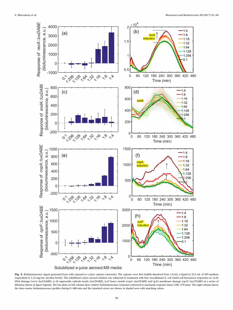
Fig. 4. Bioluminescence signal generated form cells exposed to e-juice vapours (aerosols). The vapours were first bubble-dissolved from 1.8 mL e-liquid in 22.5 mL of M9-medium(equivalent to 1.25 mg/mL nicotine levels). The solubilized e-juice aerosol solution was subjected to treatment with four recombinant E. coli whole-cell biosensors responsive to; (a-b)DNA damage (recA::luxCDABE), (c-d) superoxide radicals (sodA::luxCDABE), (e-f) heavy metals (copA::luxCDABE) and (g-h) membrane damage (oprF::luxCDABE) at a series ofdilutions shown in figure legends. The bar plots on left column show relative bioluminescence responses extracted at maximum response times (180–270 min). The right column showsthe time-course bioluminescence profiles during 0–480 min and the standard errors are shown in shaded area with matching colors.
S. Bharadwaj et al. Biosensors and Bioelectronics 90 (2017) 53–60
58

leading to activation of multiple intracellular signaling pathways andcell death (Ogura and Shimosawa, 2014). We speculate that the toxiclevels of e-juice can damage DNA or inhibit growth through arrestingnormal cellular functions, such as ROS defense system. Meanwhile, atlow concentrations, the cells may develop resistance easily in compar-ison to high concentrations and hence elicit the light emission, exceptfor E.coli-CopA. This assumption was found true when the biosensorcells were exposed to 2.5 mg/mL of nicotine in e-juice that showedsignificant growth loss as evidenced by reduced number of CFU against0.156 mg/mL concentration (Figs. S2, S4 and Table S2).
Recently, it is documented that the oxidative stress caused bynicotine is also responsible for inducing DNA damage in epithelial cells(Ginzkey et al., 2012). Therefore, it was imperative to test the DNAdamage in cells treated with e-juice that had shown significantreduction in growth. Our results revealed that high-doses of e-juicelikely to induce cellular DNA fragmentation at least in bacterial models(Fig. 6). It remains to be seen if this is also true in human cells. Thisresult provides early evidence that cell inhibition/death can be possibleat higher doses that seem to occur due to the chemical ingredients/
flavourings present in the e-juice. Furthermore, chemicals known to beharmful are present in e-cigarette aerosol and their effect could alsodepend on several variables, such as the solution used and the batteryoutput voltage (Bahl et al., 2012; Kosmider et al., 2014). Also, the levelsof toxic product may depend on the way the e-cigarette is used(Farsalinos et al., 2015).
The heated e-liquid emissions shown to contain the carcinogeniccarbonyl compounds formaldehyde and acetaldehyde (Goniewicz et al.,2014; Jensen et al., 2015; Kosmider et al., 2014), ROS, heavy metals,and volatile organic compounds, such as toluene that will havedetrimental effects on living cells (Goniewicz et al., 2014; Lerneret al., 2015; Williams and Talbot, 2011). In this study, we addressedthese effects with bacterial biosensors that were treated againstdifferent dilutions of e-juice and SEA. Our results indicated that theimpact of e-juice at least in bacterial model tend to diminish withdilutions of chemical ingredients present in e-juice (Figs. 3a-d, 4a-dand 6a-b). Moreover, the transformation of e-juice into e-vapours byheat produced more free radicals, which in turn increase the e-vapourtoxicity (Sussan et al., 2015). Hence, E. coli-SodA treated with differentdilutions of dissolved e-juice aerosols showed significant light emissionwhen compared with control cells. The E. coli-RecA induced highbioluminescence emission only at the high doses of SEA probablybecause of its ability to oxidatively damage DNA with free O2•−, whichdecreased with dilution factor and hence, low bioluminescence emis-sion from the cells (Fig. 4a). Furthermore, the recombinant E. coli-CopA showed high bioluminescence at high concentration indicates thepresence of copper in the dissolved aerosols, which reduced in themedia with the dilution. It is to be noted that the contamination ofcopper could occur by the oxidation of heating coil in e-cigarette deviceas it has been previously reported (Williams et al., 2013).
5. Conclusion
E-cigarettes deliver aerosol containing nicotine to users by heatinge-liquid/juice. Typically, e-juice contains a mixture of glycerin, propy-lene glycol and various other flavoring chemicals. The potential adverseeffects of a variety of flavoring chemical combinations used in e-juice
Fig. 5. Five E. coli strains (1.8×10⁸ cells/mL) were treated with maximum (2.5 mg/mLor 1:4 fold) and minimum (0.156 mg/mL or 1:64 fold) inhibitory doses of e-juice liquidin PBS solution were loaded along with untreated cells on an alkaline agarose gel andelectrophoresed in alkaline buffer at 10 V for 14 h. The gel was post-stained withethidium bromide after neutralization and the picture was taken under the UV-light. Thefragmented DNA due to high doses of e-juice (yellow arrow highlighted) can be seen assmears running down the gel. The non-fragmented DNA tangled with cells was seentrapped in control-wells and low-dose treated cells.
Fig. 6. Heatmaps and clustering of the bioluminescence response data measured at different time intervals as shown in x-axis against; (a) e-juice in liquid form mixed in M9-medium,and (b) Solubilized e-juice aerosol (SEA) in M9-medium. The colors red, black and green represents induction, unchanged or repressed bioluminescence responses from cells againstdifferent concentration indicated on y-axis along with strain identities, such as recA (E. coli-RecA), sodA (E. coli-SodA), copA (E. coli-CopA) and oprF (E. coli-DMO1).
S. Bharadwaj et al. Biosensors and Bioelectronics 90 (2017) 53–60
59

remains largely unexplored. Considering the fastest-growing e-cigar-ette industry, a rapid and versatile screening method is needed toassess their toxicant exposure and potential risk to ensure safety.Herein, we used four stress-specific recombinant bioluminescent E. colicells explicitly respond to single stranded DNA breaks(recA::luxCDABE), reactive oxygen species (sodA::luxCDABE), heavymetal such as copper (copA:: luxCDABE) and cell membrane damage(oprF::luxCDABE). These RBCs provided real-time biological re-sponses to liquid e-juice and e-cigarette generated SEA in limitedmineral medium with glucose as the only carbon and energy source.This allowed bioluminescent RBCs to sensitively emit specific biolu-minescence light to the e-juice/SEA toxicants. As a result, biolumines-cent RBCs provided the strong evidences that revealed sub-toxic dosesof e-juice and SEA induce cytotoxicity. We found that the liquid form ofe-juice suppressed the cellular ability to repair DNA damage, ionhomeostasis, oxidative toxicity and membrane disruption caused by thee-juice. SEA also exhibited toxicity but the level of this toxicity was notas lethal as it was with the liquid e-juice. Therefore, bioluminescentRBCs acquired the ability to adapt against the toxicants in SEA byrecruiting inducible cellular repair mechanisms, thus enabling cells topreserve their normal growth rate and reverse the SEA toxicity.
RBCs served as whole-cell living biosensors to simultaneouslydetect both cytotoxicity and intrinsic toxicity mechanisms taking placeagainst e-juice and SEA. Biological responses from these RBCs can beused as surrogate responses relevant to human health risk assessments.However, there is an intrinsic limitation in using multiple RBCsresponsive to distinct type of toxicities and thus differential biolumi-nescence patterns, respectively. Therefore, choice of appropriate cellpopulation size for each RBC type relevant to dose and intensity ofbioluminescence light emission is imperative. Further, real-time toxi-city monitoring for continuous screening with solubilized e-juiceaerosols from various other e-juice flavoring combinations is required.Our future directions are toward developing a series of other newRBCs, each specific to a different type of cytotoxicity that potentiallycontribute to a comprehensive understanding on the major cellularresponses against e-juice and SEA. For this, a multi-reactor setting maybe needed that may be equipped to continuously and simultaneouslydetect long-term cytotoxicity.
Acknowledgements
This work was supported by the Scientific and TechnologicalResearch Council of Turkey (TUBITAK), Project Grant no. 114E101.We thank Hansol Im for helping with pCopA plasmid construction.
Appendix A. Supporting information
Supplementary data associated with this article can be found in the
online version at doi:10.1016/j.bios.2016.11.026.
References
Allen, J.G., Flanigan, S.S., LeBlanc, M., Vallarino, J., MacNaughton, P., Stewart, J.H.,Christiani, D.C., 2016. Environ. Health Perspect. 124 (6), 733–739.
Arany, I., Clark, J., Reed, D.K., Juncos, L.A., 2013. Nephrol. Dial. Transpl. 28 (6),1417–1425.
Bahl, V., Lin, S., Xu, N., Davis, B., Wang, Y.H., Talbot, P., 2012. Reprod. Toxicol. 34 (4),529–537.
Brandon, T.H., Goniewicz, M.L., Hanna, N.H., Hatsukami, D.K., Herbst, R.S., Hobin,J.A., Ostroff, J.S., Shields, P.G., Toll, B.A., Tyne, C.A., Viswanath, K., Warren, G.W.,2015. J. Clin. Oncol. 33 (8), 952.
Brown, C.J., Cheng, J.M., 2014. Tob. Control 23, 4–10.Cheng, T.R., 2014. Tob. Control 23, 11–17.Chu, K.H., Unger, J.B., Allem, J.P., Pattarroyo, M., Soto, D., Cruz, T.B., Yang, H.D.,
Jiang, L., Yang, C.C., 2015. PLoS One 10, 12.Clayton, P.M., Vas, C.A., Bui, T.T., Drake, A.F., McAdam, K., 2013. Chirality 25 (5),
288–293.D'Errico, M., Parlanti, E., Dogliotti, E., 2008. Mutat. Res. 659 (1–2), 4–14.Elad, T., Seo, H.B., Belkin, S., Gu, M.B., 2015. Biosens. Bioelectron. 68, 699–704.Fan, J., de Jonge, B.L.M., MacCormack, K., Sriram, S., McLaughlin, R.E., Plant, H.,
Preston, M., Fleming, P.R., Albert, R., Foulk, M., Mills, S.D., 2014. Antimicrob.Agents Chemother. 58 (12), 7264–7272.
Farsalinos, K., Voudris, V., Poulas, K., 2015. Addiction 110 (11), 1862–1864.Ginzkey, C., Stueber, T., Friehs, G., Koehler, C., Hackenberg, S., Richter, E., Hagen, R.,
Kleinsasser, N.H., 2012. Toxicol. Lett. 208 (1), 23–29.Goniewicz, M.L., Knysak, J., Gawron, M., Kosmider, L., Sobczak, A., Kurek, J.,
Prokopowicz, A., Jablonska-Czapla, M., Rosik-Dulewska, C., Havel, C., Jacob, P.,Benowitz, N., 2014. Tob. Control 23 (2), 133–139.
Harrell, P.T., Marquinez, N.S., Correa, J.B., Meltzer, L.R., Unrod, M., Sutton, S.K.,Simmons, V.N., Brandon, T.H., 2015. Nicotine Tob. Res. 17 (2), 193–200.
He, W., Yuan, S., Zhong, W.H., Siddikee, M.A., Dai, C.C., 2016. Appl. MicrobiolBiotechnol. 100 (3), 1109–1119.
Jensen, R.P., Luo, W., Pankow, J.F., Strongin, R.M., Peyton, D.H., 2015. N. Engl. J. Med.372 (4), 392–394.
Kosmider, L., Sobczak, A., Fik, M., Knysak, J., Zaciera, M., Kurek, J., Goniewicz, M.L.,2014. Nicotine Tob. Res. 16 (10), 1319–1326.
Lee, S., Amasia, M., Madou, M., Mitchell, R.J., 2013. Biosens. Bioelectron. 46, 175–182.Lerner, C.A., Sundar, I.K., Yao, H.W., Gerloff, J., Ossip, D.J., McIntosh, S., Robinson, R.,
Rahman, I., 2015. Plos One 10, 2.Muthukumaran, S., Sudheer, A.R., Menon, V.P., Nalini, N., 2008. Toxicology 243 (1–2),
207–215.Ogura, S., Shimosawa, T., 2014. Curr. Hypertens. Rep. 16, 8.Orellana-Barrios, M.A., Payne, D., Mulkey, Z., Nugent, K., 2015. Am. J. Med 128 (7),
674–681.Sancilio, S., Gallorini, M., Cataldi, A., di Giacomo, V., 2016. Clin. Oral Investig. 20 (3),
477–483.Scheffler, S., Dieken, H., Krischenowski, O., Forster, C., Branscheid, D., Aufderheide, M.,
2015. Int J. Environ. Res. Public Health 12 (4), 3915–3925.Sussan, T.E., Gajghate, S., Thimmulappa, R.K., Ma, J.F., Kim, J.H., Sudini, K., Consolini,
N., Cormier, S.A., Lomnicki, S., Hasan, F., Pekosz, A., Biswal, S., 2015. PLoS One 10,2.
Williams, M., Talbot, P., 2011. Nicotine Tob. Res. 13 (12), 1276–1283.Williams, M., Villarreal, A., Bozhilov, K., Lin, S., Talbot, P., 2013. PLoS One 8, 3.
S. Bharadwaj et al. Biosensors and Bioelectronics 90 (2017) 53–60
60
Related Documents