ORIGINAL ARTICLE Toxicarioside A inhibits SGC-7901 proliferation, migration and invasion via NF-κB/bFGF signaling Jun-Li Guo, Shao-Jiang Zheng, Yue-Nan Li, Wei Jie, Xin-Bao Hao, Tian-Fa Li, Li-Ping Xia, Wen-Li Mei, Feng-Ying Huang, Yue-Qiong Kong, Qi-Yi He, Kun Yang, Guang-Hong Tan, Hao-Fu Dai Jun-Li Guo, Shao-Jiang Zheng, Yue-Nan Li, Xin-Bao Hao, Tian-Fa Li, Li-Ping Xia, Feng-Ying Huang, Yue-Qiong Kong, Qi-Yi He, Kun Yang, Guang-Hong Tan, Hainan Provincial Key Laboratory of Tropical Medicine, Cancer Research Insti- tute of Affiliated Hospital, Hainan Medical College, Haikou 571199, Hainan Province, China Wei Jie, Department of Pathology, Guangdong Medical Col- lege, Zhanjiang 524023, Guangdong Province, China Wen-Li Mei, Hao-Fu Dai, Key Laboratory of Tropical Crop Biotechnology, Ministry of Agriculture, Institute of Tropical Bioscience and Biotechnology, Chinese Academy of Tropical Agricultural Sciences, Haikou 571101, Hainan Province, China Author contributions: Guo JL, Zheng SJ and Li YN contrib- uted equally to this work; Guo JL and Zheng SJ designed the research; Guo JL, Zheng SJ and Li YN performed the research with Jie W, Hao XB, Li TF, Xia LP, Mei WL, Huang FY; Li YN, Kong YQ, He QY and Yang K analyzed data; Zheng SJ and Guo JL wrote the manuscript; Tan GH and Dai HF revised and finally approved the article to be published. Supported by Grants from the National Natural Scientific Foundation of China, No. 81060184; and the Natural Founda- tion of Hainan Province of China, No. 30864, 811201; and Pro- gram for New Century Excellent Talents in University of China, NCET-08-0657; and the National Basic Research Program of China, No. 2010CB534909; and Hainan Provincial Key Scien- tific Project, No. 061009 Correspondence to: Hao-Fu Dai, Professor, Key Laboratory of Tropical Crop Biotechnology, Ministry of Agriculture, Insti- tute of Tropical Bioscience and Biotechnology, Chinese Acad- emy of Tropical Agricultural Sciences, Haikou 571101, Hainan Province, China. [email protected]n Telephone: +86-898-66890292 Fax: +86-898-66890978 Received: September 10, 2011 Revised: January 17, 2012 Accepted: February 8, 2012 Published online: April 14, 2012 Abstract AIM: To investigate the inhibitory role of toxicarioside A on the gastric cancer cell line human gastric cancer cell line (SGC-7901) and determine the underlying mo- lecular mechanism. METHODS: After SGC-7901 cells were treated with toxicarioside A at various concentrations (0.5, 1.5, 4.5, 9.0 μg/mL) for 24 h or 48 h, cell viability was deter- mined by 3-(4,5-dimethyl-thiazol-2-yl)-2,5-diphenyl- 2H-tetrazolium bromide assay, and the motility and invasion of tumor cells were assessed by the Transwell chamber assay. Immunofluorescence staining, reverse transcription polymerase chain reaction and Western blotting were performed to detect the expression of basic fibroblast growth factor (bFGF) and fibroblast growth factor receptor-1 (FGFR1), and nuclear factor- kappa B (NF-κB) activation was examined by electro- phoretic mobility shift assay. RESULTS: The results showed that toxicarioside A was capable of reducing cell viability, inhibiting cell growth, and suppressing cell migration and invasion activities in a time- and dose-dependent manner in SGC-7901 cells. Further analysis revealed that not only the ex- pression of bFGF and its high-affinity receptor FGFR1 but also the NF-κB-DNA binding activity were effec- tively blocked by toxicarioside A in a dose-dependent manner compared with the control group ( P < 0.05 or P < 0.01). Interestingly, application of the NF-κB specific inhibitor, pyrrolidinedithiocarbamate (PDTC), to SGC-7901 cells significantly potentized the toxicari- oside A-induced down-regulation of bFGF compared with the control group ( P < 0.05). CONCLUSION: These findings suggest that toxicari- oside A has an anti-gastric cancer activity and this ef- fect may be achieved partly through down-regulation of NF-κB and bFGF/FGFR1 signaling. © 2012 Baishideng. All rights reserved. Key words: Anti-migration; Anti-proliferation; Basic fi- broblast growth factor; Gastric cancer; Nuclear factor- kappa B; Toxicarioside A World J Gastroenterol 2012 April 14; 18(14): 1602-1609 ISSN 1007-9327 (print) ISSN 2219-2840 (online) © 2012 Baishideng. All rights reserved. Online Submissions: http://www.wjgnet.com/1007-9327office [email protected] doi:10.3748/wjg.v18.i14.1602 1602 April 14, 2012|Volume 18|Issue 14| WJG|www.wjgnet.com

Welcome message from author
This document is posted to help you gain knowledge. Please leave a comment to let me know what you think about it! Share it to your friends and learn new things together.
Transcript
ORIGINAL ARTICLE
Toxicarioside A inhibits SGC-7901 proliferation, migration and invasion via NF-κB/bFGF signaling
Jun-Li Guo, Shao-Jiang Zheng, Yue-Nan Li, Wei Jie, Xin-Bao Hao, Tian-Fa Li, Li-Ping Xia, Wen-Li Mei, Feng-Ying Huang, Yue-Qiong Kong, Qi-Yi He, Kun Yang, Guang-Hong Tan, Hao-Fu Dai
Jun-Li Guo, Shao-Jiang Zheng, Yue-Nan Li, Xin-Bao Hao, Tian-Fa Li, Li-Ping Xia, Feng-Ying Huang, Yue-Qiong Kong, Qi-Yi He, Kun Yang, Guang-Hong Tan, Hainan Provincial Key Laboratory of Tropical Medicine, Cancer Research Insti-tute of Affiliated Hospital, Hainan Medical College, Haikou 571199, Hainan Province, China Wei Jie, Department of Pathology, Guangdong Medical Col-lege, Zhanjiang 524023, Guangdong Province, ChinaWen-Li Mei, Hao-Fu Dai, Key Laboratory of Tropical Crop Biotechnology, Ministry of Agriculture, Institute of Tropical Bioscience and Biotechnology, Chinese Academy of Tropical Agricultural Sciences, Haikou 571101, Hainan Province, ChinaAuthor contributions: Guo JL, Zheng SJ and Li YN contrib-uted equally to this work; Guo JL and Zheng SJ designed the research; Guo JL, Zheng SJ and Li YN performed the research with Jie W, Hao XB, Li TF, Xia LP, Mei WL, Huang FY; Li YN, Kong YQ, He QY and Yang K analyzed data; Zheng SJ and Guo JL wrote the manuscript; Tan GH and Dai HF revised and finally approved the article to be published.Supported by Grants from the National Natural Scientific Foundation of China, No. 81060184; and the Natural Founda-tion of Hainan Province of China, No. 30864, 811201; and Pro-gram for New Century Excellent Talents in University of China, NCET-08-0657; and the National Basic Research Program of China, No. 2010CB534909; and Hainan Provincial Key Scien-tific Project, No. 061009Correspondence to: Hao-Fu Dai, Professor, Key Laboratory of Tropical Crop Biotechnology, Ministry of Agriculture, Insti-tute of Tropical Bioscience and Biotechnology, Chinese Acad-emy of Tropical Agricultural Sciences, Haikou 571101, Hainan Province, China. [email protected]: +86-898-66890292 Fax: +86-898-66890978 Received: September 10, 2011 Revised: January 17, 2012Accepted: February 8, 2012Published online: April 14, 2012
AbstractAIM: To investigate the inhibitory role of toxicarioside A on the gastric cancer cell line human gastric cancer cell line (SGC-7901) and determine the underlying mo-lecular mechanism.
METHODS: After SGC-7901 cells were treated with toxicarioside A at various concentrations (0.5, 1.5, 4.5, 9.0 μg/mL) for 24 h or 48 h, cell viability was deter-mined by 3-(4,5-dimethyl-thiazol-2-yl)-2,5-diphenyl-2H-tetrazolium bromide assay, and the motility and invasion of tumor cells were assessed by the Transwell chamber assay. Immunofluorescence staining, reverse transcription polymerase chain reaction and Western blotting were performed to detect the expression of basic fibroblast growth factor (bFGF) and fibroblast growth factor receptor-1 (FGFR1), and nuclear factor-kappa B (NF-κB) activation was examined by electro-phoretic mobility shift assay.
RESULTS: The results showed that toxicarioside A was capable of reducing cell viability, inhibiting cell growth, and suppressing cell migration and invasion activities in a time- and dose-dependent manner in SGC-7901 cells. Further analysis revealed that not only the ex-pression of bFGF and its high-affinity receptor FGFR1 but also the NF-κB-DNA binding activity were effec-tively blocked by toxicarioside A in a dose-dependent manner compared with the control group (P < 0.05 or P < 0.01). Interestingly, application of the NF-κB specific inhibitor, pyrrolidinedithiocarbamate (PDTC), to SGC-7901 cells significantly potentized the toxicari-oside A-induced down-regulation of bFGF compared with the control group (P < 0.05).
CONCLUSION: These findings suggest that toxicari-oside A has an anti-gastric cancer activity and this ef-fect may be achieved partly through down-regulation of NF-κB and bFGF/FGFR1 signaling.
© 2012 Baishideng. All rights reserved.
Key words: Anti-migration; Anti-proliferation; Basic fi-broblast growth factor; Gastric cancer; Nuclear factor-kappa B; Toxicarioside A
World J Gastroenterol 2012 April 14; 18(14): 1602-1609 ISSN 1007-9327 (print) ISSN 2219-2840 (online)
© 2012 Baishideng. All rights reserved.
Online Submissions: http://www.wjgnet.com/[email protected]:10.3748/wjg.v18.i14.1602
1602 April 14, 2012|Volume 18|Issue 14|WJG|www.wjgnet.com
Guo JL et al . Toxicarioside A inhibits gastric cancer
Peer reviewer: Hitoshi Tsuda, MD, PhD, Diagnostic Pathology Section, Clinical Laboratory Division, National Cancer Center Hospital, 5-1-1 Tsukiji, Chuo-ku, Tokyo 104-0045, Japan
Guo JL, Zheng SJ, Li YN, Jie W, Hao XB, Li TF, Xia LP, Mei WL, Huang FY, Kong YQ, He QY, Yang K, Tan GH, Dai HF. Toxicarioside A inhibits SGC-7901 proliferation, migration and invasion via NF-κB/bFGF signaling. World J Gastroenterol 2012; 18(14): 1602-1609 Available from: URL: http://www.wjgnet.com/1007-9327/full/v18/i14/1602.htm DOI: http://dx.doi.org/10.3748/wjg.v18.i14.1602
INTRODUCTIONAntiaristoxicaria (Pers.) Lesch (Moraceae) is a well known precious species widespread in the tropical rain forest of Southeast Asia. Its latex and seeds contain a complex mixture of cardenolide glycosides and is therefore tox-ic[1]. Representative toxicariosides A-L have recently been identified from the latex and seeds of Antiaristoxicaria in our laboratory and by others[2-5]. Classically, cardenolides are used to treat congestive heart failure and arrhyth-mia[6-8]. Additionally, certain cardenolides extracted from some plants or animals have been demonstrated to be capable of blocking tumor cell proliferation through regulation of cell signal transduction[9-15].
Currently, gastric cancer is one of the leading ma-lignancies in China. However, the treatment outcome is not satisfactory because early diagnosis of gastric cancer remains difficult and most patients have already devel-oped metastatic lesions when diagnosed[16].
Basic fibroblast growth factor (bFGF) has been shown to be a multifunctional growth factor for tumor devel-opment[17-20], and it exerts its biological effects mainly through interaction with its high-affinity receptor, fibro-blast growth factor receptor-1 (FGFR1)[21-24]. Compiling evidence has demonstrated that bFGF signaling is in-volved in the development of gastric cancer[25,26].
Nuclear factor-kappa B (NF-κB) is a ubiquitous di-meric transcription factor that plays pivotal roles in regu-lating the expression of genes encoding cytokines and chemokines that are involved in tumor proliferation, an-giogenesis, and synthesis of anti-apoptotic proteins[27,28]. It has been documented that NF-κB can mediate bFGF signaling[29] and some types of cardiac glycosides can block the activation of NF-κB[30,31]. As a result, we hy-pothesize that cardiac glycosides may suppress gastric tumor growth via a decrease in NF-κB activity and blocking of the bFGF signaling pathway. In the present study, we attempted to test this hypothesis in an in vitro cell culture model.
MATERIALS AND METHODSPlant materialLatex of Antiaristoxicaria (Pers.) Lesch collected in Lingshui county of Hainan Province, China in November 2005 was identified with the assistance of Professor Zhunian
Wang at the Institute of Crops Genetic Resources, Chi-nese Academy of Tropical Agricultural Sciences. The specimen was numbered as No. AN200511.
Chemicals and reagentsRabbit-anti human bFGF and FGFR1 were purchased from Santa Cruz (Santa Cruz, CA, United States). Rho-damine (TRITC)-conjugated mouse anti-rabbit immu-noglobulin G (IgG), fluorescein isothiocyanate (FITC)-conjugated mouse anti-rabbit IgG, 3-(4,5-dimethyl-thia-zol-2-yl)-2,5-diphenyl-2H-tetrazolium bromide (MTT), trypan blue and pyrrolidinedithiocarbamate (PDTC) were obtained from Sigma (Sigma Aldrich, St Louis, MO, United States). Fetal bovine serum (FBS), RPMI 1640 medium and trypsin were procured from Gibco (Gibco, Carlsbad, CA, United States).
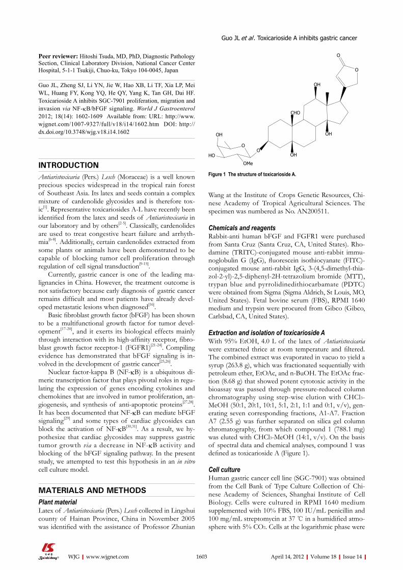
Extraction and isolation of toxicarioside AWith 95% EtOH, 4.0 L of the latex of Antiaristoxicaria were extracted thrice at room temperature and filtered. The combined extract was evaporated in vacuo to yield a syrup (263.8 g), which was fractionated sequentially with petroleum ether, EtOAc, and n-BuOH. The EtOAc frac-tion (8.68 g) that showed potent cytotoxic activity in the bioassay was passed through pressure-reduced column chromatography using step-wise elution with CHCl3-MeOH (50:1, 20:1, 10:1, 5:1, 2:1, 1:1 and 0:1, v/v), gen-erating seven corresponding fractions, A1-A7. Fraction A7 (2.55 g) was further separated on silica gel column chromatography, from which compound 1 (788.1 mg) was eluted with CHCl3-MeOH (14:1, v/v). On the basis of spectral data and chemical analyses, compound 1 was defined as toxicarioside A (Figure 1).
Cell cultureHuman gastric cancer cell line (SGC-7901) was obtained from the Cell Bank of Type Culture Collection of Chi-nese Academy of Sciences, Shanghai Institute of Cell Biology. Cells were cultured in RPMI 1640 medium supplemented with 10% FBS, 100 IU/mL penicillin and 100 mg/mL streptomycin at 37 ℃ in a humidified atmo-sphere with 5% CO2. Cells at the logarithmic phase were
1603 April 14, 2012|Volume 18|Issue 14|WJG|www.wjgnet.com
OH
OH
OH
OH
O
O
OO
CHO
OMe
HO
Figure 1 The structure of toxicarioside A.
used for experiments.
Proliferation assayMTT assay and trypanblue staining were used to deter-mine the growth and viability of SGC-7901 cells. For the MTT assay, SGC-7901 cells in logarithmic growth were trypsinized and harvested and then the cells were seeded onto a 96-well plate. After 24 h, fresh RPMI 1640 medium containing different concentrations of toxicarioside A (0.5, 1.5, 4.5, 9.0 μg/mL) was added at 100 μL per well, respectively, and 6 replicate wells were used for each of the concentrations. After incubation for different time intervals, 10 μL of MTT (5 mg/mL) was added to each well and the cells were further incubated at 37 ℃ for 4 h. The supernatant was then removed and 100 μL DMSO was added into each well. Absorbance (A value) at a wavelength of 490 nm was measured with a Bio-TekEXL808 microplate reader (Bio-Rad, Hercules, CA, United States). For trypanblue staining, SGC-7901 cells were trypsinized and seeded into 24-well plates at a density of 0.5 × 10⁴/mL. After 4.5 μg/mL toxicarioside A was added, the cells were collected and counted using trypan blue staining under an inverse light microscope for 3 consecutive days.
Invasion and migration assayInvasion assays were performed in a 24-well Transwell chamber (Corning, Lowell, MA, United States) as previ-ously described[32]. Briefly, each Transwell chamber was coated with 15 μg Matrigel, 5 × 10⁴ cells were seeded to pre-coated filters in 200 μL of serum-free medium con-taining different concentrations of toxicarioside A (0.5, 1.5, 4.5, 9.0 μg/mL) in triplicate. The lower parts of the chambers were filled with 500 μL of RPMI 1640 medi-um containing 10% FBS. After incubation in a 5% CO2 humidified incubator at 37 ℃ for 24 h, the cells on the upper surface were gently removed with a cotton swab, and the filters were fixed with 95% alcohol for 15-20 min and stained with hematoxylin-eosin for 15 min. The number of cells on the lower surface of the filters was quantified under a microscope. The same procedures were followed for the migration assay except the Tran-swell chambers were not coated with Matrigel.
Immunofluorescence staining To detect the expression of bFGF as well as its receptor FGFR1 in SGC-7901 cells, the rabbit antibody (1:100) against bFGF and FGFR1 were used. The antigenic sites were localized by TRITC-conjugated mouse anti-rabbit IgG and FITC-conjugated mouse anti-rabbit IgG, and im-ages of antigenic sites were captured under a laser scanning confocal microscope (FV500, Olympus, Tokyo, Japan).
RNA extraction and reverse transcription polymerase chain reactionTotal RNA was extracted with Trizol reagent (Invitrogen, Carlsbad, CA, United States) following the manufac-turer’s protocols. Reverse transcription polymerase chain
reaction was carried out using pairs of primers (Invitro-gen) as follows for semiquantitative assessment: bFGF (NM_002006.4) sense, 5’-AAG AGC GAC CCT CAC ATC AA-3’; anti-sense, 5’-TCG TTT CAG TGC CAC ATC CGT CAA TA CC-3’, yielding a 225 bp product; FGFR1 (M34641) sense, 5’-CTT CTGT TTC AG-3’; anti-sense, 5’-TCC ACA ATG CAG GTG TAG TT-3’, yielding a 354 bp product. β-actin (NM_001101) sense, 5’-GTT GCG TTA CAC CCT TTC TT-3’, anti-sense, 5’-CGA AGG CTC ATC ATT CAA AA-3’, yielding a 443 bp product. The products were separated by electropho-resis on a 1.5% agarose gel and visualized under UV using the gel documentation system (Bio-Rad Gel Doc1000, Bio-Rad). The mRNA levels of bFGF, FGFR1 were cal-culated based on the denstometric values of the specific bFGF, FGFR1 bands after adjustment with that of the β-actin band.
Western blotting analysis This was performed as previously described with minor modifications[33]. Cells were homogenized and separated by sodium dodecyl sulfate-polyacrylamide gel (12.5%) electrophoresis followed by electrophoretic transfer of proteins from the gel to a nitrocellulose membrane blot (Bio-Rad). The blot was incubated with a rabbit anti-bFGF antibody (1:500) or a rabbit anti-FGFR1 antibody (1:500) at 4 ℃ overnight, followed by incubation with the corresponding horseradish peroxidase-conjugated anti-biotin antibody (1:2000) at room temperature for 1 h. The immunoreactive signals were visualized with enhanced chemiluminescence reagents (Pierce, Rockford, IL, United States).
Electrophoretic mobility shift assay To determine NF-κB activation, electrophoretic mobil-ity shift assay (EMSA) was conducted essentially as de-scribed previously[34]. In brief, nuclear proteins (10 μg) were incubated with the reaction buffer for 20 min at room temperature, followed by incubation with oligo-nucleotide containing the consensus sequence for the NF-κB-DNA binding site (5’-AGAGTGGGAATT TC-CACTCA-3’)[35] (synthesized by Invitrogen, Shanghai, China). The reaction mixture was separated in a non-denaturing polyacrylamide gel (6%) that was later stained by SYBR Green EMSA staining solution from Molecular Probes (Invitrogen) with continuous, gentle agitation for about 20 min, protected from light. The gel was then washed in 150 mL of dH2O2 and the stained nucleic acids were visualized and the image documented under UV using the gel documentation system (Bio-Rad Gel Doc1000).
Statistical analysisAll data are expressed as mean ± SE. For a comparison between two groups, the Student’s t test was performed. For comparisons among multiple groups, an ANOVA was carried out, followed by a Student-Newman-Keuls test. Differences were considered significant when P < 0.05.
1604 April 14, 2012|Volume 18|Issue 14|WJG|www.wjgnet.com
Guo JL et al . Toxicarioside A inhibits gastric cancer
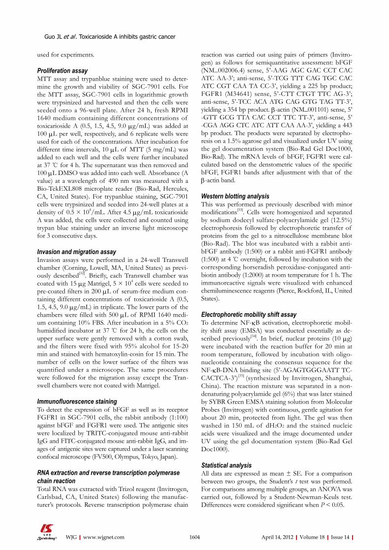
RESULTSEffect of toxicarioside A on SGC-7901 cell proliferationTo assess the effect of toxicarioside A on the growth of gastric cancer, SGC-7901 cells were treated at various concentrations (0.5, 1.5, 4.5, 9.0 μg/mL) for 24-48 h and cell viability following these treatments was determined by MTT assays. As shown in Table 1, toxicarioside A reduced SGC-7901 cell viability in a time- and dose-dependent manner. Cell growth curves also showed that toxicarioside A significantly inhibited SGC-7901 cell growth as compared with the control (Figure 2).
Effect of toxicarioside A on SGC-7901 cell migration and invasion The results of Transwell cell migration and invasion are presented in Table 2 and Figure 3. Clearly, the addition of toxicarioside A to the medium in the upper chamber resulted in significant suppression of SGC-7901 migra-tion and invasion in a dose-dependent manner at 1.5, 4.5 and 9.0 μg/mL, toxicarioside A inhibited SGC-7901 migration by 22.38% ± 10.64%, 39.58% ± 11.62% and 48.13% ± 10.12%, respectively (P < 0.05), and inhibited SGC 7901 invasion by 24.03% ± 9.06%, 44.68% ± 9.19% and 54.38% ± 8.17%, respectively (P < 0.01), as com-pared with the control group.
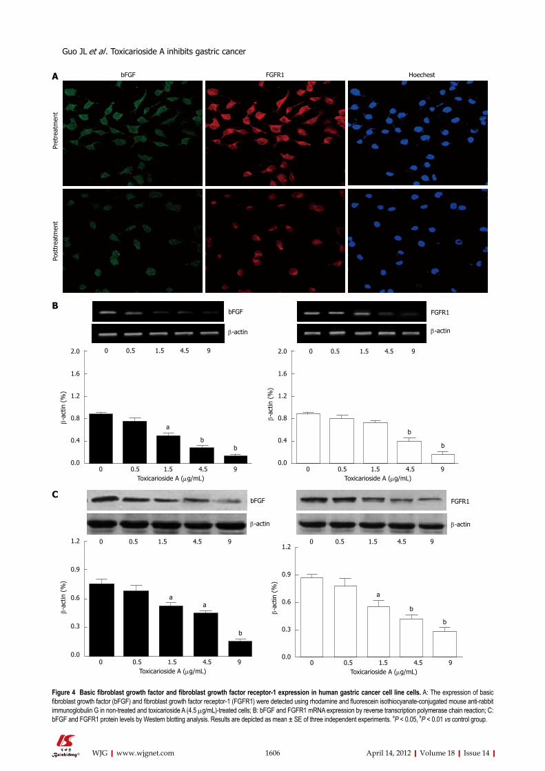
Effect of toxicarioside A on bFGF and FGFR1 in SGC- 7901 cellsAt the protein level, the expression of bFGF and FGFR1 was predominantly detected in the cytoplasm of SGC-7901 cells and toxicarioside A significantly decreased this ex-
pression, as assessed by immunofluorescence staining (Figure 4A) and Western blotting analysis (Figure 4C). At the mRNA level, the expression of bFGF and FGFR1 was decreased by toxicarioside A in a dose-dependent manner in SGC-7901 cells (Figure 4B).
Effect of toxicarioside A on NF-κB-DNA binding activity in SGC-7901 cells To determine the effect of toxicariosideA on NF-κB activation, the NF-κB-DNA binding activity was de-termined in both toxicarioside A-treated and control SGC-7901 cells by EMSA. As shown in Figure 5, after treatment with toxicarioside A at various concentrations for 48 h, the NF-κB-DNA binding activity was de-creased in a dose-dependent manner as compared with the control group (P < 0.05 or P < 0.01, Figure 5).
Effect of PDTC on toxicarioside A-induced downregulation of bFGFTo further determine whether NF-κB activation was nec-essary for bFGF expression, and was involved in toxicari-oside A-induced downregulation of bFGF, a specific in-
1605 April 14, 2012|Volume 18|Issue 14|WJG|www.wjgnet.com
Groups 24 h 48 h
A value Inhibitory rate (%) A value Inhibitory rate (%)
Control 0.879 ± 0.048 0.00 ± 0.00 0.932 ± 0.036 0.00 ± 0.00 Toxicarioside A (μg/mL) 0.5 0.793 ± 0.062 9.79 ± 7.63 0.752 ± 0.073 19.13 ± 5.38 1.5 0.646 ± 0.041 27.83 ± 4.78a 0.596 ± 0.113 36.19 ± 3.81a
4.5 0.528 ± 0.078 40.18 ± 3.32a 0.443 ± 0.056 49.32 ± 5.17b
9.0 0.352 ± 0.092 61.84 ± 6.61b 0.301 ± 0.049 66.94 ± 7.03b
Table 1 The inhibition rates of human gastric cancer cell line cells treated with different concentrations of toxicarioside A for different time intervals
Data representative of six independent experiments were expressed as mean ± SE. aP < 0.05, bP < 0.01 vs control group.
Groups Dose (μg/mL)
Migration Invasion
Cell number Inhibition rate (%)
Cell number Inhibition rate (%)
Control 0.0 69.40 ± 6.38 0.00 ± 0.00 65.60 ± 7.20 0.00 ± 0.00 Toxicarioside A 0.5 62.50 ± 8.90 10.01 ± 9.78 57.80 ± 4.32 10.89 ± 8.64
1.5 54.80 ± 7.30 22.38 ± 10.64a 49.70 ± 5.68 24.03 ± 9.06a
4.5 41.60 ± 5.88 39.58 ± 11.62a 36.40 ± 7.94 44.68 ± 9.19a
9.0 35.80 ± 8.32 48.13 ± 10.12b 30.10 ± 6.46 54.38 ± 8.17b
Table 2 Effect of toxicarioside A on cell migration and invasion
Data representative of three independent experiments were expressed as mean ± SE. aP < 0.05, bP < 0.01 vs control group.
0 12 24 36 48 60 72 Incubation time (h)
Toxicarioside AControl
2.5
2.0
1.5
1.0
0.5
0.0
Cell
num
ber
(× 1
04 )
Figure 2 Effect of toxicarioside A on the growth curve of human gastric cancer cell line cells. Cells were plated in 24-well plates at a density of 1 × 10⁴/mL and treated with 4.5 μg/mL toxicarioside A for 72 h. The results shown are representative of three independent experiments.
A B
C D
Figure 3 Representative figures of cell migration and invasion in non-treated and toxicarioside A-treated human gastric cancer cell line cells. A: Migration in the control group; B: Migration in the thetoxicarioside A-treated (4.5 μg/mL) group; C: Invasion in the control group; D: Invasion in the toxicarioside A-treated group.
Guo JL et al . Toxicarioside A inhibits gastric cancer
bFGF FGFR1 Hoechest
Post
trea
tmen
t P
retr
eatm
ent
A
BbFGF
β-actin
0 0.5 1.5 4.5 9
FGFR1
β-actin
0 0.5 1.5 4.5 9
a
bb
0 0.5 1.5 4.5 9 Toxicarioside A (μg/mL)
2.0
1.6
1.2
0.8
0.4
0.0
β-a
ctin
(%
)
b
b
0 0.5 1.5 4.5 9 Toxicarioside A (μg/mL)
2.0
1.6
1.2
0.8
0.4
0.0
β-a
ctin
(%
)
CbFGF
β-actin
0 0.5 1.5 4.5 9
FGFR1
β-actin
0 0.5 1.5 4.5 9
1606 April 14, 2012|Volume 18|Issue 14|WJG|www.wjgnet.com
aa
b
0 0.5 1.5 4.5 9 Toxicarioside A (μg/mL)
1.2
0.9
0.6
0.3
0.0
β-a
ctin
(%
)
a
b
b
0 0.5 1.5 4.5 9 Toxicarioside A (μg/mL)
1.2
0.9
0.6
0.3
0.0
β-a
ctin
(%
)
Figure 4 Basic fibroblast growth factor and fibroblast growth factor receptor-1 expression in human gastric cancer cell line cells. A: The expression of basic fibroblast growth factor (bFGF) and fibroblast growth factor receptor-1 (FGFR1) were detected using rhodamine and fluorescein isothiocyanate-conjugated mouse anti-rabbit immunoglobulin G in non-treated and toxicarioside A (4.5 μg/mL)-treated cells; B: bFGF and FGFR1 mRNA expression by reverse transcription polymerase chain reaction; C: bFGF and FGFR1 protein levels by Western blotting analysis. Results are depicted as mean ± SE of three independent experiments. aP < 0.05, bP < 0.01 vs control group.
Guo JL et al . Toxicarioside A inhibits gastric cancer
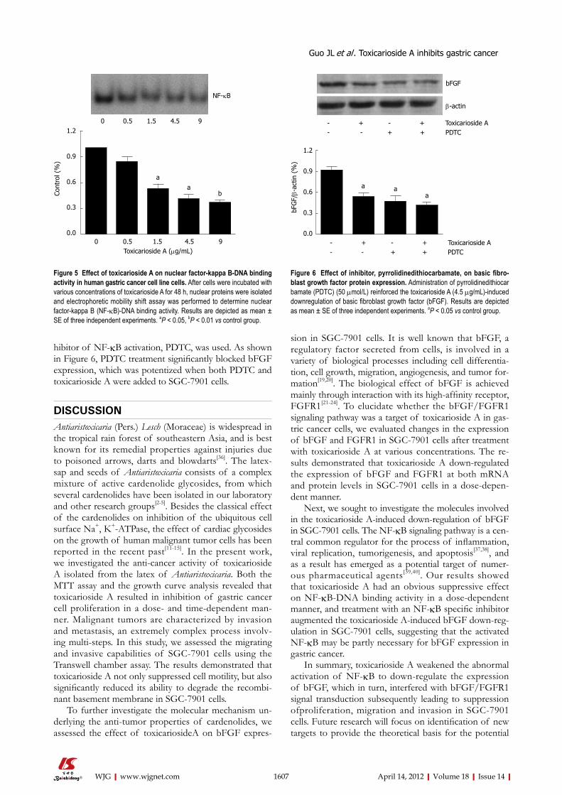
hibitor of NF-κB activation, PDTC, was used. As shown in Figure 6, PDTC treatment significantly blocked bFGF expression, which was potentized when both PDTC and toxicarioside A were added to SGC-7901 cells.
DISCUSSIONAntiaristoxicaria (Pers.) Lesch (Moraceae) is widespread in the tropical rain forest of southeastern Asia, and is best known for its remedial properties against injuries due to poisoned arrows, darts and blowdarts[36]. The latex-sap and seeds of Antiaristoxicaria consists of a complex mixture of active cardenolide glycosides, from which several cardenolides have been isolated in our laboratory and other research groups[2-5]. Besides the classical effect of the cardenolides on inhibition of the ubiquitous cell surface Na+, K+-ATPase, the effect of cardiac glycosides on the growth of human malignant tumor cells has been reported in the recent past[11-15]. In the present work, we investigated the anti-cancer activity of toxicarioside A isolated from the latex of Antiaristoxicaria. Both the MTT assay and the growth curve analysis revealed that toxicarioside A resulted in inhibition of gastric cancer cell proliferation in a dose- and time-dependent man-ner. Malignant tumors are characterized by invasion and metastasis, an extremely complex process involv-ing multi-steps. In this study, we assessed the migrating and invasive capabilities of SGC-7901 cells using the Transwell chamber assay. The results demonstrated that toxicarioside A not only suppressed cell motility, but also significantly reduced its ability to degrade the recombi-nant basement membrane in SGC-7901 cells.
To further investigate the molecular mechanism un-derlying the anti-tumor properties of cardenolides, we assessed the effect of toxicariosideA on bFGF expres-
sion in SGC-7901 cells. It is well known that bFGF, a regulatory factor secreted from cells, is involved in a variety of biological processes including cell differentia-tion, cell growth, migration, angiogenesis, and tumor for-mation[19,20]. The biological effect of bFGF is achieved mainly through interaction with its high-affinity receptor, FGFR1[21-24]. To elucidate whether the bFGF/FGFR1 signaling pathway was a target of toxicarioside A in gas-tric cancer cells, we evaluated changes in the expression of bFGF and FGFR1 in SGC-7901 cells after treatment with toxicarioside A at various concentrations. The re-sults demonstrated that toxicarioside A down-regulated the expression of bFGF and FGFR1 at both mRNA and protein levels in SGC-7901 cells in a dose-depen-dent manner.
Next, we sought to investigate the molecules involved in the toxicarioside A-induced down-regulation of bFGF in SGC-7901 cells. The NF-κB signaling pathway is a cen-tral common regulator for the process of inflammation, viral replication, tumorigenesis, and apoptosis[37,38], and as a result has emerged as a potential target of numer-ous pharmaceutical agents[39,40]. Our results showed that toxicarioside A had an obvious suppressive effect on NF-κB-DNA binding activity in a dose-dependent manner, and treatment with an NF-κB specific inhibitor augmented the toxicarioside A-induced bFGF down-reg-ulation in SGC-7901 cells, suggesting that the activated NF-κB may be partly necessary for bFGF expression in gastric cancer.
In summary, toxicarioside A weakened the abnormal activation of NF-κB to down-regulate the expression of bFGF, which in turn, interfered with bFGF/FGFR1 signal transduction subsequently leading to suppression ofproliferation, migration and invasion in SGC-7901 cells. Future research will focus on identification of new targets to provide the theoretical basis for the potential
1607 April 14, 2012|Volume 18|Issue 14|WJG|www.wjgnet.com
NF-κB
0 0.5 1.5 4.5 9
a
b
0 0.5 1.5 4.5 9 Toxicarioside A (μg/mL)
1.2
0.9
0.6
0.3
0.0
Con
trol
(%
)
a
- + - + Toxicarioside A- - + + PDTC
bFGF
β-actin
- + - + Toxicarioside A- - + + PDTC
a aa
1.2
0.9
0.6
0.3
0.0
bFG
F/β-
actin
(%
)
Figure 5 Effect of toxicarioside A on nuclear factor-kappa B-DNA binding activity in human gastric cancer cell line cells. After cells were incubated with various concentrations of toxicarioside A for 48 h, nuclear proteins were isolated and electrophoretic mobility shift assay was performed to determine nuclear factor-kappa B (NF-κB)-DNA binding activity. Results are depicted as mean ± SE of three independent experiments. aP < 0.05, bP < 0.01 vs control group.
Figure 6 Effect of inhibitor, pyrrolidinedithiocarbamate, on basic fibro-blast growth factor protein expression. Administration of pyrrolidinedithiocarbamate (PDTC) (50 μmol/L) reinforced the toxicarioside A (4.5 μg/mL)-induced downregulation of basic fibroblast growth factor (bFGF). Results are depicted as mean ± SE of three independent experiments. aP < 0.05 vs control group.
Guo JL et al . Toxicarioside A inhibits gastric cancer
application of toxicarioside A in the clinical treatment of gastric cancer.
COMMENTSBackgroundThe latex and seeds of Antiaristoxicaria contain a complex mixture of carde-nolide glycosides, and representative toxicariosides A-L have recently been identified in our laboratory and by others. Some cardenolides have been dem-onstrated to be capable of blocking tumor cell proliferation through regulation of cell signal transduction.Research frontiersGastric cancer is one of the leading malignancies in China. However, the treat-ment outcome is not satisfactory because early diagnosis of gastric cancer remains difficult and most patients have already developed metastatic lesions when diagnosed. It is important to investigate the strategies that could inhibit gastric cancer effectively.Innovations and breakthroughsTo date, little is known about the underlying mechanism regarding the anti-can-cer effects of toxicarioside A. Therefore, this study was conducted to investigate the anti-cancer activity of toxicarioside A on gastric cancer growth and migration and the underlying molecular mechanisms in vitro.ApplicationsThis study indicates the first evidence of the underlying molecular mechanisms of the anti-cancer activity of toxicarioside A in gastric cancer. These results provide the theoretical basis for the potential application of toxicarioside A in the clinical treatment of gastric cancer. TerminologyAntiaristoxicaria (Pers.) Lesch (Moraceae) is a well known precious species widespread in the tropical rain forest of Southeast Asia, and the latexsap and seeds of Antiaristoxicaria consist of a complex mixture of active cardenolide glycosides.Peer reviewThis manuscript showed toxicarioside A inhibits the proliferation, invasion and migration in a gastric cancer cell line, and these phenomena was correlated with down-regulation of nuclear factor-kappa B/basic fibroblast growth factor signal-ing. The design of study is solid and experiments were elegantly performed.
REFERENCES1 Shrestha T, Kopp B, Bisset NG. The Moraceae-based dart
poisons of South America. Cardiac glycosides of Maquira and Naucleopsis species. J Ethnopharmacol 1992; 37: 129-143
2 Dong WH, Mei WL, Zhao YX, Zeng YB, Zuo WJ, Wang H, Li XN, Dai HF. Cytotoxic cardenolide glycosides from the seeds of Antiaris toxicaria. Planta Med 2011; 77: 1730-1734
3 Dong WH, Mei WL, Zhao YX, Zeng YB, Wang H, Dai HF. A new drimane sesquiterpenoid glycoside from the seeds of Antiaris toxicaria. J Asian Nat Prod Res 2011; 13: 561-565
4 Dai HF, Gan YJ, Que DM, Wu J, Wen ZC, Mei WL. Two new cytotoxic cardenolides from the latex of Antiaris toxi-caria. J Asian Nat Prod Res 2009; 11: 832-837
5 Dai HF, Gan YJ, Que DM, Wu J, Wen ZC, Mei WL. A new cytotoxic 19-nor-cardenolide from the latex of Antiaris toxi-caria. Molecules 2009; 14: 3694-3699
6 Shi LS, Liao YR, Su MJ, Lee AS, Kuo PC, Damu AG, Kuo SC, Sun HD, Lee KH, Wu TS. Cardiac glycosides from An-tiaris toxicaria with potent cardiotonic activity. J Nat Prod 2010; 73: 1214-1222
7 Gheorghiade M, van Veldhuisen DJ, Colucci WS. Contem-porary use of digoxin in the management of cardiovascular disorders. Circulation 2006; 113: 2556-2564
8 Hamad E, Mather PJ, Srinivasan S, Rubin S, Whellan DJ, Feldman AM. Pharmacologic therapy of chronic heart fail-ure. Am J Cardiovasc Drugs 2007; 7: 235-248
9 Choedon T, Mathan G, Arya S, Kumar VL, Kumar V. An-ticancer and cytotoxic properties of the latex of Calotropis
procera in a transgenic mouse model of hepatocellular car-cinoma. World J Gastroenterol 2006; 12: 2517-2522
10 Yeh JY, Huang WJ, Kan SF, Wang PS. Effects of bufalin and cinobufagin on the proliferation of androgen dependent and independent prostate cancer cells. Prostate 2003; 54: 112-124
11 Frese S, Frese-Schaper M, Andres AC, Miescher D, Zum-kehr B, Schmid RA. Cardiac glycosides initiate Apo2L/TRAIL-induced apoptosis in non-small cell lung cancer cells by up-regulation of death receptors 4 and 5. Cancer Res 2006; 66: 5867-5874
12 Sreenivasan Y, Sarkar A, Manna SK. Oleandrin suppresses activation of nuclear transcription factor-kappa B and ac-tivator protein-1 and potentiates apoptosis induced by ce-ramide. Biochem Pharmacol 2003; 66: 2223-2239
13 McConkey DJ, Lin Y, Nutt LK, Ozel HZ, Newman RA. Car-diac glycosides stimulate Ca2+ increases and apoptosis in androgen-independent, metastatic human prostate adeno-carcinoma cells. Cancer Res 2000; 60: 3807-3812
14 Lin J, Denmeade S, Carducci MA. HIF-1alpha and calcium signaling as targets for treatment of prostate cancer by car-diac glycosides. Curr Cancer Drug Targets 2009; 9: 881-887
15 Newman RA, Yang P, Pawlus AD, Block KI. Cardiac glyco-sides as novel cancer therapeutic agents. Mol Interv 2008; 8: 36-49
16 Foukakis T, Lundell L, Gubanski M, Lind PA. Advances in the treatment of patients with gastric adenocarcinoma. Acta Oncol 2007; 46: 277-285
17 Ribatti D, Vacca A, Rusnati M, Presta M. The discovery of basic fibroblast growth factor/fibroblast growth factor-2 and its role in haematological malignancies. Cytokine Growth Factor Rev 2007; 18: 327-334
18 Shi YH, Bingle L, Gong LH, Wang YX, Corke KP, Fang WG. Basic FGF augments hypoxia induced HIF-1-alpha expres-sion and VEGF release in T47D breast cancer cells. Pathology 2007; 39: 396-400
19 Smith JA, Madden T, Vijjeswarapu M, Newman RA. Inhibi-tion of export of fibroblast growth factor-2 (FGF-2) from the prostate cancer cell lines PC3 and DU145 by Anvirzel and its cardiac glycoside component, oleandrin. Biochem Pharma-col 2001; 62: 469-472
20 Cronauer MV, Hittmair A, Eder IE, Hobisch A, Culig Z, Ramoner R, Zhang J, Bartsch G, Reissigl A, Radmayr C, Thurnher M, Klocker H. Basic fibroblast growth factor levels in cancer cells and in sera of patients suffering from prolif-erative disorders of the prostate. Prostate 1997; 31: 223-233
21 Giehl KA, Nägele U, Volkenandt M, Berking C. Protein expression of melanocyte growth factors (bFGF, SCF) and their receptors (FGFR-1, c-kit) in nevi and melanoma. J Cu-tan Pathol 2007; 34: 7-14
22 Fortin D, Rom E, Sun H, Yayon A, Bansal R. Distinct fi-broblast growth factor (FGF)/FGF receptor signaling pairs initiate diverse cellular responses in the oligodendrocyte lineage. J Neurosci 2005; 25: 7470-7479
23 Udayakumar TS, Klein RD, Maliner MS, Nagle RB, Bowden GT. Aberrant expression of fibroblast growth factor recep-tor-1 in prostate epithelial cells allows induction of prom-atrilysin expression by fibroblast growth factors. Int J Cancer 2001; 91: 187-192
24 Kamura S, Matsumoto Y, Fukushi JI, Fujiwara T, Iida K, Okada Y, Iwamoto Y. Basic fibroblast growth factor in the bone microenvironment enhances cell motility and inva-sion of Ewing’s sarcoma family of tumours by activating the FGFR1-PI3K-Rac1 pathway. Br J Cancer 2010; 103: 370-381
25 Gong J, Morishita A, Kurokohchi K, Tani J, Kato K, Miyoshi H, Inoue H, Kobayashi M, Liu S, Murota M, Muramatsu A, Izuishi K, Suzuki Y, Yoshida H, Uchida N, Deguchi K, Iwa-ma H, Ishimaru I, Masaki T. Use of protein array to investi-gate receptor tyrosine kinases activated in gastric cancer. Int J Oncol 2010; 36: 101-106
26 Katoh M, Katoh M. FGF signaling network in the gastroin-
1608 April 14, 2012|Volume 18|Issue 14|WJG|www.wjgnet.com
COMMENTS
Guo JL et al . Toxicarioside A inhibits gastric cancer
testinal tract (review). Int J Oncol 2006; 29: 163-16827 Baldwin AS. Control of oncogenesis and cancer therapy re-
sistance by the transcription factor NF-kappaB. J Clin Invest 2001; 107: 241-246
28 Nakanishi C, Toi M. Nuclear factor-kappaB inhibitors as sen-sitizers to anticancer drugs. Nat Rev Cancer 2005; 5: 297-309
29 Bushdid PB, Chen CL, Brantley DM, Yull F, Raghow R, Kerr LD, Barnett JV. NF-kappaB mediates FGF signal regu-lation of msx-1 expression. Dev Biol 2001; 237: 107-115
30 Yang Q, Huang W, Jozwik C, Lin Y, Glasman M, Caohuy H, Srivastava M, Esposito D, Gillette W, Hartley J, Pollard HB. Cardiac glycosides inhibit TNF-alpha/NF-kappaB signaling by blocking recruitment of TNF receptor-associated death domain to the TNF receptor. Proc Natl Acad Sci USA 2005; 102: 9631-9636
31 Mijatovic T, Op De Beeck A, Van Quaquebeke E, Dewelle J, Darro F, de Launoit Y, Kiss R. The cardenolide UNBS1450 is able to deactivate nuclear factor kappaB-mediated cytopro-tective effects in human non-small cell lung cancer cells. Mol Cancer Ther 2006; 5: 391-399
32 Peng L, Ran YL, Hu H, Yu L, Liu Q, Zhou Z, Sun YM, Sun LC, Pan J, Sun LX, Zhao P, Yang ZH. Secreted LOXL2 is a novel therapeutic target that promotes gastric cancer me-tastasis via the Src/FAK pathway. Carcinogenesis 2009; 30: 1660-1669
33 Zheng SJ, Zheng SP, Huang FY, Jiao CL, Wu RL. Synergistic anti-tumor effect of recombinant chicken fibroblast growth factor receptor-1-mediated anti-angiogenesis and low-dose gemcitabine in a mouse colon adenocarcinoma model.
World J Gastroenterol 2007; 13: 2484-248934 Guo J, Jie W, Kuang D, Ni J, Chen D, Ao Q, Wang G. Isch-
aemia/reperfusion induced cardiac stem cell homing to the injured myocardium by stimulating stem cell factor ex-pression via NF-kappaB pathway. Int J Exp Pathol 2009; 90: 355-364
35 Ueda A, Ishigatsubo Y, Okubo T, Yoshimura T. Transcrip-tional regulation of the human monocyte chemoattractant protein-1 gene. Cooperation of two NF-kappaB sites and NF-kappaB/Rel subunit specificity. J Biol Chem 1997; 272: 31092-31099
36 Jiang MM, Dai Y, Gao H, Zhang X, Wang GH, He JY, Hu QY, Zeng JZ, Zhang XK, Yao XS. Cardenolides from Anti-aris toxicaria as potent selective Nur77 modulators. Chem Pharm Bull (Tokyo) 2008; 56: 1005-1008
37 Baldwin AS. Series introduction: the transcription factor NF-kappaB and human disease. J Clin Invest 2001; 107: 3-6
38 Giri DK, Aggarwal BB. Constitutive activation of NF-kappaB causes resistance to apoptosis in human cutaneous T cell lymphoma HuT-78 cells. Autocrine role of tumor ne-crosis factor and reactive oxygen intermediates. J Biol Chem 1998; 273: 14008-14014
39 Namba H, Saenko V, Yamashita S. Nuclear factor-kB in thyroid carcinogenesis and progression: a novel therapeutic target for advanced thyroid cancer. Arq Bras Endocrinol Me-tabol 2007; 51: 843-851
40 Lee JI, Burckart GJ. Nuclear factor kappa B: important tran-scription factor and therapeutic target. J Clin Pharmacol 1998; 38: 981-993
S- Editor Gou SX L- Editor Webster JR E- Editor Li JY
1609 April 14, 2012|Volume 18|Issue 14|WJG|www.wjgnet.com
Guo JL et al . Toxicarioside A inhibits gastric cancer
Related Documents