Journal of Experimental Botany, Vol. 65, No. 22 pp. 6693–6709, 2014 doi:10.1093/jxb/eru389 Advance Access publication 26 September, 2014 v This paper is available online free of all access charges (see http://jxb.oxfordjournals.org/open_access.html for further details) RESEARCH PAPER Tomato Male sterile 10 35 is essential for pollen development and meiosis in anthers Hee-Jin Jeong 1,2,† , Jin-Ho Kang 1,2,† , Meiai Zhao 3 , Jin-Kyung Kwon 1,2 , Hak-Soon Choi 4 , Jung Hwan Bae 1 , Hyun-ah Lee 1,2 , Young-Hee Joung 5 , Doil Choi 1,2 and Byoung-Cheorl Kang 1,2, * 1 Department of Plant Science and Plant Genomics and Breeding Institute, College of Agriculture and Life Science, Seoul National University, 599 Gwanak-ro Gwank-gu, Seoul 151-921, Republic of Korea 2 Plant Genomics and Breeding Institute, College of Agricultural Sciences, Seoul National University, 599 Gwanak-ro Gwank-gu, Seoul 151-921, Republic of Korea 3 College of Life Science, Qingdao Agricultural University, Qingdao 266-109, PR China 4 National Institute of Horticultural and Herbal Science, Suwon 440-310, Republic of Korea 5 School of Biological Sciences and Technology, Chonnam National University, Gwangju 500-757, Republic of Korea † These authors contributed equally to this work. * To whom correspondence should be addressed: E-mail: [email protected] Received 17 July 2014; Revised 19 August 2014; Accepted 27 August 2014 Abstract Male fertility in flowering plants depends on proper cellular differentiation in anthers. Meiosis and tapetum develop- ment are particularly important processes in pollen production. In this study, we showed that the tomato male sterile (ms10 35 ) mutant of cultivated tomato (Solanum lycopersicum) exhibited dysfunctional meiosis and an abnormal tape- tum during anther development, resulting in no pollen production. We demonstrated that Ms10 35 encodes a basic helix–loop–helix transcription factor that is specifically expressed in meiocyte and tapetal tissue from pre-meiotic to tetrad stages. Transgenic expression of the Ms10 35 gene from its native promoter complemented the male sterility of the ms10 35 mutant. In addition, RNA-sequencing-based transcriptome analysis revealed that Ms10 35 regulates 246 genes involved in anther development processes such as meiosis, tapetum development, cell-wall degradation, pol- len wall formation, transport, and lipid metabolism. Our results indicate that Ms10 35 plays key roles in regulating both meiosis and programmed cell death of the tapetum during microsporogenesis. Key words: Anther, male sterility, meiosis, tapetum, tomato (Solanum lycopersicum). Introduction Pollen development is one of the most fundamental processes in the plant life cycle (Wilson and Zhang, 2009). Through pol- len, plants deliver genetic material and expand genetic diver- sity by producing recombinant progeny in the subsequent generation (Deveshwar et al., 2011). Pollen development involves an exquisite pathway supported by cellular changes and the regulation of an enormous number of genes (Honys and Twell, 2004; Wilson and Zhang, 2009; Feng et al., 2012). In Arabidopsis (Arabidopsis thaliana) and rice (Oryza sativa), anther development has been well studied and many of the genes involved have been identified. In Arabidopsis, after the floral structures are successively generated, anther cells initiate specification and differentiation to form a bilat- erally symmetrical structure with four lobes. Archesporial cells in each lobe generate five distinct cell layers (from outer to inner: epidermis, endothecium, middle layer, tapetum, and This is an Open Access article distributed under the terms of the Creative Commons Attribution License (http://creativecommons.org/licenses/by/3.0/), which permits unrestricted reuse, distribution, and reproduction in any medium, provided the original work is properly cited. © The Author 2014. Published by Oxford University Press on behalf of the Society for Experimental Biology. Abbreviations: BAC, bacterial artificial chromosome; bHLH, basic helix–loop–helix; DAPI, 4’,6-diamidino-2-phenylindole; FDA, fluorescein diacetate; HRM, high- resolution-melting; PCD, programmed cell death; PMC, pollen mother cell; RACE, rapid amplification of cDNA ends; RNA-seq, RNA sequencing; RT-PCR, reverse transcription-PCR; SEM, scanning electron microscopy; TEM, transmission electron microscopy. by guest on June 4, 2016 http://jxb.oxfordjournals.org/ Downloaded from

Welcome message from author
This document is posted to help you gain knowledge. Please leave a comment to let me know what you think about it! Share it to your friends and learn new things together.
Transcript
Journal of Experimental Botany, Vol. 65, No. 22 pp. 6693–6709, 2014doi:10.1093/jxb/eru389 Advance Access publication 26 September, 2014
vThis paper is available online free of all access charges (see http://jxb.oxfordjournals.org/open_access.html for further details)
ReseaRch PaPeR
Tomato Male sterile 1035 is essential for pollen development and meiosis in anthers
Hee-Jin Jeong1,2,†, Jin-Ho Kang1,2,†, Meiai Zhao3, Jin-Kyung Kwon1,2, Hak-Soon Choi4, Jung Hwan Bae1, Hyun-ah Lee1,2, Young-Hee Joung5, Doil Choi1,2 and Byoung-Cheorl Kang1,2,*1 Department of Plant Science and Plant Genomics and Breeding Institute, College of Agriculture and Life Science, Seoul National University, 599 Gwanak-ro Gwank-gu, Seoul 151-921, Republic of Korea2 Plant Genomics and Breeding Institute, College of Agricultural Sciences, Seoul National University, 599 Gwanak-ro Gwank-gu, Seoul 151-921, Republic of Korea3 College of Life Science, Qingdao Agricultural University, Qingdao 266-109, PR China4 National Institute of Horticultural and Herbal Science, Suwon 440-310, Republic of Korea5 School of Biological Sciences and Technology, Chonnam National University, Gwangju 500-757, Republic of Korea
† These authors contributed equally to this work.* To whom correspondence should be addressed: E-mail: [email protected]
Received 17 July 2014; Revised 19 August 2014; Accepted 27 August 2014
Abstract
Male fertility in flowering plants depends on proper cellular differentiation in anthers. Meiosis and tapetum develop-ment are particularly important processes in pollen production. In this study, we showed that the tomato male sterile (ms1035) mutant of cultivated tomato (Solanum lycopersicum) exhibited dysfunctional meiosis and an abnormal tape-tum during anther development, resulting in no pollen production. We demonstrated that Ms1035 encodes a basic helix–loop–helix transcription factor that is specifically expressed in meiocyte and tapetal tissue from pre-meiotic to tetrad stages. Transgenic expression of the Ms1035 gene from its native promoter complemented the male sterility of the ms1035 mutant. In addition, RNA-sequencing-based transcriptome analysis revealed that Ms1035 regulates 246 genes involved in anther development processes such as meiosis, tapetum development, cell-wall degradation, pol-len wall formation, transport, and lipid metabolism. Our results indicate that Ms1035 plays key roles in regulating both meiosis and programmed cell death of the tapetum during microsporogenesis.
Key words: Anther, male sterility, meiosis, tapetum, tomato (Solanum lycopersicum).
Introduction
Pollen development is one of the most fundamental processes in the plant life cycle (Wilson and Zhang, 2009). Through pol-len, plants deliver genetic material and expand genetic diver-sity by producing recombinant progeny in the subsequent generation (Deveshwar et al., 2011). Pollen development involves an exquisite pathway supported by cellular changes and the regulation of an enormous number of genes (Honys and Twell, 2004; Wilson and Zhang, 2009; Feng et al., 2012).
In Arabidopsis (Arabidopsis thaliana) and rice (Oryza sativa), anther development has been well studied and many of the genes involved have been identified. In Arabidopsis, after the floral structures are successively generated, anther cells initiate specification and differentiation to form a bilat-erally symmetrical structure with four lobes. Archesporial cells in each lobe generate five distinct cell layers (from outer to inner: epidermis, endothecium, middle layer, tapetum, and
This is an Open Access article distributed under the terms of the Creative Commons Attribution License (http://creativecommons.org/licenses/by/3.0/), which permits unrestricted reuse, distribution, and reproduction in any medium, provided the original work is properly cited.
© The Author 2014. Published by Oxford University Press on behalf of the Society for Experimental Biology.
Abbreviations: BAC, bacterial artificial chromosome; bHLH, basic helix–loop–helix; DAPI, 4’,6-diamidino-2-phenylindole; FDA, fluorescein diacetate; HRM, high-resolution-melting; PCD, programmed cell death; PMC, pollen mother cell; RACE, rapid amplification of cDNA ends; RNA-seq, RNA sequencing; RT-PCR, reverse transcription-PCR; SEM, scanning electron microscopy; TEM, transmission electron microscopy.
by guest on June 4, 2016http://jxb.oxfordjournals.org/
Dow
nloaded from
6694 | Jeong et al.
the sporogenous cell) (Smyth et al., 1990; Wilson and Zhang, 2009). At the meiotic stage, meiocytes (pollen mother cells), which are developed from sporogenous cells, undergo meiotic cell divisions and are separated from the tapetal cell layer. Tapetal cells become vacuolated and initiate programmed cell death (PCD)-triggered degradation. During meiosis I, homol-ogous chromosomes replicate, pair, synapse, and undergo recombination by exchanging DNA (Zickler and Kleckner, 1999). After that, homologous chromosomes are aligned and pulled towards opposite poles by spindle organization. Finally, dyads are produced (Ma, 2005). For example, in pro-phase I, rice HOMOLOGOUS PAIRING ABERRATION IN RICE MEIOSIS 1 (PAIR1), PAIR2, and PAIR3 are important for chromosome pairing and synapsis, respectively (Nonomura et al., 2004, 2006; Yuan et al., 2009). Arabidopsis SWITCH1 (SWI1) and rice MEIOTIC RECOMBINATION PROTEIN8 (REC8) are essential for chromatid cohesion and bivalent formation (Mercier et al., 2003; Shao et al., 2011). In metaphase I, Arabidopsis MULTIPOLAR SPINDLE1 (MPS1) plays a role in organization of the spindle and chro-mosomal segregation (Jiang et al., 2009). In anaphase, rice POLLEN SEMI-STERILITY1 (PSS1), which encodes a kinesis-1 like protein, is necessary for meiotic chromosome pulling by the spindle (Zhou et al., 2001). Mutations of these genes related to meiosis cause defective meiocyte develop-ment and male sterility.
During meiosis, tapetal cells surrounding meiocytes pro-duce various enzymes, lipids, starch, pollen wall materi-als, and other molecules required for pollen development (Goldberg et al., 1993; Zhang et al., 2006). For instance, Arabidopsis EXTRA SPOROGENOUS CELLS/EXCESS MICROSPOROCYTES1 (EMS1/EXS) and TAPETAL DETERMINANT1 (TPD1) are important for tapetal speci-fication and maintenance of tapetal cell fate, and these mutants show extra meiocytes and no tapetal cells (Canales et al., 2002; Yang et al., 2003; Zhao et al., 2002). Mutations in Arabidopsis DYSFUNCTIONAL TAPETUM1 (DYT1) and rice UNDEVELOPED TAPETUM1 (UDT1) cause abnor-mal tapetal development such as vacuolated tapetal cells, resulting in male sterility (Jung et al., 2005; Zhang et al., 2006). In addition, EMS1/EXS and TPD1 are also required for cytokinesis after chromosomal segregation (Canales et al., 2002; Yang et al., 2003; Zhao et al., 2002), suggesting that sporophytic cells and gametophytic cells coordinate with each other by cell-to-cell communication.
Tomato flowers contain five sepals that alternate with five petals, in addition to five stamens and a style formed by two fused carpels. The stamens, which house pollen production, sit inside the petals. A single tomato stamen consists of two elongated compartments, and the individual stamens are fused together to form an anther cone called the androe-cium, which surrounds the style. In the cultivated tomato, the stigma is completely covered under the staminal tube (Brukhin et al., 2003). Tomato pollen development is quite similar to that of Arabidopsis and rice (Brukhin et al., 2003, Wilson and Zhang, 2009; D. Zhang et al., 2011). Tomato stamen primordia are initiated at the early stage of anther development followed by archesporial cell differentiation.
Sporogenous and parietal cells are differentiated from arche-sporial cells. These cells give rise to microspore tetrads and tapetum, respectively, after going through meiosis. Finally, the microspores mature and become pollen grains (Rasmussen and Green, 1993; Brukhin et al., 2003). In tomato, over 50 male-sterile mutants have been reported, and they can be divided into three classes (functional, structural, and sporog-enous) based on their developmental defects (Gorman and McCormick, 1997). Sporogenous male-sterile mutants can be further classified into five groups (pre-meiotic, meiotic, tetrad, microspore, and not determined) according to the stage at which pollen development aborts or breaks down (Rick and Butler, 1956; Gorman and McCormick, 1997). For example, the functional male-sterile mutant positional sterile-2 (ps-2) is defective in pollen dehiscence. Pre-meiotic mutants such as male sterile (ms) 3 and ms15 display pollen mother cell (PMC) collapse or abortion prior to the meiotic prophase. Meiotic mutants such as ms5 and ms1035 (allelic to ms10) show defects in tapetal tissue (Rick and Butler, 1956; Gorman and McCormick, 1997). While many tomato male-sterile mutants are available, the only known underlying gene was the polygalacturonase gene responsible for the ps-2 male-sterile mutant (Gorguet et al., 2009).
The ms1035 mutant was described previously as a spon-taneous mutant with defects in tapetum development and degeneration (Rick, 1948; Zamir et al., 1980; Corral-Martínez et al., 2011). Because of its stable male sterility and lack of growth defects, it has been widely used for F1 hybrid breeding (Georgiev, 1991; Kumar and Singh, 2005). In addition, the ms1035 mutant also has been used for anther culture to generate haploid plants, because a callus can easily be induced from its anthers (Zamir et al., 1980; Corral-Martínez et al., 2011). Here, we demonstrated that the ms1035 mutant is defective in chromosome segregation at anaphase I during meiosis, as well as in tapetum devel-opment, causing male sterility. Using a map-based cloning approach, we found that Ms1035 encodes a basic helix–loop–helix (bHLH) transcription factor. RNA sequenc-ing (RNA-seq)-based transcriptome analysis revealed that Ms1035 regulates 246 genes related to meiosis, tapetum development, lipid metabolism, cell wall modification/deg-radation, and pollen wall biosynthesis. These results dem-onstrated that Ms1035 serves as a master regulator of pollen development in tomato.
Materials and methods
Plant material and plant growthA tomato male-fertile parent (T-1082) and male-sterile ms1035 (2–517), which was backcrossed to T-1082 six times, were obtained from the National Institute of Horticultural and Herbal Science (Suwon, Korea). T-1082 and the backcrossed ms1035 were used in all experiments except the mapping experiment in which the original ms1035 was crossed to T-1082. Seedlings were grown in 50-plug trays containing sterilized soil in a growth chamber maintained under 18 h light (265 mE m–2 s–1) at 27 °C and 6 h darkness at 18 °C and 60% humidity. At the eight-leaf stage, the seedlings were transplanted to a greenhouse in the farm of the College of Agriculture and Life Science at Seoul National University (Suwon, Korea).
by guest on June 4, 2016http://jxb.oxfordjournals.org/
Dow
nloaded from
Tomato Ms1035 encodes a bHLH transcription factor | 6695
MicroscopyFluorescein diacetate (FDA) was used to check pollen viability according to the protocol of Kim et al. (2013). FDA-stained pollen was examined using an Axiophot microscope (Zeiss, Oberkochen, Germany). For ultrastructure and transmission electron micros-copy (TEM) analysis, floral buds were infiltrated with Spurr’s resin according to the protocol of Kim et al. (2013). TEM images were observed using a JEM1010 transmission electron microscope (Jeol, Tokyo, Japan) at 80 kV. For scanning electron microscopy (SEM), pollen grains of mature flowers were mounted and coated with palladium-gold in a sputter coater (BAL-TEC/SCD 005; Balzers, Lichtenstein) and examined using a field emission scanning electron microscope (SUPRA 55VP; Carl Zeiss, Germany) with an accelera-tion voltage of 15 kV.
4′,6-Diamidino-2-phenylindole (DAPI) staining analysis of meiotic processesFor the observation of meiotic chromosomes, a modified PMC spreading protocol was used (Kwon and Kim, 2009). Briefly, floral buds around the meiotic stage were fixed in Carnoy’s fixative solu-tion (ethanol:acetic acid=3:1, v/v) for 48 h. Fixed buds were rinsed twice in distilled water and then once in 10 mM citrate buffer (pH 4.5). Samples were incubated at 37 °C for 3 h in a digestion mix con-taining 2% (w/v) cellulase RS, 1% (w/v) pectinase, and 0.5% (w/v) pectolyase Y23 (Sigma, St Louis, MO, USA) dissolved in 10 mM citrate buffer. After digestion, cells were fixed in 60% acetic acid on a heated slide. After air drying, fixed cells were stained and mounted with 2 μg ml–1 of DAPI solution in Vectashield anti-fade mounting medium (Vector Laboratories, Burlingame, CA, USA).
DNA extractionGenomic DNA was extracted from two to three young leaves using a hexadecyltrimethyl-ammonium bromide method (Jeong et al., 2010). Leaf tissue was fragmented using TissueLyserII (Qiagen, Haan, Germany). DNA concentrations were measured with a Nanodrop spectrophotometer (NanoDrop Technologies, Wilmington, DE, USA) and diluted to a final concentration of 20 ng μl–1 in TE buffer (pH 7.0) for further experiments.
Bacterial artificial chromosome (BAC) alignment and Ms1035-linked marker developmentThe Ms1035 gene is known to be located between the PEROXIDASE-2 (PRX-2) and ANTHOCYANIN ABSENT (AA) genes, which are around the 69–78 cM region on chromosome 2 (Tanksley and Rick, 1980; Tanksley et al., 1992). Tomato BAC clones corresponding to the 69–78 cM region were aligned and assembled by Seqman soft-ware (DNA Star; DNASTAR, Madison, WI, USA). In order to develop Ms1035-linked markers, primer sets were randomly designed within the 69–78 cM region from the assembled BAC clones. The designed primers were tested for polymorphism between paren-tal DNA (ms1035 and T-1082) and F1 DNA derived from a cross between ms1035 and T-1082 plants using high-resolution-melting (HRM) analysis (Rotor-Gene 6000 thermocycler; Corbett Research, Sydney, Australia) according to a previously described method (Jeong et al. 2011).
Genetic analysis and map-based cloning of Ms1035
Fine mapping of Ms1035 was performed with an F2 population derived from a cross between ms1035 mutant and T-1082 plants, and was facilitated by the assembled BAC sequence described above and the tomato genome sequence (Tomato Genome Consortium, 2012). A population of 1100 F2 plants was scored for male sterility and subsequently genotyped with HRM markers (Supplementary Table S1 at JXB online). Linkage analysis of molecular markers
was conducted using the Carthagene 1.0 program (de Givry et al., 2005). Ms1035 was positioned to an ~80 kb region on chromosome 2 flanked by markers 762K and 843K. Putative genes in the 80 kb region were predicted using the FGENESH program (http://linux1.softberry.com/), the tomato Unigene database from SGN (http://solgenomics.net), and the BLASTP interface of the National Center for Biotechnology Information (http://www.ncbi.nlm.nih.gov/).
Total RNA isolation and reverse transcription (RT)-PCRFloral buds at different stages, leaves, stems, and fruits were collected from ms1035 and T-1082 plants and quickly frozen in liquid nitrogen. Total RNA was isolated using Trizol extraction buffer (Ambion, Carlsbad, CA, USA) according to the manufacturer’s protocol. cDNA was synthesized from 2 μg of total RNA using reverse tran-scriptase (Promega, Madison, WI, USA). cDNA (200 ng) was used for RT-PCR. Amplified PCR products were separated on a 1% aga-rose gel and stained with ethidium bromide. The primer sequences used for RT-PCR are listed in Supplementary Table S1.
Rapid amplification of cDNA ends (RACE)To identify the transcription start site of the Ms1035 gene, 5ʹRACE-PCR was performed using a SMARTer™ RACE cDNA Amplification kit (Clontech Laboratories, Mountain View, CA, USA). RNA was extracted from T-1082 anthers and cDNA was syn-thesized according to the manufacturer’s instructions. Sequencing analysis was performed at the National Instrumentation Center for Environmental Management (NICEM, Seoul National University, Seoul, Korea).
Genome-walking PCRTo identify the mutated region of the ms1035 gene, genome-walk-ing PCR was performed using a Genome Walker kit (Clontech Laboratories, Mountain view, CA, USA) according to the manu-facturer’s manual. Sequence information for gene-specific prim-ers 1 and 2 is provided in Supplementary Table S1. The fragment amplified by genome-walking PCR was cloned into a pGEM-T vec-tor (pGEM®-T Easy Vector Systems, Promega, Seoul, Korea) and sequenced at the NICEM.
Complementation of the ms1035 mutationA 2992 bp genomic sequence of Ms1035, which contained the entire coding region (1002 bp) with 1.4 kb of upstream sequence and 0.6 kb of downstream sequence, was amplified with XbaI restriction site-tagged primers (Supplementary Table S1). The resulting fragment was digested with XbaI, purified, and cloned into the XbaI site of the pCAMBIA2300 binary vector to generate pCAMBIA2300::Ms1035. The resulting construct and pCAMBIA2300 were introduced into Agrobacterium tumefaciens strain LBA4404 and used to trans-form ms1035 heterozygous (Ms1035/ms1035) cotyledon explants, as described previously (Seong et al., 2007). The presence of the T-DNA insert in independent primary (T0) transformants was confirmed by PCR using a NPTII-specific primer set (Supplementary Table S1) to amplify a fragment of the NPTII gene. Subsequently, independ-ent T1 plants with the homozygous ms1035/ms1035 genotype were selected using ms1035-specific primers (Supplementary Table S1). From these, F1 plants having both NPTII and Ms1035 transgenes were selected using NPTII-specific and Ms1035 transgene-specific primers, respectively (Supplementary Table S1).
RNA transcriptome analysis by RNA-seqTotal RNA from floral buds at stages 1 to 3 was extracted in the same manner as described above. A strand-specific RNA-seq library was constructed for the synthesis of cDNA as described by Zhong et al. (2011). RNA transcriptome was obtained using Hiseq 2500
by guest on June 4, 2016http://jxb.oxfordjournals.org/
Dow
nloaded from
6696 | Jeong et al.
(Illumina/Solexa, San Diego, CA, USA) at NICEM. The RNA-seq algorithm of the CLC Genomics Workbench 6.0 was used for relative digital expression with a 98% identity threshold (CLC bio, Prismet, Denmark). Digital expression data were normalized and transformed using CLC Genomics Workbench 6.0 internal algo-rithms. ITAG2.3_CDS was used as a reference genome for read mapping (http://solgenomics.net). The mean values of three biologi-cal replicates were transformed into log2 values (ms1035/T-1082). The DESeq tool of the R package (http://www.bioconductor.org/) was used to identify differentially expressed genes in ms1035 compared with T-1082 with a false discovery rate of <0.05 (Anders et al., 2013).
Results
The ms1035 mutant does not produce pollen
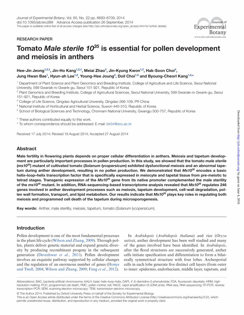
The tomato male-sterile mutant ms1035 has been described as being defective in the production of active pollen (Rick, 1948; Zamir et al., 1980). To examine developmental defects, ms1035 plants were compared with male-fertile T-1082 plants. There was no difference in development between ms1035 and T-1082 plants until the flowering stage. At the flower-ing stage, ms1035 plants showed morphological differences in flower shape. The ms1035 mutant had much smaller flow-ers compared with T-1082 plants (83% of T-1082, Fig. 1A, D, and Supplementary Fig. S1B at JXB online). In addi-tion, the anther cone and the style in ms1035 flowers were much shorter than in T-1082 flowers (60 and 86% of T-1082, Supplementary Fig. S1B). Due to the more dramatic change in anther cone length compared with style length, the styles of ms1035 flowers protruded over the anther cones (Fig. 1D and Supplementary Fig. S1B). To check whether ms1035 flowers produced viable pollen grains, an FDA assay, which measures cell viability, was performed with mature pollen grains or dust particles released from anthers at the dehiscence stage. The pollen of T-1082 flowers showed green fluorescence whereas no signals were detected from ms1035 mutant flowers (Fig. 1B, E), indicating that ms1035 flowers did not produce viable pol-len. To confirm that ms1035 flowers produced no pollen, we
used SEM. T-1082 anthers contained normal globular pollen grains, but ms1035 anthers had no pollen (Fig. 1C, F). These results demonstrated that the male sterility of the ms1035 mutant resulted from a lack of production of pollen.
Pollen development in ms1035 is arrested at the tetrad stage
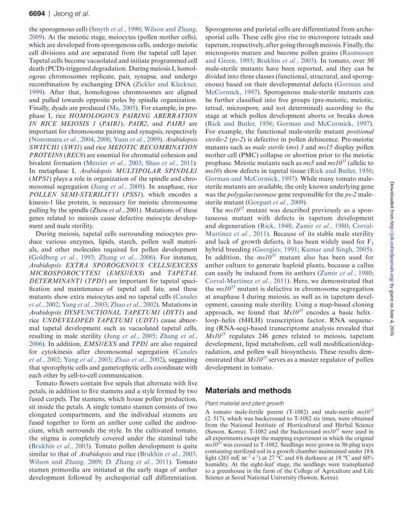
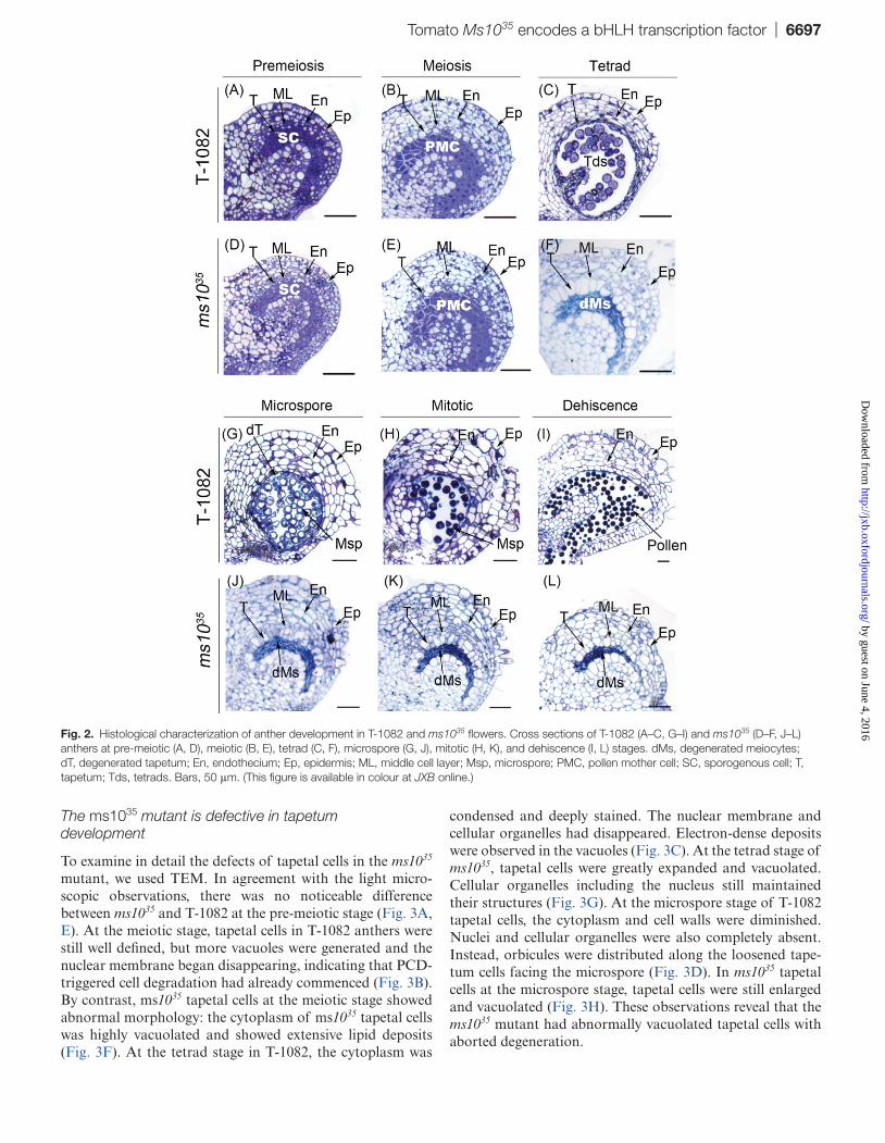
To determine the spatial and temporal occurrence of defects in ms1035 anthers, we prepared thin sections from anthers at dif-ferent stages of development and examined them using light microscopy. At the pre-meiotic and meiotic stages of T-1082 and ms1035 anthers, the five different cell layers were suc-cessfully differentiated from archesporial cells (Fig. 2A, D), and sporogenous cells developed into PMCs and underwent meiosis (Fig. 2B, E). At the tetrad stage, however, dramatic morphological differences were observed between ms1035 and T-1082 anthers. In T-1082 anthers, PMCs divided into tetrads after meiosis. Tapetal cells were greatly condensed and deeply stained. The middle cell layer was degenerated and almost invisible (Fig. 2C). In ms1035 anthers, PMCs were crushed and failed to produce tetrads. Tapetal cells and the middle cell layer were excessively enlarged and vacuolated (Fig. 2F). At the microspore stage in T-1082 anthers, free microspores were released into anther locules (Fig. 2G). In ms1035 anthers at the microspore stage, degenerated meiocytes were aggre-gated and gradually degraded. Vacuolated tapetal cells and middle cell layers were severely expanded (Fig. 2J). At the mitotic and dehiscence stages in T-1082 anthers, vacuolated microspores were deeply stained with toluidine blue due to the accumulation of collapsed tapetum fragments and nutri-ents, and the tapetum and middle cell layer had already disap-peared (Fig. 2H). Finally, anthers dehisced and pollen grains were released (Fig. 2I). By contrast, in ms1035 anthers at the mitotic and dehiscence stages, degenerated meiocytes contin-ued to dwindle, while tapetal cells remained swollen and vacu-olated without degeneration (Fig. 2K, L).
Fig. 1. Flower phenotypes and pollen production in T-1082 (male fertile) and the ms1035 mutant. (A, D) Flowers of T-1082 (A) and ms1035 plants (D). (B, E) FDA assay for pollen viability of T-1082 (B) and ms1035 (E) plants. Pollen grains or dust particles released from anthers at the dehiscence stage were used. (C, F) SEM observation of the surfaces of anthers at the dehiscence stage from T-1082 (C) and ms1035 (F) plants. The insets show a closer look at the surface of the anthers. The T-1082 anther produced pollen grains (arrow and inset). Bars, 0.5 cm (A, D); 100 µm (B, C, E, F); 10 μm (insets). (This figure is available in colour at JXB online.)
by guest on June 4, 2016http://jxb.oxfordjournals.org/
Dow
nloaded from
Tomato Ms1035 encodes a bHLH transcription factor | 6697
The ms1035 mutant is defective in tapetum development
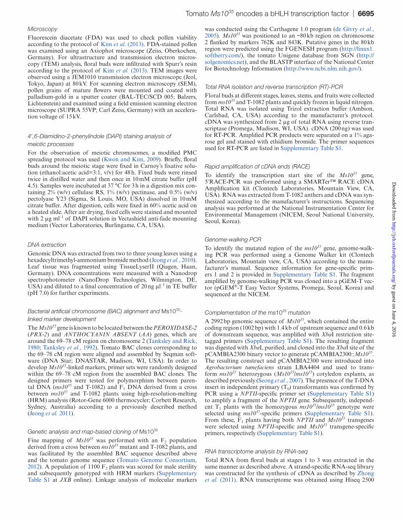
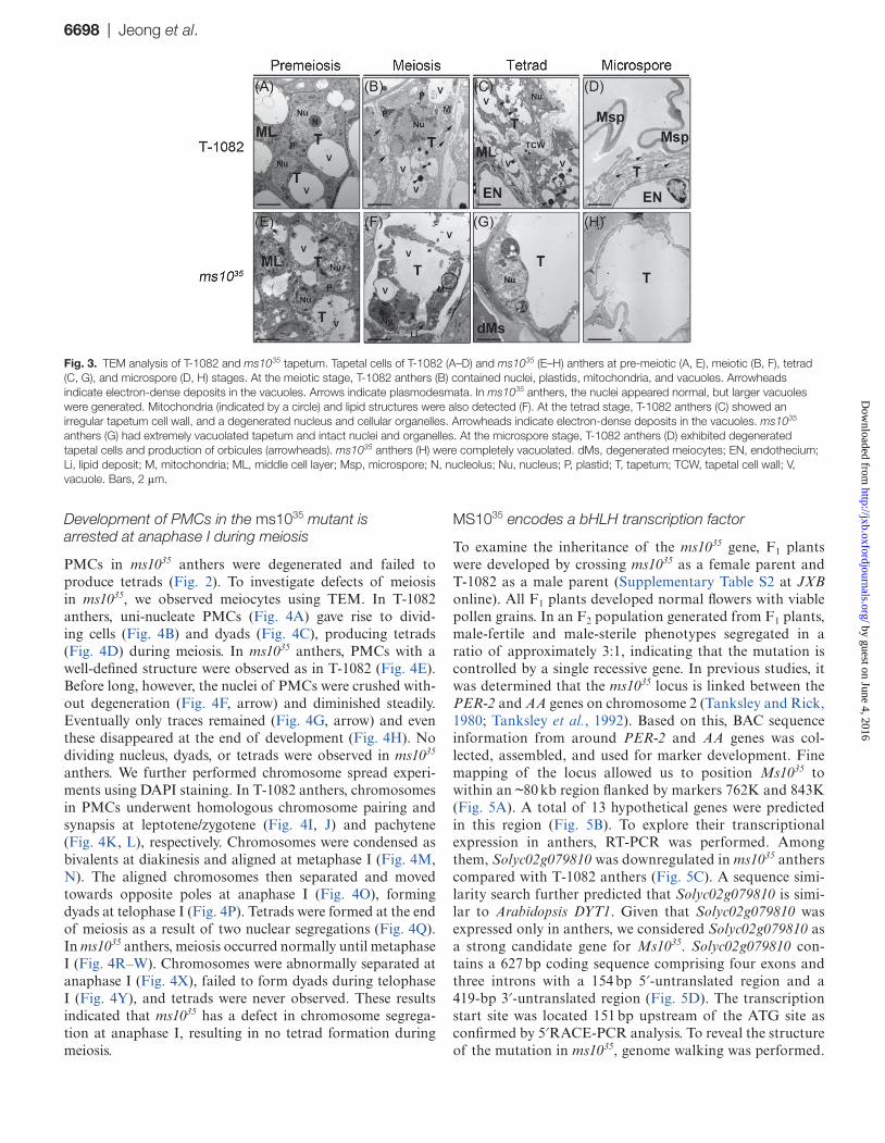
To examine in detail the defects of tapetal cells in the ms1035 mutant, we used TEM. In agreement with the light micro-scopic observations, there was no noticeable difference between ms1035 and T-1082 at the pre-meiotic stage (Fig. 3A, E). At the meiotic stage, tapetal cells in T-1082 anthers were still well defined, but more vacuoles were generated and the nuclear membrane began disappearing, indicating that PCD-triggered cell degradation had already commenced (Fig. 3B). By contrast, ms1035 tapetal cells at the meiotic stage showed abnormal morphology: the cytoplasm of ms1035 tapetal cells was highly vacuolated and showed extensive lipid deposits (Fig. 3F). At the tetrad stage in T-1082, the cytoplasm was
condensed and deeply stained. The nuclear membrane and cellular organelles had disappeared. Electron-dense deposits were observed in the vacuoles (Fig. 3C). At the tetrad stage of ms1035, tapetal cells were greatly expanded and vacuolated. Cellular organelles including the nucleus still maintained their structures (Fig. 3G). At the microspore stage of T-1082 tapetal cells, the cytoplasm and cell walls were diminished. Nuclei and cellular organelles were also completely absent. Instead, orbicules were distributed along the loosened tape-tum cells facing the microspore (Fig. 3D). In ms1035 tapetal cells at the microspore stage, tapetal cells were still enlarged and vacuolated (Fig. 3H). These observations reveal that the ms1035 mutant had abnormally vacuolated tapetal cells with aborted degeneration.
Fig. 2. Histological characterization of anther development in T-1082 and ms1035 flowers. Cross sections of T-1082 (A–C, G–I) and ms1035 (D–F, J–L) anthers at pre-meiotic (A, D), meiotic (B, E), tetrad (C, F), microspore (G, J), mitotic (H, K), and dehiscence (I, L) stages. dMs, degenerated meiocytes; dT, degenerated tapetum; En, endothecium; Ep, epidermis; ML, middle cell layer; Msp, microspore; PMC, pollen mother cell; SC, sporogenous cell; T, tapetum; Tds, tetrads. Bars, 50 μm. (This figure is available in colour at JXB online.)
by guest on June 4, 2016http://jxb.oxfordjournals.org/
Dow
nloaded from
6698 | Jeong et al.
Development of PMCs in the ms1035 mutant is arrested at anaphase I during meiosis
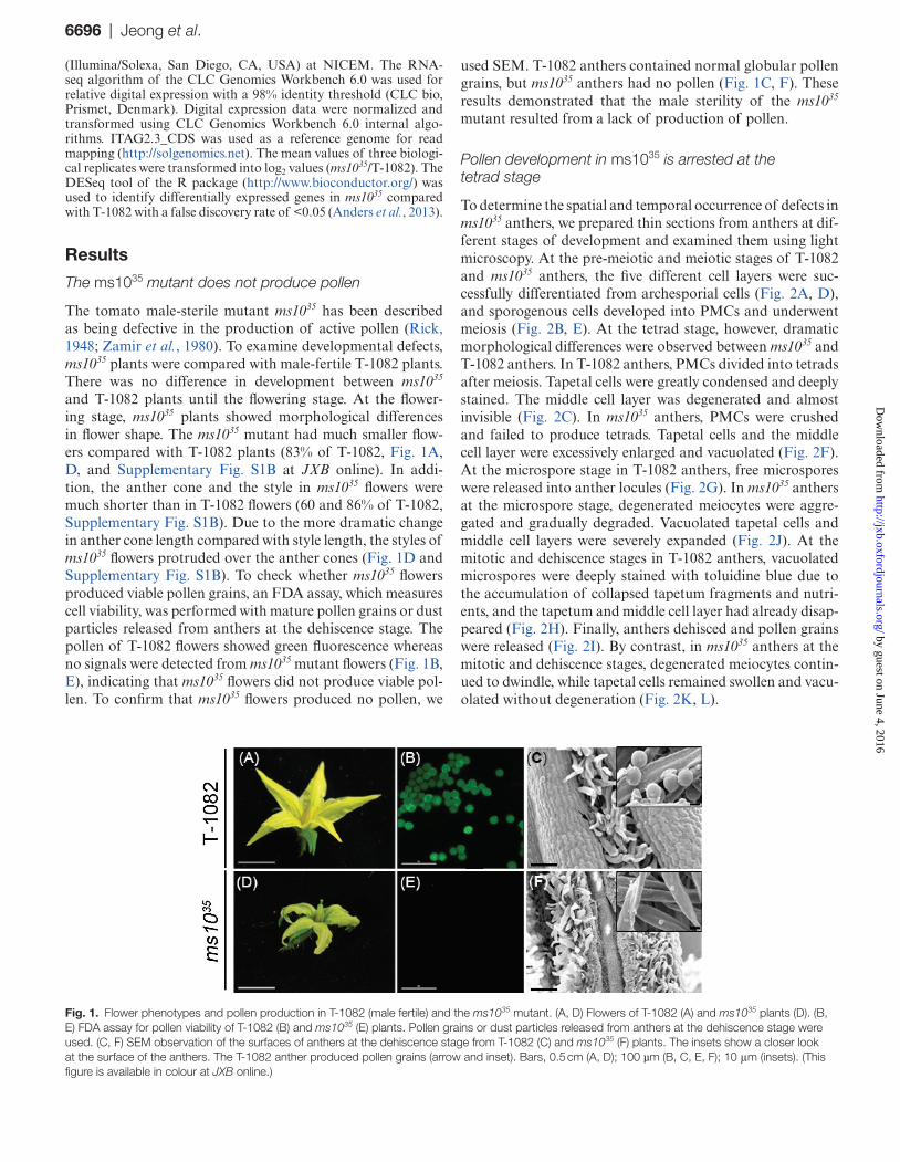
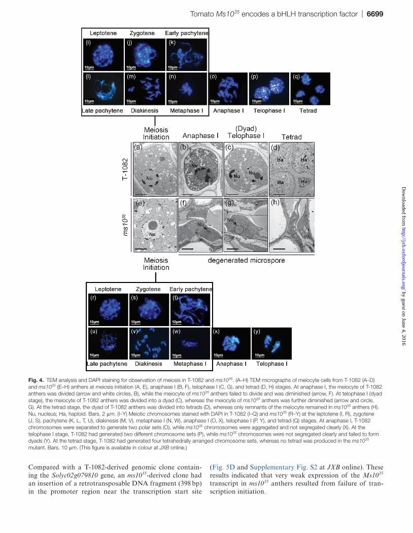
PMCs in ms1035 anthers were degenerated and failed to produce tetrads (Fig. 2). To investigate defects of meiosis in ms1035, we observed meiocytes using TEM. In T-1082 anthers, uni-nucleate PMCs (Fig. 4A) gave rise to divid-ing cells (Fig. 4B) and dyads (Fig. 4C), producing tetrads (Fig. 4D) during meiosis. In ms1035 anthers, PMCs with a well-defined structure were observed as in T-1082 (Fig. 4E). Before long, however, the nuclei of PMCs were crushed with-out degeneration (Fig. 4F, arrow) and diminished steadily. Eventually only traces remained (Fig. 4G, arrow) and even these disappeared at the end of development (Fig. 4H). No dividing nucleus, dyads, or tetrads were observed in ms1035 anthers. We further performed chromosome spread experi-ments using DAPI staining. In T-1082 anthers, chromosomes in PMCs underwent homologous chromosome pairing and synapsis at leptotene/zygotene (Fig. 4I, J) and pachytene (Fig. 4K, L), respectively. Chromosomes were condensed as bivalents at diakinesis and aligned at metaphase I (Fig. 4M, N). The aligned chromosomes then separated and moved towards opposite poles at anaphase I (Fig. 4O), forming dyads at telophase I (Fig. 4P). Tetrads were formed at the end of meiosis as a result of two nuclear segregations (Fig. 4Q). In ms1035 anthers, meiosis occurred normally until metaphase I (Fig. 4R–W). Chromosomes were abnormally separated at anaphase I (Fig. 4X), failed to form dyads during telophase I (Fig. 4Y), and tetrads were never observed. These results indicated that ms1035 has a defect in chromosome segrega-tion at anaphase I, resulting in no tetrad formation during meiosis.
MS1035 encodes a bHLH transcription factor
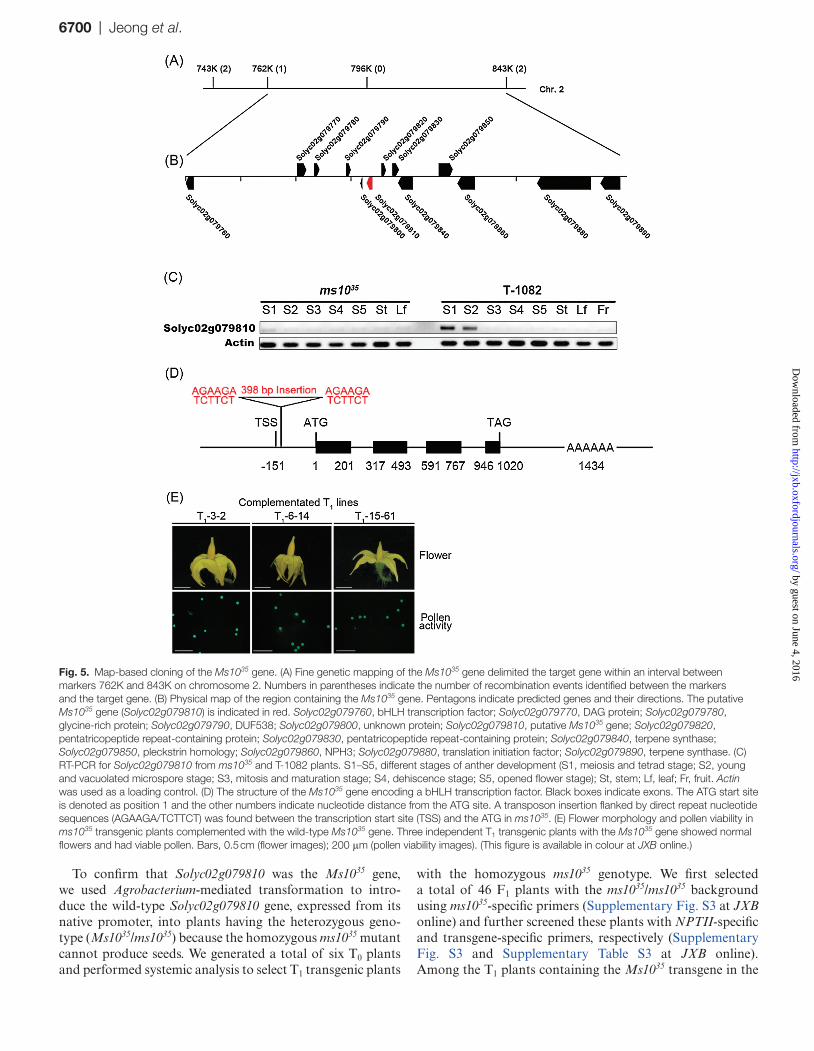
To examine the inheritance of the ms1035 gene, F1 plants were developed by crossing ms1035 as a female parent and T-1082 as a male parent (Supplementary Table S2 at JXB online). All F1 plants developed normal flowers with viable pollen grains. In an F2 population generated from F1 plants, male-fertile and male-sterile phenotypes segregated in a ratio of approximately 3:1, indicating that the mutation is controlled by a single recessive gene. In previous studies, it was determined that the ms1035 locus is linked between the PER-2 and AA genes on chromosome 2 (Tanksley and Rick, 1980; Tanksley et al., 1992). Based on this, BAC sequence information from around PER-2 and AA genes was col-lected, assembled, and used for marker development. Fine mapping of the locus allowed us to position Ms1035 to within an ~80 kb region flanked by markers 762K and 843K (Fig. 5A). A total of 13 hypothetical genes were predicted in this region (Fig. 5B). To explore their transcriptional expression in anthers, RT-PCR was performed. Among them, Solyc02g079810 was downregulated in ms1035 anthers compared with T-1082 anthers (Fig. 5C). A sequence simi-larity search further predicted that Solyc02g079810 is simi-lar to Arabidopsis DYT1. Given that Solyc02g079810 was expressed only in anthers, we considered Solyc02g079810 as a strong candidate gene for Ms1035. Solyc02g079810 con-tains a 627 bp coding sequence comprising four exons and three introns with a 154 bp 5′-untranslated region and a 419-bp 3′-untranslated region (Fig. 5D). The transcription start site was located 151 bp upstream of the ATG site as confirmed by 5′RACE-PCR analysis. To reveal the structure of the mutation in ms1035, genome walking was performed.
Fig. 3. TEM analysis of T-1082 and ms1035 tapetum. Tapetal cells of T-1082 (A–D) and ms1035 (E–H) anthers at pre-meiotic (A, E), meiotic (B, F), tetrad (C, G), and microspore (D, H) stages. At the meiotic stage, T-1082 anthers (B) contained nuclei, plastids, mitochondria, and vacuoles. Arrowheads indicate electron-dense deposits in the vacuoles. Arrows indicate plasmodesmata. In ms1035 anthers, the nuclei appeared normal, but larger vacuoles were generated. Mitochondria (indicated by a circle) and lipid structures were also detected (F). At the tetrad stage, T-1082 anthers (C) showed an irregular tapetum cell wall, and a degenerated nucleus and cellular organelles. Arrowheads indicate electron-dense deposits in the vacuoles. ms1035 anthers (G) had extremely vacuolated tapetum and intact nuclei and organelles. At the microspore stage, T-1082 anthers (D) exhibited degenerated tapetal cells and production of orbicules (arrowheads). ms1035 anthers (H) were completely vacuolated. dMs, degenerated meiocytes; EN, endothecium; Li, lipid deposit; M, mitochondria; ML, middle cell layer; Msp, microspore; N, nucleolus; Nu, nucleus; P, plastid; T, tapetum; TCW, tapetal cell wall; V, vacuole. Bars, 2 μm.
by guest on June 4, 2016http://jxb.oxfordjournals.org/
Dow
nloaded from
Tomato Ms1035 encodes a bHLH transcription factor | 6699
Compared with a T-1082-derived genomic clone contain-ing the Solyc02g079810 gene, an ms1035-derived clone had an insertion of a retrotransposable DNA fragment (398 bp) in the promoter region near the transcription start site
(Fig. 5D and Supplementary Fig. S2 at JXB online). These results indicated that very weak expression of the Ms1035 transcript in ms1035 anthers resulted from failure of tran-scription initiation.
Fig. 4. TEM analysis and DAPI staining for observation of meiosis in T-1082 and ms1035. (A–H) TEM micrographs of meiocyte cells from T-1082 (A–D) and ms1035 (E–H) anthers at meiosis initiation (A, E), anaphase I (B, F), telophase I (C, G), and tetrad (D, H) stages. At anaphase I, the meiocyte of T-1082 anthers was divided (arrow and white circles, B), while the meiocyte of ms1035 anthers failed to divide and was diminished (arrow, F). At telophase I (dyad stage), the meiocyte of T-1082 anthers was divided into a dyad (C), whereas the meiocyte of ms1035 anthers was further diminished (arrow and circle, G). At the tetrad stage, the dyad of T-1082 anthers was divided into tetrads (D), whereas only remnants of the meiocyte remained in ms1035 anthers (H). Nu, nucleus; Ha, haploid. Bars, 2 μm. (I–Y) Meiotic chromosomes stained with DAPI in T-1082 (I–Q) and ms1035 (R–Y) at the leptotene (I, R), zygotene (J, S), pachytene (K, L, T, U), diakinesis (M, V), metaphase I (N, W), anaphase I (O, X), telophase I (P, Y), and tetrad (Q) stages. At anaphase I, T-1082 chromosomes were separated to generate two polar sets (O), while ms1035 chromosomes were aggregated and not segregated clearly (X). At the telophase I stage, T-1082 had generated two different chromosome sets (P), while ms1035 chromosomes were not segregated clearly and failed to form dyads (Y). At the tetrad stage, T-1082 had generated four tetrahedrally arranged chromosome sets, whereas no tetrad was produced in the ms1035 mutant. Bars. 10 μm. (This figure is available in colour at JXB online.)
by guest on June 4, 2016http://jxb.oxfordjournals.org/
Dow
nloaded from
6700 | Jeong et al.
To confirm that Solyc02g079810 was the Ms1035 gene, we used Agrobacterium-mediated transformation to intro-duce the wild-type Solyc02g079810 gene, expressed from its native promoter, into plants having the heterozygous geno-type (Ms1035/ms1035) because the homozygous ms1035 mutant cannot produce seeds. We generated a total of six T0 plants and performed systemic analysis to select T1 transgenic plants
with the homozygous ms1035 genotype. We first selected a total of 46 F1 plants with the ms1035/ms1035 background using ms1035-specific primers (Supplementary Fig. S3 at JXB online) and further screened these plants with NPTII-specific and transgene-specific primers, respectively (Supplementary Fig. S3 and Supplementary Table S3 at JXB online). Among the T1 plants containing the Ms1035 transgene in the
Fig. 5. Map-based cloning of the Ms1035 gene. (A) Fine genetic mapping of the Ms1035 gene delimited the target gene within an interval between markers 762K and 843K on chromosome 2. Numbers in parentheses indicate the number of recombination events identified between the markers and the target gene. (B) Physical map of the region containing the Ms1035 gene. Pentagons indicate predicted genes and their directions. The putative Ms1035 gene (Solyc02g079810) is indicated in red. Solyc02g079760, bHLH transcription factor; Solyc02g079770, DAG protein; Solyc02g079780, glycine-rich protein; Solyc02g079790, DUF538; Solyc02g079800, unknown protein; Solyc02g079810, putative Ms1035 gene; Solyc02g079820, pentatricopeptide repeat-containing protein; Solyc02g079830, pentatricopeptide repeat-containing protein; Solyc02g079840, terpene synthase; Solyc02g079850, pleckstrin homology; Solyc02g079860, NPH3; Solyc02g079880, translation initiation factor; Solyc02g079890, terpene synthase. (C) RT-PCR for Solyc02g079810 from ms1035 and T-1082 plants. S1–S5, different stages of anther development (S1, meiosis and tetrad stage; S2, young and vacuolated microspore stage; S3, mitosis and maturation stage; S4, dehiscence stage; S5, opened flower stage); St, stem; Lf, leaf; Fr, fruit. Actin was used as a loading control. (D) The structure of the Ms1035 gene encoding a bHLH transcription factor. Black boxes indicate exons. The ATG start site is denoted as position 1 and the other numbers indicate nucleotide distance from the ATG site. A transposon insertion flanked by direct repeat nucleotide sequences (AGAAGA/TCTTCT) was found between the transcription start site (TSS) and the ATG in ms1035. (E) Flower morphology and pollen viability in ms1035 transgenic plants complemented with the wild-type Ms1035 gene. Three independent T1 transgenic plants with the Ms1035 gene showed normal flowers and had viable pollen. Bars, 0.5 cm (flower images); 200 μm (pollen viability images). (This figure is available in colour at JXB online.)
by guest on June 4, 2016http://jxb.oxfordjournals.org/
Dow
nloaded from
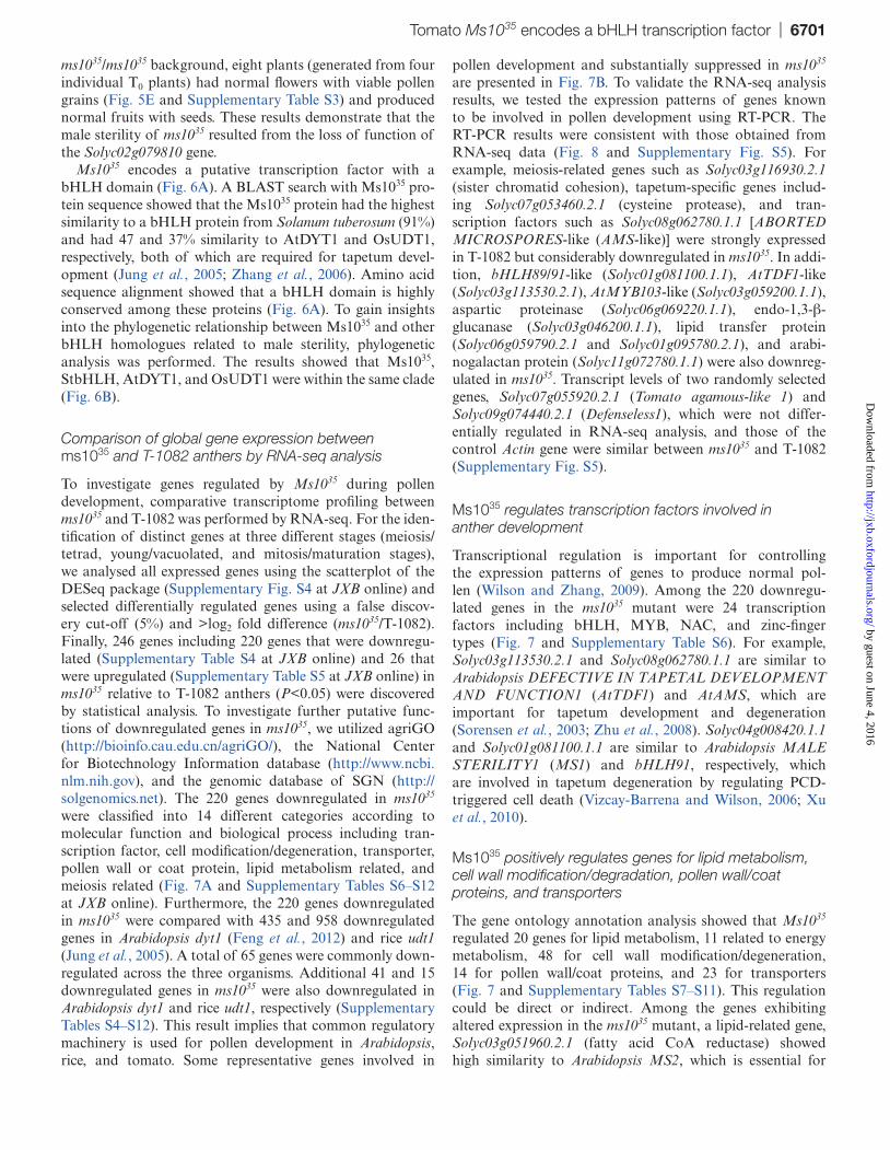
Tomato Ms1035 encodes a bHLH transcription factor | 6701
ms1035/ms1035 background, eight plants (generated from four individual T0 plants) had normal flowers with viable pollen grains (Fig. 5E and Supplementary Table S3) and produced normal fruits with seeds. These results demonstrate that the male sterility of ms1035 resulted from the loss of function of the Solyc02g079810 gene.
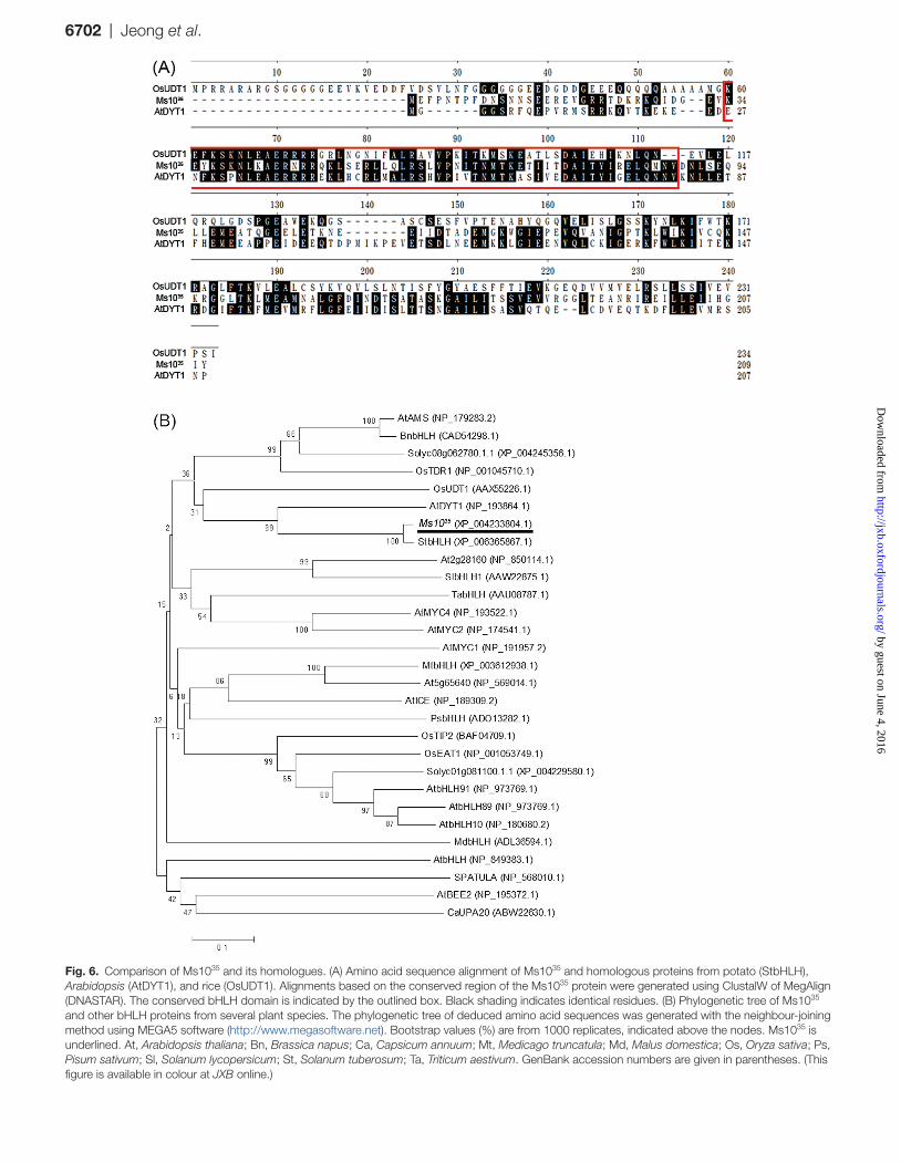
Ms1035 encodes a putative transcription factor with a bHLH domain (Fig. 6A). A BLAST search with Ms1035 pro-tein sequence showed that the Ms1035 protein had the highest similarity to a bHLH protein from Solanum tuberosum (91%) and had 47 and 37% similarity to AtDYT1 and OsUDT1, respectively, both of which are required for tapetum devel-opment (Jung et al., 2005; Zhang et al., 2006). Amino acid sequence alignment showed that a bHLH domain is highly conserved among these proteins (Fig. 6A). To gain insights into the phylogenetic relationship between Ms1035 and other bHLH homologues related to male sterility, phylogenetic analysis was performed. The results showed that Ms1035, StbHLH, AtDYT1, and OsUDT1 were within the same clade (Fig. 6B).
Comparison of global gene expression between ms1035 and T-1082 anthers by RNA-seq analysis
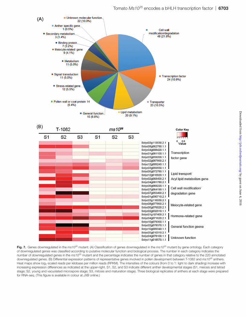
To investigate genes regulated by Ms1035 during pollen development, comparative transcriptome profiling between ms1035 and T-1082 was performed by RNA-seq. For the iden-tification of distinct genes at three different stages (meiosis/tetrad, young/vacuolated, and mitosis/maturation stages), we analysed all expressed genes using the scatterplot of the DESeq package (Supplementary Fig. S4 at JXB online) and selected differentially regulated genes using a false discov-ery cut-off (5%) and >log2 fold difference (ms1035/T-1082). Finally, 246 genes including 220 genes that were downregu-lated (Supplementary Table S4 at JXB online) and 26 that were upregulated (Supplementary Table S5 at JXB online) in ms1035 relative to T-1082 anthers (P<0.05) were discovered by statistical analysis. To investigate further putative func-tions of downregulated genes in ms1035, we utilized agriGO (http://bioinfo.cau.edu.cn/agriGO/), the National Center for Biotechnology Information database (http://www.ncbi.nlm.nih.gov), and the genomic database of SGN (http://solgenomics.net). The 220 genes downregulated in ms1035 were classified into 14 different categories according to molecular function and biological process including tran-scription factor, cell modification/degeneration, transporter, pollen wall or coat protein, lipid metabolism related, and meiosis related (Fig. 7A and Supplementary Tables S6–S12 at JXB online). Furthermore, the 220 genes downregulated in ms1035 were compared with 435 and 958 downregulated genes in Arabidopsis dyt1 (Feng et al., 2012) and rice udt1 (Jung et al., 2005). A total of 65 genes were commonly down-regulated across the three organisms. Additional 41 and 15 downregulated genes in ms1035 were also downregulated in Arabidopsis dyt1 and rice udt1, respectively (Supplementary Tables S4–S12). This result implies that common regulatory machinery is used for pollen development in Arabidopsis, rice, and tomato. Some representative genes involved in
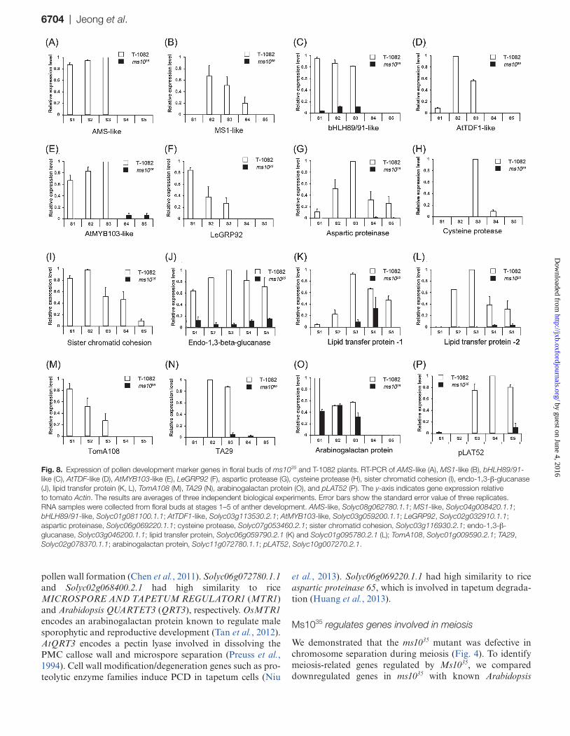
pollen development and substantially suppressed in ms1035 are presented in Fig. 7B. To validate the RNA-seq analysis results, we tested the expression patterns of genes known to be involved in pollen development using RT-PCR. The RT-PCR results were consistent with those obtained from RNA-seq data (Fig. 8 and Supplementary Fig. S5). For example, meiosis-related genes such as Solyc03g116930.2.1 (sister chromatid cohesion), tapetum-specific genes includ-ing Solyc07g053460.2.1 (cysteine protease), and tran-scription factors such as Solyc08g062780.1.1 [ABORTED MICROSPORES-like (AMS-like)] were strongly expressed in T-1082 but considerably downregulated in ms1035. In addi-tion, bHLH89/91-like (Solyc01g081100.1.1), AtTDF1-like (Solyc03g113530.2.1), AtMYB103-like (Solyc03g059200.1.1), aspartic proteinase (Solyc06g069220.1.1), endo-1,3-β-glucanase (Solyc03g046200.1.1), lipid transfer protein (Solyc06g059790.2.1 and Solyc01g095780.2.1), and arabi-nogalactan protein (Solyc11g072780.1.1) were also downreg-ulated in ms1035. Transcript levels of two randomly selected genes, Solyc07g055920.2.1 (Tomato agamous-like 1) and Solyc09g074440.2.1 (Defenseless1), which were not differ-entially regulated in RNA-seq analysis, and those of the control Actin gene were similar between ms1035 and T-1082 (Supplementary Fig. S5).
Ms1035 regulates transcription factors involved in anther development
Transcriptional regulation is important for controlling the expression patterns of genes to produce normal pol-len (Wilson and Zhang, 2009). Among the 220 downregu-lated genes in the ms1035 mutant were 24 transcription factors including bHLH, MYB, NAC, and zinc-finger types (Fig. 7 and Supplementary Table S6). For example, Solyc03g113530.2.1 and Solyc08g062780.1.1 are similar to Arabidopsis DEFECTIVE IN TAPETAL DEVELOPMENT AND FUNCTION1 (AtTDF1) and AtAMS, which are important for tapetum development and degeneration (Sorensen et al., 2003; Zhu et al., 2008). Solyc04g008420.1.1 and Solyc01g081100.1.1 are similar to Arabidopsis MALE STERILITY1 (MS1) and bHLH91, respectively, which are involved in tapetum degeneration by regulating PCD-triggered cell death (Vizcay-Barrena and Wilson, 2006; Xu et al., 2010).
Ms1035 positively regulates genes for lipid metabolism, cell wall modification/degradation, pollen wall/coat proteins, and transporters
The gene ontology annotation analysis showed that Ms1035 regulated 20 genes for lipid metabolism, 11 related to energy metabolism, 48 for cell wall modification/degeneration, 14 for pollen wall/coat proteins, and 23 for transporters (Fig. 7 and Supplementary Tables S7–S11). This regulation could be direct or indirect. Among the genes exhibiting altered expression in the ms1035 mutant, a lipid-related gene, Solyc03g051960.2.1 (fatty acid CoA reductase) showed high similarity to Arabidopsis MS2, which is essential for
by guest on June 4, 2016http://jxb.oxfordjournals.org/
Dow
nloaded from
6702 | Jeong et al.
Fig. 6. Comparison of Ms1035 and its homologues. (A) Amino acid sequence alignment of Ms1035 and homologous proteins from potato (StbHLH), Arabidopsis (AtDYT1), and rice (OsUDT1). Alignments based on the conserved region of the Ms1035 protein were generated using ClustalW of MegAlign (DNASTAR). The conserved bHLH domain is indicated by the outlined box. Black shading indicates identical residues. (B) Phylogenetic tree of Ms1035 and other bHLH proteins from several plant species. The phylogenetic tree of deduced amino acid sequences was generated with the neighbour-joining method using MEGA5 software (http://www.megasoftware.net). Bootstrap values (%) are from 1000 replicates, indicated above the nodes. Ms1035 is underlined. At, Arabidopsis thaliana; Bn, Brassica napus; Ca, Capsicum annuum; Mt, Medicago truncatula; Md, Malus domestica; Os, Oryza sativa; Ps, Pisum sativum; Sl, Solanum lycopersicum; St, Solanum tuberosum; Ta, Triticum aestivum. GenBank accession numbers are given in parentheses. (This figure is available in colour at JXB online.)
by guest on June 4, 2016http://jxb.oxfordjournals.org/
Dow
nloaded from
Tomato Ms1035 encodes a bHLH transcription factor | 6703
Fig. 7. Genes downregulated in the ms1035 mutant. (A) Classification of genes downregulated in the ms1035 mutant by gene ontology. Each category of downregulated genes was classified according to putative molecular function and biological process. The number in each category indicates the number of downregulated genes in the ms1035 mutant and the percentage indicates the number of genes in that category relative to the 220 annotated downregulated genes. (B) Differential expression patterns of representative genes involved in pollen development between T-1082 and ms1035 anthers. Heat maps show log2-scaled reads per kilobase per million reads (RPKM). The intensities of the colours (from 0 to 1: light to dark shading) increase with increasing expression differences as indicated at the upper-right. S1, S2, and S3 indicate different anther developmental stages (S1, meiosis and tetrad stage; S2, young and vacuolated microspore stage; S3, mitosis and maturation stage). Three biological replicates of anthers at each stage were prepared for RNA-seq. (This figure is available in colour at JXB online.)
by guest on June 4, 2016http://jxb.oxfordjournals.org/
Dow
nloaded from
6704 | Jeong et al.
pollen wall formation (Chen et al., 2011). Solyc06g072780.1.1 and Solyc02g068400.2.1 had high similarity to rice MICROSPORE AND TAPETUM REGULATOR1 (MTR1) and Arabidopsis QUARTET3 (QRT3), respectively. OsMTR1 encodes an arabinogalactan protein known to regulate male sporophytic and reproductive development (Tan et al., 2012). AtQRT3 encodes a pectin lyase involved in dissolving the PMC callose wall and microspore separation (Preuss et al., 1994). Cell wall modification/degeneration genes such as pro-teolytic enzyme families induce PCD in tapetum cells (Niu
et al., 2013). Solyc06g069220.1.1 had high similarity to rice aspartic proteinase 65, which is involved in tapetum degrada-tion (Huang et al., 2013).
Ms1035 regulates genes involved in meiosis
We demonstrated that the ms1035 mutant was defective in chromosome separation during meiosis (Fig. 4). To identify meiosis-related genes regulated by Ms1035, we compared downregulated genes in ms1035 with known Arabidopsis
Fig. 8. Expression of pollen development marker genes in floral buds of ms1035 and T-1082 plants. RT-PCR of AMS-like (A), MS1-like (B), bHLH89/91-like (C), AtTDF-like (D), AtMYB103-like (E), LeGRP92 (F), aspartic protease (G), cysteine protease (H), sister chromatid cohesion (I), endo-1,3-β-glucanase (J), lipid transfer protein (K, L), TomA108 (M), TA29 (N), arabinogalactan protein (O), and pLAT52 (P). The y-axis indicates gene expression relative to tomato Actin. The results are averages of three independent biological experiments. Error bars show the standard error value of three replicates. RNA samples were collected from floral buds at stages 1–5 of anther development. AMS-like, Solyc08g062780.1.1; MS1-like, Solyc04g008420.1.1; bHLH89/91-like, Solyc01g081100.1.1; AtTDF1-like, Solyc03g113530.2.1; AtMYB103-like, Solyc03g059200.1.1; LeGRP92, Solyc02g032910.1.1; aspartic proteinase, Solyc06g069220.1.1; cysteine protease, Solyc07g053460.2.1; sister chromatid cohesion, Solyc03g116930.2.1; endo-1,3-β-glucanase, Solyc03g046200.1.1; lipid transfer protein, Solyc06g059790.2.1 (K) and Solyc01g095780.2.1 (L); TomA108, Solyc01g009590.2.1; TA29, Solyc02g078370.1.1; arabinogalactan protein, Solyc11g072780.1.1; pLAT52, Solyc10g007270.2.1.
by guest on June 4, 2016http://jxb.oxfordjournals.org/
Dow
nloaded from
Tomato Ms1035 encodes a bHLH transcription factor | 6705
meiocyte-specific genes (Yang et al., 2011). We found a total of nine genes related to meiosis (Fig. 7 and Supplementary Table S12). For example, Solyc03g116930.2.1 was similar to a protein involved in sister chromatid cohesion (Jin et al., 2009). Solyc08g077860.2.1 encoded a subtilisin-related meiotic serine protease, which is expressed during meiosis and late microsporogenesis in tomato (Riggs et al., 2001). Together, these results imply that Ms1035 is a master regulator control-ling several genes involved in anther development.
Discussion
Ms1035 is important for controlling meiosis and tapetum development
Proper meiosis and development of sporophytic cell lay-ers including the tapetum are essential for successful pollen development in plants (Ma, 2005; Yuan et al., 2009; D. Zhang et al., 2011). In the ms1035 mutant, duplicated chromosomes were not separated to form dyads (Fig. 4X). This failure of dyad formation at anaphase I resulted in the degradation of meiocytes in ms1035. Similar defects were found in Arabidopsis dyt1 and rice udt1 mutants. In the dyt1 mutant, meiocytes were not able to complete cytokinesis, resulting in failure of tetrad formation (Zhang et al., 2006; Feng et al., 2012). Transcript analysis showed that expression of the meiosis-specific gene ROCK-N-ROLLER/AtMER3 (RCK/AtMER3), which is implicated in sister chromatid cohesion, was signifi-cantly reduced in dyt1 (Chen et al., 2005; Zhang et al., 2006), suggesting that DYT1 regulates the expression of prophase I-related genes. In the rice udt1 mutant, meiocytes did not pro-duce tetrads due to incomplete meiosis, and PAIR1 involved in sister chromatid cohesion was downregulated (Jung et al., 2005; Wang et al., 2011). Although these two homologues were not differentially expressed in the RNA-seq analysis, we found that several other meiosis-related genes were down-regulated in ms1035 anthers (Figs 7 and 8). For example, Solyc03g116930.2.1 is homologous to yeast PRECOCIOUS DISSOCIATION OF SISTERS PROTEIN5 (PDS5), which is important for sister chromatid cohesion such as chromo-some condensation, pairing, and synapsis in prophase I (Jin et al., 2009). Solyc02g032910.1.1 is predicted to encode a protein highly similar to glycine-rich protein, which is impor-tant for sporopollenin deposition on meiocyte and exine for-mation (McNeil and Smith, 2010). Another downregulated gene, Solyc06g076020.2.1, is similar to Heat shock protein 70 (Hsp70), which has an important role supporting cyclin-dependent kinase activity in meiosis I in animals (Eddy, 1999). In plants, Lily messages induced by meiosis 18 (LIM18), showing high similarity to eukaryotic HSP70, is specifically expressed in microsporocytes during meiosis I (Minami et al., 2000). Our results suggested that the incomplete meiosis of ms1035 may be due to downregulation of these genes.
Another significant defect in the ms1035 mutant was abnor-mal tapetum development. Degeneration of the tapetum and middle cell layer was delayed and, consequently, tapetal cells were greatly expanded and vacuolated (Fig. 3). These phe-notypic defects are also commonly observed in Arabidopsis
dyt1 and rice udt1 mutants (Jung et al., 2005; Zhang et al., 2006). Other mutants impaired in tapetum development such as Arabidopsis ams and rice tapetum degeneration retardation 1 (tdr1) also exhibit male sterility (Sorensen et al., 2003; Li et al., 2006). These results support the idea that meiosis and tapetum development are important for pollen development, and that Ms1035 and its homologues have a conserved role in the completion of meiosis and tapetum development.
RNA in situ hybridization analysis revealed that Ms1035 is expressed exclusively in the meiocyte and tapetal tissues at the early stage of anther development (Supplementary Fig. S6). These transcript results are consistent with the phenotypic defects in meiocytes and tapetal cells of the ms1035 mutant. To find putative anther-specific cis-acting regulatory elements in the Ms1035 promoter region, we searched the PLACE database (Higo et al., 1999). The Ms1035 promoter contained several putative transcription binding sites and regulatory sequences. For example, pollen-specific cis-elements such as Agamous binding site, Pollen1LeLAT52, and GTGANTG10 were found near the transcription start site (Supplementary Fig. S2), suggesting that these putative cis-elements regulate the expression of Ms1035 in anthers.
Ms1035 encodes a bHLH transcription factor
The Ms1035 protein sequence had high similarity to StbHLH, AtDYT1, and OsUDT1. Amino acid sequence alignments showed that they contain a conserved bHLH domain at the N-terminal region. The PSORT program (http://mobyle.pasteur.fr/cgi-bin/portal.py?#forms::psort) predicted these proteins to be targeted to the nucleus. Indeed, OsUDT1 is localized to the nucleus and contains a signal peptide at the N terminus (Jung et al., 2005). However, Ms1035, StbHLH, and AtDYT do not contain conventional nuclear signal peptides, and whether they are also localized to the nucleus remains to be elucidated. The phylogenetic analysis showed that Ms1035, StbHLH, AtDYT1, and OsUDT1 belonged to the same clade. Considering the conserved function of Ms1035, AtDYT1, and OsUDT1, it seems possible that StbHLH may be involved in meiosis and tapetum development in potato. A clade contain-ing OsTDR1, AtAMS, BnbHLH, and Solyc08g062780.1.1 was very close to the clade containing Ms1035. Interestingly, OsTDR1 and AtAMS are regulated by OsUDT1 and AtDYT1, respectively, and are also involved in tapetum degeneration at the post-meiotic stage (Sorensen et al., 2003; Li et al., 2006), suggesting that Solyc08g062780.1.1 (AtAMS-like), which was downregulated in ms1035, is probably involved in tapetum degeneration. In addition, OsTIP2 and OsEAT1 in rice and AtbHLH89/91 and AtbHLH10 in Arabidopsis are key regulator genes of tapetal PCD (Xu et al., 2010; Niu et al., 2013; Fu et al., 2014). Tomato Solyc01g081100.1.1 (EAT-like) belongs to the clade including these genes, sug-gesting that Solyc01g081100.1.1 could be involved in tapetal PCD. By contrast, genes in other clades were not related to anther development. For example, AtMYC2 and AtMYC4 are related to abscisic acid signalling (Abe et al., 2003) and AtICE is involved in cold stress (Chinnusamy et al., 2003). These results suggest that the bHLH subfamilies containing
by guest on June 4, 2016http://jxb.oxfordjournals.org/
Dow
nloaded from
6706 | Jeong et al.
Ms1035 and AtAMS have a conserved function and were evo-lutionarily separated from other bHLH subfamilies.
Roles of Ms1035-regulated genes during anther development
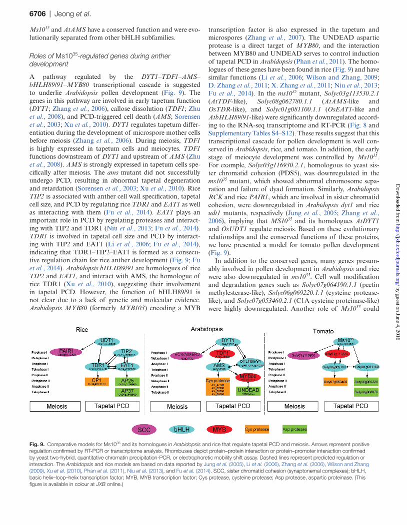
A pathway regulated by the DYT1–TDF1–AMS–bHLH89/91–MYB80 transcriptional cascade is suggested to underlie Arabidopsis pollen development (Fig. 9). The genes in this pathway are involved in early tapetum function (DYT1; Zhang et al., 2006), callose dissolution (TDF1; Zhu et al., 2008), and PCD-triggered cell death (AMS; Sorensen et al., 2003; Xu et al., 2010). DYT1 regulates tapetum differ-entiation during the development of microspore mother cells before meiosis (Zhang et al., 2006). During meiosis, TDF1 is highly expressed in tapetum cells and meiocytes. TDF1 functions downstream of DYT1 and upstream of AMS (Zhu et al., 2008). AMS is strongly expressed in tapetum cells spe-cifically after meiosis. The ams mutant did not successfully undergo PCD, resulting in abnormal tapetal degeneration and retardation (Sorensen et al., 2003; Xu et al., 2010). Rice TIP2 is associated with anther cell wall specification, tapetal cell size, and PCD by regulating rice TDR1 and EAT1 as well as interacting with them (Fu et al., 2014). EAT1 plays an important role in PCD by regulating proteases and interact-ing with TIP2 and TDR1 (Niu et al., 2013; Fu et al., 2014). TDR1 is involved in tapetal cell size and PCD by interact-ing with TIP2 and EAT1 (Li et al., 2006; Fu et al., 2014), indicating that TDR1–TIP2–EAT1 is formed as a consecu-tive regulation chain for rice anther development (Fig. 9; Fu et al., 2014). Arabidopsis bHLH89/91 are homologues of rice TIP2 and EAT1, and interact with AMS, the homologue of rice TDR1 (Xu et al., 2010), suggesting their involvement in tapetal PCD. However, the function of bHLH89/91 is not clear due to a lack of genetic and molecular evidence. Arabidopsis MYB80 (formerly MYB103) encoding a MYB
transcription factor is also expressed in the tapetum and microspores (Zhang et al., 2007). The UNDEAD aspartic protease is a direct target of MYB80, and the interaction between MYB80 and UNDEAD serves to control induction of tapetal PCD in Arabidopsis (Phan et al., 2011). The homo-logues of these genes have been found in rice (Fig. 9) and have similar functions (Li et al., 2006; Wilson and Zhang, 2009; D. Zhang et al., 2011; X. Zhang et al., 2011; Niu et al., 2013; Fu et al., 2014). In the ms1035 mutant, Solyc03g113530.2.1 (AtTDF-like), Solyc08g062780.1.1 (AtAMS-like and OsTDR-like), and Solyc01g081100.1.1 (OsEAT1-like and AtbHLH89/91-like) were significantly downregulated accord-ing to the RNA-seq transcriptome and RT-PCR (Fig. 8 and Supplementary Tables S4–S12). These results suggest that this transcriptional cascade for pollen development is well con-served in Arabidopsis, rice, and tomato. In addition, the early stage of meiocyte development was controlled by Ms1035. For example, Solyc03g116930.2.1, homologous to yeast sis-ter chromatid cohesion (PDS5), was downregulated in the ms1035 mutant, which showed abnormal chromosome sepa-ration and failure of dyad formation. Similarly, Arabidopsis RCK and rice PAIR1, which are involved in sister chromatid cohesion, were downregulated in Arabidopsis dyt1 and rice udt1 mutants, respectively (Jung et al., 2005; Zhang et al., 2006), implying that MS1035 and its homologues AtDYT1 and OsUDT1 regulate meiosis. Based on these evolutionary relationships and the conserved functions of these proteins, we have presented a model for tomato pollen development (Fig. 9).
In addition to the conserved genes, many genes presum-ably involved in pollen development in Arabidopsis and rice were also downregulated in ms1035. Cell wall modification and degradation genes such as Solyc07g064190.1.1 (pectin methylesterase-like), Solyc06g069220.1.1 (cysteine protease-like), and Solyc07g053460.2.1 (C1A cysteine proteinase-like) were highly downregulated. Another role of Ms1035 could
Fig. 9. Comparative models for Ms1035 and its homologues in Arabidopsis and rice that regulate tapetal PCD and meiosis. Arrows represent positive regulation confirmed by RT-PCR or transcriptome analysis. Rhombuses depict protein–protein interaction or protein–promoter interaction confirmed by yeast two-hybrid, quantitative chromatin precipitation-PCR, or electrophoretic mobility shift assay. Dashed lines represent predicted regulation or interaction. The Arabidopsis and rice models are based on data reported by Jung et al. (2005), Li et al. (2006), Zhang et al. (2006), Wilson and Zhang (2009), Xu et al. (2010), Phan et al. (2011), Niu et al. (2013), and Fu et al. (2014). SCC, sister chromatid cohesion (synaptonemal complexes); bHLH, basic helix–loop–helix transcription factor; MYB, MYB transcription factor; Cys protease, cysteine protease; Asp protease, aspartic proteinase. (This figure is available in colour at JXB online.)
by guest on June 4, 2016http://jxb.oxfordjournals.org/
Dow
nloaded from
Tomato Ms1035 encodes a bHLH transcription factor | 6707
be supporting pollen wall deposition by delivering materi-als and nutrients to developing microspores. For example, orbicules are known to deliver nutrients to sporopollenin of microspores. In T-1082, orbicules were successfully gen-erated along the degenerated tapetum (Fig. 3D). However, in ms1035, orbicules were not found and no normal sporo-pollenin structures were observed. In ms1035, sporopollenin biosynthesis-related genes such as Solyc12g010920.1.1 (long chain fatty acid reductases) and Solyc04g081780.2.1 (lipase) were downregulated.
ms1035 showed a protruded stigma due to significantly reduced anther cone size. Arabidopsis dyt1 also exhibits a protruded stigma phenotype (Zhang et al., 2006). Tomato Stigma exsertion (Se2.1) encoding a bHLH transcription fac-tor is the major quantitative trait locus for the development of stamen length (Chen et al., 2007). Interestingly, expression of Se2.1 (Solyc02g087860.2.1) was substantially reduced in ms1035 anthers, suggesting that Ms1035 may affect stamen length by regulating Se2.1. As we have shown here, many Ms1035-regulated genes in our RNA-seq data were directly or indirectly involved in anther development. Therefore, our RNA-seq data should provide a good basis for the identifica-tion and analysis of new genes involved in anther develop-ment in tomato.
Supplementary data
Supplementary data are available at JXB online.Supplementary Fig. S1. Differences of organ length in
ms1035 and T-1082 flowers.Supplementary Fig. S2. Nucleotide sequence of the Ms1035
promoter.Supplementary Fig. S3. Complementation of ms1035 trans-
genic plants with the wild-type Ms1035 gene.Supplementary Fig. S4. Scatterplot identification of differ-
entially expressed genes between T-1082 and ms1035 anthers.Supplementary Fig. S5. Expression patterns of genes regu-
lated by Ms1035.Supplementary Fig. S6. Localization of Ms1035 expression
in T-1082 anthers.Supplementary Table S1. Primers used in this study.Supplementary Table S2. Genetic analysis of the ms1035
gene using an F2 population derived from 2–517 (ms1035) and T-1082 (male fertile) plants.
Supplementary Table S3. Summary of ms1035 transgenic plants complemented with the wild-type Ms1035 gene.
Supplementary Table S4. 220 genes downregulated in the ms1035 mutant.
Supplementary Table S5. 26 genes upregulated in the ms1035 mutant.
Supplementary Table S6. Transcription factors downregu-lated in the ms1035 mutant.
Supplementary Table S7. Lipid metabolism genes down-regulated in the ms1035 mutant.
Supplementary Table S8. Pollen wall or coat protein genes downregulated in the ms1035 mutant.
Supplementary Table S9. Cell wall modification/degrada-tion genes downregulated in the ms1035 mutant.
Supplementary Table S10. Transporter genes downregu-lated in the ms1035 mutant.
Supplementary Table S11. Energy metabolism-related genes downregulated in the ms1035 mutant.
Supplementary Table S12. Meiosis-related genes downreg-ulated in the ms1035 mutant.
AcknowledgementsThis study was supported by a grant (project no. 710001–07) from the Vegetable Breeding Research Center through the R&D Convergence Center Support Program, Ministry of Agriculture, Food and Rural Affairs (MAFRA), Republic of Korea. This research was also supported by the Golden Seed Project (213002-04-1-CG910), Ministry of Agriculture, Food and Rural Affairs (MAFRA), Ministry of Oceans and Fisheries (MOF), Rural Development Administration (RDA), and Korea Forest Service (KFS). We acknowledge the Korea Research Institute of Bioscience and Biotechnology (KRIBB, Daejeon, Korea) for kindly providing the tomato BAC clones, Dr Young Chae for providing tomato seeds, and Dr Yeong deuk Jo for developing the perl script for RNA-seq.
ReferencesAbe H, Urao T, Ito T, Seki M, Shinozaki K, Yamaguchi-Shinozaki K. 2003. Arabidopsis AtMYC2 (bHLH) and AtMYB2 (MYB) function as transcriptional activators in abscisic acid signaling. The Plant Cell 15, 63–78.
Anders S, McCarthy DJ, Chen Y, Okoniewski M, Smyth GK, Huber W, Robinson MD. 2013. Count-based differential expression analysis of RNA sequencing data using R and Bioconductor. Nature Protocols 8, 1765–1786.
Brukhin V, Hernould M, Gonzalez N, Chevalier C, Mouras A. 2003. Flower development schedule in tomato Lycopersicon esculentum cv. sweet cherry. Sexual Plant Reproduction 15, 311–320.
Canales C, Bhatt AM, Scott R, Dickinson H. 2002. EXS, a putative LRR receptor kinase, regulates male germline cell number and tapetal identity and promotes seed development in Arabidopsis. Current Biology 12, 1718–1727.
Chen C, Zhang W, Timofejeva L, Gerardin Y, Ma H. 2005. The Arabidopsis ROCK-N-ROLLERS gene encodes a homolog of the yeast ATP-dependent DNA helicase MER3 and is required for normal meiotic crossover formation. The Plant Journal 43, 321–334.
Chen KY, Cong B, Wing R, Vrebalov J, Tanksley SD. 2007. Changes in regulation of a transcription factor lead to autogamy in cultivated tomatoes. Science 318, 643–645.
Chen W, Yu XH, Zhang K, Shi J, De Oliveira S, Schreiber L, Shanklin J, Zhang D. 2011. Male Sterile2 encodes a plastid-localized fatty acyl carrier protein reductase required for pollen exine development in Arabidopsis. Plant Physiology 157, 842–853.
Chinnusamy V, Ohta M, Kanrar S, Lee BH, Hong X, Agarwal M, Zhu JK. 2003. ICE1: a regulator of cold-induced transcriptome and freezing tolerance in Arabidopsis. Genes & Development 17, 1043–1054.
Corral-Martínez P, Nuez F, Seguí-Simarro JM. 2011. Genetic, quantitative and microscopic evidence for fusion of haploid nuclei and growth of somatic calli in cultured ms1035 tomato anthers. Euphytica 178, 215–228.
de Givry S, Bouchez M, Chabrier P, Milan D, Schiex T. 2005. CARHTA GENE: multipopulation integrated genetic and radiation hybrid mapping. Bioinformatics 21, 1703–1704.
Deveshwar P, Bovill WD, Sharma R, Able JA, Kapoor S. 2011. Analysis of anther transcriptomes to identify genes contributing to meiosis and male gametophyte development in rice. BMC Plant Biology 11, 78.
Eddy EM. 1999. Role of heat shock protein HSP70-2 in spermatogenesis. Reviews of Reproduction 4, 23–30.
Feng B, Lu D, Ma X, Peng Y, Sun Y, Ning G, Ma H. 2012. Regulation of the Arabidopsis anther transcriptome by DYT1 for pollen development. The Plant Journal 72, 612–624.
Fu Z, Yu J, Cheng X, Zong X, Xu J, Chen M, Li Z, Zhang D. 2014. The rice basic helix–loop–helix transcription factor TDR INTERACTING
by guest on June 4, 2016http://jxb.oxfordjournals.org/
Dow
nloaded from
6708 | Jeong et al.
PROTEIN2 is a central switch in early anther development. The Plant Cell 26, 1512–1524.
Georgiev H. 1991. Heterosis in tomato breeding. In: Kalloo G, ed. Genetic improvement of tomato, 14, Berlin/Heidelberg: Springer, 83–98.
Goldberg RB, Beals TP, Sanders PM. 1993. Anther development: basic principles and practical applications. The Plant Cell 5, 1217–1229.
Gorguet B, Schipper D, van Lammeren A, Visser RG, van Heusden AW. 2009. ps-2, the gene responsible for functional sterility in tomato, due to non-dehiscent anthers, is the result of a mutation in a novel polygalacturonase gene. Theoretical and Applied Genetics 118, 1199–1209.
Gorman SW, McCormick S. 1997. Male sterility in tomato. Critical Reviews in Plant Sciences 16, 31–53.
Higo K, Ugawa Y, Iwamoto M, Korenaga T. 1999. Plant cis-acting regulatory DNA elements (PLACE) database: 1999. Nucleic Acids Research 27, 297–300.
Honys D, Twell D. 2004. Transcriptome analysis of haploid male gametophyte development in Arabidopsis. Genome Biology 5, R85.
Huang J, Zhao X, Cheng K, Jiang Y, Ouyang Y, Xu C, Li X, Xiao J, Zhang Q. 2013. OsAP65, a rice aspartic protease, is essential for male fertility and plays a role in pollen germination and pollen tube growth. Journal of Experimental Botany 64, 3351–3360.
Jeong HJ, Jo YD, Park SW, Kang BC. 2010. Identification of Capsicum species using SNP markers based on high resolution melting analysis. Genome 53, 1029–1040.
Jeong HJ, Kwon JK, Pandeya D, Hwang J, Hoang NH, Bae JH, Kang BC. 2011. A survey of natural and ethyl methane sulfonate-induced variations of eIF4E using high-resolution melting analysis in Capsicum. Molecular Breeding 29, 349–360.
Jiang H, Wang FF, Wu YT, Zhou X, Huang XY, Zhu J, Gao JF, Dong RB, Cao KM, Yang ZN. 2009. MULTIPOLAR SPINDLE 1 (MPS1), a novel coiled-coil protein of Arabidopsis thaliana, is required for meiotic spindle organization. The Plant Journal 59, 1001–1010.
Jin H, Guacci V, Yu HG. 2009. Pds5 is required for homologue pairing and inhibits synapsis of sister chromatids during yeast meiosis. Journal of Cell Biology 186, 713–725.
Jung KH, Han MJ, Lee YS, Kim YW, Hwang I, Kim MJ, Kim YK, Nahm BH, An G. 2005. Rice Undeveloped Tapetum1 is a major regulator of early tapetum development. The Plant Cell 17, 2705–2722.
Kang WH, Seo JK, Chung BN, Kim KH, Kang BC. 2012. Helicase domain encoded by Cucumber mosaic virus RNA1 determines systemic infection of Cmr1 in pepper. PLos One 7, e43136.
Kim YM, Jo YD, Kwon JK, Kim IT, Kang BC. 2013. Characterization and inheritance of a novel thermo-sensitive restoration of cytoplasmic male sterility in Capsicum annuum. Scientia Horticulturae 164, 512–520.
Kumar S, Singh PK. 2005. Mechanisms for hybrid development in vegetables. Journal of New Seeds 6, 381–407.
Kwon JK, Kim BD. 2009. Localization of 5S and 25S rRNA genes on somatic and meiotic chromosomes in Capsicum species of chili pepper. Molecules and Cells 27, 205–209.
Li N, Zhang DS, Liu HS, et al. 2006. The rice tapetum degeneration retardation gene is required for tapetum degradation and anther development. The Plant Cell 18, 2999–3014.
Ma H. 2005. Molecular genetic analyses of microsporogenesis and microgametogenesis in flowering plants. Annual Review of Plant Biology 56, 393–434.
McNeil KJ, Smith AG. 2010. A glycine-rich protein that facilitates exine formation during tomato pollen development. Planta 231, 793–808.
Mercier R, Armstrong SJ, Horlow C, Jackson NP, Makaroff CA, Vezon D, Pelletier G, Jones GH, Franklin FC. 2003. The meiotic protein SWI1 is required for axial element formation and recombination initiation in Arabidopsis. Development 130, 3309–3318.
Minami M, Hiratsuka R, Ogata S, Takase H, Hotta Y, Hiratsuka K. 2000. Characterization of a meiosis-associated heat shock protein 70. Plant Biotechnology 17, 145–153.
Niu N, Liang W, Yang X, Jin W, Wilson ZA, Hu J, Zhang D. 2013. EAT1 promotes tapetal cell death by regulating aspartic proteases during male reproductive development in rice. Nature Communications 4, 1445.
Nonomura K, Nakano M, Eiguchi M, Suzuki T, Kurata N. 2006. PAIR2 is essential for homologous chromosome synapsis in rice meiosis I. Journal of Cell Science 119, 217–225.
Nonomura K, Nakano M, Fukuda T, Eiguchi M, Miyao A, Hirochika H, Kurata N. 2004. The novel gene HOMOLOGOUS PAIRING ABERRATION IN RICE MEIOSIS1 of rice encodes a putative coiled-coil protein required for homologous chromosome pairing in meiosis. The Plant Cell 16, 1008–1020.
Phan HA, Iacuone S, Li SF, Parish RW. 2011. The MYB80 transcription factor is required for pollen development and the regulation of tapetal programmed cell death in Arabidopsis thaliana. The Plant Cell 23, 2209–2224.
Preuss D, Rhee SY, Davis RW. 1994. Tetrad analysis possible in Arabidopsis with mutation of the QUARTET (QRT) genes. Science 264, 1458–1460.
Rasmussen N, Green PB. 1993. Organogenesis in flowers of the homeotic green pistillate mutant of tomato (Lycopersicon esculentum). American Journal of Botany 80, 805–813.
Rick CM. 1948. Genetics and development of nine male sterile tomato mutants. Hilgardia 18, 599–633.
Rick CM, Butler L. 1956. Cytogenetics of the tomato. Advances in Genetics 8, 267–382.
Riggs CD, Zeman K, DeGuzman R, Rzepczyk A, Taylor AA. 2001. Antisense inhibition of a tomato meiotic proteinase suggests functional redundancy of proteinases during microsporogenesis. Genome 44, 644–650.
Seong ES, Baek K-H, Oh S-K, Jo SH, Yi SY, Park JM, Joung YH, Lee S, Cho HS, Choi D. 2007. Induction of enhanced tolerance to cold stress and disease by overexpression of the pepper CaPIF1 gene in tomato. Physiologia Plantarum 129, 555–566.
Shao T, Tang D, Wang K, Wang M, Che L, Qin B, Yu H, Li M, Gu M, Cheng Z. 2011. OsREC8 is essential for chromatid cohesion and metaphase I monopolar orientation in rice meiosis. Plant Physiology 156, 1386–1396.
Smyth DR, Bowman JL, Meyerowitz EM. 1990. Early flower development in Arabidopsis. The Plant Cell 2, 755–767.
Sorensen AM, Krober S, Unte US, Huijser P, Dekker K, Saedler H. 2003. The Arabidopsis ABORTED MICROSPORES (AMS) gene encodes a MYC class transcription factor. The Plant Journal 33, 413–423.
Tan H, Liang W, Hu J, Zhang D. 2012. MTR1 encodes a secretory fasciclin glycoprotein required for male reproductive development in rice. Developmental Cell 22, 1127–1137.
Tanksley SD, Ganal MW, Prince JP, et al.. 1992. High density molecular linkage maps of the tomato and potato genomes. Genetics 132, 1141–1160.
Tanksley SD, Rick CM. 1980. Isozymic gene linkage map of the tomato: applications in genetics and breeding. Theoretical and Applied Genetics 58, 161–170.
Tomato Genome Consortium. 2012. The tomato genome sequence provides insights into fleshy fruit evolution. Nature 485, 635–641.
Vizcay-Barrena G, Wilson ZA. 2006. Altered tapetal PCD and pollen wall development in the Arabidopsis ms1 mutant. Journal of Experimental Botany 57, 2709–2717.
Wang K, Wang M, Tang D, Shen Y, Qin B, Li M, Cheng Z. 2011. PAIR3, an axis-associated protein, is essential for the recruitment of recombination elements onto meiotic chromosomes in rice. Molecular Biology of the Cell 22, 12–19.
Wilson ZA, Zhang DB. 2009. From Arabidopsis to rice: pathways in pollen development. Journal of Experimental Botany 60, 1479–1492.
Xu J, Yang C, Yuan Z, Zhang D, Gondwe MY, Ding Z, Liang W, Zhang D, Wilson ZA. 2010. The ABORTED MICROSPORES regulatory network is required for postmeiotic male reproductive development in Arabidopsis thaliana. The Plant Cell 22, 91–107.
Yang H, Lu P, Wang Y, Ma H. 2011. The transcriptome landscape of Arabidopsis male meiocytes from high-throughput sequencing: the complexity and evolution of the meiotic process. The Plant Journal 65, 503–516.
Yang SL, Xie LF, Mao HZ, Puah CS, Yang WC, Jiang L, Sundaresan V, Ye D. 2003. Tapetum determinant1 is required for cell specialization in the Arabidopsis anther. The Plant Cell 15, 2792–2804.
by guest on June 4, 2016http://jxb.oxfordjournals.org/
Dow
nloaded from
Tomato Ms1035 encodes a bHLH transcription factor | 6709
Yuan W, Li X, Chang Y, Wen R, Chen G, Zhang Q, Wu C. 2009. Mutation of the rice gene PAIR3 results in lack of bivalent formation in meiosis. The Plant Journal 59, 303–315.
Zamir D, Jones RA, Kedar N. 1980. Anther culture of male-sterile tomato (Lycopersicon esculentum Mill.) mutants. Plant Science Letters 17, 353–361.
Zhang D, Luo X, Zhu L. 2011. Cytological analysis and genetic control of rice anther development. Journal of Genetics and Genomics 38, 379–390.
Zhang W, Sun Y, Timofejeva L, Chen C, Grossniklaus U, Ma H. 2006. Regulation of Arabidopsis tapetum development and function by DYSFUNCTIONAL TAPETUM1 (DYT1) encoding a putative bHLH transcription factor. Development 133, 3085–3095.
Zhang X, Liu L, Cheng Z, Lei C, Wang J, Guo X, Wu F, Ikehashi H, Wang H, Wan J. 2011. Pollen semi-sterility1 encodes a kinesin-1-like protein important for male meiosis, anther dehiscence, and fertility in rice. The Plant Cell 23, 111–129.
Zhang ZB, Zhu J, Gao JF, et al. 2007. Transcription factor AtMYB103 is required for anther development by regulating tapetum development,
callose dissolution and exine formation in Arabidopsis. The Plant Journal 52, 528–538.
Zhao DZ, Wang GF, Speal B, Ma H. 2002. The EXCESS MICROSPOROCYTE1 gene encodes a putative leucine-rich repeat receptor protein kinase that controls somatic and reproductive cell fates in the Arabidopsis anther. Genes & Development 16, 2021–2031.
Zhong S, Joung JG, Zheng Y, Chen YR, Liu B, Shao Y, Xiang JZ, Fei Z, Giovannoni JJ. 2011. High-throughput illumina strand-specific RNA sequencing library preparation. Cold Spring Harbor Protocols 2011, 940–949.
Zhou S, Wang Y, Li W, et al. 2011. Pollen semi-sterility1 encodes a kinesin-1–like protein important for male meiosis, anther dehiscence, and fertility in rice. The Plant Cell 23, 111–129.
Zhu S, Wang Y, Li W, et al. 2008. Defective in Tapetal development and function 1 is essential for anther development and tapetal function for microspore maturation in Arabidopsis. The Plant Journal 55, 266–277.
Zickler D, Kleckner N. 1999. Meiotic chromosomes: integrating structure and function. Annual Review of Genetics 33, 603–754.
by guest on June 4, 2016http://jxb.oxfordjournals.org/
Dow
nloaded from
Related Documents


![Plant Propagation.ppt [Read-Only] - University of Arizona...1. Remove anthers from the flower of the female parent prior to pollen maturation to prevent self‐ pollination. 2. Collect](https://static.cupdf.com/doc/110x72/600e76a9ead0380480722ee1/plant-read-only-university-of-arizona-1-remove-anthers-from-the-flower.jpg)