Toll-like receptors control autophagy Mo ´ nica A Delgado, Rasha A Elmaoued, Alexander S Davis, George Kyei and Vojo Deretic* Department of Molecular Genetics and Microbiology, University of New Mexico, Health Sciences Center, Albuquerque, NM, USA Autophagy is a newly recognized innate defense mechan- ism, acting as a cell-autonomous system for elimination of intracellular pathogens. The signals and signalling pathways inducing autophagy in response to pathogen invasion are presently not known. Here we show that autophagy is controlled by recognizing conserved patho- gen-associated molecular patterns (PAMPs). We screened a PAMP library for effects on autophagy in RAW 264.7 macrophages and found that several prototype Toll-like receptor (TLR) ligands induced autophagy. Single- stranded RNA and TLR7 generated the most potent effects. Induction of autophagy via TLR7 depended on MyD88 expression. Stimulation of autophagy with TLR7 ligands was functional in eliminating intracellular microbes, even when the target pathogen was normally not associated with TLR7 signalling. These findings link two innate immunity defense systems, TLR signalling and autophagy, provide a potential molecular mechanism for induction of autophagy in response to pathogen invasion, and show that the newly recognized ability of TLR ligands to stimulate autophagy can be used to treat intracellular pathogens. The EMBO Journal (2008) 27, 1110–1121. doi:10.1038/ emboj.2008.31; Published online 13 March 2008 Subject Categories: immunology Keywords: autophagy; HIV; LC3; TLR; tuberculosis Introduction Autophagy is a fundamental cellular homeostatic process, where cells ingest and digest portions of their own cytoplasm, thus periodically cleansing their interiors (Levine and Klionsky, 2004). Autophagy is based on formation within the cytosol of double-membrane organelles termed autopha- gosomes to sequester portions of the cytoplasm earmarked for autophagic removal or turnover (Mizushima et al, 2002). Autophagosomes fuse with lysosomes to form autolyso- somes, resulting in degradation of the captured cytosolic constituents, including (i) long-lived proteins and other stable macromolecules and (ii) membranous structures such as damaged, spent or surplus organelles (Levine and Klionsky, 2004). Autophagy endows cells with a capability to adjust down their biomass and turn over their own constitu- ents at times of starvation (Lum et al, 2005). This provides amino acids and other components for the synthesis of essential proteins and other macromolecules allowing the cells to survive. Autophagy also removes faulty organelles such as spuriously damaged or leaky mitochondria lest cells undergo unscheduled apoptosis and die. All eukaryotic cells from yeast to man are capable of undergoing autophagy, and most cells in the human body can activate this process (Levine and Klionsky, 2004) or even undergo a considerable level of constitutive autophagy (Schmid et al, 2007). Since autophagy affects many cell types, it has a broad effect on a wide range of normal physiological processes, including ageing and diseases such as cancer (Levine, 2007) and neurodegeneration (Alzheimer’s, Huntington’s and Parkinson’s diseases and ataxias; Rubinsztein, 2006). It has recently been recognized that akin to the role of autophagy in eliminating toxic protein aggregates and thus protecting against neurodegeneration (Nixon, 2006), autophagy also plays a role in innate immunity against intracellular patho- gens (Deretic, 2005; Levine and Deretic, 2007; Schmid and Munz, 2007), by clearing microbes directly via ingestion into autophagosomes for subsequent degradation in autolyso- somes (Gutierrez et al, 2004; Nakagawa et al, 2004; Ogawa et al, 2005; Andrade et al, 2006; Orvedahl et al, 2007). Agonist-induced autophagosomes capture and destroy intra- cellular pathogens even when they are safely ensconced in protective vacuoles, such as the immature phagosome harbouring Mycobacterium tuberculosis (Singh et al, 2006), or parasitophorous vacuoles containing Toxoplasma gondii (Andrade et al, 2006; Ling et al, 2006). In addition to these cell-autonomous protective functions against invading patho- gens, autophagy participates in other aspects of immunity (Deretic et al, 2006; Levine and Deretic, 2007; Schmid and Munz, 2007) and is not limited to a role in direct elimination of invading bacteria, protozoans and viruses. For example, autophagy supports sequestration of endogenously synthe- sized viral or self-antigens into autophagosomes and their delivery to major histocompatibility complex (MHC) class II loading compartments, leading to MHC class II-restricted presentation of cytoplasmic antigens (Schmid and Munz, 2007). Autophagy has a role in T-cell homeostasis (Li et al, 2006), for example by controlling T-cell lifespan once they exit the thymus (Pua et al, 2007), and is an effector of Th1–Th2 polarization in defense against intracellular patho- gens (Harris et al, 2007). The role of autophagy in immunity has been further underscored by the recent recognition of a genetic association between a Chron’s disease-susceptibility locus with one of the core autophagy genes, Atg16 (Hampe et al, 2007), and with the human immunity-related GTPase IRGM (Parkes et al, 2007) implicated in autophagy (Singh et al, 2006). Two of the remaining top-tier questions in the field of autophagy are how the presence of potential autophagic Received: 5 June 2007; accepted: 7 February 2008; published online: 13 March 2008 *Corresponding author. Department of Molecular Genetics and Microbiology, University of New Mexico, Health Sciences Center, 915 Camino de Salud NE, Albuquerque, NM 87131, USA. Tel.: þ 1 505 272 0291; Fax: þ 1 505 272 5309; E-mail: [email protected] The EMBO Journal (2008) 27, 1110–1121 | & 2008 European Molecular Biology Organization | All Rights Reserved 0261-4189/08 www.embojournal.org The EMBO Journal VOL 27 | NO 7 | 2008 & 2008 European Molecular Biology Organization EMBO THE EMBO JOURNAL THE EMBO JOURNAL 1110

Welcome message from author
This document is posted to help you gain knowledge. Please leave a comment to let me know what you think about it! Share it to your friends and learn new things together.
Transcript
Toll-like receptors control autophagy
Monica A Delgado, Rasha A Elmaoued,Alexander S Davis, George Kyeiand Vojo Deretic*
Department of Molecular Genetics and Microbiology, Universityof New Mexico, Health Sciences Center, Albuquerque, NM, USA
Autophagy is a newly recognized innate defense mechan-
ism, acting as a cell-autonomous system for elimination
of intracellular pathogens. The signals and signalling
pathways inducing autophagy in response to pathogen
invasion are presently not known. Here we show that
autophagy is controlled by recognizing conserved patho-
gen-associated molecular patterns (PAMPs). We screened
a PAMP library for effects on autophagy in RAW 264.7
macrophages and found that several prototype Toll-like
receptor (TLR) ligands induced autophagy. Single-
stranded RNA and TLR7 generated the most potent effects.
Induction of autophagy via TLR7 depended on MyD88
expression. Stimulation of autophagy with TLR7 ligands
was functional in eliminating intracellular microbes, even
when the target pathogen was normally not associated
with TLR7 signalling. These findings link two innate
immunity defense systems, TLR signalling and autophagy,
provide a potential molecular mechanism for induction
of autophagy in response to pathogen invasion, and
show that the newly recognized ability of TLR ligands to
stimulate autophagy can be used to treat intracellular
pathogens.
The EMBO Journal (2008) 27, 1110–1121. doi:10.1038/
emboj.2008.31; Published online 13 March 2008
Subject Categories: immunology
Keywords: autophagy; HIV; LC3; TLR; tuberculosis
Introduction
Autophagy is a fundamental cellular homeostatic process,
where cells ingest and digest portions of their own cytoplasm,
thus periodically cleansing their interiors (Levine and
Klionsky, 2004). Autophagy is based on formation within
the cytosol of double-membrane organelles termed autopha-
gosomes to sequester portions of the cytoplasm earmarked
for autophagic removal or turnover (Mizushima et al, 2002).
Autophagosomes fuse with lysosomes to form autolyso-
somes, resulting in degradation of the captured cytosolic
constituents, including (i) long-lived proteins and other
stable macromolecules and (ii) membranous structures
such as damaged, spent or surplus organelles (Levine and
Klionsky, 2004). Autophagy endows cells with a capability to
adjust down their biomass and turn over their own constitu-
ents at times of starvation (Lum et al, 2005). This provides
amino acids and other components for the synthesis of
essential proteins and other macromolecules allowing the
cells to survive. Autophagy also removes faulty organelles
such as spuriously damaged or leaky mitochondria lest cells
undergo unscheduled apoptosis and die. All eukaryotic cells
from yeast to man are capable of undergoing autophagy, and
most cells in the human body can activate this process
(Levine and Klionsky, 2004) or even undergo a considerable
level of constitutive autophagy (Schmid et al, 2007).
Since autophagy affects many cell types, it has a broad
effect on a wide range of normal physiological processes,
including ageing and diseases such as cancer (Levine, 2007)
and neurodegeneration (Alzheimer’s, Huntington’s and
Parkinson’s diseases and ataxias; Rubinsztein, 2006). It has
recently been recognized that akin to the role of autophagy in
eliminating toxic protein aggregates and thus protecting
against neurodegeneration (Nixon, 2006), autophagy also
plays a role in innate immunity against intracellular patho-
gens (Deretic, 2005; Levine and Deretic, 2007; Schmid and
Munz, 2007), by clearing microbes directly via ingestion into
autophagosomes for subsequent degradation in autolyso-
somes (Gutierrez et al, 2004; Nakagawa et al, 2004; Ogawa
et al, 2005; Andrade et al, 2006; Orvedahl et al, 2007).
Agonist-induced autophagosomes capture and destroy intra-
cellular pathogens even when they are safely ensconced
in protective vacuoles, such as the immature phagosome
harbouring Mycobacterium tuberculosis (Singh et al, 2006),
or parasitophorous vacuoles containing Toxoplasma gondii
(Andrade et al, 2006; Ling et al, 2006). In addition to these
cell-autonomous protective functions against invading patho-
gens, autophagy participates in other aspects of immunity
(Deretic et al, 2006; Levine and Deretic, 2007; Schmid and
Munz, 2007) and is not limited to a role in direct elimination
of invading bacteria, protozoans and viruses. For example,
autophagy supports sequestration of endogenously synthe-
sized viral or self-antigens into autophagosomes and their
delivery to major histocompatibility complex (MHC) class II
loading compartments, leading to MHC class II-restricted
presentation of cytoplasmic antigens (Schmid and Munz,
2007). Autophagy has a role in T-cell homeostasis (Li et al,
2006), for example by controlling T-cell lifespan once
they exit the thymus (Pua et al, 2007), and is an effector of
Th1–Th2 polarization in defense against intracellular patho-
gens (Harris et al, 2007). The role of autophagy in immunity
has been further underscored by the recent recognition of a
genetic association between a Chron’s disease-susceptibility
locus with one of the core autophagy genes, Atg16 (Hampe
et al, 2007), and with the human immunity-related GTPase
IRGM (Parkes et al, 2007) implicated in autophagy (Singh
et al, 2006).
Two of the remaining top-tier questions in the field of
autophagy are how the presence of potential autophagicReceived: 5 June 2007; accepted: 7 February 2008; published online:13 March 2008
*Corresponding author. Department of Molecular Genetics andMicrobiology, University of New Mexico, Health Sciences Center,915 Camino de Salud NE, Albuquerque, NM 87131, USA.Tel.: þ 1 505 272 0291; Fax: þ 1 505 272 5309;E-mail: [email protected]
The EMBO Journal (2008) 27, 1110–1121 | & 2008 European Molecular Biology Organization | All Rights Reserved 0261-4189/08
www.embojournal.org
The EMBO Journal VOL 27 | NO 7 | 2008 &2008 European Molecular Biology Organization
EMBO
THE
EMBOJOURNAL
THE
EMBOJOURNAL
1110
targets is detected by the cell and how these targets become
earmarked for autophagic degradation. These issue are com-
plicated by the inherent diversity of autophagic targets,
represented by a variety of macromolecules, protein aggre-
gates and an assortment of organelles, including mitochon-
dria (Lemasters, 2005), peroxisomes (Farre and Subramani,
2004) and endoplasmic reticulum (Bernales et al, 2006). In
yeast, unique proteins select the targets for autophagy-related
processes known as the Cvt pathway (a specialized zymogen
transport and maturation pathway; Kim et al, 2001) and
pexophagy (autophagy of peroxisomes; Rayapuram and
Subramani, 2006). In mammalian cells, the initial informa-
tion points towards ubiquitylation, suggesting that ubiquitin
may be a part of the molecular recognition at least in the case
of protein aggregates (Bjorkoy et al, 2005). While this issue
has only begun to be addressed for endogenous cellular
targets, even less is known regarding signals that lead to
the recognition of intracellular pathogens. In this work, we
have considered the possibility that innate immunity pattern
recognition receptors (PRRs) have a role in activation of
autophagy upon detection of pathogen molecules. PRRs
such as Toll-like receptors (TLRs), Nods and Nod-like recep-
tors (NLRs), RIG-I and RIG-I-like receptors (RLRs) detect the
presence of microbial invaders by recognizing pathogen-
associated molecular patterns (PAMPs; Medzhitov, 2007).
PRRs specialize for a subset of cellular compartments that
they patrol and for unique classes of microbial products that
they recognize. It is generally believed that topologically
external spaces, including vacuolar compartments, are cov-
ered by one or a combination of different TLRs, whereas the
cytosol is under the surveillance by NLR and RLR molecular
complexes. It has been shown that autophagic machinery can
deliver PAMPs to endosomal TLRs (Li et al, 2006), but the
question of whether the reverse is true, that is, whether
PAMPs can stimulate autophagy once recognized by TLR
molecules, has not been addressed. Here we report a screen
with a panel of PAMPs for their capacity to induce autophagy
and show that a subset of TLR ligands can activate autop-
hagy. The TLR7 ligand single-stranded RNA (ssRNA) and
other TLR7 agonists are potent inducers of autophagy. This
validates the hypothesis that PRR-based recognition of
PAMPs serves as a detector for microbial presence and
induces autophagy as a defense mechanism against invading
pathogens, thus bridging the two seemingly separate innate-
immunity systems.
Results
TLR ligands induce formation of LC3 puncta
in macrophages
Autophagy induction in various systems is invariably mon-
itored by an assay that depends on translocation of the
autophagosome protein LC3 (Atg8) from the cytosol (diffuse
cytosolic distribution) to newly formed autophagosomes,
which appear as cytoplasmic puncta (Kabeya et al, 2000).
To test whether recognition of microbial PAMPs via TLR
molecules can induce an autophagic response, we screened
a panel of standard PAMP ligands at standard concentrations
(see Supplementary data) in macrophages for induction of
LC3 puncta. RAW 264.7 macrophages were transfected with
green fluorescent protein–LC3 (GFP–LC3) and 24 h later
incubated in starvation media (positive control for autophagy
induction), or in complete media with or without individual
PAMPs. As a measure of autophagy, GFP–LC3 puncta
(X1 mm) were quantified in 4100 cells per sample
(Figure 1; Supplementary Figure S1), in the absence of any
further manipulation (Figure 1A–C) or in the presence of
autolysosomal protease inhibitors, to prevent degradation
of formed GFP–LC3 puncta (Supplementary Figure S1).
A significant induction of GFP–LC3 puncta formation was
observed with TLR3, TLR4 and TLR7 ligands. No significant
increase in LC3 puncta was detected with synthetic bacterial
lipopeptides, Pam3CSK4 (TLR1/TLR2 ligand) and Pam2CSK4
(TLR2/TLR6 ligand), Salmonella typhimurium flagellin
(TLR5 ligand) and a CpG oligonucleotide (TLR9 ligand;
Figure 1A and B; Supplementary Figure S1). Poly(I:C),
a TLR3 ligand, and lipopolysaccharide (LPS), a TLR4 ligand,
evoked an increase in GFP–LC3 puncta (Figure 1A and B). To
ascertain TLR’s presence and their responsiveness to all
ligands tested, and thus validate those results that were
negative, we carried out an nuclear factor-kB (NF-kB)
activation assay using a NF-kB–luciferase reporter construct.
All TLR ligands tested, with the exception of poly(I:C) and
bacterial flagellin, induced NF-kB activation (Supplementary
Figure S2A). Poly(I:C) was nevertheless able to stimulate the
cells, as detected by induction of interferon-b (IFN-b) secre-
tion (Supplementary Figure S2B), although no IkB-a degrada-
tion or Janus kinase (JNK) phosphorylation were detected
(Supplementary Figure S2D and E). Whereas TLR5 (flagellin
receptor) was present in RAW 264.7 macrophages, as
shown by western blotting (Supplementary Figure S2C),
no NF-kB activation (Supplementary Figure S2A), IkB-adegradation or JNK phosphorylation was detected with
flagellin (Supplementary Figure S2D and E). Activation of
NF-kB, detected by direct measurement (Supplementary
Figure S2A) or in correlation with IkB-a degradation
(Supplementary Figure S2D), or transient JNK phosphory-
lation (Supplementary Figure S2D and E), detected for all
other TLR ligands, was however not sufficient to induce LC3
puncta formation (Figure 1A and B; Supplementary Figure
S1). Although no induction of GFP–LC3 puncta was observed
after TLR2 activation with individual lipopeptides (Figure 1B;
Supplementary Figure S3A), zymosan (a more complex TLR2
agonist engaging additional receptors) was a strong inducer
of GFP–LC3 puncta (Supplementary Figure S3A and B),
indicating that in some instances a combination of receptors
was required for induction of autophagy.
ssRNA, a TLR7 ligand, induced the most prominent
increase in LC3 puncta formation (Figure 1A and B).
Stimulation with ssRNA was comparable to starvation used
as a gold standard for autophagy induction (Seglen and
Bohley, 1992), or to the previously reported (Gutierrez
et al, 2004) induction with an immunologically relevant
agonist IFN-g (Figure 1A and B). We further examined
whether stimulation of TLR7 induced autophagy, by employ-
ing a different TLR7 ligand, imiquimod (R837), an imidazo-
quinoline amine guanosine analogue often used as a
conventional TLR7 ligand (Hemmi et al, 2002; Lee et al,
2003). Increased formation of LC3 puncta was detected upon
stimulation with imiquimod (Figure 1A and C). This was
additionally confirmed by four-dimensional live confocal
microscopy (Supplementary Movie 1).
To confirm that GFP–LC3 puncta formation induced by
ssRNA and imiquimod is a reflection of autophagy activation
Toll-like receptors control autophagyMA Delgado et al
&2008 European Molecular Biology Organization The EMBO Journal VOL 27 | NO 7 | 2008 1111
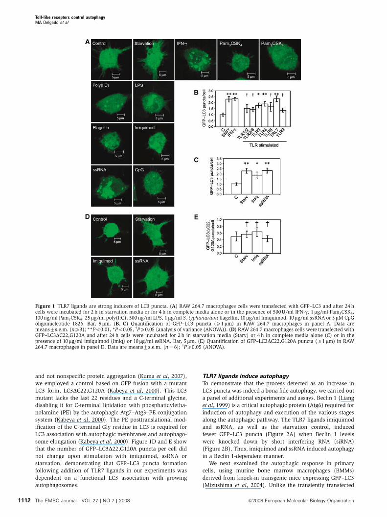
and not nonspecific protein aggregation (Kuma et al, 2007),
we employed a control based on GFP fusion with a mutant
LC3 form, LC3DC22,G120A (Kabeya et al, 2000). This LC3
mutant lacks the last 22 residues and a C-terminal glycine,
disabling it for C-terminal lipidation with phosphatidyletha-
nolamine (PE) by the autophagic Atg7–Atg3–PE conjugation
system (Kabeya et al, 2000). The PE posttranslational mod-
ification of the C-terminal Gly residue in LC3 is required for
LC3 association with autophagic membranes and autophago-
some elongation (Kabeya et al, 2000). Figure 1D and E show
that the number of GFP–LC3D22,G120A puncta per cell did
not change upon stimulation with imiquimod, ssRNA or
starvation, demonstrating that GFP–LC3 puncta formation
following addition of TLR7 ligands in our experiments was
dependent on a functional LC3 association with growing
autophagosomes.
TLR7 ligands induce autophagy
To demonstrate that the process detected as an increase in
LC3 puncta was indeed a bona fide autophagy, we carried out
a panel of additional experiments and assays. Beclin 1 (Liang
et al, 1999) is a critical autophagic protein (Atg6) required for
induction of autophagy and execution of the various stages
along the autophagic pathway. The TLR7 ligands imiquimod
and ssRNA, as well as the starvation control, induced
fewer GFP–LC3 puncta (Figure 2A) when Beclin 1 levels
were knocked down by short interfering RNA (siRNA)
(Figure 2B), Thus, imiquimod and ssRNA induced autophagy
in a Beclin 1-dependent manner.
We next examined the autophagic response in primary
cells, using murine bone marrow macrophages (BMMs)
derived from knock-in transgenic mice expressing GFP–LC3
(Mizushima et al, 2004). Unlike the transiently transfected
Figure 1 TLR7 ligands are strong inducers of LC3 puncta. (A) RAW 264.7 macrophages cells were transfected with GFP–LC3 and after 24 hcells were incubated for 2 h in starvation media or for 4 h in complete media alone or in the presence of 500 U/ml IFN-g, 1mg/ml Pam3CSK4,100 ng/ml Pam2CSK4, 25mg/ml poly(I:C), 500 ng/ml LPS, 1mg/ml S. typhimurium flagellin, 10mg/ml Imiquimod, 10mg/ml ssRNA or 3 mM CpGoligonucleotide 1826. Bar, 5mm. (B, C) Quantification of GFP–LC3 puncta (X1mm) in RAW 264.7 macrophages in panel A. Data aremeans7s.e.m. (nX3); **Po0.01, *Po0.05, wPX0.05 (analysis of variance (ANOVA)). (D) RAW 264.7 macrophages cells were transfected withGFP–LC3DC22,G120A and after 24 h cells were incubated for 2 h in starvation media (Starv) or 4 h in complete media alone (C) or in thepresence of 10mg/ml imiquimod (Imiq) or 10mg/ml ssRNA. Bar, 5mm. (E) Quantification of GFP–LC3DC22,G120A puncta (X1mm) in RAW264.7 macrophages in panel D. Data are means7s.e.m. (n¼ 6); wPX0.05 (ANOVA).
Toll-like receptors control autophagyMA Delgado et al
The EMBO Journal VOL 27 | NO 7 | 2008 &2008 European Molecular Biology Organization1112
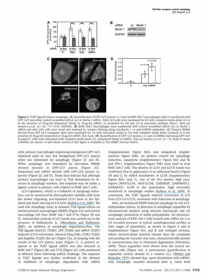
cells, primary macrophages expressing endogenous GFP–LC3
displayed none or very few background GFP–LC3 puncta
when not stimulated for autophagy (Figure 2C and D).
When autophagy was stimulated by starvation, BMMs
showed increase in GFP–LC3 puncta (Figure 2C).
Imiquimod and ssRNA elicited mild GFP–LC3 puncta re-
sponse (Figure 2C and D). These data indicate that although
primary macrophages can react to TLR stimulation by in-
crease in autophagy markers, this response may be under a
tighter control in primary cells relative to RAW 264.7 cells.
LC3-I lipidation, which is a hallmark of autophagy induc-
tion, can be monitored by electrophoretic mobility shift from
the slower migrating, non-lipidated LC3-I form to the lipi-
dated and faster moving LC3-II form (Kabeya et al, 2000). We
used this autophagy assay to examine TLR ligand effects on
murine primary macrophages (BMM) (Figure 3A) and murine
macrophage cell lines (RAW 264.7 and J774; Figure 3B and
C). Immunoblot analysis of LC3 bands was carried out in the
presence of Bafilomycin A1 (Mizushima and Yoshimori,
2007), an inhibitor of autophagic degradation/flux. The
TLR ligands Poly(I:C) (TLR3), LPS (TLR4) and ssRNA (TLR7)
induced LC3-II conversion, whereas Pam3CSK4 (TLR1/TLR2)
and flagellin (TLR5) did not (Figure 3A), in keeping with the
results of the LC3 puncta assay (Figure 1). A positive re-
sponse to the TLR7 ligand ssRNA was also detected in
RAW 264.7 (Figure 3B) and J774 (Figure 3C) cells monitored
at different times following ssRNA challenge. The response
to TLR7 ligands was further confirmed in the absence
of inhibitors of autophagic degradation with ssRNA
(Supplementary Figure S4A) and imiquimod (Supple-
mentary Figure S4B). As positive control for autophagy
induction, rapamycin (Supplementary Figure S4A and B)
and IFN-g (Supplementary Figure S4B) were used to treat
RAW 264.7 cells. The identity of LC3-I and LC3-II bands was
confirmed (due to appearance of an additional band in Figure
3B and C) by siRNA knockdown of LC3B (Supplementary
Figure S4A, lane 1), one of the five murine Atg8 para-
logues (MAP1LC3A, MAP1LC3B, GABARAP, GABARAPL1,
GABARAP); LC3B is the mammalian Atg8 invariably
monitored in autophagic studies (Kabeya et al, 2004). In
conclusion, the TLR7 ligands induced conversion of LC3
from LC3-I to LC3-II, consistent with induction of autophagy.
Next, we monitored PAMP-induced autophagy by two LC3-
independent criteria, (i) detection of autophagic organelles by
ultrastructural analysis using electron microscopy and (ii)
autophagic proteolysis of stable polypeptides. An ultrastruc-
tural analysis of RAW 264.7 cells treated with ssRNA for 2 or
4 h revealed presence of typical autophagosomal profiles at
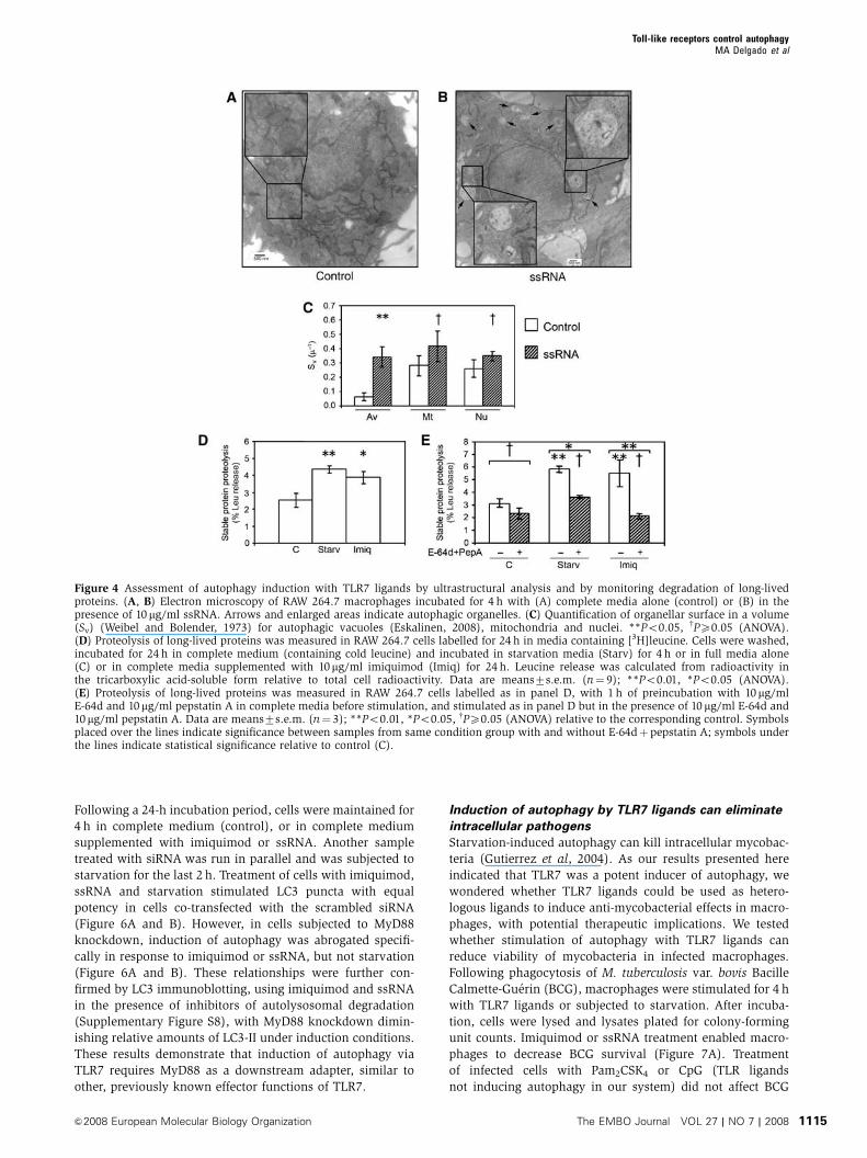
later stages of maturation, as shown in Figure 4 and in
Supplementary Figure S5A and B and enlarged sections,
where electron-dense material (denser than the cytoplasm
surrounding the vacuole) was seen, characteristically present
in autolysosomes due to ribosomal degradation (Eskalinen,
2008). These organelles were absent from the control un-
treated cells (Figure 4A). A stereometric quantification of
organellar surface density in a volume (Sv) (Weibel and
Bolender, 1973) showed that, upon stimulation with ssRNA,
only autophagic vacuoles increased their Sv value from
Figure 2 TLR7 ligands induce autophagy. (A) Quantification of GFP–LC3 puncta (X1mm) in RAW 264.7 macrophages cells co-transfected withGFP–LC3 and either control scrambled siRNA (sc) or Beclin 1 siRNA. After 22 h cells were incubated for 4 h with complete media alone (C) orin the presence of 10 mg/ml imiquimod (Imiq) or 10mg/ml ssRNA, or incubated for the last 2 h in starvation medium (Starv). Data aremeans7s.e.m. (n¼ 6); **Po0.01 (ANOVA). (B) RAW 264.7 macrophages were transfected with control scrambled siRNA (sc) or Beclin 1siRNA and after 24 h cells were lysed and analysed by western blotting using anti-Beclin 1 or anti-GAPDH antibodies. (C) Primary BMMsderived from GFP–LC3 transgenic mice were incubated for 2 h with starvation media or 4 h with complete media alone (control) or in thepresence of 10 mg/ml imiquimod or 10mg/ml ssRNA. Bar, 5mm. (D) Quantification of GFP–LC3 puncta (X1mm) in BMMs expressing GFP–LC3in panel C. Cells were stimulated with complete media alone (C), imiquimod (Imiq) or ssRNA. Data are means7s.e.m. (n¼ 3). Exact P-values(ANOVA) are shown. A full-colour version of this figure is available at The EMBO Journal Online.
Toll-like receptors control autophagyMA Delgado et al
&2008 European Molecular Biology Organization The EMBO Journal VOL 27 | NO 7 | 2008 1113

0.0670.03 to 0.3470.07m�1, whereas mitochondria or nuclei
did not show a statistically significant change in their Sv
values (Figure 4C).
Degradation of long-lived proteins represents an end-point
autophagy assay, relying on macroautophagic proteolysis of
stable polypeptides. It is often used to examine whether a
given process represents autophagy and to test whether the
autophagic pathway has been executed in full, as it depends
on conversion of autophagosomes into degradative autolyso-
somal organelles (Meley et al, 2006). Increased proteolysis
of stable polypeptides was observed when autophagy was
induced by starvation (Figure 4D). Upon stimulation of
cells with the TLR7 ligand imiquimod, a similar increase in
degradation of stable proteins was detected (Figure 4D). In
control samples, employing autophagic proteolysis inhibitors
E-64d and pepstatin A, both starvation- and imiquimod-
induced degradation of stable proteins were equally inhibited
by this treatment (Figure 4E). A full (with the exception of
LPS and zymosan) PAMP panel analysis for induction of
stable protein degradation (Supplementary Figure S6) was
in keeping with the results obtained with LC3 assays. The
results of ultrastructural analyses, proteolysis assays, LC3
puncta and LC3 lipidation tests, and dependence on Beclin
1 demonstrate that TLR7 ligands induce autophagy.
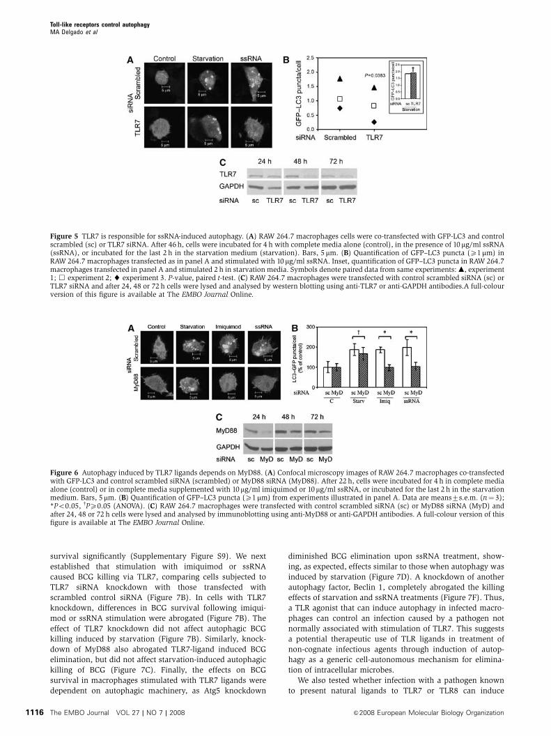
ssRNA induces autophagy through TLR7
After demonstrating that imiquimod and ssRNA induce au-
tophagy, we examined whether these compounds activated
autophagy via TLR7. TLR7 was knocked down in RAW 264.7
cells with siRNA (Figure 5A–C). Macrophages were stimu-
lated with a TLR7 ligand and autophagy induction quantified
by GFP–LC3 puncta assay (Figure 5A and B). Cells were
incubated in complete media in the absence or presence of
ssRNA and compared with starved cells. Although necessary
manipulations increased background levels of LC3 puncta in
some experiments, there was significant induction of autop-
hagy upon starvation or ssRNA stimulation in cells trans-
fected with the control scrambled siRNA compared with cells
transfected with TLR7 siRNA (Figure 5A and B). Importantly,
TLR7 knockdown did not change LC3 response to starvation
(Figure 5B, inset), demonstrating that TLR7 was required
specifically for TLR ligand-induced autophagy, but not for
starvation-induced autophagy. As a further control, cells
transfected with scrambled siRNA or with TLR7 siRNA had
the same level of GFP–LC3 puncta in the complete medium
(not shown). TLR7 knockdown, relative to scrambled siRNA
control, also diminished LC3-II levels assessed by immuno-
blotting upon stimulation with ssRNA (Supplementary
Figure S7A) or imiquimod (Supplementary Figure S7B).
Electron microscopic morphometry of autophagosomal
organelles induced by ssRNA indicated that TLR7 knockdown
by siRNA diminished Sv from 0.4970.1 to 0.1970.05m�1
(Supplementary Figure S5D). Thus, induction of autophagy
by ssRNA and imiquimod is TLR7-dependent.
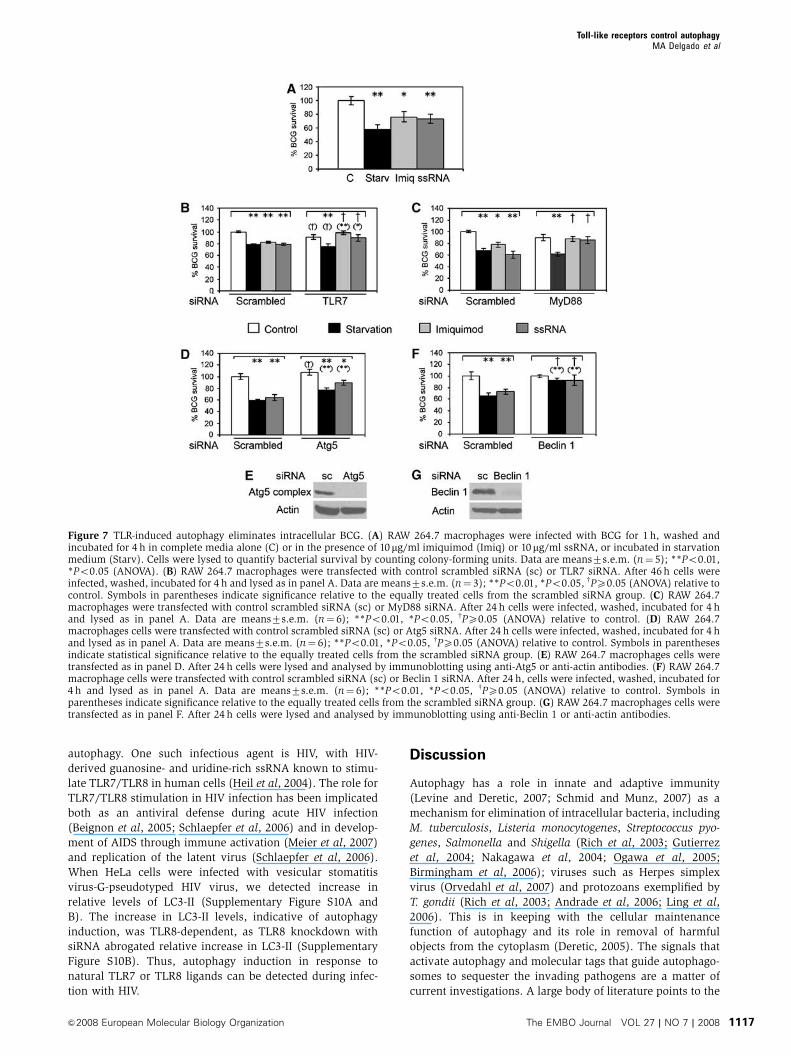
TLR7 stimulation activates autophagy via MyD88
As all known effector functions of TLR7 signalling depend on
the MyD88 adapter protein (Lee and Kim, 2007), we next
tested whether MyD88 was required for TLR7 induction of
autophagy (Figure 6A–C). Figure 6C shows a temporal ana-
lysis of MyD88 knockdown, with optimal MyD88 knockdown
being achieved early, at 24 h post siRNA transfection.
Macrophages were co-transfected with GFP–LC3 along
with control (scrambled) siRNA or MyD88 siRNA.
Figure 3 Immunoblot analysis of LC3-I to LC3-II conversion in cells stimulated with TLR ligands. (A) Primary BMMs were incubated in thepresence of 100 nM Bafilomycin A for 2 h in complete media alone (C) or in the presence of 1mg/ml Pam3CSK4, 25 mg/ml poly(I:C), 500 ng/mlLPS, 1 mg/ml S. typhimurium flagellin or 10mg/ml ssRNA. Cells were lysed and analysed by immunoblotting using anti-LC3 or anti-actinantibodies. Densitometric LC3-II/actin ratios are shown (average from three experiments). (B) RAW 264.7 macrophages were incubated in thepresence of 100 nM Bafilomycin A for 30 min, 1 h or 2 h in complete media alone (C) or in the presence of 10 mg/ml ssRNA. Cells were lysed andanalysed by immunoblotting using anti-LC3 or anti-actin antibodies. Densitometric LC3-II/actin ratios are shown as averages from two (30 minand 2 h) or three independent experiments (1 h). (C) J774 macrophages were incubated in the presence of 100 nM Bafilomycin A for 1 h incomplete media alone (C) or in the presence of 10mg/ml ssRNA. Cells were lysed and analysed by immunoblotting using anti-LC3 or anti-actinantibodies. Blot, 1 h sample. Densitometric LC3-II/actin ratios for 1 and 4 h incubation time points are shown underneath the blot.
Toll-like receptors control autophagyMA Delgado et al
The EMBO Journal VOL 27 | NO 7 | 2008 &2008 European Molecular Biology Organization1114
Following a 24-h incubation period, cells were maintained for
4 h in complete medium (control), or in complete medium
supplemented with imiquimod or ssRNA. Another sample
treated with siRNA was run in parallel and was subjected to
starvation for the last 2 h. Treatment of cells with imiquimod,
ssRNA and starvation stimulated LC3 puncta with equal
potency in cells co-transfected with the scrambled siRNA
(Figure 6A and B). However, in cells subjected to MyD88
knockdown, induction of autophagy was abrogated specifi-
cally in response to imiquimod or ssRNA, but not starvation
(Figure 6A and B). These relationships were further con-
firmed by LC3 immunoblotting, using imiquimod and ssRNA
in the presence of inhibitors of autolysosomal degradation
(Supplementary Figure S8), with MyD88 knockdown dimin-
ishing relative amounts of LC3-II under induction conditions.
These results demonstrate that induction of autophagy via
TLR7 requires MyD88 as a downstream adapter, similar to
other, previously known effector functions of TLR7.
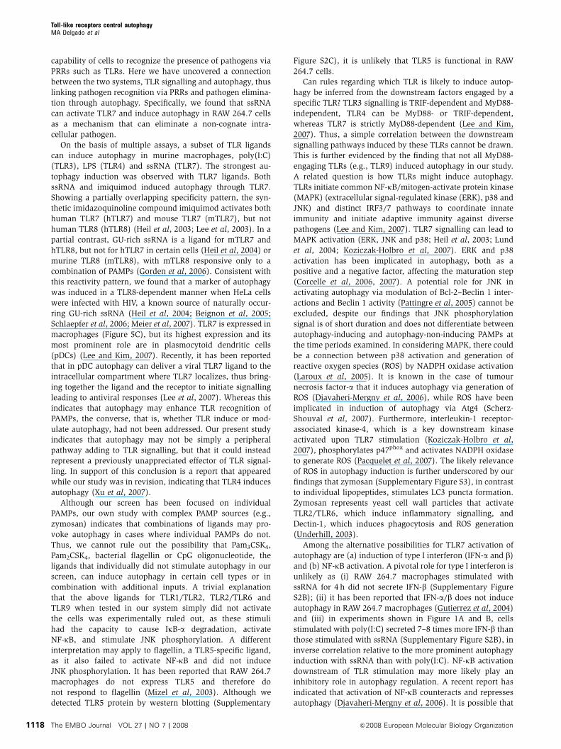
Induction of autophagy by TLR7 ligands can eliminate
intracellular pathogens
Starvation-induced autophagy can kill intracellular mycobac-
teria (Gutierrez et al, 2004). As our results presented here
indicated that TLR7 was a potent inducer of autophagy, we
wondered whether TLR7 ligands could be used as hetero-
logous ligands to induce anti-mycobacterial effects in macro-
phages, with potential therapeutic implications. We tested
whether stimulation of autophagy with TLR7 ligands can
reduce viability of mycobacteria in infected macrophages.
Following phagocytosis of M. tuberculosis var. bovis Bacille
Calmette-Guerin (BCG), macrophages were stimulated for 4 h
with TLR7 ligands or subjected to starvation. After incuba-
tion, cells were lysed and lysates plated for colony-forming
unit counts. Imiquimod or ssRNA treatment enabled macro-
phages to decrease BCG survival (Figure 7A). Treatment
of infected cells with Pam2CSK4 or CpG (TLR ligands
not inducing autophagy in our system) did not affect BCG
Figure 4 Assessment of autophagy induction with TLR7 ligands by ultrastructural analysis and by monitoring degradation of long-livedproteins. (A, B) Electron microscopy of RAW 264.7 macrophages incubated for 4 h with (A) complete media alone (control) or (B) in thepresence of 10 mg/ml ssRNA. Arrows and enlarged areas indicate autophagic organelles. (C) Quantification of organellar surface in a volume(Sv) (Weibel and Bolender, 1973) for autophagic vacuoles (Eskalinen, 2008), mitochondria and nuclei. **Po0.05, wPX0.05 (ANOVA).(D) Proteolysis of long-lived proteins was measured in RAW 264.7 cells labelled for 24 h in media containing [3H]leucine. Cells were washed,incubated for 24 h in complete medium (containing cold leucine) and incubated in starvation media (Starv) for 4 h or in full media alone(C) or in complete media supplemented with 10mg/ml imiquimod (Imiq) for 24 h. Leucine release was calculated from radioactivity inthe tricarboxylic acid-soluble form relative to total cell radioactivity. Data are means7s.e.m. (n¼ 9); **Po0.01, *Po0.05 (ANOVA).(E) Proteolysis of long-lived proteins was measured in RAW 264.7 cells labelled as in panel D, with 1 h of preincubation with 10mg/mlE-64d and 10mg/ml pepstatin A in complete media before stimulation, and stimulated as in panel D but in the presence of 10 mg/ml E-64d and10mg/ml pepstatin A. Data are means7s.e.m. (n¼ 3); **Po0.01, *Po0.05, wPX0.05 (ANOVA) relative to the corresponding control. Symbolsplaced over the lines indicate significance between samples from same condition group with and without E-64dþpepstatin A; symbols underthe lines indicate statistical significance relative to control (C).
Toll-like receptors control autophagyMA Delgado et al
&2008 European Molecular Biology Organization The EMBO Journal VOL 27 | NO 7 | 2008 1115
survival significantly (Supplementary Figure S9). We next
established that stimulation with imiquimod or ssRNA
caused BCG killing via TLR7, comparing cells subjected to
TLR7 siRNA knockdown with those transfected with
scrambled control siRNA (Figure 7B). In cells with TLR7
knockdown, differences in BCG survival following imiqui-
mod or ssRNA stimulation were abrogated (Figure 7B). The
effect of TLR7 knockdown did not affect autophagic BCG
killing induced by starvation (Figure 7B). Similarly, knock-
down of MyD88 also abrogated TLR7-ligand induced BCG
elimination, but did not affect starvation-induced autophagic
killing of BCG (Figure 7C). Finally, the effects on BCG
survival in macrophages stimulated with TLR7 ligands were
dependent on autophagic machinery, as Atg5 knockdown
diminished BCG elimination upon ssRNA treatment, show-
ing, as expected, effects similar to those when autophagy was
induced by starvation (Figure 7D). A knockdown of another
autophagy factor, Beclin 1, completely abrogated the killing
effects of starvation and ssRNA treatments (Figure 7F). Thus,
a TLR agonist that can induce autophagy in infected macro-
phages can control an infection caused by a pathogen not
normally associated with stimulation of TLR7. This suggests
a potential therapeutic use of TLR ligands in treatment of
non-cognate infectious agents through induction of autop-
hagy as a generic cell-autonomous mechanism for elimina-
tion of intracellular microbes.
We also tested whether infection with a pathogen known
to present natural ligands to TLR7 or TLR8 can induce
Figure 5 TLR7 is responsible for ssRNA-induced autophagy. (A) RAW 264.7 macrophages cells were co-transfected with GFP-LC3 and controlscrambled (sc) or TLR7 siRNA. After 46 h, cells were incubated for 4 h with complete media alone (control), in the presence of 10mg/ml ssRNA(ssRNA), or incubated for the last 2 h in the starvation medium (starvation). Bars, 5 mm. (B) Quantification of GFP–LC3 puncta (X1mm) inRAW 264.7 macrophages transfected as in panel A and stimulated with 10 mg/ml ssRNA. Inset, quantification of GFP–LC3 puncta in RAW 264.7macrophages transfected in panel A and stimulated 2 h in starvation media. Symbols denote paired data from same experiments: m, experiment1; & experiment 2; ~ experiment 3. P-value, paired t-test. (C) RAW 264.7 macrophages were transfected with control scrambled siRNA (sc) orTLR7 siRNA and after 24, 48 or 72 h cells were lysed and analysed by western blotting using anti-TLR7 or anti-GAPDH antibodies.A full-colourversion of this figure is available at The EMBO Journal Online.
Figure 6 Autophagy induced by TLR7 ligands depends on MyD88. (A) Confocal microscopy images of RAW 264.7 macrophages co-transfectedwith GFP-LC3 and control scrambled siRNA (scrambled) or MyD88 siRNA (MyD88). After 22 h, cells were incubated for 4 h in complete mediaalone (control) or in complete media supplemented with 10mg/ml imiquimod or 10mg/ml ssRNA, or incubated for the last 2 h in the starvationmedium. Bars, 5mm. (B) Quantification of GFP–LC3 puncta (X1mm) from experiments illustrated in panel A. Data are means7s.e.m. (n¼ 3);*Po0.05, wPX0.05 (ANOVA). (C) RAW 264.7 macrophages were transfected with control scrambled siRNA (sc) or MyD88 siRNA (MyD) andafter 24, 48 or 72 h cells were lysed and analysed by immunoblotting using anti-MyD88 or anti-GAPDH antibodies. A full-colour version of thisfigure is available at The EMBO Journal Online.
Toll-like receptors control autophagyMA Delgado et al
The EMBO Journal VOL 27 | NO 7 | 2008 &2008 European Molecular Biology Organization1116
autophagy. One such infectious agent is HIV, with HIV-
derived guanosine- and uridine-rich ssRNA known to stimu-
late TLR7/TLR8 in human cells (Heil et al, 2004). The role for
TLR7/TLR8 stimulation in HIV infection has been implicated
both as an antiviral defense during acute HIV infection
(Beignon et al, 2005; Schlaepfer et al, 2006) and in develop-
ment of AIDS through immune activation (Meier et al, 2007)
and replication of the latent virus (Schlaepfer et al, 2006).
When HeLa cells were infected with vesicular stomatitis
virus-G-pseudotyped HIV virus, we detected increase in
relative levels of LC3-II (Supplementary Figure S10A and
B). The increase in LC3-II levels, indicative of autophagy
induction, was TLR8-dependent, as TLR8 knockdown with
siRNA abrogated relative increase in LC3-II (Supplementary
Figure S10B). Thus, autophagy induction in response to
natural TLR7 or TLR8 ligands can be detected during infec-
tion with HIV.
Discussion
Autophagy has a role in innate and adaptive immunity
(Levine and Deretic, 2007; Schmid and Munz, 2007) as a
mechanism for elimination of intracellular bacteria, including
M. tuberculosis, Listeria monocytogenes, Streptococcus pyo-
genes, Salmonella and Shigella (Rich et al, 2003; Gutierrez
et al, 2004; Nakagawa et al, 2004; Ogawa et al, 2005;
Birmingham et al, 2006); viruses such as Herpes simplex
virus (Orvedahl et al, 2007) and protozoans exemplified by
T. gondii (Rich et al, 2003; Andrade et al, 2006; Ling et al,
2006). This is in keeping with the cellular maintenance
function of autophagy and its role in removal of harmful
objects from the cytoplasm (Deretic, 2005). The signals that
activate autophagy and molecular tags that guide autophago-
somes to sequester the invading pathogens are a matter of
current investigations. A large body of literature points to the
Figure 7 TLR-induced autophagy eliminates intracellular BCG. (A) RAW 264.7 macrophages were infected with BCG for 1 h, washed andincubated for 4 h in complete media alone (C) or in the presence of 10mg/ml imiquimod (Imiq) or 10mg/ml ssRNA, or incubated in starvationmedium (Starv). Cells were lysed to quantify bacterial survival by counting colony-forming units. Data are means7s.e.m. (n¼ 5); **Po0.01,*Po0.05 (ANOVA). (B) RAW 264.7 macrophages were transfected with control scrambled siRNA (sc) or TLR7 siRNA. After 46 h cells wereinfected, washed, incubated for 4 h and lysed as in panel A. Data are means7s.e.m. (n¼ 3); **Po0.01, *Po0.05, wPX0.05 (ANOVA) relative tocontrol. Symbols in parentheses indicate significance relative to the equally treated cells from the scrambled siRNA group. (C) RAW 264.7macrophages were transfected with control scrambled siRNA (sc) or MyD88 siRNA. After 24 h cells were infected, washed, incubated for 4 hand lysed as in panel A. Data are means7s.e.m. (n¼ 6); **Po0.01, *Po0.05, wPX0.05 (ANOVA) relative to control. (D) RAW 264.7macrophages cells were transfected with control scrambled siRNA (sc) or Atg5 siRNA. After 24 h cells were infected, washed, incubated for 4 hand lysed as in panel A. Data are means7s.e.m. (n¼ 6); **Po0.01, *Po0.05, wPX0.05 (ANOVA) relative to control. Symbols in parenthesesindicate statistical significance relative to the equally treated cells from the scrambled siRNA group. (E) RAW 264.7 macrophages cells weretransfected as in panel D. After 24 h cells were lysed and analysed by immunoblotting using anti-Atg5 or anti-actin antibodies. (F) RAW 264.7macrophage cells were transfected with control scrambled siRNA (sc) or Beclin 1 siRNA. After 24 h, cells were infected, washed, incubated for4 h and lysed as in panel A. Data are means7s.e.m. (n¼ 6); **Po0.01, *Po0.05, wPX0.05 (ANOVA) relative to control. Symbols inparentheses indicate significance relative to the equally treated cells from the scrambled siRNA group. (G) RAW 264.7 macrophages cells weretransfected as in panel F. After 24 h cells were lysed and analysed by immunoblotting using anti-Beclin 1 or anti-actin antibodies.
Toll-like receptors control autophagyMA Delgado et al
&2008 European Molecular Biology Organization The EMBO Journal VOL 27 | NO 7 | 2008 1117
capability of cells to recognize the presence of pathogens via
PRRs such as TLRs. Here we have uncovered a connection
between the two systems, TLR signalling and autophagy, thus
linking pathogen recognition via PRRs and pathogen elimina-
tion through autophagy. Specifically, we found that ssRNA
can activate TLR7 and induce autophagy in RAW 264.7 cells
as a mechanism that can eliminate a non-cognate intra-
cellular pathogen.
On the basis of multiple assays, a subset of TLR ligands
can induce autophagy in murine macrophages, poly(I:C)
(TLR3), LPS (TLR4) and ssRNA (TLR7). The strongest au-
tophagy induction was observed with TLR7 ligands. Both
ssRNA and imiquimod induced autophagy through TLR7.
Showing a partially overlapping specificity pattern, the syn-
thetic imidazoquinoline compound imiquimod activates both
human TLR7 (hTLR7) and mouse TLR7 (mTLR7), but not
human TLR8 (hTLR8) (Heil et al, 2003; Lee et al, 2003). In a
partial contrast, GU-rich ssRNA is a ligand for mTLR7 and
hTLR8, but not for hTLR7 in certain cells (Heil et al, 2004) or
murine TLR8 (mTLR8), with mTLR8 responsive only to a
combination of PAMPs (Gorden et al, 2006). Consistent with
this reactivity pattern, we found that a marker of autophagy
was induced in a TLR8-dependent manner when HeLa cells
were infected with HIV, a known source of naturally occur-
ring GU-rich ssRNA (Heil et al, 2004; Beignon et al, 2005;
Schlaepfer et al, 2006; Meier et al, 2007). TLR7 is expressed in
macrophages (Figure 5C), but its highest expression and its
most prominent role are in plasmocytoid dendritic cells
(pDCs) (Lee and Kim, 2007). Recently, it has been reported
that in pDC autophagy can deliver a viral TLR7 ligand to the
intracellular compartment where TLR7 localizes, thus bring-
ing together the ligand and the receptor to initiate signalling
leading to antiviral responses (Lee et al, 2007). Whereas this
indicates that autophagy may enhance TLR recognition of
PAMPs, the converse, that is, whether TLR induce or mod-
ulate autophagy, had not been addressed. Our present study
indicates that autophagy may not be simply a peripheral
pathway adding to TLR signalling, but that it could instead
represent a previously unappreciated effector of TLR signal-
ling. In support of this conclusion is a report that appeared
while our study was in revision, indicating that TLR4 induces
autophagy (Xu et al, 2007).
Although our screen has been focused on individual
PAMPs, our own study with complex PAMP sources (e.g.,
zymosan) indicates that combinations of ligands may pro-
voke autophagy in cases where individual PAMPs do not.
Thus, we cannot rule out the possibility that Pam3CSK4,
Pam2CSK4, bacterial flagellin or CpG oligonucleotide, the
ligands that individually did not stimulate autophagy in our
screen, can induce autophagy in certain cell types or in
combination with additional inputs. A trivial explanation
that the above ligands for TLR1/TLR2, TLR2/TLR6 and
TLR9 when tested in our system simply did not activate
the cells was experimentally ruled out, as these stimuli
had the capacity to cause IkB-a degradation, activate
NF-kB, and stimulate JNK phosphorylation. A different
interpretation may apply to flagellin, a TLR5-specific ligand,
as it also failed to activate NF-kB and did not induce
JNK phosphorylation. It has been reported that RAW 264.7
macrophages do not express TLR5 and therefore do
not respond to flagellin (Mizel et al, 2003). Although we
detected TLR5 protein by western blotting (Supplementary
Figure S2C), it is unlikely that TLR5 is functional in RAW
264.7 cells.
Can rules regarding which TLR is likely to induce autop-
hagy be inferred from the downstream factors engaged by a
specific TLR? TLR3 signalling is TRIF-dependent and MyD88-
independent, TLR4 can be MyD88- or TRIF-dependent,
whereas TLR7 is strictly MyD88-dependent (Lee and Kim,
2007). Thus, a simple correlation between the downstream
signalling pathways induced by these TLRs cannot be drawn.
This is further evidenced by the finding that not all MyD88-
engaging TLRs (e.g., TLR9) induced autophagy in our study.
A related question is how TLRs might induce autophagy.
TLRs initiate common NF-kB/mitogen-activate protein kinase
(MAPK) (extracellular signal-regulated kinase (ERK), p38 and
JNK) and distinct IRF3/7 pathways to coordinate innate
immunity and initiate adaptive immunity against diverse
pathogens (Lee and Kim, 2007). TLR7 signalling can lead to
MAPK activation (ERK, JNK and p38; Heil et al, 2003; Lund
et al, 2004; Koziczak-Holbro et al, 2007). ERK and p38
activation has been implicated in autophagy, both as a
positive and a negative factor, affecting the maturation step
(Corcelle et al, 2006, 2007). A potential role for JNK in
activating autophagy via modulation of Bcl-2–Beclin 1 inter-
actions and Beclin 1 activity (Pattingre et al, 2005) cannot be
excluded, despite our findings that JNK phosphorylation
signal is of short duration and does not differentiate between
autophagy-inducing and autophagy-non-inducing PAMPs at
the time periods examined. In considering MAPK, there could
be a connection between p38 activation and generation of
reactive oxygen species (ROS) by NADPH oxidase activation
(Laroux et al, 2005). It is known in the case of tumour
necrosis factor-a that it induces autophagy via generation of
ROS (Djavaheri-Mergny et al, 2006), while ROS have been
implicated in induction of autophagy via Atg4 (Scherz-
Shouval et al, 2007). Furthermore, interleukin-1 receptor-
associated kinase-4, which is a key downstream kinase
activated upon TLR7 stimulation (Koziczak-Holbro et al,
2007), phosphorylates p47phox and activates NADPH oxidase
to generate ROS (Pacquelet et al, 2007). The likely relevance
of ROS in autophagy induction is further underscored by our
findings that zymosan (Supplementary Figure S3), in contrast
to individual lipopeptides, stimulates LC3 puncta formation.
Zymosan represents yeast cell wall particles that activate
TLR2/TLR6, which induce inflammatory signalling, and
Dectin-1, which induces phagocytosis and ROS generation
(Underhill, 2003).
Among the alternative possibilities for TLR7 activation of
autophagy are (a) induction of type I interferon (IFN-a and b)
and (b) NF-kB activation. A pivotal role for type I interferon is
unlikely as (i) RAW 264.7 macrophages stimulated with
ssRNA for 4 h did not secrete IFN-b (Supplementary Figure
S2B); (ii) it has been reported that IFN-a/b does not induce
autophagy in RAW 264.7 macrophages (Gutierrez et al, 2004)
and (iii) in experiments shown in Figure 1A and B, cells
stimulated with poly(I:C) secreted 7–8 times more IFN-b than
those stimulated with ssRNA (Supplementary Figure S2B), in
inverse correlation relative to the more prominent autophagy
induction with ssRNA than with poly(I:C). NF-kB activation
downstream of TLR stimulation may more likely play an
inhibitory role in autophagy regulation. A recent report has
indicated that activation of NF-kB counteracts and represses
autophagy (Djavaheri-Mergny et al, 2006). It is possible that
Toll-like receptors control autophagyMA Delgado et al
The EMBO Journal VOL 27 | NO 7 | 2008 &2008 European Molecular Biology Organization1118

robust NF-kB activation or modulation of some other factors
may preclude efficient autophagy activation by TLR7 in some
cells (Z Zhao and H Virgin, personal communication).
Furthermore, several TLR ligands (Pam3CSK4, Pam2CSK4,
CpG) caused IkB-a degradation (Supplementary Figure S2D)
and activated NF-kB (Supplementary Figure S2A), but did not
induce autophagy in our study.
The connections between TLR signalling and induction of
autophagy shown here link two broad aspects of innate
immunity, TLR signalling and autophagy. In addition, the
relationships uncovered in this work open the possibility of
putting them to practical use. The TLR7 ligand imiquimod is a
prescription medication known under the trade name Aldara
with therapeutic applications in patients with viral infections
and certain cancers (Beutner et al, 1998; Edwards et al, 1998;
Syed et al, 1998). Our data show that activation of macro-
phages with imiquimod reduces mycobacterial viability in
infected macrophages. This suggests a potential application
of the relationships shown in this work as a basis for
treatment of early or latent M. tuberculosis infections. The
link between TLR signalling and autophagy may prove to be
an unanticipated but valuable application of the detailed
knowledge of TLR-signalling pathways, now expanded to
the elimination of pathogens through induction of autophagy.
Materials and methods
Cell and bacterial culturesMurine RAW 264.7 and J774 macrophage cell lines were maintainedin Dulbecco’s modified Eagle’s medium (DMEM; Invitrogen, CA,USA) supplemented with 10% fetal bovine serum (FBS) and L-glutamine (complete media). HeLa cells were maintained in DMEM,10% FBS. BMMs were derived from C57/BL6 mice or GFP–LC3mice (Dr N Mizushima, Japan) as described previously (Via et al,1998). M. tuberculosis var. bovis BCG was grown in Middlebrook7H9 broth with 0.5% Tween, 0.2% glycerol and albumin–dextrose–catalase (ADC) supplement (BD Diagnostics, Franklin Lakes, NJ,USA) and homogenized to generate a single-cell suspension, or on7H11 plates with 0.5% Tween, 0.2% glycerol and ADC.
Antibodies, TLR ligands, drugs, cytokines, siRNAs and DNAconstructsThe rabbit polyclonal antibody against LC3 (T Ueno and EKominami, Japan) was used at 1:500 dilution; mouse monoclonalanti-glyceraldehyde-3-phosphate dehydrogenase (GAPDH), rabbitpolyclonal anti-TLR7, rabbit polyclonal anti-MyD88, rabbit poly-clonal anti-GFP, mouse monoclonal anti-TLR5 antibodies andmouse monoclonal anti-actin antibodies were from Abcam Inc.(Cambridge, MA, USA); goat polyclonal anti-Beclin 1 antibody(Santa Cruz Biotechnology Inc., CA, USA) was used at 1:200dilution; mouse monoclonal anti-IkB-a, rabbit polyclonal anti-phospho-JNK and rabbit polyclonal anti-JNK antibodies werefrom Cell Signaling Technology Inc. (Danvers, MA, USA); rabbitpolyclonal anti-Atg5 was from Novus Biologicals (Littleton,CO, USA); anti-rabbit Alexa-488-conjugated antibody was fromMolecular Probes (Eugene, OR, USA). Lipopeptides Pam2CSK4 andPam3CSK4, zymosan, polyinosine–polycytidylic acid poly(I:C),imiquimod (R837), ssRNA (ssRNA40/LyoVec) and the CpGoligonocleotide ODN1826 were from InvivoGen (San Diego, CA,USA). Mouse IFN-g, LPS from Escherichia coli 026:B6, E-64d,pepstatin A, rapamycin and rabbit polyclonal anti-LC3B antibodywere from Sigma-Aldrich (St Louis, MO, USA). Bafilomycin A1 wasfrom LC Laboratories (Worburn, MA, USA). Secondary horseradishperoxidase-conjugated antibodies were from Pierce (Rockford,IL, USA). Control siRNA (siCONTROL non-targeting siRNA) andsiRNAs for mouse LC3B, mouse Beclin 1, mouse Atg5, mouse TLR7,mouse MyD88 and human TLR8 (siGENOME SMARTpool siRNA)were from Dharmacon (Chicago, IL, USA). NF-kB-responsiveluciferase reporter plasmid PathDetects NF-kB cis-ReportingSystem was from Stratagene (La Jolla, CA, USA) and theb-galactosidase construct, pEF1-Bos, was from G Nunez (Universityof Michigan).
MethodsAll other materials and methods are described in Supplementarydata.
Supplementary dataSupplementary data are available at The EMBO Journal Online(http://www.embojournal.org).
Acknowledgements
We thank Z Zhao and H Virgin for information on LC3-II response.This work was supported by National Institutes of Health grantAI069345 and in part by grants AI45148 and AI42999. ASD was anNIH T-32 AI07538 fellow.
References
Andrade RM, Wessendarp M, Gubbels MJ, Striepen B, Subauste CS(2006) CD40 induces macrophage anti-Toxoplasma gondii activityby triggering autophagy-dependent fusion of pathogen-containingvacuoles and lysosomes. J Clin Invest 116: 2366–2377
Beignon AS, McKenna K, Skoberne M, Manches O, DaSilva I,Kavanagh DG, Larsson M, Gorelick RJ, Lifson JD, Bhardwaj N(2005) Endocytosis of HIV-1 activates plasmacytoid dendritic cellsvia Toll-like receptor–viral RNA interactions. J Clin Invest 115:3265–3275
Bernales S, McDonald KL, Walter P (2006) Autophagy counter-balances endoplasmic reticulum expansion during the unfoldedprotein response. PLoS Biol 4: e423
Beutner KR, Spruance SL, Hougham AJ, Fox TL, Owens ML,Douglas Jr JM (1998) Treatment of genital warts with an im-mune-response modifier (imiquimod). J Am Acad Dermatol 38:230–239
Birmingham CL, Smith AC, Bakowski MA, Yoshimori T, Brumell JH(2006) Autophagy controls Salmonella infection in response todamage to the Salmonella-containing vacuole. J Biol Chem 281:11374–11383
Bjorkoy G, Lamark T, Brech A, Outzen H, Perander M, Overvatn A,Stenmark H, Johansen T (2005) p62/SQSTM1 forms proteinaggregates degraded by autophagy and has a protective effecton huntingtin-induced cell death. J Cell Biol 171: 603–614
Corcelle E, Djerbi N, Mari M, Nebout M, Fiorini C, Fenichel P,Hofman P, Poujeol P, Mograbi B (2007) Control of the autophagymaturation step by the MAPK ERK and p38: lessons fromenvironmental carcinogens. Autophagy 3: 57–59
Corcelle E, Nebout M, Bekri S, Gauthier N, Hofman P, Poujeol P,Fenichel P, Mograbi B (2006) Disruption of autophagy at thematuration step by the carcinogen lindane is associated with thesustained mitogen-activated protein kinase/extracellular signal-regulated kinase activity. Cancer Res 66: 6861–6870
Deretic V (2005) Autophagy in innate and adaptive immunity.Trends Immunol 26: 523–528
Deretic V, Singh S, Master S, Harris J, Roberts E, Kyei G, Davis A, deHaro S, Naylor J, Lee HH, Vergne I (2006) Mycobacteriumtuberculosis inhibition of phagolysosome biogenesis andautophagy as a host defence mechanism. Cell Microbiol 8:719–727
Djavaheri-Mergny M, Amelotti M, Mathieu J, Besancon F, Bauvy C,Souquere S, Pierron G, Codogno P (2006) NF-kappaB activationrepresses tumor necrosis factor-alpha-induced autophagy. J BiolChem 281: 30373–30382
Edwards L, Ferenczy A, Eron L, Baker D, Owens ML, Fox TL,Hougham AJ, Schmitt KA (1998) Self-administered topical 5%imiquimod cream for external anogenital warts. HPV StudyGroup. Human papillomavirus. Arch Dermatol 134: 25–30
Toll-like receptors control autophagyMA Delgado et al
&2008 European Molecular Biology Organization The EMBO Journal VOL 27 | NO 7 | 2008 1119
Eskalinen E (2008) Fine structure of the autophagosome. InAutophagosome and Phagosome, Deretic V (ed), Humana Pressvol. (in press)
Farre JC, Subramani S (2004) Peroxisome turnover by micropex-ophagy: an autophagy-related process. Trends Cell Biol 14:515–523
Gorden KK, Qiu XX, Binsfeld CC, Vasilakos JP, Alkan SS (2006)Cutting edge: activation of murine TLR8 by a combination ofimidazoquinoline immune response modifiers and polyT oligo-deoxynucleotides. J Immunol 177: 6584–6587
Gutierrez MG, Master SS, Singh SB, Taylor GA, Colombo MI, DereticV (2004) Autophagy is a defense mechanism inhibiting BCG andMycobacterium tuberculosis survival in infected macrophages.Cell 119: 753–766
Hampe J, Franke A, Rosenstiel P, Till A, Teuber M, Huse K,Albrecht M, Mayr G, De La Vega FM, Briggs J, Gunther S,Prescott NJ, Onnie CM, Hasler R, Sipos B, Folsch UR, LengauerT, Platzer M, Mathew CG, Krawczak M et al (2007) A genome-wide association scan of nonsynonymous SNPs identifies asusceptibility variant for Crohn disease in ATG16L1. Nat Genet39: 207–211
Harris J, De Haro SA, Master SS, Keane J, Roberts EA, Delgado M,Deretic V (2007) T Helper 2 cytokines inhibit autophagiccontrol of intracellular Mycobacterium tuberculosis. Immunity27: 505–517
Heil F, Ahmad-Nejad P, Hemmi H, Hochrein H, Ampenberger F,Gellert T, Dietrich H, Lipford G, Takeda K, Akira S, Wagner H,Bauer S (2003) The Toll-like receptor 7 (TLR7)-specific stimulusloxoribine uncovers a strong relationship within the TLR7, 8 and9 subfamily. Eur J Immunol 33: 2987–2997
Heil F, Hemmi H, Hochrein H, Ampenberger F, Kirschning C, AkiraS, Lipford G, Wagner H, Bauer S (2004) Species-specific recogni-tion of single-stranded RNA via toll-like receptor 7 and 8. Science303: 1526–1529
Hemmi H, Kaisho T, Takeuchi O, Sato S, Sanjo H, Hoshino K,Horiuchi T, Tomizawa H, Takeda K, Akira S (2002) Small antiviralcompounds activate immune cells via the TLR7 MyD88-depen-dent signaling pathway. Nat Immunol 3: 196–200
Kabeya Y, Mizushima N, Ueno T, Yamamoto A, Kirisako T, Noda T,Kominami E, Ohsumi Y, Yoshimori T (2000) LC3, a mammalianhomologue of yeast Apg8p, is localized in autophagosome mem-branes after processing. EMBO J 19: 5720–5728
Kabeya Y, Mizushima N, Yamamoto A, Oshitani-Okamoto S,Ohsumi Y, Yoshimori T (2004) LC3, GABARAP and GATE16localize to autophagosomal membrane depending on form-IIformation. J Cell Sci 117: 2805–2812
Kim J, Kamada Y, Stromhaug PE, Guan J, Hefner-Gravink A, BabaM, Scott SV, Ohsumi Y, Dunn Jr WA, Klionsky DJ (2001) Cvt9/Gsa9 functions in sequestering selective cytosolic cargo destinedfor the vacuole. J Cell Biol 153: 381–396
Koziczak-Holbro M, Joyce C, Gluck A, Kinzel B, Muller M, TschoppC, Mathison JC, Davis CN, Gram H (2007) IRAK-4 kinase activityis required for interleukin-1 (IL-1) receptor- and Toll-like receptor7-mediated signaling and gene expression. J Biol Chem 282:13552–13560
Kuma A, Matsui M, Mizushima N (2007) LC3, an autophagosomemarker, can be incorporated into protein aggregates independentof autophagy: caution in the interpretation of LC3 localization.Autophagy 3: 323–328
Laroux FS, Romero X, Wetzler L, Engel P, Terhorst C (2005) Cuttingedge: MyD88 controls phagocyte NADPH oxidase function andkilling of Gram-negative bacteria. J Immunol 175: 5596–5600
Lee HK, Lund JM, Ramanathan B, Mizushima N, Iwasaki A (2007)Autophagy-dependent viral recognition by plasmacytoid dendriticcells. Science 315: 1398–1401
Lee J, Chuang TH, Redecke V, She L, Pitha PM, Carson DA, Raz E,Cottam HB (2003) Molecular basis for the immunostimulatoryactivity of guanine nucleoside analogs: activation of Toll-likereceptor 7. Proc Natl Acad Sci USA 100: 6646–6651
Lee MS, Kim YJ (2007) Signaling pathways downstream of pattern-recognition receptors and their cross talk. Annu Rev Biochem 76:447–480
Lemasters JJ (2005) Selective mitochondrial autophagy, or mito-phagy, as a targeted defense against oxidative stress, mitochon-drial dysfunction, and aging. Rejuvenation Res 8: 3–5
Levine B (2007) Cell biology: autophagy and cancer. Nature 446:745–747
Levine B, Deretic V (2007) Unveiling the roles of autophagy ininnate and adaptive immunity. Nat Rev Immunol 7: 767–777
Levine B, Klionsky DJ (2004) Development by self-digestion:molecular mechanisms and biological functions of autophagy.Dev Cell 6: 463–477
Li C, Capan E, Zhao Y, Zhao J, Stolz D, Watkins SC, Jin S, Lu B (2006)Autophagy is induced in CD4+ T cells and important for thegrowth factor-withdrawal cell death. J Immunol 177: 5163–5168
Liang XH, Jackson S, Seaman M, Brown K, Kempkes B, HibshooshH, Levine B (1999) Induction of autophagy and inhibition oftumorigenesis by beclin 1. Nature 402: 672–676
Ling YM, Shaw MH, Ayala C, Coppens I, Taylor GA, Ferguson DJ,Yap GS (2006) Vacuolar and plasma membrane stripping andautophagic elimination of Toxoplasma gondii in primed effectormacrophages. J Exp Med 203: 2063–2071
Lum JJ, DeBerardinis RJ, Thompson CB (2005) Autophagy inmetazoans: cell survival in the land of plenty. Nat Rev Mol CellBiol 6: 439–448
Lund JM, Alexopoulou L, Sato A, Karow M, Adams NC, Gale NW,Iwasaki A, Flavell RA (2004) Recognition of single-strandedRNA viruses by Toll-like receptor 7. Proc Natl Acad Sci USA 101:5598–5603
Medzhitov R (2007) Recognition of microorganisms and activationof the immune response. Nature 449: 819–826
Meier A, Alter G, Frahm N, Sidhu H, Li B, Bagchi A, Teigen N,Streeck H, Stellbrink HJ, Hellman J, van Lunzen J, Altfeld M(2007) MyD88-dependent immune activation mediated by humanimmunodeficiency virus type 1-encoded Toll-like receptor li-gands. J Virol 81: 8180–8191
Meley D, Bauvy C, Houben-Weerts JH, Dubbelhuis PF, HelmondMT, Codogno P, Meijer AJ (2006) AMP-activated protein kinaseand the regulation of autophagic proteolysis. J Biol Chem 281:34870–34879
Mizel SB, Honko AN, Moors MA, Smith PS, West AP (2003)Induction of macrophage nitric oxide production by Gram-nega-tive flagellin involves signaling via heteromeric Toll-like receptor5/Toll-like receptor 4 complexes. J Immunol 170: 6217–6223
Mizushima N, Ohsumi Y, Yoshimori T (2002) Autophagosomeformation in mammalian cells. Cell Struct Funct 27: 421–429
Mizushima N, Yamamoto A, Matsui M, Yoshimori T, Ohsumi Y(2004) In vivo analysis of autophagy in response to nutrientstarvation using transgenic mice expressing a fluorescent auto-phagosome marker. Mol Biol Cell 15: 1101–1111
Mizushima N, Yoshimori T (2007) How to interpret LC3 immuno-blotting. Autophagy 3: 542–545
Nakagawa I, Amano A, Mizushima N, Yamamoto A, Yamaguchi H,Kamimoto T, Nara A, Funao J, Nakata M, Tsuda K, Hamada S,Yoshimori T (2004) Autophagy defends cells against invadinggroup A Streptococcus. Science 306: 1037–1040
Nixon RA (2006) Autophagy in neurodegenerative disease: friend,foe or turncoat? Trends Neurosci 29: 528–535
Ogawa M, Yoshimori T, Suzuki T, Sagara H, Mizushima N,Sasakawa C (2005) Escape of intracellular Shigella from auto-phagy. Science 307: 727–731
Orvedahl A, Alexander D, Talloczy Z, Sun Q, Wei Y, Zhang W, BurnsD, Leib D, Levine B (2007) HSV-1 ICP34.5 confers neurovirulenceby targeting the Beclin 1 autophagy protein. Cell Host Microbe 1:23–35
Pacquelet S, Johnson JL, Ellis BA, Brzezinska AA, Lane WS, MunafoDB, Catz SD (2007) Cross-talk between IRAK-4 and the NADPHoxidase. Biochem J 403: 451–461
Parkes M, Barrett JC, Prescott NJ, Tremelling M, Anderson CA,Fisher SA, Roberts RG, Nimmo ER, Cummings FR, Soars D,Drummond H, Lees CW, Khawaja SA, Bagnall R, Burke DA,Todhunter CE, Ahmad T, Onnie CM, McArdle W, Strachan Det al (2007) Sequence variants in the autophagy gene IRGM andmultiple other replicating loci contribute to Crohn’s diseasesusceptibility. Nat Genet 39: 830–832
Pattingre S, Tassa A, Qu X, Garuti R, Liang XH, Mizushima N,Packer M, Schneider MD, Levine B (2005) Bcl-2 antiapoptoticproteins inhibit Beclin 1-dependent autophagy. Cell 122: 927–939
Pua HH, Dzhagalov I, Chuck M, Mizushima N, He YW (2007) Acritical role for the autophagy gene Atg5 in T cell survival andproliferation. J Exp Med 204: 25–31
Rayapuram N, Subramani S (2006) The importomer—a peroxisomalmembrane complex involved in protein translocation into theperoxisome matrix. Biochim Biophys Acta 1763: 1613–1619
Toll-like receptors control autophagyMA Delgado et al
The EMBO Journal VOL 27 | NO 7 | 2008 &2008 European Molecular Biology Organization1120
Rich KA, Burkett C, Webster P (2003) Cytoplasmic bacteria can betargets for autophagy. Cell Microbiol 5: 455–468
Rubinsztein DC (2006) The roles of intracellular protein-degrada-tion pathways in neurodegeneration. Nature 443: 780–786
Scherz-Shouval R, Shvets E, Fass E, Shorer H, Gil L, Elazar Z(2007) Reactive oxygen species are essential for autophagyand specifically regulate the activity of Atg4. EMBO J 26:1749–1760
Schlaepfer E, Audige A, Joller H, Speck RF (2006) TLR7/8 triggeringexerts opposing effects in acute versus latent HIV infection.J Immunol 176: 2888–2895
Schmid D, Munz C (2007) Innate and adaptive immunity throughautophagy. Immunity 27: 11–21
Schmid D, Pypaert M, Munz C (2007) Antigen-loading compart-ments for major histocompatibility complex class II moleculescontinuously receive input from autophagosomes. Immunity 26:79–92
Seglen PO, Bohley P (1992) Autophagy and other vacuolar proteindegradation mechanisms. Experientia 48: 158–172
Singh SB, Davis AS, Taylor GA, Deretic V (2006) Human IRGMinduces autophagy to eliminate intracellular mycobacteria.Science 313: 1438–1441
Syed TA, Ahmadpour OA, Ahmad SA, Ahmad SH (1998)Management of female genital warts with an analog ofimiquimod 2% in cream: a randomized, double-blind, placebo-controlled study. J Dermatol 25: 429–433
Underhill DM (2003) Toll-like receptors: networking for success.Eur J Immunol 33: 1767–1775
Via LE, Fratti RA, McFalone M, Pagan-Ramos E, Deretic D, Deretic V(1998) Effects of cytokines on mycobacterial phagosome matura-tion. J Cell Sci 111: 897–905
Weibel E, Bolender R (1973) Stereological techniques for electronmicroscopic morphometry. In Principles and Techniques ofElectron Microscopy. Biological Applications, Hyat M (ed),Vol. 3, pp 237–296. New York: Van Nostrand Reinhold
Xu Y, Jagannath C, Liu XD, Sharafkhaneh A, Kolodziejska KE, EissaNT (2007) Toll-like receptor 4 is a sensor for autophagy associatedwith innate immunity. Immunity 27: 135–144
Toll-like receptors control autophagyMA Delgado et al
&2008 European Molecular Biology Organization The EMBO Journal VOL 27 | NO 7 | 2008 1121
Related Documents