Tinea Corporis and Tinea Capitis Rosemary Shy, MD* Author Disclosure Dr Shy did not disclose any financial relationships relevant to this article. Objectives After comp leting this articl e, readers should be able to: 1. Recogni ze the wide variation in presen tations of tinea capitis and corporis. 2. Describe that trea tment of hair, nails, and beard compare d with that for other body sites. 3. Discuss the causes and manag ement of tinea capitis and corpor is. 4. Explain why systemic thera py is necessary to eradicate tinea capitis. Introduction Tinea is a geographically widespread group of fungal infections caused by dermatophytes. Predominance of type depends on the organism, its hosts, and local factors. Infection may occu r through cont act wit h infect ed humans and animals , soi l, or ina nimate obj ect s. Tinea should be suspected in any red, scaly, pruritic, enlarging lesion or in pruritic scalp lesions that manifest scaling, folliculitis, or an inflammatory reaction. Gruby, Remak, and Schonlein described the causes of favic tinea in the early 1800s. In the early 1900s, Saboraud classified the dermatophytic fungi. In the 1950s, Kligman fur the r des cri bed the natura l course and pat hogene sis of the se inf ect ions . In 1958, treatment with oral griseofulvin was introduced, obviating the need to use epilation with radiography or thallium. More recent antifungal medications, the azoles, allylamines, and benzylamines, offer new options of shorter and more convenient dosing regimens. Definitions Tinea is a superficial infection of the skin, scalp, nails, or hair caused by dermatophytic fungi that invade the stratum corneum and use keratin as a nutrient source. Dermatophytes have three genera: Trichophyton , Epidermophyton , and Microsporum . The site of formation of arthroconidia, the spore-forming bodies of the dermatophyte, classifies the species causing tinea capitis. Ectothrix species form conidia around the hair shaft and beneath the cuticle of hair. Endothrix species have arthrospores present within the hair shaf t. The favic specie s have hyphae ar ranged in para ll el wi thin and around the hair shaft. For example, T tonsurans is endothrix, M audouini is ectothrix, and T schoenleinii is favic. Specific organisms tend to cause infection in specific geographic areas or body sites. Often the pattern of involvement depends on the anatomic site, and the specific condition is so named (Table 1). Dermatophytes are classified according to their primary host as anthropophilic, zoophilic, or geophilic. Anthropophilic dermatophytes are adapted to human keratin and affect humans primarily. These organisms often infect areas of covered or traumatized skin. Zoophilic organisms are hosted primarily by animals and commonly aff ect expose d areas suc h as the face, nec k, and arms. Ge ophili c der mat ophytes res ide in the soil. Epidemiology Although tinea infections are distributed worldwide, geography often determines the occurrence and prevalence. In the United States, tinea is second only to acne as the most frequently reported ski n dis ease. More tha n 40 spe cie s are ide nti fied, but onl y 11 Microsporum , 16 Trichophyton, and 1 Epidermophyton species are known to cause human inf ections. Tinea capiti s in the Uni ted Sta tes is caus ed ove rwh elming ly by T tonsu rans , with *Assistant Professor of Pediatrics, Wayne State University, Children’s Hospital of Michigan, Detroit, Mich. Article skin disorders 164 Pediatrics in Review Vol.28 No.5 May 2007

Welcome message from author
This document is posted to help you gain knowledge. Please leave a comment to let me know what you think about it! Share it to your friends and learn new things together.
Transcript
7/27/2019 Tinea Corporis Pedia-1
http://slidepdf.com/reader/full/tinea-corporis-pedia-1 1/11
Tinea Corporis and Tinea CapitisRosemary Shy, MD*
Author Disclosure
Dr Shy did not
disclose any financial
relationships relevant
to this article.
Objectives After completing this article, readers should be able to:
1. Recognize the wide variation in presentations of tinea capitis and corporis.
2. Describe that treatment of hair, nails, and beard compared with that for other body
sites.
3. Discuss the causes and management of tinea capitis and corporis.
4. Explain why systemic therapy is necessary to eradicate tinea capitis.
IntroductionTinea is a geographically widespread group of fungal infections caused by dermatophytes.Predominance of type depends on the organism, its hosts, and local factors. Infection may
occur through contact with infected humans and animals, soil, or inanimate objects. Tineashould be suspected in any red, scaly, pruritic, enlarging lesion or in pruritic scalp lesions
that manifest scaling, folliculitis, or an inflammatory reaction.Gruby, Remak, and Schonlein described the causes of favic tinea in the early 1800s. In
the early 1900s, Saboraud classified the dermatophytic fungi. In the 1950s, Kligmanfurther described the natural course and pathogenesis of these infections. In 1958,
treatment with oral griseofulvin was introduced, obviating the need to use epilation withradiography or thallium. More recent antifungal medications, the azoles, allylamines, and
benzylamines, offer new options of shorter and more convenient dosing regimens.
DefinitionsTinea is a superficial infection of the skin, scalp, nails, or hair caused by dermatophytic
fungi that invade the stratum corneum and use keratin as a nutrient source.Dermatophytes have three genera: Trichophyton , Epidermophyton , and Microsporum .
The site of formation of arthroconidia, the spore-forming bodies of the dermatophyte,classifies the species causing tinea capitis. Ectothrix species form conidia around the hair
shaft and beneath the cuticle of hair. Endothrix species have arthrospores present withinthe hair shaft. The favic species have hyphae arranged in parallel within and around the hair
shaft. For example, T tonsurans is endothrix, M audouini is ectothrix, and T schoenleinii isfavic.
Specific organisms tend to cause infection in specific geographic areas or body sites.Often the pattern of involvement depends on the anatomic site, and the specific condition
is so named (Table 1). Dermatophytes are classified according to their primary host as
anthropophilic, zoophilic, or geophilic. Anthropophilic dermatophytes are adapted tohuman keratin and affect humans primarily. These organisms often infect areas of covered
or traumatized skin. Zoophilic organisms are hosted primarily by animals and commonly affect exposed areas such as the face, neck, and arms. Geophilic dermatophytes reside in the
soil.
Epidemiology Although tinea infections are distributed worldwide, geography often determines theoccurrence and prevalence. In the United States, tinea is second only to acne as the most
frequently reported skin disease. More than 40 species are identified, but only 11Microsporum , 16 Trichophyton, and 1 Epidermophyton species are known to cause human
infections. Tinea capitis in the United States is caused overwhelmingly by T tonsurans, with
*Assistant Professor of Pediatrics, Wayne State University, Children’s Hospital of Michigan, Detroit, Mich.
Article skin disorders
164 Pediatrics in Review Vol.28 No.5 May 2007
7/27/2019 Tinea Corporis Pedia-1
http://slidepdf.com/reader/full/tinea-corporis-pedia-1 2/11
the occurrence among urban black children reachingepidemic proportions. Tinea corporis due to this organ-
ism occurs frequently in the same geographic areas.Before 1960, M audouinii was the predominant cause of
scalp infection in the United States, Europe, and parts of Africa, but it comprised only 0.3% of cases in the United
States in a 1979 to 1981 survey.
Tinea capitis is most common in children youngerthan 10 years of age, with a peak occurrence at 3 to 7
years. However, T tonsurans can be cultured fromasymptomatic adults in families having active disease and
from asymptomatic classmates. The age predilection isbelieved to result from the fungistatic properties of short-
and medium-chain fatty acids in postpubertal sebum and
Table 1. Dermatophyte Infections
Name Location Condition Comments
Tinea capitis Scalp Scaling, patchy alopecia; pruritus,pustules, black dots,suboccipital lymphadenopathy;kerion (see Table 2 for detailedpresentations)
Most common in prepubertal children. Causedby Trichophyton or Microsporum sp. Mostcommon in United States is T tonsurans ,M canis (from dogs and cats), M gypseum(most common geophilic species).
Tinea corporis Glabrous skin(smoothand bare)
Typical annular lesion, red andscaly with active erythematousborder, often with centralclearing; multiple lesions mayoverlap
All species of Trichophyton, Microsporum, andEpidermophyton cause infection. Mostcommon in United States is T rubrum,T tonsurans , T mentagrophytes , M canis ,and E floccosum. T violaceum common inIndia and Africa. Animals may host specificdermatophytes.*
Tinea facialeTinea faciei Face Annular, scaly plaque as above;may lack red, scaly border 3% to 4% of tinea corporis. Common inchildren. Steroids cause altered lesionsreferred to as T incognito.
Tinea cruris Groin areas Intense pruritus; multipleerythematous papulovesiclesthat have well-marginated,raised borders; spares scrotum
Primarily in adolescent and adult males.Commonly T rubrum, E flocullosum,T mentagrophytes . Organism depends onprevalence in population. Secondarychanges complicate presentation(lichenification, infection, allergy).
Tinea manuum Hands (palms) Presentations Hands and feet have similar presentations.Tinea pedis
(“athlete’s foot”)Feet Interdigital or intertriginous:
erythematous, scaly, foul odor,maceration of toe web spaces
Moccasin: erythema, fissuring,dry, scaly, hyperkeratotic on
plantar surfaces and lateralmargins
Vesicular: inflammation withvesicles or larger bullae
Less likely before puberty. T rubrum,T mentagrophytes , E floccusum mostcommon. Interdigital is the most commontype.
Tinea unguiumOnychomycosis†
Nails Causes fungal nail dystrophy inseveral forms: distal, proximal,superficial, total dystrophic;nail plate may be friable andthick, destroyed, or show whitespots
Uncommon in children; parents usually areinfected when this is found in child.Most often T rubrum, T mentagrophytes ,E floccosum.
Tinea barbae Beard Pustules; purulent papules;exudates; crusting, boggynodules; hair loss
Usually in adolescent and adult males. Mostare from zoophilic species in rural areas:T mentagrophytes, T verrucosum,T violaceum, and T rubrum.
*Examplesof zoophilicdermatophytes and theiranimal hosts: M canis in dogs andcats, T verrucosum in cattle, M equinum and M praecox in horses, M nanum in pigs, T erin in hedgehog in United Kingdom and New Zealand, T simii in monkeys in India, T mentagrophytes in many animals.†Onychomycosis refers to nail infection caused by fungus. Tinea unguium describes dermatophytic infection of the nail plate.
Adapted from Nelson M, Martin A, Heffernan M. Superficial fungal infection: dermatophytosis, onychomycosis, tinea nigra, piedra. In: Freedberg IM, Eisen AZ, Wolff K, Austen KF, Goldsmith LA, Katz A, eds. Fitzpatrick’s Dermatology in General Medicine . 6th ed. New York, NY: McGraw-Hill Professional;2003:1989–2005.
skin disorders tinea
Pediatrics in Review Vol.28 No.5 May 2007 165
7/27/2019 Tinea Corporis Pedia-1
http://slidepdf.com/reader/full/tinea-corporis-pedia-1 3/11
possibly the presence of Pityrosporum ovale , part of the
normal adult flora.
PathogenesisDermatophytes produce enzymes such as keratinase that
penetrate keratinized tissue. Their hyphae invade thestratum corneum and keratin and spread centrifugally
outward. Fungus invades the newly keratinized hair shaftas soon as it is formed at a rate of about 0.3 mm daily.
Surface factors such as trauma, pH, carbon dioxide ten-sion, and epidermal turnover may play a role in dermato-
phytic invasion. Host defenses against invasion includeincreased rate of epidermal turnover, serum inhibitory
factors, fatty acids in sebum, and immune mechanisms,
particularly T lymphocytes. Transferrins diffusing fromthe serum into the epidermis may delay fungal develop-ment by decreasing iron available for growth of the
fungus.The degree of epidermal inflammation results from an
immunologically mediated reaction to fungal antigens inthe stratum corneum. Inflammation varies among indi-
viduals and tends to be more prominent with the zoo-philic species. Scaling results from increased epidermal
replacement following inflammation.
Source of InfectionTransmission is by infective spores (arthroconidia),
which are spread by fallen hair, desquamated epithelium,and direct contact. Viable spores can be cultured from
contaminated combs, hairbrushes, barbershop instru-ments, shared hats, clothing, bedding, and furniture.
Asymptomatic carriers are common and may help per-petuate the infections. Zoophilic infection depends on
the animal source and tends to lose virulence after aboutfour human-to-human transmissions. The infected ani-
mal may be asymptomatic.
Diagnosis
The diagnosis can be made from history, clinical presen-tation, culture, and direct microscopic observation of
hyphae in infected tissue and hairs after potassium hy-droxide preparation. Culture is useful in tinea capitis and
tinea unguium and in treatment failures at other sites.Organisms can be collected by scraping or brushing with
a toothbrush or damp cotton swab at the leading edge of the infection and placing the material on a dermatophyte
medium. Most dermatophytic media contain Sabourauddextrose agar; chloramphenicol is added to inhibit bac-
terial growth and cycloheximide to inhibit saprophyticfungi. Adding phenol red changes the agar color in the
presence of alkaline dermatophyte metabolites and iden-
tifies growth. Wood lamp examination is not useful formost tinea capitis lesions because the majority of such
infections are caused by endothrix species, and only ectothrix species fluoresce green-yellow under Wood
lamp. Thus, Trichophyton, the most common species inthe United States, does not fluoresce. Actual identifica-
tion of dermatophyte species requires observation of morphology and microscopic examination in a suitable
laboratory.
Tinea CorporisClinical Presentations
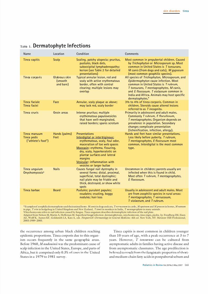
Tinea corporis often appears initially as a red, scaly papulespreading outward, with a coalescence of papules into
plaques that become scaly (Fig. 1). The lesion becomes
an annular, pruritic plaque on glabrous (smooth and
Figure 1. Tinea corporis on body and tinea facie. Note the
erythematous border and scaling.
Figure 2. Tinea corporis, with raised papular border, some
scaling, and central clearing.
skin disorders tinea
166 Pediatrics in Review Vol.28 No.5 May 2007
7/27/2019 Tinea Corporis Pedia-1
http://slidepdf.com/reader/full/tinea-corporis-pedia-1 4/11
bare) skin, with a scaly, slightly raised edge at the advanc-
ing border. The center may clear (Fig. 2), leaving a hypo-or hyperpigmented postinflammatory area as the lesion
advances.Tinea cruris causes a sharply demarcated, pruritic le-
sion that has a raised erythematous margin and thin, dry epidermal scaling that tends to spare the penis, scrotum,
vulva, and perianal area, in contrast to candidiasis, whichaffects these areas. The rash spreads from the groin down
the inner thigh. Tinea pedis or tinea unguium often areconcomitant with tinea cruris.
Tinea pedis presents most often as a web-space mac-eration, but also may have a moccasin distribution, af-
fecting the soles and lateral feet with hyperkeratotic scale.
Tinea manuum manifests similarly and often accompa-nies tinea pedis. Scaling may be finer, with white orsilvery scaling in the creases, or the typical tinea corporis
pattern may be present. Tinea unguium affects the nailplate and is the most resistant to treatment, requiring
long courses and having frequent relapses. The nail canshow dystrophy, thickening, ridging, discoloration, and
breakage or occasionally a superficial white discolorationon the nail surface. The more general term onychomy-
cosis refers to any fungal infection of the nail, including yeasts that do not respond to griseofulvin.
Atypical presentations of tinea include tinea incog-
nito, in which the characteristic scaling disappears aftertreatment of the lesion with high-potency steroid; bul-lous tinea corporis, in which herpetiform, subcorneal
vesicles appear; tinea profunda, which is characterized by subcutaneous abscesses due to T mentagrophytes ; verru-
cous lesions; and Majocchi granuloma, typified by nod-ular or kerionlike lesions not on the scalp. Tinea imbri-
cate, caused by T concentricum, has multiple concentric
rings and is found only in the western Pacific.
Differential Diagnosis
Other nondermatophytic fungi cause superficial infec-tions. Candida infection has a more erythematous pre-
sentation and has an irregular shape and satellite lesionsat the edge of an inflamed area. Tinea versicolor now is
referred to commonly as Pityrosporum versicolor to dif-ferentiate it from dermatophyte infections because it is
not a true tinea. It is caused by a filamentous form of Malassezia furfur (P orbiculare is the yeast form). Mul-
tiple oval, hypo- or hyperpigmented, macular lesions thathave very fine scales usually appear on the upper trunk,
face, and proximal arms. The condition usually is asymp-tomatic, chronic, and more prominent when sun-
exposed areas fail to tan. The rash can be pruritic, espe-
cially with sweating or warm weather, and fluoresces
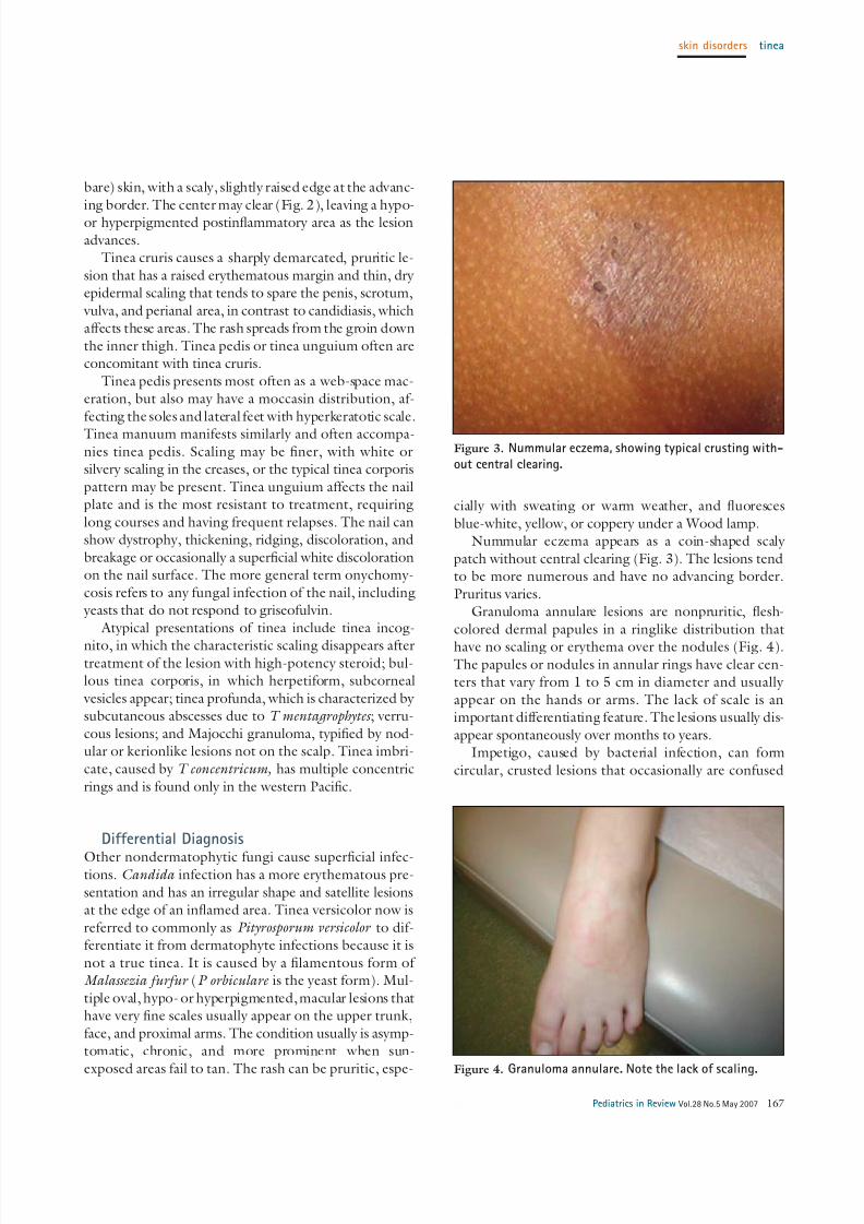
blue-white, yellow, or coppery under a Wood lamp.Nummular eczema appears as a coin-shaped scaly
patch without central clearing (Fig. 3). The lesions tendto be more numerous and have no advancing border.
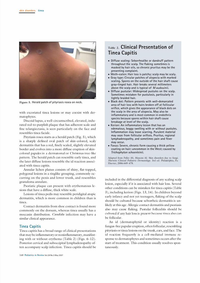
Pruritus varies.Granuloma annulare lesions are nonpruritic, flesh-
colored dermal papules in a ringlike distribution that
have no scaling or erythema over the nodules (Fig. 4).The papules or nodules in annular rings have clear cen-ters that vary from 1 to 5 cm in diameter and usually
appear on the hands or arms. The lack of scale is animportant differentiating feature. The lesions usually dis-
appear spontaneously over months to years.Impetigo, caused by bacterial infection, can form
circular, crusted lesions that occasionally are confused
Figure 3. Nummular eczema, showing typical crusting with-
out central clearing.
Figure 4. Granuloma annulare. Note the lack of scaling.
skin disorders tinea
Pediatrics in Review Vol.28 No.5 May 2007 167
7/27/2019 Tinea Corporis Pedia-1
http://slidepdf.com/reader/full/tinea-corporis-pedia-1 5/11
with excoriated tinea lesions or may coexist with der-matophytes.
Discoid lupus, a well-circumscribed, elevated, indu-rated red-to-purplish plaque that has adherent scale and
fine telangiectasia, is seen particularly on the face andresembles tinea faciale.
Pityriasis rosea starts as a herald patch (Fig. 5), whichis a sharply defined oval patch of skin-colored, scaly
dermatitis that has a red, finely scaled, slightly elevated
border and evolves into a more diffuse eruption of skin-colored papules in a dermatomal or Christmas tree-likepattern. The herald patch can resemble early tinea, and
the later diffuse lesions resemble the id reaction associ-ated with tinea capitis.
Annular lichen planus consists of shiny, flat-topped,polygonal lesions in a ringlike grouping, commonly oc-
curring on the penis and lower trunk, and resemblesgranuloma annulare.
Psoriatic plaque can present with erythematous le-sions that have a diffuse, thick white scale.
Lesions of tinea pedis may resemble peridigital atopic
dermatitis, which is more common in children than istinea.
Contact dermatitis from shoe contact is found morecommonly on the dorsum, whereas tinea usually has a
moccasin distribution. Candida infection may have asimilar clinical appearance.
Tinea CapitisTinea capitis has a broad range of clinical presentationsthat may be inflammatory or noninflammatory, manifest-
ing with or without erythema (Table 2) (Figs. 6–12).Posterior cervical and suboccipital lymphadenopathy of-
ten accompany scalp infection. Tinea capitis should be
included in the differential diagnosis of any scaling scalp
lesion, especially if it is associated with hair loss. Several
other conditions can be mistaken for tinea capitis (Table
3), including kerion (Figs. 13, 14). In children beyond
early infancy and not yet teenagers, flaking of the scalp
should be cultured because seborrheic dermatitis is un-
likely at this age. Allergic contact dermatitis and psoriasis
also may cause flaking. Pustular folliculitis should be
cultured if any hair loss is present because tinea also can
be follicular.
An id (dermatophytid or identity) reaction is a
fungus-free papular eruption, often follicular, resembling
pityriasis or tinea lesions on the trunk, ears, and face. The
id reaction frequently is a cell-mediated immune re-
sponse to dermatophytes and sometimes occurs after the
start of treatment. This condition usually resolves spon-
taneously.
Figure 5. Herald patch of pityriasis rosea on neck.
Table 2.
Clinical Presentation of Tinea Capitis
● Diffuse scaling: Seborrhealike or dandruff patternthroughout the scalp. The flaking sometimes ismasked by hair oils, so chronic pruritus may be thepresenting complaint.
● Moth-eaten: Hair loss is patchy; scalp may be scaly.● Gray type: Circular patches of alopecia with marked
scaling. Spores on the outside of the hair shaft causegray-tinged hair. Hair breaks several millimetersabove the scalp and is typical of M audouinii.
● Diffuse pustular: Widespread pustules on the scalp.Sometimes mistaken for pustulosis, particularly in
tightly braided hair.● Black dot: Pattern presents with well-demarcated
area of hair loss with hairs broken off at follicularorifice, which gives the appearance of black dots onthe scalp in the area of alopecia. May also beinflammatory and is most common in endothrixspecies because spores within hair shaft causebreakage at level of the scalp.
● Kerion: An inflammatory lesion that has anedematous, boggy swelling with or without pustules.Inflammation may leave scarring. Purulent materialmay ooze from follicular orifices. Pruritus, regionallymphadenopathy, and sometimes pain and fevermay occur.
● Favus: Severe, chronic form causing a thick yellow
coating on hair; uncommon in the West; caused byTrichophyton schoenleinii.
Adapted from Paller AS, Mancini AJ. Skin disorders due to fungi.Hurwitz Clinical Pediatric Dermatology. 3rd ed. Philadelphia, Pa:Elsevier; 2006:449–478.
skin disorders tinea
168 Pediatrics in Review Vol.28 No.5 May 2007
7/27/2019 Tinea Corporis Pedia-1
http://slidepdf.com/reader/full/tinea-corporis-pedia-1 6/11
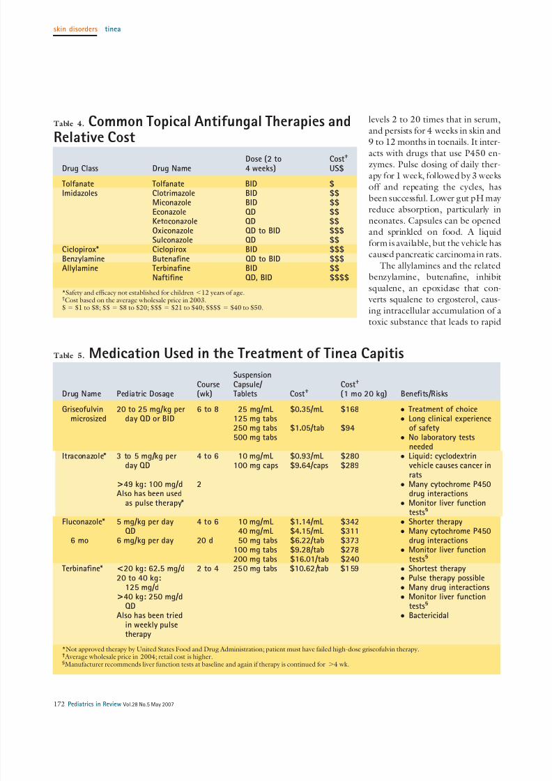
Management of Tinea InfectionsTinea corporis usually is treated with topical antifungalpreparations (Table 4). Tinea capitis always requires oral
therapy.
Topical treatment usually is continued for 1 to2 weeks after the lesion has resolved. Tineas are resistantto nystatin, an antifungal commonly used for Candida
infections. Folk remedies, such as creating a chemical
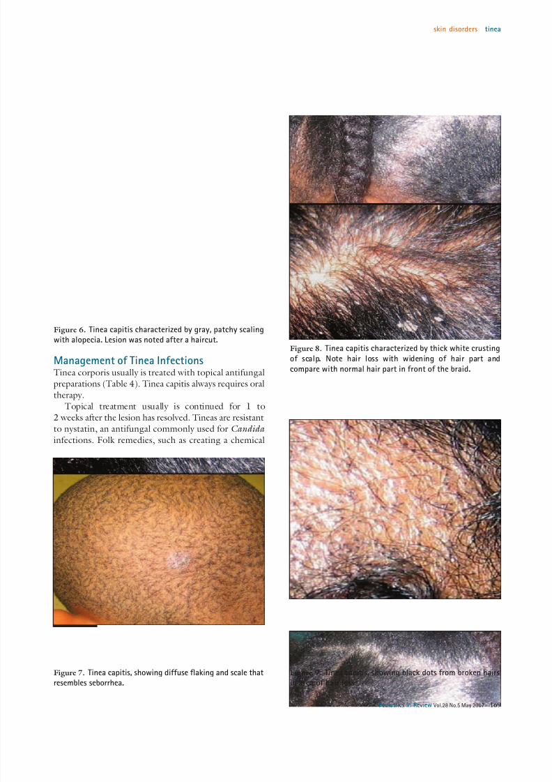
Figure 6. Tinea capitis characterized by gray, patchy scalingwith alopecia. Lesion was noted after a haircut.
Figure 7. Tinea capitis, showing diffuse flaking and scale that
resembles seborrhea.
Figure 8. Tinea capitis characterized by thick white crusting
of scalp. Note hair loss with widening of hair part and
compare with normal hair part in front of the braid.
Figure 9. Tinea capitis, showing black dots from broken hairs
in area of hair loss.
skin disorders tinea
Pediatrics in Review Vol.28 No.5 May 2007 169

7/27/2019 Tinea Corporis Pedia-1
http://slidepdf.com/reader/full/tinea-corporis-pedia-1 7/11
burn at the site with undiluted bleach, should be discour-aged because they can cause infection and scarring. Top-
ical therapy should be applied at least 2 cm beyond theedge of the lesions once or twice daily for 2 weeks,
depending on the agent. If the lesion persists after4 weeks of topical therapy, it is considered a treatment
failure. Extensive, severe, or resistant cases of tinea cor-poris may require systemic treatment (Table 5).
Infection of hair or nails in tinea capitis, tinea barbae,and tinea unguium requires systemic treatment because
topical antifungal medications do not penetrate into the
Figure 10. Tinea capitis in close up, showing white crustingaround hair root and some broken hairs.
Figure 11. Tinea capitis that was resistant to high-dose
griseofulvin after 6 weeks of treatment. Some of the smaller
lesions are new. Culture grew T tonsurans .
Figure 12. Tinea capitis, showing both white scales and
yellow, purulent exudates with hair loss that was very pruritic.
Child had been scratching her head constantly during aroutine physical examination. Culture grew T tonsurans.
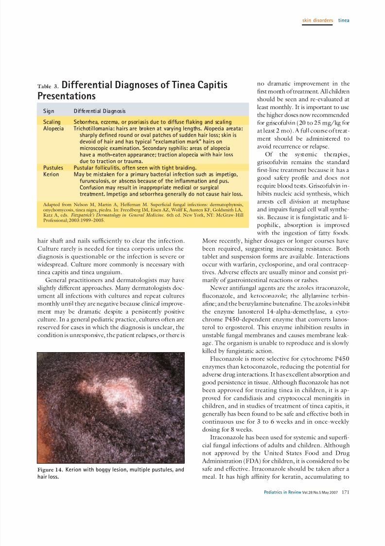
Figure 13. Kerion with boggy early lesion and multiple broken
hairs. Few small pustules are forming.
skin disorders tinea
170 Pediatrics in Review Vol.28 No.5 May 2007
7/27/2019 Tinea Corporis Pedia-1
http://slidepdf.com/reader/full/tinea-corporis-pedia-1 8/11
hair shaft and nails sufficiently to clear the infection.
Culture rarely is needed for tinea corporis unless thediagnosis is questionable or the infection is severe or
widespread. Culture more commonly is necessary withtinea capitis and tinea unguium.
General practitioners and dermatologists may have
slightly different approaches. Many dermatologists doc-ument all infections with cultures and repeat culturesmonthly until they are negative because clinical improve-
ment may be dramatic despite a persistently positiveculture. In a general pediatric practice, cultures often are
reserved for cases in which the diagnosis is unclear, thecondition is unresponsive, the patient relapses, or there is
no dramatic improvement in the
first month of treatment. All childrenshould be seen and re-evaluated at
least monthly. It is important to usethe higher doses now recommended
for griseofulvin (20 to 25 mg/kg forat least 2 mo). A full course of treat-
ment should be administered toavoid recurrence or relapse.
Of the systemic therapies,griseofulvin remains the standard
first-line treatment because it has agood safety profile and does not
require blood tests. Griseofulvin in-
hibits nucleic acid synthesis, whicharrests cell division at metaphaseand impairs fungal cell wall synthe-
sis. Because it is fungistatic and li-pophilic, absorption is improved
with the ingestion of fatty foods.More recently, higher dosages or longer courses have
been required, suggesting increasing resistance. Bothtablet and suspension forms are available. Interactions
occur with warfarin, cyclosporine, and oral contracep-tives. Adverse effects are usually minor and consist pri-
marily of gastrointestinal reactions or rashes.
Newer antifungal agents are the azoles itraconazole,fluconazole, and ketoconazole; the allylamine terbin-afine; and the benzylamine butenafine. The azoles inhibit
the enzyme lanosterol 14-alpha-demethylase, a cyto-chrome P450-dependent enzyme that converts lanos-
terol to ergosterol. This enzyme inhibition results in
unstable fungal membranes and causes membrane leak-age. The organism is unable to reproduce and is slowly
killed by fungistatic action.Fluconazole is more selective for cytochrome P450
enzymes than ketoconazole, reducing the potential foradverse drug interactions. It has excellent absorption and
good persistence in tissue. Although fluconazole has notbeen approved for treating tinea in children, it is ap-
proved for candidiasis and cryptococcal meningitis inchildren, and in studies of treatment of tinea capitis, it
generally has been found to be safe and effective both incontinuous use for 3 to 6 weeks and in once-weekly
dosing for 8 weeks.Itraconazole has been used for systemic and superfi-
cial fungal infections of adults and children. Althoughnot approved by the United States Food and Drug
Administration (FDA) for children, it is considered to besafe and effective. Itraconazole should be taken after a
meal. It has high affinity for keratin, accumulating to
Figure 14. Kerion with boggy lesion, multiple pustules, and
hair loss.
Table 3. Differential Diagnoses of Tinea Capitis
PresentationsSign Differential Diagnosis
Scaling Seborrhea, eczema, or psoriasis due to diffuse flaking and scalingAlopecia Trichotillomania: hairs are broken at varying lengths. Alopecia areata:
sharply defined round or oval patches of sudden hair loss; skin isdevoid of hair and has typical “exclamation mark” hairs onmicroscopic examination. Secondary syphilis: areas of alopeciahave a moth-eaten appearance; traction alopecia with hair lossdue to traction or trauma.
Pustules Pustular folliculitis, often seen with tight braiding.Kerion May be mistaken for a primary bacterial infection such as impetigo,
furunculosis, or abscess because of the inflammation and pus.Confusion may result in inappropriate medical or surgicaltreatment. Impetigo and seborrhea generally do not cause hair loss.
Adapted from Nelson M, Martin A, Heffernan M. Superficial fungal infections: dermatophytosis,onychomycosis, tinea nigra, piedra. In: Freedberg IM, Eisen AZ, Wolff K, Austen KF, Goldsmith LA,Katz A, eds. Fitzpatrick’s Dermatology in General Medicine. 6th ed. New York, NY: McGraw-HillProfessional; 2003:1989–2005.
skin disorders tinea
Pediatrics in Review Vol.28 No.5 May 2007 171
7/27/2019 Tinea Corporis Pedia-1
http://slidepdf.com/reader/full/tinea-corporis-pedia-1 9/11
levels 2 to 20 times that in serum,
and persists for 4 weeks in skin and9 to 12 months in toenails. It inter-
acts with drugs that use P450 en-zymes. Pulse dosing of daily ther-
apy for 1 week, followed by 3 weeksoff and repeating the cycles, has
been successful. Lower gut pH may reduce absorption, particularly in
neonates. Capsules can be openedand sprinkled on food. A liquid
form is available, but the vehicle hascaused pancreatic carcinoma in rats.
The allylamines and the related
benzylamine, butenafine, inhibitsqualene, an epoxidase that con-
verts squalene to ergosterol, caus-
ing intracellular accumulation of atoxic substance that leads to rapid
Table 4. Common Topical Antifungal Therapies and
Relative Cost
Drug Class Drug NameDose (2 to4 weeks)
Cost†
US$
Tolfanate Tolfanate BID $Imidazoles Clotrimazole BID $$
Miconazole BID $$Econazole QD $$Ketoconazole QD $$Oxiconazole QD to BID $$$Sulconazole QD $$
Ciclopirox* Ciclopirox BID $$$Benzylamine Butenafine QD to BID $$$Allylamine Terbinafine BID $$
Naftifine QD, BID $$$$
*Safety and efficacy not established for children Ͻ12 years of age.†Cost based on the average wholesale price in 2003.$ ϭ $1 to $8; $$ ϭ $8 to $20; $$$ ϭ $21 to $40; $$$$ ϭ $40 to $50.
Table 5. Medication Used in the Treatment of Tinea Capitis
Drug Name Pediatric DosageCourse(wk)
SuspensionCapsule/ Tablets Cost†
Cost†
(1 mo 20 kg) Benefits/Risks
Griseofulvinmicrosized
20 to 25 mg/kg perday QD or BID
6 to 8 25 mg/mL125 mg tabs250 mg tabs500 mg tabs
$0.35/mL
$1.05/tab
$168
$94
● Treatment of choice● Long clinical experience
of safety● No laboratory tests
neededItraconazole* 3 to 5 mg/kg per
day QD
>49 kg: 100 mg/dAlso has been used
as pulse therapy*
4 to 6
2
10 mg/mL100 mg caps
$0.93/mL$9.64/caps
$280$289
● Liquid: cyclodextrinvehicle causes cancer inrats
● Many cytochrome P450drug interactions
● Monitor liver functiontests§
Fluconazole*
6 mo
5 mg/kg per dayQD
6 mg/kg per day
4 to 6
20 d
10 mg/mL40 mg/mL50 mg tabs
100 mg tabs200 mg tabs
$1.14/mL$4.15/mL$6.22/tab
$9.28/tab$16.01/tab
$342$311$373
$278$240
● Shorter therapy● Many cytochrome P450
drug interactions● Monitor liver function
tests§
Terbinafine* <20 kg: 62.5 mg/d20 to 40 kg:
125 mg/d>40 kg: 250 mg/d
QDAlso has been tried
in weekly pulsetherapy
2 to 4 250 mg tabs $10.62/tab $159 ● Shortest therapy● Pulse therapy possible● Many drug interactions● Monitor liver function
tests§
● Bactericidal
*Not approved therapy by United States Food and Drug Administration; patient must have failed high-dose griseofulvin therapy.† Average wholesale price in 2004; retail cost is higher.§Manufacturer recommends liver function tests at baseline and again if therapy is continued for Ͼ4 wk.
skin disorders tinea
172 Pediatrics in Review Vol.28 No.5 May 2007
7/27/2019 Tinea Corporis Pedia-1
http://slidepdf.com/reader/full/tinea-corporis-pedia-1 10/11
fungal cell death. Ciclopirox accumulates inside cells,
interfering with amino acid transport and causing fungalcell membrane instability.
Terbinafine, a well-absorbed fungicide, accumulatesin skin at higher concentrations than in plasma and has a
long half-life. In comparative studies, terbinafine treat-ment for 4 weeks gave similar cure rates of Trichophyton
tinea capitis as 8 weeks of griseofulvin.Onychomycosis in children in North America has no
FDA-approved treatment. However, terbinafine is the rec-ommended drug for treating nail dermatophytes; dosage is
based on the child’s weight, and the agent is taken for6 to 12 weeks. Clearance is about 40% higher in children
than in adults. Terbinafine is available only in tablets, but
they can be broken, pulverized, and added to food. Adjunctive therapy with antifungal shampoo 2 to
3 times weekly for the patient who hastinea capitis as well
as for family members is recommended to reduce thenumber of spores. The most commonly recommended is
selenium sulfide 2.5% shampoo, which adheres to thescalp after rinsing. Ketoconazole shampoo seems less
drying because of its coconut oil base but does not work as well because it rinses away more easily. This shampoo
is more effective if left on for 5 minutes. Hair groomingpractices do not appear to play a major role in acquiring
tinea capitis. Hygienic practices such as not sharing combs,
brushes, or hats are important to decrease transmission.Exclusion from school is not recommended because
spores can shed for months. Additionally, asymptomatic
carriers within the class who can be vectors are common.
SummaryTinea capitis and corporis are very common in children.
Tinea capitis usually is caused by nonfluorescent T ton-
surans in the United States. It has many presentations,
from mild flaking to purulent infected areas, but usually
causes pruritus, hair loss, and often, suboccipital adenop-
athy. Oral antifungal treatment always is required for
tinea capitis, although shampoos may decrease the risk of
transmission. Children should not be excluded from
school because they remain infective and there likely are
asymptomatic carriers in the class. Tinea corporis may
vary in presentation on different parts of the body. Some
children manifest an id reaction, which is an allergic
response to the fungal antigens rather than to the medi-
cation.
Suggested ReadingClayton YM. Superficial fungal infections. In: Harper J, Oranje A,
Prose N, eds. Textbook of Pediatric Dermatology . Oxford,
United Kingdom: Blackwell; 2000:447– 459
Gupta AK, Cooper EA, Ginter G. Fluconazole, terbinafine and
itraconazole in the treatment of superficial fungal. Dermatol
Clin . 2003;21:511–542
Higgins EM, Fuller LC,Smith CH. Guidelines forthe management
of tinea capitis. British Association of Dermatologists. Br J
Dermatol . 2000;143:53–58
Kao GF. Tinea capitis. eMedicine Dermatology Fungal Infections .
2006. Available at: http://www.emedicine.com/derm/
topic420.htmLeyden JJ. Update on tinea capitis and new antifungal therapies.
Pediatr Infect Dis J. 1999;18:180–215
Rushing ME, Johnson A, Zember G, Lesher JL. Tinea corporis.
eMedicine Dermatology Fungal Infections . 2006. Available at:
http://www.emedicine.com/derm/topic421.htm
Stein DH. Fungal, protozoan and helminthe infections. In:
Schachner LA, Hansen RC, eds. Pediatric Dermatology . 2nd ed.
New York, NY: Churchill Livingtstone; 1998:1295–1311
skin disorders tinea
Pediatrics in Review Vol.28 No.5 May 2007 173
7/27/2019 Tinea Corporis Pedia-1
http://slidepdf.com/reader/full/tinea-corporis-pedia-1 11/11
PIR QuizQuiz also available online at www.pedsinreview.org.
1. The dermatophyte infections in humans can have a variety of clinical presentations and appearances thatcan provide the clinician with some diagnostic challenges. A black dot skin appearance is most likely seenin:
A. Tinea capitis.B. Tinea corporis.C. Tinea cruris.D. Tinea faciei.E. Tinea pedis.
2. A 5-year-old boy enjoys playing with the cats that inhabit his neighborhood. He presents to your officewith several raised, circular lesions on his arm and leg. You suspect that the child has tinea corporisacquired from his close contact with the cats. You scrape the lesions for a confirming fungal culture. Themost likely zoophilic dermophyte to cause the lesions is:
A. Microsporum canis.B. M nanum.C. M praecox.D. Trichophyton erin.E. T simii.
3. A previously useful diagnostic test for helping to diagnose suspected cases of tinea capitis was thedetection of green-yellow fluorescence in the hair roots with a Wood lamp. The primary reason this test isless helpful today is the declining incidence of:
A. Ectothrix infections.
B. Endothrix infections.C. Geophilic infections.D. M canis infections.E. Trychophyton infections.
4. A number of skin conditions must be considered in the differential diagnosis of tinea corporis. One disorderthat resembles tinea corporis but is notable for its lack of scale is:
A. Discoid lupus.B. Granuloma annulare.C. Nummular eczema.D. Pityriasis rosea.E. Psoriasis.
skin disorders tinea
174 Pediatrics in Review Vol.28 No.5 May 2007
Related Documents