Time dependence of material properties of polyethylene glycol hydrogels chain extended with short hydroxy acid segments Danial Barati, Seyedsina Moeinzadeh, Ozan Karaman, Esmaiel Jabbari * Biomimetic Materials and Tissue Engineering Laboratory, Department of Chemical Engineering, University of South Carolina, Columbia, SC 29208, USA article info Article history: Received 7 March 2014 Received in revised form 9 May 2014 Accepted 17 May 2014 Available online 24 May 2014 Keywords: Hydrogel chain extension Hydroxy acids Cell encapsulation abstract The objective of this work was to investigate the effect of chemical composition and segment number (n) on gelation, stiffness, and degradation of hydroxy acid-chain-extended star polyethylene glycol acrylate (SPEXA) gels. The hydroxy acids included glycolide (G), L-lactide (L), p-dioxanone (D) and 3 -caprolactone (C). Chain-extension generated water soluble macromers with faster gelation rates, lower sol fractions, higher compressive moduli, and a wide-ranging degradation times when crosslinked into a hydrogel. SPEGA gels with the highest fraction of inter-molecular crosslinks had the most increase in compressive modulus with n whereas SPELA and SPECA had the lowest increase in modulus. SPEXA gels exhibited a wide range of degradation times from a few days for SPEGA to a few weeks for SPELA, a few months for SPEDA, and many months for SPECA. Marrow stromal cells and endothelial progenitor cells had the highest expression of vasculogenic markers when co-encapsulated in the faster degrading SPELA gel. © 2014 Elsevier Ltd. All rights reserved. 1. Introduction The delivery of stem cells in a supportive carrier is an exciting approach to the regeneration of damaged biological tissues. In that approach the carrier acts as a temporary matrix for immobilization of the cells in the site of regeneration which is gradually displaced with the extracellular matrix (ECM) secreted by the encapsulated cells. The temporary matrix should provide a three-dimensional hydrophilic and aqueous environment to support complex cellematrix interactions but gradually degrade concurrent with ECM formation [1e4]. Due to their high water content, hydrogels have high permeability to oxygen and nutrients required for cell survival and function [5]. Further, cell-loaded hydrogel precursor solutions can be injected and gelled in situ to fill defects with irregular shapes. Inter-diffusion of hydrogel chains to tissues proximal to the defect leads to superior integration of the carrier with the surrounding tissue after gelation [6]. Due to their non-immunogenic and inert nature, polyethylene glycol (PEG) hydrogels are very attractive as a matrix for stem cell delivery to the site of regeneration [6e8]. Adhesion and differen- tiation of cells encapsulated in PEG gels can be tuned by conjugation with integrin-binding ligands and morphogenetic peptides, respectively [9e11]. A major drawback to the wide-spread use of PEG gels is their persistence in the site of regeneration. Although copolymerization of PEG macromers with resorbable monomers can be used to impart degradability to the hydrogel [12,13], degradability is offset by lower macromer solubility in aqueous solution for cell encapsulation and low gel stiffness [14,15]. Recently, we reported that the extension of star PEG macromers with very short hydroxy acid segments imparted degradability to the hydrogel without changing macromer solubility in aqueous solution and hydrogel stiffness [14,16]. Molecular dynamic simu- lations demonstrated the formation of micellar structures (<3 nm in size) that dramatically increased the local concentration of reactive groups, leading to a potentially higher modulus with little change in the overall water content of the hydrogel [17]. Further, human MSCs encapsulated in the high-stiffness, relatively slow- degrading lactide-chain-extended PEG hydrogels had higher extent of differentiation to the osteogenic lineage than those encapsulated in non-degradable PEG gels [16]. It is well-established that the fate of encapsulated cells is determined by the physical and mechanical properties of their microenvironment including degradation rate and stiffness. For example, marrow stem cells (MSCs) encapsulated in a non- degradable hyaluronic acid (HA) gel had a rounded morphology and underwent adipogenic differentiation whereas those MSCs encapsulated in a degradable HA gel spread and differentiated to the osteogenic lineage [18]. Similarly, the C2C12 mouse myoblast * Corresponding author. Swearingen Engineering Center, Rm 2C11, University of South Carolina, Columbia, SC 29208, USA. Tel.: þ1 803 777 8022; fax: þ1 803 777 0973. E-mail addresses: [email protected], [email protected] (E. Jabbari). Contents lists available at ScienceDirect Polymer journal homepage: www.elsevier.com/locate/polymer http://dx.doi.org/10.1016/j.polymer.2014.05.045 0032-3861/© 2014 Elsevier Ltd. All rights reserved. Polymer 55 (2014) 3894e3904

Welcome message from author
This document is posted to help you gain knowledge. Please leave a comment to let me know what you think about it! Share it to your friends and learn new things together.
Transcript
Time dependence of material properties of polyethylene glycolhydrogels chain extended with short hydroxy acid segments
Danial Barati, Seyedsina Moeinzadeh, Ozan Karaman, Esmaiel Jabbari*
Biomimetic Materials and Tissue Engineering Laboratory, Department of Chemical Engineering, University of South Carolina, Columbia, SC 29208, USA
a r t i c l e i n f o
Article history:Received 7 March 2014Received in revised form9 May 2014Accepted 17 May 2014Available online 24 May 2014
Keywords:Hydrogel chain extensionHydroxy acidsCell encapsulation
a b s t r a c t
The objective of this work was to investigate the effect of chemical composition and segment number (n)on gelation, stiffness, and degradation of hydroxy acid-chain-extended star polyethylene glycol acrylate(SPEXA) gels. The hydroxy acids included glycolide (G), L-lactide (L), p-dioxanone (D) and 3-caprolactone(C). Chain-extension generated water soluble macromers with faster gelation rates, lower sol fractions,higher compressive moduli, and a wide-ranging degradation times when crosslinked into a hydrogel.SPEGA gels with the highest fraction of inter-molecular crosslinks had the most increase in compressivemodulus with n whereas SPELA and SPECA had the lowest increase in modulus. SPEXA gels exhibited awide range of degradation times from a few days for SPEGA to a few weeks for SPELA, a few months forSPEDA, and many months for SPECA. Marrow stromal cells and endothelial progenitor cells had thehighest expression of vasculogenic markers when co-encapsulated in the faster degrading SPELA gel.
© 2014 Elsevier Ltd. All rights reserved.
1. Introduction
The delivery of stem cells in a supportive carrier is an excitingapproach to the regeneration of damaged biological tissues. In thatapproach the carrier acts as a temporary matrix for immobilizationof the cells in the site of regeneration which is gradually displacedwith the extracellular matrix (ECM) secreted by the encapsulatedcells. The temporary matrix should provide a three-dimensionalhydrophilic and aqueous environment to support complexcellematrix interactions but gradually degrade concurrent withECM formation [1e4]. Due to their high water content, hydrogelshave high permeability to oxygen and nutrients required for cellsurvival and function [5]. Further, cell-loaded hydrogel precursorsolutions can be injected and gelled in situ to fill defects withirregular shapes. Inter-diffusion of hydrogel chains to tissuesproximal to the defect leads to superior integration of the carrierwith the surrounding tissue after gelation [6].
Due to their non-immunogenic and inert nature, polyethyleneglycol (PEG) hydrogels are very attractive as a matrix for stem celldelivery to the site of regeneration [6e8]. Adhesion and differen-tiation of cells encapsulated in PEG gels can be tuned by
conjugation with integrin-binding ligands and morphogeneticpeptides, respectively [9e11]. Amajor drawback to thewide-spreaduse of PEG gels is their persistence in the site of regeneration.Although copolymerization of PEG macromers with resorbablemonomers can be used to impart degradability to the hydrogel[12,13], degradability is offset by lower macromer solubility inaqueous solution for cell encapsulation and lowgel stiffness [14,15].
Recently, we reported that the extension of star PEG macromerswith very short hydroxy acid segments imparted degradability tothe hydrogel without changing macromer solubility in aqueoussolution and hydrogel stiffness [14,16]. Molecular dynamic simu-lations demonstrated the formation of micellar structures (<3 nmin size) that dramatically increased the local concentration ofreactive groups, leading to a potentially higher modulus with littlechange in the overall water content of the hydrogel [17]. Further,human MSCs encapsulated in the high-stiffness, relatively slow-degrading lactide-chain-extended PEG hydrogels had higherextent of differentiation to the osteogenic lineage than thoseencapsulated in non-degradable PEG gels [16].
It is well-established that the fate of encapsulated cells isdetermined by the physical and mechanical properties of theirmicroenvironment including degradation rate and stiffness. Forexample, marrow stem cells (MSCs) encapsulated in a non-degradable hyaluronic acid (HA) gel had a rounded morphologyand underwent adipogenic differentiation whereas those MSCsencapsulated in a degradable HA gel spread and differentiated tothe osteogenic lineage [18]. Similarly, the C2C12 mouse myoblast
* Corresponding author. Swearingen Engineering Center, Rm 2C11, University ofSouth Carolina, Columbia, SC 29208, USA. Tel.: þ1 803 777 8022; fax: þ1 803 7770973.
E-mail addresses: [email protected], [email protected] (E. Jabbari).
Contents lists available at ScienceDirect
Polymer
journal homepage: www.elsevier .com/locate/polymer
http://dx.doi.org/10.1016/j.polymer.2014.05.0450032-3861/© 2014 Elsevier Ltd. All rights reserved.
Polymer 55 (2014) 3894e3904
cells encapsulated in a soft degradable alginate gel (13 kPamodulus) had a higher extent of differentiation to myotubes andlower proliferation than those encapsulated in a non-degradablestiff gel (45 kPa modulus) [19]. Tissue engineered constructs oftentimes require multiphase hydrogels with different but comple-mentary microenvironments. For example, osteogenic differentia-tion of MSCs requires a supporting matrix with high compressivemodulus and slow degradation [20] whereas vasculogenic differ-entiation of progenitor endothelial cells necessitates a lowmodulus, relatively fast-degrading matrix [21,22]. Therefore, thereis a need to develop synthetic hydrogels with tunable degradationand stiffness for wide-ranging applications in regenerativemedicine.
The objective of this work was to investigate the effect ofchemical composition and length of the hydroxy acid in hydroxyacid-chain-extended star PEG acrylate macromer on gelationcharacteristics, water content, compressive modulus, and degra-dation of the hydrogels with incubation time. The hydroxy acidmonomers included the least hydrophobic glycolide, L-lactide, p-dioxanone, and the most hydrophobic 3-caprolactone. The findingsof this work demonstrate that chain extension of star PEG macro-mers with the above hydroxy acids produces hydrogels with awiderange of physical and mechanical properties to serve as cell carriersin regenerative medicine from the compliant vascular tissue to thestiff bone tissue.
2. Experimental
2.1. Materials
Lactide, glycolide and p-dioxanone monomers (L, G, D; >99.5%purity) were purchased from Ortec (Easley, SC) and 3-Caprolactone(C) was purchased from Alfa Aesar (UK). The monomers were driedunder vacuum at 40 "C for at least 12 h prior to polymerization.Calcium hydride, tetrahydrofuran (THF), deuterated chloroform(99.8% deuterated), trimethylsilane (TMS), triethylamine (TEA),tin(II) 2-ethylhexanoate (TOC), acryloyl chloride, and dime-thylsulfoxide (DMSO) were purchased from SigmaeAldrich (St.Louis, MO). 4-arm PEG (SPEG, Mw ¼ 5000) was purchased fromSigmaeAldrich. The protected amino acids and Rink Amide Nova-Gel resin for the synthesis of acrylamide-terminated GRGD peptidewere purchased from EMD Biosciences (San Diego, CA). Dichloro-methane (DCM, Acros Organics, Pittsburg, PA) was dried by distil-lation over calcium hydride. Diethyl ether and hexane wereobtained from VWR (Bristol, CT). The dialysis tube (molecularweight cutoff 3.5 kDa) was purchased from Spectrum Laboratories(Rancho Dominquez, CA). EBM-2 medium, vascular endothelialgrowth factor (VEGF), human fibroblast growth factor-B (hFGF-B),R3-insulin-like growth factor (R3-IGF-1), human epidermal growthfactor (hEGF), ascorbic acid hydrocortisone, gentamycin, andamphotericin-B were purchased from Lonza (Hopkinton, MA).Dulbecco's Modified Eagle Medium (DMEM; 4.5 g/L glucose with L-glutamine without sodium pyruvate) was purchased from Medi-atech (Herndon, VA). Medium 199with L-glutaminewas purchasedfrom SigmaeAldrich. 3-(4,5-dimethyl-2-thiazolyl)-2,5-diphenyl-2H-tetrazolium bromide (MTT) was purchased from Calbiochem(EMD Millipore, Billerica, MA).
2.2. Macromer synthesis
The 4-arm poly(ethylene glycol) (SPEG) macromer wasextended with D, C, G and L monomers via Ring Opening Poly-merization (ROP) to synthesize SPED, SPEC, SPEG and SPEL mac-romers, respectively, as we previously described [15,16]. Briefly, forthe synthesis of SPEC, SPEG and SPEL macromers, the PEG and
monomer were added to a three-neck reaction flask equipped witha stirrer and immersed in an oil bath. The feed molar ratio of thePEG tomonomerwas based on the desired length of the degradablesegment on each macromer arm. Next, the flask was heated to120 "C under nitrogen flow and maintained at that temperature for1 h before the addition of TOC catalyst. The polymerization reactionwas allowed to proceed for 12 h at 140 "C, 160 "C and 140 "C forSPEC, SPEG and SPEL, respectively. For the synthesis of SPED, thePEG and catalyst mixture was heated to 130 "C to remove moisture,cooled to 85 "C for addition of D monomer, and the reaction wasallowed to proceed for 48 h at 85 "C. The reaction product wasprecipitated in ice-cold hexane to remove the unreacted monomerand catalyst.
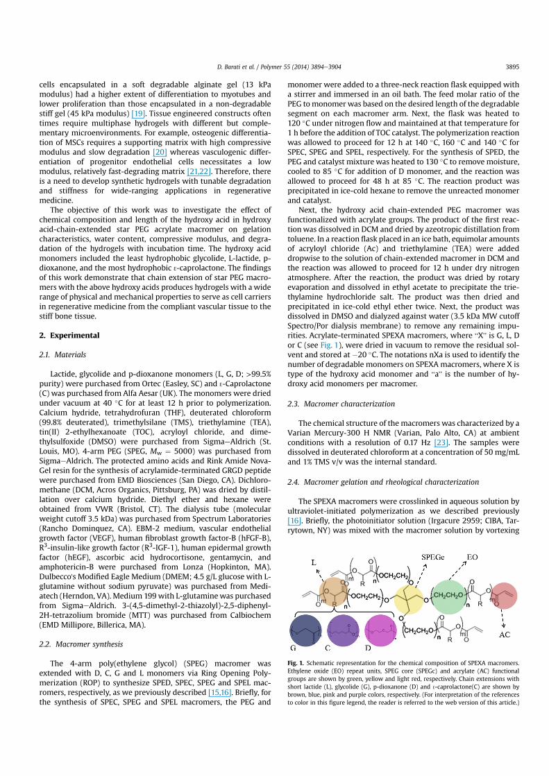
Next, the hydroxy acid chain-extended PEG macromer wasfunctionalized with acrylate groups. The product of the first reac-tionwas dissolved in DCM and dried by azeotropic distillation fromtoluene. In a reaction flask placed in an ice bath, equimolar amountsof acryloyl chloride (Ac) and triethylamine (TEA) were addeddropwise to the solution of chain-extended macromer in DCM andthe reaction was allowed to proceed for 12 h under dry nitrogenatmosphere. After the reaction, the product was dried by rotaryevaporation and dissolved in ethyl acetate to precipitate the trie-thylamine hydrochloride salt. The product was then dried andprecipitated in ice-cold ethyl ether twice. Next, the product wasdissolved in DMSO and dialyzed against water (3.5 kDa MW cutoffSpectro/Por dialysis membrane) to remove any remaining impu-rities. Acrylate-terminated SPEXA macromers, where “X” is G, L, Dor C (see Fig. 1), were dried in vacuum to remove the residual sol-vent and stored at $20 "C. The notations nXa is used to identify thenumber of degradable monomers on SPEXA macromers, where X istype of the hydroxy acid monomer and “a” is the number of hy-droxy acid monomers per macromer.
2.3. Macromer characterization
The chemical structure of the macromers was characterized by aVarian Mercury-300 H NMR (Varian, Palo Alto, CA) at ambientconditions with a resolution of 0.17 Hz [23]. The samples weredissolved in deuterated chloroform at a concentration of 50 mg/mLand 1% TMS v/v was the internal standard.
2.4. Macromer gelation and rheological characterization
The SPEXA macromers were crosslinked in aqueous solution byultraviolet-initiated polymerization as we described previously[16]. Briefly, the photoinitiator solution (Irgacure 2959; CIBA, Tar-rytown, NY) was mixed with the macromer solution by vortexing
Fig. 1. Schematic representation for the chemical composition of SPEXA macromers.Ethylene oxide (EO) repeat units, SPEG core (SPEGc) and acrylate (AC) functionalgroups are shown by green, yellow and light red, respectively. Chain extensions withshort lactide (L), glycolide (G), p-dioxanone (D) and 3-caprolactone(C) are shown bybrown, blue, pink and purple colors, respectively. (For interpretation of the referencesto color in this figure legend, the reader is referred to the web version of this article.)
D. Barati et al. / Polymer 55 (2014) 3894e3904 3895
for 5 min at 50 "C. Based on our previous results [14], the initiatorconcentration of 0.75 wt% (based on the macromer weight) wasused in the hydrogel precursor solution to minimize cell toxicityand optimize the extent of gelation. To measure compressivemodulus of the gels, the hydrogel precursor solutions weredegassed and transferred to a polytetrafluoroethylene (PTFE) mold(5 cm% 3 cm% 750 mm), coveredwith a transparent glass plate andfastened with clips. Next, the samples were irradiated with a BLAK-RAY 100Wmercury, long-wavelength (365 nm) UV lamp (B100-AP;UVP, Upland, CA) for 10 min. Disk-shaped samples were cut fromthe gel using an 8-mm cork borer and loaded on the Peltier plate ofthe rheometer (TA Instruments, New Castle, DE) and subjected to auniaxial compressive force at a displacement rate of 7.5 mm/s. Theslope of the linear fit to the stressestrain curve for 5%e10% strainwas taken as the elastic modulus (E) of the gels, as we previouslydescribed [24].
To measure gelation time, the hydrogel precursor solution wasdirectly loaded on the peltier plate of an AR-2000 rheometer (TAInstruments, New Castle, DE) and irradiated at a distance of 10 cmfrom the sample. A sinusoidal shear strain with frequency of 1 Hzand amplitude of 1%was exerted on the sample and the storage (G0)and loss moduli (G00) were recorded with time. The time at whichG0 ¼ G00 was recorded as the gelation time, as we previouslydescribed [15].
2.5. Measurement of swelling ratio, sol fraction and mass loss
Disk shape hydrogel samples with a diameter of 8 mm and athickness of 750 mm were dried at ambient conditions for 12 hfollowed by drying in vacuum for 1 h at 40 "C. After drying, the dryweights (wi) were recorded. Next, the dry samples were swollen inDI water for 24 h at 37 "C with a change of swelling medium every6 h. After swelling, the surface water was removed and the swollenweights (ws) weremeasured. Then, the swollen samples were driedas described above and the dry weights (wd) were recorded. Theswelling ratio (Q) and sol fraction (S) were calculated from the dryand swollen weights using previously described equations [25]. Tomeasure mass loss, the dried samples disk were incubated in 5 mLbasal mediumwithout fetal bovine [16] serum at 37 "C under mildagitation. At each time point, samples were washed with DI waterto remove excess electrolytes, dried under vacuum, and the drysample weights were measured and compared with the initial dryweights to determine fractional mass remaining as previouslydescribed [14].
2.6. Cell isolation and culture
MSCs were isolated from the bone marrow of young adult maleWistar rats (6e8 weeks) as we previously described [10,23,26].Cell isolations were performed under a protocol approved by theInstitutional Animal Care and Use Committee of the University ofSouth Carolina. The marrow of the removed tibias and femurswere flushed with cell isolation medium [23], the cell suspensionwas centrifuged at 200 g for 5 min. Next, the cell pellets were re-suspended in primary medium (DMEM supplemented with 10%FBS [23]), aliquoted into tissue culture flasks and maintained in ahumidified 5% CO2 incubator at 37 "C. The medium was replacedwith fresh medium at 3 and 7 days to remove unattached cells.The enzymatically lifted MSCs were used for cell culture experi-ments. To isolate endothelial progenitor cells (EPCs) from the ratbone marrow, the marrow cavity of the tibias and femurs wereflushed with Medium 199 supplemented with 20% FBS, 1% Pen/Strep and 22.5 mg/ml heparin [27,28]. The cell suspension wascentrifuged at 200 g for 5 min and the cell pellet was re-suspended in Medium-199 and plated on rat-derived
fibronectin-coated 6-well plates (1 mg/mL). After 24 h, the non-adherent cells in the medium were recovered and transferred toa new fibronectin-coated plate. This procedure was repeated toremove rapidly adherent hematopoietic cells or mature endothe-lial cells in the aspirate [27,28]. Only the non-adherent cell pop-ulation was harvested after 48 h and used for cell cultureexperiments.
The MSC/EPCs (1:1 cell ratio) were encapsulated in PEGDA,nL6.4, nD6.8, and nC7.2 hydrogels. MSCs in the co-culture act aspericytes to facilitate maturation and stabilization of tubularstructures formed by multi-cellular assembly of EPCs [29,30].Recent reports indicate that the extent of vasculogenic differenti-ation of EPCs shows a decreasing trend with increasing matrixstiffness [30]. Therefore, the lowest compressive modulus of 5 kPaattainable with SPEXA gels was used in all cell encapsulation ex-periments. The SPEGA gel was not used for cell encapsulationbecause the gel completely degraded in <3 days incubation invasculogenic medium.
To facilitate cell adhesion to the hydrogel matrix, acrylamide-terminated GRGD peptide (Ac-GRGD) was synthesized on RinkAmide NovaGel resin and characterized by mass spectrometry aswe previously described [11]. For cell encapsulation, MSCs(5 % 106 cells/mL) and EPCs (5 % 106 cells/mL) were suspended inthe sterile hydrogel precursor solution composed of SPEXAmacromer, Ac-GRGD (2 wt% of the macromer), and photoinitiator(0.75% of the macromer) in phosphate buffer saline (PBS) as wepreviously described [14]. The cell suspension was injected be-tween two sterile glass slides and crosslinked by exposure to UVlight for 200 s, the minimum exposure time for the gel to reachits plateau modulus. After gelation, the samples were incubatedin basal medium for 1 h with two medium changes to remove theunreacted macromers. Next, the medium was replaced withcomplete vasculogenic medium (EBM-2 medium containingVEGF, human hFGF-b, IGF-1, human EGF, ascorbic acid, hydro-cortisone, gentamycin, and amphotericin B) and cultured in ahumidified 5% CO2 incubator for up to 12 days as we previouslydescribed [27].
2.7. Biochemical and mRNA analysis
For measurement of cell viability, at each time point (1, 3 and 7days), the gel samples were placed in a 24 well plate and 500 mL ofserum-free DMEM containing 0.5 mg/mL of MTT was added to eachwell. The MMT assay is based on the conversion of MTT into insol-uble formazan crystals by oxidoreductase enzymes in the mito-chondria of the cell. After 3 h incubation at 37 "C, the mediumwasreplaced with 500 mL of DMSO for dissolution of the formazancrystals. The plate was incubated for 4 h in dark and the absorbancewasmeasured at the peakwavelength of 560 nmwith a plate reader(Synergy HT, Bio-Tek, Winooski, VT). Results were expressed asrelative cell viability compared to day 1 for the control PEGDA gel.For DNA content, collagen content and mRNA measurements, ateach timepoint (3, 6,12days), gel sampleswerewashedwith serum-free DMEM for 8 h to remove serum components, washed with PBS,lysedwith lysis buffer (10mMtris and2% triton) and sonicated. Aftercentrifugation, the supernatant was used for measurement of totalDNA and collagen content. The double-stranded DNA content of thesamples was determined using a Quant-it PicoGreen assay (Invi-trogen, Carlsbad, CA) at emission and excitationwavelengths of 485and 528 nm, respectively, according to the manufacturer's in-structions as described [10]. Total collagen content was measuredwith a Sircol collagen assay based on selective binding of G-X-Yamino acid sequence of collagen to Sircol dye at absorbance wave-length of 555 nm, according to manufacturer's instructions asdescribed [16,31]. For RNA analysis, at each time point, total cellular
D. Barati et al. / Polymer 55 (2014) 3894e39043896

RNAwas isolatedusingTRIzol (Invitrogen, Carlsbad, CA) plusRNeasyMini-Kit (Qiagen, Valencia, CA) according to the manufacturer's in-structions as described [16]. A 1 mg of the extracted RNA was sub-jected to cDNA conversion using the Reverse Transcription System(Promega, Madison, WI). Primers for real-time PCR (RT-qPCR)analysis were designed and selected using the Primer3 web-basedsoftware as described elsewhere [32]. RT-qPCR was performed toanalyze the differential expression of PECAM-1, vWF and VE-cadherin genes with SYBR green RealMasterMix (Eppendorf,Hamburg, Germany) using a Bio-Rad iCycler machine (Bio-Rad,Hercules, CA) and iCycler optical interface version 2.3 software. Theforward and reverse primers (synthesized by Integrated DNA tech-nologies, Coralville, IA) were as follows: PECAM-1: forward 50-CGAAATCTAGGCCTCAGC AC-30 and reverse 50-CTTTTTGTCCACGGTCACCT-30; vWF: forward 50-GCTCCAGCA AGTTGAAGACC-30 andreverse 50-GCA AGTCACTGTGTGGCACTe30; VE-cadherin: forward50-GCACCAGTTTGGCCAATATA-30 and reverse 50-GGGTTTTTGCA-TAATAAGCAGG-30; S16: forward 50-AGTCTTCGGACG CAAGAAAA-30
and reverse 50-AGCCACCAGAGCTTTTGAGA-30 [10,33]. The expres-sion ratio of the gene of interest to that of S16 housekeeping genewas determined using the Pfaffl model and normalized to the firsttime point [34].
2.8. Statistical analysis
Data are expressed as means ± standard deviation. All experi-ments were done in triplicate. Significant differences betweengroups were evaluated using a two-way ANOVA with replicationtest followed by a two-tailed Student's t-test. A value of p< 0.05wasconsidered statistically significant.
3. Results and discussion
3.1. Macromer characterization
The average number of degradable monomers as well as thenumber of acrylate groups per macromer arm was measured by1H NMR spectrometry as we previously described [14]. Theaverage number of degradable monomers per macromer arm wasdetermined from the ratio of the shifts centered at 1.4, 1.7, 2.3and 4.1 ppm ( 3-caprolactone hydrogens), 3.7, 4.2 and 4.4 ppm (p-dioxanone hydrogens), 1.6 and 5.2 ppm (L-lactide hydrogens) and4.8e4.9 ppm (glycolide hydrogens) to those at 3.6 and 4.3 ppm(PEG hydrogens). The average number of acrylate groups permacromer arm was determined from the ratio of the shifts be-tween 5.85 and 6.55 ppm (acrylate hydrogens) to those at 3.6 and4.2 ppm (PEG hydrogens). The average number of monomers andacrylate groups incorporated on each arm of the macromers andnumber average molecular weight ðMnÞ of the SPEXA macromersare shown in Table 1. The number of monomers per end groupvaried from 0.7 to 2.8 for SPECA, 0.6 to 2.9 for SPEDA, 0.8 to 2.9for SPELA and 0.7 to 2.8 for SPEGA as the monomer to PEG feedmolar ratio was increased from 5 to 15. Further, the averagenumber of acrylates per SPEXA end group ranged from 0.71 to0.87 (Table 1).
3.2. Effect of acrylate concentration on SPEXA gelation kinetics
The effect of acrylate concentration in the precursor solution,which was varied by changing the macromer concentration(Table 1), on compressive modulus, gelation time, swelling ratioand sol fraction of SPECA nC7.2, SPEDA nD6.8, SPEGA nG6.4 andSPELA nL6.4 macromers is shown in Fig. 2aed, respectively. Thecompressive modulus of SPECA, SPEDA, SPEGA and SPELA gelsincreased from 5 kPa to 480, 560, 710 and 460 kPa, respectively,
with increasing acrylate concentration from 0.02 to 0.13 mol/L(Fig. 2a). Gelation times of SPEDA and SPEGA hydrogels decreasedfrom 140 to 63 s and 128 to 60 s, respectively, with increasingacrylate concentration from 0.02 to 0.13 mol/L (Fig. 2b). Gelationtimes of SPECA and SPELA gels ranged from 77 to 30 s and 64 to28 s, respectively, which were significantly lower than those ofSPEDA and SPEGA. The SPECA and SPEGA gels had the lowest andhighest swelling ratio, respectively, for all acrylate concentrations(Fig. 2c). The swelling ratio of SPECA and SPEGA gels decreasedfrom710 to 300% and from 830 to 430%, respectively, as the acrylateconcentration increased from 0.02 to 0.13 mol/Lit. The swellingratio of SPEDA and SPELA gels was between those of SPECA andSPEGA and decreased from 750 to 730 to 340%, respectively, withincreasing acrylate concentration from 0.02 to 0.13 mol/L. The solfraction of SPECA, SPEDA and SPELA gels decreased from 13 to 6%,14 to 5.5% and 11 to 5%, respectively, with increasing acrylateconcentration from 0.02 to 0.13 mol/L (Fig. 2d). The sol fraction ofSPEGA gel was significantly lower than the other gels and rangedbetween 8 and 2% in the acrylate concentration range of0.02e0.13 mol/L. According to the theory of rubber elasticity, theelastic modulus of a crosslinked network is [35]
E ¼ nERT (1)
where vE, R and T are the density of elastically active chains, gasconstant and absolute temperature, respectively. The density ofacrylateeacrylate bonds increased with increasing the acrylateconcentration in the precursor solution. Furthermore, the proba-bility of reaction between two acrylates on the same macromer(intra-molecular acrylates) decreased with increasing the acrylateconcentration. It is well established that intra-molecular reactionslead to the formation of loops that do not contribute to the networkelasticity [25,36]. As a result of that the compressive modulus ofSPEXA gels increased monotonically with increasing acrylate con-centration (Fig. 2a). The difference between the elastic modulus ofSPEXA gels at a given acrylate concentration is related to thechemical composition of degradable monomers. We have recentlydemonstrated that SPEXA macromers aggregate and form micellar
Table 1Number of hydroxy acid monomers per macromer (nX), number average molecularweight ðMnÞ, monomer to PEG molar feed ratio, average number of hydroxy acidmonomers per macromer arm, and average number of acrylates per macromere armfor SPEXA macromers.
Macromonomer Mn(±100)
Monomer/PEGfeed molar ratio
Monomersper endgroup (±0.1)
Acrylatesper endgroup (±0.05)
SPEXA-nX0 5300 0 0 0.85SPECA-nC2.8 5500 5 0.7 0.85SPECA-nC4.8 5700 7.5 1.2 0.81SPECA-nC7.2 6000 10 1.8 0.78SPECA-nC11.2 6400 15 2.8 0.75SPEDA-nD2.4 5400 5 0.6 0.82SPEDA-nD4.4 5600 7.5 1.1 0.79SPEDA-nD6.8 5900 10 1.7 0.75SPEDA-nD11.6 6300 15 2.9 0.73SPEGA-nG2.4 5600 5 0.8 0.87SPEGA-nG4.4 5700 7.5 1.2 0.82SPEGA-nG6.4 5900 10 1.6 0.81SPEGA-nG11.2 6500 15 2.8 0.78SPELA-nL3.2 5700 5 0.8 0.86SPELA-nL4.4 5800 7.5 1.1 0.83SPELA-nL6.4 6200 10 1.6 0.82SPELA-nL11.6 6900 15 2.9 0.74LPELA-nL3.6 5200 5 1.8 0.85LPELA-nL7.4 5800 10 3.7 0.87LPELA-nL9.6 6100 15 4.8 0.77LPELA-nL14.8 6800 20 7.4 0.71
D. Barati et al. / Polymer 55 (2014) 3894e3904 3897

structures in the hydrogel precursor solution [16,17] with the hy-drophobic acrylate-terminated degradable segments and the hy-drophilic PEG segments forming the core and corona of themicelles, respectively. Further, the propensity for micelle formationand micelle size increased with increasing hydrophobicity of thedegradable segment from G to D, L and C [16]. Furthermore, thefraction of acrylate groups in the aqueous solution free from themicelle core increased from SPECA to SPELA and SPEDA, withSPEGA having the highest fraction of free acrylates [16]. As a result,a major part of the crosslinking reaction occurred in the aqueousphase for the less hydrophobic SPEGA and SPEDAmacromerswhichlead to the formation of a uniform network at the molecular scaleand longer gelation times (Fig. 2b). On the contrary, the crosslinkingreaction occurred predominantly in the micellar structures for themore hydrophobic SPELA and SPECA macromers with higher localconcentrations of acrylates, which lead to a faster crosslinking andshorter gelation times (Fig. 2b). The diffusivity of unreacted acry-lates was limited in the crosslinked hydrophobic domains whichreduced the extent of crosslinking in the micelles for SPELA andSPECA gels. Consequently, the gelation times of SPEGA and SPEDAprecursor solutions (Fig. 2b) and compressive modulus of their gels(Fig. 2a) were higher than those of SPELA and SPECA. The decreasein swelling ratio of SPEXA gels with acrylate concentration in Fig. 2cwas related to the increase in crosslink density at higher acrylateconcentrations. The water content of a hydrogel is controlled bytwo opposing free energies, namely free energy of mixing andelastic free energy. The free energy of mixing tends to increase thewater content of the gel by attractive interactions between watermolecules and the network chains [37]. The elastic free energy onthe other hand tends to decrease thewater content of the gel due tothe extension of random coil chain conformation. Hydrophobicityof the macromers affects the free energy of mixing whereas
crosslink density affects the elastic free energy. Therefore, at a givenmacromer concentration, the swelling ratio of SEPXA gelsdecreased with increasing macromer hydrophobicity due to thedecrease in free energy of mixing (Fig. 2c). Further, the swellingratio of the gels decreased with increasing acrylate concentrationdue to the formation of a network with higher crosslink density.
3.3. Effect of hydrophobic monomers per macromer arm on SPEXAgelation kinetics
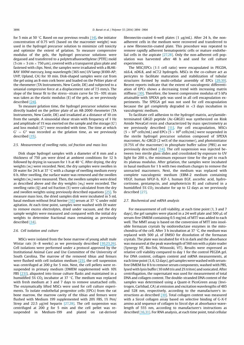
The effect of the number of degradable hydrophobic monomersper macromer arm (n) on compressive modulus, gelation time,swelling ratio and sol faction of SPECA, SPEDA, SPEGA and SPELAhydrogels at constant acrylate concentration of 0.1 (mol/L) is shownin Fig. 3aed, respectively. The compressive modulus of SPELA,SPECA, SPEDA and SPEGA gels increased significantly from 330 to390, 420, 500 and 620 kPa, respectively, as the number of mono-mers per end group (n/4) was increased from 0 to 2.9 (2.8 for C). Theaforementioned increase was related to a change in the structure ofthe gel at the molecular scale as we previously described [16,17].The reactive acrylates are uniformly distributed in aqueous solutionin the absence of chain extension of PEG with hydrophobic seg-ments [16]. With hydroxy acid chain extension, micellar structuresare formed which increase the local concentration of acrylates inthe micellar domains with increasing n at constant bulk acrylateconcentration [16]. The probability of intra-molecular reaction for apendant double bond (j) in the free radical crosslinking reaction ofmulti-functional macromers is [38]
j ¼ 1$ exp!
$34pNA½DB)r0l2
"(2)
Fig. 2. Effect of acrylate concentration in SPEXA hydrogel precursor solution on (a) compressive modulus, (b) gelation time, (c) swelling ratio and (d) sol fraction of SPEXA hydrogels.Error bars correspond to means ± 1 SD for n ¼ 3.
D. Barati et al. / Polymer 55 (2014) 3894e39043898
where [DB] is the concentration of double bonds, r0 is the averagedistance between double bonds on a macromer, l is the statisticallength of a repeating unit and NA is the Avogadro's number. Ac-cording to equation (2), the extent of cyclization decreased withincreasing n due to the increase in local acrylate concentration inthe micellar structures and the increase in end-to-end distance ofthe macromers. The compressive modulus of SPEXA gels increasedwith increasing n (Fig. 3a) because the number of intra-molecularloops that did not contribute to network elasticity was reducedwith increasing n. There was an increasing trend in compressivemodulus of SPEXA gels with SPEGA and SPELA as the stiffest andsoftest gels, respectively, at a given n value. Further, the gelationtime of SPEDA and SPEGA decreased from 150 to 64 and 61 s whenthe number of monomers per end group increased from 0 to 2.9and 2.8, respectively, whereas the gelation time of SPECA andSPELA decreased from 150 to 34 and 38 s with increasing thenumber of monomers per end group from 0 to 2.8 and 2.9 (Fig. 3b).The gelation times of SPECA and SPELA were significantly lowerthan those of SPEGA or SPEDA for all n values. The difference incompressive modulus of SPEXA gels at the same n can be attributedto differences in diffusivity and residence time of the reactive ac-rylates in the micellar structures. The residence time of the acry-lates in the micelles' core was directly correlated to the effectiveinterfacial tension between the chain ends and water (g) [39,40] inwhich g increased with hydrophobicity of the monomers from G toD, L and C. Therefore, the residence time of the acrylates in SPELAand SPECA was higher than those of SPEDA and SPEGA which leadto the higher entrapment of reactive acrylates in the crosslinkedmicellar domains and faster gelation times for SPELA and SPECA(Fig. 3b). The lower residence time of the acrylates in SPEGA andSPEDA led to higher diffusivity of the acrylates, slower but more
uniform crosslinking throughout the solution, resulting in thehigher compressive modulus of the gels.
The swelling ratio of SPECA, SPELA, SPEDA and SPEGA hydrogelsdecreased from 490 to 350, 390, 370 and 430%, respectively, withincreasing the number of monomers per end group from 0 to 2.9(2.8 for X ¼ G, C). SPECA and SPEGA hydrogels had the lowest andhighest swelling ratios, respectively, for all n values (Fig. 3c). The solfraction of SPECA, SPELA, SPEDA and SPEGA varied from 9.6 to 6.4,6.3, 7.2 and 3.1%, respectively, when the number of monomers perend group was increased from 0 to 2.9 (2.8 for X ¼ G, C). The solfraction of SPEGA was significantly lower than the other three gelsfor all n values (Fig. 3d).
3.4. Effect of hydrophobic monomers per macromer arm on SPEXAgel degradation
The mass remaining for SPEXA gels after 1, 5, 10, 20, 30 and 40days of incubation in PBS is shown in Fig. 4. The mass loss of SPEGAhydrogels increased from8 to 19, 31 and 46%with increasing n from2.8 to 4.8, 6.4 and 11.2, respectively, after one day incubation. TheSPEGA gels with n of 6.4 and 11.2 completely degraded after 5 daysincubation. Since the SPEGA had the highest water content amongSPEXA gels and each G monomer had two ester groups, SPEGA gelshad the fastest mass loss with incubation on the time scale of aweek. On the other hand, the remaining mass of SPELA gels did notsignificantly change after 1 day incubation. The remaining mass ofSPELA gels with n of 7.2 and 11.4 after 5 days incubation decreasedto 84 and 71%, respectively; it decreased to 69 and 27% after 20 daysand the gels completely degraded after 30 and 40 days. Theremaining mass of SPELA gels with n of 2.7 and 4.8 after 40 daysincubation was 51 and 45%, respectively. Since SPEDA and SPECA
Fig. 3. Effect of number of monomers in the short hydroxy acid segment on each macromer arm on (a) compressive modulus, (b) gelation time, (c) swelling ratio and (d) sol fractionof SPEXA hydrogels. Error bars correspond to means ± 1 SD for n ¼ 3.
D. Barati et al. / Polymer 55 (2014) 3894e3904 3899
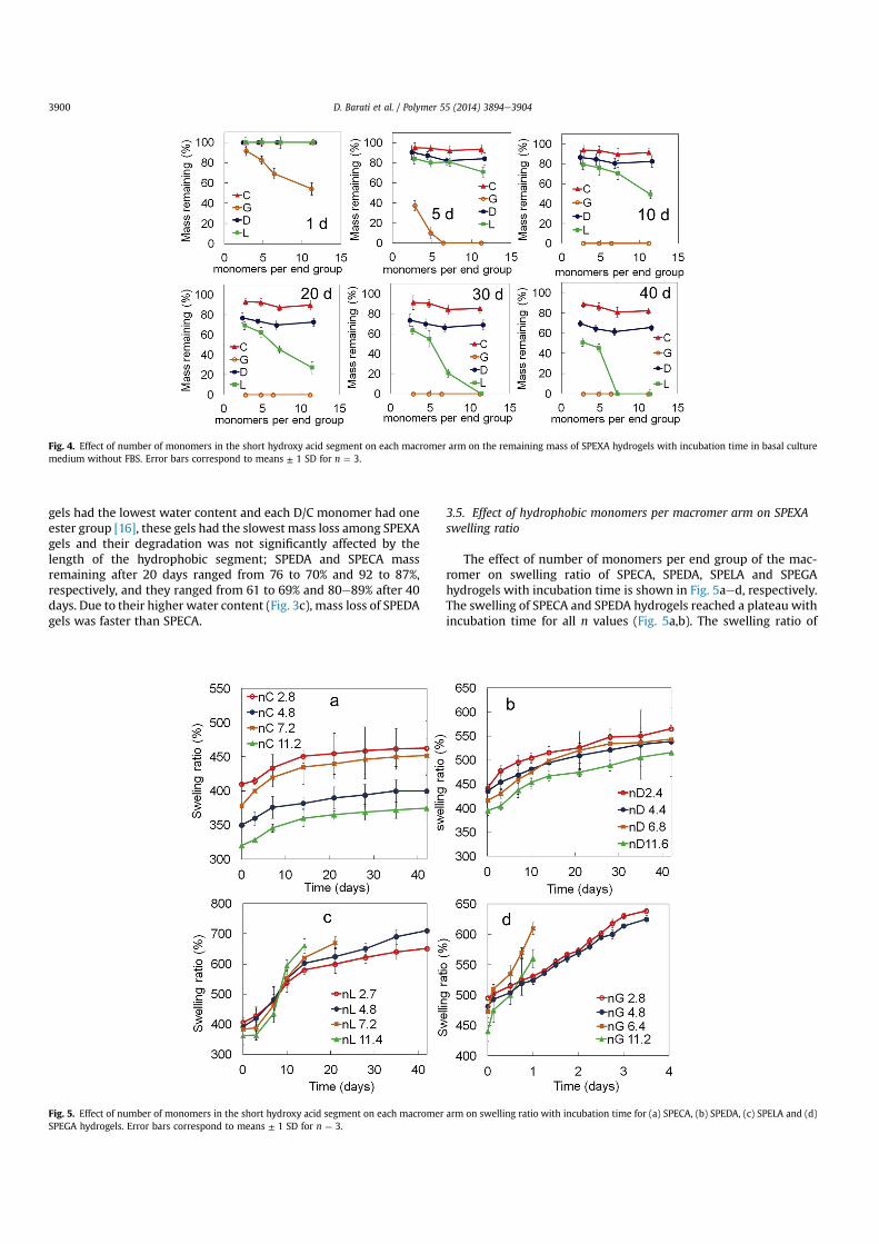
gels had the lowest water content and each D/C monomer had oneester group [16], these gels had the slowest mass loss among SPEXAgels and their degradation was not significantly affected by thelength of the hydrophobic segment; SPEDA and SPECA massremaining after 20 days ranged from 76 to 70% and 92 to 87%,respectively, and they ranged from 61 to 69% and 80e89% after 40days. Due to their higher water content (Fig. 3c), mass loss of SPEDAgels was faster than SPECA.
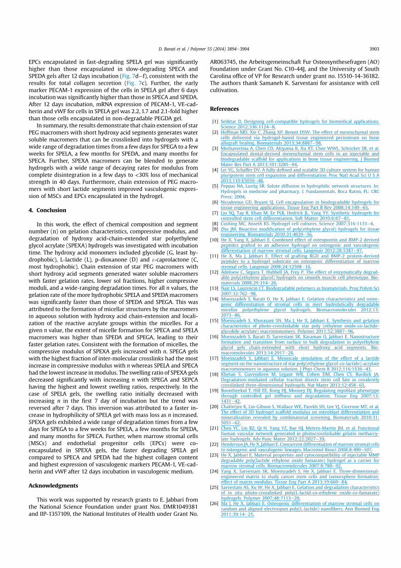
3.5. Effect of hydrophobic monomers per macromer arm on SPEXAswelling ratio
The effect of number of monomers per end group of the mac-romer on swelling ratio of SPECA, SPEDA, SPELA and SPEGAhydrogels with incubation time is shown in Fig. 5aed, respectively.The swelling of SPECA and SPEDA hydrogels reached a plateau withincubation time for all n values (Fig. 5a,b). The swelling ratio of
Fig. 4. Effect of number of monomers in the short hydroxy acid segment on each macromer arm on the remaining mass of SPEXA hydrogels with incubation time in basal culturemedium without FBS. Error bars correspond to means ± 1 SD for n ¼ 3.
Fig. 5. Effect of number of monomers in the short hydroxy acid segment on each macromer arm on swelling ratio with incubation time for (a) SPECA, (b) SPEDA, (c) SPELA and (d)SPEGA hydrogels. Error bars correspond to means ± 1 SD for n ¼ 3.
D. Barati et al. / Polymer 55 (2014) 3894e39043900

SPEDA gel increased with decreasing n from 11.6 to 6.8 for all in-cubation times but swelling was unaffected for n < 6.8 (Fig. 5b). Theswelling ratio of SPELA gels decreased with increasing n in the first7 days of incubation. However, that trend was reversed after 7 daysand swelling ratio increased with increasing n (Fig. 5c). Due to theirhigh water content, SPELA gels with n of 7.2 and 11.4 disintegratedafter 14 and 20 days, respectively. The SPELA gel with n of 11.4rapidly swelled from 360 to 660% from day zero to 14 whereasthose with n of 7.2, 4.8 and 2.7 swelled to 620, 600 and 580%,respectively (Fig. 5c). The inversion in SPELA gel swelling wasattributed to a higher increase in hydrophilicity of the gel withmass loss as n was increased. Since mass loss was proportional tothe hydrophobic segment length and crosslink density decreasedwith mass loss [41], there was a faster decrease in crosslink densitywith incubation time for SPELA gels with higher n, leading to higherswelling ratios for those gels with higher n. The nG11.2 and nG6.4SPEGA gels disintegrated one day after incubation due to highswelling ratios whereas the nG2.8 and nG4.8 gels disintegratedafter 4 days. The swelling ratios of SPEGA gels initially increasedwith increasing n from 2.8 to 6.4 in the first day of incubation andthen decreased with increasing n from 6.4 to 11.2 (Fig. 5d).
3.6. Effect of hydrophobic monomers per macromer arm oncompressive modulus of SPEXA gels
The effect of number of hydrophobic monomers per macromerarm on compressive modulus of SPECA, SPEDA, SPELA and SPEGAgels with incubation time is shown in Fig. 6aed, respectively. Thecompressive modulus of SPEXA gels decreased with incubationtime as the gel crosslink density decreased with mass loss. Theinitial compressive modulus of SPEXA gels increased with n for agiven macromer concentration. Assuming a pseudo first order re-action for hydrolysis of SPEXA gels, the crosslink density should
decay exponentially with time [42]. Based on the theory of rubberelasticity, the change in compressive modulus with incubation timeshould correlate with the rate constant for decay in crosslinkdensity (k) as follows [43]
Eee$65 kt (3)
where t is incubation time. To compare crosslink decay for SPEXAgels, the k values were determined by fitting the data in Fig. 6[ln(E) versus time] to the linearized form of equation (3). The kvalue for SPECA gels increased from 2.9 % 10$6 to5.2 % 10$6 min$1 with increasing n from 2.8 to 11.2 which lead toa difference of 1.3 and 1.1-fold in compressive modulus betweenthe two SPECA gels at day zero and after 42 days incubation,respectively (Fig. 6a). The difference between the compressivemodulus of nD11.6 and nD2.4 SPEDA gels was 1.5-fold and 1.1-fold at day zero and after 42 days incubation, respectively,which was attributed to the higher rate of crosslink decay innD11.6 gel (1.3 % 10$5 min$1) compared to nD2.4 gel (7.5 % 10$6
min$1) (Fig. 6b). Although the initial compressive modulus ofSPELA gels increased with n, that trend was reversed after 3 daysincubation (Fig. 6c). For example, the compressive modulus ofSPELA gels after 10 days incubation decreased from 235 to 220,110 and 50 kPa as n increased from 2.7 to 4.8, 7.2 and 11.4. Therapid decline in compressive moduli of nL11.4 and nL7.2compared to nL4.8 and nL2.7 gels was attributed to the signifi-cantly higher k values of nL11.4 (1.2 % 10$4 min$1) and nL7.2(7.0 % 10$5 min$1) compared to those of nL4.8 (1.2 % 10$5 min$1)and nL2.7 (8.7 % 10$6 min$1). The k values of SPEGA gelsincreased from 5.4 % 10$4 to 2.2 % 10$3 min$1 with increasing nfrom 2.8 to 11.2. The k value of the slowest degrading nC2.8SPECA gel was three orders of magnitude lower than that of thefastest degrading nG11.2 SPEGA gel.
Fig. 6. Effect of number of monomers in the short hydroxy acid segment on each macromer on compressive modulus with incubation time for (a) SPECA, (b) SPEDA, (c) SPELA and(d) SPEGA hydrogels. Error bars correspond to means ± 1 SD for n ¼ 3.
D. Barati et al. / Polymer 55 (2014) 3894e3904 3901

3.7. Effect of gel degradation on differentiation of endothelialprogenitor cells
Early and rapid induction of vascularization in engineeredcellular constructs after implantation is critical to successfulregeneration of tissues [44]. The process of vascularization byendothelial cells requires a compliant and permissive microenvi-ronment [21]. In addition to those requirements, the generation ofmicrovessels requires the presence of mural cells, vascular smoothmuscle cells and pericytes, in the vicinity of endothelial cells forstabilization of microvessels [45]. In that respect, a mixture of MSCsand EPCs encapsulated in a methacrylated gelatin produced morestable microvessels compared to encapsulation of EPCs only [21]and the extent of microvessel formation was strongly dependenton the gel's compressive modulus and degradation. In this work, ratMSCs and EPCs were encapsulated in SPEXA gels with the sameinitial compressive modulus of 5 kPa and the effect of degradationon vasculogenic expression of the encapsulated MSC//EPCs wasevaluated with incubation time. The SPEGA gel due to its completedegradation in <5 days was not used for cell encapsulation.
Fig. 7aef shows the cell viability, DNA content, total collagencontent, mRNA expression of vasculogenic markers PECAM-1, VE-cadherin, and von Willebrand Factor (vWF), respectively, for MSC/EPCs encapsulated in SPEXA gels with incubation time. The MSC/EPCs encapsulated in the non-degradable PEGDA gel was used asthe control. Cell viability of SPEXA gels decreased slightly from day1 to day 7. The MSC/EPCs encapsulated in the PEGDA gel had aslightly higher viability than those in SPEXA gels for all time points.However, the difference in cell viability between SPEXA and PEGDA
gels was not statistically significant. A slight decrease in cellviability of SPELA gels with incubation time in the first 7 days wasconsistent with DNA content of the gels (Fig. 7b). The slightdecrease in cell viability of SPELA gels with incubation time wasmost likely related to its faster degradation compared to SPECA andSPEDA, which led to the higher extent of differentiation of MSC/EPCs in the SPELA gel. DNA content of the gels decreased as the cellsdifferentiated to the vasculogenic lineage with incubation time,consistent with the previously reported results [21]. There was nota significant difference between DNA content of the cells encap-sulated in SPEXA gels for all time points and those in the controlPEGDA gel. Total collagen content of MSC/EPCs in the fasterdegrading SPELA gel after 12 days incubation was significantlyhigher than those in the other gels. The total collagen content ofPEGDA, SPEDA, SPECA and SPELA gels was 59, 59, 67 and 94 mg/mgDNA, respectively, after 12 days incubation. This was consistentwith previous reports that collagen secretion by encapsulated cellsis related to the rate of matrix turnover [46,47]. The slowerdegrading SPEDA and SPECA gels hindered collagen secretion bythe encapsulated cells whereas the faster degrading SPELA gelenhanced collagen secretion. The platelet/endothelial cell adhesionmolecule, PECAM-1 or CD31, is a transmembrane protein expressedearly in vascular development [48]. VE-cadherin, a member of thecadherin family of adhesion receptors, is a constitutive marker ofendothelial cells and plays an important role in early vascular as-sembly. Mature endothelial cells in the late phase of vasculogenesissecret vWF, a plasma protein that mediates platelet adhesion todamaged blood vessels and stabilizes blood coagulation factor VIII[49,50]. PECAM-1, VE-cadherin and vWF mRNA expression of MSC/
Fig. 7. (a) Cell viability (b) DNA content, (c) total collagen content, mRNA expression levels of Pecam-1 (d), VE-cadherin (e) and vWF (f) for MSC/EPCs encapsulated in SPELA (L),SPEDA (D), SPECA (C) and PEGDA (P) hydrogels with incubation time in vasculogenic medium. One star indicates a statistically significant difference between the test group and allother groups at the same time point. Error bars correspond to means ± 1 SD for n ¼ 3.
D. Barati et al. / Polymer 55 (2014) 3894e39043902
EPCs encapsulated in fast-degrading SPELA gel was significantlyhigher than those encapsulated in slow-degrading SPECA andSPEDA gels after 12 days incubation (Fig. 7def), consistent with theresults for total collagen secretion (Fig. 7c). Further, the earlymarker PECAM-1 expression of the cells in SPELA gel after 6 daysincubationwas significantly higher than those in SPECA and SPEDA.After 12 days incubation, mRNA expression of PECAM-1, VE-cad-herin and vWF for cells in SPELA gel was 2.2, 1.7 and 2.1-fold higherthan those cells encapsulated in non-degradable PEGDA gel.
In summary, the results demonstrate that chain extension of starPEG macromers with short hydroxy acid segments generates watersoluble macromers that can be crosslinked into hydrogels with awide range of degradation times from a few days for SPEGA to a fewweeks for SPELA, a few months for SPEDA, and many months forSPECA. Further, SPEXA macromers can be blended to generatehydrogels with a wide range of decaying rates for modulus fromcomplete disintegration in a few days to <30% loss of mechanicalstrength in 40 days. Furthermore, chain extension of PEG macro-mers with short lactide segments improved vasculogenic expres-sion of MSCs and EPCs encapsulated in the hydrogel.
4. Conclusion
In this work, the effect of chemical composition and segmentnumber (n) on gelation characteristics, compressive modulus, anddegradation of hydroxy acid-chain-extended star polyethyleneglycol acrylate (SPEXA) hydrogels was investigated with incubationtime. The hydroxy acid monomers included glycolide (G, least hy-drophobic), L-lactide (L), p-dioxanone (D) and 3-caprolactone (C,most hydrophobic). Chain extension of star PEG macromers withshort hydroxy acid segments generated water soluble macromerswith faster gelation rates, lower sol fractions, higher compressivemoduli, and a wide-ranging degradation times. For all n values, thegelation rate of themore hydrophobic SPELA and SPEDAmacromerswas significantly faster than those of SPEDA and SPEGA. This wasattributed to the formation of micellar structures by the macromersin aqueous solution with hydroxy acid chain-extension and locali-zation of the reactive acrylate groups within the micelles. For agiven n value, the extent of micelle formation for SPECA and SPELAmacromers was higher than SPEDA and SPEGA, leading to theirfaster gelation rates. Consistent with the formation of micelles, thecompressive modulus of SPEXA gels increased with n. SPEGA gelswith the highest fraction of inter-molecular crosslinks had the mostincrease in compressive modulus with nwhereas SPELA and SPECAhad the lowest increase inmodulus. The swelling ratio of SPEXA gelsdecreased significantly with increasing n with SPEGA and SEPCAhaving the highest and lowest swelling ratios, respectively. In thecase of SPELA gels, the swelling ratio initially decreased withincreasing n in the first 7 day of incubation but the trend wasreversed after 7 days. This inversion was attributed to a faster in-crease in hydrophilicity of SPELA gel with mass loss as n increased.SPEXA gels exhibited a wide range of degradation times from a fewdays for SPEGA to a few weeks for SPELA, a few months for SPEDA,and many months for SPECA. Further, when marrow stromal cells(MSCs) and endothelial progenitor cells (EPCs) were co-encapsulated in SPEXA gels, the faster degrading SPELA gelcompared to SPECA and SPEDA had the highest collagen contentand highest expression of vasculogenic markers PECAM-1, VE-cad-herin and vWF after 12 days incubation in vasculogenic medium.
Acknowledgments
This work was supported by research grants to E. Jabbari fromthe National Science Foundation under grant Nos. DMR1049381and IIP-1357109, the National Institutes of Health under Grant No.
AR063745, the Arbeitsgemeinschaft Fur Osteosynthesefragen (AO)Foundation under Grant No. C10-44J, and the University of SouthCarolina office of VP for Research under grant no. 15510-14-36182.The authors thank Samaneh K. Sarvestani for assistance with cellcultivation.
References
[1] Seliktar D. Designing cell-compatible hydrogels for biomedical applications.Science 2012;336:1124e8.
[2] Hoffman MD, Xie C, Zhang XP, Benoit DSW. The effect of mesenchymal stemcells delivered via hydrogel-based tissue engineered periosteum on boneallograft healing. Biomaterials 2013;34:8887e98.
[3] Moshaverinia A, Chen CD, Akiyama K, Xu XT, Chee WWL, Schricker SR, et al.Encapsulated dental-derived mesenchymal stem cells in an injectable andbiodegradable scaffold for applications in bone tissue engineering. J BiomedMater Res Part A 2013;101:3285e94.
[4] Lei YG, Schaffer DV. A fully defined and scalable 3D culture system for humanpluripotent stem cell expansion and differentiation. Proc Natl Acad Sci U S A2013;110:E5039e48.
[5] Peppas NA, Lustig SR. Solute diffusion in hydrophilic network structures. In:Hydrogels in medicine and pharmacy. I. Fundamentals. Boca Raton, FL: CRCPress; 2004.
[6] Nicodemus GD, Bryant SJ. Cell encapsulation in biodegradable hydrogels fortissue engineering applications. Tissue Eng Part B Rev 2008;14:149e65.
[7] Liu SQ, Tay R, Khan M, Ee PLR, Hedrick JL, Yang YY. Synthetic hydrogels forcontrolled stem cell differentiation. Soft Matter 2010;6:67e81.
[8] Cushing MC, Anseth KS. Hydrogel cell cultures. Science 2007;316:1133e4.[9] Zhu JM. Bioactive modification of poly(ethylene glycol) hydrogels for tissue
engineering. Biomaterials 2010;31:4639e56.[10] He X, Yang X, Jabbari E. Combined effect of osteopontin and BMP-2 derived
peptides grafted to an adhesive hydrogel on osteogenic and vasculogenicdifferentiation of marrow stromal cells. Langmuir 2012;28:5387e97.
[11] He X, Ma J, Jabbari E. Effect of grafting RGD and BMP-2 protein-derivedpeptides to a hydrogel substrate on osteogenic differentiation of marrowstromal cells. Langmuir 2008;24:12508e16.
[12] Adeloew C, Segura T, Hubbell JA, Frey P. The effect of enzymatically degrad-able poly(ethylene glycol) hydrogels on smooth muscle cell phenotype. Bio-materials 2008;29:314e26.
[13] Nair LS, Laurencin CT. Biodegradable polymers as biomaterials. Prog Polym Sci2007;32:762e98.
[14] Moeinzadeh S, Barati D, He X, Jabbari E. Gelation characteristics and osteo-genic differentiation of stromal cells in inert hydrolytically degradablemicellar polyethylene glycol hydrogels. Biomacromolecules 2012;13:2073e86.
[15] Moeinzadeh S, Khorasani SN, Ma J, He X, Jabbari E. Synthesis and gelationcharacteristics of photo-crosslinkable star poly (ethylene oxide-co-lactide-glycolide acrylate) macromonomers. Polymer 2011;52:3887e96.
[16] Moeinzadeh S, Barati D, Sarvestani SK, Karaman O, Jabbari E. Nanostructureformation and transition from surface to bulk degradation in polyethyleneglycol gels chain-extended with short hydroxy acid segments. Bio-macromolecules 2013;14:2917e28.
[17] Moeinzadeh S, Jabbari E. Mesoscale simulation of the effect of a lactidesegment on the nanostructure of star poly(ethylene glycol-co-lactide)-acrylatemacromonomers in aqueous solution. J Phys Chem B 2012;116:1536e43.
[18] Khetan S, Guvendiren M, Legant WR, Cohen DM, Chen CS, Burdick JA.Degradation-mediated cellular traction directs stem cell fate in covalentlycrosslinked three-dimensional hydrogels. Nat Mater 2013;12:458e65.
[19] Boontheekul T, Hill EE, Kong HJ, Mooney DJ. Regulating myoblast phenotypethrough controlled gel stiffness and degradation. Tissue Eng 2007;13:1431e42.
[20] Chatterjee K, Lin-Gibson S, Wallace WE, Parekh SH, Lee YJ, Cicerone MT, et al.The effect of 3D hydrogel scaffold modulus on osteoblast differentiation andmineralization revealed by combinatorial screening. Biomaterials 2010;31:5051e62.
[21] Chen YC, Lin RZ, Qi H, Yang YZ, Bae HJ, Melero-Martin JM, et al. Functionalhuman vascular network generated in photocrosslinkable gelatin methacry-late hydrogels. Adv Func Mater 2012;22:2027e39.
[22] Henderson JA, HeX, Jabbari E. Concurrent differentiation ofmarrow stromal cellsto osteogenic and vasculogenic lineages. Macromol Biosci 2008;8:499e507.
[23] He X, Jabbari E. Material properties and cytocompatibility of injectable MMPdegradable poly(lactide ethylene oxide fumarate) hydrogel as a carrier formarrow stromal cells. Biomacromolecules 2007;8:780e92.
[24] Yang X, Sarvestani SK, Moeinzadeh S, He X, Jabbari E. Three-dimensional-engineered matrix to study cancer stem cells and tumorsphere formation:effect of matrix modulus. Tissue Eng Part A 2013;19:669e84.
[25] Sarvestani AS, Xu W, He X, Jabbari E. Gelation and degradation characteristicsof in situ photo-crosslinked poly(L-lactid-co-ethylene oxide-co-fumarate)hydrogels. Polymer 2007;48:7113e20.
[26] Ma J, He X, Jabbari E. Osteogenic differentiation of marrow stromal cells onrandom and aligned electrospun poly(L-lactide) nanofibers. Ann Biomed Eng2011;39:14e25.
D. Barati et al. / Polymer 55 (2014) 3894e3904 3903
[27] Kahler CM, Wechselberger J, Hilbe W, Gschwendtner A, Colleselli D,Niederegger H, et al. Peripheral infusion of rat bone marrow derived endo-thelial progenitor cells leads to homing in acute lung injury. Respir Res2007;8:1e17.
[28] Yang NN, Li DW, Jiao P, Chen B, Yao ST, Sang H, et al. The characteristics ofendothelial progenitor cells derived from mononuclear cells of rat bonemarrow in different culture conditions. Cytotechnology 2011;63:217e26.
[29] Ghajar CM, Kachgal S, Kniazeva E, Mori H, Costes SV, George SC, et al.Mesenchymal cells stimulate capillary morphogenesis via distinct proteolyticmechanisms. Exp Cell Res 2010;316:813e25.
[30] Rao RR, Peterson AW, Ceccarelli J, Putnam AJ, Stegemann JP. Matrix compo-sition regulates three-dimensional network formation by endothelial cellsand mesenchymal stem cells in collagen/fibrin materials. Angiogenesis2012;15:253e64.
[31] Chen KL, Sahoo S, He PF, Ng KS, Toh SL, Goh JCH. A hybrid silk/RADA-basedfibrous scaffold with triple hierarchy for ligament regeneration. Tissue EngPart A 2012;18:1399e409.
[32] Jabbari E, He X, Valarmathi MT, Sarvestani AS, Xu W. Material properties andbone marrow stromal cells response to in situ crosslinkable RGD-functionlized lactide-co-glycolide scaffolds. J Biomed Mater Res Part A2009;89A:124e37.
[33] Kiran MS, Viji RI, Kumar SV, Prabhakaran AA, Sudhakaran PR. Changes inexpression of VE-cadherin and MMPa in endothelial cells: Implications forangiogenesis. Vasc Cell 2011;3(6):1e12.
[34] Pfaffl MW. A new mathematical model for relative quantification in real-timeRT-PCR. Nucleic Acids Res 2001;29:e45.
[35] Cima LG, Lopina ST. Network structures of radiation-cross-linked star polymergels. Macromolecules 1995;28:6787e94.
[36] Sanson N, Rieger J. Synthesis of nanogels/microgels by conventional andcontrolled radical crosslinking copolymerization. Polym Chem 2010;1:965e77.
[37] Flory PJ. Principles of polymer chemistry. New York: Cornell University Press;1953.
[38] Elliott JE, Bowman CN. Kinetics of primary cyclization reactions in cross-linked polymers: an analytical and numerical approach to heterogeneity innetwork formation. Macromolecules 1999;32:8621e8.
[39] Li ZL, Dormidontova EE. Equilibrium chain exchange kinetics in block copol-ymer micelle solutions by dissipative particle dynamics simulations. SoftMatter 2011;7:4179e88.
[40] Nicolai T, Colombani O, Chassenieux C. Dynamic polymeric micelles versusfrozen nanoparticles formed by block copolymers. Soft Matter 2010;6:3111e8.
[41] Metters AT, Anseth KS, Bowman CN. Fundamental studies of a novel, biode-gradable PEG-b-PLA hydrogel. Polymer 2000;41:3993e4004.
[42] Martens P, Metters AT, Anseth KS, Bowman CN. A generalized bulk-degradation model for hydrogel networks formed from multivinyl cross-linking molecules. J Phys Chem B 2001;105:5131e8.
[43] Martens PJ, Bowman CN, Anseth KS. Degradable networks formed from multi-functional poly(vinyl alcohol) macromers: comparison of results from ageneralized bulk-degradation model for polymer networks and experimentaldata. Polymer 2004;45:3377e87.
[44] Tsigkou O, Pomerantseva I, Spencer JA, Redondo PA, Hart AR, O'Doherty E,et al. Engineered vascularized bone grafts. Proc Natl Acad Sci U S A 2010;107:3311e6.
[45] Levenberg S, Rouwkema J, Macdonald M, Garfein ES, Kohane DS, Darland DC,et al. Engineering vascularized skeletal muscle tissue. Nat Biotechnol 2005;23:879e84.
[46] Bryant SJ, Anseth KS. Hydrogel properties influence ECM production bychondrocytes photoencapsulated in poly(ethylene glycol) hydrogels. J BiomedMater Res 2002;59:63e72.
[47] Bryant SJ, Anseth KS. Controlling the spatial distribution of ECM componentsin degradable PEG hydrogels for tissue engineering cartilage. J Biomed MaterRes Part A 2003;64A:70e9.
[48] Albelda SM, Muller WA, Buck CA, Newman PJ. Molecular and cellular prop-erties of PECAM-1 (endocam/CDF31) e a novel vascular cell cell-adhesionmolecule. J Cell Biol 1991;114:1059e68.
[49] Bautch VL, Redick SD, Scalia A, Harmaty M, Carmeliet P, Rapoport R. Charac-terization of the vasculogenic block in the absence of vascular endothelialgrowth factor-A. Blood 2000;95:1979e87.
[50] Sadler JE. Biochemistry and genetics of von Willebrand factor. Annu RevBiochem 1998;67:395e424.
D. Barati et al. / Polymer 55 (2014) 3894e39043904
Related Documents