PRIORITY PUBLICATION Thrombospondin-1 Plays a Critical Role in the Induction of Hair Follicle Involution and Vascular Regression During the Catagen Phase KiichiroYano, Lawrence F. Brown, n Jack Lawler, n Tokichi Miyakawa,w and Michael Detmar Cutaneous Biology Research Center, Department of Dermatology, Massachusetts General Hospital and Harvard Medical School, Charlestown, Massachusetts, U.S.A.; n Department of Pathology, Beth Israel Deaconess Medical Center and Harvard Medical School, Boston, Massachusetts, U.S.A.; wDepartment of Molecular Biotechnology, Hiroshima University, Higashi-Hiroshima, Japan Hair growth is associated with pronounced vascular-en- dothelial-growth-factor-induced perifollicular angio- genesis, whereas the catagen regression phase is characterized by apoptosis-driven blood vessel regres- sion. The biologic relevance of endogenous inhibitors of angiogenesis in the control of hair cycling, however, has remained unknown. We studied the expression and biologic role of the angiogenesis inhibitor thrombos- pondin-1 (TSP-1) during the induced adult hair follicle- cycle in wild-type, TSP-1 de¢cient, and TSP-1 overexpressing transgenic mice. TSP-1 expression was absent from hair bulb and dermal papilla cells during early to mid-anagen but was highly upregulated throughout the catagen involution phase. In TSP-1 de¢cient mice, the follicle growth phase was signi¢- cantly prolonged, associated with increased perifollicu- lar vascularization and vascular proliferation. Conversely, hair follicle growth was delayed in K14/ TSP-1 transgenic mice that expressed high levels of TSP-1 in outer root sheath keratinocytes, associated with reduced perifollicular vascularization. These ef- fects were most probably mediated via its antiangio- genic e¡ects because TSP-1 did not a¡ect the growth of cultured murine vibrissae in the absence of a func- tional vascular system. These results identify a critical role of TSP-1 in the induction of anagen follicle involu- tion, with potential implications for the therapeutic modulation of hair follicle growth. Key words: TSP-1/ angiogenesis/VEGF. J Invest Dermatol 120:14 ^19, 2003 T he hair follicle undergoes a life-long cyclic transfor- mation from the resting phase (telogen) to the growth phase (anagen), associated with rapid proliferation of follicular keratinocytes and with elongation and thickening of the hair shafts, followed by the regression phase (catagen) that leads to involution of hair follicles (Chase, 1954; Hardy, 1992). These changes involve rapid remodeling of both epithelial and dermal components and have led to the establishment of the murine hair follicle cycle as a prime model for studies of epithelial^mesenchymal interactions, leading to the identi¢cation of several important molecular med- iators that control epithelial morphogenesis and growth (Paus and Cotsarelis, 1999). We have previously reported a pronounced increase in perifollicular vascularization during the anagen growth phase, accompanied by upregulation of vascular endothe- lial growth factor (VEGF) mRNA expression in follicular kerati- nocytes of the outer root sheath (Yano et al , 2001). Conversely, during the catagen follicular involution phase, a rapid regression of perifollicular blood vessels is associated with downregulation of follicular VEGF expression (Yano et al, 2001). Moreover, en- hanced levels of VEGF in outer root sheath keratinocytes resulted in accelerated follicle growth and in increased thickness of hair follicles and hair shafts in K14/VEGF transgenic mice, whereas systemic inhibition of VEGF led to a reduced thickness of hair follicles (Yano et al , 2001). During the catagen involution phase, hair follicles undergo a highly controlled process of involution that re£ects a burst of programmed cell death (apoptosis) of follicular keratinocytes and of perifollicular endothelial cells (Paus and Cotsarelis, 1999; Mecklenburg et al , 2000; Yano et al , 2001). Our previous ¢ndings suggested that the regression of perifollicular blood vessels might represent a key factor that triggers the regression of hair follicles, resulting in reduced nutritional supply of proliferating hair ma- trix cells (Yano et al , 2001). Importantly, reduced vascularization of hair follicles has been suggested to play a critical role in disor- ders characterized by hair loss (Levy-Frankel, 1931; Beurey et al , 1971; Goldman et al, 1995), including androgenetic alopecia (male pattern baldness), which is associated with progressive shortening of successive anagen cycles and with miniaturization of geneti- cally predisposed follicles (Cormia and Ernyey, 1961). Whereas the vascular expansion during anagen is probably due to upregu- lation of follicular VEGF expression, it has remained unknown whether endogenous inhibitors of angiogenesis might contribute to the vascular regression during catagen, and whether angiogen- esis inhibition might represent a key event in the induction of hair follicle involution. Thrombospondin-1 (TSP-1) is a 450 kDa matricellular protein that inhibits endothelial cell proliferation and migration in vitro Reprint requests to: Michael Detmar, M.D., CBRC/Department of Dermatology, Massachusetts General Hospital, Building 149, 13th Street, Charlestown, MA 02129; Email: [email protected] Abbreviations: TSP-1, thrombospondin-1; VEGF, vascular endothelial growth factor. Manuscript received September 11, 2002; revised October 7, 2002; accepted for publication October 15, 2002 0022-202X/03/$15.00 Copyright r 2003 by The Society for Investigative Dermatology, Inc. 14

Welcome message from author
This document is posted to help you gain knowledge. Please leave a comment to let me know what you think about it! Share it to your friends and learn new things together.
Transcript

PRIORITY PUBLICATION
Thrombospondin-1 Plays a Critical Role in the Inductionof Hair Follicle Involution and Vascular Regression During theCatagen Phase
KiichiroYano, Lawrence F. Brown,n Jack Lawler,nTokichi Miyakawa,w and Michael DetmarCutaneous Biology Research Center, Department of Dermatology, Massachusetts General Hospital and Harvard Medical School, Charlestown,Massachusetts, U.S.A.; nDepartment of Pathology, Beth Israel Deaconess Medical Center and Harvard Medical School, Boston, Massachusetts, U.S.A.;wDepartment of Molecular Biotechnology, Hiroshima University, Higashi-Hiroshima, Japan
Hair growth is associated with pronounced vascular-en-dothelial-growth-factor-induced perifollicular angio-genesis, whereas the catagen regression phase ischaracterized by apoptosis-driven blood vessel regres-sion. The biologic relevance of endogenous inhibitorsof angiogenesis in the control of hair cycling, however,has remained unknown.We studied the expression andbiologic role of the angiogenesis inhibitor thrombos-pondin-1 (TSP-1) during the induced adult hair follicle-cycle in wild-type, TSP-1 de¢cient, and TSP-1overexpressing transgenic mice. TSP-1 expression wasabsent from hair bulb and dermal papilla cells duringearly to mid-anagen but was highly upregulatedthroughout the catagen involution phase. In TSP-1de¢cient mice, the follicle growth phase was signi¢-
cantly prolonged, associated with increased perifollicu-lar vascularization and vascular proliferation.Conversely, hair follicle growth was delayed in K14/TSP-1 transgenic mice that expressed high levels ofTSP-1 in outer root sheath keratinocytes, associatedwith reduced perifollicular vascularization. These ef-fects were most probably mediated via its antiangio-genic e¡ects because TSP-1 did not a¡ect the growthof cultured murine vibrissae in the absence of a func-tional vascular system. These results identify a criticalrole of TSP-1 in the induction of anagen follicle involu-tion, with potential implications for the therapeuticmodulation of hair follicle growth. Key words: TSP-1/angiogenesis/VEGF. J Invest Dermatol 120:14 ^19, 2003
The hair follicle undergoes a life-long cyclic transfor-mation from the resting phase (telogen) to thegrowth phase (anagen), associated with rapidproliferation of follicular keratinocytes and withelongation and thickening of the hair shafts, followed
by the regression phase (catagen) that leads to involution of hairfollicles (Chase, 1954; Hardy, 1992). These changes involve rapidremodeling of both epithelial and dermal components and haveled to the establishment of the murine hair follicle cycle as aprime model for studies of epithelial^mesenchymal interactions,leading to the identi¢cation of several important molecular med-iators that control epithelial morphogenesis and growth (Pausand Cotsarelis, 1999).We have previously reported a pronouncedincrease in perifollicular vascularization during the anagengrowth phase, accompanied by upregulation of vascular endothe-lial growth factor (VEGF) mRNA expression in follicular kerati-nocytes of the outer root sheath (Yano et al, 2001). Conversely,during the catagen follicular involution phase, a rapid regressionof perifollicular blood vessels is associated with downregulationof follicular VEGF expression (Yano et al, 2001). Moreover, en-
hanced levels of VEGF in outer root sheath keratinocytes resultedin accelerated follicle growth and in increased thickness of hairfollicles and hair shafts in K14/VEGF transgenic mice, whereassystemic inhibition of VEGF led to a reduced thickness of hairfollicles (Yano et al, 2001).During the catagen involution phase, hair follicles undergo a
highly controlled process of involution that re£ects a burst ofprogrammed cell death (apoptosis) of follicular keratinocytesand of perifollicular endothelial cells (Paus and Cotsarelis, 1999;Mecklenburg et al, 2000; Yano et al, 2001). Our previous ¢ndingssuggested that the regression of perifollicular blood vessels mightrepresent a key factor that triggers the regression of hair follicles,resulting in reduced nutritional supply of proliferating hair ma-trix cells (Yano et al, 2001). Importantly, reduced vascularizationof hair follicles has been suggested to play a critical role in disor-ders characterized by hair loss (Levy-Frankel, 1931; Beurey et al,1971; Goldman et al, 1995), including androgenetic alopecia (malepattern baldness), which is associated with progressive shorteningof successive anagen cycles and with miniaturization of geneti-cally predisposed follicles (Cormia and Ernyey, 1961). Whereasthe vascular expansion during anagen is probably due to upregu-lation of follicular VEGF expression, it has remained unknownwhether endogenous inhibitors of angiogenesis might contributeto the vascular regression during catagen, and whether angiogen-esis inhibition might represent a key event in the induction ofhair follicle involution.Thrombospondin-1 (TSP-1) is a 450 kDa matricellular protein
that inhibits endothelial cell proliferation and migration in vitro
Reprint requests to: Michael Detmar, M.D., CBRC/Department ofDermatology, Massachusetts General Hospital, Building 149, 13th Street,Charlestown, MA 02129; Email: [email protected]: TSP-1, thrombospondin-1; VEGF, vascular endothelial
growth factor.
Manuscript received September 11, 2002; revised October 7, 2002;accepted for publication October 15, 2002
0022-202X/03/$15.00 � Copyrightr 2003 by The Society for Investigative Dermatology, Inc.
14

and angiogenesis associated with tumor growth and granulationtissue formation in vivo (Tolsma et al, 1993; Bleuel et al, 1999;Streit et al, 1999; 2000; Bornstein, 2001). TSP-1 is expressed in hu-man and murine skin and contributes to the normal quiescenceof the cutaneous vasculature (Detmar, 2000). To directly investi-gate the biologic role of TSP-1 in the control of hair follicle re-gression and vascular involution during catagen, we studied theinduced adult hair follicle cycle in transgenic mice overexpressingTSP-1 in outer sheath follicular keratinocytes (Streit et al, 2000;Hawighorst et al, 2002; Yano et al, 2002) and in TSP-1 de¢cientmice (Lawler et al, 1998).Here, we report that TSP-1 mRNA and protein expression are
highly upregulated in the dermal papilla and in epithelial cellsduring the catagen and telogen phases, whereas TSP-1 expressionis absent from the hair bulb during the anagen growth phase. Im-portantly, transgenic overexpression of TSP-1 in follicular kerati-nocytes resulted in retarded hair growth, associated withdecreased perifollicular vascularization. Conversely, TSP-1 de¢-cient mice showed a prolongation of the anagen growth phasewith increased perifollicular vascularization, accompanied by in-creased proliferation of perifollicular endothelial cells and hairmatrix cells. Together with the absence of direct e¡ects of TSP-1in vibrissa organ cultures, our results identify a major role ofTSP-1-mediated angiogenesis inhibition in the control of hairgrowth and cycling.
METHODS
Induced adult hair follicle cycle The adult hair follicle cycle wasinduced in the back skin of 8-wk-old female C57BL/6 mice (CharlesRiver Laboratories, Wilmington, MA) by depilation as describedpreviously (Paus et al, 1990), resulting in a synchronized induction ofanagen follicle growth. Tissue samples were obtained at days 1, 3, 5, 8, 12,15, 18, 19, 20, and 22 after depilation as described previously (three mice pertime point), encompassing hair development from early anagen to telogen(Yano et al, 2001). Back skin was harvested parallel to the paravertebral lineand the distinct phases of hair follicle development were determined asdescribed earlier (Dry, 1926; Hardy, 1992). Skin samples were either snapfrozen in liquid nitrogen or ¢xed in 4% paraformaldehyde andembedded in para⁄n as described previously (Streit et al, 2000). Allanimal studies were approved by the Massachusetts General HospitalSubcommittee on Research Animal Care.
Hair follicle cycle studies in TSP-1 transgenic mice and in TSP-1de¢cient mice TSP-1 transgenic mice were established by using akeratin 14 (K14) promoter expression cassette to target human TSP-1expression to basal epidermal keratinocytes and outer root sheathkeratinocytes of hair follicles. The establishment and the phenotypiccharacterization of TSP-1 transgenic mice have been previously reported(Streit et al, 2000). The induced hair follicle cycle was studied in 8-wk-oldTSP-1 transgenic mice (n¼ 30) and in age-matched wild-type littermates(n¼ 30 per group) as described above. Hair follicle cycling was alsoinvestigated in TSP-1 de¢cient mice (Lawler et al, 1998) and their wild-type controls (n¼10 per group).
Immunohistochemistry and computer-assisted morphometricvessel analysis Immunohistochemical stainings were performed on 7mm frozen sections as described previously (Streit et al, 2000), using amonoclonal rat antimouse CD31 antibody (Pharmingen, San Diego, CA)and a secondary antirat IgG antibody labeled with AlexaFluor488(Molecular Probes, Eugene, OR). Representative sections obtained fromthree mice for each time point were analyzed, using a Nikon E-600microscope (Nikon, Melville, NY). Images were captured with a Spotdigital camera (Diagnostic Instruments, Sterling Heights, MI) andmorphometric analyses were performed using the IP LAB software(Scanalytics, Fairfax, VA) as described previously (Streit et al, 1999). Threedi¡erent ¢elds in each section were examined at 60�magni¢cation, andthe average vessel size and the relative area occupied by blood vessels weredetermined within an area of 30 mm distance of individual hair follicles asdescribed previously (Yano et al, 2001). The two-sided unpaired t test wasused to analyze di¡erences between the di¡erent genotypes. Endothelialcell proliferation was studied by intraperitoneal injection of mice with 5-bromodeoxyuridine (BrdU; 250 mg per kg of body weight) 2 h prior tosacri¢ce, followed by double immuno£uorescence staining with anti-BrdU(Pharmingen) and anti-CD31 antibodies (Streit et al, 2000).
Measurement of hair follicle length Hematoxylin�eosin stains wereperformed on 7 mm frozen sections of tissues obtained from the di¡erentstages of the induced adolescent hair follicle cycle (three mice each per timepoint). Five representative sections for each sample were analyzed. Thelength of hair follicles was measured as the distance from the bottom ofhair bulbs with clearly visible dermal papilla to the epidermis (50 hairfollicles for each time point), using the IP LAB software. Statisticalanalysis was performed using the two-sided unpaired Student’s t test.
In situ hybridization In situ hybridization was performed on 5 mmpara⁄n sections as described previously (Streit et al, 2000). Sense andantisense RNA probes to human TSP-1 were transcribed from apBluescript II KSþvector containing a 240 bp PCR fragment of thecoding region of human TSP-1. This probe recognizes both mouse andhuman TSP-1 (Streit et al, 2000). Transcription reactions were carried outusing the Riboprobe Gemini II kit (Promega Madison, WI) in thepresence of (a-35S) UTP. TSP-1 mRNA expression was quanti¢ed insections obtained from three mice for each time point by countingindividual photographic grains over follicular keratinocytes as describedby Brown et al (1995), and samples were scored as negative (0^1 grains percell; score 0), weakly positive (2^5 grains per cell; score 1), moderatelypositive (6^10 grains per cell; score 2), or strongly positive (410 grains percell; score 3). Data are shown as percentage of maximum score.
Vibrissa organ culture Whisker pads were isolated from 5-wk-oldfemale C57/BL mice as described previously (Yano et al, 2001) and wereshortly immersed in 70% ethanol in phosphate-bu¡ered saline, followedby incubation inWilliam’s E medium containing 400 U per ml penicillin,400 mg per ml streptomycin, and 1 mg per ml fungizone (Life SciencesRockville, MD) for 10 min. Vibrissa follicles in the early or mid-anagengrowth phase were isolated under a dissecting microscope, and the part ofthe hair shaft that extended over the epidermal surface was cut o¡. Follicleswere then plated on nylon membranes (ICN, Irvine, CA) in serum-freeWilliam’s E medium alone (n¼ 8), in medium containing 10 mg per mlhumanTSP-1 (n¼ 7), or in medium containing 10 ng per ml transforminggrowth factor b2 (n¼ 8; Sigma, St. Louis, MO). Media were replaced byfresh medium every 2 d, and follicles were incubated over a total period of7 d. After 7 d, the length of the outgrowing hair shafts was determined byimage analysis of digital pictures, using the IP LAB software. Data areexpressed as means7SD of in vitro hair growth; statistical analyses wereperformed using the two-sided unpaired Student’s t test.
RESULTS
Upregulation of TSP-1 mRNA expression during thecatagen follicle involution phase In situ hybridization studiesrevealed low levels of TSP-1 mRNA expression in follicularkeratinocytes of the outer root sheath during the anagen growth
Figure1. Upregulation of TSP-1 expression during catagen. In situhybridization with TSP-1 antisense riboprobe reveals moderate TSP-1mRNA expression in follicular keratinocytes of the outer root sheath (ar-rows), but not in the hair bulb, during the anagen follicle growth phase inwild-type mice (A). In contrast,TSP-1 expression was strongly upregulatedin the dermal papilla and the epithelial strand throughout the catagenphase (days 18 and 19) (B, C) and was maintained during the telogen restingphase (D). Sense control hybridizations demonstrate low background sig-nal (E�G). Scale bars: 50 mm.
TSP-1 AND CATAGEN FOLLICLE INVOLUTION 15VOL. 120, NO. 1 JANUARY 2003
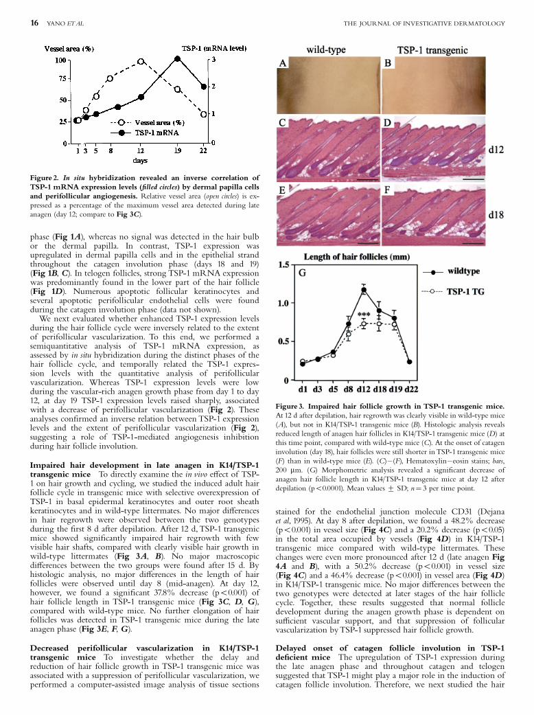
phase (Fig 1A), whereas no signal was detected in the hair bulbor the dermal papilla. In contrast, TSP-1 expression wasupregulated in dermal papilla cells and in the epithelial strandthroughout the catagen involution phase (days 18 and 19)(Fig 1B, C). In telogen follicles, strong TSP-1 mRNA expressionwas predominantly found in the lower part of the hair follicle(Fig 1D). Numerous apoptotic follicular keratinocytes andseveral apoptotic perifollicular endothelial cells were foundduring the catagen involution phase (data not shown).We next evaluated whether enhanced TSP-1 expression levels
during the hair follicle cycle were inversely related to the extentof perifollicular vascularization. To this end, we performed asemiquantitative analysis of TSP-1 mRNA expression, asassessed by in situ hybridization during the distinct phases of thehair follicle cycle, and temporally related the TSP-1 expres-sion levels with the quantitative analysis of perifollicularvascularization. Whereas TSP-1 expression levels were lowduring the vascular-rich anagen growth phase from day 1 to day12, at day 19 TSP-1 expression levels raised sharply, associatedwith a decrease of perifollicular vascularization (Fig 2). Theseanalyses con¢rmed an inverse relation between TSP-1 expressionlevels and the extent of perifollicular vascularization (Fig 2),suggesting a role of TSP-1-mediated angiogenesis inhibitionduring hair follicle involution.
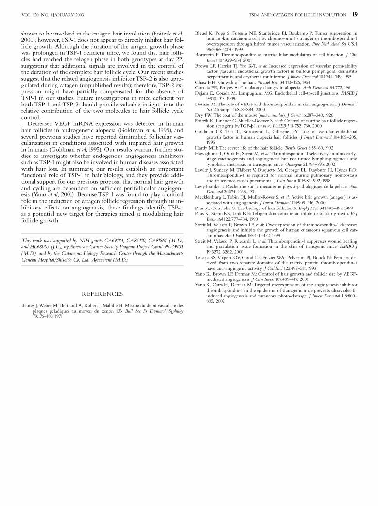
Impaired hair development in late anagen in K14/TSP-1transgenic mice To directly examine the in vivo e¡ect of TSP-1 on hair growth and cycling, we studied the induced adult hairfollicle cycle in transgenic mice with selective overexpression ofTSP-1 in basal epidermal keratinocytes and outer root sheathkeratinocytes and in wild-type littermates. No major di¡erencesin hair regrowth were observed between the two genotypesduring the ¢rst 8 d after depilation. After 12 d,TSP-1 transgenicmice showed signi¢cantly impaired hair regrowth with fewvisible hair shafts, compared with clearly visible hair growth inwild-type littermates (Fig 3A, B). No major macroscopicdi¡erences between the two groups were found after 15 d. Byhistologic analysis, no major di¡erences in the length of hairfollicles were observed until day 8 (mid-anagen). At day 12,however, we found a signi¢cant 37.8% decrease (po0.001) ofhair follicle length in TSP-1 transgenic mice (Fig 3C, D, G),compared with wild-type mice. No further elongation of hairfollicles was detected in TSP-1 transgenic mice during the lateanagen phase (Fig 3E, F, G).
Decreased perifollicular vascularization in K14/TSP-1transgenic mice To investigate whether the delay andreduction of hair follicle growth in TSP-1 transgenic mice wasassociated with a suppression of perifollicular vascularization, weperformed a computer-assisted image analysis of tissue sections
stained for the endothelial junction molecule CD31 (Dejanaet al, 1995). At day 8 after depilation, we found a 48.2% decrease(po0.001) in vessel size (Fig 4C) and a 20.2% decrease (po0.05)in the total area occupied by vessels (Fig 4D) in K14/TSP-1transgenic mice compared with wild-type littermates. Thesechanges were even more pronounced after 12 d (late anagen Fig4A and B), with a 50.2% decrease (po0.001) in vessel size(Fig 4C) and a 46.4% decrease (po0.001) in vessel area (Fig 4D)in K14/TSP-1 transgenic mice. No major di¡erences between thetwo genotypes were detected at later stages of the hair folliclecycle. Together, these results suggested that normal follicledevelopment during the anagen growth phase is dependent onsu⁄cient vascular support, and that suppression of follicularvascularization byTSP-1 suppressed hair follicle growth.
Delayed onset of catagen follicle involution in TSP-1de¢cient mice The upregulation of TSP-1 expression duringthe late anagen phase and throughout catagen and telogensuggested that TSP-1 might play a major role in the induction ofcatagen follicle involution. Therefore, we next studied the hair
Figure 2. In situ hybridization revealed an inverse correlation ofTSP-1 mRNA expression levels (¢lled circles) by dermal papilla cellsand perifollicular angiogenesis. Relative vessel area (open circles) is ex-pressed as a percentage of the maximum vessel area detected during lateanagen (day 12; compare to Fig 3C).
Figure 3. Impaired hair follicle growth in TSP-1 transgenic mice.At 12 d after depilation, hair regrowth was clearly visible in wild-type mice(A), but not in K14/TSP-1 transgenic mice (B). Histologic analysis revealsreduced length of anagen hair follicles in K14/TSP-1 transgenic mice (D) atthis time point, compared with wild-type mice (C). At the onset of catageninvolution (day 18), hair follicles were still shorter inTSP-1 transgenic mice(F) than in wild-type mice (E). (C)�(F), Hematoxylin�eosin stains; bars,200 mm. (G) Morphometric analysis revealed a signi¢cant decrease ofanagen hair follicle length in K14/TSP-1 transgenic mice at day 12 afterdepilation (po0.0001). Mean values7 SD; n¼ 3 per time point.
16 YANO ETAL THE JOURNAL OF INVESTIGATIVE DERMATOLOGY

follicle cycle in mice with targeted gene disruption of the TSP-1gene. We found comparable growth of hair follicles in TSP-1de¢cient and in wild-type mice during the anagen growth phase(Fig 5A, B). The onset of catagen involution was signi¢cantlydelayed in TSP-1 de¢cient mice, however. Whereas all follicleshad initiated catagen involution at day 19 in wild-type mice,only anagen follicles with a clearly recognizable dermal papillawere detected in TSP-1 de¢cient mice at this time point(Fig 5C, D). After 21 d, catagen involution was also detectablein TSP-1 de¢cient mice (Fig 5F); however, the follicle lengthwas still signi¢cantly increased (po0.001) compared with wild-type mice (Fig 5E, F). After 22 d, all follicles in wild-type andin TSP-1 de¢cient mice were in the telogen resting phase.
Increased perifollicular vascularization in TSP-1 de¢cientmice Analysis of CD31-stained tissue sections revealedelongated and nlarged perifollicular blood vessels at day 19 inTSP-1 de¢cient mice, compared with small and fragmentedvessels in wild-type mice. By quantitative image analysis ofCD31-stained vessels, no major di¡erences in vessel size andcutaneous area covered by blood vessels were observed until day12 after depilation. We found a 110.3% increase (po0.001) invessel size (Fig 6C) and a 72.5% increase (po0.05) in total vesselarea (Fig 6D) during catagen at day 19, however, and a60.3% increase (po0.001) in vessel size (Fig 6C) and a20.5% increase (n.s.) in vessel area (Fig 6D) at day 21 in TSP-1de¢cient mice, compared with wild-type littermates. Doubleimmuno£uorescence stains for CD31 and for the proliferationmarker BrdU revealed persistent endothelial cell proliferation inTSP-1 de¢cient mice at day 19 (Fig 6B), whereas no proliferatingendothelial cells were detected in wild-type follicles that hadalready undergone catagen involution at this time point (Fig 6A).The di¡erences in vascularization between the two genotypes atday 19 were probably due to the prolongation of anagen growthin TSP-1 de¢cient mice, because no signi¢cant di¡erences in the
perifollicular vascularization were found in anagen phase follicles(Fig 6D).
Absence of TSP-1 e¡ects on hair growth and follicle size invibrissa follicle cultures in vitro To investigate whether theobserved inhibitory e¡ects of TSP-1 on hair growth might havebeen mediated, in addition to its inhibitory e¡ects onperifollicular angiogenesis, by direct e¡ects of TSP-1 on hairfollicle cells, we isolated mouse vibrissae and investigated hairgrowth in organ cultures in vitro, in the absence of a functioningvascular system. In organ culture, untreated mouse vibrissaeshowed an average hair shaft outgrowth of approximately 2.3mm over a period of 7 d (Fig 7). Addition of 50 ng per mltransforming growth factor b to vibrissa cultures, used as apositive control, resulted in a signi¢cant (po0.01) inhibition ofhair shaft growth, demonstrating the sensitivity of hair growthto inhibition in this experimental system (Fig 7). In contrast,addition of TSP-1 did not in£uence the hair growth rate inorgan culture, suggesting that a functional perifollicular vascular
Figure 4. Diminished perifollicular angiogenesis in TSP-1transgenic mice. CD31 immunostains (red) of wild-type (A) and TSP-1transgenic (B) mice demonstrate diminished perifollicular vascularizationof anagen follicles (day 12) in TSP-1 transgenic mice. Scale bars: 100 mm.Quantitative image analysis of CD31-stained vessels revealed a signi¢cantlydecreased average vessel size (po0.001) at 8 and 12 d after depilation (C) inTSP-1 transgenic mice (¢lled bars) compared with wild-type mice (open bars)and a signi¢cant decrease of the relative area covered by vessels (D) at days8 and 12 in K14/TSP-1 transgenic mice. npo0.05; nnnpo0.001. Mean values7 SD; n¼ 3 per time point.
Figure 5. Prolonged anagen hair growth in TSP-1 de¢cient mice.No major di¡erences in the length of hair follicles were detected in TSP-1de¢cient mice and wild-type mice until day 12 of the induced hair cycle(A, B). At day 19, wild-type follicles had initiated catagen involution (C),whereas TSP-1 de¢cient follicles were still in the anagen growth phase (D).After 21 d, only catagen follicles were detected in both genotypes (E, F),although TSP-1 de¢cient follicle length was still increased over wild-typefollicles (G). Morphometric analysis revealed a signi¢cant increase of hairfollicle length in TSP-1 de¢cient mice at days 19 and 21 after depilation.nnnpo0.001. Mean values7 SD; n¼ 3 per time point. Scale bar: 100 mm.
TSP-1 AND CATAGEN FOLLICLE INVOLUTION 17VOL. 120, NO. 1 JANUARY 2003

system is needed in order for TSP-1 to exert its inhibitory e¡ectson hair growth.
DISCUSSION
Previously, we have reported that murine hair follicle growth isassociated with pronounced perifollicular angiogenesis andVEGF expression by follicular keratinocytes of the outer rootsheath (Yano et al, 2001). Moreover, increased VEGF levels inK14/VEGF transgenic mice resulted in increased hair shaft andfollicle size, whereas systemic blockade of VEGF impaired hairfollicle growth (Yano et al, 2001).The biologic role of endogenousinhibitors of angiogenesis for hair follicle growth and cycling hasremained unknown, however. Based on our previous identi¢ca-tion of TSP-1 as a major endogenous inhibitor of skin angiogen-esis (Detmar, 2000; Streit et al, 2000; Hawighorst et al, 2002;Yano et al, 2002), we hypothesized that TSP-1 might also play animportant role in the regulation of cyclic angiogenesis associatedwith hair follicle growth and involution. Semiquantitative in situhybridization studies revealed that TSP-1 mRNA expression wasrelatively low during the anagen growth phase but was stronglyupregulated in the dermal papilla and in follicular epithelial cellsat the end of anagen and throughout the catagen involution phaseand the telogen resting phase of the follicle. The inverse relationof TSP-1 mRNA expression levels with the levels of perifollicu-lar angiogenesis suggests that TSP-1 represents a major antiangio-genic stimulus responsible for vascular involution during thecatagen follicle involution phase.To directly evaluate the biologic consequences of enhanced
TSP-1 expression for hair growth, we analyzed the induced ado-lescent hair follicle cycle in our previously characterized trans-genic mouse model for the selective overexpression of TSP-1 inthe epidermis and outer root sheath follicular keratinocytes, using
a K14 promoter to target transgene expression (Streit et al, 2000).We have previously reported decreased vascularization of granu-lation tissue during wound healing (Streit et al, 2000), reducedultraviolet-B-induced cutaneous photodamage (Yano et al, 2002),and diminished epithelial carcinogenesis (Hawighorst et al, 2002)in these mice; however, hair growth had not been studied indetail. Our ¢ndings that targeted overexpression of TSP-1 infollicular keratinocytes resulted in delayed and diminishedhair regrowth, associated with diminished perifollicular vascular-ization and increased levels of endothelial apoptosis, provide the¢rst direct evidence that endogenous inhibitors of angiogenesisnegatively regulate hair growth. Because TSP-1 treatment didnot a¡ect hair growth in organ cultures of mouse vibrissae, inthe absence of a functional vascular system, our results suggestthat the e¡ects of TSP-1 are predominantly mediated through in-hibition of perifollicular angiogenesis rather than by direct e¡ectson hair follicles. These ¢ndings are in agreement with our pre-vious results that systemic blockade of the angiogenesis factorVEGF with neutralizing antibody caused impaired anagen hairgrowth and decreased angiogenesis (Yano et al, 2001).We cannotexclude the possibility that TSP-1, at doses higher than thosetested (10 mg per ml), might a¡ect hair growth in vibrissa cul-tures, however, and that hair follicles might react di¡erently toTSP-1, compared with vibrissa follicles. Moreover, because thelocalization of TSP-1 receptors in keratinocytes of the regressinghair follicles is unknown, potential direct e¡ects of TSP-1 on in-trafollicular cells cannot be completely ruled out.The important role of TSP-1 in the control of hair follicle
cycling was con¢rmed in additional studies on TSP-1 de¢cientmice that showed delayed induction of the catagen involutionphase associated with maintained perifollicular angiogenesis, in-dicating that TSP-1 is a major part of the molecular program thatinitiates catagen follicle involution. In contrast to other mediatorssuch as transforming growth factor b that have been previously
Figure 6. Increased perifollicular vascularization at day 19 in TSP-1de¢cient mice. Double immuno£uorescence stains for CD31 (red) andthe proliferation marker BrdU (green) revealed proliferating endothelialcells (arrows) in the skin of TSP-1 de¢cient mice (B), but not in wild-typemice (A), at day 19 of the hair cycle. Scale bar: 100 mm. Increased perifolli-cular vascularization at day 19 in TSP-1 de¢cient mice. Computer-assistedmorphometric analysis of CD31-stained sections revealed a signi¢cant in-crease (po0.001) in perifollicular vessel size (C) and relative vessel area (D)at day 19 in TSP-1 de¢cient mice (¢lled bars), compared with wild-type lit-termates (open bars). npo0.05; nnnpo0.001. Mean values 7 SD; n¼ 3 pertime point.
Figure 7. Absence of e¡ects of TSP-1 treatment on the in vitro hairgrowth rate in mouse vibrissa organ cultures. Quantitative analysis ofin vitro hair growth demonstrates signi¢cant inhibition of in vitro hairgrowth by 50 ng per ml transforming growth factor b (positive control)but lack of e⁄ciency of TSP-1 (10 mg per ml) treatment. Data are expressedas mean7SD. n.s., no signi¢cant di¡erences; nnpo0.01.
18 YANO ETAL THE JOURNAL OF INVESTIGATIVE DERMATOLOGY
shown to be involved in the catagen hair involution (Foitzik et al,2000), however,TSP-1 does not appear to directly inhibit hair fol-licle growth. Although the duration of the anagen growth phasewas prolonged in TSP-1 de¢cient mice, we found that hair folli-cles had reached the telogen phase in both genotypes at day 22,suggesting that additional signals are involved in the control ofthe duration of the complete hair follicle cycle. Our recent studiessuggest that the related angiogenesis inhibitor TSP-2 is also upre-gulated during catagen (unpublished results); therefore,TSP-2 ex-pression might have partially compensated for the absence ofTSP-1 in our studies. Future investigations in mice de¢cient forboth TSP-1 and TSP-2 should provide valuable insights into therelative contribution of the two molecules to hair follicle cyclecontrol.Decreased VEGF mRNA expression was detected in human
hair follicles in androgenetic alopecia (Goldman et al, 1995), andseveral previous studies have reported diminished follicular vas-cularization in conditions associated with impaired hair growthin humans (Goldman et al, 1995). Our results warrant further stu-dies to investigate whether endogenous angiogenesis inhibitorssuch asTSP-1might also be involved in human diseases associatedwith hair loss. In summary, our results establish an importantfunctional role of TSP-1 in hair biology, and they provide addi-tional support for our previous proposal that normal hair growthand cycling are dependent on su⁄cient perifollicular angiogen-esis (Yano et al, 2001). Because TSP-1 was found to play a criticalrole in the induction of catagen follicle regression through its in-hibitory e¡ects on angiogenesis, these ¢ndings identify TSP-1as a potential new target for therapies aimed at modulating hairfollicle growth.
This work was supported by NIH grants CA69184, CA86410, CA91861 (M.D.)and HL68003 (J.L.), byAmerican Cancer Society Program Project Grant 99-23901(M.D.), and by the Cutaneous Biology Research Center through the MassachusettsGeneral Hospital/Shiseido Co. Ltd. Agreement (M.D.).
REFERENCES
Beurey J,Weber M, Bertrand A, Robert J, Mabille H: Mesure du debit vasculaire desplaques peladiques au moyen du xenon 133. Bull Soc Fr Dermatol Syphiligr79:176^180, 1971
Bleuel K, Popp S, Fusenig NE, Stanbridge EJ, Boukamp P: Tumor suppression inhuman skin carcinoma cells by chromosome 15 transfer or thrombospondin-1overexpression through halted tumor vascularization. Proc Natl Acad Sci USA96:2065^2070, 1999
Bornstein P: Thrombospondins as matricellular modulators of cell function. J ClinInvest 107:929^934, 2001
Brown LF, Harrist TJ, Yeo K-T, et al: Increased expression of vascular permeabilityfactor (vascular endothelial growth factor) in bullous pemphigoid, dermatitisherpetiformis, and erythema multiforme. J Invest Dermatol 104:744^749, 1995
Chase HH: Growth of the hair. Physiol Rev 34:113^126, 1954Cormia FE, ErnyeyA: Circulatory changes in alopecia. Arch Dermatol 84:772, 1961Dejana E, Corada M, Lampugnani MG: Endothelial cell-to-cell junctions. FASEB J
9:910^918, 1995Detmar M:The role of VEGF and thrombospondins in skin angiogenesis. J Dermatol
Sci 24(Suppl. 1):S78^S84, 2000Dry FW:The coat of the mouse (mus musculus). J Genet 16:287^340, 1926Foitzik K, Lindner G, Mueller-Roever S, et al: Control of murine hair follicle regres-
sion (catagen) byTGF-b1 in vivo. FASEB J 14:752^760, 2000Goldman CK, Tsai JC, Soroceanu L, Gillespie GY: Loss of vascular endothelial
growth factor in human alopecia hair follicles. J Invest Dermatol 104:18S^20S,1995
Hardy MH:The secret life of the hair follicle.Trends Genet 8:55^60, 1992Hawighorst T, Oura H, Streit M, et al: Thrombospondin-1 selectively inhibits early-
stage carcinogenesis and angiogenesis but not tumor lymphangiogenesis andlymphatic metastasis in transgenic mice. Oncogene 21:794^795, 2002
Lawler J, Sunday M, Thibert V, Duquette M, George EL, Rayburn H, Hynes RO:Thrombospondin-1 is required for normal murine pulmonary homeostasisand its absence causes pneumonia. J Clin Invest 101:982^992, 1998
Levy-Frankel J: Recherche sur le mecanisme physio-pathologique de la pelade. AnnDermatol 2:1074^1088, 1931
Mecklenburg L,Tobin DJ, Muller-Rover S, et al: Active hair growth (anagen) is as-sociated with angiogenesis. J Invest Dermatol 114:909^916, 2000
Paus R, Cotsarelis G: The biology of hair follicles. N Engl J Med 341:491^497, 1999Paus R, Stenn KS, Link RE: Telogen skin contains an inhibitor of hair growth. Br J
Dermatol 122:777^784, 1990Streit M,Velasco P, Brown LF, et al: Overexpression of thrombospondin-1 decreases
angiogenesis and inhibits the growth of human cutaneous squamous cell car-cinomas. AmJ Pathol 155:441^452, 1999
Streit M, Velasco P, Riccardi L, et al: Thrombospondin-1 suppresses wound healingand granulation tissue formation in the skin of transgenic mice. EMBO J19:3272^3282, 2000
Tolsma SS,Volpert OV, Good DJ, Frazier WA, Polverini PJ, Bouck N: Peptides de-rived from two separate domains of the matrix protein thrombospondin-1have anti-angiogenic activity. J Cell Biol 122:497^511, 1993
Yano K, Brown LF, Detmar M: Control of hair growth and follicle size by VEGF-mediated angiogenesis. J Clin Invest 107:409^417, 2001
Yano K, Oura H, Detmar M: Targeted overexpression of the angiogenesis inhibitorthrombospondin-1 in the epidermis of transgenic mice prevents ultraviolet-B-induced angiogenesis and cutaneous photo-damage. J Invest Dermatol 118:800^805, 2002
TSP-1 AND CATAGEN FOLLICLE INVOLUTION 19VOL. 120, NO. 1 JANUARY 2003
Related Documents


