The Angiogenesis Inhibitor Thrombospondin-1 Inhibits Acute Cutaneous Hypersensitivity Reactions Paula Velasco 1,5 , Rainer Huegel 1,5 , Jochen Brasch 1 , Jens M. Schro ¨der 1 , Michael Weichenthal 1 , Eggert Stockfleth 2 , Thomas Schwarz 1 , Jack Lawler 3 , Michael Detmar 4 and Bernhard Lange-Asschenfeldt 1,2 There is increasing evidence that vascular remodeling and endothelial cell activation promote acute and chronic inflammation. Thrombospondin 1 (TSP-1) is a potent endogenous angiogenesis inhibitor thought to play an important role in maintaining cutaneous vascular quiescence. We first investigated TSP-1 expression in human and contact hypersensitivity (CHS) reactions and found that TSP-1 was upregulated in the inflamed skin of patients and in mice. To elucidate the function of TSP-1 in cutaneous inflammation, we induced CHS reactions in the skin of mice with targeted epidermal TSP-1 overexpression in TSP-1-deficient mice and in wild- type mice. We found decreased edema formation, angiogenesis, and inflammatory infiltrate in the inflamed skin of TSP-1 transgenic mice. Conversely, TSP-1-deficient mice exhibited an enhanced and prolonged inflammation, characterized by increased edema formation, enhanced vascular remodeling, and increased neutrophilic infiltrate, when compared with wild-type mice. Moreover, we found strong upregulation of the proinflamma- tory cytokines IL-1b, macrophage inflammatory protein 2, and tumor necrosis factor-a in the inflamed skin of TSP-1-deficient mice. Our results indicate that TSP-1 downregulates cutaneous delayed-type hypersensitivity reactions by acting on several distinct pathways mediating skin inflammation. Journal of Investigative Dermatology (2009) 129, 2022–2030; doi:10.1038/jid.2008.447; published online 5 February 2009 INTRODUCTION There is increasing evidence that vascular endothelial cells are critically involved in the pathogenesis of inflammatory diseases, as angiogenesis, vascular hyperpermeability, plas- ma leakage, leukocyte recruitment, and extravasation are prevalent features of inflamed tissues. A variety of endogen- ous pro- and antiangiogenic factors are involved in inflam- mation. For example, chronic transgenic delivery of vascular endothelial growth factor (VEGF)-A to the skin results in chronic, psoriasis-like skin inflammation (Xia et al., 2003; Kunstfeld et al., 2004), and systemic treatment with the angiogenesis inhibitor vasostatin inhibits experimentally induced skin inflammation (Huegel et al., 2007). In human skin, thrombospondin (TSP)-1, derived from epidermal keratinocytes and TSP-2, is thought to contribute to the antiangiogenic barrier that separates the avascular epidermis from the highly vascularized dermis (Detmar, 2000) and to maintain cutaneous vascular quiescence. Earlier studies have shown that TSP-2 is induced in murine cutaneous hypersensitivity (CHS) reactions, whereas TSP-2 deficiency was associated with enhanced and prolonged experimental CHS reactions (Lange-Asschenfeldt et al., 2002). Moreover, TSP-2 was shown to protect the synovial microenvironment in rheumatoid arthritis from excessive angiogenesis and tissue inflammation (Park et al., 2004). In contrast, the role of TSP-1 during inflammatory processes remains unclear. TSP-1, a secreted homotrimeric glycoprotein (Lawler, 2002), was the first protein shown to inhibit angiogenesis in vivo (Good et al., 1990) and to suppress vascular endothelial cell proliferation and migration in vitro (Tolsma et al., 1997; Streit et al., 2000). Although TSP-1 expression is upregulated in rheumatoid arthritis (Koch et al., 1993), in wounds (DiPietro et al., 1996), and in atherosclerotic plaques (Riessen et al., 1998), TSP-1-derived peptides de- creased neovascularization, leukocyte infiltration, and thicken- ing of the synovial lining in a murine arthritis model (Rico et al., 2007). TSP-1 was also involved in the phagocytic clearance of senescent neutrophils (Savill et al., 1992), thereby contributing to the resolution phase of inflammation. We hypothesized that TSP-1 might act as an endogenous negative regulator of inflammation by antagonizing inflam- mation-associated angiogenesis. Therefore, we investigated the biological role of TSP-1 in experimental CHS reactions ORIGINAL ARTICLE 2022 Journal of Investigative Dermatology (2009), Volume 129 & 2009 The Society for Investigative Dermatology Received 14 March 2008; revised 15 August 2008; accepted 29 September 2008; published online 5 February 2009 1 Department of Dermatology, University Hospital Schleswig-Holstein, Campus Kiel, Kiel, Germany; 2 Institute of Pharmaceutical Sciences, Swiss Federal Institute of Technology, ETH Zurich, Zu ¨ rich, Switzerland; 3 Department of Dermatology, Charite ´ University Medicine, Berlin, Germany and 4 Department of Pathology, Division of Cancer Biology and Angiogenesis, Beth Israel Deaconess Medical Center and Harvard Medical School, Boston, Massachusetts, USA Correspondence: Dr Bernhard Lange-Asschenfeldt, Department of Dermatology, Charite ´ University Medicine, Charite ´platz 1 10117 Berlin, Germany. E-mail: [email protected] 5 These authors contributed equally to this work Abbreviations: Ab, antibody; CHS, contact hypersensitivity; MIP-2, macrophage inflammatory protein 2; PlGF, placental growth factor; RT-PCR, reverse transcription PCR; TNF-a, tumor necrosis factor-a; TSP, thrombospondin; VEGF, vascular endothelial growth factor

Welcome message from author
This document is posted to help you gain knowledge. Please leave a comment to let me know what you think about it! Share it to your friends and learn new things together.
Transcript

The Angiogenesis Inhibitor Thrombospondin-1Inhibits Acute Cutaneous Hypersensitivity ReactionsPaula Velasco1,5, Rainer Huegel1,5, Jochen Brasch1, Jens M. Schroder1, Michael Weichenthal1,Eggert Stockfleth2, Thomas Schwarz1, Jack Lawler3, Michael Detmar4 and Bernhard Lange-Asschenfeldt1,2
There is increasing evidence that vascular remodeling and endothelial cell activation promote acute andchronic inflammation. Thrombospondin 1 (TSP-1) is a potent endogenous angiogenesis inhibitor thought toplay an important role in maintaining cutaneous vascular quiescence. We first investigated TSP-1 expression inhuman and contact hypersensitivity (CHS) reactions and found that TSP-1 was upregulated in the inflamed skinof patients and in mice. To elucidate the function of TSP-1 in cutaneous inflammation, we induced CHSreactions in the skin of mice with targeted epidermal TSP-1 overexpression in TSP-1-deficient mice and in wild-type mice. We found decreased edema formation, angiogenesis, and inflammatory infiltrate in the inflamed skinof TSP-1 transgenic mice. Conversely, TSP-1-deficient mice exhibited an enhanced and prolonged inflammation,characterized by increased edema formation, enhanced vascular remodeling, and increased neutrophilicinfiltrate, when compared with wild-type mice. Moreover, we found strong upregulation of the proinflamma-tory cytokines IL-1b, macrophage inflammatory protein 2, and tumor necrosis factor-a in the inflamed skin ofTSP-1-deficient mice. Our results indicate that TSP-1 downregulates cutaneous delayed-type hypersensitivityreactions by acting on several distinct pathways mediating skin inflammation.
Journal of Investigative Dermatology (2009) 129, 2022–2030; doi:10.1038/jid.2008.447; published online 5 February 2009
INTRODUCTIONThere is increasing evidence that vascular endothelial cellsare critically involved in the pathogenesis of inflammatorydiseases, as angiogenesis, vascular hyperpermeability, plas-ma leakage, leukocyte recruitment, and extravasation areprevalent features of inflamed tissues. A variety of endogen-ous pro- and antiangiogenic factors are involved in inflam-mation. For example, chronic transgenic delivery of vascularendothelial growth factor (VEGF)-A to the skin results inchronic, psoriasis-like skin inflammation (Xia et al., 2003;Kunstfeld et al., 2004), and systemic treatment with theangiogenesis inhibitor vasostatin inhibits experimentallyinduced skin inflammation (Huegel et al., 2007).
In human skin, thrombospondin (TSP)-1, derived fromepidermal keratinocytes and TSP-2, is thought to contributeto the antiangiogenic barrier that separates the avascularepidermis from the highly vascularized dermis (Detmar,2000) and to maintain cutaneous vascular quiescence. Earlierstudies have shown that TSP-2 is induced in murinecutaneous hypersensitivity (CHS) reactions, whereas TSP-2deficiency was associated with enhanced and prolongedexperimental CHS reactions (Lange-Asschenfeldt et al.,2002). Moreover, TSP-2 was shown to protect the synovialmicroenvironment in rheumatoid arthritis from excessiveangiogenesis and tissue inflammation (Park et al., 2004).
In contrast, the role of TSP-1 during inflammatory processesremains unclear. TSP-1, a secreted homotrimeric glycoprotein(Lawler, 2002), was the first protein shown to inhibitangiogenesis in vivo (Good et al., 1990) and to suppressvascular endothelial cell proliferation and migration in vitro(Tolsma et al., 1997; Streit et al., 2000). Although TSP-1expression is upregulated in rheumatoid arthritis (Koch et al.,1993), in wounds (DiPietro et al., 1996), and in atheroscleroticplaques (Riessen et al., 1998), TSP-1-derived peptides de-creased neovascularization, leukocyte infiltration, and thicken-ing of the synovial lining in a murine arthritis model (Rico et al.,2007). TSP-1 was also involved in the phagocytic clearance ofsenescent neutrophils (Savill et al., 1992), thereby contributingto the resolution phase of inflammation.
We hypothesized that TSP-1 might act as an endogenousnegative regulator of inflammation by antagonizing inflam-mation-associated angiogenesis. Therefore, we investigatedthe biological role of TSP-1 in experimental CHS reactions
ORIGINAL ARTICLE
2022 Journal of Investigative Dermatology (2009), Volume 129 & 2009 The Society for Investigative Dermatology
Received 14 March 2008; revised 15 August 2008; accepted 29 September2008; published online 5 February 2009
1Department of Dermatology, University Hospital Schleswig-Holstein,Campus Kiel, Kiel, Germany; 2Institute of Pharmaceutical Sciences, SwissFederal Institute of Technology, ETH Zurich, Zurich, Switzerland;3Department of Dermatology, Charite University Medicine, Berlin, Germanyand 4Department of Pathology, Division of Cancer Biology and Angiogenesis,Beth Israel Deaconess Medical Center and Harvard Medical School, Boston,Massachusetts, USA
Correspondence: Dr Bernhard Lange-Asschenfeldt, Department ofDermatology, Charite University Medicine, Chariteplatz 1 10117 Berlin,Germany. E-mail: [email protected]
5These authors contributed equally to this work
Abbreviations: Ab, antibody; CHS, contact hypersensitivity; MIP-2,macrophage inflammatory protein 2; PlGF, placental growth factor; RT-PCR,reverse transcription PCR; TNF-a, tumor necrosis factor-a; TSP,thrombospondin; VEGF, vascular endothelial growth factor

elicited in TSP-1 transgenic, TSP-1-deficient, and wild-typemice, and we also studied the expression of TSP-1 in humanand murine contact dermatitis. We propose that TSP-1 acts asan important endogenous negative regulator of inflammationby antagonizing inflammation-associated angiogenesis, leu-kocyte recruitment, and vascular leakage.
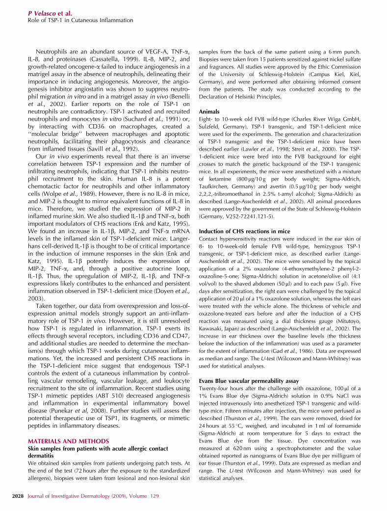
RESULTSTSP-1 expression is upregulated in human acute allergiccontact dermatitis
To determine the tissue localization of TSP-1 protein and oneof its primary receptors, CD36, in normal and inflamed
human skin, immunohistochemical stains with antibodies(Abs) against human TSP-1 and CD36 were performed.In the normal uninflamed skin, TSP-1 protein was depositedin the basement membrane zone of the dermal–epidermaljunction (Figure 1a), in accordance with its describedexpression pattern (Wight et al., 1985). In the inflamedskin, we observed increased TSP-1 expression stronglyassociated with blood vessels and in the epidermis(Figure 1b). Combined immunohistochemical stains withAbs against TSP-1 and laminin showed the colocalization ofTSP-1 expression and blood vessels in the inflamed skin(Figure 1e).
a
Figure 1. Upregulation of TSP-1 expression and its receptor CD36 in human acute contact dermatitis. Immunohistochemical stains of (a) uninflamed and (b)
inflamed back skin at 72 hours after the onset of a human ACD (acute allergic contact dermatitis) using an antibody directed against human TSP-1. Non-
radioactive in situ hybridization (ISH) using a riboprobe specific for human TSP-1 in the uninflamed human skin (c). In situ hybridization for TSP-1 (red)
combined with an immunostain for collagen IV (green) revealed TSP-1 mRNA-positive vessels (arrows) in the inflamed skin (d). Double immunofluorescence
stain with antibodies directed against laminin (red) and TSP-1 (green) revealed TSP-1-labeled vessels (yellow, arrowheads) (e). Immunohistochemical stains of
uninflamed (f) and inflamed skin (g) using an antibody directed against CD36. Double immunofluorescence stains of inflamed skin with antibodies directed
against laminin (red) and CD36 (green) reveal a scattered staining pattern in the dermis and the epidermis (arrows) (h). White dots delineate the dermoepidermal
junction. Twelve uninflamed and 15 inflamed skin samples were analyzed. Bars¼ 200mm (a, b, and d–h), 100mm (c).
www.jidonline.org 2023
P Velasco et al.Role of TSP-1 in Cutaneous Inflammation

To investigate whether TSP-1 mRNA expression ismodulated during acute allergic contact dermatitis, non-radioactive in situ hybridizations with a riboprobe againsthuman TSP-1 were performed on uninflamed and inflamedhuman skin samples. In the healthy skin, the signal wasconfined to the outer root sheath keratinocytes of the hairfollicle (Figure 1c) and mesenchymal cells. The combinationof in situ hybridizations with a probe specific for TSP-1mRNA and with immunohistochemical stains with an Abagainst collagen IV confirmed the co-expression of TSP-1 byblood vessels in the inflamed human skin. The CD36expression was also upregulated in inflamed lesions (Figure1g) when compared with normal skin (Figure 1f). In theuninflamed skin, CD36 was expressed by cells in the basallayer of the epidermis (Figure 1f). In the inflamed skin, wefound CD36 expressed by single cells distributed within thedermis and in the basal layer of the epidermis (Figure 1g).Combined immunofluorescence stains for laminin and CD36showed that single cells that were positive for CD36 in thedermis of the inflamed skin rarely associated with endothelialcells (Figure 1h). However, it is conceivable that CD36 onendothelial cells was below the detection level usingimmunohistochemical stains, as it was strongly expressedby other cells in the inflamed tissue. Twelve uninflamed and15 inflamed human skin biopsies were analyzed.
TSP-1 expression is upregulated in contact hypersensitivityreactions in murine skin
To determine the dynamics of TSP-1 regulation duringcutaneous inflammation, we induced CHS reactions in theskin of wild-type mice sensitized with oxazolone. The skintissues were harvested 24, 48, and 96 hours after the onset ofinflammation. Immunohistochemical stains of uninflamedskin of wild-type mice showed TSP-1 protein expression inthe epidermis (Figure 2a). In the inflamed skin of wild-typemice, an increased TSP-1 expression was found in epidermalkeratinocytes (Figure 2b). We then performed quantitativereal-time reverse transcription PCR (RT-PCR) analyses ofcDNA generated from inflamed and uninflamed murine skinusing primers specific for murine TSP-1. This assay revealed afour-fold increase (Po0.01) in the level of TSP-1 mRNA at24 hours and a more than three-fold increase (Po0.01) at48 hours after the onset of inflammation. By 96 hours, TSP-1mRNA levels returned to baseline (uninflamed skin: median
0.29, range 0.09–0.68; inflamed 24 hours: median 1.19,range: 1.04–1.34; and inflamed 48 hours: median 0.94, range0.62–2.18).
Decreased edema formation in the ear skin of TSP-1 transgenicmice during CHS reactions
We next induced CHS reactions in the ear skin of TSP-1transgenic mice, with targeted epidermal overexpression ofhuman TSP-1, and in wild-type mice, using oxazolone as asensitizing agent. We confirmed the TSP-1 transgene expres-sion by using real-time PCR with a probe specific for humanTSP-1 and found a 3.8-fold increase in TSP-1 mRNAexpression in the inflamed skin (median 0.31, range0.05–1.16) compared with the uninflamed skin (median0.08, range 0.03–0.19) of TSP-1 transgenic mice. No humanTSP-1 mRNA was detectable in the uninflamed and inflamedskin of wild-type mice. In addition, non-radioactive in situhybridizations with a riboprobe specific for human TSP-1(Figure S1) confirmed the expression of TSP-1 mRNA in theskin of TSP-1 transgenic mice 24 hours after the onset ofinflammation. Ear thickness measurements performed 24 and48 hours after the initial challenge revealed 22% (Po0.01)and 23% (Po0.01) reduction in ear thickness in the inflamedears of TSP-1 transgenic mice when compared with that ofwild-type mice (Figure 3a–c). This trend was observedthroughout the investigated period of 5 days (Figure 3c).
Using a modified Evans blue vascular leakage assay, wequantified the amount of plasma that extravasated into thetissue during the experimental inflammation. Twenty-fourhours after the onset of inflammation, 46% (Po0.05) of lessEvans Blue dye leaked into the inflamed tissue of TSP-1transgenic mice (median 90 ng/mg, range 46–138) comparedwith wild-type mice (median 165 ng/mg, range 137–208). Inthe uninflamed skin, no significant difference in the vascularleakage between TSP-1 transgenic mice (median 26 ng/mg,range 12–46 ) and wild-type mice (median 40 ng/mg range,11–84) was observed.
Decreased vascular remodeling in the inflamed skin of TSP-1transgenic mice
Computer-assisted morphometric image analyses of CD31-stained vessels revealed a significant decrease (28%,Po0.01) in the average vessel size in the inflamed ears ofTSP-1 transgenic mice 24 hours after oxazolone challenge
Figure 2. Strong expression of TSP-1 protein in the inflamed murine skin. Immunohistochemical stains of uninflamed (a) and inflamed (b) skin of
wild-type mice revealed TSP-1 protein expression in the epidermis. Bars¼ 200mm.
2024 Journal of Investigative Dermatology (2009), Volume 129
P Velasco et al.Role of TSP-1 in Cutaneous Inflammation

when compared with that of wild-type mice (Figure 3f). Therewas no difference in the average vessel size or the number ofvessels in the uninflamed skin of TSP-1 transgenic and wild-type mice.
Decreased inflammatory cell infiltration in the inflamed skin ofTSP-1 transgenic mice
In addition to reduced edema formation, the skin of TSP-1transgenic mice was characterized by a decrease in theinflammatory infiltrate at 24 hours after the onset of the CHSreaction when compared with wild-type mice. Immunohis-tochemical stains for Ly-6G (neutrophil granulocytes)
revealed 49.8% fewer neutrophils (Table 1, Po0.05) in theinflamed skin of TSP-1 transgenic mice when compared withthat of inflamed wild-type mice. No significant differencewas observed in the density of macrophages, CD8þ T cells,or CD4þ T cells in the inflamed skins (Table 1).
Enhanced and prolonged edema formation during CHSreactions in TSP-1-deficient mice
We next studied the CHS response in TSP-1-deficient mice.Twenty-four hours after the induction of a CHS reaction, wefound increased edema formation in the inflamed skin of TSP-1-deficient mice (Figure 4b) compared with wild-type mice(Figure 4a). Ear thickness measurements revealed a 20.4%(Po0.01) increase in ear swelling in TSP-1-deficient mice24 hours after the challenge when compared with wild-typemice (Figure 4c). Furthermore, we observed a prolongedinflammation in TSP-1-deficient mice. At day 7, after theonset of the inflammation, TSP-1-deficient mice exhibited a189% (Po0.05) increase in ear swelling as compared withwild-type mice.
Enhanced vascular remodeling in the CHS response of TSP-1-deficient mice
Computer-assisted morphometric image analyses of CD31-stained cryosections showed enlarged vessels (42.9%
∗∗∗∗
WT
TSP-1 TG
∗∗ ∗∗
Change (μm)
Days
WT
∗∗
TSP-1 TG
Uninflamed Inflamed
1,200
1,000
800
600
400
200
0
Average vessel size (μm2)
1 2 3 4
250
200
150
100
50
0
Figure 3. Reduced vascular leakage and vascularity in the inflamed skin of TSP-1 transgenic mice. In (a) wild-type and (b) TSP-1 transgenic mice, a mouse ear
swelling test (c) was performed after the induction of CHS reactions. At least six TSP-1 transgenic and six wild-type mice per time point were used for the
experiment. Ear swelling is expressed as the increase (change) over the original ear thickness in micrometers. Based on CD 31-immunostained vessels in the
tissue of wild-type (d) and TSP-1 transgenic mice (e) computer-assisted morphometric image analyses were performed (f ) at 24 hours after the onset of
inflammation. Skin samples from at least six TSP-1 transgenic and six wild-type mice were analyzed. Data are representative of three independent experiments.
Data are expressed as median and range. **Po0.01 (U-test). Bars¼ 200mm.
Table 1. Analysis of the inflammatory infiltrate ofinflamed ears
WT TSP-1 TG TSP-1�/�
Neutrophil
granulocytes
769 (425–1,036) 383 (178–718) 1,044 (817–1,341)
Macrophages 490 (462–519) 466 (364–623) 518 (382–736)
CD 4 108 (87–119) 141 (78–190) 118 (111–122)
CD 8 15 (12–18) 24 (6–37) 19 (7–24)
TSP-1, thrombospondin 1; WT, wild type.
www.jidonline.org 2025
P Velasco et al.Role of TSP-1 in Cutaneous Inflammation

Po0.05) in the inflamed skin of TSP-1-deficient micewhen compared with those of wild-type mice 24 hoursafter antigen challenge. Seventy-two hours after theonset of the inflammation, the vessels in TSP-1-deficientmice were still enlarged (48% Po0.05) compared withthose in wild-type mice (Figure 4f). No difference inthe number of vessels between TSP-1-deficient andwild-type mice was observed in the inflamed or uninflamedskin.
Increased inflammatory cell infiltration in the inflamed skin ofTSP-1-deficient mice
We next analyzed the inflammatory infiltrate in the skinof TSP-1-deficient and wild-type mice, using immuno-histochemical stains for F4/80, Ly-6G, CD4, andCD8. Image analyses at 24 hours after the onset ofinflammation revealed a 35.8% increase (Po0.01) in thenumber of neutrophils infiltrating the inflamed skin ofTSP-1-deficient mice (Table 1) as compared with that ofwild-type mice. No difference in the number of macro-phages, CD8þ T cells, or CD4þ T cells was observed in theinflamed skins.
TSP-1 deficiency results in increased expression of IL-1b, MIP-2,and TNF-aThe enhanced inflammatory response and vascular remodel-ing, together with increased vascular leakage in TSP-1-deficient mice, suggested the involvement of proangiogenicfactors with known influence on vascular leakage (forexample, PlGF and VEGF-A) and mediators of cutaneousinflammation, such as macrophage inflammatory protein 2(MIP-2, which is widely thought to mirror the functions ofIL-8 in mice), IL-1b, and tumor necrosis factor-a (TNF-a).Quantitative real-time RT-PCR analyses of cDNA generatedfrom inflamed and uninflamed skin using primers for PlGF,VEGF-A, TNF-a, IL-1b, and MIP-2 revealed a significantincrease in the transcript levels of IL-1b (4.5-fold, Po0.05,WT: median 0.35, range 0.35–1.36; TSP-1-deficient mice:median 1.56, range 1.55–6.15), MIP-2 (7.9-fold, Po0.01,WT: median 1.21, range 0.47–2.73; TSP-1-deficient mice:median 9.52, range 5.43–26.56), and TNF-a (9.5-foldPo0.05, WT: median 0.42, range 0.22–1.79; TSP-1-deficientmice: median 3.99, range 2.26–8.68) 48 hours after the onsetof inflammation in TSP-1-deficient mice, as compared withwild-type mice. There was no difference in the expression of
∗∗ WT
TSP-1–/–
∗∗∗
Change (μm)
∗
∗
∗
WT
TSP-1–/–
Average vessel size (μm2)
1,500
1,000
500
00H 24H 72H
500
400
300
200
100
0
Days1 2 3 4 7
Figure 4. Enhanced inflammation and increased vascular remodeling in the skin of TSP-1-deficient mice Ear swelling of (a) wild-type and (b) TSP-1-deficient
mice after the induction of CHS reactions (c). Ear swelling is expressed as the increase (change) over the original ear thickness in micrometers (c). Based on
CD31-stained cryosections of the inflamed ear skin of wild-type (d) and TSP-1-deficient mice (e) morphometric image analyses were performed (f ) 24 hours after
the induction of a CHS reaction. Six TSP-1-deficient and six wild-type mice per time point were analyzed. Reported data are representative of three independent
experiments. Data are expressed as median and range. *Po0.05; **Po0.01 (U-test). Bar¼200 mm.
2026 Journal of Investigative Dermatology (2009), Volume 129
P Velasco et al.Role of TSP-1 in Cutaneous Inflammation

VEGF-A or PlGF in the inflamed skins of TSP-1-deficient andwild-type mice (data not shown). Moreover, there was nodifference in the expression of VEGF-A, PlGF, MIP-2, IL-1b,or TNF-a in the uninflamed skins of the mice (data notshown).
DISCUSSIONAlthough inhibiting angiogenesis as a therapeutic target hasbeen well studied and clinically implemented in tumorbiology, this is not the case in inflammatory diseases.Moreover, the role of endogenous angiogenesis inhibitorshas not been studied well enough to assess their significancein inflammatory diseases. Therefore, we initially studied theexpression of TSP-1 in the skin of patients suffering from CHSreactions. We found increased expression of TSP-1 in theskins of these patients and in mice during experimental CHSreactions. This suggested a role of TSP-1 in the control of theinflammatory response. Accordingly, we found that trans-genic overexpression of TSP-1 in the skin decreasedinflammation, edema formation, vascular permeability,vascular remodeling, and neutrophil infiltration in CHSreactions in mice. Conversely, TSP-1 deficiency resulted inincreased and prolonged inflammation, vascular remodeling,and neutrophil infiltration associated with the upregulation ofkey proinflammatory cytokines in the inflamed skin.
In normal skin, blood vessels permit minimal extravasationof plasma proteins. However, during inflammation, vesselsbecome hyperpermeable, leading to edema formation and tothe activation of proteolytic enzymes that promote vascularremodeling (Senger et al., 1994). The proangiogenic factorsVEGF-A (Thurston et al., 1999; Kunstfeld et al., 2004) andPlGF (Carmeliet et al., 2001; Oura et al., 2002) have beenshown to increase vascular permeability in experimental skininflammation in mice. Conversely, our study indicates thatTSP-1 reduces vascular permeability during inflammation.This finding is in agreement with earlier work that showedTSP-2, which shares several functions with TSP-1, involved incontrolling vascular leakage (Lange-Asschenfeldt et al.,2002). Similarly, TSP-2 was upregulated in the inflamed skinduring murine CHS reactions leakage (Lange-Asschenfeldtet al., 2002). Together, these results underscore the impor-tance of TSPs in the maintenance of blood vessel integrity.
In the skin, targeted TSP-1 transgene expression has beenassociated with decreased angiogenesis in several experi-mental models. In the same TSP-1 transgenic animal modelthat was used in earlier studies, transgenic TSP-1 suppressedangiogenesis in a full-thickness wound-healing model (Streitet al., 2000), led to impaired hair cycling as a consequence ofreduced vascularization of the hair follicles (Yano et al.,2003), and diminished UVB-induced photodamage (Yanoet al., 2002). Moreover, transgenic TSP-1 inhibited skincarcinogenesis in a chemical, multistep skin carcinogenesismodel, primarily by inhibiting angiogenesis (Hawighorstet al, 2002). However, TSP-1 has also been reported toinduce neovascularization and endothelial cell migrationin vitro, and to enhance tumor angiogenesis in severalcarcinomas (Qian et al., 2001; Motegi et al., 2002; Zhanget al., 2003). The effect of TSP-1 on angiogenesis seems to
depend on its proteolytic state, as TSP-1 fragments exerteither pro- or antiangiogenic effects (Tuszynski and Nicosia,1996; Sargiannidou et al., 2001).
Our results reveal that epidermal TSP-1 overexpressionpotently suppresses inflammation-associated vascular remo-deling. Conversely, TSP-1 deficiency resulted in a significantincrease in the vessel size during the inflammation and in apersistence of vascular activation. Angiogenesis in inflamedskin consists predominantly of the enlargement of existingvessels—frequently associated with enhanced endothelialcell proliferation—as opposed to the sprouting of new vesselsthat is observed during tumor growth (Braverman and Sibley,1982; Carmeliet and Jain, 2000; Oura et al., 2002; Kunstfeldet al., 2004). The resulting increase in the number ofendothelial cells and the endothelial surface area translatesinto an increase in the production of proinflammatory andproangiogenic factors (endothelial cells are an importantsource of these proteins), to an increase in the expression ofadhesion molecules, and to a greater area for leukocyteadhesion and transmigration (Jackson et al., 1997). Indeed,selective epidermal overexpression of VEGF-A resulted inchronic skin inflammation that closely resembled psoriasis(Xia et al., 2003; Kunstfeld et al., 2004). Thus, TSP-1-mediated inhibition of vascular remodeling likely contributesto the attenuated inflammatory response observed in TSP-1transgenic mice, and might also explain the exaggeratedinflammatory response in TSP-1-deficient mice. Overall, ourresults indicate that the upregulation of TSP-1 expression inCHS is responsible for the downregulation of the vascularactivation mediated by proangiogenic, proleakage, andproinflammatory factors, thereby limiting the extent ofinflammation.
It remains to be investigated whether TSP-1 might also beinvolved in chronic inflammatory skin diseases such aspsoriasis, as has been shown for TSP-1 and TSP-2 inrheumatoid arthritis (Park et al., 2004; Rico et al., 2007).Furthermore, the role of platelet-derived TSP-1 during theinflammation remains to be elucidated. Activated plateletsare an important source of TSP-1 during tissue repair. Leakyvessels in the inflamed skin may induce platelet activationand contribute to the elevated expression of TSP-1 observedin the inflamed tissue.
Inflammatory cell infiltrates are a hallmark of cutaneousinflammation. The inflamed skin of TSP-1 transgenic micerevealed a decrease in the number of infiltrating neutrophils,whereas there was an increase in neutrophils in TSP-1-deficient mice. This is in agreement with earlier findings thatTSP-1-deficient mice have increased numbers of neutrophilsand macrophages infiltrating the lung tissue, and anincreased number of circulating white blood cells (Lawleret al., 1998; Ludlow et al., 2005). TSP-1-deficient mice alsosuffer from chronic inflammation of the pancreas and lungs(Lawler et al., 1998). This appears to be a consequence of theloss of TSP-1-mediated activation of the latent transforminggrowth factor b1 (TGF-b1), as treatment with a synthetic, TSP-1-derived peptide that activates TGF-b1 resolved the chroniclung inflammation (Meade et al., 1992; Crawford et al.,1998).
www.jidonline.org 2027
P Velasco et al.Role of TSP-1 in Cutaneous Inflammation
Neutrophils are an abundant source of VEGF-A, TNF-a,IL-8, and proteinases (Cassatella, 1999). IL-8, MIP-2, andgrowth-related oncogene-a failed to induce angiogenesis in amatrigel assay in the absence of neutrophils, delineating theirimportance in inducing angiogenesis. Moreover, the angio-genesis inhibitor angiostatin was shown to suppress neutro-phil migration in vitro and in a matrigel assay in vivo (Benelliet al., 2002). Earlier reports on the role of TSP-1 onneutrophils are contradictory. TSP-1 activated and recruitedneutrophils and monocytes in vitro (Suchard et al., 1991) or,by interacting with CD36 on macrophages, created a‘‘molecular bridge’’ between macrophages and apoptoticneutrophils, facilitating their phagocytosis and clearancefrom inflamed tissues (Savill et al., 1992).
Our in vivo experiments reveal that there is an inversecorrelation between TSP-1 expression and the number ofinfiltrating neutrophils, indicating that TSP-1 inhibits neutro-phil recruitment to the skin. Human IL-8 is a potentchemotactic factor for neutrophils and other inflammatorycells (Wolpe et al., 1989). However, there is no IL-8 in mice,and MIP-2 is thought to mirror equivalent functions of IL-8 inmice. Therefore, we studied the expression of MIP-2 ininflamed murine skin. We also studied IL-1b and TNF-a, bothimportant modulators of CHS reactions (Enk and Katz, 1995).We found an increase in IL-1b, MIP-2, and TNF-a mRNAlevels in the inflamed skin of TSP-1-deficient mice. Langer-hans cell-derived IL-1b is thought to be of critical importancein the induction of immune responses in the skin (Enk andKatz, 1995). IL-1b potently induces the expression ofMIP-2, TNF-a, and, through a positive autocrine loop,IL-1b. Thus, the upregulation of MIP-2, IL-1b, and TNF-aexpressions likely contributes to the enhanced and persistentinflammation observed in TSP-1-deficient mice (Doyen et al.,2003).
Taken together, our data from overexpression and loss-of-expression animal models strongly support an anti-inflam-matory role of TSP-1 in vivo. However, it is still unresolvedhow TSP-1 is regulated in inflammation. TSP-1 exerts itseffects through several receptors, including CD36 and CD47,and additional studies are needed to determine the mechan-ism(s) through which TSP-1 works during cutaneous inflam-mations. Yet, the increased and persistent CHS reactions inthe TSP-1-deficient mice suggest that endogenous TSP-1controls the extent of a cutaneous inflammation by control-ling vascular remodeling, vascular leakage, and leukocyterecruitment to the site of inflammation. Recent studies usingTSP-1 mimetic peptides (ABT 510) decreased angiogenesisand inflammation in experimental inflammatory boweldisease (Punekar et al, 2008). Further studies will assess thepotential therapeutic use of TSP1, its fragments, or mimeticpeptides in inflammatory diseases.
MATERIALS AND METHODSSkin samples from patients with acute allergic contactdermatitis
We obtained skin samples from patients undergoing patch tests. At
the end of the test (72 hours after the exposure to the standardized
allergens), biopsies were taken from lesional and non-lesional skin
samples from the back of the same patient using a 6 mm punch.
Biopsies were taken from 15 patients sensitized against nickel sulfate
and fragrances. All studies were approved by the Ethic Commission
of the University of Schleswig-Holstein (Campus Kiel, Kiel,
Germany), and were performed after obtaining informed consent
from the patients. The study was conducted according to the
Declaration of Helsinki Principles.
Animals
Eight- to 10-week old FVB wild-type (Charles River Wiga GmbH,
Sulzfeld, Germany), TSP-1 transgenic, and TSP-1-deficient mice
were used for the experiments. The generation and characterization
of TSP-1 transgenic and the TSP-1-deficient mice have been
described earlier (Lawler et al., 1998; Streit et al., 2000). The TSP-
1-deficient mice were bred into the FVB background for eight
crosses to match the genetic background of the TSP-1 transgenic
mice. In all experiments, the mice were anesthetized with a mixture
of ketamine (800 mg/10 g per body weight; Sigma-Aldrich,
Taufkirchen, Germany) and avertin (0.5 mg/10 g per body weight
2,2,2,-tribromoethanol in 2.5% t-amyl alcohol; Sigma-Aldrich) as
described (Lange-Asschenfeldt et al., 2002). All animal procedures
were approved by the government of the State of Schleswig-Holstein
(Germany, V252-72241.121-5).
Induction of CHS reactions in mice
Contact hypersensitivity reactions were induced in the ear skin of
8- to 10-week-old female FVB wild-type, hemizygous TSP-1
transgenic, or TSP-1-deficient mice, as described earlier (Lange-
Asschenfeldt et al., 2002). The mice were sensitized by the topical
application of a 2% oxazolone (4-ethoxymethylene-2 phenyl-2-
oxazoline-5-one; Sigma-Aldrich) solution in acetone/olive oil (4:1
vol/vol) to the shaved abdomen (50 ml) and to each paw (5 ml). Five
days after sensitization, the right ears were challenged by the topical
application of 20 ml of a 1% oxazolone solution, whereas the left ears
were treated with the vehicle alone. The thickness of vehicle and
oxazolone-treated ears before and after the induction of a CHS
reaction was measured using a dial thickness gauge (Mitutoyo,
Kawasaki, Japan) as described (Lange-Asschenfeldt et al., 2002). The
increase in ear thickness over the baseline levels (the thickness
before the induction of the inflammation) was used as a parameter
for the extent of inflammation (Gad et al., 1986). Data are expressed
as median and range. The U-test (Wilcoxon and Mann–Whitney) was
used for statistical analyses.
Evans Blue vascular permeability assayTwenty-four hours after the challenge with oxazolone, 100 ml of a
1% Evans Blue dye (Sigma-Aldrich) solution in 0.9% NaCl was
injected intravenously into anesthetized TSP-1 transgenic and wild-
type mice. Fifteen minutes after injection, the mice were perfused as
described (Thurston et al., 1999). The ears were removed, dried for
24 hours at 55 1C, weighed, and incubated in 1 ml of formamide
(Sigma-Aldrich) at room temperature for 5 days to extract the
Evans Blue dye from the tissue. Dye concentration was
measured at 620 nm using a spectrophotometer and the value
obtained reported as nanograms of Evans Blue dye per milligram of
ear tissue (Thurston et al., 1999). Data are expressed as median and
range. The U-test (Wilcoxon and Mann–Whitney) was used for
statistical analyses.
2028 Journal of Investigative Dermatology (2009), Volume 129
P Velasco et al.Role of TSP-1 in Cutaneous Inflammation

Immunohistochemistry and non-radioactive in situhybridization
Human skin biopsies and murine skin samples were embedded in an
optimal cutting temperature compound (Sakura Finetek Europe BV,
Zoeterwoude, The Netherlands) and were frozen in liquid nitrogen.
Hematoxylin/eosin stains were performed following standard proto-
cols. Immunohistochemical and immunofluorescent stains were
performed on 10 mm cryostat sections using a rabbit anti-laminin Ab
(Sigma-Aldrich), a mouse anti-human collagen IV Ab (clone CIV22;
DakoCytomation GmbH, Hamburg, Germany), a mouse anti-human
TSP-1 Ab (clone HB8432; Lab Vision (UK) Ltd., Newmarket Suffolk,
England, UK), or a mouse anti-human CD36 Ab (clone 185-1G2;
Lab Vision). The murine skin sections were stained using mono-
clonal rat Abs against CD31 (clone MEC 13.3), Ly-6G (clone RB6-
8C5), CD4 (clone H129.19), CD8 (clone 53–6.7; all from BD
Biosciences Pharmingen, Erembodem, Belgium) or against F4/80
(clone CI:A3-1; Caltag Laboratories GmbH, Hamburg, Germany).
Corresponding biotinylated secondary Abs, the horseradish perox-
idase-conjugated ABC elite kit (Vector Laboratories, Burlingame,
CA), and the AEC (3-amino-9-ethylcarbazole) substrate (Biologo,
Kiel, Germany) or corresponding Alexa-fluor-labeled secondary Abs
(Molecular Probes Europe BV, Leiden, Netherlands) were used as
described earlier (Skobe et al, 2001, Streit et al, 2000). For the anti-
human TSP-1 and the anti-mouse CD4 and CD8 immunostains, the
TSA Biotin System (PerkinElmer, LAS GmbH, Rodgau-Jugesheim,
Germany) was used according to the manufacturer’s instructions.
Non-radioactive in situ hybridizations were performed as
described (Croix et al., 2000) using an mRNA hybridization buffer
(DakoCytomation) containing 200 ng/ml digoxigenim (DIG)-labeled
riboprobe that was hybridized overnight at 55 1C. Sense and anti-
sense riboprobes for human TSP-1 were obtained using human
dermal microvascular endothelial cell-derived cDNA as a template.
To test for the activation of the human TSP-1 transgene during the
experimental CHS reaction, we also performed in situ hybridizations
on the inflamed ear skin of TSP-1 transgenic and wild-type mice at
24 hours after the onset of the inflammation using the same protocol
and human TSP-1 riboprobes.
For the combined TSP-1 in situ hybridization/collagen IV
immunostain, we incubated the sections with a horseradish
peroxidase-conjugated rabbit anti-DIG Ab and a mouse anti-human
collagen IV Ab; thereafter, sections were incubated with biotinyl
tyramide followed by incubation with a goat anti-mouse Alexa-fluor
green reagent (Molecular Probes Europe BV, Leiden, Netherlands) to
visualize collagen IV, and a horseradish peroxidase-conjugated
Alexa-fluor red reagent (Molecular Probes Europe BV) to visualize
the TSP-1 transcript.
RNA isolation and real-time RT-PCR analysesTotal cellular RNA was isolated from murine skin samples using Tri-
reagent (Sigma-Aldrich) according to the manufacturer’s instructions.
DNase digestion was performed, using the RNase-Free DNase Set
(Qiagen GmbH, Hilden, Germany) following the instructions of the
manufacturer. Real-time RT-PCR analyses were performed in a
fluorescence temperature cycler (LightCycler, Roche Applied
Science, Mannheim, Germany), as described earlier (Harder and
Schroder, 2002) using the First-Strand cDNA Synthesis Kit for RT-
PCR (Roche Applied Science) according to the manufacturer’s
instructions. The cDNA corresponding to 10 ng of RNA served as a
template in a 20 ml reaction containing 4 mM MgCl2, 0.5 mM of each
primer, and a 2ml LightCycler-FastStart DNA Master SYBR Green I
mixture (Roche Applied Science). We used intron-spanning primers
to exclude amplification resulting from genomic DNA contamina-
tion (annealing temperatures: 60 1C). Melting curves were generated
after each run to confirm amplification of specific transcripts. For the
quantification of mRNA, standard curves were obtained for each
primer set with serial dilutions of murine skin cDNA, and all
quantifications were normalized to the housekeeping gene porpho-
bilinogen deaminase (PBGD). All PCR results are expressed as
relative transcript level. All primers used are summarized in Table
S1. Data are expressed as median and range. The U-test (Wilcoxon
and Mann–Whitney) was used for statistical analyses.
Computer-assisted morphometric analyses of the vascularityand inflammatory infiltrate
We analyzed the vascular morphology and the extent of the
inflammatory infiltrate in the inflamed and uninflamed skin samples
of TSP-1 transgenic, TSP-1-deficient, and wild-type mice. Cryostat
sections (10 mm) were stained using Abs directed against CD31,
CD4, CD8, Ly-6G, or F4/80. Tissue sections were examined using an
Axiotech 100 Upright Microscope (Zeiss, Jena, Germany) and
images were captured with a PowerShot G2 digital camera (Canon
Deutschland GmbH, Krefeld, Germany). Computer-assisted mor-
phometric analyses of digital images were performed using the
IP-LAB software (Scanalytics Inc., Fairfax, VA). At least three
individual fields per section were examined at original magnification
� 100 (Kunstfeld et al., 2004). Data are expressed as median and
range. The U-test (Wilcoxon and Mann–Whitney) was used for
statistical analyses.
CONFLICT OF INTERESTThe authors state no conflict of interest.
ACKNOWLEDGMENTSWe thank N. Tuexen for excellent technical assistance. This work wassupported by the Deutsche Forschungsgemeinschaft (La1219/2-1), theDeutscher Akademischer Austauschdienst (DAAD) (R.H., A/03/35561), theNational Institutes of Health Grant nos. CA69184 (MD), CA86410 (M.D.),CA92644 (J.L. and M.D.), American Cancer Society Research Project Grantno 99-23901 (M.D.), the Hensel-Stiftung of the University of Kiel, Germanyand the Werner and Klara Kreitz Foundation (B.L.-A.).
SUPPLEMENTARY MATERIAL
Table S1. Primers used for real-time RT-PCR analyses and non-radioactivein situ hybridization.
Figure S1. In situ hybridization for TSP-1 in the inflamed skin of (a) wild-typeand (b) TSP-1 transgenic mice at 24 hours after the induction of a CHSreaction.
REFERENCES
Benelli R, Morini M, Carrozzino F, Ferrari N, Minghelli S, Santi L et al. (2002)Neutrophils as a key cellular target for angiostatin: implications forregulation of angiogenesis and inflammation. FASEB J 16:267–9
Braverman IM, Sibley J (1982) Role of the microcirculation in the treatmentand pathogenesis of psoriasis. J Invest Dermatol 78:12–7
Carmeliet P, Jain RK (2000) Angiogenesis in cancer and other diseases. Nature407:249–57
Carmeliet P, Moons L, Luttun A, Vincenti V, Compernolle V, De Mol M et al.(2001) Synergism between vascular endothelial growth factor andplacental growth factor contributes to angiogenesis and plasmaextravasation in pathological conditions. Nat Med 7:575–83
www.jidonline.org 2029
P Velasco et al.Role of TSP-1 in Cutaneous Inflammation

Cassatella MA (1999) Neutrophil-derived proteins: selling cytokines by thepound. Adv Immunol 73:369–509
Crawford SE, Stellmach V, Murphy-Ullrich JE, Ribeiro SM, Lawler J, HynesRO et al. (1998) Thrombospondin-1 is a major activator of TGF-beta1in vivo. Cell 93:1159–70
Croix B, Rago C, Velculescu V, Traverso G, Romans K, Montgomery E et al.(2000) Genes expressed in human tumor endothelium. Science289:1197–202
Detmar M (2000) The role of VEGF and thrombospondins in skinangiogenesis. J Dermatol Sci 24(Suppl 1):S78–84
DiPietro LA, Nissen NN, Gamelli RL, Koch AE, Pyle JM, Polverini PJ (1996)Thrombospondin 1 synthesis and function in wound repair. Am J Pathol148:1851–60
Doyen V, Rubio M, Braun D, Nakajima T, Abe J, Saito H et al. (2003)Thrombospondin 1 is an autocrine negative regulator of human dendriticcell activation. J Exp Med 198:1277–83
Enk AH, Katz SI (1995) Contact sensitivity as a model for T-cell activation inskin. J Invest Dermatol 105:80S–3S
Gad SC, Dunn BJ, Dobbs DW, Reilly C, Walsh RD (1986) Development andvalidation of an alternative dermal sensitization test: the mouse earswelling test (MEST). Toxicol Appl Pharmacol 84:93–114
Good DJ, Polverini PJ, Rastinejad F, Le BM, Lemons RS, Frazier WA et al.(1990) A tumor suppressor-dependent inhibitor of angiogenesis isimmunologically and functionally indistinguishable from a fragment ofthrombospondin. Proc Natl Acad Sci USA 87:6624–8
Harder J, Schroder JM (2002) RNase 7, a novel innate immune defenseantimicrobial protein of healthy human skin. J Biol Chem 277:46779–84
Hawighorst T, Oura H, Streit M, Janes L, Nguyen L, Brown LF et al. (2002)Thrombospondin-1 selectively inhibits early-stage carcinogenesis andangiogenesis but not tumor lymphangiogenesis and lymphatic metastasisin transgenic mice. Oncogene 21:7945–56
Huegel R, Velasco P, De la Luz Sierra M, Christophers E, Schroder JM,Schwarz T et al. (2007) Novel anti-inflammatory properties of theangiogenesis inhibitor vasostatin. J Invest Dermatol 127:65–74
Jackson JR, Seed MP, Kircher CH, Willoughby DA, Winkler JD (1997) Thecodependence of angiogenesis and chronic inflammation. FASEB J11:457–65
Koch AE, Friedman J, Burrows JC, Haines GK, Bouck NP (1993) Localizationof the angiogenesis inhibitor thrombospondin in human synovial tissues.Pathobiology 61:1–6
Kunstfeld R, Hirakawa S, Hong YK, Schacht V, Lange-Asschenfeldt B, VelascoP et al. (2004) Induction of cutaneous delayed-type hypersensitivityreactions in VEGF-A transgenic mice results in chronic skin inflammationassociated with persistent lymphatic hyperplasia. Blood 104:1048–57
Lange-Asschenfeldt B, Weninger W, Velasco P, Kyriakides TR, von AndrianUH, Bornstein P et al. (2002) Increased and prolonged inflammation andangiogenesis in delayed-type hypersensitivity reactions elicited in theskin of thrombospondin-2-deficient mice. Blood 99:538–45
Lawler J (2002) Thrombospondin-1 as an endogenous inhibitor of angiogen-esis and tumor growth. J Cell Mol Med 6:1–12
Lawler J, Sunday M, Thibert V, Duquette M, George EL, Rayburn H et al.(1998) Thrombospondin-1 is required for normal murine pulmonaryhomeostasis and its absence causes pneumonia. J Clin Invest 101:982–92
Ludlow A, Yee KO, Lipman R, Bronson R, Weinreb P, Huang X et al. (2005)Characterization of integrin beta6 and thrombospondin-1 double-nullmice. J Cell Mol Med 9:421–37
Meade R, Askenase PW, Geba GP, Neddermann K, Jacoby RO, Pasternak RD(1992) Transforming growth factor-beta 1 inhibits murine immediate anddelayed type hypersensitivity. J Immunol 149:521–8
Motegi K, Harada K, Pazouki S, Baillie R, Schor AM (2002) Evidence of abi-phasic effect of thrombospondin-1 on angiogenesis. HistochemJ 34:411–21
Oura H, Silva J, Velasco P, Brown LF, Carmeliet P, Detmar M (2002) A criticalrole of placental growth factor in the induction of inflammation andedema formation. Blood 5:560–7
Park YW, Kang YM, Butterfield J, Detmar M, Goronzy JJ, Weyand CM(2004) Thrombospondin 2 functions as an endogenous regulator ofangiogenesis and inflammation in rheumatoid arthritis. Am J Pathol165:2087–98
Punekar S, Zak S, Kalter VG, Dobransky L, Punekar I, Lawler JW et al. (2008)Thrombospondin 1 and its mimetic peptide ABT-510 decrease angio-genesis and inflammation in a murine model of inflammatory boweldisease. Pathobiology 75:9–21
Qian X, Rothman VL, Nicosia RF, Tuszynski GP (2001) Expression ofthrombospondin-1 in human pancreatic adenocarcinomas: role in matrixmetalloproteinase-9 production. Pathol Oncol Res 7:251–9
Rico MC, Castaneda JL, Manns JM, Uknis AB, Sainz IM, Safadi FF et al. (2007)Amelioration of inflammation, angiogenesis and CTGF expression in anarthritis model by a TSP1-derived peptide treatment. J Cell Physiol211:504–12
Riessen R, Kearney M, Lawler J, Isner JM (1998) Immunolocalization ofthrombospondin-1 in human atherosclerotic and restenotic arteries. AmHeart J 135(2 Part 1):357–64
Sargiannidou I, Zhou J, Tuszynski GP (2001) The role of thrombospondin-1 intumor progression. Exp Biol Med (Maywood) 226:726–33
Savill J, Hogg N, Ren Y, Haslett C (1992) Thrombospondin cooperates withCD36 and the vitronectin receptor in macrophage recognition ofneutrophils undergoing apoptosis. J Clin Invest 90:1513–22
Senger DR, Brown LF, Claffey KP, Dvorak HF (1994) Vascular permeabilityfactor, tumor angiogenesis and stroma generation. Invasion Metastasis14:385–94
Skobe M, Hamberg LM, Hawighorst T, Schirner M, Wolf GL, Alitalo K et al.(2001) Concurrent induction of lymphangiogenesis, angiogenesis, andmacrophage recruitment by vascular endothelial growth factor-C inmelanoma. Am J Pathol 159:893–903
Streit M, Velasco P, Riccardi L, Spencer L, Brown LF, Janes L et al. (2000)Thrombospondin-1 suppresses wound healing and granulation tissueformation in the skin of transgenic mice. EMBO J 19:3272–82
Suchard SJ, Burton MJ, Dixit VM, Boxer LA (1991) Human neutrophiladherence to thrombospondin occurs through a CD11/CD18-indepen-dent mechanism. J Immunol 146:3945–52
Thurston G, Suri C, Smith K, McClain J, Sato TN, Yancopoulos GD et al.(1999) Leakage-resistant blood vessels in mice transgenically over-expressing angiopoietin-1. Science 286:2511–4
Tolsma SS, Volpert OV, Good DJ, Frazier WA, Polverini PJ, Bouck N (1997)Lumen formation and other angiogenic activities of cultured capillaryendothelial cells are inhibited by thrombospondin-1. Microvascular Res54:13–26
Tuszynski GP, Nicosia RF (1996) The role of thrombospondin-1 in tumorprogression and angiogenesis. Bioessays 18:71–6
Wight TN, Raugi GJ, Mumby SM, Bornstein P (1985) Light microscopicimmunolocation of thrombospondin in human tissues. J HistochemCytochem 33:295–302
Wolpe SD, Sherry B, Juers D, Davatelis G, Yurt RW, Cerami A (1989)Identification and characterization of macrophage inflammatory protein2. Proc Natl Acad Sci USA 86:612–6
Xia YP, Li B, Hylton D, Detmar M, Yancopoulos GD, Rudge JS (2003)Transgenic delivery of VEGF to mouse skin leads to an inflammatorycondition resembling human psoriasis. Blood 102:161–8
Yano K, Brown LF, Lawler J, Miyakawa T, Detmar M (2003) Thrombospondin-1 plays a critical role in the induction of hair follicle involutionand vascular regression during the catagen phase. J Invest Dermatol120:14–9
Yano K, Oura H, Detmar M (2002) Targeted overexpression of theangiogenesis inhibitor thrombospondin-1 in the epidermis of transgenicmice prevents ultraviolet-B-induced angiogenesis and cutaneous photo-damage. J Invest Dermatol 118:800–5
Zhang J, Ito R, Oue N, Zhu X, Kitadai Y, Yoshida K et al. (2003) Expression ofthrombospondin-1 is correlated with microvessel density in gastriccarcinoma. Virchows Arch 442:563–8
2030 Journal of Investigative Dermatology (2009), Volume 129
P Velasco et al.Role of TSP-1 in Cutaneous Inflammation
Related Documents