HIT-MED Guidance for Patients with Medulloblastoma Ependymoma CNS-PNET and Pineoblastoma Version 3.0 Attention: The guidance given here only contains general therapy principals and recommendations and always needs to be adapted to the individual patient. Medical therapy of medulloblastomas, ependymomas, pineoblastomas and CNS-PNETs must be conducted only by individuals with the appropriate training and qualifications and in appropriate paediatric-oncology- centres. Although the guidance has been written and checked carefully, the authors cannot guarantee correctness and do not assume any liability for the content. If you detect a mistake or if you have any doubts, please contact the HIT-MED trial office. The guidance does not substitute individual responsibility of the treating physicians for the selection of drug, dose and timing. It also does not replace an independent medical assessment of every patient nor the requirement of fully informing the patient about the treatment. The sole responsibility for the treatment lies with the treating physicians. All rights reserved. The guidance is the property of the authors and protected by copyright. Universitätsklinikum Hamburg-Eppendorf © Stand 09/2015

Welcome message from author
This document is posted to help you gain knowledge. Please leave a comment to let me know what you think about it! Share it to your friends and learn new things together.
Transcript

HIT-MED Guidance for Patients with Medulloblastoma
Ependymoma
CNS-PNET and
Pineoblastoma
Version 3.0
Attention: The guidance given here only contains general therapy principals and recommendations and
always needs to be adapted to the individual patient.
Medical therapy of medulloblastomas, ependymomas, pineoblastomas and CNS-PNETs must be conducted
only by individuals with the appropriate training and qualifications and in appropriate paediatric-oncology-
centres.
Although the guidance has been written and checked carefully, the authors cannot guarantee correctness
and do not assume any liability for the content. If you detect a mistake or if you have any doubts, please
contact the HIT-MED trial office.
The guidance does not substitute individual responsibility of the treating physicians for the selection of
drug, dose and timing. It also does not replace an independent medical assessment of every patient nor the
requirement of fully informing the patient about the treatment. The sole responsibility for the treatment
lies with the treating physicians.
All rights reserved. The guidance is the property of the authors and protected by copyright.
Universitätsklinikum Hamburg-Eppendorf © Stand 09/2015

HIT-MED Therapy Guidance Version 3.0 – 16 Sep 2015 1
Authors: B.-Ole Juhnke
Martin Mynarek
Katja von Hoff
Rolf-D. Kortmann (radiotherapy) and
Stefan Rutkowski
for the HIT-MED trial commitee
The HIT-MED trial office is supported by
Deutsche Kinderkrebsstiftung
Damp Stiftung
Fördergemeinschaft Kinderkrebs-Zentrum Hamburg e. V.

HIT-MED Therapy Guidance Version 3.0 – 16 Sep 2015 2
Preface
This document is provided by the HIT-MED trial office in Hamburg, Germany as is. The HIT-MED trial office does not guarantee correctness of all doses or timing and does not assume any liability for the content of this guidance.
Treatment recommendations in paediatric oncology are subject to constant change and update frequently. Please register at [email protected] in order to make sure you will receive all updates of this document.
The administration of the therapies described in this document requires expert experience in paediatric oncology in order to prevent the patient from an unjustified risk of complications. Improper patient management or application errors may result in the patients’ death. Moreover, serious complications may occur even if the therapy is correctly administered. Therefore, sufficient measures have to be undertaken by the treating institution to prevent, recognize and treat these complications. By using these treatment recommendations, the user accepts full responsibility for the consequences of the treatment and declares that he/she has sufficient knowledge and sufficient institutional support to manage this therapy and its complications.
This document focuses on the description of postoperative chemotherapy and radiotherapy. Supportive therapy (including fluid management, antiemetic treatment and infection prophylaxis) has to be defined by the responsible physician.
All described therapeutic schedules and therapeutic elements are based on the German multicentre experience with the HIT-2000, HIT-91 and HIT-SKK therapy studies. They may serve as a guidance for the treatment of patients, who cannot be treated within a prospective trial. They are described for informative reasons only. Selection and application of the appropriate treatment can only be made by the responsibility of the treating paediatric oncologist. For many of the drugs described here, formal approval by the drug regulatory affairs is not available for children with brain tumour.
Surveillance of toxicity and efficacy is an important task in rare diseases. Physicians are encouraged to use available systems for the documentation of these effects, e.g. national cancer registries or drug safety committees (e.g. Drug Commission of the German Medical Association in Germany).
Contact details HIT MED trial office: Prof. Dr. Stefan Rutkowski (head), Dr. Katja von Hoff (deputy) Dr. Martin Mynarek, B.-Ole Juhnke Clinical Trial Manager: Regine Riechers Data Manager: Susanne Becker Antje Stiegmann Address: Clinic for Paediatric Haematology and Oncology University Clinical Centre Hamburg-Eppendorf Martinistraße 52 20246 Hamburg GERMANY Phone: +49-40/7410-58200 Fax: +49-40/7410-58300 Email: [email protected]

HIT-MED Therapy Guidance Version 3.0 – 16 Sep 2015 3
Table of contents
Preface ........................................................................................................................................ 2
Table of contents ........................................................................................................................ 3
Abbreviations .............................................................................................................................. 6
1. Introduction......................................................................................................................... 8
2. Stratification and standard treatment recommendations .................................................. 9
2.1. Medulloblastoma ........................................................................................................ 9
2.2. CNS-PNET and pineoblastoma .................................................................................. 11
2.3. Ependymoma (intracranial) ...................................................................................... 12
3. Results of other prospective clinical trials and planned international studies (SIOP-E) .... 14
3.1. Medulloblastoma ...................................................................................................... 14
3.1.1. Standard risk medulloblastoma: ......................................................................... 14
3.1.2. High risk medulloblastoma ................................................................................. 15
3.1.3. Medulloblastoma of young children ................................................................... 15
3.2. Ependymoma ............................................................................................................ 15
3.3. CNS-PNET and pineoblastoma .................................................................................. 16
4. Therapy flow sheets .......................................................................................................... 18
4.1. SKK chemotherapy .................................................................................................... 19
4.2. Intensified induction chemotherapy with response-adjusted consolidation ........... 20
4.3. Conventional RT (23.4 Gy CSI + boost) with maintenance chemotherapy ............... 21
4.4. Conventional RT, enhanced dose (35.2 Gy CSI + boost) with maintenance
chemotherapy ....................................................................................................................... 22
4.5. Two cycles SKK chemotherapy with hyperfractionated RT (40 Gy CSI + boost) and
maintenance chemotherapy ................................................................................................. 23
4.6. Carboplatin/VP16 96h-induction with response-adjusted consolidation ................. 24
4.7. Hyperfractionated RT (36 Gy CSI + boost) with maintenance chemotherapy .......... 25
4.8. Two cycles modified SKK chemotherapy with local RT and response-adjusted
maintenance ......................................................................................................................... 26
4.9. Five cycles SKK chemotherapy with local RT (54 Gy) ................................................ 27
4.10. Hyperfractionated local RT (68 Gy) with or without maintenance chemotherapy
(conventional local RT (54.0 Gy) for children < 4 years) ....................................................... 28
5. Chemotherapy documentation sheets .............................................................................. 29
5.1. Documentation sheet SKK chemotherapy ................................................................ 30
5.2. Documentation sheet modified SKK chemotherapy ................................................. 31

HIT-MED Therapy Guidance Version 3.0 – 16 Sep 2015 4
5.3. Documentation sheet maintenance chemotherapy ................................................. 32
5.4. Documentation sheet intensified induction therapy ................................................ 33
5.5. Documentation sheet Carbo/VP16 96h infusion ...................................................... 34
5.6. Documentation sheet tandem high-dose chemotherapy (1. HDCT and 2. HDCT) .... 35
5.7. Documentation sheet temozolomide maintenance ................................................. 36
6. Surgery recommendations ................................................................................................ 37
6.1. Interventions for elevated intracranial pressure ...................................................... 37
6.2. Tumour resection ..................................................................................................... 37
6.3. Further surgery/surgery for residual tumours .......................................................... 38
6.4. Implantation of Rickham or Ommaya reservoir ........................................................ 38
7. Description of radiotherapy elements .............................................................................. 40
7.1. Medulloblastoma, CNS-PNET, and Pineoblastoma ................................................... 40
7.1.1. Timing of Radiotherapy (RT) ............................................................................... 40
7.1.2. Equipment .......................................................................................................... 40
7.1.3. Energy (craniospinal/local RT) ............................................................................ 40
7.1.4. Treatment position ............................................................................................. 41
7.1.5. RT planning ......................................................................................................... 41
7.1.6. Treatment volume, anatomical description and dose ........................................ 41
7.1.7. Organs at risk (OAR) ............................................................................................ 43
7.1.8. Dose Specification .............................................................................................. 44
7.1.9. Reference Points ................................................................................................. 44
7.1.10. Treatment verifications ................................................................................... 45
7.1.11. Rests ................................................................................................................ 45
7.1.12. Treatment Technique ..................................................................................... 45
7.1.13. Treatment Modifications due to Haematological Toxicity .............................. 46
7.1.14. Dose prescriptions .......................................................................................... 46
7.1.15. Fractionation ................................................................................................... 49
7.2. Ependymoma ............................................................................................................ 49
7.2.1. Timing of radiotherapy ....................................................................................... 49
7.2.2. Equipment .......................................................................................................... 49
7.2.3. Conformal/ 3D Radiotherapy .............................................................................. 49
7.2.4. Target Volumes ................................................................................................... 49
Metastatic deposits (Ependymoma) ..................................................................................... 51

HIT-MED Therapy Guidance Version 3.0 – 16 Sep 2015 5
7.2.5. Organs at Risk (OAR) ........................................................................................... 51
7.2.6. Dosimetry ........................................................................................................... 51
7.2.7. Treatment technique .......................................................................................... 52
7.2.8. Dose prescriptions .............................................................................................. 53
7.2.9. Dose to Organs at Risk ........................................................................................ 54
8. Description of chemotherapy elements ............................................................................ 55
8.1. Maintenance chemotherapy cisplatin/CCNU/vincristine ......................................... 55
8.2. SKK chemotherapy .................................................................................................... 58
8.2.1. Overview ............................................................................................................. 58
8.2.2. Block SKK – cyclophosphamide / vincristine ....................................................... 59
8.2.3. Block SKK - high-dose methotrexate / vincristine ............................................... 61
8.2.4. Block SKK - Carboplatin/Etoposide (conventional dose) ..................................... 63
8.3. Intensified induction ................................................................................................. 66
8.4. Carboplatin/ VP16 (etoposide) 96h infusion ............................................................ 69
8.5. Tandem High-dose chemotherapy ........................................................................... 71
8.5.1. 1. High dose chemotherapy (1. HDCT) ............................................................... 71
8.5.2. 2. High dose chemotherapy (2. HDCT) ............................................................... 73
8.6. Temozolomide maintenance .................................................................................... 75
8.7. Intraventricular MTX ................................................................................................. 76
8.7.1. Guidelines for the intraventricular application of Methotrexate ....................... 79
8.7.2. Guidelines for elevated MTX CSF levels after intraventricular application ......... 81
8.8. Leucovorin rescue after intravenous high-dose MTX ............................................... 81
9. References ......................................................................................................................... 85
10. Members of the HIT-MED trial committee .................................................................... 89
11. History of changes ......................................................................................................... 90
11.1. Version 1.0 ................................................................................................................ 90
11.2. Version 2.0 ................................................................................................................ 90
11.3. Version 3.0 ................................................................................................................ 90
Appendix A – National Documents .......................................................................................... 92

HIT-MED Therapy Guidance Version 3.0 – 16 Sep 2015 6
Abbreviations AMB Anaplastic medulloblastoma
CMB Classic medulloblastoma
CNS-PNET Central nervous system primitive neuroectodermal tumour
CRF Case record form
CSF Cerebrospinal fluid
CT Computer tomography
CR Complete response
CSI Craniospinal irradiation
CSRT Craniospinal radiotherapy
CTV Clinical target volume
DMB Desmoplastic medulloblastoma
DRBE Direct radiobiological effectiveness
DTI Diffusion tensor imaging
DTPA Radiolabeled somatostatin analogue
DVH Dose volume histogram
EC European Committee
EDC Electronic data capture
EFS Event free survival
FDA Food and Drug Administration
FLAIR Fluid attenuated inversion recovery
FSD Focal spot to skin distance
GCP Good Clinical Practice
GPOH Gesellschaft für Pädiatrische Onkologie und Hämatologie
GTV Gross tumour volume
Gy Gray
HART Hyperfractionated accelerated radiotherapy
HFRT Hyperfractionated radiotherapy
HIT Hirntumoure
ICH International Conference on Harmonisation
ICRU International Commission on Radiation Units and Measurements
IMP Improvement
IMRT Intensity modulated radiation therapy
i.v. intra venous
i.vtr. Intra ventricular
LCMB Large cell medulloblastoma
M0 No metastasis
M+ Metastasis
M1 Cerebrospinal fluid metastasis
M2 Cranial metastasis
M3 Spinal metastasis
M4 Metastasis outside central nervous system

HIT-MED Therapy Guidance Version 3.0 – 16 Sep 2015 7
MB Medulloblastoma
MBEN Medulloblastoma with extensive nodularity
MED Medulloblastoma, ependymoma, diverse
MeV Mega-electronvolt
MRI Magnetic resonance imaging
MRS Magnetic resonance spectroscopy
MTX Methotrexat
MYC Human regulator gene for transcription factor c-myc
MYCN Human regulator gene for transcription factor n-myc
PD Progressive disease
PNET Primitive neuroectodermal tumour
PR Partial response
PTV Planning target volume
R0 Without residual disease
R+ With residual disease
RBE Radiobiological effectiveness
RDE Remote data entry
SD Stable disease
SIOP-E European Society for Paediatric Oncology
SKK Säuglinge und Kleinkinder
TMZ Temozolomide
TSE Turbo spin echo
VCR Vincristine
VP16 Etoposide
VP-Shunt Ventriculoperitoneal shunt
WBC White blood cells
WHO World Health Organization
WNT WNT signal transduction pathway

HIT-MED Therapy Guidance Version 3.0 – 16 Sep 2015 8
1. Introduction
The therapy recommendations given here are based on the experience obtained during the
HIT-2000 trial, which was open for recruitment between 01.01.2001 and 31.12.2011 in
Austria, Germany and Switzerland. Feasibility of these treatment concepts has been shown in
a multi-institutional setting with far more than 50 contributing centres.
After the end of the inclusion period of HIT-2000, the HIT-MED study group committee agreed
on these therapy recommendations with the aim to give an update on current treatment
strategies to the participating centres of HIT-2000. The recommendations were originally
written to guide physicians in Austria, Germany and Switzerland and were most recently
reviewed in 2013. These recommendations for historical reasons base on the therapy
elements described and used in HIT-2000 (and the preceding trials HIT-91 and HIT-SKK92) and
have been modified according to published results of HIT-2000 and other brain tumour trials
and according to unpublished preliminary results of HIT-2000. Evidence for most
recommendations is limited, as well-designed, randomised controlled trials are not feasible
within such rare diseases. Results of case series and sometimes personal opinion of the
members of the HIT-MED trial committee were also considered, where no better evidence
was available.
The HIT-2000 trial included central review for histopathology, neuroradiology and CSF
cytology as well as central quality control for radiotherapy. These measures also have proven
their feasibility in this setting and are considered standard of care by the brain tumour group
of the German society of paediatric oncology and haematology (GPOH). Therefore, a central
review of these critical results by independent experienced specialists is strongly
recommended for all patients with brain tumours.

HIT-MED Therapy Guidance Version 3.0 – 16 Sep 2015 9
2. Stratification and standard treatment recommendations
2.1. Medulloblastoma
Please note: SIOP-E PNET5 Medulloblastoma will soon be open for recruitment. Please note
that all patients of participating national groups should be registered and treated within SIOP-
E PNET5 Medulloblastoma after start of recruitment.
Age Histologic subtype
Metastasis Therapy recommendation Page
< 5y
DMB/ MBEN
M0 / M+ SKK chemotherapy with i.vtr. MTX (*1) 19
< 3y CMB/ AMB/ LCMB
M0 SKK chemotherapy without i.vtr. MTX
CSI at relapse (*1)
19
< 4y M+ Intensified induction chemotherapy with response-adjusted consolidation (*2)
20
3-4y AMB/ LCMB/ myc
pos CMB
M0 Intensified induction chemotherapy with response-adjusted consolidation (*2)
20
3-4 CMB M0R+
≥ 3-5y**
until 21y
CMB/DMB M0 Conventional RT (23.4 Gy CSI + boost) with maintenance chemotherapy (*3)
– check eligibility for PNET5 MB -
21
M0R+ (>1.5cm²)
Conventional RT, enhanced dose (35.2 Gy CSI + boost) with maintenance chemotherapy (*4)
22
AMB/ LCMB/ myc
pos CMB
M0 Conventional RT, enhanced dose (35.2 Gy CSI + boost) with maintenance chemotherapy (*4)
22
any M1 Conventional RT, enhanced dose (35.2 Gy CSI + boost) with maintenance chemotherapy (*4)
22
M2-4 Two cycles SKK chemotherapy with hyperfractionated RT (40 Gy CSI + boost) and maintenance chemotherapy (*5)
23
** age cut-of for DMB: 5 years; age cut-of for non-metastatic CMB: 3 years; age cut.-of for
myc-pos CMB, CMB M0R+, AMB, LCMB, and metastatic CMB,: 4 years
(*1) SKK chemotherapy
This therapy was first introduced in the SKK92 trial with the intention to avoid radiotherapy
for young children.1 The HIT-2000 trial was designed to validate the positive results of the
SKK92 trial. High efficiency of this regimen was confirmed for patients with
desmoplastic/nodular histology.2 The finding that the histological subtype is an independent
predictor of outcomes in young children with medulloblastoma in a meta-analysis of several

HIT-MED Therapy Guidance Version 3.0 – 16 Sep 2015 10
European trials3 resulted in a subtype-specific therapy stratification recommendation for
medulloblastoma. An attempt to increase the prognosis of young patients with classic (or
anaplastic / large cell) medulloblastoma by addition of local radiotherapy to SKK
chemotherapy was not successful (Ashley 20124 and HIT-2000 unpublished results), but
complicated relapse treatment because of the difficulty to administer craniospinal
radiotherapy after local radiotherapy.
(*2) Intensified induction chemotherapy with response-adjusted consolidation for young
children with metastasized medulloblastoma
In the first years of the HIT-2000 study, continuous 96h infusions of cisplatin and etoposide
were evaluated as induction regimen for young children with metastatic medulloblastoma.
The response rate with this regimen was relatively low. Due to the poor outcome of this
patient group3 and the promising results obtained by the HeadStart group,5 the HIT-2000
protocol was amended in 2005. An intensified induction regimen based on the Head Start
regimen was introduced for all patients younger than 4 years at diagnosis with initially
disseminated disease. Preliminary analyses show enhanced response rates, as well as
enhanced EFS rates. However, this treatment regimen was associated with toxic events, and
may only be applied by experienced teams in paediatric oncology centres.
Patients between 3 and 4 with high-risk features such as large residual tumour, anaplastic or
large-cell histology or myc-amplification might have insufficient outcomes with both SKK-
chemotherapy (with or without intraventricular MTX) and PNET4-like therapy (Conventional
RT with 23.4Gy CSI + Boost + maintenance chemotherapy). In these patients, this regimen
might be alternative to avoid high-dose craniospinal radiotherapy (35.2Gy).
(*3) Conventional RT (23.4 Gy CSI + boost) with maintenance chemotherapy (PNET 4
standard regimen)
The multinational European trial PNET4 demonstrated that in children older than 4 years with
standard-risk medulloblastoma (classical or desmoplastic histology, no dissemination),
hyperfractionated radiotherapy (HFRT) did not have a benefit compared to conventional
radiotherapy, in combination with 8 cycles of maintenance chemotherapy. EFS-rates were
satisfactory even after lowering the dose of craniospinal irradiation from 35.2 Gy to 23.4 Gy
in the standard radiotherapy arm.6
The prospective study PNET5 MB will soon open for recruitment for patients with standard
risk or low risk medulloblastoma. Please check eligibility for PNET 5 MB.
(*4) Conventional RT, enhanced dose (35.2 Gy CSI + boost) with maintenance chemotherapy
(HIT-91 maintenance regimen)
The HIT-91 trial compared outcomes of postoperative radiotherapy (35.2 Gy CSI + boost)
followed by maintenance chemotherapy versus “sandwich” chemotherapy (i.e. postoperative
chemotherapy followed by radiotherapy followed by chemotherapy). Radiotherapy followed
by maintenance chemotherapy was superior, especially in patients older than 6 years at
diagnosis and in patients with M1-disease,7,8 while in M2/M3 disease a difference was not

HIT-MED Therapy Guidance Version 3.0 – 16 Sep 2015 11
detected.8 Due to the inferior outcome of patients with large-cell or anaplastic histology, the
reduced CSI dose used in the PNET4 trial (23.4 Gy) was not regarded sufficient for the
treatment of these patients.9 Therefore, patients with large-cell or anaplastic histology receive
the higher CSI dose. The same was true for patients with M1 disease. Due to the inferior
outcome compared to M0 CMB/DMB patients, M1 patients were considered at higher risk for
relapse7 and a reduction of the CSI dose was not regarded justifiable so far.
(*5) Two cycles SKK chemotherapy with hyperfractionated RT (40 Gy CSI + boost) and
maintenance chemotherapy (MET-HIT AB4 regimen)
The HIT-2000 trial evaluated the effectivity of a combination of induction chemotherapy with
SKK, hyperfractionated radiotherapy and maintenance chemotherapy in patients with
disseminated medulloblastoma. The observed survival rates were in the range of the rates
observed by other groups.10 Unlike treatment strategies for localized medulloblastoma,
strategies for disseminated medulloblastoma are very diverse between the different groups
worldwide. Currently, attempts are being made to harmonise treatment between the
European groups.
2.2. CNS-PNET and pineoblastoma
Age Metastasis Therapy recommendation Page
< 4 y M0 / M+ Carboplatin/VP16 96h-induction with response-adjusted consolidation (*6)
24
≥ 4 y until 21y
M0 Hyperfractionated RT (36 Gy CSI + boost) with maintenance chemotherapy (*7)
25
M+ Two cycles SKK chemotherapy with hyperfractionated RT (40 Gy CSI + boost) and maintenance chemotherapy (*8)
23
(*6) Carboplatin / VP16 96h-induction with response-adjusted consolidation followed by
tandem HDCT for young patients with CNS-PNET or pineoblastoma
Effectivity of SKK chemotherapy was insufficient for young patients with CNS-PNET or
pineoblastoma, who were treated within the SKK92 study or HIT-2000 study before
amendment.11-13 After an amendment in 2005 all patients received an intensified induction
treatment with continuous 96h infusions of carboplatin and etoposide (VP16) followed by
tandem high dose chemotherapy with autologous stem cell rescue for responders to the
induction regimen. Preliminary results from HIT-2000 show enhanced survival rates with this
strategy.
(*7) Hyperfractionated radiotherapy (36 Gy CSI + boost) with maintenance chemotherapy
for older patients with localized CNS-PNET or pineoblastoma
In HIT-91 radiotherapy dose has been shown to have a relevant influence on the outcome of
patients with CNS-PNET.14 In HIT-2000 primary hyperfractionated RT (HFRT) followed by
Packer maintenance chemotherapy was introduced for all older patients with M0 CNS-PNET

HIT-MED Therapy Guidance Version 3.0 – 16 Sep 2015 12
and pineoblastoma. In the second phase of the HIT-2000 trial, the evaluation of an alternative
treatment with primary postoperative SKK chemotherapy, followed by HFRT and maintenance
chemotherapy (as used for treatment of metastatic medulloblastoma patients) did not show
a superior effectivity (preliminary analysis, unpublished). Therefore we would use the initial
strategy with primary HFRT as treatment for M0 patients.
(*8) Two cycles SKK chemotherapy with hyperfractionated RT (40 Gy CSI + boost) and
maintenance chemotherapy (MET-HIT AB4 regimen) for patients with metastatic CNS-PNET
or pineoblastoma
For older patients with metastatic CNS-PNET or pineoblastoma the same treatment strategy
was used in HIT-2000 as for patients with metastatic medulloblastoma: primary postoperative
SKK chemotherapy followed by HFRT and maintenance chemotherapy. The preliminary results
of this strategy were in the same range as published results from other series.
2.3. Ependymoma (intracranial)
Please note: The SIOP-E Ependymoma II trial will soon be open for recruitment. Please note
that all patients of participating national groups should be registered and treated within SIOP-
E Ependymoma II after start of recruitment.
Age Metastasis Residual tumour
Therapy recommendation Page
All until 21y
M0 R+ Two cycles modified SKK chemotherapy with local RT and response-adjusted maintenance (*9)
26
< 1.5y M0 R0 Five cycles SKK chemotherapy with local RT (54 Gy) (*10)
27
≥ 1.5y until 21y
M0 R0 Hyperfractionated local RT (68 Gy) with or without maintenance chemotherapy (*11) [Conventional RT (54.0 Gy) for children <4 years]
28
All until 21y
M+ all individual (*12)
(*9) Primary modified SKK chemotherapy, evaluation for second surgery, local radiotherapy
and stratified post irradiation chemotherapy for ependymoma patients with measurable
residual tumour (E-HIT R+)
This treatment strategy was introduced in the HIT-2000 amendment in 2005. Before, therapy
stratification was only based on age. All children were treated with either primary SKK
chemotherapy and delayed radiotherapy or with primary radiotherapy, irrespective of the
surgical result. As young R+ patients who were treated with primary SKK were mainly
progressing before the start of the radiotherapy, the pre-RT chemotherapy was shortened in

HIT-MED Therapy Guidance Version 3.0 – 16 Sep 2015 13
case of R+. On the other hand, a short postoperative chemotherapy phase was regarded
reasonable for evaluation also for older R+ patients, to evaluate the response of the residual
tumour for later therapy stratification, and to possibly render the residual tumour more
amenable for second surgery before the start of radiotherapy. Experiences in HIT-2000
showed that diligent evaluation of second surgery is very important for the success of this
approach.
(*10) Primary chemotherapy and delayed local radiotherapy (54 Gy) for young patients with
localised and completely resected ependymoma
Within HIT-2000, young children with ependymoma were treated with primary SKK
chemotherapy (without i.vtr. MTX) and delayed local radiotherapy, if they were younger than
4 years at primary diagnosis. The preliminary results for R0 patients treated with this strategy
compared favourably with the results of older patients, who received primary radiotherapy.
However, international data as well as own unpublished data show that local radiotherapy is
well tolerated in young patients15,16 and the SKK chemotherapy represents a high burden for
the patients and their families. Therefore we would recommend this strategy only for very
young patients (younger than 1.5 years at diagnosis).
(*11) Primary local HFRT (68 Gy) with or without maintenance chemotherapy for older
patients with localised and completely resected ependymoma. (Children < 4 years, or
children in which HFRT cannot be applied receive conventional fractionated RT, 59.4 Gy)
Primary local radiotherapy can be considered as treatment standard for patients with
ependymoma. In HIT-2000 the younger age limit for the application of primary local
radiotherapy was 4 years at first diagnosis. We recommend lowering this age limit to 1.5 years
for treatment outside of prospective studies (see paragraph above). Regarding
hyperfractionation the impact cannot be defined yet. It is only recommended for patients,
where application of hyperfractionation is practicable. Also the role of adjuvant chemotherapy
has yet to be defined. It is currently evaluated within a COG trial, and will be evaluated within
the upcoming SIOP Ependymoma II trial. In HIT-2000 adjuvant chemotherapy was applied to
all patients with WHO grade III ependymomas. Stratification of treatment regimens according
to tumour grading cannot be further recommended, as the prognostic significance of the
ependymoma grading could not be confirmed.17 The evidence for effectivity of chemotherapy
after radiotherapy is limited in ependymoma and chemotherapy is not used by some
international groups.18 However, it has been part of the HIT-2000 strategy and evidence for
the safety of omission is equally unavailable.
(*12) Individual Disseminated Ependymoma is rare at initial presentation, although it is more frequently seen
at relapse. Due to the low number of patients with heterogenous manifestation, standardized
therapy recommendations cannot be given. Please contact the HIT-MED trial office if you wish
to discuss individual treatment options.

HIT-MED Therapy Guidance Version 3.0 – 16 Sep 2015 14
3. Results of other prospective clinical trials and planned international studies (SIOP-E)
Please note that the scope of this chapter is to give a shortcut of relevant information. The
mentioned literature is just a selection and cannot give an encompassing overview on the
topic.
3.1. Medulloblastoma
Much progress has recently been made on elucidation of the tumour biology of
medulloblastoma. According to current consensus, at least 4 different biological groups can
be differentiated. 19 Despite the stratification of young medulloblastoma patients according
to histology (which overlaps with the biological group), the application of biological
subgrouping or prognostic markers for therapy stratification currently cannot be
recommended outside of clinical trials. In the upcoming PNET5 study the on-time evaluation
of the biological prognostic markers myc and mycn will be evaluated, and diagnosis of WNT
pathway activation will be integrated for therapy stratification.
For therapy decisions outside of clinical trials, stratification should be based on the clinical risk
group.
3.1.1. Standard risk medulloblastoma:
According to current understanding, medulloblastoma can be considered clinically standard
risk in the absence of metastases, and the absence of significant residual tumour. Clinical
trials, which have evaluated therapy strategies for treatment of standard risk
medulloblastoma are:
- PNET 4, evaluation of hyperfractionated vs. conventionally fractionated radiotherapy
followed by maintenance chemotherapy9
- COG 9961, evaluation of 2 different postradiotherapy chemotherapies20
- SJMB96, risk adapted craniospinal radiotherapy followed by high dose chemotherapy21
- SFOP HFRT, online quality control, hyperfractionated radiotherapy alone, and reduced
boost volume for standard risk medulloblastoma22
- PNET3, pre-irradiation chemotherapy versus radiotherapy alone23
These trials have shown good survival rates (3-y EFS about 80%) with CSI doses of 23.4 Gy
combined with post-irradiation chemotherapy. The application of hyperfractionated
radiotherapy was not associated with improved survival rates in PNET4.
The upcoming SIOP-E trial PNET 5 MB will include clinically standard risk patients, and stratify
treatment due to the biological profile.

HIT-MED Therapy Guidance Version 3.0 – 16 Sep 2015 15
3.1.2. High risk medulloblastoma
According to current understanding, medulloblastoma can be considered clinically high risk in
case of metastatic presentation, and in case of large residual tumour. Clinical trials, which have
evaluated therapy strategies for treatment of high risk medulloblastoma are:
- POG 9031, chemotherapy before or after radiotherapy24
- HART, hyperfractionated, accelerated radiotherapy25
- SJMB96, risk adapted craniospinal radiotherapy followed by high dose chemotherapy21
- SFOP, 8in1 and VP16/Carbo chemotherapy and radiotherapy26
- PNET3, pre-irradiation chemotherapy and radiotherapy27
Improved survival rates have been achieved with intensified treatment.
A SIOP-E multinational trial for high risk medulloblastoma patients is currently in the phase
of conceptualization.
3.1.3. Medulloblastoma of young children
The prognostic influence of desmoplastic histology (and extensive nodularity as the extreme
end of nodular desmoplastic differentiation) was demonstrated in several young children
brain tumour studies and an international meta-analysis:
- SKK 92, postoperative chemotherapy alone: SKK + i.vtr. MTX1
- HIT-2000, postoperative chemotherapy alone: SKK + i.vtr. MTX2
- COG 9934, induction chemotherapy and conformal irradiation4
- CCG 9921, multiagent chemotherapy and deferred radiotherapy28
- Head Start, induction chemotherapy and myeloablative chemotherapy29
- international meta-analysis3
For most young children with DMB / MBEN primary treatment with chemotherapy and
avoidance of radiotherapy is effective.
For young patients with CMB, AMB, LC MB none of the above mentioned strategies have
shown clear superiority, and the effectivity of these standard treatments was limited. As a
high percentage of young patients with CMB, AMB or LCMB relapse after a treatment which
avoids craniospinal irradiation, the ability and toxicity of relapse treatment must be taken into
account for planning of treatment.
A SIOP-E multinational trial on young children with medulloblastoma is currently in the phase
of conceptualization.
3.2. Ependymoma
Progress has also been achieved on the understanding of biology of ependymoma. After it has
become evident that the biological characteristics differ between ependymoma with different

HIT-MED Therapy Guidance Version 3.0 – 16 Sep 2015 16
primary location, distinct subgroups have been defined.30,31 The impact of tumour location
and the biological group on therapy stratification has not been prospectively evaluated yet.
Standard treatment for children with ependymoma is primary local radiotherapy. For young
patients and patients with postoperative residual tumour, primary chemotherapy strategies
have been evaluated. Recent clinical trials, which have evaluated treatment strategies for
treatment of ependymoma are:
- primary proton radiotherapy32
- St Jude, conformal radiotherapy after surgery for children18
- SFOP, postoperative hyperfractionated radiotherapy33
- AIEOP, hyperfractionated radiotherapy and chemotherapy34
- CCG 9942, primary chemotherapy for R+ patients35
- Head Start, induction chemotherapy and myeloablative chemotherapy36,37
- AIEOP, second look surgery for ependymoma38
- AIEOP, omitted or deferred radiotherapy for infant ependymoma39
- UKCCSG, primary postoperative chemotherapy without RT40
- BBSFOP, postoperative chemotherapy without irradiation41
These trials have shown good survival rates with primary local radiotherapy. The role of
hyperfractionation remains unclear. Application of conformal local radiotherapy is tolerable
at a young age. Chemotherapy has been used for deferral of radiotherapy for young children
with different success. The role of chemotherapy in the treatment of patients with residual
tumour also remains to be clarified. Second surgery was effective for improvement of survival
of patients with residual tumour after first surgery.
In the upcoming multinational SIOP Ependymoma II trial, different therapy strategies will be
evaluated: the role of chemotherapy after irradiation for R0 patients, postoperative
chemotherapy window for R+ patients, and primary baby type chemotherapy for very young
patients.
3.3. CNS-PNET and pineoblastoma
There are only few studies on the rare diseases CNS-PNET and Pineoblastoma. For the
interpretation of these results, one has to regard that CNS-PNET are a heterogeneous group
of tumours, and the histological diagnosis has been refined in the meantime.
For older patients, combined radio- and chemotherapy regimens were evaluated. Young
children have been treated with primary baby type chemotherapy, and also with intensified
regimens:
- COG 99701, use of carboplatin as radiosensitizer in combination with radiotherapy,
followed by chemotherapy42
- PNET3, evaluation of pre-irradiation chemotherapy43
- HIT-91, pre- or post-irradiation chemotherapy14

HIT-MED Therapy Guidance Version 3.0 – 16 Sep 2015 17
- CCG 921, pre- or post-irradiation chemotherapy44
- HIT-2000, conventional SKK chemotherapy for young children with pineoblastoma or
CNS PNET13
- SKK92/HIT-2000, pineoblastoma11
- CCG9921, multiagent chemotherapy and deferred radiotherapy45
- Head Start, induction chemotherapy and myeloablative chemotherapy46
With combined radio/chemotherapy regimens, survival was superior for older pineoblastoma
patients, compared to CNS-PNET in some of the series. The opposite has been demonstrated
for young patients, where the diagnosis of pineoblastoma is associated with poor survival.
Compared to conventional baby type chemotherapy, intensified treatment regimens seem to
be superior for treatment of young patients.

HIT-MED Therapy Guidance Version 3.0 – 16 Sep 2015 18
4. Therapy flow sheets
4.1 SKK chemotherapy
4.2 Intensified induction chemotherapy with response-adjusted consolidation
4.3 Conventional RT (23.4 Gy CSI + boost) with maintenance chemotherapy
4.4 Conventional RT, enhanced dose (35.2 Gy CSI + boost) with maintenance chemotherapy
4.5 Two cycles SKK chemotherapy with hyperfractionated RT (40 Gy CSI + boost) and
maintenance chemotherapy
4.6 Carboplatin/VP16 96h-induction with response-adjusted consolidation
4.7 Hyperfractionated RT (36 Gy CSI + boost) with maintenance chemotherapy
4.8 Two cycles modified SKK chemotherapy with local RT and response-adjusted
maintenance
4.9 Five cycles SKK chemotherapy with local RT (54 Gy)
4.10 Hyperfractionated local RT (68 Gy) with or without maintenance chemotherapy
(conventional local RT (54.0 Gy) for children < 4 years)

HIT-MED Therapy Guidance Version 3.0 – 16 Sep 2015 19
4.1. SKK chemotherapy
MRI MRI MRI
CR
No
MRI
Yes
MRI
Yes
MRIMRI MRI MRI
No
Surgery
MRI
CR
Yes
No
End of therapy
MRI MRI
>18 Months
Maintenance cycles until age 18 months
Radiotherapy
Radiotherapy
MRI
MRIMRI3 Cycles of SKK
4 Cycles of maintenance
Re-surgery
Pre-surgicaldiagnostics
Post-surgicaldiagnostics
Re-surgery
Re-surgery
2 Cycles of modified SKK*
Notes
- Consider special surgery recommendations for “grape-like” MBEN
- i. vtr. MTX should only be given in DMB or MBEN patients
- In case of progression/ relapse in DMB or MBEN patients, please contact HIT-MED trial
office
- In case of early progression/ relapse in CMB patients, radiotherapy should be initiated
immediately
- In case of late relapse in CMB patients re-induction chemotherapy + radiotherapy might
be considered
- i.vtr. MTX must only be used in centres with respective experience (see guidelines
chapter 8.7)
- please consider genetic counselling in case of SHH-pathway-activation before
radiotherapy, according to national laws (for Gorlin-Goltz and Li-Fraumeni syndrome)
SKK (5.1) Cyclophosphamide/ vincristine 2 x High-dose MTX/ vincristine Carboplatin/ etoposide Intraventricular MTX (only for patients with DMB or MBEN)
Modified SKK (5.2) Cyclophosphamide/ vincristine Carboplatin/ etoposide * No i.vtr. MTX
Maintenance (5.3) Cisplatin/ lomustine (CCNU)/ vincristine
Radiotherapy CSI 24.0 Gy
Posterior fossa boost 30.6 Gy (total 54.6 Gy)
Metastasis boost 25.2 Gy
No vincristine
Indications
Age Histology Metastasis 0 - 5 years DMB, MBEN M0 and M+ 0 - 3 years CMB, AMB, LCMB M0
DMB: desmoplastic medulloblastoma, MBEN: medulloblastoma with extensive nodularity,
CMB: classical medulloblastoma, AMB: anaplastic medulloblastoma, LCMB: large cell medulloblastoma

HIT-MED Therapy Guidance Version 3.0 – 16 Sep 2015 20
4.2. Intensified induction chemotherapy with response-adjusted consolidation
MRI
ASC collection
MRI No
Yes
1 Cycle intensified inductionSurgery
MRIPre-surgicaldiagnostics
Post-surgicaldiagnostics
2 Cycles intensified induction
1. HDCT 2. HDCT
MRI
CR End of therapy Yes
No MRI Radiotherapy
MRI
Radiotherapy
MRIMRI MRIMRI
6 Cycles of maintenance
Re-surgery
Re-surgery
Re-surgery
Goodresponse
Indications
Age Histology Metastasis 0 - 4 years CMB, AMB, LCMB M+ 3 – 4 years CMB M0R+ (>1.5cm²)
3 – 4 years AMB, LCMB, CMB MYCC or
MYCN pos
M0 and M+
CMB: classical medulloblastoma, AMB: anaplastic medulloblastoma, LCMB: large cell medulloblastoma
Intensified Induction (5.4) Cisplatin/ vincristine/ etoposide/
cyclophosphamide/ high-dose-MTX, / i.vtr. MTX
1. HDCT (5.6) Carboplatin/ etoposide/ i.vtr. MTX/ ASCT
2. HDCT Thiotepa/ cyclophosphamide/ i.vtr. MTX/ ASCT
Maintenance (5.3) Cisplatin/ lomustine (CCNU)/ vincristine
Radiotherapy CSI 24.0 Gy
Posterior fossa boost 30.6 Gy (total 54.6 Gy)
Metastasis boost 25.2 Gy
No vincristine
Notes
- in case of WNT-pathway-activation, please check eligibility for trials such as PNET 5 MB! - High toxicity -> only experienced, specialized oncologic centres! - for patients with isolated M1R0 disease (no macroscopic tumour manifestions) at
diagnosis are two options for treatment: - induction without i. vtr. MTX followed by CSI or - induction with i. vtr. MTX followed by HDCT; CSI in case of non-CR/relapse
- In patients with macroscopic disease at presentation, good response is defined as CR or PR (>50% tumour reduction) of postoperative tumour/metastasis AND negative CSF-cytology for tumour cells
- Patients with positive CSF-cytology after induction are defined poor responders - Therapy according to regimens scheduled for patients >4 years (see chapter 4.4 or 4.5)
might be more efficient in patients >3 years but may be associated with poorer neuropsychological outcome. If you opt for this treatment, consider not giving i.vtr. MTX!
- i.vtr. MTX must only be used in centres with respective experience (see guidelines chapter 8.7)
- please consider genetic counselling in case of SHH-pathway-activation before radiotherapy, according to national laws (for Gorlin-Goltz and Li-Fraumeni syndrome)

HIT-MED Therapy Guidance Version 3.0 – 16 Sep 2015 21
4.3. Conventional RT (23.4 Gy CSI + boost) with maintenance chemotherapy
MRI MRI MRI MRI MRIMRI
8 Cycles of maintenanceRadiotherapySurgery
MRIPre-surgicaldiagnostics
Post-surgicaldiagnostics
Re-surgery Re-surgery
Indications
Age Histology Metastasis 3 – 21 years CMB, MYCC and MYCN neg,
or MYCC / MYCN not eval. M0R0 (residual tumour <1.5cm²)
5 – 21 years DMB, MYCC and MYCN neg,
or MYCC / MYCN not eval.
M0R0 (residual tumour <1.5cm²)
CMB: classical medulloblastoma , DMB: desmoplastic medulloblastoma
Notes
- Please check eligibility for PNET5 MB!
- treatment reductions according to PNET5 should only be performed in clinical trials
- experimental treatments are only to be performed in clinical trials
- Note that there are differences to PNET5-treatment: Chemotherapy during RT,
maintenance chemotherapy, dose volume of RT-boost and CSI-dose for PNET5-LR (WNT-
arm) patients (PNET5 Version 11+)
- Maintenance chemotherapy may be toxic. Monitoring of toxicity is crucial and dose
modifications are frequently required (see guidelines). Dose reduction was not
associated with inferior prognosis in HIT-91 (von Hoff Eur J Cancer 2009). Alternative
maintenance chemotherapy courses according to POG9031 (Tarbell JCO 2013) or Packer
JCO 2006 may be alternatives in case of prolonged / severe toxicity.
- evidence for efficacy of vincristine during RT is limited
- please consider genetic counselling in case of SHH-pathway-activation before
radiotherapy, according to national laws (for Gorlin-Goltz and Li-Fraumeni syndrome)
Maintenance (5.3) Cisplatin/ lomustine (CCNU)/ vincristine
Radiotherapy CSI 23.4 Gy
Posterior fossa (only PNET5: tumour site): 30.6 Gy (total 54.0 Gy)
Vincristine

HIT-MED Therapy Guidance Version 3.0 – 16 Sep 2015 22
4.4. Conventional RT, enhanced dose (35.2 Gy CSI + boost) with maintenance chemotherapy
MRI MRI MRI MRI MRIMRI
8 Cycles of maintenanceRadiotherapySurgery
MRIPre-surgicaldiagnostics
Post-surgicaldiagnostics
Re-surgery Re-surgery
Notes
- in case of WNT-pathway-activation, please check eligibility for trials such as PNET 5 MB!
- Maintenance chemotherapy may be toxic. Monitoring of toxicity is crucial and dose
modifications are frequently required (see guidelines). Dose reduction was not
associated with inferior prognosis in HIT-91 (von Hoff Eur J Cancer 2009). Maintenance
chemotherapy according to POG9031 (Tarbell JCO 2013) or Packer JCO 2006 may be
alternatives.
- Evidence for efficacy of vincristine during RT is limited.
- please consider genetic counselling in case of SHH-pathway-activation before
radiotherapy, according to national laws (for Gorlin-Goltz and Li-Fraumeni syndrome)
Maintenance (5.3) Cisplatin/ lomustine (CCNU)/ vincristine
Radiotherapy CSI 35.2 Gy
Posterior fossa boost 19.8 Gy (total 55.0 Gy)
Vincristine
Indications
Age Histology Metastasis 5 - 21 years DMB M0R+ (>1.5cm²) or M1 DMB MYCC or MYCN pos M0R0
4 - 21 years CMB, AMB, LCMB M1
4- 21 years CMB M0R+ (>1.5cm²)
4 - 21 years AMB, LCMB M0
4 - 21 years CMB MYCC or MYCN pos M0
DMB: desmoplastic medulloblastoma, CMB: classical medulloblastoma, AMB: anaplastic
medulloblastoma, LCMB: large cell medulloblastoma

HIT-MED Therapy Guidance Version 3.0 – 16 Sep 2015 23
4.5. Two cycles SKK chemotherapy with hyperfractionated RT (40 Gy CSI + boost) and maintenance chemotherapy
MRI MRI MRI
Surgery
Pre-surgicaldiagnostics
Post-surgicaldiagnostics
Radiotherapy
MRI MRIMRI MRI
4 Cycles of maintenance2 Cycles of SKK Re-surgery Re-surgery
if PD after 1 cycle SKK, proceed with RT
SKK (5.1) Cyclophosphamide/ vincristine
2 x High-dose-MTX/ vincristine
Carboplatin/ etoposide
i.vtr. MTX
Maintenance (5.3) Cisplatin/ lomustine (CCNU)/ vincristine
Radiotherapy Hyperfractionated CSI 40.0 Gy
Posterior fossa boost 20.0 Gy (total 60.0 Gy)
Tumour boost 28.0 Gy (total 68.0 Gy)
Supratentorial metastasis boost 20.0 Gy
Spinal metastasis boost 10.0 Gy
Vincristine
Indications
Age Histology Metastasis 5 - 21 years DMB M2/M3 4 - 21 years CMB, AMB, LCMB M2/M3
4 - 21 years CNS-PNET Pineoblastoma
M+
DMB: desmoplastic medulloblastoma, CMB: classical medulloblastoma, AMB: anaplastic
medulloblastoma, LCMB: large cell medulloblastoma, CNS-PNET: central nervous system primitive
neuroectodermal tumour
Notes
- if available, confirm diagnosis of CNS-PNET by additional biological parameters
- Intraventricular MTX must only be used by centres with respective experience (see
guidelines chapter 8.7)
- Maintenance chemotherapy may be toxic. Monitoring of toxicity is crucial and dose
modifications are frequently required (see guidelines). Maintenance chemotherapy
according to POG9031 (Tarbell JCO 2013) or Packer (JCO 2006) may be alternatives in
MB-patients.
- If hyperfractionated radiotherapy is impossible, conventional fractionated radiotherapy
should be applied (dose: CSI 35.2Gy + boost).
- The analysis of the impact of optional HDCT for CNS-PNET patients with CR/PR after 1st
cycle SKK-chemotherapy is ongoing
- Evidence for efficacy of vincristine during RT is limited.
- please consider genetic counselling in case of SHH-pathway-activation before
radiotherapy, according to national laws (for Gorlin-Goltz and Li-Fraumeni syndrome)

HIT-MED Therapy Guidance Version 3.0 – 16 Sep 2015 24
4.6. Carboplatin/VP16 96h-induction with response-adjusted consolidation
Indications
Age Histology Metastasis 0 - 4 years CNS-PNET
Pineoblastoma M0 and M+
CNS-PNET: central nervous system primitive neuroectodermal tumour
Carbo/VP16 (5.5) Carboplatin/etoposide 96h infusion
i.vtr. MTX only for M+ patients
Maintenance (5.3) Cisplatin/ lomustine (CCNU)/ vincristine
1. HDCT (5.6) Carboplatin/ etoposide/ i.vtr. MTX only for M+ patients/ ASCT
2. HDCT Thiotepa/ cyclophosphamide/ i.vtr. MTX only for M+ patients/ ASCT
Radiotherapy CSI 24.0 Gy
Tumour boost 30.6 Gy (total 54.6 Gy)
No vincristine
SurgerySurgery
MRI
ASC collection
No
Yes MRIMRI
Re-Surgery
MRI
MRI
CR
Yes 1. HDCT 2. HDCT
No 1. HDCT 2. HDCT
MRI
MRI
CR
End of therapy
Re-Surgery
MRI
No
Yes
Re-surgery
Pre-surgicaldiagnostics
Post-surgicaldiagnostics
Re-surgery
Radiotherapy
Radiotherapy
Radiotherapy
MRIMRI MRIMRI
6 Cycles of maintenance
Goodresponse
2 Cycles Carbo/ VP16 96h
1 Cycle Carbo/ VP16 96h
Notes
- if available, confirm diagnosis of CNS-PNET by additional biological parameters
- i.vtr. MTX is only recommended in patients with metastatic disease and must only be
used by centres with respective experience (see guidelines chapter 8.7)
- Patients with isolated M1 disease are treated like “poor responders” because response
to induction chemotherapy cannot be evaluated
- In patients M2/M3 disease, response is defined as CR or PR (>50% tumour reduction) of
postoperative tumour/metastasis AND negative CSF cytology for tumour cells
- Patients with positive CSF cytology after induction are defined poor responders
- Patients who are not in CR before HDCT but reach CR after HDCT may optionally receive
CSI (data unclear)
- Maintenance chemotherapy may be toxic. Monitoring of toxicity is crucial and dose
modifications are frequently required (see guidelines).
- please consider genetic counselling in case of somatic DICER1 or RB1 mutations,
according to national laws

HIT-MED Therapy Guidance Version 3.0 – 16 Sep 2015 25
4.7. Hyperfractionated RT (36 Gy CSI + boost) with maintenance chemotherapy
Surgery
MRI
Re-surgery
Pre-surgicaldiagnostics
Post-surgicaldiagnostics
Radiotherapy
MRI MRI MRI MRIMRIMRI
Re-surgery 8 Cycles of maintenance
Indications
Age Histology Metastasis 4 - 21 years CNS-PNET
Pineoblastoma M0
CNS-PNET: central nervous system primitive neuroectodermal tumour
Notes
- if available, confirm diagnosis of CNS-PNET by additional biological parameters
- Maintenance chemotherapy may be toxic. Monitoring of toxicity is crucial and dose
modifications are frequently required (see guidelines).
- Evidence for efficacy of vincristine during RT is limited.
- evidence for superiority of hyperfractionated vs. convential fractionated RT is limited
- please consider genetic counselling in case of somatic DICER1 or RB1 mutations,
according to national laws
Maintenance (5.3) Cisplatin/ lomustine (CCNU)/ vincristine
Radiotherapy Hyperfractionated CSI 36.0 Gy
Tumour boost 32.0 Gy (total 68.0 Gy)
Vincristine

HIT-MED Therapy Guidance Version 3.0 – 16 Sep 2015 26
4.8. Two cycles modified SKK chemotherapy with local RT and response-adjusted maintenance
Indications
Age Histology Metastasis 0 – 21 years Ependymoma M0R+
Measurable (nodular) residual tumour PR: reduction of tumour volume equal or more than 50 % compared to the previous staging-MRI
IMP: reduction of tumour volume between 50 % and equal or more than 25 %
Notes
- Check eligibility for SIOP-Ependymoma II
- Patients with residual tumour should undergo consequent evaluation for second surgery
- Patients with small, not measurable residual tumour may be treated according to the
protocol for R0 patients
- Chemotherapy after radiotherapy should be stratified according to response to pre-RT
chemotherapy: patients responding well (PR/CR) to pre-RT chemo will receive further
SKK chemotherapy, patients with poor or no response (IMP, SD) will receive
temozolomide
- Evidence for efficacy of vincristine during RT is limited.
Surgery
MRIMRIMRI MRI No
MRIMRI MRI
Yes
No
Yes
Yes
MRI
No
Yes
MRIMRI
MRI
Re-surgery
Pre-surgicaldiagnostics
Post-surgicaldiagnostics
Re-surgery >18 Months
> 4 Years
Re-surgery
MRI MRI
Re-surgery
Goodresponse
>18 Months
Radiotherapy#
Radiotherapy##
Max. 3 cycles of SKK*
2 Cycles of modified SKK*
3 Cycles of modified SKK*
Temozolomide maintenance
Modified SKK (5.2) Cyclophosphamide/ vincristine
Carboplatin/ etoposide
* No i.vtr. MTX
SKK (5.1) Cyclophosphamide/ vincristine
2 x High-dose-MTX/ vincristine
Carboplatin/ etoposide
* No i.vtr. MTX
Radiotherapy# Hyperfractionated local RT 68.0 Gy (tumour site)/ 72.0 Gy (stereotactic RT)
Vincristine
Radiotherapy## Local RT 54.0 Gy
Vincristine

HIT-MED Therapy Guidance Version 3.0 – 16 Sep 2015 27
4.9. Five cycles SKK chemotherapy with local RT (54 Gy)
Surgery
MRI MRI MRI MRI
Re-surgery Re-surgery
Pre-surgicaldiagnostics
Post-surgicaldiagnostics
2 Cycles of SKK* 1 Cycle of SKK*
MRI
2 Cycles of modified SKK*
MRI MRI MRI
Re-surgery Radiotherapy
MRI
SKK (5.1) Cyclophosphamide/ vincristine
2 x High-dose-MTX
Carboplatin/ etoposide
*No i.vtr. MTX
Modified SKK (5.2) Cyclophosphamide/ vincristine
Carboplatin/ etoposide
*No i.vtr. MTX
Radiotherapy Local RT 54.0 Gy
No vincristine
(lower age limit for radiotherapy: 18 months)
Indications
Age Histology Metastasis
0 - 18 months Ependymoma M0R0 (no or small, not measurable, residual tumour )
Notes
- Includes patients that reach R0 after second (third) surgery
- Patients with small, not measurable residual tumour may be treated according to this
protocol
- Check eligibility for SIOP-Ependymoma II
- some groups start radiotherapy also in younger children (≥ 1 year)

HIT-MED Therapy Guidance Version 3.0 – 16 Sep 2015 28
4.10. Hyperfractionated local RT (68 Gy) with or without maintenance chemotherapy (conventional local RT (54.0 Gy) for children < 4 years)
Surgery
Pre-surgicaldiagnostics
Post-surgicaldiagnostics
MRI MRIMRI MRI MRI MRI MRI
Radiotherapy 5 Cycles of modified SKK*
Modified SKK (5.2) Cyclophosphamide/ vincristine
Carboplatin/ etoposide
No i.vtr. MTX
Radiotherapy Age > 18 months to < 4 years 54.0 Gy local RT
no vincristine
Age > 4 years hyperfractionated local RT 68.0 Gy
Vincristine
Indications
Age Histology Metastasis
18 months - 21 years Ependymoma M0R0 (no or small, not measurable, residual tumour )
Notes
- Includes patients that reach R0 after second (third) surgery
- Patients with small, not measurable residual tumour may be treated according to this
protocol
- Check eligibility for SIOP-Ependymoma II
- patients younger than 4 years should receive conventionally fractionated radiotherapy
without vincristine during RT
- Evidence for post-RT chemotherapy is limited. It has been used in HIT-2000 for WHO III
patients.
- evidence for efficacy of vincristine during RT is limited

HIT-MED Therapy Guidance Version 3.0 – 16 Sep 2015 29
5. Chemotherapy documentation sheets
5.1 Documentation sheet SKK chemotherapy
5.2 Documentation sheet modified SKK chemotherapy
5.3 Documentation sheet maintenance chemotherapy
5.4 Documentation sheet intensified induction therapy
5.5 Documentation sheet Carbo/VP16 96h infusion
5.6 Documentation sheet tandem high-dose chemotherapy (1. HDCT and 2. HDCT)
5.7 Documentation sheet temozolomide maintenance

HIT-MED Therapy Guidance Version 3.0 – 16 Sep 2015 30
5.1. Documentation sheet SKK chemotherapy
Previous therapy element:________________________________ End date:___________________
Cyclophosphamide (1x 800mg/m²/d x 3d) Mesna Vincristine (1x 1 . 5mg/m²/d x
1d, max 2 . 0mg)
i.vtr. MTX* ( 1x (1-) 2mg/d x 4d not indicated)
Methotrexate i.v. (1x 5g/m² x 1d)
Leucovorin rescue Vincristine (1x 1 . 5mg/m²/d x
1d, max 2 . 0mg)
i.vtr. MTX* ( 1x (1-) 2mg/d x 2d not indicated)
Methotrexate i.v. (1x 5g/m² x1d)
Leucovorin rescue Vincristine (1x 1 . 5mg/m²/d x
1d, max 2 . 0mg)
i.vtr. MTX* ( 1x (1-) 2mg/d x 2d not indicated)
Carboplatin (1x 200mg/m²/d x 3d)
Etoposide (1x 150mg/m²/d x 3d)
i.vtr. MTX* ( 1x (1-) 2mg/d x 4d not indicated)
MRI Outcome
Cycle 1 Weight: ___.___kg Height: _____cm Body surface ___.______m²
Date: ___.___._____ CPM: __x__.__ mg/d VCR: __.__ mg i.vtr. MTX: __x__mg Full / reduced dose
Date: ___.___._____ MTX: __.__ g VCR: __.__ mg i.vtr. MTX: __x__mg Full / reduced dose
Date: ___.___._____ MTX: __.__ g VCR: __.__ mg i.vtr. MTX: __x__mg Full / reduced dose
Date: ___.___._____ Carbo: __x__.__ mg/d Eto: __x__.__ mg/d i.vtr. MTX: __x__mg Full / reduced dose
___.___._____ CR PR SD PD
Cycle 2 Weight: ___.___kg Height: _____cm Body surface ___.______m²
Date: ___.___._____ CPM: __x__.__ mg/d VCR: __.__ mg i.vtr. MTX: __x__mg Full / reduced dose
Date: ___.___._____ MTX: __.__ g VCR: __.__ mg i.vtr. MTX: __x__mg Full / reduced dose
Date: ___.___._____ MTX: __.__ g VCR: __.__ mg i.vtr. MTX: __x__mg Full / reduced dose
Date: ___.___._____ Carbo: __x__.__ mg/d Eto: __x__.__ mg/d i.vtr. MTX: __x__mg Full / reduced dose
___.___._____ CR PR SD PD
Cycle 3** Weight: ___.___kg Height: _____cm Body surface ___.______m²
Date: ___.___._____ CPM: __x__.__ mg/d VCR: __.__ mg i.vtr. MTX: __x__mg Full / reduced dose
Date: ___.___._____ MTX: __.__ g VCR: __.__ mg i.vtr. MTX: __x__mg Full / reduced dose
Date: ___.___._____ MTX: __.__ g VCR: __.__ mg i.vtr. MTX: __x__mg Full / reduced dose
Date: ___.___._____ Carbo: __x__.__ mg/d Eto: __x__.__ mg/d i.vtr. MTX: __x__mg Full / reduced dose
___.___._____ CR PR SD PD
Re-Surgery: Date:
___.____._______ Procedure:
___.___._____ CR PR SD PD
Continue therapy with:___________________________
*i.vtr. MTX must only be used by centres with respective experience (see guidelines chapter 8.7)
** Cycle 3 is not indicated in all schedules, cross out if not indicated

HIT-MED Therapy Guidance Version 3.0 – 16 Sep 2015 31
5.2. Documentation sheet modified SKK chemotherapy
Previous therapy element:________________________________ End date:___________________
Cyclophosphamide
(1x 800mg/m²/d x 3d) Mesna Vincristine (1x 1 . 5mg/m²/d x
1d, max 2 . 0mg)
No HD-MTX in modified SKK
chemotherapy!
Carboplatin (1x 200mg/m²/d x 3d)
Etoposide (1x 150mg/m²/d x 3d)
MRI Outcome
Cycle 1 Weight: ___.___kg Height: _____cm Body surface ___.______m²
Date: ___.___._____ CPM: __x__.__ mg/d VCR: __.__ mg Full / reduced dose
Date: ___.___._____ Carbo: __x__.__ mg/d Eto: __x__.__ mg/d Full / reduced dose
___.___._____
CR PR SD PD
Cycle 2 Weight: ___.___kg Height: _____cm Body surface ___.______m²
Date: ___.___._____ CPM: __x__.__ mg/d VCR: __.__ mg Full / reduced dose
Date: ___.___._____ Carbo: __x__.__ mg/d Eto: __x__.__ mg/d Full / reduced dose
___.___._____
CR PR SD PD
Cycle 3* Weight: ___.___kg Height: _____cm Body surface ___.______m²
Date: ___.___._____ CPM: __x__.__ mg/d VCR: __.__ mg Full / reduced dose
Date: ___.___._____ Carbo: __x__.__ mg/d Eto: __x__.__ mg/d Full / reduced dose
___.___._____
CR PR SD PD
Cycle 4* Weight: ___.___kg Height: _____cm Body surface ___.______m²
Date: ___.___._____ CPM: __x__.__ mg/d VCR: __.__ mg Full / reduced dose
Date: ___.___._____ Carbo: __x__.__ mg/d Eto: __x__.__ mg/d Full / reduced dose
___.___._____
CR PR SD PD
Cycle 5* Weight: ___.___kg Height: _____cm Body surface ___.______m²
Date: ___.___._____ CPM: __x__.__ mg/d VCR: __.__ mg Full / reduced dose
Date: ___.___._____ Carbo: __x__.__ mg/d Eto: __x__.__ mg/d Full / reduced dose
___.___._____
CR PR SD PD
Re-Surgery: Date: ___.____._______
Procedure:
___.___._____
CR PR SD PD
Continue therapy with:___________________________
* Cycles 3–5 are not indicated in all schedules, cross out if not indicated

HIT-MED Therapy Guidance Version 3.0 – 16 Sep 2015 32
5.3. Documentation sheet maintenance chemotherapy
Previous therapy element:________________________________ End date:___________________
Cisplatin (1 x 70mg/m²/d x 1d)
CCNU (1 x 75mg/m²/d x 1d)
VCR (1 x 1 . 5mg/m²/d x 1d, max
2 . 0mg)
VCR (1 x 1 . 5mg/m²/d x 1d, max 2 . 0mg)
VCR (1 x 1 . 5mg/m²/d x 1d, max 2 . 0mg)
MRI Outcome
Cycle 1 Weight: ___.___kg Height: _____cm Body surface ___.______m²
Date:___.___._____ Date:___.___._____ Date:___.___._____ Full / reduced dose: Cisplatin: _____.___mg CCNU: _____.___mg VCR: ___x ___.___mg
Cycle 2 Weight: ___.___kg Height: _____cm Body surface ___.______m²
Date:___.___._____ Date:___.___._____ Date:___.___._____ Full / reduced dose:: Cisplatin: _____.___mg CCNU: _____.___mg VCR: ___x ___.___mg
___.___._____ CR PR SD PD
Cycle 3 Weight: ___.___kg Height: _____cm Body surface ___.______m²
Date:___.___._____ Date:___.___._____ Date:___.___._____ Full / reduced dose:: Cisplatin: _____.___mg CCNU: _____.___mg VCR: ___x ___.___mg
Cycle 4 Weight: ___.___kg Height: _____cm Body surface ___.______m²
Date:___.___._____ Date:___.___._____ Date:___.___._____ Full / reduced dose: Cisplatin: _____.___mg CCNU: _____.___mg VCR: ___x ___.___mg
___.___._____ CR PR SD PD
Cycle 5* Weight: ___.___kg Height: _____cm Body surface ___.______m²
Date:___.___._____ Date:___.___._____ Date:___.___._____ Full / reduced dose: Cisplatin: _____.___mg CCNU: _____.___mg VCR: ___x ___.___mg
Cycle 6* Weight: ___.___kg Height: _____cm Body surface ___.______m²
Date:___.___._____ Date:___.___._____ Date:___.___._____ Full / reduced dose: Cisplatin: _____.___mg CCNU: _____.___mg VCR: ___x ___.___mg
___.___._____ CR PR SD PD
Cycle 7* Weight: ___.___kg Height: _____cm Body surface ___.______m²
Date:___.___._____ Date:___.___._____ Date:___.___._____ Full / reduced dose: Cisplatin: _____.___mg CCNU: _____.___mg VCR: ___x ___.___mg
Cycle 8* Weight: ___.___kg Height: _____cm Body surface ___.______m²
Date:___.___._____ Date:___.___._____ Date:___.___._____ Full / reduced dose:: Cisplatin: _____.___mg CCNU: _____.___mg VCR: ___x ___.___mg
___.___._____ CR PR SD PD
Continue therapy with:___________________________
* Cycle 5-8 is not indicated in all schedules, cross out if not indicated

HIT-MED Therapy Guidance Version 3.0 – 16 Sep 2015 33
5.4. Documentation sheet intensified induction therapy
Previous therapy element:________________________________ End date:___________________
Cisplatin (1 x 3 . 5 mg/kg/d x 1d) VCR (1x 0 . 05mg/kg/d x 1d, max. 2 . 0mg)
Etoposide (1x 4mg/kg/d x 2d)
Cyclophosphamide (1x 65mg/kg/d x 2d)
Mesna i.vtr. MTX (1 x (1-) 2mg/d x 3d)*
Methotrexate (1x 5g/m²/d x 1d)
Leucovorin rescue Vincristine (1x 0 . 05mg/m²/d x 1 d, max.
2 . 0mg)
i.vtr. MTX (1 x (1-) 2mg/d x 2d) )*
MRI Outcome
Cycle 1 Weight: ___.___kg Height: _____cm Body surface ___.______m²
Date:___.___._____ Cisplatin: ___ x ___.___mg VCR: ___ x ___.___mg Eto: ___ x ______mg Cyclo: ___ x ______mg i.vtr. MTX: ___ x ______mg Full / reduced dose
Date:___.___._____ MTX: ____.___g VCR: ____.___mg/kg i.vtr. MTX: ___ x ______mg Full / reduced dose
___.___._____ CR PR SD PD
Autologous stem cell collection: ___.____.________ ____x108 nucleated cells /kg ____x106 CD34+ cells /kg
Cycle 2 Weight: ___.___kg Height: _____cm Body surface ___.______m²
Date:___.___._____ Cisplatin: ___ x ___.___mg VCR: ___ x ___.___mg Eto: ___ x ______mg Cyclo: ___ x ______mg i.vtr. MTX: ___ x ______mg Full / reduced dose
Date:___.___._____ MTX: ____.___g VCR: ____.___mg/kg i.vtr. MTX: ___ x ______mg Full / reduced dose
Cycle 3 Weight: ___.___kg Height: _____cm Body surface ___.______m²
Date:___.___._____ Cisplatin: ___ x ___.___mg VCR: ___ x ___.___mg Eto: ___ x ______mg Cyclo: ___ x ______mg i.vtr. MTX: ___ x ______mg Full / reduced dose
Date:___.___._____ MTX: ____.___g VCR: ____.___mg/kg i.vtr. MTX: ___ x ______mg Full / reduced dose
___.___._____ CR PR SD PD
Continue with high-dose chemotherapy only if good response!
Continue therapy with:___________________________
*i.vtr. MTX must only be used by centres with respective experience (see guidelines chapter 8.7)

HIT-MED Therapy Guidance Version 3.0 – 16 Sep 2015 34
5.5. Documentation sheet Carbo/VP16 96h infusion
Previous therapy element:________________________________ End date:___________________
Carboplatin (1 x 200 mg/m²/d x 4d)
Etoposide (1 x 100 mg/m²/d x 4d)
i.vtr. MTX (1 x (1-) 2 mg/d x 4d) )*
MRI Outcome
Cycle 1 Weight: ___.___kg Height: _____cm Body surface ___.______m²
Date:___.___._____ Carboplatin: ___ x ______mg/d Eto: ___ x ______mg/d i.vtr. MTX: ___ x ______mg Full / reduced dose
Cycle 2 Weight: ___.___kg Height: _____cm Body surface ___.______m²
Date:___.___._____ Carboplatin: ___ x ______mg/d Eto: ___ x ______mg/d i.vtr. MTX: ___ x ______mg Full / reduced dose
___.___._____ CR PR SD PD
Autologous stem cell collection: ___.____.________ ____x108 nucleated cells /kg ____x106 CD34+ cells /kg
Cycle 3 Weight: ___.___kg Height: _____cm Body surface ___.______m²
Date:___.___._____ Carboplatin: ___ x ______mg/d Eto: ___ x ______mg/d i.vtr. MTX: ___ x ______mg Full / reduced dose
___.___._____ CR PR SD PD
Continue with high-dose chemotherapy only if good response!
Continue therapy with:___________________________
*i.vtr. MTX must only be used by centres with respective experience (see guidelines chapter 8.7)

HIT-MED Therapy Guidance Version 3.0 – 16 Sep 2015 35
5.6. Documentation sheet tandem high-dose chemotherapy (1. HDCT and 2. HDCT)
Previous therapy element:________________________________ End date:___________________
1. HDCT: Carboplatin (1 x 500 mg/m²/d x 4d) Etoposide (1 x 250 mg/m²/d x 4d)
i.vtr. MTX (1 x (1-) 2 mg/d x 4d) )* ASCT G-CSF
MRI Outcome
1. HDCT Weight: ___.___kg Height: _____cm Body surface ___.______m²
Start date: ___.___._____ Carboplatin ___ x _______mg/d Etoposide ___ x _______mg/d i.vtr. MTX: ___ x _______mg Full dose / reduced dose: ASCT date: ___.___._____
2. HDCT: Thiotepa (1 x 300mg/m²/d x 3d) Cyclophosphamide (1 x 1500mg/m²/d x 3d)
Mesna i.vtr. MTX: (1 x (1-) 2mg/d x 4d) )* ASCT G-CSF
2. HDCT Weight: ___.___kg Height: _____cm Body surface ___.______m²
Start date: ___.___._____ Thiotepa ___ x _______mg/d Cyclophosphamide ___ x _______mg/d i.vtr. MTX: ___ x _______mg Full dose / reduced dose: ASCT date: ___.___._____
___.___._____ CR PR SD PD
Continue therapy with:___________________________
*i.vtr. MTX must only be used by centres with respective experience (see guidelines chapter 8.7)

HIT-MED Therapy Guidance Version 3.0 – 16 Sep 2015 36
5.7. Documentation sheet temozolomide maintenance
Previous therapy element:________________________________ End date:___________________
Temozolomide (5 x 150 or 200 or 250mg/m²/d)
MRI Outcome
Cycle 1 Weight: ___.___kg Height: _____cm Body surface: ___.______m²
Date first dose: ___.___._____ Scheduled dose: 150 / 200 / 250 mg/m²/d: Temozolomide: _____.___mg/ d
Cycle 2 Weight: ___.___kg Height: _____cm Body surface: ___.______m²
Date first dose: ___.___._____ Scheduled dose: 150 / 200 / 250 mg/m²/d: Temozolomide: _____.___mg/ d
Cycle 3 Weight: ___.___kg Height: _____cm Body surface: ___.______m²
Date first dose: ___.___._____ Scheduled dose: 150 / 200 / 250 mg/m²/d: Temozolomide: _____.___mg/ d
___.___._____ CR PR SD PD
Cycle 4 Weight: ___.___kg Height: _____cm Body surface: ___.______m²
Date first dose: ___.___._____ Scheduled dose: 150 / 200 / 250 mg/m²/d: Temozolomide: _____.___mg/ d
Cycle 5 Weight: ___.___kg Height: _____cm Body surface: ___.______m²
Date first dose: ___.___._____ Scheduled dose: 150 / 200 / 250 mg/m²/d: Temozolomide: _____.___mg/ d
Cycle 6 Weight: ___.___kg Height: _____cm Body surface: ___.______m²
Date first dose: ___.___._____ Scheduled dose: 150 / 200 / 250 mg/m²/d: Temozolomide: _____.___mg/ d
___.___._____ CR PR SD PD
Cycle 7 Weight: ___.___kg Height: _____cm Body surface: ___.______m²
Date first dose: ___.___._____ Scheduled dose: 150 / 200 / 250 mg/m²/d: Temozolomide: _____.___mg/ d
Cycle 8 Weight: ___.___kg Height: _____cm Body surface: ___.______m²
Date first dose: ___.___._____ Scheduled dose: 150 / 200 / 250 mg/m²/d: Temozolomide: _____.___mg/ d
Cycle 9 Weight: ___.___kg Height: _____cm Body surface: ___.______m²
Date first dose: ___.___._____ Scheduled dose: 150 / 200 / 250 mg/m²/d: Temozolomide: _____.___mg/ d
___.___._____ CR PR SD PD
Continue therapy with:___________________________

HIT-MED Therapy Guidance Version 3.0 – 16 Sep 2015 37
6. Surgery recommendations
6.1. Interventions for elevated intracranial pressure
In case of hydrocephalus at presentation, a preoperative third ventriculostomy might be
helpful to normalise CSF flow and to allow tumour surgery with normalised CSF pressure. An
transient alternative way to treat this condition would be the external ventricular drain.
The implantation of a permanent VP-shunt should be avoided whenever possible.
6.2. Tumour resection
Maximal safe surgery is recommended at diagnosis. The tumour resection must not incur any
excessive risk of postoperative neurological deficits. Preservation of function should be
weighted higher than complete resection.
Tumour resection in patients with medulloblastoma, ependymoma, CNS-PNET or
pineoblastoma should only be conducted in centres with respective experience in this field.
As postoperative adjuvant treatment modalities are always required in these histologies, the
availability or referral to an appropriate paediatric oncology section must be timely
considered.
Whenever possible, fresh frozen tissue for further pathological and biological examinations
should be stored.
There is no evidence for a prognostic difference between gross and near total resection in the
surgery of medulloblastomas, therefore not in every case a gross total resection must be
aimed for.
A neurosurgical second opinion might be helpful in doubtful cases.
Note:
MBEN are very sensitive to chemotherapy and typically present with a “grape like”
appearance on the MRI in young children (mostly <3 years) with posterior fossa tumour. In
this case surgery should consider good prognosis even in incompletely resected tumours.

HIT-MED Therapy Guidance Version 3.0 – 16 Sep 2015 38
MRI with typical “grape like” appearance of MBEN. Note the typical frontal bossing, which can typically also be observed clinically. Courtesy of M. Warmuth-Metz, Department of Neuroradiology, University of Wuerzburg.
6.3. Further surgery/surgery for residual tumours
In case of residual postoperative tumour or nodular metastatic disease, the feasibility of a
resection should be evaluated regularly during the course of the treatment. Re-surgery should
be considered if a significant reduction of tumour mass is deemed realistic with adequate
risks. If possible, gross total resection should be aimed at, but subtotal resection may be
acceptable in large tumours.
Second surgery planned before the onset of postoperative adjuvant treatment (either
radiotherapy of chemotherapy) should be performed early after first surgery. Moreover,
further surgery should be considered during the treatment course. The time points given in
the description of therapy regimens should be considered as suggestions. Surgery is possible
at any time during the treatment.
Please note that re-surgery is a very effective treatment for patients with ependymoma and
residual tumour. As prognosis for patients with persistent residual tumour is impaired, the
possibility of re-surgery should diligently be evaluated for patients with ependymoma.
6.4. Implantation of Rickham or Ommaya reservoir
Please note, that i.vtr. MTX treatment should only be considered in centres with respective
experience with this treatment.

HIT-MED Therapy Guidance Version 3.0 – 16 Sep 2015 39
Patients with scheduled i.vtr. treatment need a Rickham or Ommaya reservoir. The reservoir
should be implanted postoperatively as soon as the patients’ condition allows the operation,
which sometimes may be after initiation of postoperative adjuvant therapy.
A perioperative single shot antibiotic treatment with e. g. cefuroxime should be considered.
Small reservoir sizes should be preferred for implantation to avoid dead space in the system.
Ample incisions and placement of the suture besides the reservoir may help to prevent wound
dehiscence. The perforated area at the end of the used catheters should be as short as
possible. It occurs that the reservoir catheter is misplaced, even if cerebrospinal fluid can be
aspirated. Therefore, a radiological control of the catheter placement by CT or MRI is
recommended.
In case of questionable CSF flow functionality (diffuse and widespread metastases, high CSF
pressure, high CSF protein, and clinical signs of hydrocephalus are known risk factors), a CSF
flow scintigraphy can be performed.47 If there is access to the ventricular system prior to
reservoir implantation, due to e. g. an external ventricular drain, also the administration of a
contrast agent and subsequent imaging is possible to examine the CSF circulation. Given
clinical or neuroradiological signs of a compartmentalised or otherwise obstructive
hydrocephalus, the reservoir implantation should not be performed. Non-metastatic patients
with permanent shunts should not receive i.vtr. MTX.
The presence of a permanent shunt does not exclude i.vtr. MTX application as such, and is
therefore no contraindication for reservoir implantation. If a permanent shunt is needed, the
free circulation of CSF should be assessed either by scintigraphy or contrast agent
administration. If allowed by national regulations, devices with on-off-valves should be
preferred and may assure higher methotrexate CSF-levels. Programmable shunts do not
guarantee that no methotrexate is distributed into the peritoneum and tend to break down
after several adjustments. On-off-valve reservoirs should only be implanted frontally as an
occipital implanted device might be unintentionally deactivated. Either way, the families
should be informed about the possibility of accidental closure of this kind of reservoir system
which might lead to increased intracranial pressure.
Another strategy to handle intraventricular administration of methotrexate in patients with a
permanent shunt is to withdraw 10 ml CSF prior to the injection of methotrexate. This is only
possible if the ventricles are wide enough to allow the removal of this amount CSF, but will
prevent an opening of the shunt valve until the withdrawn CSF is reproduced.
After therapy the reservoir may maintain in the patient or be explanted according to the
guidelines of the treatment centre. If an explantation is planned, it should take place within
one year after treatment, as the possibility of complications as a result of explantation may
increase after a longer time span.

HIT-MED Therapy Guidance Version 3.0 – 16 Sep 2015 40
7. Description of radiotherapy elements
7.1. Medulloblastoma, CNS-PNET, and Pineoblastoma
7.1.1. Timing of Radiotherapy (RT)
For all patients older than 3 years with CMB, AMB and LCMB (older than 5 years for DMB)
without metastases, radiotherapy should start less than 4 weeks after surgery. Patients to
adjuvant PNET 5 radiotherapy treatment should start as soon as possible. If there is a progress
under chemotherapy, radiotherapy should start immediately.
For all patients with CNS-PNET (M0) older 4 years without metastases, radiotherapy should
start less than 42 days after surgery. Patients younger than 3 years (CMB, AMB, LCMB),
younger than 5 years (DMB, MBEN) without metastases and patients with metastases should
start with chemotherapy first, followed by radiotherapy.
7.1.2. Equipment
Patients should be treated using conformal radiation therapy treatment planning and delivery
techniques. IMRT techniques will be allowed assuming that appropriate departmental QA
procedures are available and prospectively approved by the national co-ordinator. A primary
IMRT approach (including arcing techniques e.g. tomotherapy, VMAT, RapidArc) should
ensure adequate irradiation of the target volume allowing for tissue heterogeneity and the
junction between the cranial fields and spinal field can be precisely calculated and
implemented and a sufficient dose gradient is employed over the vertebral bodies to ensure
symmetrical bone growth arrest. All patients should be treated on isocentric linear
accelerators with a minimum source-to-axis distance (SAD) of 80 cm. Megavoltage photons
with a nominal energy ≥ 4 MV should be used. Treatment with 60Co should be avoided.
The use of electron spinal fields may be acceptable provided a beam of sufficient energy is
available to ensure adequate irradiation of the target volume allowing for tissue heterogeneity
and the junction between the photon cranial fields and spinal electron field can be precisely
calculated and implemented.
Equally primary proton therapy is acceptable assuming an adequate coverage of the entire
vertebral body to minimise the risk of asymmetrical bone growth arrest.
7.1.3. Energy (craniospinal/local RT)
Cranial (whole brain) fields shall be treated with megavoltage photons with energies in the
range of 4-6 MeV. Energies higher than 6 MeV should be avoided due to the risk of under-
dosing the lateral meninges. Tumour bed RT can be given with a higher energy if deemed
dosimetrically beneficial. Photons of 4-6 MeV will generally be used for spinal irradiation but
electrons of suitable energy or protons can be used as an alternative.

HIT-MED Therapy Guidance Version 3.0 – 16 Sep 2015 41
7.1.4. Treatment position
Patients should be immobilised using an immobilisation device according to departmental
policies. The patient should be maintained in the same position for the cranial and spinal
components of craniospinal radiotherapy (CSRT) for the duration of this treatment phase. For
local radiotherapy patients should be treated in the supine position. Use of immobilization
devices is mandatory. In selected cases boost to the posterior fossa can also be applied in
prone position provided that an adequate head fixation can be achieved.
For young children deep sedation or general anesthesia is strongly recommended.
7.1.5. RT planning
A planning CT should be done for the definition of the target volumes of both craniospinal axis
and posterior fossa (tumour bed SIOP PNET 5). It is strongly recommended that the CT slice
thickness should be no greater than 0.5 cm in the region of the cribriform fossa, base of skull,
posterior fossa and cranio-cervical field junction (ideally 2.5 mm or smaller), and no greater
than 1.0 cm elsewhere within the craniospinal axis.
If the spinal field is treated with electron beams the dose along the entire spinal axis should
be calculated with an appropriate correction for tissue heterogeneity.
Treatment planning images for boost: A treatment planning CT (or MR) with the patient in the treatment position is required. CT
slices thickness should be no greater than 2 mm. Planning CT images should be registered with
diagnostic MRI images obtained in the same time point (last week of conformal radiotherapy
phase).
7.1.6. Treatment volume, anatomical description and dose
Target Volume: Target volumes should be defined according to ICRU 50/62 guidelines. Delineation of all target
volumes is based on a planning CT with i.v. contrast and or CT-MR image fusion and will be
outlined on each slice of the planning scan.
Craniospinal Axis: The clinical target volume (CTV) for CSRT comprises the whole brain as well as the spinal cord and thecal sac. Whole Brain Volume: The whole brain CTV should extend anteriorly to include the entire frontal lobe and cribriform
plate region. In order to include the cribriform fossa within the CTV, and allowing an additional
appropriate margin for (PTV), the edge of the field (i.e. the geometric edge of the shielding
block) would in many cases include the lenses.

HIT-MED Therapy Guidance Version 3.0 – 16 Sep 2015 42
The geometric edge of the shielding should extend at least 0.5 cm inferiorly below the
cribriform plate and at least 1 cm elsewhere below the base of the skull. The margin between
the shielding and the anterior border of the upper cervical vertebrae should be 0.5 cm. The
lower border of the cranial fields should form a precise match with the upper border of the
spinal field.
The CTV should include any herniation of the meninges through the craniotomy scar.
Cervical Spinal Volume The spinal field should extend superiorly to form an accurate match with the border with the
lower borders of the cranial fields.
Dorso-Lumbar Spine Volume The inferior limit of the spinal CTV should be determined by imaging the lower limit of the
thecal sac on a spinal MR performed as part of the staging process. The treatment field edge
should be set 1 cm below the lowest point of the thecal sac as visualised on MRI.
Width of the Spinal Volume The aim is to include the entire subarachnoid space including the extensions along the nerve
roots as far as the intervertebral foramena. Thus the spinal CTV should extend laterally to
cover the intervertebral foramina. An additional margin, generally 1.0 cm on either side should
be added for PTV, and an appropriate field width chosen to allow for this. The use of a ‘spade’
shaped field to treat the lumbo-sacral spine is not recommended.
Tumour Bed Volume (e.g. SIOP PNET 5, CNS-PNET/Pineoblastoma) In the SIOP PNET 5 study the local boost (tumour site only) will be investigated. Patients with
medulloblastoma, which are not included in clinical trials, should receive posterior fossa
irradiation. Patients with CNS-PNET or pineoblastoma receive tumour bed volume boost.
The GTV includes all gross residual tumour and/or the walls of the resection cavity at the
primary site, based on the initial imaging examination that defines the tissue initially involved
with disease anatomically and the post-operative and pre-irradiation neuro-imaging
examinations. The GTV will have to take into account any anatomical shift or changes after
surgery.
The CTV includes the GTV with an added margin that is meant to treat sub-clinical microscopic
disease and is anatomically confined (i.e. the CTV is limited to the confines of the bony
calvarium and tentorium where applicable). The CTV is defined as the GTV plus a 1.0 cm
margin except at bone or tentorial interface where it remains within the confines of the
posterior fossa.
The PTV is defined as the CTV plus an additional 0.3-0.5 cm margin. The size of the required
margin will depend on the quality of the immobilisation device chosen and the departmental
reproducibility records for the patient position and chosen device. If the treating oncologist
feels that a 0.5 cm margin is insufficient as a CTV/PTV margin, the national reference centre
should be informed and the case should be discussed. CAVE: The final PTV should be not

HIT-MED Therapy Guidance Version 3.0 – 16 Sep 2015 43
extending beyond the boundaries of a “classical” PTV when the entire posterior fossa is
defined as CTV unless clinically indicated.
A field arrangement using 3D conformal planning is a useful requirement. At least the use of
posterior oblique fields is strongly recommended. The purpose of this is to minimise the RT
dose to the middle ears and temporal lobes. A beam arrangement of a parallel opposed pair
is not permitted.
By using a reproducible head fixation system the safety margins between CTV and PTV for the
posterior fossa irradiation can be reduced down to 2-3 mm.
Metastatic deposits (MB/stPNET/ Pineoblastoma) For intracranial metastasis: It is strongly recommended that the CTV for metastatic deposits
should be determined on a planning CT including MR imaging/image fusion. The safety margin
for CTV circulating sites is 0,5 cm considering anatomical borders. Definition of CTV is based
on post-chemotherapeutical or postoperative imaging. For PTV, an additional margin should
be allowed according to departmental policy depending on the geometric precision. The field
arrangement will be chosen to provide a high conformity index, avoiding OAR where possible.
For spinal sites the safety margins to visible tumour in cranio-caudal direction should be the
lenght of one vertebral body. Postoperative imaging should be used in case of surgical
resection. Laterally the filed border should encompass the pedicles.
7.1.7. Organs at risk (OAR)
The following minimum number of OAR should be defined for 3-D conformal radiation therapy
or IMRT planning:
Brain stem
Hypophysis
Hypothalamus
Cochlea, left and right
Lens, left and right
Optic chiasm
Optic nerves, left and right
Supratentorial brain
Posterior fossa (infratentorial brain)
Temporal lobes, left and right
Hippocampus, left and right Pituitary
Spinal cord
Thyroid
The supratentorial volume for OAR definition is defined as whole brain volume (down to the
foramen magnum) minus the posterior fossa volume.

HIT-MED Therapy Guidance Version 3.0 – 16 Sep 2015 44
The posterior fossa volume for OAR definition is defined as:
Superiorly - the tentorium
Inferiorly - the extension of the spinal meninges 2 cm below the lower limit of the tumour as defined on the pre-operative scan. The resulting inferior field edge should at least include the outer table of the skull at the foramen magnum.
Anteriorly - the anterior edge of the brain stem.
Posteriorly - the posterior extension of the meninges to the inner table of the skull. The CTV does not need to include any herniation of the meninges through the craniotomy defect.
Laterally - the lateral extension of the meninges around the cerebellum
7.1.8. Dose Specification
Dose Definition: All doses will be specified according to ICRU 50/ICRU 62.
7.1.9. Reference Points
Brain If the brain is treated by a pair of parallel opposed fields, the dose should be defined at the
midpoint of the central axis.
Spine The dose to the spine should be prescribed along the central axis at a depth representing the
posterior margin of the vertebral bodies.
In the case of electron RT to the spine the anterior border of the target volume (posterior
aspect of the vertebral bodies) must be encompassed within the 85% isodose.
Tumour bed boost The primary tumour bed should be treated, using a suitable technique that allows for the least
amount of normal brain tissue and organs to be at risk from exposure to high dose irradiation.
The prescription point should be the isocentre unless an IMRT technique is used.
Dose uniformity Dose variations across the target volume should be within + 7% and – 5% of the prescription
point according to ICRU 50/62 recommendations. If technically achievable, the dose variation
should preferentially be kept within ± 5%. An effort should be made to spare the cochlea and
middle ear especially in combination with subsequent platinum based consolidation
chemotherapy.
Field shaping The use of customised divergent beam blocks or multi-leaf collimators using beam’s eye view
facilities is strongly recommended.

HIT-MED Therapy Guidance Version 3.0 – 16 Sep 2015 45
Metastatic deposits The Prescription point is at or near the isocentre unless an IMRT technique is adopted.
7.1.10. Treatment verifications
Regular treatment verification according to institutional policies is required. As a minimum
standard, weekly portal images should be performed and the set-up variations recorded.
7.1.11. Rests
There should be no planned rests. Delays due to machine services and bank holidays should
be avoided wherever possible.
7.1.12. Treatment Technique
Cranial RT The cranial fields will be treated with lateral opposed fields.
Spine Irradiation If possible the spinal volume should be treated with a single posterior field. If necessary the
spinal field can be treated at an extended FSD. The exit from the spinal field should not include
the teeth and jaw.
Junctions Junctions of abutting fields should be moved on a regular basis either intra fractionally, daily
or by other predefined time points (moving junction technique).
Primary Tumour Bed Volume It is strongly recommended that this volume is treated conformal. The field arrangement
should be chosen to provide a high conformity index and to minimise the RT dose to OARs.
Intensity Modulated Radiotherapy (IMRT) It is likely that IMRT planning and delivery techniques will be increasingly employed. As an
example, this may be used as an option for reducing the radiation dose to the cochlea. IMRT
has also been used to improve homogeneity of spinal RT. If centres employ IMRT then it will
be essential to observe strict criteria for immobilization and departmental quality assurance.
Proton Beam Therapy Proton beam Therapy will be increasingly available and is attractive to reduce dose to normal
tissues i.e. cochlea, lenses, non-involved brain or pituitary. Due to smaller volume receiving
low and medium dose, theoretically also the secondary malignancies risk may decease. Proton
technology is still evolving and the delivery of craniospinal treatment will be provided only in
few places worldwide. For posterior fossa treatment rotating gantries seem to be advisable as
compared to horizontal beam lines only capable of achieving lateral beam arrangements. As

HIT-MED Therapy Guidance Version 3.0 – 16 Sep 2015 46
there is some uncertainty about increased RBE (relative biological effectiveness) as the distal
Bragg peak, weighting of spots and bragg peaks need to be carefully evaluated. The use of
multiple field techniques might be preferable if high weighted spots cumulate in critical areas.
As with conventional treatment, organ tolerances as well as target coverage are to be
respected.
7.1.13. Treatment Modifications due to Haematological Toxicity
Whenever tolerable for the patient, treatment should not be interrupted for anemia,
leucopenia or thrombocytopenia unless life threatening. Blood product or growth factor
support should be instituted according to institutional guidelines (e.g. SIOP PNET 5). Irradiated
blood products should be used at all times. Transfusions are recommended when the
hemoglobin levels fall below 10 g/l. Platelets should be transfused as clinically indicated when
counts are ≤ 25 x 109. In case of low absolute neutrophil count (< = 0.5 x 10 9) growth factors
should be considered and given preferably during the weekends. Any treatment interruption
should be compensated according to national or institutional policies.
7.1.14. Dose prescriptions
7.1.14.1. Radiotherapy in therapy flow sheet 4.1. (SKK chemotherapy)
a) < 5 years, DMB, MBEN, with or without metastasis (M0/M+) and < 3 years, CMB, AMB, LCMB without metastasis (M0) if not in CR after 3 cycles of SKK chemotherapy
< 3 years CMB, AMB, LCMB without metastasis (M0) in case of relapse or progressive disease during chemotherapy
Number
fractions Dose per fraction
Schedule Dose Duration [weeks; days]
CSI 15 1.6 Gy 1x daily 24.0 Gy 3; 0
Posterior fossa 17 1.8 Gy 1x daily +30.6 Gy +3; 4
Metastasis 14 1.8 Gy 1x daily +25.2 Gy +2; 4
Total: 32 54.6 Gy 6; 4
b) < 5 years, DMB/MBEN, with or without metastasis (M0/M+), radiotherapy in case of relapse or progressive disease during chemotherapy: Please contact the HIT-MED trial office.

HIT-MED Therapy Guidance Version 3.0 – 16 Sep 2015 47
7.1.14.2. Radiotherapy in therapy flow sheet 4.2. (Intensified induction chemotherapy with
response-adjusted consolidation)
<4 years, CMB/AMB/LCMB, with metastasis (M+) and less than good response after induction chemotherapy
Number fractions
Dose per fraction
Schedule Dose Duration [weeks; days]
CSI 15 1.6 Gy 1x daily 24.0 Gy 3; 0
Posterior fossa +17 1.8 Gy 1x daily + 30.6 Gy +3; 2
Metastasis* +14 1.8 Gy 1x daily + 25.2 Gy + 2; 4
Total 32 54.6 Gy 6; 2 * In case of diffuse spinal metastasis, please contact the HIT-MED trial office
7.1.14.3. Radiotherapy in therapy flow sheet 4.3. (Conventional RT 23.4 Gy + boost with
maintenance chemotherapy)
> 3 years CMB, > 5 years DMB without metastasis (M0), without postoperative residual tumour (R0) Please check eligibility for PNET5 MB in this cohort
Number fractions
Dose per fraction
Schedule Dose Duration [weeks; days]
CSI 13 1.8 Gy 1x daily 23.4 Gy 2; 3
Posterior fossa (PNET5 patients: tumour site)
+17 1.8 Gy 1x daily + 30.6 Gy +3; 2
Total 30 54.0 Gy 6; 0 **age cut off for DMB (M0): 5 years; age cut off for CMB/AMB/LCMB (M0): 3 years
7.1.14.4. Radiotherapy in therapy flow sheet 4.4. (Conventional RT 36 Gy + boost with
maintenance chemotherapy)
>3 years AMB or LCMB with or without isolated liquor-metastasis (M0/ M1) and >3 years CMB or > 5 years DMB with isolated liquor-metastasis (M1)
Number fractions
Dose per fraction
Schedule Dose Duration [weeks; days]
CSI 22 1.6 Gy 1x daily 35.2 Gy 4; 2
Posterior fossa +11 1.8 Gy 1x daily + 19.8 Gy +2; 1
Total 33 55.0 Gy 6; 3

HIT-MED Therapy Guidance Version 3.0 – 16 Sep 2015 48
7.1.14.5. Radiotherapy in therapy flow sheet 4.5. (Two cycles of SKK chemotherapy with
hyperfractionated RT and maintenance chemotherapy)
> 5 years DMB with M2-M4 > 4 years CMB, AMB, LCMB with M2-M4 > 4 years, CNS-PNET and Pineoblastoma with metastasis (M+)
Number fractions
Dose per fraction
Schedule Dose Duration [weeks; days]
CSI 40 1.0 Gy 2x daily 40.0 Gy 4; 0
Tumour site +28 (kum. 68 Gy) 1.0 Gy 2x daily +28 Gy +2; 4
Posterior fossa +20 (kum. 60 Gy) 1.0 Gy 2x daily +20 Gy + 2;0
Supratentorial metastasis
+20 1.0 Gy 2x daily +20 Gy +2; 0
Spinal metastasis +10 1.0 Gy 2x daily +10 Gy +1; 0
Total 68 68 Gy (residual tumour: 72 Gy)
6; 4 (7; 1)
7.1.14.6. Radiotherapy in therapy flow sheet 4.6. (Carboplatin/ VP16-induction with response-
adjusted consolidation)
< 4 years, CNS-PNET and pineoblastoma, with/or without metastasis (M0/M+) in case of partial response or progressive disease during chemotherapy
Number fractions
Dose per fraction
Schedule Dose Duration [weeks; days]
CSI 15 1.6 Gy 1x daily 24.0 Gy 3; 0
Tumour site +17 1.8 Gy 1x daily +30.6 Gy +3; 2
Total 32 54.6 Gy (metastasis: 49.2 Gy)
6; 0
7.1.14.7. Radiotherapy in therapy flow sheet 4.7. (Hyperfractionated RT with maintenance
chemotherapy)
> 4 years, CNS-PNET and pineoblastoma, without metastasis (M0)
Number fractions
Dose per fraction
Schedule Dose Duration [weeks; days]
CSI 36 1.0 Gy 2x daily 36.0 Gy 3; 0
Tumour site +32 1.0 Gy 2x daily +32.0 Gy +3; 2
Total 68 68.0 Gy (residual tumour: 72.0 Gy)
6; 8 (7; 1)

HIT-MED Therapy Guidance Version 3.0 – 16 Sep 2015 49
7.1.15. Fractionation
All fields should be treated daily 5 days per week.
7.2. Ependymoma
7.2.1. Timing of radiotherapy
The interval between surgery and radiotherapy should be less than 6 weeks.
7.2.2. Equipment
Use of megavoltage photons with a nominal energy ≥ 6MV are strongly encouraged and obligatory in the SIOP-Ependymoma II Protocol.
Treatment with 60 Co is discouraged and not permitted in the SIOP-Ependymoma II Protocol
IMRT techniques are allowed.
Proton beams may be used.
7.2.3. Conformal/ 3D Radiotherapy
Radiotherapy should be delivered using 3D-conformal treatment planning and delivery
techniques that require:
1. 3-D imaging data (CT and/or MR) acquisition with the patient in the treatment position.
2. Imaging data are utilized to delineate and reconstruct in three dimensions the gross
tumour volume (GTV), the clinical target volume (CTV), the planning target volume
(PTV), critical structures, and patient anatomy.
3. Beam geometry in three dimensions is defined on an individual base taking into
account structures traversed by each single beam as visualized by the beam’s eye view.
4. Dose distribution to the target and organs at risk is calculated on a point-by-point basis
in the three dimensions.
7.2.4. Target Volumes
Target volumes are defined according to the ICRU 50/62/78/83 Reports.
MRI obtained immediately before radiotherapy should be used for treatment planning. To
properly delineate target volumes, complete information defining the extent of disease before
and after surgery are needed. Pre- and post-operative MRI, in particular pre- and post-
gadolinium contrast T1, T2, and FLAIR sequences, should be reviewed. Sequences that best

HIT-MED Therapy Guidance Version 3.0 – 16 Sep 2015 50
define post-operative tumour bed and residual disease at each time point should be utilized
to define the GTV and registered to planning CT.
Photon definition for GTV, CTV, and PTV
Gross Tumour Volume (GTV): the GTV includes the tumour bed at the primary site and
macroscopic residual tumour after surgery. The pre-operative imaging defines all the tissues
and anatomical areas initially involved with disease. The post-operative and pre-irradiation
MRI define the residual disease and the possibly collapsed post-surgical tumour bed witch is
the edge of the resection cavity. It is strongly suggested to register the best pre-operative MRI
sequence showing tumour extension and to contour a structure denominated GTV pre-op to
better identify relations between tumour and surrounding normal tissue anatomically
involved. Tissue defects from surgical procedures should not be included in GTV if not involved
by tumour. In case of discrepancies between intra-operative finding and imaging, the larger
volume will define the GTV
Clinical Target Volume (CTV): the CTV includes the GTV with an added margin to treat
subclinical microscopic disease and is anatomically confined (i.e. the CTV is limited to the
confines of the bony calvarium, falx and tentorium or extend up to but not beyond
neuroanatomic structures surely not invaded by tumour). The CTV margin will be 0.5 cm for
all patients.
CTV for the stereotactic boost if persistent macroscopic disease ( 2x 4.0 Gy) is CTV = GTV i.e.
no additional margin to be applied to GTV for CTV. This is to restrict the boost volume as far
as possible. The stereotactic boost, however, should only be given within protocol SIOP
Ependymoma II.
Planning Target Volume (PTV): the PTV is a geometric expansion of the CTV to take into
account for uncertainties in immobilisation, daily patient positioning and image registration.
The PTV margin will be 0.3 cm - 0.5 cm in all directions. The size of the required margins will
depend on the quality of the immobilization device chosen and the departmental
reproducibility records for the patient position and chosen device.
Proton definition for GTV, CTV, and PTV
Gross Tumour Volume (GTV): is the same for photons and protons
Clinical Target Volume (CTV): is the same for photons and protons
Planning Target Volume (PTV): for protons both lateral margins and the margin in depth
(relative to proximal and distal tumour surface) have to be considered. Consequently for each
beam orientation one would need a separate PTV with different margins laterally and along
the beam direction, that will take into account range uncertainties and tissue in
homogeneities. Any individual adaptation is at the discretion of the local team (radiotherapist
and physicist).

HIT-MED Therapy Guidance Version 3.0 – 16 Sep 2015 51
Metastatic deposits (Ependymoma)
For intracranial metastasis: It is strongly recommended, that the CTV for metastatic deposits
should be determined on a planning CT including MR imaging/image fusion. The safety margin
for CTV circulating sites is 0.5 cm considering anatomical borders. Definition of CTV is based
on post-chemotherapeutical or postoperative imaging. For PTV, an additional margin should
be allowed according to departmental policy depending on the geometric precision. The field
arrangement will be chosen to provide a high conformity index, avoiding OAR, where possible.
For spinal sites the safety margins to visible tumour in cranio-caudal direction should be the
length of one vertebral body. Postoperative imaging should be used in case of surgical
resection. Laterally the field border should encompass the pedicles.
7.2.5. Organs at Risk (OAR)
All possible organs at risk must be identified and contoured. In particular:
Brain stem
Hypophysis
Hypothalamus
Cochlea, left and right
Lens, left and right
Optic chiasm
Optic nerves, left and right
Supratentorial brain
Posterior fossa (infratentorial brain
Temporal lobes, left and right
Hippocampus, left and right Pituitary
Spinal cord
Thyroid
7.2.6. Dosimetry
Photon dose is specified in dose-to-water (Gy).
Proton dose is prescribed in cobalt Gray equivalent (Gy1.1) using a recommended value of
radiobiological effectiveness (RBE) of 1.1 [Paganetti 2002]. For simplicity all proton doses
referred to in this protocol are the RBE-weighted proton absorbed dose (DRBE) and are the
dose of photons that would produce the same therapeutic effect as a proton absorbed-dose,
given under identical circumstances [ICRU 2007].
Prescription point: for photon beams the prescription point is at or near the isocentre unless
an IMRT technique is adopted.

HIT-MED Therapy Guidance Version 3.0 – 16 Sep 2015 52
Children younger than 18 months (and older than 12 months) at irradiation without post-
surgical residual disease or children with risk factors, namely multiple surgeries (more than 2)
or poor neurological status should receive 54 Gy
Dose uniformity: at least 95% of the protocol specified dose should encompass 100% of the
PTV and no more than 10% of the PTV should receive 110% of the protocol dose as evaluated
by DVH.
N.B.: Attention should be paid to avoid 110% isodose involving normal tissue outside the PTV
110% isodose distribution should be confined to the PTV
Metastatic deposits The prescription point is at or near the isocentre unless an IMRT technique is adopted.
7.2.7. Treatment technique
Patient position: patients may be treated in the supine or prone position. Use of
immobilization devices is mandatory. Deep sedation or general anesthesia is strongly
recommended for younger children.
Treatment planning images: a treatment planning CT with the patient in the treatment
position is required. CT slices thickness should be ≤4 mm, preferably 2 mm, and the study
should be obtained as close as possible to the start of radiotherapy.
MR registration: registration of MR to treatment planning CT is strongly recommended for all
patients.
Treatment planning: computerized treatment planning systems for 3D conformal
radiotherapy should be utilized (DVH, BEV, DRR). Beam geometries and treatment techniques
must be selected to minimize dose to critical organs without compromising dose homogeneity
to PTV.
Proton beams: for posterior fossa treatment, rotating gantries seem to be advisable as
compared to horizontal beam lines only capable of achieving lateral beam arrangements. As
there is some uncertainty about increased RBE at the distal bragg peak, weighthing of spots
and bragg peaks need to be carefully evaluated. The use of multiple field techniques might be
preferred if high weighted spots cumulate in critical areas. As with conventional treatment,
organ tolerances as well as target coverage are to be respected.
Beam shaping: all photon beams should be individually shaped with shielding at least 5HVL
thick or multi-leaf collimation.
Treatment verification: regular treatment verification according to institutional policies is
required. As a minimum standard, weekly portal images should be performed and the set-up
variations recorded.

HIT-MED Therapy Guidance Version 3.0 – 16 Sep 2015 53
Rest: There will be no planned rest or breaks of treatment.
7.2.8. Dose prescriptions
7.2.7.1. Radiotherapy in therapy flow sheet 4.8. (Two cycles modified SKK chemotherapy with
local RT and response-adjusted maintenance)
All ages, ependymoma, without metastasis (M0), with residual disease (R+)
Number fractions
Dose per fraction
Schedule Dose Duration [weeks; days]
< 4 years
Tumour Site 30 1.8 Gy 1x daily 54.0 Gy 6; 0
≥ 4 years
Tumour site/ stereotactic RT
68/ 72 1.0 2x daily 68.0/ 72.0 Gy
6; 4/ 7; 1
A stereotactic boost according to the SIOP Ependymoma II protocol should only be given within the protocol (2 x 4.0 Gy).
7.2.7.2. Radiotherapy in therapy flow sheet 4.9. (Five cycles SKK chemotherapy with local RT)
12 months to 18 months, ependymoma, without metastasis (M0), without residual disease (R0)
Number fractions
Dose per fraction
Schedule Dose Duration [weeks; days]
> 12 months to < 18 months
Tumour Site 30 1.8 Gy 1x daily 54.0 Gy 6; 0
7.2.7.3. Radiotherapy in therapy flow sheet 4.10. (Hyperfractionated local RT with or without
maintenance chemotherapy(conventional local RT (59,4 Gy) for children < 4 years))
> 18 months, ependymoma, without metastasis (M0), without residual disease (R0)
Number fractions
Dose per fraction
Schedule Dose Duration [weeks; days]
> 18 months to 4 years
Tumour Site 30 1.8 Gy 1x daily 54.0 Gy 6; 0
≥ 4 years
Tumour site 68 1.0 2x daily 68.0 6; 4
7.2.7.4. Radiotherapy in Ependymoma, all ages, with metastasis (M+), with/or without residual
disease (R0/R+)
Individual therapy discussion is recommended. Please contact the HIT-MED trial office.

HIT-MED Therapy Guidance Version 3.0 – 16 Sep 2015 54
7.2.9. Dose to Organs at Risk
Spinal cord: the spinal cord should be contoured from the inferior border of the
foramen magnum to a length of at least 6 cm caudally. For infratentorial tumours
extending beyond the foramen magnum, the corresponding spinal cord will be excluded
at a cumulative physical dose of 54 Gy. In all other cases, the cervical spinal cord possibly
included in the PTV will be excluded at a cumulative physical dose of 50 Gy.
Brain Stem: the brain stem should be contoured from the superior border of the
foramen magnum to the upper aspect of the mesencencephalon as detected on the
most cranial planning CT slice. For patients receiving prescriptions in excess of 68 Gy an
IMRT approach is in the attempt to spare as much as possible portion of the brain stem
at least from the highest dose levels. No dose constraints at the brainstem at
hyperfractionation.
Optic chiasm: should be contoured on at least 2 successive planning CT slices. Optic
chiasm should be excluded at a cumulative treatment dose of 54 Gy or 60 Gy at
hyperfractionation.
Cochlea: each cochlea should be contoured on at least 2 successive slices of planning
CT as a circular structure within the petrous portion of the temporal bone. The mean
dose to at least one cochlea should be limited to 30 Gy.

HIT-MED Therapy Guidance Version 3.0 – 16 Sep 2015 55
8. Description of chemotherapy elements
8.1. Maintenance chemotherapy cisplatin/CCNU/vincristine
Timing:
Start approximately 6 weeks after end of radiotherapy
Recommended examinations and entry criteria (dose modifications see below):
Physical examination Good clinical conditions No organ dysfunction
Blood count
Haematological regeneration: WBC >2000/µl Neutrophils >500/µl Platelets >100 000/µl
Creatinine (additional renal function parameters if indicated)
Normal renal function
Audiometry No major hearing loss
Transaminases No organ dysfunction
Additional examinations upon local policy and patient’s requirements
Dosing:
Day Drug Dose Route
Day 1 Cisplatin 70 mg/m² Continuous infusion over 6h
CCNU (lomustine) 75 mg/m² Oral
Vincristine 1.5 mg/m² (max: 2 mg)
i.v./short infusion*
Day 8 Vincristine 1.5 mg/m² (max: 2 mg)
i.v./short infusion*
Day 15 Vincristine 1.5 mg/m² (max: 2 mg)
i.v./short infusion*
Day 43 = Day 1
Minimum supportive care:
Hyperhydration 3000ml/m²/d during cisplatin treatment
Use mannit during cisplatin treatment to regulate diuresis
Antiemesis
Infection prophylaxis
* Please consider WHO-recommendation to deliver Vinca alkaloids only in minibags in order to avoid accidental
intrathecal administration [either in the same or in another patient!] (WHO Information Exchange System Alert
No. 115, 2007)

HIT-MED Therapy Guidance Version 3.0 – 16 Sep 2015 56
Dose modifications:
Haematological toxicity
Before initiation of the cycle: WBC <2000/µl or Neutrophils <500/µl or Platelets <100 000/µl
Postpone cycle for at least one week or until haematological regeneration
After the cycle: In case of first sepsis AND WBC < 500/µl OR Neutrophils <50/µl
Reduce CCNU to 50 mg/m² and give G-CSF support
In case of second sepsis AND WBC < 500/µl OR Neutrophils <50/µl
Reduce cisplatin to 50 mg/m²
thrombocytopenia < 30 000/µl AND substitution required
reduce CCNU to 50 mg/m²
thrombocytopenia < 30 000/µl AND substitution required despite reduction of CCNU to 50 mg/m²
discontinue CCNU
Delayed haematological regeneration results in delay of chemotherapy >2 weeks for the first time
Do not give CCNU in this cycle Reduce CCNU to 50 mg/m² in subsequent cycle
Delayed haematological regeneration results in delay of chemotherapy >2 weeks for the second time
Discontinue CCNU
Neurotoxicity Seizure after VCR Omit VCR in this cycle Reduce VCR in the subsequent cycle to 1 mg/m² Give full dose thereafter if completely recovered
Ileus Omit VCR in this cycle Reduce VCR in the subsequent cycle to 1 mg/m² Give full dose thereafter if completely recovered
Dysesthesia Muscular weakness Severe abdominal pain
Reduce total dose of VCR by reducing the number of doses in each cycle (give only 1st and 2nd, only the 1st or even no VCR according to severity of symptoms) Increase number of doses per cycle upon recovery

HIT-MED Therapy Guidance Version 3.0 – 16 Sep 2015 57
Renal toxicity Creatinine >1.2 mg/dl [>105 µmol/l] OR Creatinine >1.5 x baseline / normal OR Creatinine-Clearance <80 ml/min/1.73 m² if no regeneration: GFR > 60 ml/min/1.73 m² GFR <60 ml/min/1.73 m²
Postpone chemotherapy for 1 week nephrological diagnostics replace cisplatin by carboplatin (400 mg/m²) discontinue platinum-based chemotherapy
Ototoxicity Hearing loss 16-30db @ 1-3 kHz OR >40db @ >4kHz
Replace cisplatin by carboplatin (400 mg/m²)
Hearing loss >30db @ 1-3kHz
Discontinue platinum-based chemotherapy
Weight loss Weight loss >20% during maintenance chemotherapy
Reduce CCNU to 50 mg/m²
Further weight loss with reduced CCNU
Discontinue CCNU

HIT-MED Therapy Guidance Version 3.0 – 16 Sep 2015 58
8.2. SKK chemotherapy
8.2.1. Overview
SKK chemotherapy and modified SKK chemotherapy are modular chemotherapy cycles
consisting of four or two blocks, respectively.
One regular SKK chemotherapy cycle consists of four subsequent blocks:
1.) SKK cyclophosphamide / vincristine
2.) SKK high dose - methotrexate / vincristine (1)
3.) SKK high dose - methotrexate / vincristine (2)
4.) SKK carboplatin / etoposide (conventional dose)
These four blocks may be given WITH or WITHOUT i.vtr. methotrexate via an intraventricular
access decive (Ommaya/Rickham), depending on stratification and depending on the
expertise of the centre with this treatment.
Please note, that i.vtr. MTX treatment should only be considered in centres with respective
experience with this treatment
During SKK-chemotherapy, i.vtr. MTX is recommended only for patients with:
non-metastatic and metastatic DMB, who are younger than 5 years at diagnosis,
during the first three cycles of SKK chemotherapy
metastatic DMB, who are older than 5 years at diagnosis during the first two cycles
of SKK chemotherapy
metastatic CMB, LCMB or AMB, who are older than 3 years at diagnosis, during the
first two cycles of SKK chemotherapy
metastatic CNS-PNET older than 4 years at diagnosis during the first two cycles of
SKK chemotherapy
One modified SKK chemotherapy cycle consists of two subsequent blocks:
1.) SKK Cyclophosphamide / Vincristine
2.) SKK Carboplatin / Etoposide (conventional dose)
These two blocks are always given WITHOUT i.vtr. MTX.

HIT-MED Therapy Guidance Version 3.0 – 16 Sep 2015 59
8.2.2. Block SKK – cyclophosphamide / vincristine
Timing:
if used prior to radiotherapy start as early after operation as appropriate, ideally within 2–4 weeks
if used as maintenance chemotherapy post radiotherapy
start ideally within 4 weeks after radiotherapy
if used after previous cycle SKK chemotherapy
start approximately 3 weeks after initiation of the previous block
Recommended examinations and entry criteria (dose modifications see below):
Physical examination Good clinical conditions, no organ dysfunction
Blood count
Haematological regeneration: WBC: >2000/µl Neutrophils: >500/µl Platelets: >80 000/µl
Creatinine (additional renal function parameters if indicated)
Normal renal function
Transaminases No organ dysfunction
Additional examinations upon local policy and patients requirements
Dosing:
Day Drug Dose Route
1 Cyclophosphamide 800 mg/m2/day 1 h i.v.
Mesna 250 mg/m² i.v. (directly before 1st Cyclophosphamide)
Mesna 750 mg/m2/day 24 h i.v.
Vincristine 1,5 mg/m2 (max. 2 mg) i.v./short infusion*
Optional intraventricular therapy (depending on stratification):
Methotrexate § 2 mg/day i.vtr.
2 Cyclophosphamide 800 mg/m2/day 1 h i.v.
Mesna 750 mg/m2/day 24 h i.v.
Optional intraventriular therapy (depending on stratification):
Methotrexate§ 2 mg/day i.vtr.
3 Cyclophosphamide 800 mg/m2/day 1 h i.v.
Mesna 750 mg/m2/day 24 h i.v.
Optional intraventriular therapy (depending on stratification):
Methotrexate§ 2 mg/day i.vtr.
4 Mesna 750 mg/m2/day 24 h i.v.
Optional intraventriular therapy (depending on stratification):
Methotrexate§ 2 mg/day i.vtr.
15 Continue with next block
* Please consider WHO-recommendation to deliver Vinca alkaloids only in minibags in order to avoid accidental intrathecal administration [either in the same or in another patient!] (WHO Information Exchange System Alert No. 115, 2007)

HIT-MED Therapy Guidance Version 3.0 – 16 Sep 2015 60
§See separate application guidelines for i.vtr. MTX (chapter 8.7), Please note, that i.vtr. MTX treatment should only be considered in centres with respective experience with this treatment and that i.vtr. MTX is only scheduled during max. 3 SKK-chemotherapy-cycles within the treatment course.
Minimum supportive care:
Hyperhydration 3000ml/m²/d
Antiemesis
Infection prophylaxis
Indications for intraventricular MTX
Intraventricular MTX during SKK chemotherapy is only recommended in patients with
non-metastatic and metastatic DMB, who are younger than 5 years at diagnosis, during the first three cycles of SKK chemotherapy
metastatic DMB, who are older than 5 years at diagnosis during the first two cycles of SKK chemotherapy
metastatic CMB, LCMB or AMB, who are older than 3 years at diagnosis, during the first two cycles of SKK chemotherapy
metastatic CNS-PNET older than 4 years at diagnosis during the first two cycles of SKK chemotherapy
CAUTION
Age specific dose reductions are required for cyclophosphamide and vincristine. < 6 month : 2/3 of the m² dosage 7 to 12 month age: 4/5 of the m² dosage >12 month: full m² dosage. Intraventricular MTX dose is 1mg/day in children < 6 months.
Dose modifications:
Haematological toxicity
Before initiation of the block: WBC <2000/µl or Neutrophils <500/µl or Platelets <80 000/µl
Postpone block for at least one week or until haematological regeneration
After the block: In case of first sepsis AND WBC < 500/µl OR Neutrophils <50/µl
Give G-CSF support in the next block
In case of second sepsis AND WBC < 500/µl OR Neutrophils <50/µl
Omit day 3 of the block

HIT-MED Therapy Guidance Version 3.0 – 16 Sep 2015 61
8.2.3. Block SKK - high-dose methotrexate / vincristine
Timing:
Start 2 weeks after initiation of the previous block. Complete haematological regeneration is not essential to start high-dose MTX.
Recommended examinations and entry criteria (dose modifications see below):
Physical examination Good clinical conditions No organ dysfunction No mucositis
Blood count
Haematological regeneration:
Platelets: >30 000/µl
Creatinine (additional renal function parameters if indicated)
Normal renal function
Transaminases No organ dysfunction
Additional examinations upon local policy and patient’s requirements
Dosing:
Day Drug Dose Route
1
Methotrexate 5 g/m² Divide in 2 doses: 0.5 g/m² (= 10%) 4.5 g/m² (= 90%)
0.5 h i.v. 23.5 h i.v.
Vincristine 1,5 mg/m2 (max. 2 mg) i.v./short infusion*
Optional intraventricular therapy (depending on stratification): Methotrexate§ 2 mg/day i.vtr.
2 Leukovorin rescue† 15 mg/m2 x6q6h i.v., Start h42
Optional intraventriular therapy (depending on stratification): Methotrexate§ 2 mg/day i.vtr.
15 Continue with next block †for details of leucovorin rescue see chapter 8.8 Leucovorin rescue after intravenous high-dose MTX. §See separate application guidelines for i.vtr. MTX (chapter 8.7)! Please note, that i.vtr. MTX treatment should only be considered in centres with respective experience with this treatment and that i.vtr. MTX is only scheduled during max. 3 SKK-chemotherapy-cycles within the treatment course.
Minimum supportive care:
Hyperhydration 3000ml/m²/d (or higher if MTX underexcretion, see chapter 8.8)
Consider urine alkalysation
For detailed description of leucovorin-rescue see chapter 8.8.
Antiemesis
Infection prophylaxis
* Please consider WHO-recommendation to deliver Vinca alkaloids only in minibags in order to avoid accidental intrathecal administration [either in the same or in another patient!] (WHO Information Exchange System Alert No. 115, 2007)

HIT-MED Therapy Guidance Version 3.0 – 16 Sep 2015 62
Indications for intraventricular MTX
Intraventricular MTX during SKK chemotherapy is only recommended in patients with
non-metastatic and metastatic DMB, who are younger than 5 years at diagnosis, during the first three cycles of SKK chemotherapy
metastatic DMB, who are older than 5 years at diagnosis during the first two cycles of SKK chemotherapy
metastatic CMB, LCMB or AMB, who are older than 3 years at diagnosis, during the first two cycles of SKK chemotherapy
metastatic CNS-PNET older than 4 years at diagnosis during the first two cycles of SKK chemotherapy
CAUTION
Age specific dose reduction is required for vincristine. < 6 month : 2/3 of the m² dosage 7 to 12 month age: 4/5 of the m² dosage >12 month: full m² dosage Intraventricular MTX dose is 1mg/day in children < 6 months.
Dose modifications:
Haematological toxicity
Before initiation of the block Platelets <30 000 /µl
Postpone block until thrombocytes > 30 000 /µl
Liver toxicity Before initiation of the block ASAT [AST/GOT] > 500 U/l or ALAT [ALT/GPT] > 500 U/l
Postpone block until values below the limit

HIT-MED Therapy Guidance Version 3.0 – 16 Sep 2015 63
8.2.4. Block SKK - Carboplatin/Etoposide (conventional dose)
Timing:
Start 2 weeks after day 1 of the previous block.
Recommended examinations and entry criteria (dose modifications see below):
Physical examination Good clinical conditions No organ dysfunction No mucositis
Blood count
Haematological regeneration: WBC: >2000/µl Neutrophils: >500/µl Platelets: >80 000/µl
Creatinine (additional renal function parameters if indicated)
Normal renal function
Transaminases No organ dysfunction
Audiometry No hearing disability
Additional examinations upon local policy and patient’s requirements
Dosing:
Day Drug Dose Route
1 Carboplatin 200 mg/m2/day 1 h i.v.
Etoposide 150 mg/m2/day ½ h i.v.
Optional intraventriular therapy (depending on stratification):
Methotrexate§ 2 mg/day i.vtr.
2 Carboplatin 200 mg/m2/day 1 h i.v.
Etoposide 150 mg/m2/day ½ h i.v.
Optional intraventriular therapy (depending on stratification):
Methotrexate§ 2 mg/day i.vtr.
3 Carboplatin 200 mg/m2/day 1 h i.v.
Etoposide 150 mg/m2/day ½ h i.v.
Optional intraventriular therapy (depending on stratification):
Methotrexate§ 2 mg/day i.vtr.
4 Optional intraventriular therapy (depending on stratification):
Methotrexate§ 2 mg/day i.vtr.
§See separate application guidelines for i.vtr. MTX (chapter 8.7)! Please note, that i.vtr. MTX treatment should only be considered in centres with respective experience with this treatment and that i.vtr. MTX is only scheduled during max. 3 SKK-chemotherapy-cycles within the treatment course.
Minimum supportive care:
Hyperhydration 3000ml/m²/d
Use mannit during carboplatin treatment to regulate diuresis
Antiemesis
Infection prophylaxis

HIT-MED Therapy Guidance Version 3.0 – 16 Sep 2015 64
Indications for intraventricular MTX
Intraventricular MTX during SKK chemotherapy is only recommended in patients with
non-metastatic and metastatic DMB, who are younger than 5 years at diagnosis, during the first three cycles of SKK chemotherapy
metastatic DMB, who are older than 5 years at diagnosis during the first two cycles of SKK chemotherapy
metastatic CMB, LCMB or AMB, who are older than 3 years at diagnosis, during the first two cycles of SKK chemotherapy
metastatic CNS-PNET older than 4 years at diagnosis during the first two cycles of SKK chemotherapy
CAUTION
Age specific dose reduction is required for carboplatin, and may be considered for etoposide. <6 month: 2/3 of the m² dosage 7 to 12 month: 4/5 of the m² dosage >12 month: full m² dosage Intraventricular MTX dose is 1mg/day in children < 6 months.
Dose modifications:
Haematological toxicity
Before initiation of the block: WBC <2000/µl or Neutrophils <500/µl or Platelets <80 000/µl
Postpone block for at least one week or until haematological regeneration
After the block: In case of first sepsis AND WBC <500/µl OR Neutrophils <50/µl
Give G-CSF support in the next block
In case of second sepsis AND WBC <500/µl OR Neutrophils <50/µl
Give only day 1 and 2 of the block
Renal toxicity Creatinine >1.2 mg/dl [>105 µmol/l] OR Creatinine >1.5 x baseline / norm OR Creatinine-Clearance <80 ml/min/1.73m² if no improvement of renal impairment after 1 week GFR >60 ml/min/1.73m² GFR <60 ml/min/1.73m²
Postpone chemotherapy for 1 week Nephrological diagnostics Reduce carboplatin to 3 x 125 mg/m² Replace carboplatin by cyclophosphamide (1 x 800 mg/m²/d day 1-3 + mesna)

HIT-MED Therapy Guidance Version 3.0 – 16 Sep 2015 65
Ototoxicity Hearing loss 16-30db @ 1-3kHz OR >40db @ 4-8kHz
Reduce carboplatin to 3 x 125 mg/m²
Hearing loss >30db @ 1-3kHz
Replace carboplatin by cyclophosphamide (1 x 800 mg/m²/d day 1-3 + mesna)

HIT-MED Therapy Guidance Version 3.0 – 16 Sep 2015 66
8.3. Intensified induction
Timing:
Start with the first cycle upon recovery from the surgery, ideally 2-4 weeks postoperatively. The interval between the cycles should be at least 4 weeks (day 29 = day 1).
This cycle is associated with relevant toxicity (in particular liver toxicity). Therapy should be performed only in experienced centres.
Recommended examinations and entry criteria (dose modifications see below):
Physical examination Good clinical conditions No organ dysfunction No mucositis
Blood count
Haematological regeneration:
WBC: >2000/µl
Neutrophils: >500/µl
Platelets: >80 000/µl
Creatinine (additional renal function parameters if indicated)
Normal renal function
Transaminases No organ dysfunction
Audiometry No hearing disability
Additional examinations upon local policy and patient’s requirements
Dosing:
Day Drug Dose Route
1 Cisplatin 3,5 mg/kg 6 h i.v.
Vincristine* 0.05 mg/kg i.v. / short infusion*
Intraventricular therapy
Methotrexate§ 2 mg i.vtr.
2 Etoposide 4 mg/kg 1 h i.v.
Cyclophosphamide 65 mg/kg 3 h i.v.
Mesna Mesna
25 mg/kg 65 mg/kg
i.v. (directly before 1st cyclophosphamide) 24 h i.v.
Intraventricular therapy
Methotrexate§ 2 mg i.vtr.
3 Etoposide 4 mg/kg 1 h i.v.
Cyclophosphamide 65 mg/kg/d 3 h i.v.
Mesna 25 mg/kg 65 mg/kg/d
i.v. bolus 24 h i.v.
Intraventricular therapy
Methotrexate§ 2 mg i.vtr.
6-13 G-CSF 5 µg/kg i.v. / s.c.
* Please consider WHO-recommendation to deliver Vinca alkaloids only in minibags in order to avoid accidental intrathecal administration [either in the same or in another patient!] (WHO Information Exchange System Alert No. 115, 2007)

HIT-MED Therapy Guidance Version 3.0 – 16 Sep 2015 67
§ See separate application guidelines for i.vtr. MTX (chapter 8.7)! Please note, that i.vtr. MTX treatment should only be considered in centres with respective experience with this treatment.
Proceed with day 15 only if
Neutrophils > 500/µl
Transaminases < 1.5x upper limit of normal (ULN)
Normal renal function
Good clinical conditions/no infection Interval between last dose of G-CSF and start of intravenous MTX should be at least 48 hours. G-CSF treatment can be continued 3 days after start of MTX, if MTX level is < 0.25 µmol / l at this time.
Day Drug Dose Route
15 Methotrexate 5 g/m² Divide in 2 doses: 0.5 g/m² (=10 %) 4.5 g/m² (=90 %)
0.5 h i.v. 23.5 h i.v.
Vincristine* 0,05 mg/kg (max. 2mg) i.v. / short infusion Intraventricular therapy:
Methotrexate§ 2 mg i.vtr.
16 Leukovorin rescue† 15 mg/m2 x6q6h i.v. q6h, start h42 Intraventricular therapy:
Methotrexate§ 2 mg i.vtr.
18 if MTX levels <0.25 µmol/l
G-CSF 5 µg/kg i.v. / s.c.
29 = Day 1 Minimum interval since first day of MTX = 14 days
§ See separate application guidelines for i.vtr. MTX (chapter 8.7)! Please note, that i.vtr. MTX treatment should only be considered in centres with respective experience with this treatment. †for details of leucovorin rescue see chapter 8.8 Leucovorin rescue after intravenous high-dose MTX. Continue G-CSF until neutrophils >500 /µl or until stem cell collection (after the first cycle; consider increased dose of 10 µg/kg upon neutrophil regeneration in order to accelerate stem cell collection).
Minimum supportive care:
Hyperhydration 3000ml/m²/d (or higher if MTX underexcretion, see chapter 8.8)
Consider urine alkalysation during MTX-administration
For detailed description of leucovorin-rescue see chapter 8.8.
Antiemesis
Infection prophylaxis
CAUTION
* Please consider WHO-recommendation to deliver Vinca alkaloids only in minibags in order to avoid accidental intrathecal administration [either in the same or in another patient!] (WHO Information Exchange System Alert No. 115, 2007)

HIT-MED Therapy Guidance Version 3.0 – 16 Sep 2015 68
NO age specific dose reductions are required in this cycle for intravenous drugs! Intraventricular MTX dose is 1mg/day in children < 6 months.
Dose modifications:
Haematological toxicity
Before initiation of the cycle: WBC <2000/µl or Neutrophils <500/µl or Platelets <80 000/µl
Postpone cycle for at least one week or until haematological regeneration
At day 15: WBC <500/µl
Postpone day 15 up to 7 days (until day 22)
Renal toxicity Creatinine >1.2 mg/dl [>105µmol/l] OR Creatinine >1.5 x baseline / norm OR Creatinine-Clearance <80 ml/min/1.73m² if no improvement or renal impairment after 1 week: GFR > 60 ml/min/1.73m² GFR <60 ml/min/1.73m²
Postpone chemotherapy Nephrological diagnostics Replace cisplatin by carboplatin [12mg/kg] Omit all platinum-containing agents
Ototoxicity Hearing loss 16-30db @ 1-3kHz OR >40db @ 4-8kHz
Replace cisplatin by carboplatin [12mg/kg]
Hearing loss >30db @ 1-3kHz
Omit all platinum-containing agents
Liver toxicity Transaminases > 1.5 ULN Postpone chemotherapy until regeneration; consider defibrotide in case of suspicion of hepatic veno-occlusive disease (VOD)

HIT-MED Therapy Guidance Version 3.0 – 16 Sep 2015 69
8.4. Carboplatin/ VP16 (etoposide) 96h infusion
Timing:
Start with the first cycle upon recovery from the surgery, ideally 2-4 weeks postoperatively.
The interval between the cycles should be at least 4 weeks (day 29 = day 1).
Recommended examinations and entry criteria (dose modifications see below):
Physical examination Good clinical conditions No organ dysfunction No mucositis
Blood count
Haematological regeneration: WBC: >2000/µl Neutrophils: >500/µl Platelets: >80 000/µl
Creatinine
(additional renal function parameters if indicated)
Creatinine Clearance >60 ml/min/1.73m²
Transaminases No organ dysfunction
Audiometry Hearing loss >30db @ 2-4 kHz
Additional examinations upon local policy and patient’s requirements
Dosing:
Day Drug Dose Route
1 Carboplatin 200 mg/m² 24h i.v.
Etoposide 100 mg/m² 24h i.v. (start 6h after Start of carboplatin)
Optional intraventriular therapy (depending on stratification):
Methotrexate§ 2 mg i.vtr.
2 Carboplatin 200 mg/m² 24h i.v.
Etoposide 100 mg/m² 24h i.v.
Optional intraventriular therapy (depending on stratification):
Methotrexate§ 2 mg i.vtr.
3 Carboplatin 200 mg/m² 24h i.v.
Etoposide 100 mg/m² 24h i.v.
Optional intraventriular therapy (depending on stratification):
Methotrexate§ 2 mg i.vtr.
4 Carboplatin 200 mg/m² 24h i.v.
Etoposide 100 mg/m² 24h i.v.
Optional intraventriular therapy (depending on stratification):
Methotrexate§ 2 mg I.vtr.
Day 22-29 = Day 1
§ See separate application guidelines for i.vtr. MTX (chapter 8.7)! Please note, that i.vtr. MTX treatment should only be considered in centres with respective experience with this treatment.

HIT-MED Therapy Guidance Version 3.0 – 16 Sep 2015 70
Minimum supportive care:
Hyperhydration 3000ml/m²/d
Antiemesis
Infection prophylaxis
Dose modifications:
Haematological toxicity
Before initiation of the cycle: WBC <2000/µl or Neutrophils <500/µl or Platelets <80 000/µl
Postpone cycle until haematological regeneration
Nephrotoxicity Creatinine-Clearance <60 ml/min/1.73m²
Dose modifications for renal toxicity have not been established for this cycle and should be done upon individual consideration
Ototoxicity Hearing loss >16db @ 1-3 kHz >40db @ 4-8 kHz
Dose modifications for ototoxicity have not been established for this cycle and should be done upon individual consideration

HIT-MED Therapy Guidance Version 3.0 – 16 Sep 2015 71
8.5. Tandem High-dose chemotherapy
8.5.1. 1. High dose chemotherapy (1. HDCT)
High dose chemotherapy requires autologous stem cell support!
Severe, life threatening complications must be expected. Therefore high dose chemotherapy may only be performed in centres with experience in autologous stem
cell transplantation.
Centres need to make sure that toxicity rates do not exceed potential benefits of HDCT!
Recommended examinations and entry criteria (dose modifications see below):
Physical examination Good clinical conditions, no organ dysfunction, no mucositis
Blood count
Haematological regeneration: WBC: >2000/µl Neutrophils: >500/µl Platelets: >50 000/µl
Creatinine (additional renal function parameters if indicated)
Creatinine Clearance >50ml/min/1.73m²
Liver function parameters No liver dysfunction
Chest X-ray, Spirometry No severe lung disorder
Echokardiography and electrocardiography No severe cardiac disorder No severe neurological dysfunction No therapy resistant seizure disorder
Additional examinations upon local policy and patient’s requirements
Dosing:
Day Drug Dose Route
-8 Carboplatin 500 mg/m² 24h i.v.
Etoposide 250 mg/m² 24h i.v. (start 6h after start carboplatin)
Intraventricular therapy:
Methotrexate§ 2 mg i.vtr.
-7 Carboplatin 500 mg/m² 24h i.v.
Etoposide 250 mg/m² 24h i.v.
Intraventricular therapy:
Methotrexate§ 2 mg i.vtr.
-6 Carboplatin 500 mg/m² 24h i.v.
Etoposide 250 mg/m² 24h i.v.
Intraventricular therapy:
Methotrexate§ 2 mg i.vtr.
-5 Carboplatin 500 mg/m² 24h i.v.
Etoposide 250 mg/m² 24h i.v.
Intraventricular therapy:
Methotrexate§ 2 mg i.vtr.
0 Autologous stem cell transplantation
+5 Start G-CSF 5 µg/kg i.v. or s.c.

HIT-MED Therapy Guidance Version 3.0 – 16 Sep 2015 72
§ See separate application guidelines for i.vtr. MTX (chapter 8.7)! Please note, that i.vtr. MTX treatment should only be considered in centres with respective experience with this treatment.
Minimum supportive care:
Sufficient autologous stem cell support available
Hyperhydration 3000ml/m²/d
Antiemesis
Infection prophylaxis
Do high-dose chemotherapy with stem cell rescue only in centres with sufficient
experience in stem cell transplantation

HIT-MED Therapy Guidance Version 3.0 – 16 Sep 2015 73
8.5.2. 2. High dose chemotherapy (2. HDCT)
High dose chemotherapy requires autologous stem cell support!
Severe, life threatening complications must be expected. Therefore high dose chemotherapy may only be performed in centres with experience in autologous stem
cell transplantation!
Centres must make sure that toxicity rates do not exceed potential benefits of HDCT!
Recommended examinations and entry criteria (dose modifications see below):
Physical examination Good clinical conditions No organ dysfunction No mucositis
Blood count
Haematological regeneration: WBC: >2000/µl Neutrophils: >500/µl Platelets: >50 000/µl
Creatinine (additional renal function parameters if indicated)
Creatinine Clearance >50ml/min/1.73m² No evidence of fanconi syndrome
Liver function parameters No liver dysfunction
Chest X-ray, Spirometry No severe lung disorder
Echokardiography and electrocardiography No severe cardiac disorder No severe neurological dysfunction No therapy resistant seizure disorder
Additional examinations upon local policy and patient’s requirements

HIT-MED Therapy Guidance Version 3.0 – 16 Sep 2015 74
Dosing:
Day Drug Dose Route
-4 Thiotepa 300 mg/m² i.v. (1 hour) Cyclophosphamide 1500 mg/m² i.v. (1 hour) Mesna 500 mg/m² i.v. (directly before 1st Cyclophosphamide) Mesna 1500 mg/m² Continuously i.v.(24 h) Intraventricular therapy: Methotrexate§ 2 mg I.vtr.
-3 Thiotepa 300 mg/m² i.v. (1 hour) Cyclophosphamide 1500 mg/m² i.v. (1 hour) Mesna 1500 mg/m² Continuously i.v.(24 h) Intraventricular therapy: Methotrexate§ 2 mg I.vtr.
-2 Thiotepa 300 mg/m² i.v. (1 hour) Cyclophosphamide 1500 mg/m² i.v. (1 hour) Mesna 1500 mg/m² Continuously i.v.(24 h) Intraventricular therapy: Methotrexate§ 2 mg I.vtr.
-1 Mesna 1500 mg/m² Continuously i.v.(24 h) Intraventricular therapy: Methotrexate§ 2 mg I.vtr.
0 Autologous stem cell transplantation +5 G-CSF 5 µg/kg i.v. or s.c.
§ See separate application guidelines for i.vtr. MTX (chapter 8.7)! Please note, that i.vtr. MTX treatment should only be considered in centres with respective experience with this treatment. Minimum supportive care:
Sufficient autologous stem cell support available
Hyperhydration 3000ml/m²/d
Antiemesis
Infection prophylaxis
Do high-dose chemotherapy with stem cell rescue only in centres with sufficient
experience in stem cell transplantation

HIT-MED Therapy Guidance Version 3.0 – 16 Sep 2015 75
8.6. Temozolomide maintenance
Timing:
Repeat the cycle of temozolomide every 3 (to 4) weeks.
Recommended examinations and entry criteria (dose modifications see below):
Physical examination Good clinical conditions No organ dysfunction No mucositis
Blood count
Haematological regeneration: WBC: >2000/µl Neutrophils: >1000/µl Platelets: >70 000/µl
Creatinine (additional renal function parameters if indicated)
No major organ dysfunction
Transaminases No organ dysfunction
Additional examinations upon local policy and patient’s requirements Monitor blood counts weekly during chemotherapy with temozolomide
Dosing:
Day Drug Dose Route
1 - 5 Temozolomide 150 mg/m²/d Orally, once daily
Increase dose to 200 mg/m²/d or 250 mg/m²/d if well tolerated
Day 22 (-29) = Day 1
Dose modifications:
Haematological toxicity
At day 22 of the cycle: Neutrophils <500/µl or Platelets <50 000/µl
Postpone cycle until haematological regeneration and decrease dose by 50 mg/m²/d
At day 22 of the cycle: Neutrophils <1000/µl (>500/µl) or Platelets <70 000/µl (>50 000/µl)
Postpone cycle until haematological regeneration and use the same dose as in last cycle
At day 22 of the cycle: Neutrophils <1500/µl (>1000/µl) or Platelets <100 000/µl (>70 000/µl)
Initiate cycle and use the same dose as in last cycle
At day 22 of the cycle: Neutrophils >1500/µl and Platelets >100 000/µl
Initiate cycle and increase dose by 50 mg/m²/d (max dose 250 mg/m²/d)
Other toxicities Any infection CTC III° or any toxicity CTC III°
Reduce dose to 50% in the subsequent cycle
Any infection CTC IV° or any toxicity CTC IV°
Omit therapy

HIT-MED Therapy Guidance Version 3.0 – 16 Sep 2015 76
8.7. Intraventricular MTX
Intraventricular MTX must only be used in experienced centres by trained physicians.
Despite good outcomes and acceptable toxicity in published clinical trials using
intraventricular MTX in combination therapy, there are no formal phase I/II trials and
must still be considered experimental.
Timing:
Intraventricular MTX is part of different treatment regimens and depends on stratification.
See corresponding guidelines for timing.
When used in patients after radiotherapy, i.vtr. MTX is associated with reduced
neuropsychological outcomes. Taking into account the lack of randomized evidence for a
survival benefit, its use requires good explanation and consent of parents and/or their legal
representatives.
Intraventricular MTX requires implantation of an intraventricular access device (e.g. Ommaya,
Rickham). Repeated lumbar punctions are not equivalent!
Recommended examinations and entry criteria:
Please note that administration of intraventricular MTX has only been evaluated within
combination therapies within the clinical trials (HIT-2000 and preceeding HIT-SKK92 trials).
Formal phase I/II trials have not been performed. Effectiveness is not proven and specific
toxicity associated with intraventricular MTX cannot be determined. Therefore, a general
recommendation outside clinical trials is difficult. However, in centres with long-lasting
experience we encourage to consider intraventricular MTX in
a) patients with favourable risk profile, who are likely not to require radiotherapy during
their treatment course
b) patients with very poor risk profile, for whom high treatment intensity is desired to
maximise survival.

HIT-MED Therapy Guidance Version 3.0 – 16 Sep 2015 77
The HIT-MED trial commission suggests to consider intraventricular MTX for patients
with:
non-metastatic and metastatic DMB, who are younger than 5 years at diagnosis,
during the first three cycles of SKK chemotherapy
metastatic DMB, who are older than 5 years at diagnosis during the first two cycles
of SKK chemotherapy
metastatic CMB, LCMB or AMB younger than 3 years at diagnosis during intensified
induction chemotherapy
metastatic CMB, LCMB or AMB, who are older than 3 years at diagnosis, during the
first two cycles of SKK chemotherapy
metastatic CNS-PNET younger than 4 years at diagnosis during induction
chemotherapy
metastatic CNS-PNET older than 4 years at diagnosis during the first two cycles of
SKK chemotherapy
Improved stratification will likely lead to identification of more precisely defined risk groups
within the next years. In good prognosis patients, a combination of intraventricular MTX
and radiotherapy should be avoided.
Recommended examinations and entry criteria:
I.vtr. therapy must only be administered if there is no evidence for dislocation of the
reservoir/ventricular catheter. Correct placement may be checked routinely before 1st use by
native CT scan or other imaging.
Preventive measures have to be established to strictly exclude i.vtr. application of any other
drug than methotrexate.
Contraindications for i.vtr. applications are:
Clinical or laboratory signs for CNS infection
Platelets < 30 000 /µl
Clinical or neuroradiological signs for disturbed CSF flow (including CSF malabsorption)
– be careful in case of malabsorption associated with meningeosis carcinomatosa!
Careful initiation of intraventricular treatment might be justified (reduced dose,
frequent measurements of CSF-MTX concentrations), but data is very limited.
Hygroma in connection to the ventricular system
Elevated MTX CSF level of ≥ 5 µmol/l (measurement required on day 2 before i.vtr.
MTX application)
CSF protein > 800 mg/l (measurement required on day 1 and day 2 before i.vtr. MTX
application)
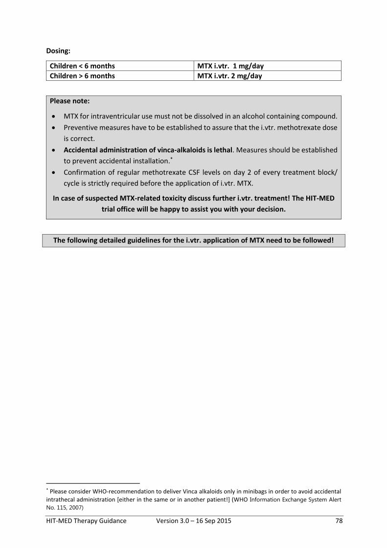
HIT-MED Therapy Guidance Version 3.0 – 16 Sep 2015 78
Dosing:
Children < 6 months MTX i.vtr. 1 mg/day
Children > 6 months MTX i.vtr. 2 mg/day
Please note:
MTX for intraventricular use must not be dissolved in an alcohol containing compound.
Preventive measures have to be established to assure that the i.vtr. methotrexate dose
is correct.
Accidental administration of vinca-alkaloids is lethal. Measures should be established
to prevent accidental installation.*
Confirmation of regular methotrexate CSF levels on day 2 of every treatment block/
cycle is strictly required before the application of i.vtr. MTX.
In case of suspected MTX-related toxicity discuss further i.vtr. treatment! The HIT-MED
trial office will be happy to assist you with your decision.
The following detailed guidelines for the i.vtr. application of MTX need to be followed!
* Please consider WHO-recommendation to deliver Vinca alkaloids only in minibags in order to avoid accidental
intrathecal administration [either in the same or in another patient!] (WHO Information Exchange System Alert
No. 115, 2007)

HIT-MED Therapy Guidance Version 3.0 – 16 Sep 2015 79
8.7.1. Guidelines for the intraventricular application of Methotrexate
Injection of i.vtr. MTX should be performed by an experienced physician under sterile
conditions using a mask, sterile gloves and a sterile cloth. The patient should lay down (upper
body raised to 45 degrees), and should wear a mask. Skin disinfection should be done
thoroughly (e.g. 3 times with 0,1% Octenidin/2%g Phenoxyethanol [e.g. Octenisept ®]) and let
it dry. Injection can be done with a 27-G Butterfly. (If you use an older Ommaya device, check
if it is necessary to rinse repeatedly before and after injection using a sterile swab). After the
puncture use a sterile patch with or without a Providon-Jod salve.
MTX must not be administered if CSF aspiration is disturbed (check dislocation)!
Make sure not to administer any other drug than MTX intraventricularly! Establish
preventive measures (e.g. color-codes/different injection systems) to prevent accidental
injection of vincristine!
Day 1:
1) Aspirate 2 ml CSF (4 ml in case of Ommaya) Store syringe with CSF in a sterile place to be able to use it to flush after MTX injection
2) Aspirate 2 ml CSF for MTX and protein CSF concentration Aspirate 4 ml CSF for cytology
3) Dilute MTX with CSF to a total volume of 2 ml (2mg MTX = 0.8ml) 4) Inject MTX under sterile conditions 5) Inject the 2 ml CSF (4 ml in case of Ommaya) you have aspirated and stored in step
1
Day 2: 2 punctures!
Puncture 1 (approx. 20-24 hours after first injection)
1) Aspirate 2 ml CSF (ca. 4 ml in case of Ommaya) Store syringe with CSF in a sterile place
2) Aspirate 2 ml CSF for MTX and protein CSF concentration 3) Inject the 2 ml CSF (4 ml in case of Ommaya) you have aspirated and stored in step
1
Only proceed if MTX CSF levels are below 5 µmol/l
If > 5 µmol/l: see guidelines for elevated MTX CSF levels
Puncture 2
1) Aspirate 2 ml CSF (ca. 4 ml in case of Ommaya) Store syringe with CSF in a sterile place to be able to use it to flush after MTX injection
2) Dilute MTX with CSF to a total volume of 2 ml (2mg MTX = 0.8ml) 3) Inject MTX under sterile conditions 4) Inject the 2 ml CSF (4 ml in case of Ommaya) you have aspirated and stored in step
1

HIT-MED Therapy Guidance Version 3.0 – 16 Sep 2015 80
Days 3 and 4 (the same as day 1, but without cytology!):
1) Aspirate 2 ml CSF (ca. 4 ml in case of Ommaya) Store syringe with CSF in a sterile place to be able to use it to flush after MTX injection
2) Aspirate 2 ml CSF for MTX and protein CSF concentration 3) Dilute MTX with CSF to a total volume of 2 ml (2mg MTX = 0.8ml) 4) Inject MTX under sterile conditions 5) Inject the 2 ml CSF (4 ml in case of Ommaya) you have aspirated and stored in step
1

HIT-MED Therapy Guidance Version 3.0 – 16 Sep 2015 81
8.7.2. Guidelines for elevated MTX CSF levels after intraventricular application
Elevated CSF MTX levels after intraventricular application of methotrexate are an emergency situation and require urgent intervention!
The most common reason for MTX CSF levels ≥ 5 μmol/l are sampling errors, especially if there
is no evidence of disturbed circulation of the CSF and the child is good clinical condition
without any neurological symptoms. Please recheck CSF-level.The amount of CSF used to flush
after the MTX injection may have been too low, leaving some MTX in the device. Therefore
please aspirate first 4 ml CSF, discarde the syringe and aspirate again 2 ml for detection of the
MTX CSF level.
If rechecked CSF concentrations remain elevated, the patient has neurological symptoms
compatible with CNS toxicity, or MTX CSF concentrations continue to be elevated after 24
hours > 5 μmol/L, please consider emergency measures as follows:
1) Aspirate at least 20-30 ml CSF (may be substituted by sterile NaCl)
2) As next step please consider:
- Leucovorin intravenously, caution: do not inject leucovorin into the CSF!
- Dexamethasone intravenously/orally
3) Contact the HIT-MED office for advice
4) As further steps consider (experimental!):
- Ventriculolumbar CSF lavage
- Carboxypeptidase intrathecally [There are case reports on the intrathecal administration of carboxypeptidase.48]
8.8. Leucovorin rescue after intravenous high-dose MTX
Expected MTX-levels and standard leukovorin-rescue after high-dose MTX 5g/m²:
Time after start of MTX [hours]
Routine creatinine measurements
MTX serum concentration [µmol/l]
Leucovorin dose [mg/m²]
24 X < 150 -
36* < 3* -
42 X ≤ 1 15
48 X ≤ 0,4 15
54 ≤ 0,25 15
60 ** 15
66 ** 15
72 ** 15
* MTX serum concentration 36 h after start of MTX only needs to be determined if the level
24 h after start of MTX is >150 µmol/l. For improved security, consider measurement of MTX
serum concentration 36h after start in all patients treated with intensified induction.

HIT-MED Therapy Guidance Version 3.0 – 16 Sep 2015 82
** if the serum concentration of MTX is below the upper expected limit at 42, 48, and 54
hours after start of MTX infusion, further evaluations of MTX serum levels are not necessary.
Leukovorin-rescue has to be continued until hour 72.
In case of excessively increased MTX levels, consider additional individual interventions
(e.g. higher dose of leucovorin, application of Carboxypeptidase-G2, dialysis).
See chapter “MTX underexcretion” for more information.
Monitoring of serum MTX concentrations and consequences of elevated MTX levels
1.) Measure MTX serum concentrations at 24 hours after start of i.v. MTX If the MTX serum concentration is > 150 µmol/l please
a) Raise fluid intake to 4500 ml/m²/24 h with consequent urine alkalinisation (urine pH ≥ 7.0) and tight fluid equilibration upon receipt of the result If the urine pH drops < 7.0, give sodium bicarbonate (1 mmol/kg) [e.g. sodium bicarbonate 8.4% 1 ml/kg [= 1 mmol/kg] mixed together with aqua ad iniectabilia 1 ml/kg as short infusion in 30-60 minutes]
b) Determine MTX serum concentration additionally at hour 36. c) If the MTX serum concentration is > 3 µmol/l at 36 hours after start of MTX
Continue increased fluid intake (4500 ml/m²/24 h) with consequent urine alkalinisation (urine pH ≥ 7.0) and tight fluid equilibration (if not already done before) If the urine pH drops < 7.0, give sodium bicarbonate (1 mmol/kg) [e.g. sodium bicarbonate 8.4% 1ml/kg [= 1 mmol/kg] mixed together with aqua ad iniectabilia 1 ml/kg as short infusion in 30-60 minutes]
Give leucovorin upon receipt of the result according to the diagram depicted below, i.e. 60mg/m² if serum MTX concentration is between 3 and 4µmol/l and 75mg/m² of serum MTX concentration is between 4 and 5µmol/l. If above 5µmol/l see calculation below. Remember to give very high doses of leucovorin only as short infusion and to check serum calcium concentrations (for details see below).
Consider repeating leucovorin 42 hours after start of MTX: either repeat the same dose as given before, or adapt dose to result of MTX serum concentration measurement at 42 hours (depending on time to receipt of the result, MTX serum level and local policy)
2.) Measure MTX serum concentration at 42 hours, 48 hours and 54 hours after start of i.v.
MTX. If the MTX- serum concentration is above the upper expected limits a) Adjust the leucovorin dose for the subsequent time point to the following:
If the MTX serum concentration is < 5 µmol/l, upon receipt of the result, the leukovorin dose should be determined according to the diagram

HIT-MED Therapy Guidance Version 3.0 – 16 Sep 2015 83
MTX mol/l
5 75 mg/m2
4 60 mg/m2
3 45 mg/m2
2 30 mg/m2
1 15 mg/m2
0,25 NO RESCUE
0 24 36 42 48 54 60 66 72 78 84
Hours after Start of the MTX-Infusion
If the MTX serum concentration is > 5 µmol/l, upon receipt of the result, the leukovorin dose can be calculated by the formula:
Leukovorin [mg] = MTX level in serum [µmol/l] x Body weight [kg]
Caution: Leukovorin doses > 600 mg/m² (or 20 mg/kg) should be administered as short infusion (1 h). Shorter infusion times cannot be recommended because of the high content of Calcium. Repeated determination of serum Calcium levels may be necessary. Oral administration is not recommended because of saturable enteral resorption.
b) Raise fluid intake to 4500 ml/m²/24 h with consequent urine alkalinisation (urine pH ≥ 7.0) and tight fluid equilibration. If the urine pH drops < 7.0, give sodium bicarbonate (1 mmol/kg; e.g. sodium bicarbonate 8.4% 1ml/kg [= 1mmol/kg] mixed together with aqua ad iniectabilia 1ml/kg as short infusion in 30-60 minutes).
c) Continue MTX level determination and leucovorin rescue every 6 h until the MTX level is below 0.25 µmol/l.

HIT-MED Therapy Guidance Version 3.0 – 16 Sep 2015 84
Renal MTX underexcretion
A MTX induced renal dysfunction with prolonged MTX clearance is a medical emergency.
Potential signs of renal MTX underexcretion are one or more of the following symptoms that
occur during the first day of MTX therapy:
- Drop of urine pH < 6.0
- Rise of the creatinine (serum) > 50% of the basic level
- Dropping diuresis despite adequate therapy with furosemide
- 24 h MTX level above the target level
Typical clinical signs of acute MTX intoxication are severe, therapy resistant vomiting during
the first 24-48 h after exposition to MTX, yellow diarrhoea (the same colour as MTX) and
neurological symptoms (disorientation, impaired vision, seizures).
In case of a severe disruption of the MTX excretion it is possible to decrease the MTX serum
level by application of carboxypeptidase-G2 (CPD-G2) very rapidly. MTX will be hydrolysed by
the bacterial enzyme (CPD-G2) to the inactive metabolite 2,4-diamino-N10-methylpteroinacid
(DAMPA). CPD-G2 does not cross the blood-brain barrier. In the event of a severe disruption
of the MTX excretion, please contact the HIT-MED office in order to discuss the next safety
measures.
Carboxypeptidase is expensive and may not be available without pre-organized contacts to
hospitals/pharmacies who store this drug for emergency use. Physicians using this
treatment schedule have to make sure in advance that supply of caboxypeptidase is
available in case of an emergency

HIT-MED Therapy Guidance Version 3.0 – 16 Sep 2015 85
9. References
1. Rutkowski S, Bode U, Deinlein F, et al: Treatment of early childhood medulloblastoma by postoperative chemotherapy alone. N Engl J Med 352:978-86, 2005
2. von Bueren AO, von Hoff K, Pietsch T, et al: Treatment of young children with localized medulloblastoma by chemotherapy alone: results of the prospective, multicenter trial HIT 2000 confirming the prognostic impact of histology. Neuro Oncol 13:669-79, 2011
3. Rutkowski S, von Hoff K, Emser A, et al: Survival and prognostic factors of early childhood medulloblastoma: an international meta-analysis. J Clin Oncol 28:4961-8, 2010
4. Ashley DM, Merchant TE, Strother D, et al: Induction chemotherapy and conformal radiation therapy for very young children with nonmetastatic medulloblastoma: Children's Oncology Group study P9934. J Clin Oncol 30:3181-6, 2012
5. Chi SN, Gardner SL, Levy AS, et al: Feasibility and response to induction chemotherapy intensified with high-dose methotrexate for young children with newly diagnosed high-risk disseminated medulloblastoma. J Clin Oncol 22:4881-7, 2004
6. Lannering B, Rutkowski S, Doz F, et al: Hyperfractionated versus conventional radiotherapy followed by chemotherapy in standard-risk medulloblastoma: results from the randomized multicenter HIT-SIOP PNET 4 trial. J Clin Oncol 30:3187-93, 2012
7. von Hoff K, Hinkes B, Gerber NU, et al: Long-term outcome and clinical prognostic factors in children with medulloblastoma treated in the prospective randomised multicentre trial HIT'91. Eur J Cancer 45:1209-17, 2009
8. Kortmann RD, Kuhl J, Timmermann B, et al: Postoperative neoadjuvant chemotherapy before radiotherapy as compared to immediate radiotherapy followed by maintenance chemotherapy in the treatment of medulloblastoma in childhood: results of the German prospective randomized trial HIT '91. Int J Radiat Oncol Biol Phys 46:269-79, 2000
9. Lannering B, Rutkowski S, doz F: HIT-SIOP PNET-4 - A randomised multicentre study of hyperfractionated (HFRT) versus standard radiotherapy (STRT) in children with standard risk medulloblastoma. Neuro Oncol 12:ii5, 2010
10. Rutkowski S, von Bueren AO, Warmuth-Metz M, et al: Treatment of children and adolsecents with metastatic medulloblastoma by conventional chemotherapy and hyperfractionated radiotherapy. Prelininary results of the prospective GPOH-trial HIT2000 (MET-HIT 2000 AB4). Neuro Oncol 12:ii5, 2010
11. Hinkes BG, von Hoff K, Deinlein F, et al: Childhood pineoblastoma: experiences from the prospective multicenter trials HIT-SKK87, HIT-SKK92 and HIT91. J Neurooncol 81:217-23, 2007
12. Timmermann B, Kortmann RD, Kuhl J, et al: Role of radiotherapy in supratentorial primitive neuroectodermal tumour in young children: results of the German HIT-SKK87 and HIT-SKK92 trials. J Clin Oncol 24:1554-60, 2006
13. Friedrich C, von Bueren AO, von Hoff K, et al: Treatment of young children with CNS-primitive neuroectodermal tumours/pineoblastomas in the prospective multicenter trial HIT 2000 using different chemotherapy regimens and radiotherapy. Neuro Oncol 15:224-34, 2013
14. Timmermann B, Kortmann RD, Kuhl J, et al: Role of radiotherapy in the treatment of supratentorial primitive neuroectodermal tumours in childhood: results of the prospective German brain tumour trials HIT 88/89 and 91. J Clin Oncol 20:842-9, 2002

HIT-MED Therapy Guidance Version 3.0 – 16 Sep 2015 86
15. Merchant TE, Kiehna EN, Li C, et al: Radiation dosimetry predicts IQ after conformal radiation therapy in pediatric patients with localized ependymoma. Int J Radiat Oncol Biol Phys 63:1546-54, 2005
16. Fouladi M, Gilger E, Kocak M, et al: Intellectual and functional outcome of children 3 years old or younger who have CNS malignancies. J Clin Oncol 23:7152-60, 2005
17. Ellison DW, Kocak M, Figarella-Branger D, et al: Histopathological grading of pediatric ependymoma: reproducibility and clinical relevance in European trial cohorts. J Negat Results Biomed 10:7, 2011
18. Merchant TE, Li C, Xiong X, et al: Conformal radiotherapy after surgery for paediatric ependymoma: a prospective study. Lancet Oncol 10:258-66, 2009
19. Taylor MD, Northcott PA, Korshunov A, et al: Molecular subgroups of medulloblastoma: the current consensus. Acta Neuropathol 123:465-72, 2012
20. Packer RJ, Gajjar A, Vezina G, et al: Phase III study of craniospinal radiation therapy followed by adjuvant chemotherapy for newly diagnosed average-risk medulloblastoma. J Clin Oncol 24:4202-8, 2006
21. Gajjar A, Chintagumpala M, Ashley D, et al: Risk-adapted craniospinal radiotherapy followed by high-dose chemotherapy and stem-cell rescue in children with newly diagnosed medulloblastoma (St Jude Medulloblastoma-96): long-term results from a prospective, multicentre trial. Lancet Oncol 7:813-20, 2006
22. Carrie C, Grill J, Figarella-Branger D, et al: Online quality control, hyperfractionated radiotherapy alone and reduced boost volume for standard risk medulloblastoma: long-term results of MSFOP 98. J Clin Oncol 27:1879-83, 2009
23. Taylor RE, Bailey CC, Robinson K, et al: Results of a randomized study of preradiation chemotherapy versus radiotherapy alone for nonmetastatic medulloblastoma: The International Society of Paediatric Oncology/United Kingdom Children's Cancer Study Group PNET-3 Study. J Clin Oncol 21:1581-91, 2003
24. Tarbell NJ, Friedman H, Polkinghorn WR, et al: High-risk medulloblastoma: a pediatric oncology group randomized trial of chemotherapy before or after radiation therapy (POG 9031). J Clin Oncol 31:2936-41, 2013
25. Gandola L, Massimino M, Cefalo G, et al: Hyperfractionated accelerated radiotherapy in the Milan strategy for metastatic medulloblastoma. J Clin Oncol 27:566-71, 2009
26. Verlooy J, Mosseri V, Bracard S, et al: Treatment of high risk medulloblastomas in children above the age of 3 years: a SFOP study. Eur J Cancer 42:3004-14, 2006
27. Taylor RE, Bailey CC, Robinson KJ, et al: Outcome for patients with metastatic (M2-3) medulloblastoma treated with SIOP/UKCCSG PNET-3 chemotherapy. Eur J Cancer 41:727-34, 2005
28. Leary SE, Zhou T, Holmes E, et al: Histology predicts a favorable outcome in young children with desmoplastic medulloblastoma: a report from the children's oncology group. Cancer 117:3262-7, 2011
29. Dhall G, Grodman H, Ji L, et al: Outcome of children less than three years old at diagnosis with non-metastatic medulloblastoma treated with chemotherapy on the "Head Start" I and II protocols. Pediatr Blood Cancer, 2008

HIT-MED Therapy Guidance Version 3.0 – 16 Sep 2015 87
30. Taylor MD, Poppleton H, Fuller C, et al: Radial glia cells are candidate stem cells of ependymoma. Cancer Cell 8:323-35, 2005
31. Witt H, Mack SC, Ryzhova M, et al: Delineation of two clinically and molecularly distinct subgroups of posterior fossa ependymoma. Cancer Cell 20:143-57, 2011
32. Macdonald SM, Sethi R, Lavally B, et al: Proton radiotherapy for pediatric central nervous system ependymoma: clinical outcomes for 70 patients. Neuro Oncol 15:1552-9, 2013
33. Conter C, Carrie C, Bernier V, et al: Intracranial ependymomas in children: society of pediatric oncology experience with postoperative hyperfractionated local radiotherapy. Int J Radiat Oncol Biol Phys 74:1536-42, 2009
34. Massimino M, Gandola L, Giangaspero F, et al: Hyperfractionated radiotherapy and chemotherapy for childhood ependymoma: final results of the first prospective AIEOP (Associazione Italiana di Ematologia-Oncologia Pediatrica) study. Int J Radiat Oncol Biol Phys 58:1336-45, 2004
35. Garvin JH, Jr., Selch MT, Holmes E, et al: Phase II study of pre-irradiation chemotherapy for childhood intracranial ependymoma. Children's Cancer Group protocol 9942: a report from the Children's Oncology Group. Pediatr Blood Cancer 59:1183-9, 2012
36. Zacharoulis S, Levy A, Chi SN, et al: Outcome for young children newly diagnosed with ependymoma, treated with intensive induction chemotherapy followed by myeloablative chemotherapy and autologous stem cell rescue. Pediatr Blood Cancer 49:34-40, 2007
37. Venkatramani R, Ji L, Lasky J, et al: Outcome of infants and young children with newly diagnosed ependymoma treated on the "Head Start" III prospective clinical trial. J Neurooncol 113:285-91, 2013
38. Massimino M, Solero CL, Garre ML, et al: Second-look surgery for ependymoma: the Italian experience. J Neurosurg Pediatr 8:246-50, 2011
39. Massimino M, Gandola L, Barra S, et al: Infant Ependymoma in a 10-year AIEOP (Associazione Italiana Ematologia Oncologia Pediatrica) Experience with Omitted or Deferred Radiotherapy. Int J Radiat Oncol Biol Phys, 2010
40. Grundy RG, Wilne SA, Weston CL, et al: Primary postoperative chemotherapy without radiotherapy for intracranial ependymoma in children: the UKCCSG/SIOP prospective study. Lancet Oncol 8:696-705, 2007
41. Grill J, Le Deley MC, Gambarelli D, et al: Postoperative chemotherapy without irradiation for ependymoma in children under 5 years of age: a multicenter trial of the French Society of Pediatric Oncology. J Clin Oncol 19:1288-96, 2001
42. Jakacki R, Zhou T, Holmes E, et al: Outcome for Patients with Non-pineal Supratentorial PNET Treated with Carboplatin as a Radiosensitizer during Radiotherapy (RT) Followed by Adjuvant Cyclophosphamide (CPM) and Vincristine (VCR): Preliminary Results of COG 99701. Neuro Oncol 10:485, 2008
43. Pizer BL, Weston CL, Robinson KJ, et al: Analysis of patients with supratentorial primitive neuro-ectodermal tumours entered into the SIOP/UKCCSG PNET 3 study. Eur J Cancer 42:1120-8, 2006
44. Cohen BH, Zeltzer PM, Boyett JM, et al: Prognostic factors and treatment results for supratentorial primitive neuroectodermal tumours in children using radiation and chemotherapy: a Childrens Cancer Group randomized trial. J Clin Oncol 13:1687-96, 1995

HIT-MED Therapy Guidance Version 3.0 – 16 Sep 2015 88
45. Geyer JR, Sposto R, Jennings M, et al: Multiagent chemotherapy and deferred radiotherapy in infants with malignant brain tumours: a report from the Children's Cancer Group. J Clin Oncol 23:7621-31, 2005
46. Fangusaro J, Finlay J, Sposto R, et al: Intensive chemotherapy followed by consolidative myeloablative chemotherapy with autologous hematopoietic cell rescue (AuHCR) in young children with newly diagnosed supratentorial primitive neuroectodermal tumours (sPNETs): report of the Head Start I and II experience. Pediatr Blood Cancer 50:312-8, 2008
47. Chamberlain MC: Radioisotope CSF flow studies in leptomeningeal metastases. J Neurooncol 38:135-40, 1998
48. Widemann BC, Balis FM, Shalabi A, et al: Treatment of accidental intrathecal methotrexate overdose with intrathecal carboxypeptidase G2. J Natl Cancer Inst 96:1557-9, 2004

HIT-MED Therapy Guidance Version 3.0 – 16 Sep 2015 89
10. Members of the HIT-MED trial committee
Paediatric Oncology Radiotherapy Prof. Dr. med. Stefan Rutkowski Prof. Dr. med. R.-D. Kortmann Dr. med. Katja von Hoff Dr. med. Beate Timmermann Dr. med. Martin Mynarek Dr. med. Dagmar Hornung Prof. Dr. med. Norbert Graf Dr. med. Jutta Welzel Prof. Dr. med. Gudrun Fleischhack Prof. Dr. med. Anca Grosu Dr. med. Astrid Gnekow Dr. med. Stephanie Combs Prof. Dr. Dr. med. Michael Frühwald Prof. Dr. med. Paul-Gerhardt Schlegel Prof. Dr. med. Wolfram Scheurlen Dr. med. Frank Deinlein Neuroradiology Neuropathology Prof. Dr. med. Monika Warmuth-Metz Prof. Dr. med. Torsten Pietsch Dr. med. Ulrike Löbel Neurosurgery Neuropaediatrics: Dr. med. Jürgen Krauß Dr. med. Pablo Hernáiz-Driever Prof. Dr. med. Martin Schuhmann Dr. med. Ronald Sträter Dr. med. Ulrich Thomale Dr. med. Jonas Denecke Dr. med. Gertrud Kammler Biology Human genetics: Prof. Dr. med. Stefan Pfister Prof. Dr. med. Stefan Aretz Prof. Dr. med. Olaf Witt Late effects Quality of Survival Prof. Dr. med. Thorsten Langer Dr. med. Gabriele Calaminus Neuropsychology International Collaborations Anika Resch/ Hamburg Prof. Dr. med. Karin Dieckmann/ Österreich Dr. rer. nat. Holger Ottensmeier Dr. med. Martin Benesch/ Österreich Dr. med. Nicolas Gerber/ Schweiz Biometry Prof. Dr. rer. nat. et med. habil. Andreas Faldum

HIT-MED Therapy Guidance Version 3.0 – 16 Sep 2015 90
11. History of changes
Substantial changes marked in bold
11.1. Version 1.0
HIT-MED guidance established and published
11.2. Version 2.0
Chapter Change
2.1, 2.2, 2.3 “until 21” deleted where not indicated
2.1, 4.2, 4,4 Change in stratification algorithm: Patients 3 to 4 years with myc-pos.
CMB, CMB M0R+, AMB, LCMB, that before were stratified into treatment
with conventional RT with 35.2Gy CSI + boost + maintenance chemotherapy
should receive intensified induction chemotherapy followed by response-
adapted high-dose chemotherapy +/- radiotherapy
(Decision made by HIT-MED trial committee 23 Jan 2015)
4.3 Information on differences to PNET5 chemotherapy added
Appendix A
(Germany)
Added information for 2 cycles SKK-chemotherapy
Replaced “Hickman” by “Rickham” where misspelled.
11.3. Version 3.0
Chapter Change
2.3 replaced “59.4 Gy” by “54.0 Gy”, where mistyped
4.2 changed decision algorithm for treatment options: patients up to 4 years
with CMB, AMB, LCMB, that present isolated M1-dissemination can be
treated either by high-dose-chemotherapy or CSI after the intensified
induction chemotherapy but should not receive i. vtr. methotrexate during
induction in case a CSI is planned
4.1, 4.2, 4.3,
4.4, 4.5
note on genetic counselling in case of SHH-pathway-activation added
4.6, 4.7 notes on diagnosis-verification and DICER1/RB1 mutations added
5.2 another two cycles on documentation sheet modified SKK added
6.1, 6.2, 6.4 surgery recommendations revised
8.1 information on dose modifications of lomustine added

HIT-MED Therapy Guidance Version 3.0 – 16 Sep 2015 91
8.2 information on timing of 1st block SKK chemotherapy added
8.3 start of G-CSF in the interval between cisplatin/vincristine/etoposide/
cyclophosphamide and high-dose methotrexate changed from day 5 to day
6 (transcription error)

HIT-MED Therapy Guidance Version 3.0 – 16 Sep 2015 92
Appendix A – National Documents
Related Documents