NEUROLOGICAL REVIEW SECTION EDITOR: DAVID E. PLEASURE, MD Therapeutic Considerations for Disease Progression in Multiple Sclerosis Evidence, Experience, and Future Expectations Elliot M. Frohman, MD, PhD; Olaf Stüve, MD, PhD; Eva Havrdova, MD; John Corboy, MD; Anat Achiron, MD, PhD; Robert Zivadinov, MD, PhD; Per Soelberg Sorensen, MD; J. Theodore Phillips, MD, PhD; Brian Weinshenker, MD; Kathleen Hawker, MD; Hans-Peter Hartung, MD, PhD; Lawrence Steinman, MD; Scott Zamvil, MD, PhD; Bruce A. C. Cree, MD, PhD; Stephen Hauser, MD; Howard Weiner, MD; Michael K. Racke, MD; Massimo Filippi, MD I n the management of patients with multiple sclerosis (MS), providers are all faced with the highly formidable challenge of ascertaining whether, and to what degree, disease-modifying therapy is effective in the individual patient. While much has been learned in randomized, con- trolled clinical trials, we cannot simply extrapolate the outcomes of these initiatives and apply them to the care of a single patient. In the future, the application of pharmacogenetic techniques, pro- teomics, and microarray analysis will yield novel profiling information on individual patients that will substantially refine the specific therapeutic questions of relevance: (1) What is the best treatment for an individual patient? (2) Which patients require intensive therapeutic combination regimens to op- timize control of the disease process? (3) What are the appropriate drug dosing targets for an indi- vidual patient? and (4) Which patients will be predisposed to the development of drug-related ad- verse events? Such data may provide a novel variable of drug responsiveness that will mandate its inclusion into the process of covariate analyses for clinical trials. In the management of individual patients, subjective judgments will, no doubt, be made in trying to determine whether treatment should be changed or intensified. Relevant to the process of analyzing for breakthrough disease is the consideration of different treat- ment effects, such as control of relapse rate, disability, and magnetic resonance imaging (MRI) measures of disease activity. Each of these domains of efficacy will be determined by a number of highly relevant factors that potentially dictate how and when we com- mence the process of therapy reassessment. For example, what is the latency period be- tween treatment inception and the onset of significant effects on these various domains of disease activity? Phase III clinical trials do provide us with some level of evidence-based outcomes on which to make some prelimi- nary assumptions. Nevertheless, in formu- lating a treatment strategy for an individual patient, additional information is required to address particular disease characteristics. Pharmacogenomic approaches are needed to assist clinicians in understanding the fac- tors that underlie differences in treatment response. This appears to be particularly im- portant in MS given the heterogeneity of the potential clinical and radiographic pheno- types. ASSESSMENT OF DISEASE ACTIVITY Patient Adherence It is important to recognize that the fidel- ity of treatment adherence achieved by pa- Author Affiliations: Departments of Ophthalmology (Dr Frohman) and Neurology (Drs Frohman, Stüve, Hawker, and Racke) and Center for Immunology (Dr Racke), University of Texas Southwestern Medical Center, Dallas; Department of Neurology, Heinrich Heine University, Düsseldorf, Germany (Drs Stüve and Hartung); Department of Neurology, Charles University, Prague, Czech Republic (Dr Havrdova); Department of Neurology, University of Colorado, Denver (Dr Corboy); Multiple Sclerosis Center, Sheba Medical Center, Tel-Hashomer, Israel (Dr Achiron); Department of Neurology, University of Buffalo, NY (Dr Zivadinov); Department of Neurology, Copenhagen University Hospital, Copenhagen, Denmark (Dr Sorensen); Texas Neurology, Dallas (Dr Phillips); Department of Neurology, Mayo Clinic, Rochester, Minn (Dr Weinshenker); Department of Neurology, Stanford University, Stanford, Calif (Dr Steinman); Department of Neurology, University of California, San Francisco (Drs Zamvil, Cree, and Hauser); Department of Neurology, Brigham and Women’s Hospital, Boston, Mass (Dr Weiner); and Department of Neurology, Scientific Institute and University Ospedale San Raffaele, Milan, Italy (Dr Filippi). (REPRINTED) ARCH NEUROL / VOL 62, OCT 2005 WWW.ARCHNEUROL.COM 1519 ©2005 American Medical Association. All rights reserved. by JohnFeibel, on October 10, 2005 www.archneurol.com Downloaded from

Welcome message from author
This document is posted to help you gain knowledge. Please leave a comment to let me know what you think about it! Share it to your friends and learn new things together.
Transcript
NEUROLOGICAL REVIEW
SECTION EDITOR: DAVID E. PLEASURE, MD
Therapeutic Considerations for Disease Progressionin Multiple Sclerosis
Evidence, Experience, and Future Expectations
Elliot M. Frohman, MD, PhD; Olaf Stüve, MD, PhD; Eva Havrdova, MD; John Corboy, MD; Anat Achiron, MD, PhD;Robert Zivadinov, MD, PhD; Per Soelberg Sorensen, MD; J. Theodore Phillips, MD, PhD; Brian Weinshenker, MD;Kathleen Hawker, MD; Hans-Peter Hartung, MD, PhD; Lawrence Steinman, MD; Scott Zamvil, MD, PhD;Bruce A. C. Cree, MD, PhD; Stephen Hauser, MD; Howard Weiner, MD; Michael K. Racke, MD; Massimo Filippi, MD
I n the management of patients with multiple sclerosis (MS), providers are all faced with thehighly formidable challenge of ascertaining whether, and to what degree, disease-modifyingtherapy is effective in the individual patient. While much has been learned in randomized, con-trolled clinical trials, we cannot simply extrapolate the outcomes of these initiatives and apply
them to the care of a single patient. In the future, the application of pharmacogenetic techniques, pro-teomics, and microarray analysis will yield novel profiling information on individual patients that willsubstantially refine the specific therapeutic questions of relevance: (1) What is the best treatment foran individual patient? (2) Which patients require intensive therapeutic combination regimens to op-timize control of the disease process? (3) What are the appropriate drug dosing targets for an indi-vidual patient? and (4) Which patients will be predisposed to the development of drug-related ad-verse events? Such data may provide a novel variable of drug responsiveness that will mandate its inclusioninto the process of covariate analyses for clinical trials.
In the management of individual patients,subjectivejudgmentswill,nodoubt,bemadein trying to determine whether treatmentshould be changed or intensified. Relevanttotheprocessofanalyzingforbreakthroughdiseaseis theconsiderationofdifferenttreat-ment effects, such as control of relapse rate,disability, andmagnetic resonance imaging(MRI) measures of disease activity. Each ofthesedomainsofefficacywillbedeterminedby a number of highly relevant factors that
potentially dictate how and when we com-mence theprocessof therapyreassessment.For example, what is the latency period be-tween treatment inception and the onset ofsignificanteffectson thesevariousdomainsofdiseaseactivity?PhaseIIIclinical trialsdoprovideuswithsomelevelofevidence-basedoutcomes on which to make some prelimi-nary assumptions. Nevertheless, in formu-lating a treatment strategy for an individualpatient, additional information is requiredtoaddressparticulardiseasecharacteristics.Pharmacogenomic approaches are neededtoassist clinicians inunderstanding the fac-tors that underlie differences in treatmentresponse.Thisappears tobeparticularly im-portant inMSgiventheheterogeneityof thepotential clinical and radiographic pheno-types.
ASSESSMENT OF DISEASE ACTIVITY
Patient Adherence
It is important to recognize that the fidel-ity of treatment adherence achieved by pa-
Author Affiliations: Departments of Ophthalmology (Dr Frohman) and Neurology(Drs Frohman, Stüve, Hawker, and Racke) and Center for Immunology (Dr Racke),University of Texas Southwestern Medical Center, Dallas; Department of Neurology,Heinrich Heine University, Düsseldorf, Germany (Drs Stüve and Hartung);Department of Neurology, Charles University, Prague, Czech Republic (Dr Havrdova);Department of Neurology, University of Colorado, Denver (Dr Corboy); MultipleSclerosis Center, Sheba Medical Center, Tel-Hashomer, Israel (Dr Achiron);Department of Neurology, University of Buffalo, NY (Dr Zivadinov); Department ofNeurology, Copenhagen University Hospital, Copenhagen, Denmark (Dr Sorensen);Texas Neurology, Dallas (Dr Phillips); Department of Neurology, Mayo Clinic,Rochester, Minn (Dr Weinshenker); Department of Neurology, Stanford University,Stanford, Calif (Dr Steinman); Department of Neurology, University of California,San Francisco (Drs Zamvil, Cree, and Hauser); Department of Neurology, Brighamand Women’s Hospital, Boston, Mass (Dr Weiner); and Department of Neurology,Scientific Institute and University Ospedale San Raffaele, Milan, Italy (Dr Filippi).
(REPRINTED) ARCH NEUROL / VOL 62, OCT 2005 WWW.ARCHNEUROL.COM1519
©2005 American Medical Association. All rights reserved. by JohnFeibel, on October 10, 2005 www.archneurol.comDownloaded from
tients in a clinical trial does not necessarily equate withbehavior in general practice. In a clinical trial, patients fre-quently visit the study site, receive substantial support andeducation, and are accountable for the return of the medi-cation vials as well as unused medication. Clear expecta-tions are outlined to patients in the study about the con-duct of the trial and the specific responsibilities expectedof patients. It is also patently clear that patients with MSare a highly motivated group of trial participants.
Adherence rates have been very high in the contextof well-designed clinical trials. In contrast, there is littledata to suggest that these drug usage rates can be simplyextrapolated to the expected use in clinical practice. Wehave found that when we actively query patients duringeach visit or during calls to the clinic about their use ofinjectable disease-modifying agents, up to one third ofour patients admit to frequent nonadherence. This un-derscores the importance of assessing treatment adher-ence and compliance as part of any strategy to diagnosebreakthrough disease in MS. Education, support, and ac-countability (eg, use of administration logs) to ensure thesuggested use of medications may be one strategy to im-prove patient adherence.
Neurological Examination
The periodic assessment of the neurological examina-tion continues to represent an important cornerstone oftracking changes in disease progression. Repeat evalua-tion will frequently reveal definitive changes that are notnecessarily appreciated by our patients. One of the mostformidable challenges faced by clinicians relates to whichexamination techniques should be included. Certainly,the routine neurological examination is standard prac-tice in the periodic assessment of patients with MS. Re-cently, a number of ancillary techniques have been usedin clinical trials that have been validated as useful in-struments to detect meaningful change in discrete func-tions. These include the MS Functional Composite thatcombines a timed 25-foot walk, the 9-hole peg test (a mea-sure of upper extremity function), and the paced serialauditory addition test (a measure of information pro-cessing speed).1 While apparently useful as outcome mea-sures in clinical trials, we lack data to corroborate theusefulness of these techniques to follow changes in theclinical course of our individual patients with MS in re-sponse to individually tailored therapy regimens. The sameargument could be made for the use of the Expanded Dis-ability Status Scale (EDSS) in clinical practice. While thisdisability assessment technique has been of use in clini-cal trials, most clinicians do not have sufficient time orstaffing to perform the complete neurological examina-tion, including the attempted 500-m walk, at every clinicvisit. Many neurologists who treat MS can, however, rou-tinely measure the 25-foot timed walk. This measure doescorrelate with the ambulation index and EDSS, and it pro-vides us with a useful serial assessment strategy for fol-lowing up gait mechanics and safety.2
Even when neurologists are equipped with adequateresources to perform these assessments, there are still anumber of confounders that must always be consideredwhen evaluating patients with MS: (1) What is the time
of day? (2) Is the patient being examined at the same timeat each visit (which is unlikely)? (3) What is the weatherlike (as physical function does change in patients withMS in response to temperature variations)? and (4) Doesthe patient have an infection or fever, or are they havinga particularly stressful day? There is a high degree of vari-ability relating to the conditions under which we rou-tinely follow up patients in the clinic. Despite these limi-tations, the astute neurologist is capable of translatingroutine assessments over time into a judgment concern-ing disease stability or instability. This judgment is re-fined by integrating information concerning relapses, MRIactivity, and even the patient’s subjective report of their“own examination” (analysis of activities of daily living,work performance, etc).
Patient’s Examination(Activities of Daily Living)
The office-based examination is a highly stereotypic andstandardized (as well as very artificial) approach to ob-jectively documenting meaningful change in neurologi-cal capabilities. The inherent strength of this approachhas been obvious and highly useful in the context of clini-cal trial initiatives. Despite these strengths, the “real” ex-amination takes place where patients live their lives. Inthe assessment of breakthrough disease, patients shouldbe asked about toileting, showering, dressing, ambula-tion, and eating. How are they functioning at work? Arethey having more difficulty driving? Are cognitive changesoccurring? Does depression cloud this assessment? A veryimportant area of inquiry concerns the level of our pa-tient’s physical conditioning. The deconditioned pa-tient with MS often perceives that his or her disease coursehas deteriorated. Correspondingly, the physician may like-wise document a change in the course of disease pro-gression secondary to breakthrough disease when a re-duction of exercise tolerance may have compromised thepatient’s activities of daily living.
DEFINING BREAKTHROUGH DISEASE
Breakthrough disease may be characterized by unaccept-able clinical or radiographic evidence of disease activity thatis not sufficiently controlled by current treatment inter-vention. Detection of such activity is, in reality, not nec-essarily a true reflection of the actual level of disease ac-tivity. Defining breakthrough is contingent on the sensitivityof the assessment strategies being used to detect disease ac-tivity. As such, inherent limitations of subjective evalua-tions (clinical examination, patient self-report of activi-ties of daily living) and even information derived fromquantitative studies (MRI, immunological profiling, physi-ological examination) will continue to make the establish-ment of threshold limits for the confirmation of constitu-tive disease activity in MS inadequate. Hopefully, the useof newly emerging MRI applications, including magneticresonance spectroscopy, magnetization transfer, and dif-fusion tensor imaging, will help to refine our ability to gen-erate more realistic burden-of-disease measurements thatcan be used to monitor the disease process and to detecttreatment effects in controlled clinical trials.
(REPRINTED) ARCH NEUROL / VOL 62, OCT 2005 WWW.ARCHNEUROL.COM1520
©2005 American Medical Association. All rights reserved. by JohnFeibel, on October 10, 2005 www.archneurol.comDownloaded from
Progression, whether clinically or radiographically evi-dent to the physician or patient with MS, likely repre-sents an ongoing process that, while modifiable with treat-ment intervention, is not currently amenable to aconfirmable therapy-induced remission. Insights from so-phisticated imaging and histopathological investiga-tions in brain and spinal cord tissue samples from pa-tients with MS indicate that a very active constitutiveprocess of molecular and cellular events operates withinthe microenvironment of the central nervous system(CNS), generally without interruption throughout thecourse of the disease.2-11 Disease activity may be esca-lated or mitigated depending on yet to be elucidated “driv-ing” or “braking” factors or treatment interventions. Whatconstitutes breakthrough activity in one patient mightbe considered adequate control in another. The featuresthat characterize the pretreatment level of disease activ-ity may be important to consider when formulating rea-sonable expectations to be derived from therapy inter-vention in the individual patient. For many (if not most)patients, some level of ongoing disease activity might beexpected.
Despite the substantial and inherent limitations in de-fining breakthrough disease in MS, it does appear rea-sonable to begin the process of ascertaining changes indisease activity across multiple domains and comparingsuch changes over time with respect to baseline and pre-baseline measures. For instance, ascertaining the num-ber of bona fide relapses in the preceding year or 2 yearsbefore treatment initiation may constitute an importantbenchmark from which to compare relapse rate after treat-ment inception (even though subclinical relapses oftengo undetected). While there is certainly no magic num-ber of relapses that should be considered as a thresholdfor altering therapy, control over the disease process with
drug therapy should be attempted to limit the numberand severity of attacks.
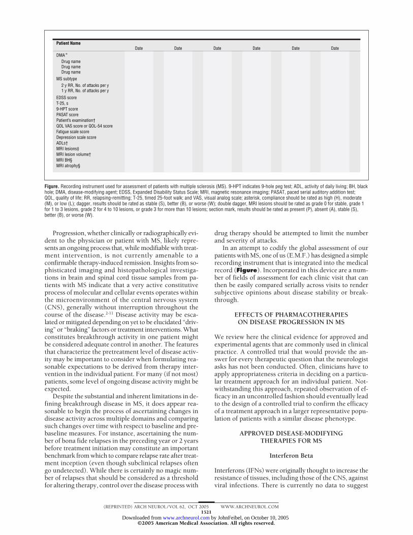
In an attempt to codify the global assessment of ourpatients with MS, one of us (E.M.F.) has designed a simplerecording instrument that is integrated into the medicalrecord (Figure). Incorporated in this device are a num-ber of fields of assessment for each clinic visit that canthen be easily compared serially across visits to rendersubjective opinions about disease stability or break-through.
EFFECTS OF PHARMACOTHERAPIESON DISEASE PROGRESSION IN MS
We review here the clinical evidence for approved andexperimental agents that are commonly used in clinicalpractice. A controlled trial that would provide the an-swer for every therapeutic question that the neurologistasks has not been conducted. Often, clinicians have toapply appropriateness criteria in deciding on a particu-lar treatment approach for an individual patient. Not-withstanding this approach, repeated observation of ef-ficacy in an uncontrolled fashion should eventually leadto the design of a controlled trial to confirm the efficacyof a treatment approach in a larger representative popu-lation of patients with a similar disease phenotype.
APPROVED DISEASE-MODIFYINGTHERAPIES FOR MS
Interferon Beta
Interferons (IFNs) were originally thought to increase theresistance of tissues, including those of the CNS, againstviral infections. There is currently no data to suggest
Patient Name
DMA ∗
MS subtype
EDSS scoreT-25, s9-HPT score
MRI lesions‡MRI lesion volume†MRI BH§MRI atrophy§
PASAT scorePatient’s examination†QOL VAS score or QOL-54 scoreFatigue scale scoreDepression scale scoreADLs†
Drug nameDrug nameDrug name
2 y RR, No. of attacks per y1 y RR, No. of attacks per y
Date Date Date Date Date Date
Figure. Recording instrument used for assessment of patients with multiple sclerosis (MS). 9-HPT indicates 9-hole peg test; ADL, activity of daily living; BH, blackhole; DMA, disease-modifying agent; EDSS, Expanded Disability Status Scale; MRI, magnetic resonance imaging; PASAT, paced serial auditory addition test;QOL, quality of life; RR, relapsing-remitting; T-25, timed 25-foot walk; and VAS, visual analog scale; asterisk, compliance should be rated as high (H), moderate(M), or low (L); dagger, results should be rated as stable (S), better (B), or worse (W); double dagger, MRI lesions should be rated as grade 0 for stable, grade 1for 1 to 3 lesions, grade 2 for 4 to 10 lesions, or grade 3 for more than 10 lesions; section mark, results should be rated as present (P), absent (A), stable (S),better (B), or worse (W).
(REPRINTED) ARCH NEUROL / VOL 62, OCT 2005 WWW.ARCHNEUROL.COM1521
©2005 American Medical Association. All rights reserved. by JohnFeibel, on October 10, 2005 www.archneurol.comDownloaded from
that viral inhibition underlies IFN-� effects on MS in anyway. Several mechanisms of action have been de-scribed. Interferon � increases levels of vascular adhe-sion molecule 1 in the sera of patients with MS.12 In 1996,2 studies13,14 showed that the migration of activated T lym-phocytes across an artificial blood-brain barrier was partlymediated by metalloproteinase-9, and that IFN-� treat-ment reduced the production of metalloproteinase-9by activated T cells as well as the migration of the T cellsin vitro. It was also shown that IFN-� down-regulatesIFN-�–inducible major histocompatibility complexclass II expression on nonprofessional CNS antigen-presenting cells such as astrocytes15 and microglial cells,16
and it may therefore reduce T-cell activation in the CNS.Furthermore, production of the proinflammatory cyto-kine tumor necrosis factor � by T cells activated by mi-croglial antigen-presenting cells was also significantlydecreased by IFN-�.17
Evidence. The first study to show that a pharmacothera-peutic intervention could improve the clinical course ofMS was published in 1993, when IFN-�-1b (Betaseron;Berlex Inc, Montville, NJ) reduced the rate of exacerba-tions of relapsing-remitting (RR) MS in a multicentertrial.18,19 Furthermore, the number and frequency of le-sions on brain MRI were decreased in the high-dose–treated patient population.18,19 There was a trend to lessdisability, though it was not statistically significant. Theresults of another multicenter placebo-controlled trial us-ing IFN-�-1a (Avonex; Biogen Idec Inc, Cambridge, Mass)administered intramuscularly once weekly demon-strated that IFN-�-1a significantly delayed the time tosustained clinical disability in RRMS. A reduction in theexacerbation rate and time to sustained change in clini-cal disability was also seen.20 Yet another preparation ofIFN-�-1a (Rebif; Serono Inc, Rockland, Mass) was alsoshown to decrease the number of clinical exacerbations,to decrease the percentage of T2 active MRI scans, andto delay sustained disease progression.17 In that particu-lar multicenter, placebo-controlled trial, the relapse ratewas significantly lower at 1 and 2 years with both a lowdose (22 µg, 3 times per week) and a high dose (44 µg,3 times per week) of IFN-�-1a than with placebo. A re-cently published study21 indicates that IFN-�-1a (Avonex)has significant beneficial effects for patients with RRMSwith regard to cognitive function.
In 1999, a double-blinded, placebo-controlled trial22
conducted in Europe revealed a highly significant delayof progression in patients with secondary progressive (SP)MS who were treated with IFN-�-1b. Unfortunately, asecond SPMS trial in North America showed no statisti-cally significant benefit of IFN-�-1b as comparedwith placebo.23 Findings from a study24,25 of IFN-�-1a(Rebif) in SPMS were also disappointing. In a study26 us-ing IFN-�-1a (Avonex), a significant reduction in dis-ability was demonstrated. The inconsistency with re-gard to the outcome of these trials may be partly owingto differences in the patient populations, both with re-gard to disability and disease activity.
Future Perspectives. Clearly, the effect of IFN-� on dis-ease progression is only modest. This may be owing to the
fact that our current dosing is insufficient, or that IFN-�givenasmonotherapy is simplynoteffectiveenough insig-nificantly slowing the natural course of MS. Combinationtherapy with other approved or experimental agents maygreatly improve the effect of IFN-� on disease progression.
Role of IFN-Neutralizing Antibodiesin MS Progression
The use of biotherapeutic agents is frequently associ-ated with the generation of antibodies. Interferon � is afirst-line treatment for RRMS, and occurrence of anti-bodies against IFN-� were described in several clinicaltrials.18,20,27-29 While binding antibodies were found in asmany as 78% of patients treated with IFN-�, the fre-quency of neutralizing antibodies (NAbs) has varied from2% to 42%.
Evidence. Neutralizing antibodies hamper the biologicresponse to IFN-� and have a detrimental effect on thetreatment response after 18 to 24 months of therapy.30-32
In the pivotal trials of the 3 commercially available IFN-�preparations (Betaseron, Avonex, and Rebif), all of the3 studies found initially no relationship between NAbsand clinical efficacy in a 2-year study period.18,20,33 How-ever, in the trial of IFN-�-1b (Betaseron), NAbs oc-curred in approximately 35% of the patients; between 13and 36 months of treatment, the exacerbation rate in NAb-positive patients was similar to that seen in placebo-treated patients. Further, the number of active lesions onMRI increased significantly in NAb-positive patients.34
In the extension phase of the Prevention of Relapses andDisability by Interferon-�-1a Subcutaneously in Mul-tiple Sclerosis study,35 NAbs to IFN-�-1a caused a sig-nificant reduction in efficacy during treatment years 3and 4. Patients who were positive for NAbs experienceda significantly higher relapse rate than NAb-negative pa-tients (relapse rate, 0.81 vs 0.50, respectively; P=.002).The disease burden on MRI decreased by 9% from base-line to year 4 in NAb-negative patients whereas it in-creased by nearly 18% in NAb-positive patients (P�.01).In a recent Danish study36 of IFN-�-1a and IFN-�-1b prod-ucts, the presence of NAbs was studied in 541 patientswith RRMS treated with IFN-�, and an evaluation of dif-ferent concentrations of NAbs on the treatment effect wasassessed. It was found that across all of the IFN-� treat-ments, even moderate concentrations of Nabs caused asignificant difference in the relapse rates. The yearly re-lapse rate was 0.64 during NAb-positive periods as com-pared with 0.43 in NAb-negative periods, yielding an oddsratio of 1.53 (95% confidence interval, 1.31-1.92) for hav-ing relapses during NAb-positive periods. Patients whowere positive for NAbs showed an increase in the meanEDSS score after 48 months, though the difference be-tween the 2 groups did not reach statistical significance.Time to confirmed progression of 1 point on the EDSSsustained for at least 6 months showed a trend in favorof NAb-negative patients (P=.14).
Future Perspectives. The biologic activity of IFN-� canbe assessed in vivo by analyses of MxA gene expres-sion.37 Measurement of IFN-� bioactivity in all of the pa-
(REPRINTED) ARCH NEUROL / VOL 62, OCT 2005 WWW.ARCHNEUROL.COM1522
©2005 American Medical Association. All rights reserved. by JohnFeibel, on October 10, 2005 www.archneurol.comDownloaded from
tients with MS who receive IFN-� therapy might be thefuture method for detecting antibody-mediated hamper-ing of treatment effect, specifically with regard to dis-ease progression.
Glatiramer Acetate
Glatiramer acetate (GA) is a random, synthetic, basiccopolymer of L-alanine, L-glutamic acid, L-lysine, andL-tyrosine, the most prevalent amino acids in myelinbasic protein.38-40 This agent is administered as a dailysubcutaneous injection that is well tolerated by mostpatients.
Evidence. In 1995, a multicenter, randomized, 2-yearstudy41 demonstrated that the clinical benefits in GA-treated patients were similar to those demonstrated inthe IFN-�-1b trial. The frequency of clinical MS attackswas reduced by 29%. After approximately 6 years of evalu-ation, GA showed sustained efficacy in reducing the rateof clinical exacerbations in patients with RRMS.42 Mag-netic resonance imaging studies of a cohort of patientsincluded in the original trial were performed, and theydemonstrated that GA treatment significantly decreasedthe percentages of annual MRI lesion volume and lossof brain tissue.43
Future Perspectives. The effects of GA on disease pro-gression seem modest. Future trials should assess the ef-fects of GA in combination with other approved or ex-perimental agents.
Mitoxantrone
Mitoxantrone is an antineoplastic agent that interca-lates DNA, resulting in DNA strand breaks and inter-strand crosslinks. The major limitation associated withthe use of mitoxantrone is related to potential cardio-toxic effects. Mitoxantrone can produce a vacuolar car-diomyopathy, producing a reduction in ejection frac-tion. Further, there is an increased incidence of leukemiaassociated with this agent that should be completely dis-cussed with candidate patients before commencingtherapy.44 Most MS specialists primarily use mitoxan-trone for patients who exhibit either inflammatory de-myelinating syndromes not responding to corticoste-roids or plasma exchange, and for those patients whoexhibit neurological deterioration and progression in dis-ability despite first-line therapy interventions.
Evidence. Based on the results of a phase II trial45 and aphase III trial,46 mitoxantrone was the first drug ap-proved for the treatment of SPMS with worsening re-lapsing and progressive relapsing disease course. In thephase III trial, the greater of 2 mitoxantrone doses (12mg/m2) resulted in a 64% reduction in sustained diseaseprogression and a 69% reduction in the number of treatedrelapses as compared with the placebo control group.
Future Perspectives. Like all of the other approvedmedications, mitoxantrone should be assessed in com-bination with other approved or experimental agents in
controlled clinical trials. Similarly, studies are under-way to determine whether a strategy is effective in re-ducing the risk of cardiotoxic effects when administer-ing this agent.
Natalizumab
Natalizumab is a selective adhesion molecule inhibitorused for the treatment of relapsing forms of MS, and ithad only recently been approved by the Food and DrugAdministration in November 2004. Biogen Idec Inc andElan Corp, Dublin, Ireland, the manufacturers of natali-zumab, then announced the voluntary withdrawal of thisagent from the market because of the development of pro-gressive multifocal leukoencophalopathy in 2 patients whohad been treated with a combination therapy of natali-zumab and IFN-�-1a (Avonex). In addition, the compa-nies have stopped using the drug in clinical trials. It isunclear when and whether this agent will be reintro-duced for MS therapy.
EXPERIMENTAL ANTI-INFLAMMATORY ANDIMMUNOMODULATORY AGENTS
Currently, clinical trials for numerous pharmacologicalagents are in the planning stage or are under way. Thisreview will only discuss agents that are already com-monly being used in clinical practice. There is no defini-tive evidence that any of these medications alter the natu-ral course of MS. Particularly, it is unknown whether anyof these agents slow disease progression. This point un-derscores the enormous need for a broader number ofrandomized, controlled trials to carefully and systemati-cally assess the use of these agents in the managementof MS.
Corticosteroids
During the past 2 decades, the use of glucocorticoste-roids to treat MS relapses has gained increasing accep-tance. There is general consensus that intravenous (IV)methylprednisolone (MP) (administered usually as 500-1000 mg daily for 3-5 days) hastens recovery from MSrelapses.47,48 It has been found that short-term treat-ment with IVMP reduces tissue damage and promoteslesion recovery in patients with RRMS.49 Moreover, it hasbeen suggested that pulsed IVMP could favorably affectevents responsible for early preenhancing lesion forma-tion.50 Different mechanisms may explain this hypoth-esis. There is evidence showing that IVMP restores theblood-brain barrier by down-regulating adhesion mol-ecule expression, inhibits proinflammatory cytokines, re-duces matrix metalloproteinase secretion, induces lym-phocyte apoptosis, and promotes remyelination.51
Evidence. There is some suggestion that MP treatmentmay change the natural course of RRMS.52 Results of theOptic Neuritis Treatment Trial53 suggested that IVMP de-lays development of clinically definite MS following op-tic neuritis in the long run. However, it was unclearwhether the results could be generalized to clinically iso-lated syndromes other than optic neuritis, or to patients
(REPRINTED) ARCH NEUROL / VOL 62, OCT 2005 WWW.ARCHNEUROL.COM1523
©2005 American Medical Association. All rights reserved. by JohnFeibel, on October 10, 2005 www.archneurol.comDownloaded from
with RRMS. A randomized, controlled, single-blind,5-year, phase II clinical trial of IVMP in patients withRRMS demonstrated that prolonged treatment with pulsedIVMP slowed the development of destructive lesions (T1black holes), the rate of whole-brain atrophy progres-sion, and the development of sustained physical disabil-ity.50 A phase II, double-blind, dose-comparison studyof bimonthly IVMP pulses in patients with SPMS showedno significant improvement related to difference in pri-mary outcome, which was the proportion of patients withsustained disability worsening over 24 months.54 How-ever, a beneficial effect was detected with the high-doseIVMP regimen as measured by the preplanned second-ary analysis, a comparison of time to onset of sustainedprogression of disability. Both studies50,54 demonstratedthat prolonged use of pulsed IVMP was safe and well tol-erated, and they concluded that phase III trials of corti-costeroids in RRMS and SPMS are warranted to more de-finitively establish the role of pulsed IVMP as a disease-modifying therapy, either alone or in combination withother agents.
A randomized, double-blind, placebo-controlled pilotstudy of IV immunoglobulins (IVIgs) in combination withIVMP did not demonstrate superiority of IVMP-IVIg in thetreatment of moderate-to-severe acute relapses in MS.55
Future Perspectives. Two recently published stud-ies55,56 investigated the effect of glucocorticosteroids asadd-on therapy to standard disease-modifying therapy inpatients with MS. Two other multicenter combinationtrials have been launched to investigate the efficacy ofIVMP as an add-on therapy to standard treatments. TheAvonex Combination Therapy study will assess the ben-efit of IFN-�-1a (Avonex) combined with bimonthlyIVMP in patients with RR breakthrough disease. An-other double-blind, controlled trial will evaluate the ef-ficacy of IFN-�-1b (Betaseron) alone or in combinationwith bimonthly IVMP in patients with SPMS.
IVIg
Some patients fail to respond to standard treatments andcontinue to worsen over time, with the occurrence of ad-ditional relapses associated with neurological deteriora-tion and no apparent effect of the immunomodulatorytreatment. Other groups of patients who are not suit-able for standard treatments include (1) patients who de-velop intolerable adverse events, (2) patients who are non-compliant to self-injections or are reluctant to takeinjectable medications, and (3) female patients who arecontemplating becoming pregnant.
Intravenous Ig modulates the immune system by vari-ous mechanisms, such as macrophage Fc receptor block-ade, idiotypic–anti-idiotypic networking, decreasingT-cell activation, and enhancing remyelination in virus-induced experimental encephalomyelitis, which are allrelevant to MS.
Evidence. Intravenous Ig treatment has been describedto be beneficial for patients with RRMS.57-60 Relapse rate,relapse severity, progression of disability, and disease ac-tivity evaluated by brain MRI were all found to be posi-
tively affected by IVIg treatment. Four double-blindtrials61,62 in RRMS have demonstrated that IVIg reducesthe relapse rate and the number of gadolinium-enhancing lesions, and in this respect, IVIg seems com-parable to the established therapies, ie, IFN-� and GA.61,62
Owing to the relatively small sample size of these stud-ies, a meta-analysis63 was recently undertaken, and it dem-onstrated a significant beneficial effect of IVIg on the an-nual relapse rate (effect size, −0.5; P�.001) as well as onthe proportion of relapse-free patients and change in neu-rological disability by the EDSS score. In another small-sample study,64 quantitative brain MRI analysis showeda statistically significant decrease in the volume of le-sions in IVIg-treated patients with RRMS as comparedwith patients treated with placebo, at follow-up at both3 and 6 months. Taken together, these studies supportthe possibility of using IVIg to treat patients with MS whodo not respond to standard treatments.
Future Perspectives. The definite role of IVIg and theextent of its efficacy in the management of patients withprogression and/or breakthrough disease should be es-tablished in large and long-term double-blind studies. Fur-ther, given the heterogeneity of treatment response andthe high cost of IVIg, it will be important to elucidate fac-tors that can stratify patients into groups who are eitherinappropriate or appropriate candidates for treatment.
Azathioprine
Azathioprine (AZA) is a nonspecific immunosuppres-sant that was first proposed in MS treatment 30 years ago.65
It interferes with the biosynthesis of nucleic acids, par-ticularly during the S phase of the mitotic cycle. Aza-thioprine is believed to primarily affect immature im-munocytes and to have little or no effect on maturecomponents of antigenic memory.66 One in 300 sub-jects will experience intolerance to AZA, characterizedby effects toxic to bone marrow secondary to a homo-zygous polymorphism for thiopurine methyltransferasedeficiency. Eleven percent of subjects are heterozygousand have intermediate levels of thiopurine methyltrans-ferase activity; the remaining 89% of subjects are homo-zygous for the allele for high activity. Human thiopu-rine methyltransferase activity can be easily measured inred blood cells.67 In cases of repeated viral infection, im-munodeficiency should be excluded, and the dose of AZAshould be reduced or the therapy should be changed, es-pecially in cases of herpetic infection. The most concern-ing risk of AZA treatment has been the putative risk ofcancer. In fact, no significant risk was observed duringthe first years of treatment, and an increased risk was sug-gested only after approximately 10 years of continuoususe, especially in patients who also have other risk fac-tors for cancer.68
Evidence. In 1991, a meta-analysis69 of all of the pub-lished randomized, controlled trials of AZA in MS sug-gested that AZA significantly decreased the relapse rateand marginally significantly reduced the increase in dis-ability after 2 and 3 years of treatment. Not all of thesetrials had acceptable methodology, and all of them were
(REPRINTED) ARCH NEUROL / VOL 62, OCT 2005 WWW.ARCHNEUROL.COM1524
©2005 American Medical Association. All rights reserved. by JohnFeibel, on October 10, 2005 www.archneurol.comDownloaded from
performed in the pre-MRI era. At the present time, how-ever, there is an increasing number of articles support-ing positive effects of AZA on the number of T2, new T1,and gadolinium-enhancing lesions.70-72 Most studies car-ried out in recent years have been designed to test AZAin combination with other approved drugs. In terms ofsafety and tolerability, 10% to 20% of patients may com-plain of gastrointestinal discomfort at the beginning oftreatment, and in some, this may limit the use of AZA.
Future Perspectives. Azathioprine has been proposed asa suitable candidate drug in combination with IFN-� orIVIg. Small pilot trials have demonstrated acceptable safetyand possibly efficacy commensurate with already estab-lished monotherapies. Larger, adequately blinded, con-trolled trials are in progress.
Methotrexate
Methotrexate is an inhibitor of dihydrofolate reductase,which results in anti-inflammatory effects by reducingthe release of TH1 cytokines. Traditionally used in sub-stantial doses for malignancies, low weekly dose regi-mens have been applied to a number of immune-mediated disorders.
Evidence. A randomized, double-blinded, placebo-controlled trial73 of low-dose methotrexate (7.5 mg/wk)was performed in 45 patients with MS (of various types).This small study provided some evidence to suggest a ben-eficial effect in those with a relapsing disease course butnot in those with progressive disease. In a study74 con-ducted to demonstrate the therapeutic benefit in pa-tients with either primary MS or SPMS, patients wererandomized to receive either placebo or low-dose metho-trexate (7.5 mg/wk). A composite outcome measure us-ing measures of ambulation (EDSS and ambulation in-dex) and upper extremity function (9-hole peg test andbox-and-block test) demonstrated benefit in patientstreated with methotrexate. The beneficial effect on thecomposite score was principally driven by change on the9-hole peg test.
Future Perspectives. A new combination clinical trial ini-tiative (Avonex Combination Therapy study) is under wayto assess the merits of using methotrexate with cortico-steroids and IFN-�.
Mycophenolate Mofetil
Mycophenolate mofetil (MMF) is a selective inhibitor ofinosine 5�-monophosphate dehydrogenase type II that isa potent immunosuppressant principally used in trans-plant medicine as an antirejection agent.75 This enzymeis responsible for the de novo synthesis of the purinenucleotide guanine within activated T and B lympho-cytes and macrophages without affecting purine salvagepathways. Mycophenolate mofetil exhibits the capabil-ity to suppress lymphocyte proliferation and the expres-sion of T-cell surface antigens in whole-blood lympho-cyte analysis derived from treated allograft recipients.75-78
In activated lymphocytes, metabolites of MMF inter-
rupt cytokine-dependent signals that control the cell cycle,and they block activation of T cells in the mid-G(1) phase.Humoral effects have also been observed, with MMF sup-pressing anti–blood-type IgG antibodies in patients re-ceiving ABO-mismatched renal transplants.79
Evidence. A small, open-label surveillance study80 in-volving 7 patients with MS who were treated with MMFwas described in 2001, and it suggested evidence of tol-erability and potential efficacy in this small cohort. Wehave recently extended this observation with our open-label, exploratory surveillance safety experience with MMFin 79 patients with MS.81
Future Perspectives. The favorable safety profile, novelmechanism of action, and ease of administration makeMMF a potentially useful agent to be used as mono-therapy or in conjunction with IFN-� or GA. Mycophe-nolate mofetil has a specific molecular target throughwhich it mediates its therapeutic effect. Characterizingpolymorphisms of the inosine monophosphate dehydro-genase gene may provide for the opportunity to identifypatient populations that are more or less likely to re-spond favorably to this agent.
Statins
Several studies82-85 indicate that cholesterol-lowering 3-hy-droxy-3-methylglutaryl coenzyme A reductase inhibi-tors (statins) have immunomodulatory properties that maybe beneficial in the treatment of MS. In contrast with cur-rently approved medications used in MS therapy, whichare administered parenterally and are associated with ad-verse and potentially toxic effects, statins are given orallyand, in general, are well tolerated. Several years ago, itwas observed that statins inhibited CNS glial cells fromproducing proinflammatory molecules, including tu-mor necrosis factor �, nitric oxide synthase, and inter-leukin 1�.86 More recently, it was observed that in vivotreatment with atorvastatin calcium (Lipitor; Pfizer Inc,New York, NY) promoted a protective TH2 bias and couldreverse relapsing and chronic paralysis in experimentalautoimmune encephalomyelitis in the MS model, a modelof inflammatory demyelination.83 Atorvastatin calciuminhibited the up-regulation of major histocompatibilitycomplex class II and costimulatory molecules on antigen-presenting macrophages and microglial cells. Meva-lonic acid, the product of 3-hydroxy-3-methylglutarylcoenzyme A reductase, prevented atorvastatin calcium–induced TH2 differentiation and reversed the statin-induced effects on macrophages and microglial cells, in-dicating that the immunomodulatory effects are mediatedthrough inhibition of the mevalonate pathway, which in-volves isoprenoid intermediates that participate in post-translational modification of key proteins that directT-cell differentiation and activation.
Evidence. The beneficial immunomodulatory effects ob-served in experimental autoimmune encephalomyelitisand other autoimmune models have provided the impe-tus to test statins in MS and other TH1-mediated inflam-matory diseases. An open-label phase II trial87 tested sim-
(REPRINTED) ARCH NEUROL / VOL 62, OCT 2005 WWW.ARCHNEUROL.COM1525
©2005 American Medical Association. All rights reserved. by JohnFeibel, on October 10, 2005 www.archneurol.comDownloaded from
vastatin (80 mg; Zocor; Merck & Co Inc, WhitehouseStation, NJ) in 30 patients with RRMS for 6 months, anda 44% reduction in the number of new gadolinium-enhancing lesions and a 41% reduction in volume of newenhancing lesions were observed. It is not known whetherthis modest reduction in new lesions represented a trueeffect of Zocor or regression to the mean, as patients en-rolled in this trial were selected for MRI activity.
Future Perspectives. It is clear that larger, placebo-controlled statin trials will be advantageous in evaluat-ing the clinical and immunomodulatory effects of stat-ins in MS. A placebo-controlled 152-patient phase II trialthat will test whether 80 mg of Lipitor will reduce MSactivity in patients who have experienced their first CNSdemyelinating attack, a “clinically isolated syndrome,”will begin shortly.
Plasma Exchange
Plasma exchange is an effective, short-term treatment foracute inflammatory demyelinating polyneuropathy. The un-derlying hypothesis is that humoral factors, including butnot necessarily restricted to immunoglobulins, account forthe ongoing inflammatory demyelination in steroid-refractory attacks of MS and other CNS inflammatory de-myelinating diseases.88-90 By removing these humoral fac-tors, recovery from acute, severe attacks might be facilitated.
Evidence. A single previous sham-controlled clinical trial91
in patients with attacks of MS gave equivocal results. Arandomized, sham-controlled clinical trial92 of 22 pa-tients with acute steroid-refractory attacks of MS (n=12)or other idiopathic inflammatory demyelinating diseases(n=10; including acute transverse myelitis, neuromyeli-tis optica, acute disseminated encephalomyelitis, and fo-cal cerebral demyelinating disease) showed that plasmaexchange was effective in producing moderate or greaterrecovery within 2 weeks. Uncontrolled experience by thesame group and subsequent case reports have further docu-mented the benefits of plasma exchange.93,94
Future Perspectives. The biological basis of the improve-ment is under continued evaluation by correlative stud-ies including the histopathology of lesions (specifically,the presence of immunoglobulin and markers of comple-ment activation in brain tissue).95 Passive transfer of thedemyelinating activity would confirm the humoral basisof the effect and would provide a bioassay that would en-able further isolation and identification of the specific fac-tors that are responsible for the demyelinating activity.
Cyclophosphamide
Cyclophosphamide, first tested in MS in 1966,96 is an al-kylating agent used to treat malignancy. Cyclophospha-mide has pronounced immunologic effects that involvenot only the suppression of TH1-type responses, but alsoimmunomodulation associated with increases in inter-leukin 4, interleukin 10, and transforming growth fac-tor �. The adverse effects of cyclophosphamide are wellknown and include toxic effects in the bladder, infertil-
ity, infection, and cancer risk. Toxic effects in the blad-der are generally well managed by adequate fluid in-take. The maximum recommended total dose is 80 g.Cyclophosphamide can also be used for short periods oftime, eg, pulses monthly for 6 months in patients whoneed better control on injectable therapy. Data from thelupus nephritis literature suggest that the drug is moreeffective if given with steroids, and another study97 dem-onstrated that pulsed cyclophosphamide given with ste-roids is superior in decreasing inflammation as com-pared with steroids given alone to patients who do notrespond to IFN.
Evidence. During the past 30 years, cyclophosphamidehas been used for the treatment of selected patients withMS. There have been more than 40 articles on the clinicaland immunologic effects of cyclophosphamide in MS. Ini-tial trials suggested a clinical benefit in patients with RRMSand relapsing forms of this disease.98-102 However, 2 ran-domized, clinical trials103,104 in patients with SPMS did notdemonstrate any effect on the progression of neurologi-cal disability. Not surprisingly, the results of these stud-ies initially led to conflicting opinions regarding the useof cyclophosphamide in the treatment of MS.
Future Perspectives. A recent large study105 of 490 pa-tients suggests that response at 6 months following treat-ment may predict who in the progressive stage of the dis-ease may be helped by such therapy. Our experiencecorroborates the observation that those patients who ex-hibit ongoing clinical and radiographic evidence of dis-ease activity are the most likely to benefit from thistherapy.
NEW TREATMENT STRATEGIES
Novel Therapeutic Targets
In the past, our treatment efforts have focused on modu-lating immunological responses to presumed foreignantigens or self-antigens. This strategy has been success-ful with regard to treating disease relapses and inflamma-tion. However, there is now a broad consensus amongMS specialists that neurodegeneration and the failure torepair damaged CNS tissue may play a critical role in ac-cumulating clinical disability. The Nogo-A protein is amember of the reticulon family present in myelin. Ithas been demonstrated that Nogo-A inhibits neurite re-growth,106,107 which may be a relevant mechanism in in-complete recovery from an MS attack. Several therapeu-tic strategies aimed at improving axonal regeneration havebeen used to try to block interactions between Nogo-A andits receptors.108-112 Another potential therapeutic target maybe glutamate and its receptors. The possible role of glu-tamate excitotoxicity in MS was recently demonstrated inexperimental autoimmune encephalomyelitis.113 It was alsodemonstrated that imbalanced glutamate homeostasis maycontribute to axonal and oligodendroglial pathological ab-normalities in MS.114 A rational future pharmacotherapyto prevent disease progression may be the combination ofanti-inflammatory agents with compounds that reduce neu-rodegeneration.
(REPRINTED) ARCH NEUROL / VOL 62, OCT 2005 WWW.ARCHNEUROL.COM1526
©2005 American Medical Association. All rights reserved. by JohnFeibel, on October 10, 2005 www.archneurol.comDownloaded from
Combination Therapy
Combination therapy constitutes treatment with 2 or moremedications to improve clinical outcomes. In numerousautoimmune diseases, combination therapy is the stan-dard of care, especially for patients who continue toprogress while receiving monotherapy. Ideally, medica-tions chosen for combination therapy should (1) pro-duce an additive or synergistic effect, (2) have nonover-lapping toxic effects, and (3) have different modes of action.While there is currently no evidence that any particularcombination of approved or experimental agents wouldimprove the clinical outcome in MS, the recognition thatenhanced control of the disease process may be betterachieved by instituting multicomponent treatment regi-mens has been recognized by neurologists and scientists.The ability to down-regulate different “switch points” alongthe injury cascade in MS could potentially uncouple thecoordinated interplay of pathogenetic steps that ulti-mately culminate in inflammatory demyelination, neuro-degeneration, and irreversible physical and cognitive dis-abilities.
EARLY TREATMENT INITIATIVESSUPPORT EARLIEST INTERVENTION
Remarkable changes have occurred in our ability to di-agnose and treat MS. The presentation of a clinically iso-lated syndrome of inflammatory demyelination in con-junction with the presence of characteristic demyelinatinglesions disseminated in regions other than that which hasproduced the clinical syndrome strongly predicts futureconversion to clinically definite MS (multiple events inspace and time).115 It would appear that such patients al-ready have MS, given that the histopathological profilesof the lesions present at baseline are virtually indistin-guishable from those in patients with confirmed MS bytraditional diagnostic criteria.116,117 Equipped with thisnew information, we are faced with the prospect of set-ting a new precedent in the way we approach the diag-nosis and treatment of MS.
There have now been 3 Class I early treatment trialsfor patients with clinically isolated syndromes.27,118,119 Inthese studies, substantial clinical and radiographic ben-efits were achieved in those randomly assigned to activetreatment (IFN-� or IVIg) vs those who received pla-cebo. These observations confirm the suspicion that suchpatients appear to benefit from MS disease-altering therapy,even before the diagnosis of clinically definite MS is con-firmed by conventional approaches. Almost all of these pa-tients have MS at the time of the initial clinical presenta-tion.115 In fact, up to 80% of patients with a clinically isolatedsyndrome already have radiographic evidence of diseaseactivity (T2 or fluid-attenuated inversion recovery le-sions without gadolinium enhancement) predating the on-set of the first clinical presentation.120-122
In the near future, a new standard of care for MS willevolve, particularly one focused aggressively on the ear-liest possible identification so that immediate treatmentintervention can ensue. It appears that the early phaseof the disease (characterized by relapses and new MRIlesions) is more responsive to anti-inflammatory agents
as compared with the disease in those patients with longer-standing disease or progressive forms of the illness. Thetransition from relapsing to progressive disease likely sig-nals important changes in the pathological cascade andin treatment responsiveness.
COMMENT
Understanding the underlying mechanisms that consti-tute progression in MS represents one of the most for-midable challenges in modern neurobiology. Corre-spondingly, without such insights, it will be equallychallenging to rationally design combination therapyregimens to target the various injury cascades that un-derlie the final pathways that culminate in changes inbrain and spinal cord tissue architecture and the conse-quent changes in neurological capabilities. Elucidatingthe genetic, pharmacogenetic, and proteomic rudi-ments of MS will translate into great dividends for pa-tients with MS and for those who have a predilection forthe development of this most common disabling neuro-logical disease.
Accepted for Publication: July 14, 2005.Correspondence: Elliot M. Frohman, MD, PhD, Depart-ment of Neurology, University of Texas SouthwesternMedical Center at Dallas, 5323 Harry Hines Blvd, Dal-las, TX 75390 ([email protected]).Author Contributions: Study concept and design: Frohman,Stüve, Corboy, Zivadinov, Phillips, Hawker, Hartung,Steinman, Zamvil, Hauser, Weiner, Racke, and Filippi.Acquisition of data: Frohman, Achiron, Sorensen, Wein-shenker, Hawker, Zamvil, and Hauser. Analysis and in-terpretation of data: Frohman, Havrdova, Achiron, So-rensen, Phillips, Hawker, Hartung, Zamvil, Cree, andHauser. Drafting of the manuscript: Frohman, Stüve, Havr-dova, Achiron, Zivadinov, Sorensen, Phillips, Hawker,Steinman, Zamvil, Hauser, Racke, and Filippi. Critical re-vision of the manuscript for important intellectual content:Frohman, Stüve, Corboy, Sorensen, Phillips, Weinshen-ker, Hawker, Hartung, Zamvil, Cree, Hauser, Weiner, andRacke. Statistical analysis: Zamvil, Cree, and Hauser. Ob-tained funding: Steinman and Zamvil. Administrative, tech-nical, and material support: Frohman, Stüve, Zivadinov,Hawker, Hartung, Zamvil, and Filippi. Study supervi-sion: Frohman, Stüve, Corboy, Phillips, Hartung, Zam-vil, and Weiner.Financial Disclosure: Dr Sorenson has received hono-raria for lecturing and advisory councils, travel expensesfor attending meetings, and financial support for his de-partment from Biogen Idec Inc, Serono Inc, Schering Co,Turku, Finland, Teva Pharmaceutical Industries Ltd,Netanya, Israel, and Bayer AG, Leverkusin, Germany.Funding/Support: This work was supported by the OnceUpon a Time Foundation, Fort Worth, Tex (Dr Frohman),and by the National Institutes of Health, Bethesda, Md(Dr Racke).
REFERENCES
1. Hobart J, Kalkers N, Barkhof F, Uitdehaag B, Polman C, Thompson A. Outcomemeasures for multiple sclerosis clinical trials: relative measurement precision
(REPRINTED) ARCH NEUROL / VOL 62, OCT 2005 WWW.ARCHNEUROL.COM1527
©2005 American Medical Association. All rights reserved. by JohnFeibel, on October 10, 2005 www.archneurol.comDownloaded from
of the Expanded Disability Status Scale and Multiple Sclerosis FunctionalComposite. Mult Scler. 2004;10:41-46.
2. Simon JH, Jacobs LD, Campion MK, et al; Multiple Sclerosis Collaborative Re-search Group (MSCRG). A longitudinal study of brain atrophy in relapsing mul-tiple sclerosis. Neurology. 1999;53:139-148.
3. Stevenson VL, Leary SM, Losseff NA, et al. Spinal cord atrophy and disabilityin MS: a longitudinal study. Neurology. 1998;51:234-238.
4. Trapp BD, Peterson J, Ransohoff RM, Rudick R, Mork S, Bo L. Axonal transec-tion in the lesions of multiple sclerosis. N Engl J Med. 1998;338:278-285.
5. Truyen L, van Waesberghe JH, van Walderveen MA, et al. Accumulation of hy-pointense lesions (“black holes”) on T1 spin-echo MRI correlates with diseaseprogression in multiple sclerosis. Neurology. 1996;47:1469-1476.
6. Lucchinetti CF, Brueck W, Rodriguez M, Lassmann H. Multiple sclerosis: les-sons from neuropathology. Semin Neurol. 1998;18:337-349.
7. Kornek B, Lassmann H. Axonal pathology in multiple sclerosis: a historical note.Brain Pathol. 1999;9:651-656.
8. Ferguson B, Matyszak MK, Esiri MM, Perry VH. Axonal damage in acute mul-tiple sclerosis lesions. Brain. 1997;120:393-399.
9. Bitsch A, Schuchardt J, Bunkowski S, Kuhlmann T, Bruck W. Acute axonal in-jury in multiple sclerosis: correlation with demyelination and inflammation. Brain.2000;123:1174-1183.
10. Rivera-Quinones C, McGavern D, Schmelzer JD, Hunter SF, Low PA, Rod-riguez M. Absence of neurological deficits following extensive demyelinationin a class I-deficient murine model of multiple sclerosis. Nat Med. 1998;4:187-193.
11. Kuhlmann T, Lingfeld G, Bitsch A, Schuchardt J, Bruck W. Acute axonal dam-age in multiple sclerosis is most extensive in early disease stages and de-creases over time. Brain. 2002;125:2202-2212.
12. Matsuda M, Tsukada N, Miyagi K, Yanagisawa N. Increased levels of solublevascular cell adhesion molecule-1 (VCAM-1) in the cerebrospinal fluid and seraof patients with multiple sclerosis and human T lymphotropic virus type-1-associated myelopathy. J Neuroimmunol. 1995;59:35-40.
13. Leppert D, Waubant E, Burk MR, Oksenberg JR, Hauser SL. Interferon beta-1binhibits gelatinase secretion and in vitro migration of human T cells: a possiblemechanism for treatment efficacy in multiple sclerosis. Ann Neurol. 1996;40:846-852.
14. Stuve O, Dooley NP, Uhm JH, et al. Interferon beta-1b decreases the migrationof T lymphocytes in vitro: effects on matrix metalloproteinase-9. Ann Neurol.1996;40:853-863.
15. Barna BP, Chou SM, Jacobs B, Yen-Lieberman B, Ransohoff RM. Interferon-beta impairs induction of HLA-DR antigen expression in cultured adult humanastrocytes. J Neuroimmunol. 1989;23:45-53.
16. Hall GL, Wing MG, Compston DA, Scolding NJ. Beta-interferon regulates theimmunomodulatory activity of neonatal rodent microglia. J Neuroimmunol. 1997;72:11-19.
17. Chabot S, Williams G, Yong VW. Microglial production of TNF-alpha is inducedby activated T lymphocytes: involvement of VLA-4 and inhibition byinterferon-beta-1b. J Clin Invest. 1997;100:604-612.
18. IFNB Multiple Sclerosis Study Group. Interferon beta-1b is effective in relapsing-remitting multiple sclerosis, I: clinical results of a multicenter, randomized, double-blind, placebo-controlled trial. Neurology. 1993;43:655-661.
19. Paty DW, Li DK; UBC MS/MRI Study Group; IFNB Multiple Sclerosis Study Group.Interferon beta-1b is effective in relapsing-remitting multiple sclerosis, II: MRIanalysis results of a multicenter, randomized, double-blind, placebo-controlled trial. Neurology. 1993;43:662-667.
20. Jacobs LD, Cookfair DL, Rudick RA, et al; Multiple Sclerosis Collaborative Re-search Group (MSCRG). Intramuscular interferon beta-1a for disease progres-sion in relapsing multiple sclerosis. Ann Neurol. 1996;39:285-294.
21. Fischer JS, Priore RL, Jacobs LD, et al; Multiple Sclerosis Collaborative Re-search Group. Neuropsychological effects of interferon beta-1a in relapsing mul-tiple sclerosis. Ann Neurol. 2000;48:885-892.
22. European Study Group on Interferon Beta-1b in Secondary Progressive MS.Placebo-controlled multicentre randomised trial of interferon beta-1b in treat-ment of secondary progressive multiple sclerosis. Lancet. 1998;352:1491-1497.
23. Panitch H, Miller A, Paty D, Weinshenker B; North American Study Group onInterferon beta-1b in Secondary Progressive MS. Interferon beta-1b in second-ary progressive MS: results from a 3-year controlled study. Neurology. 2004;63:1788-1795.
24. Secondary Progressive Efficacy Clinical Trial of Recombinant Interferon-beta-1a in MS (SPECTRIMS) Study Group. Randomized controlled trial of in-terferon-beta-1a in secondary progressive MS: clinical results. Neurology. 2001;56:1496-1504.
25. Li DK, Zhao GJ, Paty DW; University of British Columbia MS/MRI Analysis Re-search Group; SPECTRIMS Study Group. Randomized controlled trial of inter-
feron-beta-1a in secondary progressive MS: MRI results. Neurology. 2001;56:1505-1513.
26. Cohen JA, Cutter GR, Fischer JS, et al; IMPACT Investigators. Benefit of inter-feron beta-1a on MSFC progression in secondary progressive MS. Neurology.2002;59:679-687.
27. Jacobs LD, Beck RW, Simon JH, et al; CHAMPS Study Group. Intramuscularinterferon beta-1a therapy initiated during a first demyelinating event in mul-tiple sclerosis. N Engl J Med. 2000;343:898-904.
28. Abdul-Ahad AK, Galazka AR, Revel M, Biffoni M, Borden EC. Incidence of an-tibodies to interferon-beta in patients treated with recombinant human interferon-beta 1a from mammalian cells. Cytokines Cell Mol Ther. 1997;3:27-32.
29. Rudick RA, Simonian NA, Alam JA, et al; Multiple Sclerosis Collaborative Re-search Group (MSCRG). Incidence and significance of neutralizing antibodiesto interferon beta-1a in multiple sclerosis. Neurology. 1998;50:1266-1272.
30. Sorensen PS, Koch-Henriksen N, Ross C, Clemmesen KM, Bendtzen K; DanishMultiple Sclerosis Study Group. Appearance and disappearance of neutralizingantibodies during interferon-beta therapy. Neurology. 2005;65:33-39.
31. Kappos L, Clanet M, Sandberg-Wollheim M, et al; European InterferonBeta-1a IM Dose-Comparison Study Investigators. Neutralizing antibodies andefficacy of interferon beta-1a: a 4-year controlled study. Neurology. 2005;65:40-47.
32. Francis GS, Rice GP, Alsop JC; PRISMS Study Group. Interferon beta-1a in MS:results following development of neutralizing antibodies in PRISMS. Neurology.2005;65:48-55.
33. PRISMS (Prevention of Relapses and Disability by Interferon beta-1a Subcu-taneously in Multiple Sclerosis) Study Group. Randomised double-blind placebo-controlled study of interferon beta-1a in relapsing/remitting multiple sclerosis.Lancet. 1998;352:1498-1504.
34. IFNB Multiple Sclerosis Study Group; University of British Columbia MS/MRIAnalysis Group. Interferon beta-1b in the treatment of multiple sclerosis: finaloutcome of the randomized controlled trial. Neurology. 1995;45:1277-1285.
35. PRISMS Study Group; University of British Columbia MS/MRI Analysis Group.PRISMS-4: long-term efficacy of interferon-beta-1a in relapsing MS. Neurology.2001;56:1628-1636.
36. Sorensen PS, Ross C, Clemmesen KM, et al; Danish Multiple Sclerosis StudyGroup. Clinical importance of neutralising antibodies against interferon beta inpatients with relapsing-remitting multiple sclerosis. Lancet. 2003;362:1184-1191.
37. Pachner AR, Bertolotto A, Deisenhammer F. Measurement of MxA mRNA orprotein as a biomarker of IFNbeta bioactivity: detection of antibody-mediateddecreased bioactivity (ADB). Neurology. 2003;61:S24-S26.
38. Arnon R. The development of Cop 1 (Copaxone), an innovative drug for the treat-ment of multiple sclerosis: personal reflections. Immunol Lett. 1996;50:1-15.
39. Teitelbaum D, Sela M, Arnon R. Copolymer 1 from the laboratory to FDA. Isr JMed Sci. 1997;33:280-284.
40. Neuhaus O, Farina C, Wekerle H, Hohlfeld R. Mechanisms of action of glat-iramer acetate in multiple sclerosis. Neurology. 2001;56:702-708.
41. Johnson KP, Brooks BR, Cohen JA, et al; Copolymer 1 Multiple Sclerosis StudyGroup. Copolymer 1 reduces relapse rate and improves disability in relapsing-remitting multiple sclerosis: results of a phase III multicenter, double-blind pla-cebo-controlled trial. Neurology. 1995;45:1268-1276.
42. Johnson KP, Brooks BR, Ford CC, et al; Copolymer 1 Multiple Sclerosis StudyGroup. Sustained clinical benefits of glatiramer acetate in relapsing multiple scle-rosis patients observed for 6 years. Mult Scler. 2000;6:255-266.
43. Ge Y, Grossman RI, Udupa JK, et al. Glatiramer acetate (Copaxone) treatmentin relapsing-remitting MS: quantitative MR assessment. Neurology. 2000;54:813-817.
44. Goodkin DE. Therapy-related leukemia in mitozantrone treated patients. MultScler. 2003;9:426.
45. Edan G, Miller D, Clanet M, et al. Therapeutic effect of mitoxantrone combinedwith methylprednisolone in multiple sclerosis: a randomised multicentre studyof active disease using MRI and clinical criteria. J Neurol Neurosurg Psychiatry.1997;62:112-118.
46. Hartung HP, Gonsette R, Konig N, et al. Mitoxantrone in progressive multiplesclerosis: a placebo-controlled, double-blind, randomised, multicentre trial. Lancet.2002;360:2018-2025.
47. Filippini G, Brusaferri F, Sibley WA, et al. Corticosteroids or ACTH for acute ex-acerbations in multiple sclerosis. Cochrane Database Syst Rev. 20004:CD001331.
48. Miller DM, Weinstock-Guttman B, Bethoux F, et al. A meta-analysis of methyl-prednisolone in recovery from multiple sclerosis exacerbations. Mult Scler. 2000;6:267-273.
49. Richert ND, Ostuni JL, Bash CN, Leist TP, McFarland HF, Frank JA. Interferonbeta-1b and intravenous methylprednisolone promote lesion recovery in mul-tiple sclerosis. Mult Scler. 2001;7:49-58.
(REPRINTED) ARCH NEUROL / VOL 62, OCT 2005 WWW.ARCHNEUROL.COM1528
©2005 American Medical Association. All rights reserved. by JohnFeibel, on October 10, 2005 www.archneurol.comDownloaded from
50. Zivadinov R, Rudick RA, De Masi R, et al. Effects of IV methylprednisolone onbrain atrophy in relapsing-remitting MS. Neurology. 2001;57:1239-1247.
51. Andersson PB, Goodkin DE. Glucocorticosteroid therapy for multiple sclero-sis: a critical review. J Neurol Sci. 1998;160:16-25.
52. Beck RW, Cleary PA, Trobe JD, et al; Optic Neuritis Study Group. The effect ofcorticosteroids for acute optic neuritis on the subsequent development of mul-tiple sclerosis. N Engl J Med. 1993;329:1764-1769.
53. Optic Neuritis Study Group. The 5-year risk of MS after optic neuritis: experi-ence of the optic neuritis treatment trial. Neurology. 1997;49:1404-1413.
54. Goodkin DE, Kinkel RP, Weinstock-Guttman B, et al. A phase II study of IV meth-ylprednisolone in secondary-progressive multiple sclerosis. Neurology. 1998;51:239-245.
55. Visser LH, Beekman R, Tijssen CC, et al. A randomized, double-blind, placebo-controlled pilot study of IV immune globulins in combination with IV methyl-prednisolone in the treatment of relapses in patients with MS. Mult Scler. 2004;10:89-91.
56. Salama HH, Kolar OJ, Zang YC, Zhang J. Effects of combination therapy of beta-interferon 1a and prednisone on serum immunologic markers in patients withmultiple sclerosis. Mult Scler. 2003;9:28-31.
57. Sorensen PS, Wanscher B, Jensen CV, et al. Intravenous immunoglobulin Greduces MRI activity in relapsing multiple sclerosis. Neurology. 1998;50:1273-1281.
58. Achiron A, Gabbay U, Gilad R, et al. Intravenous immunoglobulin treatment inmultiple sclerosis. Effect on relapses. Neurology. 1998;50:398-402.
59. Fazekas F, Deisenhammer F, Strasser-Fuchs S, Nahler G, Mamoli B; AustrianImmunoglobulin in Multiple Sclerosis Study Group. Randomised placebo-controlled trial of monthly intravenous immunoglobulin therapy in relapsing-remitting multiple sclerosis. Lancet. 1997;349:589-593.
60. Lewanska M, Siger-Zajdel M, Selmaj K. No difference in efficacy of 2 differentdoses of intravenous immunoglobulins in MS: clinical and MRI assessment.Eur J Neurol. 2002;9:565-572.
61. Sorensen PS. The role of intravenous immunoglobulin in the treatment of mul-tiple sclerosis. J Neurol Sci. 2003;206:123-130.
62. Achiron A, Miron S. Immunoglobulins treatment in multiple sclerosis and ex-perimental autoimmune encephalomyelitis. Mult Scler. 2000;6(suppl 2):S6-S8.
63. Sorensen PS, Fazekas F, Lee M. Intravenous immunoglobulin G for the treat-ment of relapsing-remitting multiple sclerosis: a meta-analysis. Eur J Neurol.2002;9:557-563.
64. Teksam M, Tali T, Kocer B, Isik S. Qualitative and quantitative volumetric evalu-ation of the efficacy of intravenous immunoglobulin in multiple sclerosis: pre-liminary report. Neuroradiology. 2000;42:885-889.
65. Cendrowski WS. Therapeutic trial of Imuran (azathioprine) in multiple sclerosis.Acta Neurol Scand. 1971;47:256-260.
66. Corsini E, La Mantia L, Gelati M, et al. Long-term immunological changes inazathioprine-treated MS patients. Neurol Sci. 2000;21:87-91.
67. Thomas FJ, Hughes TA, Anstey A. Azathioprine treatment in multiple sclerosis:pretreatment assessment of metaboliser status. J Neurol Neurosurg Psychiatry.2001;70:815.
68. Confavreux C, Moreau T. Emerging treatments in multiple sclerosis: azathio-prine and mofetil. Mult Scler. 1996;1:379-384.
69. Yudkin PL, Ellison GW, Ghezzi A, et al. Overview of azathioprine treatment inmultiple sclerosis. Lancet. 1991;338:1051-1055.
70. Cavazzuti M, Merelli E, Tassone G, Mavilla L. Lesion load quantification in se-rial MR of early relapsing multiple sclerosis patients in azathioprine treatment:a retrospective study. Eur Neurol. 1997;38:284-290.
71. Lus G, Romano F, Scuotto A, Accardo C, Cotrufo R. Azathioprine and inter-feron beta(1a) in relapsing-remitting multiple sclerosis patients: increasing ef-ficacy of combined treatment. Eur Neurol. 2004;51:15-20.
72. Markovic-Plese S, Bielekova B, Kadom N, et al. Longitudinal MRI study: the ef-fects of azathioprine in MS patients refractory to interferon beta-1b. Neurology.2003;60:1849-1851.
73. Currier RD, Haerer AF, Meydrech EF. Low dose oral methotrexate treatment ofmultiple sclerosis: a pilot study. J Neurol Neurosurg Psychiatry. 1993;56:1217-1218.
74. Goodkin DE, Rudick RA, VanderBrug Medendorp S, et al. Low-dose (7.5 mg)oral methotrexate reduces the rate of progression in chronic progressive mul-tiple sclerosis. Ann Neurol. 1995;37:30-40.
75. Jonsson CA, Carlsten H. Mycophenolic acid inhibits inosine 5�-monophosphatedehydrogenase and suppresses production of pro-inflammatory cytokines, nitricoxide, and LDH in macrophages. Cell Immunol. 2002;216:93-101.
76. Barten MJ, van Gelder T, Gummert JF, et al. Pharmacodynamics of mycophe-nolate mofetil after heart transplantation: new mechanisms of action and cor-relations with histologic severity of graft rejection. Am J Transplant. 2002;2:719-732.
77. Barten MJ, van Gelder T, Gummert JF, Shorthouse R, Morris RE. Novel assaysof multiple lymphocyte functions in whole blood measure: new mechanismsof action of mycophenolate mofetil in vivo. Transpl Immunol. 2002;10:1-14.
78. Quemeneur L, Flacher M, Gerland LM, Ffrench M, Revillard JP, Bonnefoy-Berard N. Mycophenolic acid inhibits IL-2-dependent T cell proliferation, butnot IL-2-dependent survival and sensitization to apoptosis. J Immunol. 2002;169:2747-2755.
79. Ishida H, Tanabe K, Furusawa M, et al. Mycophenolate mofetil suppresses theproduction of anti-blood type antibodies after renal transplantation across theabo blood barrier: ELISA to detect humoral activity. Transplantation. 2002;74:1187-1189.
80. Ahrens N, Salama A, Haas J. Mycophenolate-mofetil in the treatment of refrac-tory multiple sclerosis. J Neurol. 2001;248:713-714.
81. Frohman EM, Brannon K, Racke MK, Hawker K. Mycophenolate mofetil in mul-tiple sclerosis. Clin Neuropharmacol. 2004;27:80-83.
82. Neuhaus O, Strasser-Fuchs S, Fazekas F, et al. Statins as immunomodulators:comparison with interferon-beta 1b in MS. Neurology. 2002;59:990-997.
83. Youssef S, Stuve O, Patarroyo JC, et al. The HMG-CoA reductase inhibitor, ator-vastatin, promotes a Th2 bias and reverses paralysis in central nervous sys-tem autoimmune disease. Nature. 2002;420:78-84.
84. Aktas O, Waiczies S, Smorodchenko A, et al. Treatment of relapsing paralysisin experimental encephalomyelitis by targeting Th1 cells through atorvastatin.J Exp Med. 2003;197:725-733.
85. Greenwood J, Walters CE, Pryce G, et al. Lovastatin inhibits brain endothelialcell Rho-mediated lymphocyte migration and attenuates experimental autoim-mune encephalomyelitis. FASEB J. 2003;17:905-907.
86. Pahan K, Sheikh FG, Namboodiri AM, Singh I. Lovastatin and phenylacetateinhibit the induction of nitric oxide synthase and cytokines in rat primaryastrocytes, microglia, and macrophages. J Clin Invest. 1997;100:2671-2679.
87. Vollmer T, Key L, Durkalski V, et al. Oral simvastatin treatment in relapsing-remitting multiple sclerosis. Lancet. 2004;363:1607-1608.
88. Storch MK, Piddlesden S, Haltia M, Iivanainen M, Morgan P, Lassmann H.Multiple sclerosis: in situ evidence for antibody- and complement-mediateddemyelination. Ann Neurol. 1998;43:465-471.
89. Genain CP, Cannella B, Hauser SL, Raine CS. Identification of autoantibodiesassociated with myelin damage in multiple sclerosis. Nat Med. 1999;5:170-175.
90. Lucchinetti CF, Mandler RN, McGavern D, et al. A role for humoral mecha-nisms in the pathogenesis of Devic’s neuromyelitis optica. Brain. 2002;125:1450-1461.
91. Weiner HL, Dau PC, Khatri BO, et al. Double-blind study of true vs sham plasmaexchange in patients treated with immunosuppression for acute attacks of mul-tiple sclerosis. Neurology. 1989;39:1143-1149.
92. Weinshenker BG, O’Brien PC, Petterson TM, et al. A randomized trial of plasmaexchange in acute central nervous system inflammatory demyelinating disease.Ann Neurol. 1999;46:878-886.
93. Keegan M, Pineda AA, McClelland RL, Darby CH, Rodriguez M, WeinshenkerBG. Plasma exchange for severe attacks of CNS demyelination: predictors ofresponse. Neurology. 2002;58:143-146.
94. Mao-Draayer Y, Braff S, Pendlebury W, Panitch H. Treatment of steroid-unresponsive tumefactive demyelinating disease with plasma exchange.Neurology. 2002;59:1074-1077.
95. Lucchinetti C, Bruck W, Parisi J, Scheithauer B, Rodriguez M, Lassmann H.Heterogeneity of multiple sclerosis lesions: implications for the pathogenesisof demyelination. Ann Neurol. 2000;47:707-717.
96. Aimard G, Girard PF, Raveau J. Multiple sclerosis and the autoimmunizationprocess: treatment by antimitotics [in French]. Lyon Med. 1966;215:345-352.
97. Weiner HL, Cohen JA. Treatment of multiple sclerosis with cyclophosphamide:critical review of clinical and immunologic effects. Mult Scler. 2002;8:142-154.
98. Hommes OR, Prick JJ, Lamers KJ. Treatment of the chronic progressive formof multiple sclerosis with a combination of cyclophosphamide and prednisone.Clin Neurol Neurosurg. 1975;78:59-72.
99. Theys P, Gosseye-Lissoir F, Ketelaer P, Carton H. Short-term intensive cyclo-phosphamide treatment in multiple sclerosis: a retrospective controlled study.J Neurol. 1981;225:119-133.
100. Gonsette RE, Demonty L, Delmotte P. Intensive immunosuppression with cy-clophosphamide in multiple sclerosis: follow up of 110 patients for 2-6 years.J Neurol. 1977;214:173-181.
101. Hauser SL, Dawson DM, Lehrich JR, et al. Intensive immunosuppression inprogressive multiple sclerosis: a randomized, 3-arm study of high-dose
(REPRINTED) ARCH NEUROL / VOL 62, OCT 2005 WWW.ARCHNEUROL.COM1529
©2005 American Medical Association. All rights reserved. by JohnFeibel, on October 10, 2005 www.archneurol.comDownloaded from
intravenous cyclophosphamide, plasma exchange, and ACTH. N Engl J Med.1983;308:173-180.
102. Carter JL, Hafler DA, Dawson DM, Orav J, Weiner HL. Immunosuppressionwith high-dose IV cyclophosphamide and ACTH in progressive multiple scle-rosis: cumulative 6-year experience in 164 patients. Neurology. 1988;38:9-14.
103. Likosky WH, Fireman B, Elmore R, et al. Intense immunosuppression in chronicprogressive multiple sclerosis: the Kaiser study. J Neurol Neurosurg Psychiatry.1991;54:1055-1060.
104. Canadian Cooperative Multiple Sclerosis Study Group. The Canadian coopera-tive trial of cyclophosphamide and plasma exchange in progressive multiplesclerosis. Lancet. 1991;337:441-446.
105. Zephir H, de Seze J, Duhamel A, et al. Treatment of progressive forms of mul-tiple sclerosis by cyclophosphamide: a cohort study of 490 patients. J NeurolSci. 2004;218:73-77.
106. Chen MS, Huber AB, van der Haar ME, et al. Nogo-A is a myelin-associated neu-rite outgrowth inhibitor and an antigen for monoclonal antibody IN-1. Nature.2000;403:434-439.
107. GrandPre T, Nakamura F, Vartanian T, Strittmatter SM. Identification of the Nogoinhibitor of axon regeneration as a Reticulon protein. Nature. 2000;403:439-444.
108. Merkler D, Metz GA, Raineteau O, Dietz V, Schwab ME, Fouad K. Locomotorrecovery in spinal cord-injured rats treated with an antibody neutralizing themyelin-associated neurite growth inhibitor Nogo-A. J Neurosci. 2001;21:3665-3673.
109. Brosamle C, Huber AB, Fiedler M, Skerra A, Schwab ME. Regeneration of le-sioned corticospinal tract fibers in the adult rat induced by a recombinant, hu-manized IN-1 antibody fragment. J Neurosci. 2000;20:8061-8068.
110. GrandPre T, Li S, Strittmatter SM. Nogo-66 receptor antagonist peptide pro-motes axonal regeneration. Nature. 2002;417:547-551.
111. Li S, Strittmatter SM. Delayed systemic Nogo-66 receptor antagonist pro-motes recovery from spinal cord injury. J Neurosci. 2003;23:4219-4227.
112. Fournier AE, Gould GC, Liu BP, Strittmatter SM. Truncated soluble Nogo re-
ceptor binds Nogo-66 and blocks inhibition of axon growth by myelin. J Neurosci.2002;22:8876-8883.
113. Pitt D, Werner P, Raine CS. Glutamate excitotoxicity in a model of multiplesclerosis. Nat Med. 2000;6:67-70.
114. Werner P, Pitt D, Raine CS. Multiple sclerosis: altered glutamate homeostasisin lesions correlates with oligodendrocyte and axonal damage. Ann Neurol. 2001;50:169-180.
115. Frohman EM, Goodin DS, Calabresi PA, et al; Therapeutics and Technology As-sessment Subcommittee of the American Academy of Neurology. The utility ofMRI in suspected MS: report of the Therapeutics and Technology AssessmentSubcommittee of the American Academy of Neurology. Neurology. 2003;61:602-611.
116. Poser CM, Paty DW, Scheinberg L, et al. New diagnostic criteria for multiplesclerosis: guidelines for research protocols. Ann Neurol. 1983;13:227-231.
117. McDonald WI, Compston A, Edan G, et al. Recommended diagnostic criteriafor multiple sclerosis: guidelines from the International Panel on the diagnosisof multiple sclerosis. Ann Neurol. 2001;50:121-127.
118. Comi G, Filippi M, Barkhof F, et al; Early Treatment of Multiple Sclerosis StudyGroup. Effect of early interferon treatment on conversion to definite multiplesclerosis: a randomised study. Lancet. 2001;357:1576-1582.
119. Achiron A, Kishner I, Sarova-Pinhas I, et al. Intravenous immunoglobulin treat-ment following the first demyelinating event suggestive of multiple sclerosis: arandomized, double-blind, placebo-controlled trial. Arch Neurol. 2004;61:1515-1520.
120. Ormerod IE, McDonald WI, du Boulay GH, et al. Disseminated lesions at pre-sentation in patients with optic neuritis. J Neurol Neurosurg Psychiatry. 1986;49:124-127.
121. Jacobs L, Kinkel PR, Kinkel WR. Silent brain lesions in patients with isolatedidiopathic optic neuritis: a clinical and nuclear magnetic resonance imaging study.Arch Neurol. 1986;43:452-455.
122. Ormerod IE, Miller DH, McDonald WI, et al. The role of NMR imaging in theassessment of multiple sclerosis and isolated neurological lesions: a quantita-tive study. Brain. 1987;110:1579-1616.
(REPRINTED) ARCH NEUROL / VOL 62, OCT 2005 WWW.ARCHNEUROL.COM1530
©2005 American Medical Association. All rights reserved. by JohnFeibel, on October 10, 2005 www.archneurol.comDownloaded from
Related Documents