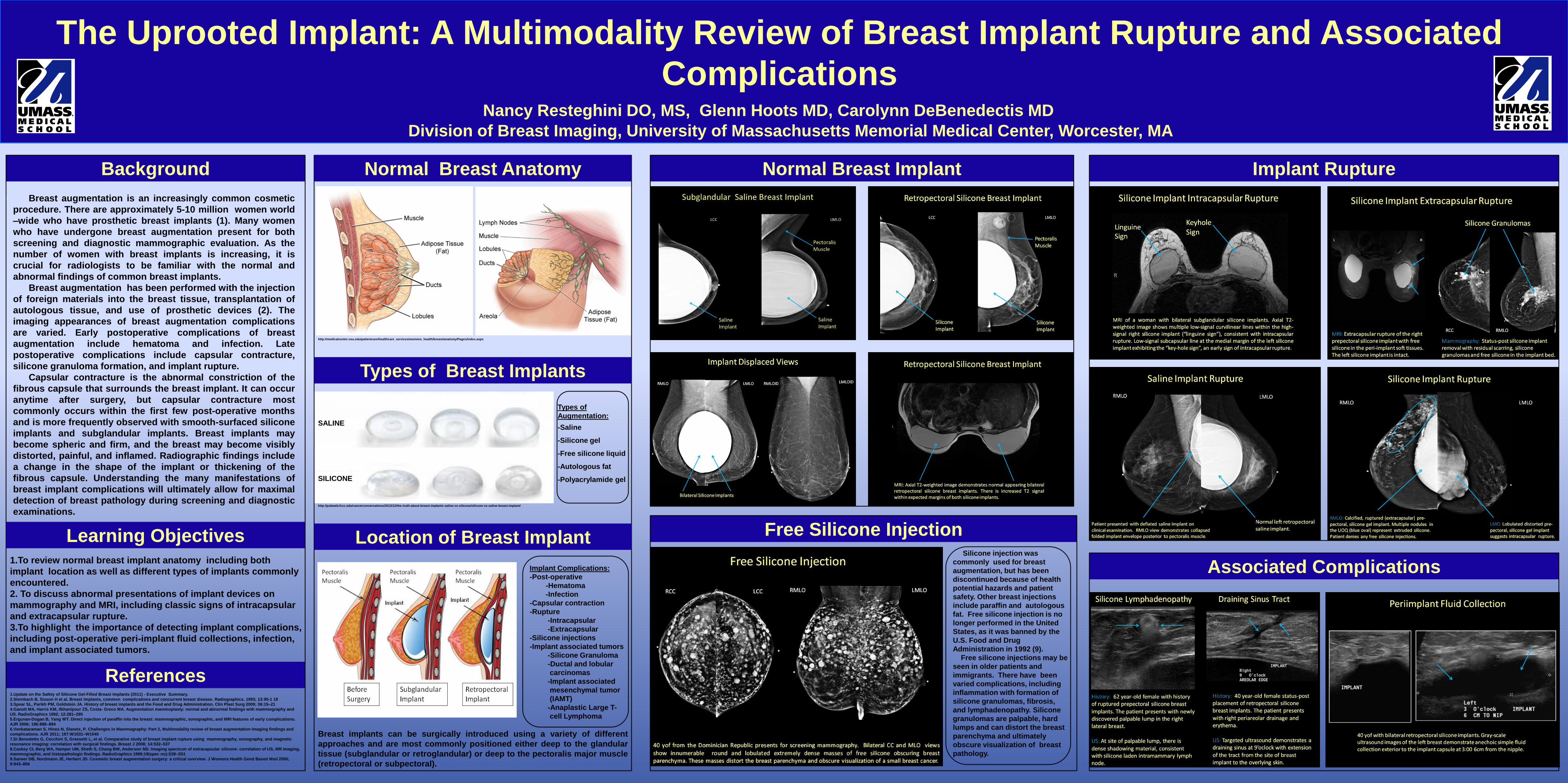
The Uprooted Implant: A Multimodality Review of Breast Implant Rupture and Associated Complications Nancy Resteghini DO, MS, Glenn Hoots MD, Carolynn DeBenedectis MD Division of Breast Imaging, University of Massachusetts Memorial Medical Center, Worcester, MA http://pubweb.fccc.edu/cancerconversations/2013/12/the-truth-about-breast-implants-saline-vs-silicone/silicone-vs-saline-breast-implant/ Breast augmentation is an increasingly common cosmetic procedure. There are approximately 5-10 million women world –wide who have prosthetic breast implants (1). Many women who have undergone breast augmentation present for both screening and diagnostic mammographic evaluation. As the number of women with breast implants is increasing, it is crucial for radiologists to be familiar with the normal and abnormal findings of common breast implants. Breast augmentation has been performed with the injection of foreign materials into the breast tissue, transplantation of autologous tissue, and use of prosthetic devices (2). The imaging appearances of breast augmentation complications are varied. Early postoperative complications of breast augmentation include hematoma and infection. Late postoperative complications include capsular contracture, silicone granuloma formation, and implant rupture. Capsular contracture is the abnormal constriction of the fibrous capsule that surrounds the breast implant. It can occur anytime after surgery, but capsular contracture most commonly occurs within the first few post-operative months and is more frequently observed with smooth-surfaced silicone implants and subglandular implants. Breast implants may become spheric and firm, and the breast may become visibly distorted, painful, and inflamed. Radiographic findings include a change in the shape of the implant or thickening of the fibrous capsule. Understanding the many manifestations of breast implant complications will ultimately allow for maximal detection of breast pathology during screening and diagnostic examinations. Background References Normal Breast Implant 1.To review normal breast implant anatomy including both implant location as well as different types of implants commonly encountered. 2. To discuss abnormal presentations of implant devices on mammography and MRI, including classic signs of intracapsular and extracapsular rupture. 3.To highlight the importance of detecting implant complications, including post-operative peri-implant fluid collections, infection, and implant associated tumors. SALINE SILICONE Learning Objectives Normal Breast Anatomy Types of Breast Implants Location of Breast Implant http://medicalcenter.osu.edu/patientcare/healthcare_services/womens_health/breast/anatomy/Pages/index.aspx Types of Augmentation: -Saline -Silicone gel -Free silicone liquid -Autologous fat -Polyacrylamide gel Breast implants can be surgically introduced using a variety of different approaches and are most commonly positioned either deep to the glandular tissue (subglandular or retroglandular) or deep to the pectoralis major muscle (retropectoral or subpectoral). Implant Complications: -Post-operative -Hematoma -Infection -Capsular contraction -Rupture -Intracapsular -Extracapsular -Silicone injections -Implant associated tumors -Silicone Granuloma -Ductal and lobular carcinomas -Implant associated mesenchymal tumor (IAMT) -Anaplastic Large T- cell Lymphoma 1.Update on the Safety of Silicone Gel-Filled Breast Implants (2011) - Executive Summary. 2.Steinbach B, Sisson H et al. Breast Implants, common complications and concurrent breast disease. Radiographics. 1993; 13:95-1 18 3.Spear SL, Parikh PM, Goldstein JA. History of breast implants and the Food and Drug Administration. Clin Plast Surg 2009; 36:15–21 4.Ganott MA, Harris KM, Ilkhanipour ZS, Costa- Greco MA. Augmentation mammoplasty: normal and abnormal findings with mammography and US. RadioGraphics 1992; 12:281–295 5.Erguvan-Dogan B, Yang WT. Direct injection of paraffin into the breast: mammographic, sonographic, and MRI features of early complications. AJR 2006; 186:888–894 6.Venkataraman S, Hines N, Slanetz, P. Challenges in Mammography: Part 2, Multimodality review of breast augmentation-Imaging findings and complications. AJR 2011; 197:W1031–W1045 7.Di Benedetto G, Cecchini S, Grassetti L, et al. Comparative study of breast implant rupture using mammography, sonography, and magnetic resonance imaging: correlation with surgical findings. Breast J 2008; 14:532–537 8.Caskey CI, Berg WA, Hamper UM, Sheth S, Chang BW, Anderson ND. Imaging spectrum of extracapsular silicone: correlation of US, MR imaging, mammographic, and histopathologic findings. RadioGraphics 1999;19(spec no):S39–S51 9.Sarwer DB, Nordmann JE, Herbert JD. Cosmetic breast augmentation surgery: a critical overview. J Womens Health Gend Based Med 2000; 9:843–856 Implant Rupture Free Silicone Injection Silicone injection was commonly used for breast augmentation, but has been discontinued because of health potential hazards and patient safety. Other breast injections include paraffin and autologous fat. Free silicone injection is no longer performed in the United States, as it was banned by the U.S. Food and Drug Administration in 1992 (9). Free silicone injections may be seen in older patients and immigrants. There have been varied complications, including inflammation with formation of silicone granulomas, fibrosis, and lymphadenopathy. Silicone granulomas are palpable, hard lumps and can distort the breast parenchyma and ultimately obscure visualization of breast pathology. Associated Complications

Welcome message from author
This document is posted to help you gain knowledge. Please leave a comment to let me know what you think about it! Share it to your friends and learn new things together.
Transcript
The Uprooted Implant: A Multimodality Review of Breast Implant Rupture and Associated
Complications
Nancy Resteghini DO, MS, Glenn Hoots MD, Carolynn DeBenedectis MD
Division of Breast Imaging, University of Massachusetts Memorial Medical Center, Worcester, MA
http://pubweb.fccc.edu/cancerconversations/2013/12/the-truth-about-breast-implants-saline-vs-silicone/silicone-vs-saline-breast-implant/
Breast augmentation is an increasingly common cosmetic
procedure. There are approximately 5-10 million women world
–wide who have prosthetic breast implants (1). Many women
who have undergone breast augmentation present for both
screening and diagnostic mammographic evaluation. As the
number of women with breast implants is increasing, it is
crucial for radiologists to be familiar with the normal and
abnormal findings of common breast implants.
Breast augmentation has been performed with the injection
of foreign materials into the breast tissue, transplantation of
autologous tissue, and use of prosthetic devices (2). The
imaging appearances of breast augmentation complications
are varied. Early postoperative complications of breast
augmentation include hematoma and infection. Late
postoperative complications include capsular contracture,
silicone granuloma formation, and implant rupture.
Capsular contracture is the abnormal constriction of the
fibrous capsule that surrounds the breast implant. It can occur
anytime after surgery, but capsular contracture most
commonly occurs within the first few post-operative months
and is more frequently observed with smooth-surfaced silicone
implants and subglandular implants. Breast implants may
become spheric and firm, and the breast may become visibly
distorted, painful, and inflamed. Radiographic findings include
a change in the shape of the implant or thickening of the
fibrous capsule. Understanding the many manifestations of
breast implant complications will ultimately allow for maximal
detection of breast pathology during screening and diagnostic
examinations.
Background
References
Normal Breast Implant
1.To review normal breast implant anatomy including both
implant location as well as different types of implants commonly
encountered.
2. To discuss abnormal presentations of implant devices on
mammography and MRI, including classic signs of intracapsular
and extracapsular rupture.
3.To highlight the importance of detecting implant complications,
including post-operative peri-implant fluid collections, infection,
and implant associated tumors.
SALINE
SILICONE
Learning Objectives
Normal Breast Anatomy
Types of Breast Implants
Location of Breast Implant
http://medicalcenter.osu.edu/patientcare/healthcare_services/womens_health/breast/anatomy/Pages/index.aspx
Types of
Augmentation:
-Saline
-Silicone gel
-Free silicone liquid
-Autologous fat
-Polyacrylamide gel
Breast implants can be surgically introduced using a variety of different
approaches and are most commonly positioned either deep to the glandular
tissue (subglandular or retroglandular) or deep to the pectoralis major muscle
(retropectoral or subpectoral).
Implant Complications:
-Post-operative
-Hematoma
-Infection
-Capsular contraction
-Rupture
-Intracapsular
-Extracapsular
-Silicone injections
-Implant associated tumors
-Silicone Granuloma
-Ductal and lobular
carcinomas
-Implant associated
mesenchymal tumor
(IAMT)
-Anaplastic Large T-
cell Lymphoma
1.Update on the Safety of Silicone Gel-Filled Breast Implants (2011) - Executive Summary.
2.Steinbach B, Sisson H et al. Breast Implants, common complications and concurrent breast disease. Radiographics. 1993; 13:95-1 18
3.Spear SL, Parikh PM, Goldstein JA. History of breast implants and the Food and Drug Administration. Clin Plast Surg 2009; 36:15–21
4.Ganott MA, Harris KM, Ilkhanipour ZS, Costa- Greco MA. Augmentation mammoplasty: normal and abnormal findings with mammography and
US. RadioGraphics 1992; 12:281–295
5.Erguvan-Dogan B, Yang WT. Direct injection of paraffin into the breast: mammographic, sonographic, and MRI features of early complications.
AJR 2006; 186:888–894
6.Venkataraman S, Hines N, Slanetz, P. Challenges in Mammography: Part 2, Multimodality review of breast augmentation-Imaging findings and
complications. AJR 2011; 197:W1031–W1045
7.Di Benedetto G, Cecchini S, Grassetti L, et al. Comparative study of breast implant rupture using mammography, sonography, and magnetic
resonance imaging: correlation with surgical findings. Breast J 2008; 14:532–537
8.Caskey CI, Berg WA, Hamper UM, Sheth S, Chang BW, Anderson ND. Imaging spectrum of extracapsular silicone: correlation of US, MR imaging,
mammographic, and histopathologic findings. RadioGraphics 1999;19(spec no):S39–S51
9.Sarwer DB, Nordmann JE, Herbert JD. Cosmetic breast augmentation surgery: a critical overview. J Womens Health Gend Based Med 2000;
9:843–856
Implant Rupture
Free Silicone Injection Silicone injection was
commonly used for breast
augmentation, but has been
discontinued because of health
potential hazards and patient
safety. Other breast injections
include paraffin and autologous
fat. Free silicone injection is no
longer performed in the United
States, as it was banned by the
U.S. Food and Drug
Administration in 1992 (9).
Free silicone injections may be
seen in older patients and
immigrants. There have been
varied complications, including
inflammation with formation of
silicone granulomas, fibrosis,
and lymphadenopathy. Silicone
granulomas are palpable, hard
lumps and can distort the breast
parenchyma and ultimately
obscure visualization of breast
pathology.
Associated Complications
Related Documents